Shikimate Kinase: A Potential Target for Development of Novel Antitubercular Agents
10
Current Drug Targets, 2007, 8, 459-468 459 1389-4501/07 $50.00+.00 © 2007 Bentham Science Publishers Ltd. Shikimate Kinase: A Potential Target for Development of Novel Antitubercular Agents José H. Pereira † , Igor B. Vasconcelos § , Jaim S. Oliveira § , Rafael A. Caceres ¤ , Walter F. de Azevedo Jr. ¤ , Luis A. Basso §* and Diógenes S. Santos §* † Programa de Pós-Graduação em Biofísica Molecular-Departamento de Física, UNESP, São José do Rio Preto, SP, 15054-000; § Centro de Pesquisas em Biologia Molecular e Funcional, Faculdade de Farmácia, Instituto de Pesquisas Biomédicas, Pontifícia Universidade Católica do Rio Grande do Sul. Avenida Ipiranga 6681, Tecnopuc, Partenon 90619-900, Porto Alegre, RS, Brazil and ¤ Faculdade de Biociências-PUCRS. Avenida Ipiranga 6681, 90619-900, Porto Alegre, RS, Brazil Abstract: Tuberculosis (TB) remains the leading cause of mortality due to a bacterial pathogen, Mycobacterium tuberculosis. However, no new classes of drugs for TB have been developed in the past 30 years. Therefore there is an urgent need to develop faster acting and effective new antitubercular agents, preferably belonging to new structural classes, to better combat TB, including MDR-TB, to shorten the duration of current treatment to improve patient compliance, and to provide effective treatment of latent tuberculosis infection. The enzymes in the shikimate pathway are potential targets for development of a new generation of antitubercular drugs. The shikimate path- way has been shown by disruption of aroK gene to be essential for the Mycobacterium tuberculosis. The shikimate kinase (SK) catalyses the phosphorylation of the 3-hydroxyl group of shikimic acid (shikimate) using ATP as a co-substrate. SK belongs to family of nucleo- side monophosphate (NMP) kinases. The enzyme is an / protein consisting of a central sheet of five parallel -strands flanked by - helices. The shikimate kinases are composed of three domains: Core domain, Lid domain and Shikimate-binding domain. The Lid and Shikimate-binding domains are responsible for large conformational changes during catalysis. More recently, the precise interactions be- tween SK and substrate have been elucidated, showing the binding of shikimate with three charged residues conserved among the SK se- quences. The elucidation of interactions between MtSK and their substrates is crucial for the development of a new generation of drugs against tuberculosis through rational drug design. Key Words: tuberculosis, malaria, shikimate kinase, shikimate pathway, drug design, drug target, Mycobacterium tuberculosis, Plasmodium sp 1. THE SHIKIMATE PATHWAY The shikimate pathway was discovered as a biosynthetic route through the studies of Bernhard Davis and David Sprinson and their collaborators [1,2]. The shikimate pathway links metabolism of carbohydrates to biosynthesis of aromatic compounds through seven metabolic steps, where phosphoenolpyruvate and erythrose 4- phosphate are converted to chorismic acid [3,4]. Chorismic acid is a common precursor for the synthesis of aromatic compounds, such as aromatic amino acids, folate, ubiquinone and menaquinones. Amongst them, the only ones that can be synthesized by humans are tyrosine, which is synthesized from phenylalanine through a reaction catalyzed by phenylalanine hidroxylase enzyme, and ubiquinone from tyrosine through a cascade of eight aromatic pre- cursors [5]. The molecular organization of the shikimate pathway enzymes varies between taxonomic groups [6]. Bacteria have seven individ- ual polypeptides, which are encoded by separate genes. Plants have a molecular arrangement similar to bacteria [7], with the exception of dehydroquinase (DHQase, third enzyme) and shikimate dehy- drogenase (fourth enzyme) which have been shown to be present as separate domains on a bifunctional polypeptide [8]. In fungi and apicomplexan parasites (Toxoplasma gondii) the shikimate pathway has been shown to include monofunctional 3-deoxy-D-arabino- heptulosonate 7-phosphate (DAHP) synthase and chorismate *Address correspondence to this author at the Centro de Pesquisas em Bio- logia Molecular e Funcional, Faculdade de Farmácia, Instituto de Pesquisas Biomédicas, Pontifícia Universidade Católica do Rio Grande do Sul. Aveni- da Ipiranga 6681, Tecnopuc, Partenon 90619-900, Porto Alegre, RS, Brazil; Tel: +55-51-33203629; Fax, +55-51-3320-3629; E-mail: [email protected] synthase (CS) enzymes and a pentafunctional polypeptide termed AROM, which accounts for the remaining five shikimate pathway reactions [9]. The shikimate pathway enzymes are attractive targets for de- velopment of non-toxic antibacterial [10] and herbicides [11], be- cause this pathway is essential for algae, higher plants, bacteria, fungi, whereas it is absent from mammals [12]. Thus, in the case of bacterial diseases, inhibition of any of shikimate pathway enzymes is unlikely to cause toxic side effects on the host. In addition, the importance of shikimate pathway can be indicated by the finding that deletion of the aroA gene, which codes EPSPS, causes Strep- tomyces pneumoniae and Bordetella bronchiseptica strains to be attenuated for virulence [13,14]. The shikimate pathway has also been discovered in apicom- plexan parasites providing several targets for the development of new antiparasite drugs [15]. In vitro growth of apicomplexan para- sites such as Plasmodium falciparum (malaria), Toxoplasma gondii (toxoplasmosis), and Cryptosporidium parvum (cryptosporidiosis) was inhibited by the herbicide glyphosate, a well-characterized inhibitor [16] of the shikimate pathway enzyme 5-enolpyruvyl shikimate 3-phosphate synthase (EPSPS), at concentration of 1-6 mM [15]. In P. falciparum and T. gondii, this inhibitory effect was reversed by co-addition o p-aminobenzoate (PABA) or folate sug- gesting that this pathway is essential for the biosynthesis of folate precursors. Moreover, several shikimate pathway enzymes has been detected in T. gondii and P. falciparum extracts [15,17] and a gene encoding a pentafunctional polypeptide AROM has been described in T. goondii indicating that a complete shikimate pathway is pre- sent in this parasite [18]. Whereas the shikimate pathway gives an array of compounds in bacteria, fungi and plants, only its folate biosynthesis function has been established is in apicomplexan para- sites. All folate pathway enzymes, which convert GTP to deriva-
-
Upload
independent -
Category
Documents
-
view
0 -
download
0
Transcript of Shikimate Kinase: A Potential Target for Development of Novel Antitubercular Agents

Current Drug Targets, 2007, 8, 459-468 459
1389-4501/07 $50.00+.00 © 2007 Bentham Science Publishers Ltd.
Shikimate Kinase: A Potential Target for Development of Novel Antitubercular Agents
José H. Pereira†, Igor B. Vasconcelos§, Jaim S. Oliveira§, Rafael A. Caceres¤, Walter F. de Azevedo Jr.¤, Luis A. Basso§* and Diógenes S. Santos§*
†Programa de Pós-Graduação em Biofísica Molecular-Departamento de Física, UNESP, São José do Rio Preto, SP, 15054-000;
§Centro de Pesquisas em Biologia Molecular e Funcional, Faculdade de Farmácia, Instituto de Pesquisas Biomédicas, Pontifícia
Universidade Católica do Rio Grande do Sul. Avenida Ipiranga 6681, Tecnopuc, Partenon 90619-900, Porto Alegre, RS, Brazil and ¤Faculdade de Biociências-PUCRS. Avenida Ipiranga 6681, 90619-900, Porto Alegre, RS, Brazil
Abstract: Tuberculosis (TB) remains the leading cause of mortality due to a bacterial pathogen, Mycobacterium tuberculosis. However, no new classes of drugs for TB have been developed in the past 30 years. Therefore there is an urgent need to develop faster acting and effective new antitubercular agents, preferably belonging to new structural classes, to better combat TB, including MDR-TB, to shorten the duration of current treatment to improve patient compliance, and to provide effective treatment of latent tuberculosis infection. The enzymes in the shikimate pathway are potential targets for development of a new generation of antitubercular drugs. The shikimate path-way has been shown by disruption of aroK gene to be essential for the Mycobacterium tuberculosis. The shikimate kinase (SK) catalyses the phosphorylation of the 3-hydroxyl group of shikimic acid (shikimate) using ATP as a co-substrate. SK belongs to family of nucleo-side monophosphate (NMP) kinases. The enzyme is an / protein consisting of a central sheet of five parallel -strands flanked by -helices. The shikimate kinases are composed of three domains: Core domain, Lid domain and Shikimate-binding domain. The Lid and Shikimate-binding domains are responsible for large conformational changes during catalysis. More recently, the precise interactions be-tween SK and substrate have been elucidated, showing the binding of shikimate with three charged residues conserved among the SK se-quences. The elucidation of interactions between MtSK and their substrates is crucial for the development of a new generation of drugs against tuberculosis through rational drug design.
Key Words: tuberculosis, malaria, shikimate kinase, shikimate pathway, drug design, drug target, Mycobacterium tuberculosis, Plasmodium sp
1. THE SHIKIMATE PATHWAY
The shikimate pathway was discovered as a biosynthetic route through the studies of Bernhard Davis and David Sprinson and their collaborators [1,2]. The shikimate pathway links metabolism of carbohydrates to biosynthesis of aromatic compounds through seven metabolic steps, where phosphoenolpyruvate and erythrose 4-phosphate are converted to chorismic acid [3,4]. Chorismic acid is a common precursor for the synthesis of aromatic compounds, such as aromatic amino acids, folate, ubiquinone and menaquinones. Amongst them, the only ones that can be synthesized by humans are tyrosine, which is synthesized from phenylalanine through a reaction catalyzed by phenylalanine hidroxylase enzyme, and ubiquinone from tyrosine through a cascade of eight aromatic pre-cursors [5].
The molecular organization of the shikimate pathway enzymes varies between taxonomic groups [6]. Bacteria have seven individ-ual polypeptides, which are encoded by separate genes. Plants have a molecular arrangement similar to bacteria [7], with the exception of dehydroquinase (DHQase, third enzyme) and shikimate dehy-drogenase (fourth enzyme) which have been shown to be present as separate domains on a bifunctional polypeptide [8]. In fungi and apicomplexan parasites (Toxoplasma gondii) the shikimate pathway has been shown to include monofunctional 3-deoxy-D-arabino-heptulosonate 7-phosphate (DAHP) synthase and chorismate
*Address correspondence to this author at the Centro de Pesquisas em Bio-logia Molecular e Funcional, Faculdade de Farmácia, Instituto de Pesquisas Biomédicas, Pontifícia Universidade Católica do Rio Grande do Sul. Aveni-da Ipiranga 6681, Tecnopuc, Partenon 90619-900, Porto Alegre, RS, Brazil; Tel: +55-51-33203629; Fax, +55-51-3320-3629; E-mail: [email protected]
synthase (CS) enzymes and a pentafunctional polypeptide termed AROM, which accounts for the remaining five shikimate pathway reactions [9].
The shikimate pathway enzymes are attractive targets for de-velopment of non-toxic antibacterial [10] and herbicides [11], be-cause this pathway is essential for algae, higher plants, bacteria, fungi, whereas it is absent from mammals [12]. Thus, in the case of bacterial diseases, inhibition of any of shikimate pathway enzymes is unlikely to cause toxic side effects on the host. In addition, the importance of shikimate pathway can be indicated by the finding that deletion of the aroA gene, which codes EPSPS, causes Strep-tomyces pneumoniae and Bordetella bronchiseptica strains to be attenuated for virulence [13,14].
The shikimate pathway has also been discovered in apicom-plexan parasites providing several targets for the development of new antiparasite drugs [15]. In vitro growth of apicomplexan para-sites such as Plasmodium falciparum (malaria), Toxoplasma gondii (toxoplasmosis), and Cryptosporidium parvum (cryptosporidiosis) was inhibited by the herbicide glyphosate, a well-characterized inhibitor [16] of the shikimate pathway enzyme 5-enolpyruvyl shikimate 3-phosphate synthase (EPSPS), at concentration of 1-6 mM [15]. In P. falciparum and T. gondii, this inhibitory effect was reversed by co-addition o p-aminobenzoate (PABA) or folate sug-gesting that this pathway is essential for the biosynthesis of folate precursors. Moreover, several shikimate pathway enzymes has been detected in T. gondii and P. falciparum extracts [15,17] and a gene encoding a pentafunctional polypeptide AROM has been described in T. goondii indicating that a complete shikimate pathway is pre-sent in this parasite [18]. Whereas the shikimate pathway gives an array of compounds in bacteria, fungi and plants, only its folate biosynthesis function has been established is in apicomplexan para-sites. All folate pathway enzymes, which convert GTP to deriva-

460 Current Drug Targets, 2007, Vol. 8, No. 3 Pereira et al.
tives of tetrahydrofolate, were found in Apicomplexa when the complete genome was sequenced [19].
Two shikimic acid analogs, 6-S-fluorshikimate and 6-R-fluorshikimate, have been shown to inhibit P. falciparum growth and inhibition shown to be specific to the shikimate pathway [20]. Despite the completion of P.falciparum genome sequence [21], only a single gene encoding the chorismate synthase enzyme and a potential bifunctional SK/EPSP synthase protein has been identified in the genome annotation [22]. The coding DNA sequence of P. falciparum chorismate synthase has been cloned and the protein has been shown to be located on cytosol by immunological studies [23]. Moreover, chorismate synthase, which catalyzes the last step of shikimate pathway, has been shown to be required for Plasmodium falciparum growth, as disruption of expression by RNA interfer-ence decreased parasite growth [19]. It has been proposed that the missing shikimate pathway enzymes are either substituted by non-homologous enzymes that catalyze the same reaction or that the enzymes are homologous but too divergent to be identified [22].
In mycobacteria, the chorismic acid intermediate is a precursor for the synthesis of naphthoquinones, menaquinones, and myco-bactins, besides aromatic amino acids [24]. The salycilate-derived mycobactins siderophores have been shown to be essential for M. tuberculosis growth in macrophages [25]. Particularly, in Mycobac-
terium tuberculosis, the shikimate pathway has been shown to be essential for the bacterial viability. The disruption of aroK gene, which codes for the shikimate kinase enzyme (SK), was only possi-ble when the second functional copy of aroK was integrated into the chromosome. Moreover, excision of the second integrated copy of aroK by the L5 excisionase could not be achieved in a M. tuber-
culosis strain carrying the disrupted copy of aroK gene, but was possible in a strain carrying a wild-type copy [26].
2. SHIKIMATE KINASE
The shikimate kinase (SK; EC 2.7.1.71), the fifth enzyme of the pathway, catalyses the regiospecific phosphorylation of the 3-hydroxyl group of shikimic acid (shikimate) using ATP as a co-substrate. In Escherichia coli, the SK reaction is catalyzed by two isoforms: SK I encoded by the aroK gene [27] and SK II encoded by the aroL gene [28]. The major difference between the isoen-zymes is their Km for shikimate, 20 mM for the SK I and 0.2 mM for the SK II enzyme [29]. The SK II isoform appears to play a dominant role in the shikimate pathway, its expression is controlled by the tyrR regulator, and it is repressed by tyrosine and tryptophan [30,31]. The physiological role of SK I in E.coli is not clear. Since mutations in SK I are associated with sensitivity to the antibiotic mecillinam [32] it has been suggested that SK I may have an alter-native biological role that is distinct and unrelated to its shikimate kinase activity [29]. As pointed out by Parish and Stoker [26], if M. tuberculosis aroK-encoded SK I possess a similar activity it is pos-sible that disruption of this activity can account for the observed inability of M. tuberculosis to grow in the absence of a functional copy of aroK gene. However, the actual nature of second activity of aroK gene product remains to be established. Contrary to the pres-ence of isoenzymes in E. coli, complete genome sequences of a number of bacteria, for example, Haemophilus influenzae and My-cobacterium tuberculosis, have revealed the presence of only one SK-coding gene. Most of these SKs appear to be encoded by aroK rather than aroL because their amino acid sequences have higher degree of identity with E.coli SK I. The kinetic parameters for aroK-encoded M.tuberculosis SK (MtSK) are more similar to those of aroL-encoded E.coli SK II than to those of aroK-encoded E.coli SK I. Thus, the MtSK Km value (0.41mM) for shikimate suggests that not all aroK-encoded SKs have high Km values for shikimate [33].
Fig. (1). Overall structure of Shikimate kinase from M. tuberculosis [61]. MtSK displays an / -fold and the precise ordering of the strands 23145 in the paral-lel -sheet classifies MtSK as belonging to the family as the NMP kinases. The SKs are composed of three domains: CORE, LID and Shikimate-binding (SB) domains. The positions of the ADP, Mg2+ and shikimate are shown in the MtSK structure.
Shikimate Kinase: A Potential Target for Development of Novel Current Drug Targets, 2007, Vol. 8, No. 3 461
2.1. The Enzyme Fold
Shikimate kinase displays an / -fold and consists of five cen-tral parallel -sheet with the strand order 23145, flanked by -helices [34] (Fig. (1)). The three crystal structures of SK from Er-winia chrysanthemi (ErcSK) [34,35] showed that SK belongs to the same structural family of nucleoside monophosphate (NMP) kinases for which structures are known for adenylate kinase (AK) [36,37], guanylate kinase [38], uridylate kinase [39] and thymidine kinase [40].
The NMP kinases are composed of three domains: CORE, LID and NMP-binding (NMPB) domains [41]. A characteristic feature of the NMP kinases is that they undergo large conformational changes during catalysis, for which AK is the most extensively studied [41]. There are two flexible regions of the structures that are responsible for movement: one is the NMP-binding site which is formed by a series helices between strands 2 and 3 of parallel -sheet and the other is the LID domain, a region of varied size and structure following the fourth -strand of the sheet [42,43]. In SK, the shikimate-binding (SB) domain corresponds to the NMPB do-main of NMP kinases.
2.2. Functional Motifs
Three functional motifs of nucleotide-binding enzymes are rec-ognizable in shikimate kinase, including a Walker A-motif (A-motif), a Walker B-motif (B-motif), and an adenine-binding loop. The Walker A-motif is located between the first -strand ( 1) and
first -helix ( 1), containing the GXXXXGKT/S conserved se-quence [44], where X represents any residue. This motif forms the phosphate-binding loop (P-loop), a giant anion hole which accom-modates the -phosphate of the ADP by donating hydrogen bonds from several backbone amides [45]. The side chain of P-loop lysine have a catalytic role of stabilizing the pentavalent transition state of the -phosphoryl group as has been shown for adenylate kinase [46] and p21ras [47].
In addition to the Walker A-motif, it is observed a second con-served sequence ZZDXXG called Walker B-motif [44], where Z represents a hydrophobic residue. More recently, however, se-quence and structural comparisons for all P-loop-fold proteins clas-sified Shikimate Kinase in the DxD group of enzymes [48], which has a conserved DxD motif in strand 2. The Walker B motif con-sensus in shikimate kinases is ZZZTGGG and the second glycine has been implicated in hydrogen bonding to the phosphate of ATP. This motif is located on the C-terminal segment of the third strand ( 3) of the central -sheet The adenine-binding loop motif may be described as a sequence stretch of I/VDXXX(X)XP [33]. This motif forms a loop that wraps around the adenine moiety of ATP, connecting the 5-strand with the C-terminal -helix.
3. TUBERCULOSIS AND MYCOBACTERIUM TUBERCULO-SIS SHIKIMATE KINASE
Tuberculosis (TB) remains the leading cause of mortality due to a bacterial pathogen, Mycobacterium tuberculosis. The interruption
Fig. (2). Interactions involved in the binding of shikimate to the MtSK active site (PDB access code: 1WE2) [61]. Hydrogen bonds are represented as broken lines. For clarity, the the water320-mediated hydrogens bonds between Gly79, Gly80, Gly81, Ala82, Arg58, and Glu61 protein residues and 3-hydroxyl group of shikimate are not shown.

462 Current Drug Targets, 2007, Vol. 8, No. 3 Pereira et al.
of centuries of decline in case rates of TB occurred, in most cases, in the late 1980s and involved the USA and some European coun-tries due to increased poverty in urban settings and the immigration from TB high-burden countries [49]. Thus, no sustainable control of TB epidemics can be reached in any country without properly ad-dressing the global epidemic. It is estimated that 8.2 million new TB cases occurred worldwide in the year 2000, with approximately 1.8 million deaths in the same year, and more than 95 % of those were in developing countries [50]. Approximately 2 billion indi-viduals are believed to harbor latent TB based on tuberculin skin test surveys [51], which represents a considerable reservoir of ba-cilli. According to a recent report compiled by the World Health Organization (WHO), the total number of new cases of tuberculosis worldwide in 2002 had risen to approximately 9 million [52]. A key driver of the increase is the synergy with the HIV epidemic, which is having a devastating impact on some parts of the world mostly in the African Region, where 31% of new TB cases were attributable to HIV co-infection [50]. Another problem is the proliferation of multi-drug resistant (MDR) strains, defined as resistants to at least isoniazid and rifampicin, which are the most effective first-line drugs [53]. According to the 2004 Global TB Control Report of the World Health Organization, there are 300,000 new cases per year of MDR-TB worldwide, and 79 % of MDR-TB cases are now “super strains”, resistant to at least three of the four main drugs used to treat TB [52]. The factors that most influence the emergence of drug-resistant strains include inappropriate treatment regimens, and patient noncompliance in completing the prescribed courses of therapy due to the lengthy standard “short-course” treatment or when the side effects become unbearable [54]. No new classes of drugs for TB have been developed in the past 30 years, reflecting the inherent difficulties in discovery and clinical testing of new agents and the lack of pharmaceutical industry in investing money and manpower for research in the area [55]. Hence, there is an ur-gent need to developing faster acting and effective new antitubercu-lar agents, preferably belonging to new structural classes, to better combat TB, including MDR-TB, to shorten the duration of current treatment to improve patient compliance, and to provide effective treatment of latent tuberculosis infection [53].
In M.tuberculosis, the presence of the genes involved in the shikimate pathway began to be elucidated in the early 90s, with the cloning and characterization of aroA gene product, which codifies the EPSPS synthase [56]. Nevertheless, the complete genome se-quence from M.tuberculosis, strain H37Rv reported by Cole et al. in 1998 [57] allowed the identification by sequence homology of all genes coding for shikimate pathway enzymes.
Four homologues to the shikimate pathway enzymes were lo-cated in a cluster containing the aroD-encoded type II DHQ dehy-dratase (Rv2537c), aroB-encoded DHQ synthase (Rv2538c), aroK-encoded type I shikimate kinase (Rv2539c), and aroF-encoded chorismate synthase (Rv2540c). The remaining homologues to shikimate pathway enzymes were annotated as follows: aroG-encoded class II phenylalanine-regulated DAHPS (Rv2178c), aroE-encoded shikimate dehydrogenase (Rv2552c), and aroA-encoded EPSP synthase (Rv3227).
The aroK structural gene is composed by 531 bp and codes a protein of 176 amino acids. The theoretical molecular mass of MtSK enzyme subunit is 18.58 KDa. The first report of cloning and overexpression in soluble and functional form of MtSK occurred in 2001 [58], where has been reported the PCR amplification aroK gene from genomic DNA of M. tuberculosis H37Rv strain, cloning in the plasmid pET-23a(+), and overexpression of aroK-encoded MtSK protein in Escherichia coli BL21(DE3) host cells, without IPTG induction.
The crystal structure of a protein complexed with its substrates is of crucial importance for the rational design of inhibitors that target the enzyme. Although two crystal structures of SK from M.tuberculosis [33] have revealed the dynamic role of LID Domain
in catalysis and the position of Mg2+ and ADP ligands in the struc-ture, the precise positions and interactions between shikimate and MtSK was not demonstrated because the shikimate-binding site was not occupied by the substrate or the electron density was not suffi-cient clear to position the shikimate molecule in the complex. In case of SK enzymes, a likely drawback of ATP-binding-site-based SK inhibitors would be their lack of specificity, owing to the com-mon fold and similar ATP-binding site shared by many P-loop kinases [48]. Hence, trying to obtain a description of molecular interaction between MtSK and shikimate, molecular-modeling and docking studies has been carried out and its results reported [59]. Despite these studies failed to predict all molecular interactions between MtSK and shikimate [60], Asp34 and Arg136 residues and its hydrogen bonds implicated on shikimate binding are in agree-ment with the crystal structure of MtSK-MgADP-Shikimate ternary complex reported afterwards [61]. The crystal structure of MtSK-MgADP-Shikimate was the first crystallographic structure of SK with bound shikimate deposited in Protein Data Bank (PDB access code: 1WE2) [61]. Crystals were obtained by the hanging-drop vapour-diffusion method, and larger final concentration of ADP and shikimate in the drop (8.0mM) were used than those used by Gu et al. (2002) to obtain the crystals (4.0mM), in order to accurately determine the position of shikimate binding in the active site of MtSK. The structure has been determined at 2.3 Å resolution, clearly reveling the amino-acids residues involved in shikimate binding. The molecular replacement method was used, using as a search model the structure of MtSK-MgADP [33]. Almost at the same time of publishing of our article describing the structure of MtSK-MgADP-Shikimate ternary complex, another article describ-ing a similar structure of the ternary complex has been published (PDB access code: 1U8A) [60]. The crystal structures of MtSK-MgADP-Shikimate [61] and MtSK-ADP-Shikimate [60] ternary complexes have unequivocally revealed in detail the interactions of amino acid residues with bound shikimate and conformational changes upon substrate binding.
3.1. ADP/Mg2+
Interaction
Essentially all kinases require a Mg2+-nucleotide complex as one of the enzyme substrates [62], an exception being the first par-tial reaction catalyzed by nucleoside diphosphate kinase, which can proceed independently of Mg2+ [63]. Nucleophilic attack on the -phosphate group of ATP will be most facilitated by meta-ion bind-ing (Mg2+) to the - and -phosphoryl groups, whereas departure of the leaving group will be most favoured in a structure with metal binding to the - and -phosphoryl groups [64]. Presumably Mg2+
also assists in orienting the -phosphoryl group of ‘inline’ with respect to the second substrate, creating the correct geometry to complete phosphoryl transfer [62]. The binding of this cation in many P-loop proteins such as myosin [65], elongation factor EF-Tu [66], p21-Ras [67] and the heterotrimeric G-proteins [68] involves hexa-coordination of the Mg2+ by two oxygen atoms (from the and -phosphates of the bound nucleotide), two water molecules, and two protein ligands. As in the MtSK-MgADP structure (PDB code 1L4Y) [33], a typical six-coordination has been observed for Mg2+ in the MtSK-MgADP-Shikimate structure, with some minor differences between the position of Mg2+ in the structures. In the MtSK-MgADP-Shikimate structure Mg2+ interacts with a -phosphate oxygen of ADP, Ser16 OG of the Walker A motif and four water molecules [61]. Superimposition of shikimate-free MtSK-MgADP structure on shikimate-complexed MtSK-MgADP structure showed that in the ternary complex water1 and water4 are in equivalent positions but that shifts of 1.32 and 2.75 Å are ob-served for water2 and water3, respectively. In the MtSK-MgADP structure, the interaction between Asp32 and Ser16 of the Walker A motif is via a bridging water molecule (water6), whereas in the ternary complex structure this interaction occurs directly via a hy-drogen bond between the two residues [61], which accounts for the exclusion of water6 from the magnesium-binding site in the ternary

Shikimate Kinase: A Potential Target for Development of Novel Current Drug Targets, 2007, Vol. 8, No. 3 463
structure. In the ternary complex, the water1 molecule coordinated to the Mg2+ interacts directly with the chloride ion instead of inter-acting with Asp34 via a bridging water molecule (water5) as ob-served in the MtSK-MgADP structure [33]. This chloride ion also interacts with the 3-hydroxyl group of shikimate and the backbone amide of Gly80 [61]. Thus, the different mode of interaction ob-served for residue Asp34 arises from the presence of shikimate, which leads to the exclusion of water5 from MtSK active site [61]. The Mg2+ cation was not included in the final structure of MtSK-ADP-Shikimate ternary complex reported by Dhaliwal et. al.(2004), since the best diffracting crystals grew in the absence of MgCl2 [60]. Accordingly, the chloride ion observed in the active sites of both MtSK binary complex [33] and MtSK-MgADP-Shikimate ternary complex [61is absent in the Mg2+-free structure of MtSK-ADP-Shikimate ternary complex [60]. Thus, the absence of MgCl2 in crystallization mixture probably have accounted for the small differences observed in conformation, position and molecular inter-actions of shikimate when the structure reported by Dhaliwal et al. is compared with MtSK-MgADP-Shikimate-Cl structure [61]. In Mg2+-free MtSK structure the 3-hydroxyl group of shikimate is closer to -phosphoryl group of ADP than it is in the MtSK-MgADP-Shikimate structure. Furthermore, two additional water-mediated interactions between protein residues and shikimate have been shown. The NZ atom of Lys15 residue forms a 2.6 Å hydro-gen-bond with oxygen atom of another water molecule, which in turn interacts with the 3-hydroxyl group of shikimate, and the main-chain nitrogen of Leu119 residue forms a 2.9 Å hydrogen bond with the oxygen atom of a water molecule, which in turn interacts with 5-hydroxyl group of substrate [60]. In the MtSK-MgADP-Shikimate structure, NZ atom of Lys15 cannot form a water-mediated interaction with 3-hydroxyl group of shikimate, since the chloride ion is bound to the active site cavity between them. The distance between NZ atom of Lys15 and the chloride ion is 3.94 Å, which in turn forms a 3.36-Å hydrogen bond with 3-hydroxyl group of shikimate [61].
In the MtSK-MgADP structures [33], Lys15 forms a hydrogen bond with a -phosphate O1B atom and the chloride ion. The main-chain NH of Gly80 is hydrogen bonded to the chloride ion the bi-nary (1L4Y) [33] and ternary complex structures [60,61]. These residues are located in vicinity of where the chemical reaction occur a may thus play a critical role in the transition state stabilization.
The molecular interactions that describe the ADP binding mode on enzyme are very similar in all available structures of MtSK. The adenine moiety of ADP is sandwiched between Arg110 and Pro155 [33] and this interaction has also been observed in ErcSK [34], and in Adenilate kinase [69] and isoenzyme II [70]. Arg110 is located at the C-terminus of 6 where the LID domain starts. In MtSK, Arg110 and Arg117 residues represent, respectively, the first and the last residue of a conserved motif of LID domain observed for P-loop kinases (typically RXX(X)R) [48]. In P-loop shikimate kinases the conserved motif of the LID domain has been proposed to be R(X)6-9R [61]. The Arg117 has been shown to interact with the
and -phosphate groups of ADP [33,60,61], it generally inter-acts with the -phosphate of ATP bound to enzymes, and it may stabilize the transition state by neutralizing the developing negative charge on the - bridge O atom [71]. The Pro155 is the last residue of the adenine-binding loop motif (residues 148-155 in MtSK), which was first recognized in Adenilate kinase and ErcSK [34] and has been described as an I/VDXXX(X)XP sequence stretch [33]. However, the second (aspartate) residue and the last (proline) resi-due are not conserved in the adenine-binding loops of aroK-encoded SKs from Escherichia coli [72] and Campylobacter jejuni (PDB acess code: 1VIA). However, they are recognizable from structural alignment analysis using MtSK-MgADP as a reference structure [61]. In fact, it has been pointed out that the main-chain contacts with the adenine base and the presence of a structural motif independent of sequence may be the most important features for
adenine binding in kinases [62]. The adenine-binding loop motif (residues 148-155 in MtSK) forms a loop that wraps around the adenine moiety of ATP, connecting the 5-strand with the C-terminal 8-helix.
For catalysis, the three protein interacting residues Lys15, Arg117, and Arg136 have been proposed to be the most important [33]. Consistent with this proposal, the K15M mutant of ErcSK showed no detectable enzyme activity [35], although it was in fact a K15M/P115L double mutant. Contrary to the previously proposed, analysis of the MtSK ternary complexes have revealed that Lys15 and Arg117 are the only positively charged residues located in the vicinity of where the reaction may occur and may therefore play critical roles in the stabilization of the transition state [33]. The Arg136 residue, instead, appears to interact with the carboxyl group of shikimate and probably, it is not involved in catalysis [60,61].
3.2. Interaction with Shikimate
The shikimate-binding domain, which follows strand 2, con-sists of helices 2 and 3 and the N-terminal region of helix 4 (Fig. (1)). In particular for MtSK, the precise interactions between shikimate and SK have been elucidated, showing that the guanidi-nium groups of Arg58 and Arg136 and the NH backbone group of Gly81 interact with the carboxyl group of shikimate. The 3-hydroxyl group of shikimate forms hydrogen bonds with the car-boxyl group of Asp34 and the main-chain NH group of Gly80 and a water molecule. The 2-hydroxyl group of shikimate hydrogen bonds to the side chain of Asp34 (Fig. (2)). The Glu61 is conserved in both aroK and aroL-encoded shikimate kinase enzymes. This residue is not directly involved in substrate binding, but it forms a hydrogen bond and a salt bridge with the conserved Arg58 and assists in positioning the guanidinium group of Arg58 for shikimate binding. In the ternary structure, Glu61 side chain also forms a water-mediated interaction with the 3-hydroxyl group of shikimate. Therefore, Glu61 plays an important role in the substrate-binding site [61]. The Glu54 carboxylate group also appears to anchor the guanidinium group of Arg58 for interaction with the shikimate carboxylate group; however, Glu54 is not conserved, except for aroK-encoded shikimate kinases [61]. In the MtSK-ADP-shikimate structure [60], the main-chain nitrogen of Leu119 forms a 2.9 A H-bond with the water3 oxygen atom, which in turns interacts with the 5-hydroxyl group of shikimate, and the NZ atom of Lys15 forms a 2.6 A H-bond with the oxygen atom of water2, which also interacts with the 3-hydroxyl group of the substrate.
3.3. Conformational Changes Upon Substrate Binding
As pointed out above, NMP Kinases undergoes large conforma-tional changes during catalysis, because their LID domain and the NMP binding-site are very flexible and can make large movements upon substrate binding. These structural changes act to position enzyme sidechains appropriately around the substrates and to se-quester the substrates so as to prevent the hydrolysis of bound ATP or other phosphoryl-containing substrates prior to catalysis [41,62,64]. In addition, it has also been shown that these two do-mains are capable to make independently moves towards each other [41]. Previous studies have shown that hexokinases [73] and adeny-late kinases [42,74], which are classified as NMP kinases, undergo a large conformational change during catalysis.
Several structural and spectroscopic studies have demonstrated that SKs undergo conformational changes on ligand binding. Circu-lar dichroism spectra of unliganded and liganded ErcSK enzyme in the presence of 2mM shikimate or 2mM N-ATP (an non-hydrolisable ATP analogue) have shown that SK undergoes con-formational changes upon ligand binding [34]. Moreover, fluores-cence studies were performed using a single tryptophan residue (W54) as a report group, which is positioned close to the shikimate binding site. The addition of shikimate to protein solution caused a quenching in ErcSK protein fluorescence and a blue shift of 3 nm
464 Current Drug Targets, 2007, Vol. 8, No. 3 Pereira et al.
in the emission maximum, consistent with the loop containing the tryptophan residue becoming more deeply buried within the protein following ligand-binding [75]. In addition, the averaged B-factor for all residues in crystal structure of ErcSK showed clear evidence of the flexibility of the molecule, where the temperature factors for both ErcSK molecules in the asymmetric unit (one with bound shikimate and other with unbound shikimate) indicate two regions of high mobility, corresponding to the shikimate binding-site and the LID domain and its flanking regions [34]. A comparison of the residue-averaged B factors between the crystal structures of ErcSK-MgADP, MtSK-MgADP (1L4Y), and MtSK-MgADP Pt-derivative (1L4U) binary complexes [33] shows that the enzyme from M. tuberculosis follows the same pattern of flexibility in the LID and SB domains as previously observed for ErcSK.
Another method used to evaluate conformational changes in proteins is the superposition of structures in order to compare dif-ferent complexes of the same protein. To demonstrate conforma-tional changes upon shikimate binding in MtSK, alignment of C positions of the MtSK-MgADP-Shikimate dead-end ternary com-plex and the MtSK-MgADP binary complex structures were made [61], showing that the LID and SB domains undergoes noticeable concerted movements towards each other. In this alignment, which included all residues, were verified r.m.s. deviation values of 0.56 and 0.54 Å for 1L4U and 1L4Y, respectively. Residues 112-124, that comprises the LID domain, showed a r.m.s. deviation of 1.33 Å, and the SB doamin shift was somewhat smaller, with an r.m.s. deviation value of 0.74 Å for residues 33-61. Similar values were found when the same procedure was applied to the binary complex and the MtSK-ADP-Shikimate structure [60], with a value of 0.7 Å for the overall structure (excluding residues 114-115), 1.5 Å for the LID domain and 0.4 Å for the SB domain. The residues directly involved in these movements are Val116, Pro118 and Leu119 (LID domain), Ile45, Ala46, Glu54, Phe57 and Arg58 (SB domain), where its side chains shifted upon shikimate binding [61]. A com-parison of MtSK-ADP-Shikimate ternary complex to MtSK-MgADP binary complex showed, within of SB region, a shift of 0.9 Å of Arg58 residue towards the carboxylate group of shikimate. Phe49 residue moves approximately ~1.7 Å away from Phe57 and closer to the substrate, translating towards shikimate [60]. In addi-tion, it has been proposed by Dhaliwal et al. (2004) that the ob-served changes in the orientation and position of Phe49 and Phe57 residues disrupt their strong ring stacking interactions and probably result from the Van Der Waals contacts made between shikimate with both phenylalanines [60]. However, in MtSK-MgADP-Shikimate the strong ring stacking interaction between the phenyla-
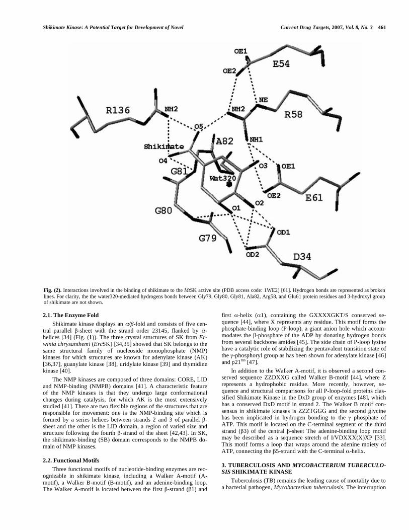
lanines is not disrupted in spite of the shift of Phe49 and Phe57 residues towards shikimate of, 0.49 Å and 0.79 Å, respectively. Moreover, there is a reduction in molecular surface area value the ternary complex compared to binary complex. The calculated value for MtSK complexed with MgADP (1L4U) was approximately 7246 A2 and for MtSK complexed with MgADP and shikimate a value of 6915 A2 was found. Based on these results, the difference between the complexes molecular surface areas is 330 A2, which are buried on shikimate binding (Fig. (3)) [61]. A reduction in mo-lecular surface area value has also been observed in the Mg2+-free crystal structure of MtSK-ADP-Shikimate ternary complex (Fig. (3)). Nevertheless, in this complex a molecular surface of only 206 A2 is buried on shikimate binding, indicating that Mg2+ binding has a role in the closure of MtSK active site. It is important to point out that both the MtSK-MgADP bynary complexes [33] and MtSK-MgADP-Shikimate [61] and MtSK-ADP-Shikimate [60] ternary complexes have been crystallized in the same space group with similar unit-cell parameters and residues from LID and SB domains form no crystal contacts with symmetry-related MtSK molecules. Thus, the conformational changes that have been described for MtSK cannot merely be a reflection of the different crystal-packing arrangements.
The incomplete LID-domain closure observed in crystal struc-tures of both MtSK-MgADP-Shikimate [61] and MtSK-ADP-Shikimate [60] ternary complexes suggests that -phosphate of ATP is necessary for the completion of the domain movement [34]. This find is not unexpected, since total active-site closure upon dead-end ternary complex formation would result in locking the enzyme ac-tive site in an inactive form in which shikimate substrate binding to MtSK enzyme prior to MgADP dissociation from its active site would result in an inactive abortive complex. Several previously reported results are consistent with these structural data. Measure-ments of ErcSK intrinsic tryptophan fluorescence (Trp54) on shikimate binding to either ErcSK or the ErcSK-MgADP binary complex showed a modest synergism of binding between these substrates, since the dissociation constant value for shikimate (Kd = 0.72 mM) decreased to 0.3 mM in the presence of 1.5 mM ADP [75]. In addition, measurements of the quenching of protein fluo-rescence of the aroL-encoded ErcSK upon nucleotide binding dem-onstrated the dissociation-constant values for ADP and ATP to be 1.7 and 2.6 mM, respectively [35]. The Km for ATP (620 M) was found to be approximately four times lower than the dissociation constant in the absence of shikimate [35]. These results prompted the proposal that the conformational changes in the ErcSK enzyme associated with the binding of the first substrate led to an increase
Fig. (3). Molecular surface of (A) MtSK-MgADP (1L4U) [33], (B) MtSK-MgADP-Shikimate (1WE2) [61], and (C) MtSK-ADP-Shikimate (1U8A) [60] struc-tures The molecular surface areas have been calculated using the program Swiss-PDBViewer v3.7 (www.expasy.org/spdbv), probe radius of 1.4 Å, and a fixed radius for all atoms. Calculated values are approximately 7246 Å2 for MtSK complexed with only MgADP (A), 6915 Å2 for structure complexed with MgADP and Shikimate (B), and 7040 Å2 for the Mg2+-free structure complexed with ADP and Shikimate. Thus, approximately 330 Å2 and 206 Å2 of solvent-acessible surface are buried on shikimate binding to, respectively, MtSK-MgADP-Shikimate and MtSK-ADP-Shikimate ternary complexes. The shikimate binding leads to a counter movement of LID and SB domains and, consequently, to a partial closure of shikimate-binding pocket. Probably, the Mg2+ cation has a role in the closure of MtSK active site. All atoms of shikimate and ADP, Mg+2 cation, and oxygen atoms of waters are colored grey.
Shikimate Kinase: A Potential Target for Development of Novel Current Drug Targets, 2007, Vol. 8, No. 3 465
in the affinity for the second substrate. However, this synergism on substrate binding does not appear to hold for MtSK since the appar-ent dissociation-constant values for ATP (89 M) and shikimate (440 M) are similar to their Km values, 83 M for ATP and 410
M for shikimate considering either a rapid equilibrium random-order bi-bi enzyme mechanism or a steady-state-ordered bi-bi en-zyme mechanism [33]. Moreover, no evidence for synergism be-
tween shikimate and ATP could be observed in substrate binding to ErcSK in a chloride buffer system [76].
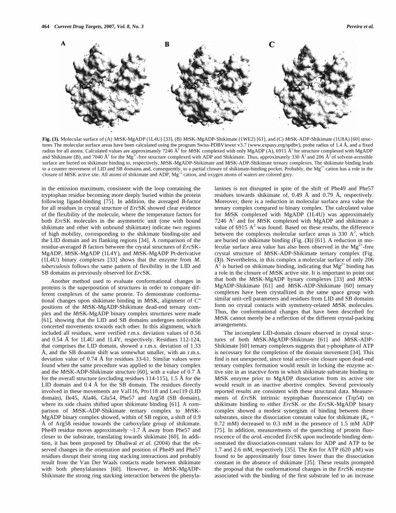
The K15M ErcSK mutant has been crystallized in an open con-formation that is proposed to presumably be equivalent to an apo-enzyme structure in which neither ADP (or ATP) nor shikimate would be bound [35]. This mutant was produced to evaluate the role of the conserved Lys15 of the Walker A motif. However, an unwanted mutation point mutation in the LID domain (Pro115Leu) was detected during the refinement of the model. Furthermore, the enzyme was crystallized with two independent molecules in the asymmetric unit and extensive contacts of neighboring LID do-mains lead to a stabilization of this part of the molecule that is not visible in the native crystal structure [34,35]. It therefore appears unwarranted to consider the double K15M/P115L ErcSK mutant a model for the apo enzyme. Since a better model for the apo-enzyme is not currently available, here we considered the double K15M/P115L ErcSK mutant as a model for the apo-SK. The super-position of MtSK-MgADP-Shikimate and apo ErcSK (K15M+ P115L) show the large conformational changes in the LID and SB domains associated with the binding of ADP and shikimate on MtSK (Fig. (4)).
4. MOLECULAR DYNAMICS SIMULATIONS OF THE SHIKIMATE KINASE STRUCTURE
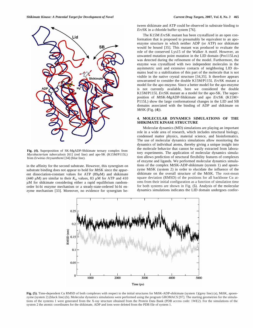
Molecular dynamics (MD) simulations are playing an important role in a wide area of research, which includes structural biology, condensed matter physics, material science, and bioinformatics. The use of molecular dynamics simulations allow monitoring the dynamics of individual atoms, thereby giving a unique insight into the molecule behavior that cannot be easily extracted from labora-tory experiments. The application of molecular dynamics simula-tion allows prediction of structural flexibility features of complexes of enzyme and ligands. We performed molecular dynamics simula-tions of the complex MtSK-ADP-shikimate (system 1) and apoen-zyme MtSK (system 2) in order to elucidate the influence of the shikimate on the overall structure of the MtSK. The root-mean square deviation (RMSD) of the positions for all backbone C at-oms from their initial configuration as a function of simulation time for both systems are shown in Fig. (5). Analysis of the molecular dynamics simulations indicates the LID domain undergoes confor-
Fig. (4). Superposition of SK-MgADP-Shikimate ternary complex from Mycobacterium tuberculosis [61] (red line) and apo-SK (K15M/P115L) from Erwinia chrysanthemi [34] (blue line).
Fig. (5). Time-dependent C RMSD of both complexes with respect to the initial structures for MtSK-ADP-shikimate (system 1)(grey line) (a), MtSK, apoen-zyme (system 2) (black line) (b). Molecular dynamics simulations were performed using the program GROMACS [97]. The starting geometries for the simula-tions of the systems 1 were generated from the X-ray structure obtained from the Protein Data Bank (PDB access code: 1WE2). For the simulations of the system 2 the atomic coordinates for the shikimate, ADP and ions were deleted from the PDB file of system 1.

466 Current Drug Targets, 2007, Vol. 8, No. 3 Pereira et al.
mation change in the system 2 (without shikimate). The structure without the molecule of MtSK presents high flexibility for the resi-dues involved in the LID domain in the system 2. Analysis of the residues participating in the LID domain in the system 1 indicates the presence of a hydrogen bond network involving residues 112-124 and the shikimate, which promotes the stabilization of the LID domain in the closed conformation. Molecular dynamics simula-tions of the system 2 for 5ns show the movement of the LID do-main in the MtSK structure. Superposition of the atomic coordi-nates of the initial and final structures for system 2 after 5ns of molecular dynamics simulation is shown in Fig. (6). The LID do-main is moved towards the solvent in the structure at the final of the 5ns molecular dynamics simulation, demonstrating that the molecu-lar dynamics simulation results are in agreement with experimental X-ray crystallographic data (Fig. (4)), confirming that the binding of the shikimate to the SK structure is enough to promote the clo-sure of the LID domain. On the other hand, the lacking of the shikimate in the MtSK promotes the opening of the LID domain. This structural feature should be considered in any structure-based design of SK inhibitors, which are competitive against shikimate. A detailed analysis of the molecular dynamics simulations for MtSK with different ligand is underway and will be published elsewhere.
5. CONCLUDING REMARKS
The disruption of gene arok, which codes for the shikimate kinase, has been shown to be essential for the viability of M. tuber-culosis. The evidence that shikimate pathway is essential for M. tuberculosis even in the presence of exogenous supplements as p-aminobenzoate, p-hydroxibenzoate and aromatic amino acids rein-forces its attractiveness as a drug target [26]. It is interesting to note that shikimic acid can be used to supply the aromatic amino acid and aromatic vitamin requirements of E. coli blocked in any of the first three steps of the shikimate pathway [77]. This ability has been related to the presence of the shikimic acid transport system en-coded by the gene shiA [78]. Thus, there is the possibility that
shikimic acid could be used as a supplement to allow the growth in vitro of mutant M. tuberculosis strains disrupted on aroG, aroB, aroD or aroE genes, which code for first four shikimate pathway enzymes in M. tuberculosis. These aro-disrupted M. tuberculosis strains could be used in experiments to determine if shikimate pathway is essential to in vivo growth of M. tuberculosis.
A comprehensive structural picture of the interactions between M. tuberculosis SK enzyme and Mg2+, ADP and shikimate sub-strates have been obtained from crystallographic studies [33,60,61]. In addition, the LID and SB domains conformational changes upon ADP and shikimate substrates binding, which result in a partial closure of and solvent expulsion from MtSK active site, has been described.
There has been considerable debate within the literature as whether enzyme-catalyzed phosphoryl transfer reactions operate primarily with an associative (SN2-like) or dissociative (SN1-like) transition state [62]. A criterion that has been used for distinguish-ing between these mechanisms is based on the geometry and reac-tion coordinate distances between the terminal phosphoryl group of ATP and the acceptor substrate in ground state enzyme-substrate or enzyme inhibitor complexes [79]. For the transition state to have some degree of associative character, these distances are expected to be less than the sum of a P-O van der Waals contacts and a P-O single bond (i.e in the order of approximately 4.9 Å) [62]. In disso- ciative mechanisms these distances would be expected to be longer than this to allow space for the intermediate monomeric metaphos-phate to exist between the leaving group (ADP) and the entering group [79]. A crystal structure of MtSK in complex with MgADP, Shikimate, and AlF3 (a structural analog of -phosphoryl group transfered in the reaction) could permit a snapshot of the transition-state like structure of MtSK. As it has been shown to other kinases [80-93], this structural study could distinguish between associative and dissociative transition states in the reaction catalyzed by MtSK.
It has been recently described the structural alterations caused by binding of the shikimate, magnesium and chloride ions in the SK from Mycobacterium tuberculosis (MtSK). These new findings indicate that both ions and the shikimate influence the structural conformation of MtSK. The magnesium ion also appears to influ-ence both the hydroxyl groups of shikimate molecule and some residues from active site. The chloride ion can influence the affinity of shikimate for MtSK, in the binding position of ADP in MtSK active site and also in the opening length of LID domain of the MtSK structure. The shikimate binding causes a closing of LID domain, which appears to influence drastically the crystallographic packing of the protein [94-95].
Structures of SKs deposited in the protein data bank (PDB) were superimposed and positions of residues involved in binding (Lys15, Asp34, Arg58, Glu61, Gly79, Gly80, Gly81, Arg136) be-tween shikimate and SK are highly conserved [61]. The precise interactions between MtSK and shikimate have been elucidated, showing the residues involved in the binding of substrate and the conformational changes upon substrate binding. Accordingly, the availability of the M. tuberculosis shikimate kinase structures com-plexed with shikimate provide crucial information for the design of non-promiscuous SK inhibitors that target both the shikimate- and ATP-binding pockets or uniquely, the shikimate-binding site. Moreover, the knowledge of functional factors that lead to active site closure could be used for designing inhibitors that force MtSK to a closed conformation that would be unable to catalyze the phos-phoryl transfer to shikimate. These inhibitors could block the bio-synthesis of aromatic aminoacids [96] and other compounds (folate, micobactins, etc), which are essential for the growth and viability of the microorganism.
ACKNOWLEDGEMENTS
Financial support for this work was provided by FAPESP (Proc. 02/05347-4, 01/07532-0, 04/00217-0) and Millennium Initiative
Fig. (6). Superposition of the atomic coordinates of the initial (light grey) and final (grey) structures for system 2. The atomic coordinates for the final structure were those at the end of 5ns molecular dynamics simulation.
Shikimate Kinase: A Potential Target for Development of Novel Current Drug Targets, 2007, Vol. 8, No. 3 467
Program MCT-CNPq, Ministry of Health - Secretary of Health Policy (Brazil) to DSS and LAB. DSS (304051/1975-06) WFA (CNPq, 300851/98-7), and LAB (520182/99-5) are research career awarded from the National Research Council of Brazil (CNPq).
ABBREVIATIONS
TB = Tuberculosis
Mt = Mycobacterium tuberculosis
SK = Shikimate kinase
DAHPS = 3-deoxy-D-arabino-heptulosonate-7-phosphate synthase
CS = Chorismate synthase
EPSPS = 5-enolpyruvylshikimate-3-phosphate synthase
PABA = p-aminobenzoate
GTP = Guanosine triphosphate
ATP = Adenosine triphosphate
ADP = Adenosine diphosphate
Km = Michaellis-Menten constant
NMP = Nucleoside monophosphate
AK = Adenylate kinase
WHO = World Health Organization
HIV = Human immunodeficiency vírus
MDR = Multidrug-resistant
DHQ = Dehydroquinase
PCR = Polymerase chain reaction
IPTG = Isopropyl-beta-D-thiogalactoside
Mg = Magnesium
r.m.s. = Root mean square
Erc = Erwinia chrysantemi
AMP-PCP = Adenosine - , -methyleneadenosine 5'-triphosphate
AMP-PCP = , -imidoadenosine 5'-triphosphate
REFERENCES
[1] Davis, B.D. and Mingioli, E. S. (1953) J. Bacteriol. 66(2), 129-136. [2] Sprinson, D.B. (1960) Adv. Carbohydrate Chem. 15, 235-270. [3] Pittard, A.J. (1987) In F.C. Neidhardt, J.L. Ingraham, K.B. Low, B.
Magasanik, M. Schaechter, and H.E. Umbarger (ed.), Escherichia coli and Salmonella typhimurium: cellular and molecular biology, vol.1, pp. 368-394. American Society for Microbiology, Washing-ton, D.C.
[4] Haslam, E. (1993) Shikimic acid: metabolism and metabolites. John Wiley & Sons, Chichester, United Kingdom.
[5] Folkers, K.(1996). Biochem. Biophys. Res. Commun. 224(2), 358-361.
[6] Coggins, J.R., Duncan, K., Anton, I.A., Boocock, M.R., Chaudhuri, S., Lambert, J.M., Lewendon, A., Millar, G., Mousdale, D.M., Smith, D.D., (1987) Biochem. Soc. Trans. 15(4), 754-759.
[7] Butler, J.R., Alworth, W.L., Nugent, M.J., (1974) J. Am. Chem.
Soc. 96, 1617-1618. [8] Mousdale, D.M., Campbell, M.S., Coggins, J.R. (1987) Phyto-
chemistry 26(10), 2665-2670. [9] Duncan, K., Edwards, R.M., Coggins, J.R. (1987) Biochem. J.
246(2), 375-386. [10] Davies, G.M., Barret-Bee, K.J., Jude, D.A., Lehan, M., Nichols,
W.W., Pinder, P.E., Thain, J.L., Watkins, W.J., Wilson, R.G. (1994) Antimicrob. Agents Chemother. 38(2), 403-406.
[11] Coggins, J.R. (1989) The shikimate pathway as a target for herbi-cides. In Herbicides and Plant Metabolism (Dodge, A., ed.), Cam-bridge University Press, Cambridge, UK, pp. 97-112.
[12] Bentley, R. (1990). Crit. Rev. Biochem. Mol. Biol. 25(5), 307-384. [13] McDevitt, D., Payne, D.J., Holmes, D.J., and Rosenberg, M. (2002)
J. Appl. Microbiol. 92, 28S-34S.
[14] McArthur, J.D., West, N.P., Cole, J.N., Jungnitz, H., Guzman,C.A., Chin, J., Lehrbach, P.R., Djordjevic, S.P., and Walker, M.J. (2003) FEMS Microbiol. Lett. 221(1), 7-16.
[15] Roberts, F., Roberts, C.W., Johnson, J.J., Kyle, D.E., Krell, T., Coggins, J.R., Coombs, G.H., Milhous, W.K., Tzipori, S., Fergun-son, D.J.P., Chakrebarti, D., and McLeod, R. (1998). Nature 393, 801-805.
[16] Kishore, G.M. & Shah, D.M. (1988) Annu. Rev. Biochem. 57, 627-663.
[17] Dieckmann, A., and Jung, A. (1986) Mol. Biochem. Parasitol. 19(2), 143-147.
[18] Campbell, S.A., Richards, T.A., Mui, E.J., Samuel, B.U., Coggins, J.R., McLeod, R., Roberts, C.W. (2004) Int. J. Parasitol. 34(1), 5-13.
[19] McRobert, L., McConkey, G.A. (2002) Mol. Biochem. Parasitol. 119(2), 273-278.
[20] McConkey G.A. (1999) Antimicrob. Agents Chemother. 43(1), 175-177.
[21] Gardner, M.J., Shallom, S.J., Carlton, J.M., Salzberg, S.L., Nene, V., Shoaibi, A., Ciecko, A., Lynn, J., Rizzo, M., Weaver, B., Jar-rahi, B., Brenner, M., Parvizi, B., Tallon, L., Moazzez, A., Granger, D., Fujii, C., Hansen, C., Pederson, J., Feldblyum, T., Peterson, J., Suh, B., Angiuoli, S., Pertea, M., Allen, J., Selengut, J., White, O., Cummings, L.M., Smith, H.O., Adams, M.D., Venter, J.C., Ca-rucci, D.J., Hoffman, S.L., Fraser, C.M. (2002) Nature 419, 498-511.
[22] McConkey, G.A., Pinney, J.W., Westhead, D.R. (2004) Trends
Parasitol. 20(2), 60-65. [23] Fitzpatrick T, Ricken S, Lanzer M, Amrhein N, Macheroux P,
Kappes B. (2001) Mol. Microbiol. 40(1), 65-75. [24] Ratledge, C. (1982). The Biology of the Mycobacteria. Academic
Press, London 1, 185-271. [25] Voos, J.J., Rutter, K., Schroder, B.G., Su, H., Zhu, Y., and Barry,
C.B. III. (2000) Proc. Natl. Acad. Sci. USA, 97, 1252-1257. [26] Parish, T., and Stoker, N. G. (2002) Microbiology 148, 3069-3077. [27] Whipp, M.J. & Pittard, A.J. (1995) J. Bacterial. 177(6), 1627-1629. [28] Millar, G., Lewendon, A., Hunter, M.G. & Coggins, J.R. (1986).
Biochem. J. 237(2), 427-437. [29] De Feyter, R.C. & Pittard, J. (1986) J. Bacteriol. 165(1), 331-333. [30] Ely, B. and Pittard, J. (1979) J. Bacteriol. 138(3), 933-943. [31] De Feyter, R.C., Davidson, B.E., and Pittard, J. (1986) J. Bacteriol.
165(1), 233-239. [32] Vinella, D., Gagny, B., Joseleau-Petit, D., D’Ardi, R. & Cashel, M.
(1996) J. Bacteriol. 178(13), 3818-3828. [33] Gu, Y., Reshetnikova, L., Li, Y., Wu, Y., Yan, H., Singh, S., Ji, X.
(2002) J. Mol. Biol. 319(3), 779-789. [34] Krell, T., Coggins, J.R. & Lapthorn, A.J. (1998) J. Mol. Biol.
278(5), 983-997. [35] Krell, T., Maclean, J., Boam, D.J., Cooper, A., Resmini, M., Brock-
lehurst, K. et al. (2001) Protein Sci. 10(6), 1137-1149. [36] Dreusicke, D., Karplus, A. & Schulz, G.E. (1988) J. Mol. Biol.
199(2), 359-371. [37] Schlauderer, G.J. & Schulz, G.E. (1996) Protein Sci. 5(3), 434-441. [38] Stehle, T. & Schulz, G.E. (1990) J. Mol. Biol. 211(1), 249-254. [39] Müller-Dieckmann, H.-J. & Schulz, G.E. (1994) J. Mol. Biol.
236(1), 361-367. [40] Wild, K., Bohner, T., Aubry, A., Folkers, G. & Schulz, G.E. (1995)
FEBS Lett. 368(2), 289-292. [41] Vonrhein, C., Schlauderer, G. J. & Schulz, G. E. (1995) Structure,
3(5), 483-490. [42] Müller, C.W., Schlauderer, G.J., Reinstein, J. & Schulz, G.E.
(1996) Structure, 4(2), 147-156. [43] Gerstein, M., Schulz, G.E. & Chothia, C. (1993) J. Mol. Biol.
229(2), 494-501. [44] Walker, J.E., Saraste, M., Runswick, M.J. & Gay, N.J. (1982).
EMBO J. 1(8), 945-951. [45] Smith, C.A. & Rayment, I. (1996) Biophys. J. 70(4), 1590-1602. [46] Reinstein, J., Schlichting, I., and Wittinghofer, A. (1990) Bioche-
mistry 29(32), 7451-7459. [47] Sigal, I.S., Gibbs, J.B., D’Alonzo, J.S., Temeles, G.L., Wolanski,
B.S., Socher, S.H., and Scolnick, E.M. (1986) Proc. Natl. Acad.
Sci. USA 83(4), 952-956. [48] Leipe, D.D., Koonin, E.V., and Aravind, L. (2003) J. Mol. Biol.
333(4), 781-815. [49] Raviglione, M.C. (2003) Tuberculosis, 83(1-3), 4-14.

468 Current Drug Targets, 2007, Vol. 8, No. 3 Pereira et al.
[50] Corbett, E.L., Watt, C.J., Walker, N., Maher, D., Willians, B.G., Raviglione, M.C., Dye, C. (2003). Arch. Intern. Med. 163(9), 1009-1021.
[51] Dye C, Scheele S, Dolin P, Pathania V, Raviglione, M.C. (1999) JAMA 282(7), 677-686.
[52] World Health Organization: Global Tuberculosis Control. WHO Report 2004. ISBN 92 4 156264 1.
[53] Basso, L.A., and Santos, D.S. (2005) Med. Chem. Rev. Online 2, 393-413.
[54] Duncan, K. (2003) Tuberculosis, 83(1-3), 201-207. [55] O’Brien, R.J., Nunn, P.P. (2001) Am. J. Resp. Crit. Care Med.
163(5), 1055-1058. [56] Garbe, T., Joens, C., Charles, I., Dougan, G., Young, D. (1990) J.
Bacteriol. 172(12), 6774-6782. [57] Cole, S.T.; Brosch, R.; Parkhill, J.; Garnier, T.; Churcher, C.; Har-
ris, D.; Gordon, S.V.; Eiglmeier, K.; Gas, S.; Barry III, C.E.; Tekaia, F.; Badcock, K.; Basham, D.; Brown, D.; Chillingworth, T.; Connor, R.; Davies, R.; Devlin, K.; Feltwell, T.; Gentles, S.; Hamlin, N.; Holroyd, S.; Hornsby, T.; Jagels, K.; Krogh, A.; McLean, J.; Moule, S.; Murphy, L.; Oliver, K.; Osborne, J.; Quail, M.A.; Rajandream, M.-A.; Rogers, J.; Rutter, S.; Seeger, K.; Skel-ton, J.; Squares, R.; Squares, S.; Sulston, J.E.; Taylor, K.; White-head, S. and Barrell, B.G. (1998) Nature 393, 537-544.
[58] Oliveira, J.S., Pinto, C.A., Basso, L.A., Santos, D.S. (2001) Protein Expr. Purif. 22(3), 430-435.
[59] De Azevedo, W. F. Jr., de Oliveira, J. S., Basso, L. A., Palma, M.S., Pereira, J. H., Canduri, F., and Santos, D. S. (2002) Biochem.
Biophys. Res. Commun, 295(1), 142-148. [60] Dhaliwal, B., Nichols, C.E., Ren, J., Lockyer, M., Charles, I.,
Hawkins, A.R., Stammers, D.K. (2004) FEBS Lett. 574(1-3), 49-54.
[61] Pereira, J.H., Oliveira, J.S, Canduri, F., Dias, M.V.B., Palma, M.S., Basso, L.A., Santos, D.S., and Azevedo Jr., W.F. (2004) Acta
Cryst. Section D 60, 2310-2319. [62] Matte, A., Tari, L.W., and Delbaere, T.J. (1998) Structure, 6(4),
413-419. [63] Williams, R.L., Oren, D.A., Muñoz-Dorado, J., Inouye, S., Inouye,
M., and Arnold, M. (1993) J. Mol. Biol. 234(4), 1230-1247. [64] Jencks, W.P. (1975) Adv. Enzymol. 43, 219-410. [65] Smith, C.A., Reyment, I. (1995) Biochemistry, 34(28), 8973-8981. [66] Berchthold, H., Reshetnikovca, L., Reiser, C.O.A., Schrimer, N.K.,
Sprinzl, M., Hilgenfeld, R. Nature (1993) 365(6442), 126-132. [67] Pai, E.F., Krengel, U., Petski, G.A., Goody, R.S., Kabsch, W.,
Wittinghofer, A. . (1990) EMBO J. 9(8), 2351-2359. [68] Coleman, D.E., Berghuis A.M., Lee, E., Linder, M.E., Gilman,
A.G., Sprang, S.R. (1994) Science 265(5177), 1405-1412. [69] Abele, U. and Schulz, G. E. (1995) Protein Sci. 4(7), 1262-1271. [70] Schlauderer, G.J. & Schulz, G.E. (1996) Protein Sci. 5(3), 434-441. [71] Hasemann, C. A., Istvan, E. S., Uyeda, K. & Deisenhofer, J. (1996)
Structure 4(9), 1017-1029. [72] Romanowski, M.J., Burley, S.K. (2002) Proteins: Structure, Func-
tion Genet. 47(4), 558-562. [73] Bennett, W.S. and Steitz, T.A. (1980) J. Mol.Biol. 140(2), 211-230. [74] Schulz, G.E., Müller, C.W. and Diederichs, K. (1990) J. Mol. Biol.
213(4), 627-630.
[75] Idziak, C., Price, N.C., Kelly, S.M., Krell, T., Boam, D.J., Lapthorn, A.J., and Coggins, J.R. (1997) Biochem. Soc. Trans. 25(4), S627.
[76] Cerasoli, E., Kelly, S.M., Coggins, J.R., Lapthorn, A.J., Clarke D.T. (2003) Biochim. Biophys.Acta, 1648(1-2), 43-54.
[77] Pittard, A.J., and Wallace, B.J. (1966) J. Bacteriol. 92(4), 1070-1075.
[78] Whipp, M.J., Camakaris, H., and Pittard, A.J. (1998) Gene 209(1-2), 185-192.
[79] Mildvan, A.S. (1979) Adv. Enzymol. Relat. Areas Mol. Biol. 49, 103-126.
[80] Schlichting, I., and Reinstein, J. (1997) Biochemistry 36(31), 9290-9296.
[81] Xu, Y.-W., Moréra, S., Janin, J., and Cherfils, J. (1997) Proc. Natl. Acad. Sci. USA, 94(8), 3579-3583.
[82] Scheffzek, K., and Wittinghofer, A. (1997) Science, 277, 333-338. [83] Basso, L. A., Pereira da Silva, L. H., Fett-Neto, A. G., de Azevedo
Jr., W. F., Moreira, I. S., Palma, M. S., Calixto, J. B., Astolfi Filho, S., dos Santos, R. R., Soares, M. B. P., Santos, D. S. (2005) Mem.
Inst. Oswaldo Cruz, 100(6): 475-506. [84] da Silveira, N. J. F., Uchoa, H. B., Canduri, F.,Pereira, J. H., Cam-
era Jr., J. C., Basso, L. A., Palma, M. S., Santos, D. S., de Azevedo Jr., W. F. (2004) Biochem Biophys. Res. Commun. 322(1), 100-104.
[85] Uchoa, H. B., Jorge, G. E., da Silveira, N. J., Camera J. C., Can-duri, F., De Azevedo, W.F. (2004) Biochem. Biophys. Res. Com-
mun. 325(4), 1481-1486. [86] da Silveira, N. J. F., Bonalumi, C. A., Uchoa, H. B., Pereira, J. H.,
Canduri, F.,Pereira, J. H., de Azevedo Jr., W. F. (2006) Cell Bio-chem. Biophys. 44(3), 366-374.
[87] De Azevedo, W.F. Jr.; Mueller-Dieckmann, H.J.; Schulze-Gahmen, U.; Worland, P.J.; Sausville, E.; Kim S.-H. (1996) Proc. Natl. Acad. Sci. USA. 93(7), 2735-2740.
[88] Kim, S.-H.; Schulze-Gahmen, U.; Brandsen, J.; De Azevedo, W. F. Jr. (1996) Prog. Cell Cycle Res. 2, 137-145.
[89] Canduri, F.; Uchoa, H.B.; de Azevedo, W.F.Jr. (2004) Biochem.
Biophys. Res. Commun. 324(2), 661-666. [90] De Azevedo, W.F.Jr.; Canduri, F.; Silveira, N.J.F. (2002) Biochem
Biophys. Res. Commun. 293(1), 566-571. [91] De Azevedo, W.F.Jr.; Leclerc, S.; Meijer, L.; Havlicek, L.; Strnad,
M.; Kim, S.-H. (1997) Eur. J. Biochem. 243, 518-526. [92] De Azevedo, W.F.Jr., Gaspar, R.T., Canduri, F.; Camera, J.C.Jr.,
Silveira, N.J.F. (2002) Biochem Biophys. Res. Commun. 297(5), 1154-1158.
[93] De Azevedo, W. F. Jr., Canduri, F. (2005) Curr. Comput. Aided Drug Design 1, 53-64.
[94] Gan, J., Gu, Y., Li, Y., Yan, H., Ji, X. (2006) Biochemistry 45, 8539-8545.
[95] Dias, M. V. B., Faim, L. M., Vasconcelos, I. B., Oliveira, J. S., Basso, L. A., Santos, D. S., De Azevedo, W. F. Acta Crystal-
lographica F. (In press). [96] Ducati, R. D., Basso, L. A., Santos, D. S. (2007) Curr. Drug Tar-
gets 8(1) In press. [97] van der Spoel, D., Lindahl, E., Hess, B., Groenhof, G., Mark, A. E.
Berendsen, H. J. C. (2005) J. Comp. Chem. 26(16),1701-1718.
Received: December 12, 2006 Accepted: May 26, 2006 Updated: September 11, 2006