Sequencing of cDNA from 50 unrelated patients reveals that mutations in the triple-helical domain of...
7
Sequencing of cDNA from 50 Unrelated Patients Reveals That Mutations in the Triple-Helical Domain of Type Ill Procollagen Are an Infrequent Cause of Aortic Aneurysms Gerard Tromp, * Yuli Wu, * Darwin J. Prockop, * Swama L. Madhatheri, * Caren Kleinert, * James J. Earley, * Jiapiao Zhuang, * Orjan Norrgard,t R. Clement Darling,' William M. Abbott, C. William Cole,11 Pekka Jaakkola,' Markku Ryynanen,** William H. Pearce, James S. T. Yao, Kari Majamaa," Stanton N. Smullens,1111 Zoran Gatalica," Robert E. Ferrell,*** Sergio A. Jimenez,mt Charles E. Jackson, Of Virginia V. Michels,111111 Michael Kaye,"' and Helena Kuivaniemi* *Department of Biochemistry and Molecular Biology, Jefferson Institute of Molecular Medicine, and "Department of Pathology, Jefferson Medical College, and 111IDepartment ofSurgery, and mDivision ofRheumatology, Department ofMedicine, Thomas Jefferson University Hospital, Thomas Jefferson University, Philadelphia, Pennsylvania 19107; tDepartment of Surgery, University of Umed, S-90185 Umed, Sweden; §General Surgical Services, Massachusetts General Hospital, and Department of Surgery, Harvard Medical School, Boston, Massachusetts 02114; I1Division of Vascular Surgery, University of Ottawa, Ottawa, Ontario, Canada KJ Y 4E9; Departments of'Surgery, and **Obstetrics and Gynecology, University of Kuopio, SF-70211 Kuopio, Finland; t$Division of Vascular Surgery, Northwestern University Medical School, Chicago, Illinois 60611; *Department ofNeurology, University of Oulu, SF-90220 Oulu, Finland; ***Department ofHuman Genetics, University ofPittsburgh, Pittsburgh, Pennsylvania 15261; MDivision of Clinical and Molecular Genetics, Henry Ford Hospital, Detroit, Michigan 48202; 11111IDepartment of Medical Genetics, Mayo Clinic, Rochester, Minnesota 55905; and 'Division ofNephrology, The Montreal General Hospital, Montreal, Quebec H3G IA4, Canada Abstract Detailed DNA sequencing of the triple-helical domain of type III procollagen was carried out on cDNA prepared from 54 patients with aortic aneurysms. The 43 male and 11 female patients originated from 50 different families and five different nationalities. 43 patients had at least one additional blood rela- tive who had aneurysms. Five overlapping asymmetric PCR products, covering all the coding sequences of the triple-helical domain of type III procollagen, were sequenced with 28 specific sequencing primers. Analysis of the sequencing gels revealed only two nucleotide changes that altered the structure of the protein. One was a substitution of threonine for proline at amino acid position 501 and its functional importance was not clearly established. The other was a substitution of arginine for an obligatory glycine at amino acid position 136. In 40 of the 54 patients, detection of a polymorphism in the mRNA estab- lished that both alleles were expressed. The results indicate that mutations in type III procollagen are the cause of only about 2% of aortic aneurysms. (J. Clin. Invest. 1993. 91:2539- 2545.) Key words: familial aneurysms * polymerase chain reac- tion- polymorphisms Introduction A number of careful studies have shown that aneurysms, even when they are not associated with well-recognized, heritable syndromes such as the Marfan syndrome ( 1 ) or Ehlers-Danlos Address reprint requests to Dr. Helena Kuivaniemi, Department of Biochemistry and Molecular Medicine, Jefferson Institute of Molecu- lar Medicine, Jefferson Medical College, Thomas Jefferson University, 233 South Tenth Street, Philadelphia, PA 19107. Receivedfor publication 15 October 1992 and in revisedform 22 January 1993. syndrome (EDS)' type IV ( 1 ), are frequently familial (2-1 1 ). A recent statistical evaluation suggested that aneurysms are caused by defects in a single gene ( 12). The structural compo- nents of arteries and the gene products participating in the proteolysis of the structural components have been suggested as candidate genes for aneurysms ( 13 ). Mutations in the fibril- lin gene located in chromosome 15 (14) have been shown to cause the Marfan syndrome ( 15, 16). There is a considerable variability in the phenotype among different individuals with the Marfan syndrome within the same family and among dif- ferent families ( 17). Fibrillin 15 is, therefore, a potential candi- date gene for aortic aneurysms ( 17). Other candidate genes for aortic aneurysms are collagens and elastin, which are impor- tant structural components of the aorta. Their contribution to the strength of an artery was elegantly demonstrated by Dobrin and his co-workers ( 18) by using dog and human cadaver arter- ies in a system where pressure could be applied onto the artery. Treatment of the artery with collagenase or elastase resulted in a considerable weakness of the structure ( 18). Several different types of collagens have been isolated from blood vessels, but types I and III collagen are the major components in the arte- rial wall ( 19). They are fibril-forming collagens in which three polypeptide chains called the a chains form a stable triple-heli- cal structure (20, 21 ). Each a chain consists of about 340 re- peating tripeptide sequences of -Gly-X-Y- in which X is fre- quently proline and Y is frequently hydroxyproline (20, 21 ). A recent study on the type III procollagen secreted by cultured skin fibroblasts from patients with aortic aneurysms demon- strated that in 2 out of 14 patients the type III procollagen was thermally unstable when assayed by brief proteinase digestion, suggesting a structural defect in the type III procollagen (22). Furthermore, a mutation in the type III procollagen gene was found in a man who expired at the age of 34 yr from a rupture of the aorta (23). The man had a history of easy bruisability and bleeding, but none of the dramatic skin changes usually associated with type IV EDS ( 1, 24) such as ecchymoses, ab- normal scarring, and heavy pigmentation (23). He, therefore, 1. Abbreviations used in this paper: EDS, Ehlers-Danlos syndrome; nt, nucleotide. Aortic Aneurysms and Type III Procollagen Gene 2539 J. Clin. Invest. © The American Society for Clinical Investigation, Inc. 0021-9738/93/06/2539/07 $2.00 Volume 91, June 1993, 2539-2545
Transcript of Sequencing of cDNA from 50 unrelated patients reveals that mutations in the triple-helical domain of...
Sequencing of cDNA from 50 Unrelated Patients RevealsThat Mutations in the Triple-Helical Domain of Type Ill
Procollagen Are an Infrequent Cause of Aortic AneurysmsGerard Tromp, * Yuli Wu, * Darwin J. Prockop, * Swama L. Madhatheri, * Caren Kleinert, * James J. Earley, * Jiapiao Zhuang, *
Orjan Norrgard,t R. Clement Darling,' William M. Abbott, C. William Cole,11 Pekka Jaakkola,' Markku Ryynanen,**William H. Pearce, James S. T. Yao, Kari Majamaa," Stanton N. Smullens,1111 Zoran Gatalica," Robert E. Ferrell,***Sergio A. Jimenez,mt Charles E. Jackson,Of Virginia V. Michels,111111 Michael Kaye,"' and Helena Kuivaniemi**Department ofBiochemistry and Molecular Biology, Jefferson Institute ofMolecular Medicine, and "Department ofPathology,Jefferson Medical College, and 111IDepartment ofSurgery, and mDivision ofRheumatology, Department ofMedicine, Thomas JeffersonUniversity Hospital, Thomas Jefferson University, Philadelphia, Pennsylvania 19107; tDepartment ofSurgery, University of Umed,S-90185 Umed, Sweden; §General Surgical Services, Massachusetts General Hospital, and Department ofSurgery, Harvard MedicalSchool, Boston, Massachusetts 02114; I1Division of Vascular Surgery, University of Ottawa, Ottawa, Ontario, Canada KJ Y 4E9;Departments of'Surgery, and **Obstetrics and Gynecology, University ofKuopio, SF-70211 Kuopio, Finland; t$Division of VascularSurgery, Northwestern University Medical School, Chicago, Illinois 60611; *Department ofNeurology, University ofOulu, SF-90220Oulu, Finland; ***Department ofHuman Genetics, University ofPittsburgh, Pittsburgh, Pennsylvania 15261; MDivision ofClinical andMolecular Genetics, Henry Ford Hospital, Detroit, Michigan 48202; 11111IDepartment ofMedical Genetics, Mayo Clinic, Rochester,Minnesota 55905; and 'Division ofNephrology, The Montreal General Hospital, Montreal, Quebec H3G IA4, Canada
Abstract
Detailed DNA sequencing of the triple-helical domain of typeIII procollagen was carried out on cDNA prepared from 54patients with aortic aneurysms. The 43 male and 11 femalepatients originated from 50 different families and five differentnationalities. 43 patients had at least one additional blood rela-tive who had aneurysms. Five overlapping asymmetric PCRproducts, covering all the coding sequences of the triple-helicaldomain of type III procollagen, were sequenced with 28 specificsequencing primers. Analysis of the sequencing gels revealedonly two nucleotide changes that altered the structure of theprotein. One was a substitution of threonine for proline atamino acid position 501 and its functional importance was notclearly established. The other was a substitution of arginine foran obligatory glycine at amino acid position 136. In 40 ofthe 54patients, detection of a polymorphism in the mRNA estab-lished that both alleles were expressed. The results indicatethat mutations in type III procollagen are the cause of onlyabout 2% of aortic aneurysms. (J. Clin. Invest. 1993. 91:2539-2545.) Key words: familial aneurysms * polymerase chain reac-
tion- polymorphisms
Introduction
A number of careful studies have shown that aneurysms, even
when they are not associated with well-recognized, heritablesyndromes such as the Marfan syndrome ( 1 ) or Ehlers-Danlos
Address reprint requests to Dr. Helena Kuivaniemi, Department ofBiochemistry and Molecular Medicine, Jefferson Institute of Molecu-lar Medicine, Jefferson Medical College, Thomas Jefferson University,233 South Tenth Street, Philadelphia, PA 19107.
Receivedfor publication 15 October 1992 and in revisedform 22January 1993.
syndrome (EDS)' type IV ( 1 ), are frequently familial (2-1 1 ).A recent statistical evaluation suggested that aneurysms arecaused by defects in a single gene ( 12). The structural compo-nents of arteries and the gene products participating in theproteolysis of the structural components have been suggestedas candidate genes for aneurysms ( 13 ). Mutations in the fibril-lin gene located in chromosome 15 (14) have been shown tocause the Marfan syndrome ( 15, 16). There is a considerablevariability in the phenotype among different individuals withthe Marfan syndrome within the same family and among dif-ferent families ( 17). Fibrillin 15 is, therefore, a potential candi-date gene for aortic aneurysms ( 17). Other candidate genes foraortic aneurysms are collagens and elastin, which are impor-tant structural components of the aorta. Their contribution tothe strength ofan artery was elegantly demonstrated by Dobrinand his co-workers ( 18) by using dog and human cadaver arter-ies in a system where pressure could be applied onto the artery.Treatment of the artery with collagenase or elastase resulted ina considerable weakness ofthe structure ( 18). Several differenttypes of collagens have been isolated from blood vessels, buttypes I and III collagen are the major components in the arte-rial wall ( 19). They are fibril-forming collagens in which threepolypeptide chains called the a chains form a stable triple-heli-cal structure (20, 21 ). Each a chain consists of about 340 re-peating tripeptide sequences of -Gly-X-Y- in which X is fre-quently proline and Y is frequently hydroxyproline (20, 21 ). Arecent study on the type III procollagen secreted by culturedskin fibroblasts from patients with aortic aneurysms demon-strated that in 2 out of 14 patients the type III procollagen wasthermally unstable when assayed by brief proteinase digestion,suggesting a structural defect in the type III procollagen (22).Furthermore, a mutation in the type III procollagen gene wasfound in a man who expired at the age of 34 yr from a ruptureof the aorta (23). The man had a history of easy bruisabilityand bleeding, but none of the dramatic skin changes usuallyassociated with type IV EDS ( 1, 24) such as ecchymoses, ab-normal scarring, and heavy pigmentation (23). He, therefore,
1. Abbreviations used in this paper: EDS, Ehlers-Danlos syndrome; nt,nucleotide.
Aortic Aneurysms and Type III Procollagen Gene 2539
J. Clin. Invest.© The American Society for Clinical Investigation, Inc.0021-9738/93/06/2539/07 $2.00Volume 91, June 1993, 2539-2545

probably presented phenotypic overlap between EDS type IVand familial aneurysms. Subsequently, a mutation in the typeIII procollagen gene was found in a 37-yr-old woman who hada strong family history of sudden death from ruptured aorticaneurysms (25) without any evidence of EDS type IV or re-lated disorder.
The hypothesis that aortic aneurysms are caused by in-creased proteolytic activity in the aorta that leads to the weaken-ing ofthe arterial wall and rupture, has also been supported byseveral studies (26-29). There are, however, very few, if any,known examples of a gene defect producing an increase in anenzyme activity. It might, therefore, be that the increased pro-teolytic activity found in aneurysmal aorta is a secondary phe-nomenon due to some other primary defect.
In conclusion, several candidate genes for aortic aneurysmshave been suggested, including (a) fibrillin 15 (17), (b) colla-gen (20, 21, 30), (c) elastin (31), (d) collagenase (18), (e)elastase (27), (f) tissue inhibitor of metalloproteinase (29),and (g) a1-antitrypsin (28). In this article we report the de-tailed DNA sequencing approach to answer the question ofwhether most aneurysms are caused by mutations in the genefor the type III procollagen.
Methods
Patient material. Skin biopsy was obtained from all patients listed inTable I after written informed consent. In some cases, blood sampleswere obtained from family members of the patient after written in-formed consent. Skin biopsies were used to establish skin fibroblastcultures. Total RNA was isolated from cultured skin fibroblasts with aprocedure involving extraction with guanidinium isothiocyanate (32).The RNA was purified by centrifugation on a cesium chloride gradientand used to synthesize first strand ofcDNA with MMVL reverse tran-scriptase (Bethesda Research Laboratories, Gaithersburg, MD). Geno-mic DNA was isolated from cultured skin fibroblasts or blood on aGenepure 341 automated DNA extractor (Applied Biosystems, Inc.,Foster City, CA).
Sequencing ofcDNA. Sets of oligonucleotides (Table II) based onthe cDNA sequence of type III procollagen (33-38) were used asprimers so as to generate five overlapping PCR products ( 1, 2A, 2B, 3,and 4 in Table II) to cover all the coding sequences for the triple-helicaldomain of 1029 amino acids of the type III procollagen. Originally,products 2A and 2B were amplified together as one longer fragment,but since results from DNA sequencing were somewhat variable, theregion was divided into two PCR fragments, after which the sequencingresults improved. To produce single-stranded DNA sequencing tem-plates, three successive PCRs, of 20 cycles each, were performed foreach region.
The first PCR (39) was carried out for 20 cycles in a 30-pl reactionvolume containing 0.6 pmol of each primer. 2 ql of the first PCRproduct was used in a second PCR that was carried out for 20 cycles in a50 pl-reaction volume containing 10 pmol ofone primer and 2.5 pmolof the other. The third PCR was carried out for 20 cycles in a 100 p1reaction volume containing 50 pmol of one primer and 1 pmol of theother primer and 2 ,l of the second PCR product. The products fromthe third PCR were run on an agarose gel, and ifthey were ofacceptablequality, additional third PCRs were performed to produce sufficienttemplate DNA for sequencing.
Typically, the final results from the third PCR showed a clean am-plification ofthe desired product on an agarose gel with two bands, onerepresenting the double-stranded DNA and the other representing thesingle-stranded DNA (not shown). Routinely, cDNA from 10 differ-ent cell lines was amplified simultaneously. Water blanks were in-cluded in every experiment and they were negative.
The PCR products were purified by adsorption to powdered glass(silica) under high salt conditions using a commercial kit (Geneclean,BIO 101, Inc., Vista, CA). Since adsorption ofDNA to silica powder inthe presence of 6 M Nal is pH-dependent (pH of 6 M Nal should bebetween 6.2 and 7.2 at room temperature), the Nal solution was buff-ered with 50mM Pipes and the pH was adjusted to 6.7 at room tempera-ture with acetic acid. In addition, the pH of the asymmetric PCR sam-ple was modified by the addition of 1 Al of I M Pipes, pH 6.7, per 100 A1of PCR product. Successful asymmetric PCRs were transferred to mi-crocentrifuge tubes and three volumes of pH-buffered Nal and 5 JA1 of"Glassmilk" per 100 pl of PCR product were added and the mixturesrotated at room temperature. Pelleting and washing of the powderedglass were performed according to the Geneclean kit instructions ex-cept that they were performed in an Autogen 540 (an integrated micro-centrifuge and pipetting station; Autogen Instruments Inc., Beverly,MA). The pellet was air-dried for 10 min at room temperature after thefinal wash and the adsorbed DNA eluted by resuspending the pellet in5-30 pAl of water and heating to 650C for 10 min. The powdered glasswas pelleted by 20 min centrifugation and the supernatant transferredto a new tube. Because adsorption of nucleic acids to powdered glass(under the conditions used here) is dependent on length with ex-tremely inefficient adsorption of fragments less than 300 nt in length,the purified asymmetrical PCR products contained almost no PCRprimers and were suitable to use as templates in dideoxynucleotideDNA sequencing.
The purified PCR products were sequenced directly with 28 se-quencing primers specific for type III collagen (Table III) using Se-quenase (U.S. Biochemical Corp., Cleveland, OH) or Taq DNA poly-merase (Perkin Elmer Cetus, Norwalk, CT) in the dideoxynucleotidemethod (40). The sequencing was carried out in sets of 12 so that DNAfrom 10 different cell lines, M 13 control DNA and M 13 clone contain-ing type III collagen sequences that corresponded to the region in thepatient sample, were sequenced simultaneously on a multiwell plate(Earley and Tromp, manuscript in preparation). The reactions wererun on 6% DNA sequencing gels (Sequagel-6, National Diagnostics,Atlanta, GA). To increase the speed and ease with which the autoradio-graphs could be interpreted, several reactions terminated with the samedideoxynucleotide were loaded in adjacent lanes (Fig. 1). Typically,the samples from 5-10 patients were loaded so that the blocks of adja-cent lanes could be interpreted like a conventional sequence ladder. Asignificant advantage was that it was easy to identify the appearance ofa new band where the other cell lines lacked the band.
Analysis ofDNA polymorphisms. Three polymorphic bases at posi-tions nucleotides (nt) 1851, 2092, and 2244 in the coding region oftype III collagen were analyzed using genomic DNA as a template inthe PCR according to conditions described previously (41, 42; Wu andKuivaniemi, manuscript in preparation). PCR products were then an-alyzed using allele-specific oligonucleotide hybridization technique orrestriction endonuclease digestion as described previously (41, 42; Wuand Kuivaniemi, manuscript in preparation).
Results
Patient material. DNA sequencing was carried out on type IIIcollagen cDNA from 54 patients with aneurysms. Among the54 patients there were four pairs of siblings (JIMM408 andJIMM409, JIMM425 and JIMM425a, JIMM425b andJIMM425c, and JIMM425d and JIMM425e in Table I). Thus,the number of unrelated individuals analyzed was 50, repre-senting 50 different families. The 50 unrelated patients werefrom five different nationalities: 1 Haitian, 3 Finnish, 4 Cana-dian, 11 Swedish, and 31 U.S. American. Most (43/50) ofthepatients had at least one blood relative who was also diagnosedwith an aneurysm. More specifically, 15 patients had one fam-ily member with the disease, 17 patients had two familymembers, 6 patients had three family members, 2 patients had
2540 Tromp et al.

Table I. Clinical Information on Patients
Location ofCode Gender Age* aneurysmt Status1 Family"l Nationality
JIMM255 M 60 AAA R 3 (2B,A)' SwedishJIMM256 M 64 AAA, IA 0, R 1(F) SwedishJIMM263 M 38 TAA CT 0 CanadianJIMM279 F 50 IA 0 2 (5, N)' SwedishJIMM280 M 55 AAA 0 0 SwedishJIMM281 M 60 AAA 0 0 SwedishJIMM285 M 54 AAA, 11A 0 1 (B) U.S.JIMM295 M 55 AAA, IA 0, R 2 (2B)' SwedishJIMM296 M 68 AAA 0 0 SwedishJIMM332 M 70 AAA 0 0 SwedishJIMM334 F 67 AAA 0 1 (B) U.S.JIMM335 M 69 AAA 0 1 (B) U.S.JIMM335d M 65 AAA 0 1 (B) U.S.JIMM335f M 67 AAA 0 2 (2S) U.S.JIMM335g M 66 AAA 0 2 (2B) U.S.JIMM335h M 64 AAA 0 1 (B) U.S.JIMM335i M 73 AAA 0 2 (B, 5) U.S.JIMM335j M 67 AAA 0 1 (B) U.S.JIMM335k M 65 AAA 0 2 (B, 5) U.S.JIMM341 M 73 AAA 0 1 (5) SwedishJIMM342 M 46 AAA, IA 0, R 2 (S, F)' SwedishJIMM350 M 66 AAA 0 1 (B) SwedishJIMM35 1 F 62 TAA R 2 (F, 5) U.S.JIMM396 M 58 AAA, IIA 0 2 (F, B) U.S.JIMM397 M 67 AAA 0 3 (B, 2C) FinnishJIMM398 M 66 AAA U 1(B) FinnishJIMM406 M 41 MAA 0 2 (D, B) U.S.JIMM407 M 18 AAA R U.S.JIMM408 M 68 AAA - (F, G, A, 2B) U.S.JIMM409 M 63 AAA - (F, G, A, 2B) U.S.JIMM419 M 59 AAA R 3 (F, 2C) U.S.JIMM421 F 47 AAA 0 4 (2B, M, F) U.S.JIMM422 F 85 AAA 0 3 (2B, F) U.S.JIMM425 M 70 AAA U 1(B) CanadianJIMM425a M 71 AAA 0 1 (B) CanadianJIMM425b M 65 AAA 0 2 (B) CanadianJIMM425c M 62 AAA 0 2 (B) CanadianJIMM425d M 69 AAA 0 1 (B) CanadianJIMM425e M 70 AAA U 1(B) CanadianJIMM425g F 65 AAA U 1(B) CanadianJIMM429 F 77 AAA - 2 (2S) U.S.JIMM430 M - AAA 0 9 (4B, 5, 2A, 2C) U.S.JIMM436 M 40 IA 0 4 (S, F, C, U) U.S.JIMM438 M 63 AAA 0 1 (B) U.S.JIMM443 F 34 AAA 0 3 (2B3, M)' U.S.JIMM445 F 68 AAA 0 2 (F, 5) U.S.JIMM449 M 59 AAA 0 2 (B, M) U.S.JIMM483 F 76 AAA U 2 (B, 5) U.S.JIMM490 M 49 TAA R 13 (6C, 2S, B, G, 2A, U) U.S.JIMM492 M 79 AAA 0 1 (B) U.S.JIMM497 F 73 TAA 0 3 (3B)' U.S.JIMM498 M 71 AAA 0 2 (F, N)' U.S.JIMM499 M 46 IA, TAA 0, R 0 HaitianJIMM543 M 25 TAA R 2 (F, 0) Finnish
*At the time of diagnosis.Abbreviations: AAA, abdominal aortic aneurysm; TAA, thoracic aortic aneurysm; MAA, multiple aortic aneurysms; IIA, iliac artery aneurysm; IA, intracranial
artery aneurysm.Sttsof aneurysm at the time of diagnosis. Abbreviations: U, dilation detected by ultrasonography; 0, elective operation; R, rupture; CT, computerized tomography.
"Number of affected family members in addition to the proband. Abbreviations: 5, sister, B, brother, F, father-, M, mother-, D, daughter, So, son; G, grandparent; C,cousin; U, uncle; A, aunt; N, niece or nephew.'Some members in the family have intracranial aneurysms.
four family members, 1 patient had five family members, 1I 60.3±14.1). Among the 54 patients studied there were 43patient had nine family members, and another patient had 13 males and 11I females. The males were between 18 and 79 yr ofaffected family members. The age of the patients at the time age at the time of diagnosis (mean±SD = 59.3±13.9). Theof diagnosis varied between 18 and 85 yr (mean±SD females were between 34 and 85 yr ofage at the time ofdiagno-
Aortic Aneurysms and Type III Procollagen Gene 2541

Table II. Oligonucleotide Primers Used to Amplify the Coding Sequences ofthe al(III) Triple-Helical Domain
PCR Primer Primer Size ofproduct name Primer sequence location* PCR product
bp
1 III-1 GCCGTCTAGACTGGTCCTCAGAACTATTCT 436-456 1026111-2 CGCAAGCTTAGCTCCTGGAAGCCCATTTGC 1423-1443
2A III-3 GCCGTCTAGAAGAATGGTGCCAAAGGAGAG 1305-1326 688III-32 CGCGAATTCTTCCCCAGGTTTTCCATTTTCT 1954-1974
2B III-25 CGCGGATCCAGTCAAGGATAAAGTGGTCGA 1666-1686 562111-4 CGCAAGCTTGACTTCCAAGACCTCCTCTTT 2189-2209
3 111-5 GCCTCTAGACCACAAGGATTACAAGGCTTG 1909-1929 1067III-6 CCCGCAAGCTTAGCTCCTGGTTTCCCACTTT 2936-2955
4 111-13 CGGAATTCTTGGGATTGCTGGGATCACT 2839-2859 933111-14 CGGAATTCATCAGGACTAATGAGGCTTTC 3736-3757
* Location in cDNA sequence, nucleotide numbers according to Ala-Kokko et al. (36).
sis (mean±SD = 63.3±15.0), and all females had at least one study because they had family members with aortic aneurysms.additional blood relative who had the disease. One ofthese cases was JIMM279 who had an intracranial aneu-
A majority (46 of 54) of the patients had aneurysm(s) in rysm at the age of 50 yr and whose sister also had an intracra-the abdominal aorta (see Table I). There were, however, six nial aneurysm and the daughter of the sister had a dissectingpatients with a thoracic aortic aneurysm. In addition, three aortic aneurysm at the age of 25 yr.patients with intracranial aneurysms were included in the Four patients had aneurysms both in cranial arteries and in
Table III. Oligonucleotide Primers Usedfor Direct Sequencing ofthe PCR Products
PCR product Primer name Primer sequence
1 SP38 CTT GCC ATC TTC GCC TTT AGC TSP12B CTC CTT TGG CAC CAT TCT TAFS8 ACC CAT TTC GCC TTT ACFS2 GCC TTC GAA TTG TCC AGG GGC ACC ATT TGASP41 CCT CGA GCA CCG TCA TTASP 1I CAT TTC GTC CAT CGA AGSP22 CTA CCT GAT TCT CAA TCT TTT
IR JEJ-1 CCT GGA CTG ATG GGA GCC CG2A SP36 TGT GTC TCC TII GTC ACC ACC A
SP39 GCC ACC TCG TTC TCC ATT CTT ASP2 CGG GCA TGC CCC TCA TTSP13B CAC 1-' CTC CTT GAC TT
2B ALA2194 GGG GAC CAG CTC CAC CTC TSP9 CAA GCC TTG TAA TCC TT
3 SP40 ACT TTC ACC CTT GAC ACC CTG ASP15B GCA CCA GGC GAT CTC TTC TCTSP30 CCT GGG TTA CCA TTA CTA CCSP4 GGG CCT CCT TCA CCT TTSP31 CCA GTT TCA CCT CTC TCA CSP19 GAC TTC CAA GAC CTC CTC TTT CSP14 CAC CCT TTC CTC CTT CG
4 SP42 CTA ATG AGG CTT TCT ATT TGTSP16 ATG GCA GCG GCT CCA ACSP43 CCT GGG GAG CCC TCA GATSP17 CCG ATT GCA CCC TGC TGSP7 CTG TTT CAC CTT TGTSP33 ACC ACG ATC ACC CTT GCC ASP44 TCC AGG TTC ACC AGC TGT A
2542 Tromp et al.
The PCR product number refers to the numbers presented in Table II. The sequencing primers used here generate antisense sequences, exceptprimer JEJ- 1 which generates sense sequences with PCR-product I R.

G1 2 3 4 5 611
A T C
2 3 4 5 6 1 2 3 4 5 6 1 2 3 4 5 6
4-Ei-I- jsuurI-nm-
m-mI~~~~~~~~~~~~~~~~~~~mmmhhI'm~~~~~~~~~~~~~~~~~~~~~~~~~~~~~~~~mim~~~~~~~~~~~~~~~mms..I.~~~~~~~~~~~~~~~~~~~~~~~~
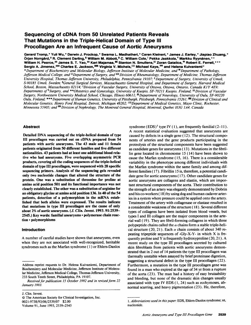
Figure 1. Autoradiogram of a DNA sequencing gel. To facilitate the interpretation of results, G reactions from six (lanes 1-6) different individ-uals were loaded adjacent to each other, followed by A, T, and C reactions, in this order, from the same individuals. Lane T4 has an extra band(arrow), indicating the presence ofG to T change in one allele of cell line JIMM498. The sequence is in antisense orientation.
the aorta. Two patients had iliac artery aneurysms. Six individ-uals with abdominal or thoracic aortic aneurysms had one ormore family members with intracranial aneurysms. At thetime ofthe diagnosis the aortic aneurysm had ruptured in 7/54patients. 36 patients underwent an elective operation for theiraortic aneurysms. Five patients were first diagnosed by ultra-sonographic examination.
DNA sequences and polymorphisms of type III collagen.The sequence analysis carried out here provided 3,232 nucleo-tides from each allele of the gene for type III procollagen. Theregion analyzed covered the coding sequences for all the 14amino acids of the amino-terminal telopeptide, all the 1,029amino acids ofthe triple-helical domain, all the 25 amino acidsof the carboxyterminal telopeptide, and 8 of the 246 aminoacids of the carboxy-terminal propeptide. The sequences forthe amino-terminal propeptide and most of the carboxy-ter-minal propeptide were not analyzed. These amino acid se-quences are cleaved off from the procollagen before the fibrilformation can occur and they are, therefore, not present in themature form of collagen (20).
Only two sequence variations were found in the 54 individ-uals (Table IV). Cell line JIMM407 had a G to A change at nt907. The nucleotide change converted the codon at amino acidposition 136 from GGG for glycine to AGG, a codon for argi-nine. The mutation was confirmed using genomic DNA iso-lated from the patient's fibroblasts (not shown). The mutationwas not found in any of additional 127 individuals sequencedin the laboratory.
The patient was an 18-yr-old black male without any priorrelevant medical history (43). He had suddenly developed par-
aparesis and bilateral loss of pulses below the waist (43). Anaortogram disclosed a dissecting aneurysm of the entire aortaand obstruction ofblood flow below the renal arteries (43). Hisautopsy findings revealed dissecting aortic aneurysm and gener-alized fibromuscular dysplasia (43). His father had died at theage of 36 yr in a car accident and no affected relatives wereavailable for DNA testing. His mother did not have the muta-tion, but the patient's three unaffected sibs were found to havethe same mutation. Ultrasound examination of the aorta onthe sibs (aged 21, 20, and 16 yr) did not reveal any abnormali-ties.
In the cell line JIMM498 a C to A change was found at nt2002, changing the codon CCT for proline at amino acid posi-tion 501 to ACT, a codon for threonine (Fig. 1 ). PCR on geno-mic DNA from the patient indicated that the sequence variantwas real and not a PCR artifact (not shown). The change wasnot found in any of the other 127 individuals sequenced in thelaboratory. The patient was 70 yr old, when he was operated onfor an abdominal aortic aneurysm. His father also had an aorticaneurysm but was deceased and no material was available forDNA testing. The patient's nephew (a son of the patient'syounger brother) was operated on for an intracranial aneurysmat the age of28 yr. This nephew had the mutation. DNA analy-sis revealed that the change was also present in the patient's twounaffected brothers aged 68 and 73 yr, the younger of whichhad been examined by sonography and no aortic aneurysm wasfound. Thus, two unaffected and two affected individuals inthe family had the DNA change. The change was, therefore,probably not the cause of the aneurysms.
When analyzing the sequencing results from the 54 individ-
Aortic Aneurysms and Type III Procollagen Gene 2543
.........k.11,- Ma. 111M..,.lNW'
"..
'OK" milim.n--, i
4
MWIMMI'm-..
Wloml~ m~~

Table IV. Sequence Variations Detected in the 54 Patients
Cell linecode Location* Naturet Occurrence
JIMM407 nt 907 (E14) Glyl36 -Arg 1/128§JIMM498 nt 2002 (E30) Pro501 - Thr I/128§
* E14, exon 14 of the gene for type III procollagen; E30, exon 30 ofthe gene for type III procollagen.$ Numbering starts at the beginning of the triple-helical domain oftype III procollagen.§ Only one patient was found to have the base change. A total of 127other unrelated individuals that included the 49 other aortic aneu-rysm patients presented here, 55 patients with intracranial aneurysms,5 patients with the EDS type IV, 4 patients with osteogenesis imper-fecta, 5 patients with the EDS (without subclassification), 8 patientswith a variety of connective tissue abnormalities without any specificdiagnosis, and one apparently healthy individual were sequenced inthe laboratory.
uals, special care was taken to record data on known polymor-phic regions of the type III collagen. The three polymorphismsthat have been found previously in the coding region oftype IIIcollagen are at positions nt 1851 (Wu and Kuivaniemi, unpub-lished results), 2092 (41), and 2244 (42). 40 ofthe 54 individ-uals were found to be heterozygous for at least one of the poly-morphisms (Table V), indicating that both of the alleles ofthegene for type III procollagen were expressed at the mRNAlevel. The polymorphism analysis was also carried out on geno-mic DNA samples isolated from the cultured skin fibroblasts ofthe patients. The results obtained from the analysis showedthat the 40 individuals that were heterozygous in their cDNAfor one or more polymorphisms were also heterozygous in theirgenomic DNA. The results also showed that the 14 individualswho did not show any heterozygosity in the cDNA sequenceswere homozygous on their genomic DNA for all three markersstudied.
Discussion
There are two general approaches using DNA techniques thatcan be used to identify the gene harboring the mutations caus-ing aortic aneurysms. The first approach is to carry out linkagestudies using markers that have been mapped to a particularlocus on the genome and are used to test whether or not themarker is co-inherited with aortic aneurysms in families. Link-age can be established whenever the phenotype and genotypeof related individuals is analyzed. Therefore, linkage can beestablished by analyzing large families. There are, however,
Table V. Heterozygosity ofthe 54 Patients at Three PolymorphicSites Located in the Coding Sequences of Type III Collagen
Location of polymorphism Number of heterozygous individuals
nt 1851 18nt 2092 22nt 2244 21
Total number of patients heterozygous for at least one of the markers:40.
several problems in the approach when studying a late-onsetdisease such as aortic aneurysms. It is rare to find large indexfamilies that have more than two generations ofliving, affectedmembers so that pertinent samples can be obtained. In addi-tion, diagnosis is a problem, inasmuch as the data on the inci-dence ofaneurysms in the population indicate that few individ-uals develop aneurysms before the age of 50 yr and that theincidence increases with age.
The second approach, the candidate gene approach, in-volves the analysis of a candidate gene for absence or presenceof mutations. The modem techniques of molecular biologyhave made the approach feasible (44). Here we were able to getdefinitive results from detailed DNA sequencing analysis ofthegene for type III procollagen of 54 individuals from 50 unre-lated families. The analysis revealed a mutation in obligatoryglycine (20, 21 ) in one patient. The substitution ofarginine fora glycine at amino acid position 136 in type III procollagen islikely to disrupt the triple-helical structure of the protein andmake the protein less stable (20, 21 ). The finding represents asecond example ofa glycine to arginine mutation in the type IIIprocollagen causing aortic aneurysms in patients without othersigns ofthe EDS. The first report was a substitution ofargininefor glycine at amino acid position 619 of the triple-helical do-main of type III procollagen (25).
The other sequence variant detected in another patient sub-stituted threonine for proline in a Y position of the Gly-X-Yrepeat of collagen triple-helical domain (20, 21 ). Substitutionofthreonine for a Y-position proline is not likely to be destabi-lizing since six ofthe 14 threonines normally found in the typeIII collagen triple-helical domain (36) occupy the Y position.In addition, threonine and proline have been exchanged duringevolution of collagens (45). Also, the mutation was present inthe patient's apparently unaffected brothers suggesting that themutation was an infrequent sequence variant. The results,therefore, indicate that mutations in the triple-helical domainof type III procollagen are the cause of - 2% (1/ 50) of aorticaneurysms. In addition, the results suggest that mutations inthe promoter region or other control regions of the gene fortype III procollagen are not a common cause for aneurysms, inthat at least 40 of the 50 patients studied here had mRNA thatwas derived from both alleles of the gene.
From a theoretical perspective, DNA sequencing provides acomplete record of all of the bases that comprise a gene or itscoding sequence. Therefore, DNA sequencing will lead to thedetection of all mutations in a target region. Furthermore, itmakes it possible to exclude definitively the region from thosethat possibly contain mutations. Our results demonstrate thatsequencing has become a feasible approach not only to estab-lish rapidly and definitively whether or not a candidate geneharbors the mutations causing a disease phenotype, but also todetermine what fraction of affected individuals have a muta-tion in the particular candidate gene. This approach could,therefore, be used to establish whether or not other genes thathave been suggested as candidate genes, such as the genes forfibrillin ( 17), elastin (31 ), collagenase (26), elastase (27), ortissue inhibitor of metalloproteinase (29), harbor mutationscausing aortic aneurysms.
Acknowledgments
The authors thank Maria Hervada-Page for collecting some of the pa-tient information, and Gi-Chung Chen, Nasrin Rafi, and Kanger Wufor culturing cells.
2544 Tromp et al.

This work was supported in part by grants from the National Insti-tutes ofHealth (HL 45996 and AR 38188), the American Heart Associ-ation, Southeastern Pennsylvania Affiliate, the Lucille P. Markey Char-itable Trust, and the March of Dimes/Birth Defects Foundation.
References1. McKusick, V. A. 1972. Heritable Disorders of Connective Tissue. C. V.
Mosby Co., St. Louis, MO. 61-223, 292-37 1.2. Clifton, M. A. 1977. Familial abdominal aortic aneurysms. Br. J. Surg.
64:765-766.3. Tilson, M. D., and M. R. Seashore. 1984. Human genetics ofthe abdominal
aortic aneurysm. Surg. Gynecol. Obstet. 158:129-132.4. Tilson, M. D., and M. R. Seashore. 1984. Fifty families with abdominal
aortic aneurysms in two or more first-order relatives. Am. J. Surg. 147:551-553.5. NorrgArd, O., 0. Rais, and K. A. Angquist. 1984. Familial occurrence of
abdominal aortic aneurysms. Surgery. 95:650-656.6. Johansen, K., and T. Koepsell. 1986. Familial tendency for abdominal
aortic aneurysms. JAMA (J. Am. Med. Assoc.). 256:1934-1936.7. Powell, J. T., and R. M. Greenhalgh. 1987. Multifactorial inheritance of
abdominal aortic aneurysm. Eur. J. Vasc. Surg. 1:29-31.8. Cole, C. W., G. G. Barber, A. G. Bouchard, N. V. McPhail, C. Roberge,
W. G. Waddell, and J. L. Wellington. 1989. Abdominal aortic aneurysm: conse-quences of a positive family history. Can. J. Surg. 32:117-120.
9. Darling, R. C. III, D. C. Brewster, R. C. Darling, G. M. LaMuraglia, A. C.Moncure, R. P. Cambria, and W. M. Abbott. 1989. Are familial abdominal aorticaneurysms different? J. Vasc. Surg. 39-43.
10. Bengtsson, H., 0. Norrgird, K. A. Angquist, 0. Ekberg, L. Oberg, and D.Bergqvist. 1989. Ultrasonographic screening of the abdominal aorta among sib-lings of patients with abdominal aortic aneurysms. Br. J. Surg. 76:589-591.
11. Webster, M. W., R. E. Ferrell, P. L. St. Jean, P. P. Majumder, S. R. Fogel,and D. L. Steed. 1991. Ultrasound screening of first-degree relatives of patientswith an abdominal aortic aneurysm. J. Vasc. Surg. 13:9-14.
12. Majumder, P. P., P. L. St. Jean, R. E. Ferrell, M. W. Webster, and D. L.Steed. 1991. On the inheritance of abdominal aortic aneurysm. Am. J. Hum.Genet. 48:164-170.
13. Reilly, J. M., and M. D. Tilson. 1989. Incidence and etiology ofabdomi-nal aortic aneurysms. Surg. Clin. North Am. 69:705-71 1.
14. Lee, B., M. Godfrey, E. Vitale, H. Hori, M.-G. Mattei, M. Sarfarazi, P.Tsipouras, F. Ramirez, and D. W. Hollister. 1991. Linkage of Marfan syndromeand a phenotypically related disorder to two different fibrillin genes. Nature(Lond.). 352:330-334.
15. Dietz, H. C., G. R. Cutting, R. E. Pyeritz, C. L. Maslen, L. Y. Sakai, G. M.Corson, E. G. Puffenberger, A. Hamosh, E. J. Nanthakumar, S. M. Curristin, etal. 1991. Marfan syndrome caused by a recurrent de novo missense mutation inthe fibrillin gene. Nature (Lond.). 352:337-339.
16. Kainulainen, K., L. Y. Sakai, M. Pope, A. Child, L. Puhakka, L.Ryynanen, A. Palotie, I. Kaitila, and L. Peltonen. 1992. Two mutations in Mar-fan syndrome resulting in truncated fibrillin polypeptides. Proc. Natl. Acad. Sci.USA. 89:5917-5921.
17. McKusick, V. A. 1991. The defect in Marfan syndrome. Nature (Lond.).352:279-281.
18. Dobrin, P. B., T. H. Schwarcz, and R. Mrkvicka. 1990. Longitudinalretractive force in pressurized dog and human arteries. J. Surg. Res. 48:116-120.
19. Mayne, R. 1987. Vascular connective tissue. Normal biology and derange-ment in human diseases. In Connective Tissue Disease. Molecular Pathology ofthe Extracellular Matrix. J. Uitto and A. J. Perejda, editors. Marcel Dekker Inc.,New York. 163-183.
20. Prockop, D. J. 1990. Mutations that alter the primary structure of type Icollagen. The perils ofa system for generating large structures by the principle ofnucleated growth. J. Biol. Chem. 265:15349-15352.
21. Kuivaniemi, H., G. Tromp, and D. J. Prockop. 1991. Mutations in colla-gen genes: causes of rare and some common diseases in humans. FASEB J.5:2052-2060.
22. Deak, S. B., J. J. Ricotta, T. J. Mariani, S. T. Deak, M. A. Zatina, J. W.Mackenzie, and C. D. Boyd. 1992. Abnormalities in the biosynthesis of type IIIprocollagen in cultured skin fibroblasts from two patients with multiple aneu-rysms. Matrix. 12:92-100.
23. Kontusaari, S., G. Tromp, H. Kuivaniemi, R. L. Ladda, and D. J.Prockop. 1990. Inheritance of an RNA splicing mutation (G"' 1vs20) in the type
III procollagen gene (COL3A I ) in a family with aortic aneurysms and easy bruis-ability: phenotypic overlap between familial arterial aneurysms and the Ehlers-Danlos syndrome type IV. Am. J. Hum. Genet. 47:112-120.
24. Beighton, P. 1970. The Ehlers-Danlos Syndrome. William Heinemann,Ltd., London. 194 pp.
25. Kontusaari, S., G. Tromp, H. Kuivaniemi, A. M. Romanic, and D. J.Prockop. 1990. A mutation in the gene for type III procollagen (COL3A I) in afamily with aortic aneurysms. J. Clin. Invest. 86:1465-1473.
26. Busuttil, R. W., A. M. Abou-Zamzam, and H. I. Machleder. 1980. Collage-nase activity of the human aorta: a comparison of patients with and withoutabdominal aortic aneurysms. Arch. Surg. 115:1373-1378.
27. Cannon, D., and R. Read. 1982. Blood elastolytic activity in patients withaortic aneurysms. Ann. Thorac. Surg. 34:10-15.
28. Cohen, J. R., I. Sarfati, I. Ratner, and M. D. Tilson. 1990. Alpha-l anti-trypsin phenotypes in patients with abdominal aortic aneurysms. J. Surg. Res.49:319-321.
29. Brophy, C. M., B. Sumpio, J. M. Reilly, and M. D. Tilson. 1991. De-creased tissue inhibitor of metalloproteinases (TIMP) in abdominal aortic aneu-rysmal tissue: a preliminary report. J. Surg. Res. 50:653-657.
30. Kuivaniemi, H., G. Tromp, and D. J. Prockop. 1991. Genetic causes ofaortic aneurysms. Unlearning at least part of what the textbooks say. J. Clin.Invest. 88:1441-1444.
31. Menashi, S., J. S. Campa, R. M. Greenhalgh, and J. T. Powell. 1987.Collagen in abdominal aortic aneurysm: typing, content, and degradation. J.Vasc. Surg. 6:578-582.
32. Maniatis, T., E. F. Fritsch, and J. Sambrook. 1982. Molecular Cloning: ALaboratory Manual. Cold Spring Harbor Laboratory, Cold Spring Harbor, NY.
33. Loidl, H. R., J. M. Brinker, M. May, T. Pihlajaniemi, S. Morrow, J.Rosenbloom, and J. C. Myers. 1984. Molecular cloning and carboxylpropeptideanalysis of human type III procollagen. Nucleic Acids Res. 12:9383-9394.
34. Chu, M.-L., D. Weil, W. de Wet, M. Bernard, M. Sippola, and F. Ramirez.1985. Isolation ofcDNA and genomic clones encoding human pro-al (III) colla-gen: partial characterization of the 3'-end region of the gene. J. Biol. Chem.260:4357-4363.
35. Miskulin, M., R. Dalgleish, B. Kluve-Beckerman, S. I. Rennard, P. Tol-stoshev, M. Brantly, and R. G. Crystal. 1986. Human type III collagen geneexpression is coordinately modulated with the type I collagen genes during fibro-blast growth. Biochemistry. 25:1408-1413.
36. Ala-Kokko, L., S. Kontusaari, C. Baldwin, H. Kuivaniemi, and D. J.Prockop. 1989. Structure of cDNA clones coding for the entire preproa 1(111)chain of human type III procollagen: differences in protein structure from type Iprocollagen and conservation of codon preferences. Biochem. J. 260:509-516.
37. Tromp, G., H. Kuivaniemi, H. Shikata, and D. J. Prockop. 1989. A singlebase mutation that substitutes serine for glycine 790 ofthe a I (III) chain oftypeIII procollagen exposes an arginine and causes Ehlers-Danlos syndrome IV. J.Biol. Chem. 264:1349-1352.
38. Tromp, G., H. Kuivaniemi, C. Stolle, F. M. Pope, and D. J. Prockop.1989. Single base mutation in the type III procollagen gene that converts thecodon for glycine 883 to aspartate in a mild variant of Ehlers-Danlos syndromeIV. J. Biol. Chem. 264:19313-19317.
39. Saiki, R. K., S. Scharf, F. Faloona, K. B. Mullis, G. T. Horn, H. A. Erlich,and N. Arnheim. 1985. Enzymatic amplification off-globin genomic sequencesand restriction site analysis for diagnosis of sickle cell anemia. Science (Wash.DC). 230:1350-1354.
40. Sanger, F., S. Nicklen, and A. R. Coulson. 1977. DNA sequencing withchain-terminating inhibitors. Proc. Nati. Acad. Sci. USA. 74:5463-5467.
41. Zafarullah, K., C. Kleinert, G. Tromp, H. Kuivaniemi, S. Kontusaari, Y.Wu, A. Ganguly, and D. J. Prockop. 1990. G to A polymorphism in exon 31 ofthe COL3A I gene. Nucleic Acids Res. 18:6180.
42. Tromp, G., C. Kleinert, H. Kuivaniemi, and D. J. Prockop. 1991. C to Tpolymorphism in exon 33 of the COL3A 1 gene. Nucleic Acids Res. 19:681.
43. Gatalica, Z., Z. Gibas, and A. Martinez-Hernandez. 1992. Dissectingaortic aneurysm as a complication ofgeneralized fibromuscular dysplasia. Hum.Pathol. 23:586-588.
44. Tromp, G., and H. Kuivaniemi. 1992. Identification of familial aorticaneurysms using DNA technique. In Technologies in Vascular Surgery. J. S. T.Yao, and W. H. Pearce, editors. W. B. Saunders Co., Philadelphia. 27-39.
45. Kuivaniemi, H., Tromp, G., Chu, M.-L., and Prockop, D. J. 1988. Struc-ture ofa full-length clone for the preproa2(I) chain ofhuman type I procollagen:comparison with the chicken gene confirms unusual patterns of gene conserva-tion. Biochem. J. 253:633-640.
Aortic Aneurysms and Type III Procollagen Gene 2545