Manganese oxide reactivity in North Sea sediments - CiteSeerX
Sensitization Prevalence, Antibody Cross-Reactivity andImmunogenic Peptide Profile of Api g 2, the Non-SpecificLipid Transfer Protein 1 of CeleryGabriele Gadermaier1*, Michael Hauser1, Matthias Egger1, Rosetta Ferrara2, Peter Briza1, Keity Souza
Santos3, Danila Zennaro2, Tamara Girbl1, Laurian Zuidmeer-Jongejan4, Adriano Mari2, Fatima Ferreira1
1 Christian Doppler Laboratory for Allergy Diagnosis and Therapy, University of Salzburg, Salzburg, Austria, 2 Center for Molecular Allergology, IDI-IRCCS, Rome, Italy,
3 Discipline of Allergy and Immunology, School of Medicine, University of Sao Paulo, Sao Paulo, Brazil, 4 Laboratory of Allergy, Department of Experimental Immunology,
Academic Medical Center, Amsterdam, The Netherlands
Abstract
Background: Celery (Apium graveolens) represents a relevant allergen source that can elicit severe reactions in the adultpopulation. To investigate the sensitization prevalence and cross-reactivity of Api g 2 from celery stalks in a Mediterraneanpopulation and in a mouse model.
Methodology: 786 non-randomized subjects from Italy were screened for IgE reactivity to rApi g 2, rArt v 3 (mugwort pollenLTP) and nPru p 3 (peach LTP) using an allergen microarray. Clinical data of 32 selected patients with reactivity to LTP underinvestigation were evaluated. Specific IgE titers and cross-inhibitions were performed in ELISA and allergen microarray. Balb/c mice were immunized with purified LTPs; IgG titers were determined in ELISA and mediator release was examined usingRBL-2H3 cells. Simulated endolysosomal digestion was performed using microsomes obtained from human DCs.
Results: IgE testing showed a sensitization prevalence of 25.6% to Api g 2, 18.6% to Art v 3, and 28.6% to Pru p 3 andfrequent co-sensitization and correlating IgE-reactivity was observed. 10/32 patients suffering from LTP-related allergyreported symptoms upon consumption of celery stalks which mainly presented as OAS. Considerable IgE cross-reactivitywas observed between Api g 2, Art v 3, and Pru p 3 with varying inhibition degrees of individual patients’ sera. SimulatingLTP mono-sensitization in a mouse model showed development of more congruent antibody specificities between Api g 2and Art v 3. Notably, biologically relevant murine IgE cross-reactivity was restricted to the latter and diverse from Pru p 3epitopes. Endolysosomal processing of LTP showed generation of similar clusters, which presumably represent T-cellpeptides.
Conclusions: Api g 2 represents a relevant celery stalk allergen in the LTP-sensitized population. The molecule displayscommon B cell epitopes and endolysosomal peptides that encompass T cell epitopes with pollen and plant-food derivedLTP.
Citation: Gadermaier G, Hauser M, Egger M, Ferrara R, Briza P, et al. (2011) Sensitization Prevalence, Antibody Cross-Reactivity and Immunogenic Peptide Profileof Api g 2, the Non-Specific Lipid Transfer Protein 1 of Celery. PLoS ONE 6(8): e24150. doi:10.1371/journal.pone.0024150
Editor: Jacques Zimmer, Centre de Recherche Public de la Sante (CRP-Sante), Luxembourg
Received May 24, 2011; Accepted August 1, 2011; Published August 29, 2011
Copyright: � 2011 Gadermaier et al. This is an open-access article distributed under the terms of the Creative Commons Attribution License, which permitsunrestricted use, distribution, and reproduction in any medium, provided the original author and source are credited.
Funding: The study was supported by the Christian-Doppler Research Association, Biomay AG, Vienna, Austria, and the Italian Ministry of Health – CurrentResearch Program 2009. The funders had no role in study design, data collection and analysis, decision to publish, or preparation of the manuscript.
Competing Interests: The role of Biomay AG in funding the research was via the Christian Doppler Research Association, a government institution partiallyfinanced by industrial partners. This does not alter the authors’ adherence to all the PLoS ONE policies on sharing data and materials.
* E-mail: [email protected]
Introduction
In the Mediterranean population, non-specific lipid transfer
proteins (LTP) represent important allergens in fruits and
vegetables and account for the majority of type I food allergies
in adults [1,2,3]. Due to their pronounced resistance to thermal
and proteolytic treatment [4], they are considered true food
allergens and were proposed to mediate sensitization via the
gastrointestinal tract [3]. Pru p 3, the major allergen from peach
seems to play an important role in LTP-mediated allergy with a
sensitization prevalence of 13% and 9.8% in a Spanish and Italian
population, respectively [5,6]. The clinical picture of an LTP-
mediated food allergy ranges from local oral allergy syndrome to
severe systemic symptoms [7]. We recently identified and
characterized an nsLTP1 in celery stalks which was included as
Api g 2.0101 in the official I.U.I.S. allergen nomenclature [8]. The
natural protein consists of a single isoallergen with a mass of
9024.5 Da. A recombinant protein was produced as non-fusion
protein in E. coli which presented equivalent physio-chemical and
immunological properties as the natural counterpart. In line with
other members of the LTP family, Api g 2 shows extreme
resistance to gastrointestinal proteolysis and thermal treatment [8].
Notably, the allergen was able to fully refold after heating in acidic
environment, a property that was previously also demonstrated for
Pru p 3 [8,9]. In addition to plant-derived foods, LTP have been
shown to constitute important allergens in pollen and ever since
PLoS ONE | www.plosone.org 1 August 2011 | Volume 6 | Issue 8 | e24150

there is a longstanding debate about the sensitizing source [7].
Generally, the classification of LTP in the field of food allergy
turns out to be extremely difficult. On the one hand, LTP fulfill all
the requirements of true food allergens, i.e. the capability to
sensitize via the gastrointestinal tract. On the other hand, they
might be regarded as pollen-associated food allergens, as allergenic
LTP are also found in plant pollen and associations between pollen
and food allergies involving LTP sensitization have been reported
[3,7,10]. In mugwort and peach allergy, both Art v 3 and Pru p 3
have been proposed as primary sensitizer [11,12], however at
present no evidence suggests a clinical association between
sensitization to plant food and pollen LTP.
Apium graveolens, celery, is considered one of the most important
plant food allergen sources in the adult Central European
population [13,14] and therefore, declaration of products that
contain celery is mandatory (European Directive 2007/68/EC,
amending Directive 2000/13/EC). Apart from the recently
identified nsLTP1, three allergens from A. graveolens have been
described and characterized at the molecular level, the PR-10
protein Api g 1 [15], the profilin Api g 4 [16], and Api g 5, a
member of the flavoprotein family [17]. Among Central European
patients, a predominant sensitization to Api g 1 (59%) and Api g 4
(23$41%) is observed [16,18]. Although celery stalks are
worldwide consumed, there is only limited information on
allergens and clinical relevance of the aerial celery tissue, since
the majority of studies focused on patients from Central Europe
[13,14,19,20], who predominately consume celery tuber (celeriac).
Noteworthy, the recently identified LTP1 Api g 2 can be
considered a celery stalk-specific allergen, since expression is
restricted to the green stalks and was not detectable in the tuber
tissue [8].
Clinical allergy to celeriac is frequently associated with
sensitization to Artemisia vulgaris and Betula verrucosa pollen in
Central European countries and thus, the terms celery-mugwort
and celery-birch syndrome have been established [10]. Association
between birch pollinosis and celery hypersensitivity is mainly
attributed to Api g 1, a Bet v 1-homologous PR-10 protein [15,21].
Moreover, Api g 4 and Api g 5 have been mentioned to play a role
as cross-reactive molecules in this population [16,22,23]. By
contrast, a heat stable molecule involved in the celery-mugwort
syndrome which might be able to trigger severe allergic reactions
in Central European celeriac sensitized patients is not yet
conspicuously determined [24].
In this study, we investigated the sensitization prevalence of Api
g 2 in allergic patients from the Mediterranean area by testing on a
microarray system in parallel with other LTP. The sensitization
pattern and IgE cross-reactivity of Api g 2 was investigated in a
selected patients’ cohort sensitized to Art v 3 and Pru p 3, model
allergens for pollen and plant food LTP, respectively. In order to
elucidate which molecule has the potential to act as primary
sensitizer, we immunized mice with pollen and plant-food derived
LTP and determined the patterns of antibody cross-reactivity.
Immunogenicity and putative T cell epitopes were determined in
vitro by simulated endolysosomal degradation assays.
Results
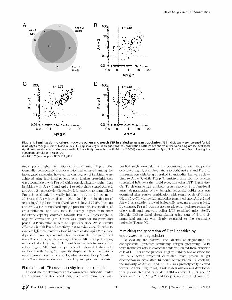
IgE profiling of celery stalks, mugwort pollen, and peachLTP in Mediterranean patients
To assess the sensitization prevalence of the novel celery stalk
allergen in an allergic Mediterranean population, sera of 786
subjects were tested in vitro by an experimental ISAC microarray.
Among them, 25.6% displayed specific IgE against celery stalk
LTP, 18.6% were sensitized to mugwort pollen LTP, and 28.6%
to peach LTP. The majority of patients (n = 111) reacted to all 3
LTPs, and associated reactivity of Api g 2 to Pru p 3 and Art v 3
was observed in 54 and 4 individuals, respectively (Figure 1A).
Isolated IgE reactivity to Api g 2 was observed in 32 patients, while
16 and 44 patients exclusively reacted to Art v 3 and Pru p 3,
respectively. In sensitized individuals, the average IgE reactivity to
Api g 2, Art v 3, and Pru p 3 was 1.03 kUA/l, 1.12 kUA/l, and
2.59 kUA/l, respectively. As shown in Figure 1B-D, significant
correlations (p,0.0001) in IgE-reactivity were observed between
all three LTP under investigation.
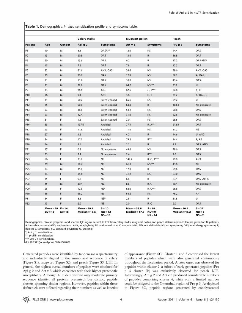
Symptoms and IgE reactivity of patients sensitized toplant food and pollen LTP
Defining Art v 3 and Pru p 3 as model allergens for pollen and
food LTP sensitization, we selected a cohort of 32 Api g 2-positive
subjects that additionally displayed in vitro IgE-reactivity to
mugwort pollen and peach LTP. Demographic data, clinical
manifestations to the respective sources and IgE reactivity to Api g
2, Art v 3, and Pru p 3 are given in Table 1. In this selected cohort,
ten patients (P1-10) reported allergic reactions upon consumption
of celery stalks which predominately presented as oral allergy
syndrome (80%). In addition, angioedema was observed in 2
patients and anaphylaxis in 1 patient. However, twelve individuals
(P11-22) could not unambiguously report about clinical manifes-
tations or tolerance upon consumption of raw celery stalks since
they were not exposed, routinely cooked the vegetable as a
precaution or actively avoided consumption due to previously
severe allergic reactions to other LTP containing food sources. To
investigate a possible relevance of other A. graveolens allergens in
our patients’ cohort, sensitization to Api g 1 was derived from a
microarray testing revealing a positive IgE reactivity for patient P1
and P29. To give further information on potentially important
cross-reactive molecules, profilin sensitization of two patients (P1
and P31) is denoted in Table 1. Notably, among the ten patients
with clinical symptoms to celery stalks, only one patient (P1)
showed additional sensitization to A. graveolens allergens. Thus, for
the remaining subjects, Api g 2 might be involved in a clinical
manifestations of celery stalk allergy, even though sensitization to
yet unknown celery allergens cannot be ruled out and would need
further investigation. Adverse reactions upon consumption of
peach and inhalant allergy symptoms to mugwort pollen were
reported by 27 and 18 patients sensitized to Pru p 3 and Art v 3,
respectively (Table 1). LTP-specific serum IgE levels were
determined in ELISA showing highest in vitro reactivity for Pru p
3 which differed significantly from Api g 2 (p,0.001) and Art v 3
(p,0.02) using the Wilcoxon signed rank sum test (Table 1).
Interestingly, heat-denaturation for 15 minutes at 95uC at a
concentration of $1 mg/ml in phosphate buffer resulted in
complete abolishment of IgE reactivity to celery stalk and
mugwort pollen LTP, whereas residual IgE-binding (range 5.5–
62.0%, median 6.9%) to Pru p 3 was still detectable in 50% of
patients (data not shown). No unspecific IgE binding to
investigated proteins was observed using sera of non-atopic
individuals.
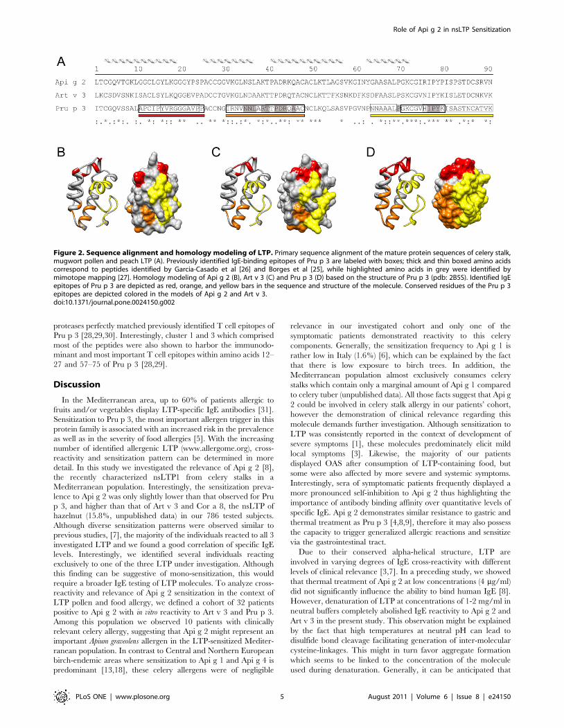
IgE cross-reactivity in LTP sensitized patientsThe primary sequence identity of Api g 2, Art v 3, and Pru p 3
ranges between 49% and 53%, and amino acid alignments and 3-
D models of investigated LTP are depicted in Figure 2. Previously
identified linear IgE-binding epitopes of Pru p 3 [25,26,27]
demonstrate highest sequence identities to Api g 2 (76%) and Art v
3 (82%) within residue 66-82 in the C-terminal epitope (Figure 2).
To assess the IgE cross-reactivity of Api g 2, Art v 3, and Pru p 3,
inhibition studies were performed using LTP-reactive sera in a
Role of Api g 2 in nsLTP Sensitization
PLoS ONE | www.plosone.org 2 August 2011 | Volume 6 | Issue 8 | e24150
single point highest inhibition-achievable assay (Figure 3A).
Generally, considerable cross-reactivity was observed among the
investigated molecules, however varying degrees of inhibition were
achieved using individual patients’ sera. Highest cross-inhibition
was accomplished with Pru p 3 which was significantly higher than
inhibition with Art v 3 and Api g 2 to solid-phase coated Api g 2
and Art v 3, respectively. Generally, IgE reactivity to immobilized
Pru p 3 could only be weakly inhibited by Api g 2 (median =
20.2%) and Art v 3 (median = 0%). Notably, pre-incubation of
sera using Api g 2 for immobilized Art v 3 showed 72.5% (median)
and Art v 3 for immobilized Api g 2 presented 43.4% (median) of
cross-inhibition, and was thus in average higher than their
inhibitory capacity observed towards Pru p 3. Interestingly, a
negative correlation (r = 20.842) was found for mugwort and
peach LTP inhibition in sera of 3 patients, since Art v 3 could
efficiently inhibit Pru p 3 reactivity, but not vice versa. In order to
evaluate IgE cross-reactivity to solid phase coated Api g 2 in a dose
dependent manner, cross-inhibition experiments were performed
using 3 sera of celery stalk allergics (Figure 3B), 2 subjects eating
only cooked celery (Figure 3C), and 5 individuals tolerating raw
celery (Figure 3D). Notably, patients who showed highest self-
inhibition with Api g 2 frequently displayed clinical symptoms
upon consumption of celery stalks, while stronger Pru p 3 and/or
Art v 3 reactivity was observed in celery asymptomatic patients.
Elucidation of LTP cross-reactivity in a mouse modelTo evaluate the development of cross-reactive antibodies under
LTP mono-sensitization conditions, mice were immunized with
purified single molecules. Art v 3-sensitized animals frequently
developed high IgG antibody titers to both, Api g 2 and Pru p 3.
Immunization with Api g 2 resulted in antibodies that were able to
bind to Art v 3, while Pru p 3 sensitized mice did not develop
substantial IgG titers that could recognize other LTP (Figure 4A–
C). To determine IgE antibody cross-reactivity in a functional
assay, degranulation of rat basophil leukemia (RBL) cells was
examined after passive sensitization with serum pools of 6 mice
(Figure 5A–C). Murine IgE antibodies generated upon Api g 2 and
Art v 3 sensitization showed biologically relevant cross-reactivity.
By contrast, Pru p 3 was not able to trigger a mediator release in
celery stalk and mugwort pollen LTP sensitized mice (5A-B).
Notably, IgE-mediated degranulation using sera of Pru p 3-
immunized animals was clearly restricted to the sensitizing
molecule (Figure 5C).
Mimicking the generation of T cell peptides byendolysosomal degradation
To evaluate the patterns and kinetics of degradation by
endolysosomal proteases simulating antigen processing, LTPs
were incubated with microsomal contents isolated from dendritic
cells of LTP-sensitized patients. Highest stability was observed for
Pru p 3, which presented detectable intact protein in gel
electrophoresis even after 48 hours of incubation. In contrast,
the majority of Art v 3 and Api g 2 was proteolytically cleaved
within 12 hours (Figure 6A). Protein degradation was densitome-
trically evaluated and calculated half-lives were 11, 18, and 32
hours for Art v 3, Api g 2, and Pru p 3, respectively (Figure 6B).
Figure 1. Sensitization to celery, mugwort pollen and peach LTP in a Mediterranean population. 786 individuals were screened for IgEreactivity to rApi g 2, rArt v 3, and nPru p 3 using an allergen microarray and co-sensitization patterns are shown in the Venn diagram (A). Statisticalsignificant correlations of allergen specific IgE reactivity presented as kUA/L (p,0.0001) were observed for Api g 2, Art v 3 and Pru p 3 using theSpearman correlation test (B-D).doi:10.1371/journal.pone.0024150.g001
Role of Api g 2 in nsLTP Sensitization
PLoS ONE | www.plosone.org 3 August 2011 | Volume 6 | Issue 8 | e24150
Generated peptides were identified by tandem mass spectrometry
and individually aligned to the amino acid sequence of celery
(Figure S1), mugwort (Figure S2), and peach (Figure S3) LTP. In
general, the highest overall numbers of peptides were obtained for
Api g 2 and Art v 3 which correlates with their higher proteolytic
susceptibility. Although LTP demonstrate only moderate primary
sequence identity, all proteins presented four distinct peptide
clusters spanning similar regions. However, peptides within those
defined clusters differed regarding their numbers as well as kinetics
of appearance (Figure 6C). Cluster 1 and 3 comprised the largest
numbers of peptides which were also generated continuously
throughout the incubation period. A later onset was observed for
peptides within cluster 2, a subset of early generated peptides (Pru
p 3 cluster 2b) was exclusively observed for peach LTP.
Interestingly, Api g 2 and Art v 3 produced considerable numbers
of peptides comprising cluster 4, while only a limited number
could be assigned to the C-terminal region of Pru p 3. As depicted
in Figure 6C, peptide regions generated by endolysosomal
Table 1. Demographics, in vitro sensitization profile and symptoms table.
Celery stalks Mugwort pollen Peach
Patient Age Gender Api g 2 Symptoms Art v 3 Symptoms Pru p 3 Symptoms
P1 10 M 8.6 OAS*,** 12.0 NS 44.4 OAS
P2 43 M 60.8 OAS 13.0 R 56.8 OAS
P3 20 M 15.6 OAS 6.2 R 17.2 OAS.ANG
P4 15 M 7.2 OAS 7.8 R 12.2 OAS
P5 22 M 21.6 ANX, OAS 24.6 NS 59.6 ANX. OAS
P6 35 M 20.0 OAS 17.8 NS 58.2 A, OAS, U
P7 11 F 11.8 OAS 10.0 NS 43.4 OAS
P8 21 M 72.8 OAS 64.2 NS*** 73.2 U
P9 23 M 20.6 ANG 67.6 C, R*** 54.8 C, R
P10 26 M 9.4 ANG 3.6 C, R 31.2 A, OAS, U
P11 14 M 50.2 Eaten cooked 65.6 NS 59.2 U
P12 15 M 90.8 Eaten cooked 63.8 R 103.4 No exposure
P13 23 M 38.6 Eaten cooked 53.2 NS 90.8 OAS
P14 23 M 42.4 Eaten cooked 31.6 NS 52.6 No exposure
P15 31 F 1.6 Eaten cooked 7.0 NS 28.6 OAS
P16 35 M 137.6 Avoided 77.4 R, A*** 212.8 OAS
P17 23 F 11.8 Avoided 11.0 NS 11.2 NS
P18 27 F 4.6 Avoided 4.2 R 44.6 U, ANG
P19 34 M 17.0 Avoided 79.2 R*** 14.4 R, AB
P20 54 F 3.6 Avoided 2.2 R 4.2 OAS, ANG
P21 17 F 6.2 No exposure 49.6 NS 78.6 OAS
P22 33 F 3.4 No exposure 2.4 R*** 2.0 OAS
P23 56 F 33.8 NS 149.4 R, C, A*** 29.0 ANX
P24 39 M 50.4 NS 61.8 NS*** 43.8 NS
P25 24 M 35.8 NS 17.8 R 59.6 OAS
P26 14 F 25.6 NS 41.2 NS 63.0 OAS
P27 35 F 9.8 NS 6.6 R 23.4 OAS, AP, A
P28 45 M 39.4 NS 8.8 R, C 80.4 No exposure
P29 25 F 12.8 NS* 62.0 R, C*** 26.8 OAS
P30 36 F 66.2 NS 54.2 NS 76.2 AP
P31 54 F 8.6 NS** 2.8 R 51.8 U
P32 43 F 2.0 NS 2.0 R, C 6.0 OAS
Mean = 29SD = 13
F = 14M = 18
Mean = 29.4Median = 18.5
S = 10ND = 12NS = 10
Mean = 33.8Median = 17.8
S = 18ND = 0NS = 14
Mean = 50.4Median = 48.2
S = 27ND = 3NS = 2
Demographics, clinical symptoms and specific IgE (ng/ml serum) to LTP from celery stalks, mugwort pollen and peach determined in ELISA are given for 32 patients.A, bronchial asthma; ANG, angioedema; ANX, anaphylaxis; AP, abdominal pain; C, conjunctivitis; ND, not definable; NS, no symptoms; OAS, oral allergy syndrome; R,rhinitis; S, symptoms; SD, standard deviation; U, urticaria;*, Api g 1 sensitization;**, profilin sensitization;***, Art v 1 sensitization.doi:10.1371/journal.pone.0024150.t001
Role of Api g 2 in nsLTP Sensitization
PLoS ONE | www.plosone.org 4 August 2011 | Volume 6 | Issue 8 | e24150
proteases perfectly matched previously identified T cell epitopes of
Pru p 3 [28,29,30]. Interestingly, cluster 1 and 3 which comprised
most of the peptides were also shown to harbor the immunodo-
minant and most important T cell epitopes within amino acids 12–
27 and 57–75 of Pru p 3 [28,29].
Discussion
In the Mediterranean area, up to 60% of patients allergic to
fruits and/or vegetables display LTP-specific IgE antibodies [31].
Sensitization to Pru p 3, the most important allergen trigger in this
protein family is associated with an increased risk in the prevalence
as well as in the severity of food allergies [5]. With the increasing
number of identified allergenic LTP (www.allergome.org), cross-
reactivity and sensitization pattern can be determined in more
detail. In this study we investigated the relevance of Api g 2 [8],
the recently characterized nsLTP1 from celery stalks in a
Mediterranean population. Interestingly, the sensitization preva-
lence to Api g 2 was only slightly lower than that observed for Pru
p 3, and higher than that of Art v 3 and Cor a 8, the nsLTP of
hazelnut (15.8%, unpublished data) in our 786 tested subjects.
Although diverse sensitization patterns were observed similar to
previous studies, [7], the majority of the individuals reacted to all 3
investigated LTP and we found a good correlation of specific IgE
levels. Interestingly, we identified several individuals reacting
exclusively to one of the three LTP under investigation. Although
this finding can be suggestive of mono-sensitization, this would
require a broader IgE testing of LTP molecules. To analyze cross-
reactivity and relevance of Api g 2 sensitization in the context of
LTP pollen and food allergy, we defined a cohort of 32 patients
positive to Api g 2 with in vitro reactivity to Art v 3 and Pru p 3.
Among this population we observed 10 patients with clinically
relevant celery allergy, suggesting that Api g 2 might represent an
important Apium graveolens allergen in the LTP-sensitized Mediter-
ranean population. In contrast to Central and Northern European
birch-endemic areas where sensitization to Api g 1 and Api g 4 is
predominant [13,18], these celery allergens were of negligible
relevance in our investigated cohort and only one of the
symptomatic patients demonstrated reactivity to this celery
components. Generally, the sensitization frequency to Api g 1 is
rather low in Italy (1.6%) [6], which can be explained by the fact
that there is low exposure to birch trees. In addition, the
Mediterranean population almost exclusively consumes celery
stalks which contain only a marginal amount of Api g 1 compared
to celery tuber (unpublished data). All those facts suggest that Api g
2 could be involved in celery stalk allergy in our patients’ cohort,
however the demonstration of clinical relevance regarding this
molecule demands further investigation. Although sensitization to
LTP was consistently reported in the context of development of
severe symptoms [1], these molecules predominately elicit mild
local symptoms [3]. Likewise, the majority of our patients
displayed OAS after consumption of LTP-containing food, but
some were also affected by more severe and systemic symptoms.
Interestingly, sera of symptomatic patients frequently displayed a
more pronounced self-inhibition to Api g 2 thus highlighting the
importance of antibody binding affinity over quantitative levels of
specific IgE. Api g 2 demonstrates similar resistance to gastric and
thermal treatment as Pru p 3 [4,8,9], therefore it may also possess
the capacity to trigger generalized allergic reactions and sensitize
via the gastrointestinal tract.
Due to their conserved alpha-helical structure, LTP are
involved in varying degrees of IgE cross-reactivity with different
levels of clinical relevance [3,7]. In a preceding study, we showed
that thermal treatment of Api g 2 at low concentrations (4 mg/ml)
did not significantly influence the ability to bind human IgE [8].
However, denaturation of LTP at concentrations of 1-2 mg/ml in
neutral buffers completely abolished IgE reactivity to Api g 2 and
Art v 3 in the present study. This observation might be explained
by the fact that high temperatures at neutral pH can lead to
disulfide bond cleavage facilitating generation of inter-molecular
cysteine-linkages. This might in turn favor aggregate formation
which seems to be linked to the concentration of the molecule
used during denaturation. Generally, it can be anticipated that
Figure 2. Sequence alignment and homology modeling of LTP. Primary sequence alignment of the mature protein sequences of celery stalk,mugwort pollen and peach LTP (A). Previously identified IgE-binding epitopes of Pru p 3 are labeled with boxes; thick and thin boxed amino acidscorrespond to peptides identified by Garcia-Casado et al [26] and Borges et al [25], while highlighted amino acids in grey were identified bymimotope mapping [27]. Homology modeling of Api g 2 (B), Art v 3 (C) and Pru p 3 (D) based on the structure of Pru p 3 (pdb: 2B5S). Identified IgEepitopes of Pru p 3 are depicted as red, orange, and yellow bars in the sequence and structure of the molecule. Conserved residues of the Pru p 3epitopes are depicted colored in the models of Api g 2 and Art v 3.doi:10.1371/journal.pone.0024150.g002
Role of Api g 2 in nsLTP Sensitization
PLoS ONE | www.plosone.org 5 August 2011 | Volume 6 | Issue 8 | e24150
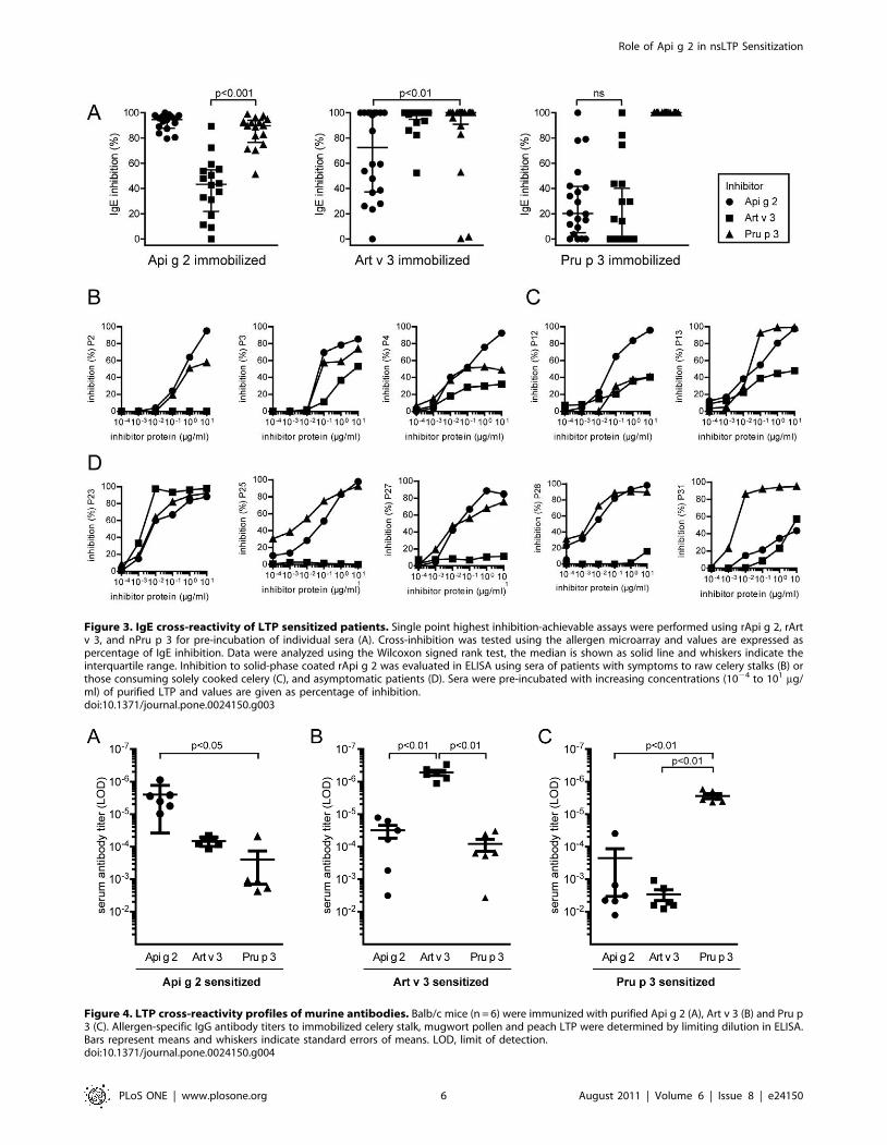
Figure 3. IgE cross-reactivity of LTP sensitized patients. Single point highest inhibition-achievable assays were performed using rApi g 2, rArtv 3, and nPru p 3 for pre-incubation of individual sera (A). Cross-inhibition was tested using the allergen microarray and values are expressed aspercentage of IgE inhibition. Data were analyzed using the Wilcoxon signed rank test, the median is shown as solid line and whiskers indicate theinterquartile range. Inhibition to solid-phase coated rApi g 2 was evaluated in ELISA using sera of patients with symptoms to raw celery stalks (B) orthose consuming solely cooked celery (C), and asymptomatic patients (D). Sera were pre-incubated with increasing concentrations (1024 to 101 mg/ml) of purified LTP and values are given as percentage of inhibition.doi:10.1371/journal.pone.0024150.g003
Figure 4. LTP cross-reactivity profiles of murine antibodies. Balb/c mice (n = 6) were immunized with purified Api g 2 (A), Art v 3 (B) and Pru p3 (C). Allergen-specific IgG antibody titers to immobilized celery stalk, mugwort pollen and peach LTP were determined by limiting dilution in ELISA.Bars represent means and whiskers indicate standard errors of means. LOD, limit of detection.doi:10.1371/journal.pone.0024150.g004
Role of Api g 2 in nsLTP Sensitization
PLoS ONE | www.plosone.org 6 August 2011 | Volume 6 | Issue 8 | e24150
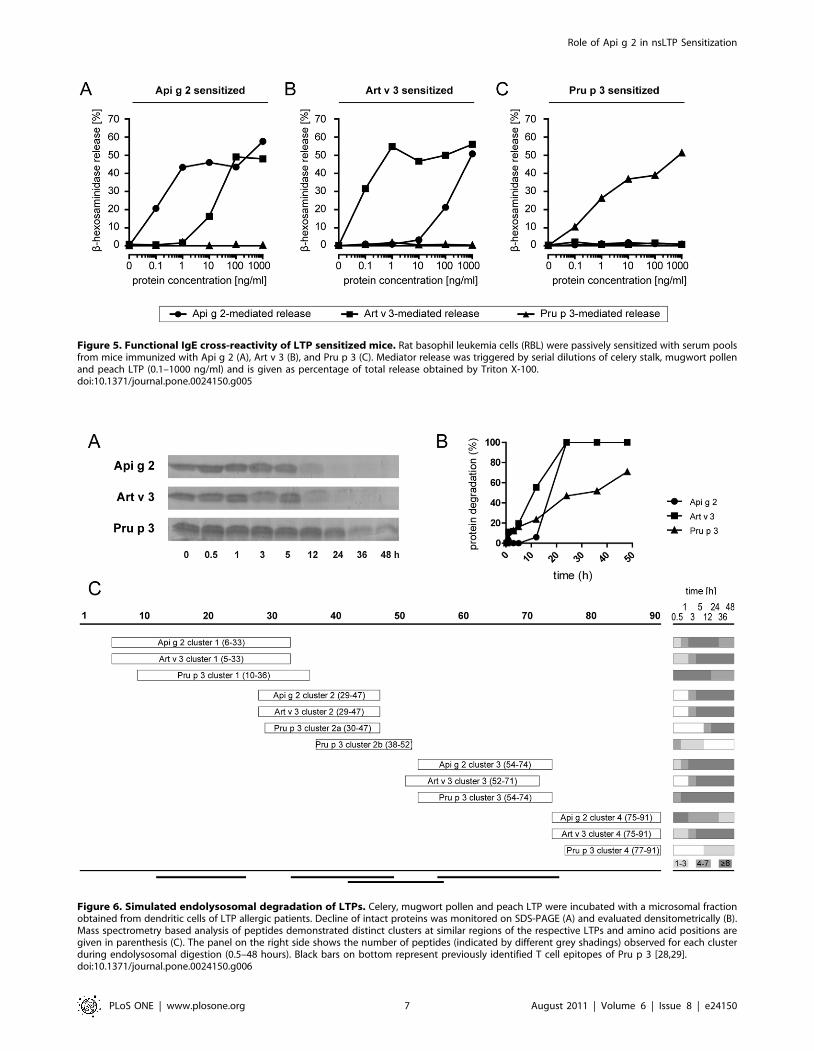
Figure 5. Functional IgE cross-reactivity of LTP sensitized mice. Rat basophil leukemia cells (RBL) were passively sensitized with serum poolsfrom mice immunized with Api g 2 (A), Art v 3 (B), and Pru p 3 (C). Mediator release was triggered by serial dilutions of celery stalk, mugwort pollenand peach LTP (0.1–1000 ng/ml) and is given as percentage of total release obtained by Triton X-100.doi:10.1371/journal.pone.0024150.g005
Figure 6. Simulated endolysosomal degradation of LTPs. Celery, mugwort pollen and peach LTP were incubated with a microsomal fractionobtained from dendritic cells of LTP allergic patients. Decline of intact proteins was monitored on SDS-PAGE (A) and evaluated densitometrically (B).Mass spectrometry based analysis of peptides demonstrated distinct clusters at similar regions of the respective LTPs and amino acid positions aregiven in parenthesis (C). The panel on the right side shows the number of peptides (indicated by different grey shadings) observed for each clusterduring endolysosomal digestion (0.5–48 hours). Black bars on bottom represent previously identified T cell epitopes of Pru p 3 [28,29].doi:10.1371/journal.pone.0024150.g006
Role of Api g 2 in nsLTP Sensitization
PLoS ONE | www.plosone.org 7 August 2011 | Volume 6 | Issue 8 | e24150

the majority of LTP epitopes are dependent on an intact 3-
dimensional fold as loss of the disulfide stabilized structure by
reduction and alkylation resulted in complete unfolding of Pru p
3 and subsequent loss of allergenicity [32]. Even though Pru p 3
was able to maintain minor parts of its IgE-binding activity upon
treatment at 95uC in our study, this might be due to higher
resistance to thermal processing which could have left some
conformational epitopes still intact. In previous studies, several
IgE-binding epitopes of Pru p 3 were identified by peptide and
mimotope mapping [25,26,27]. However, the relevance of IgE-
binding to linear epitopes remains unclear, given the fact that
unfolding of Pru p 3 led to completely abrogated allergenicity
[32]. Nevertheless, highest sequence identities of Pru p 3 to Api g
2 and Art v 3 are found in the C-terminal region which could
therefore constitute a generally LTP cross-reactive epitope
(Figure 2). The identified N-terminal epitope seems to be less
conserved and likely represents a Rosaceae-specific epitope [25],
with lower homology to LTP from other families. A striking
sequence similarity between Api g 2 and Art v 3 was observed
within residue 27–44, which might encompass a cross-reactive
celery stalk/mugwort epitope thus explaining the higher inhib-
itory capacity of Api g 2 to mugwort when compared to peach
LTP. Interestingly, the IgE-binding to Art v 3 was not inhibited
by Pru p 3 in 3/20 patients’ sera, suggesting that these individuals
were primarily sensitized to mugwort pollen Art v 3 and IgE
reactivity to Pru p 3 might be due to cross-reactivity. These
patients were also reactive to Art v 1, a genuine marker allergen
for Artemisia sensitization [33], which further corroborates this
idea. The distinction between LTP co-sensitization and cross-
reactivity is generally difficult, explaining the contradictory results
and divergent interpretation of clinical manifestations in previous
studies [11,12]. As Art v 3 and Pru p 3 seem to harbor common
but also distinct allergen-specific IgE-epitopes, both pollen and/
or food LTP were suggested as primary sensitizer [3]. Since the
sensitizer question cannot be unequivocally answered in human
studies, we used a mouse model to investigate antibody cross-
reactivity under mono-sensitization conditions. By immunizing
purified recombinant molecules, biased antibody responses due to
trace amounts of allergen impurities in a natural LTP preparation
(e. g. profilins, PR-10 proteins) could be entirely ruled out [8].
Based on our in vivo data, immunization with Api g 2 or Art v 3
seems to induce antibodies that were able to recognize the
homologous LTP of mugwort and celery stalks, and furthermore
trigger biologically relevant mediator release via IgE cross-
linking. Notably, antibodies raised against Pru p 3 were not able
to trigger mediator releases when tested with Api g 2 and Art v 3.
Results obtained from this murine model cannot be unequivo-
cally translated to the human situation, e. g. due to diverse HLA
repertoires. However, it remains still unclear if only one
particular LTP is involved in primary sensitization, since we
identified a considerable number of individuals reacting to Art v 3
and/or Api g 2 in the absence of a Pru p 3 sensitization.
Therefore, one might speculate that LTP allergy could coexist as
at least two independent branches, (i) sensitization via food Pru p
3 and (ii) sensitization via pollen, e.g. Art v 3. Such a multi-
sensitization may result in broadening of the spectrum of IgE
reactivity to different LTP, while sensitization could also take
place through an LTP that was not yet identified. The fact that
some studies showed that pollen LTP can act as primary
sensitizer [11,34,35,36] is supporting the concept and may help
to understand contradictory study outcomes of food and pollen
LTP [7]. This idea is further supported by our mouse data which
showed Pru p 3 to be independent of Art v 3 and Api g 2
antibody cross-reactivity.
The previously reported association of sensitization to Apium
graveolens and Artemisia vulgaris pollen [10] could be partially
attributed to the IgE cross-reactivity between Api g 2 and Art v 3.
However, commercially available products for allergy diagnosis
often do not indicate the specific tissue of Apium graveolens
ingredients used for extract preparation. Notably, there is a
tremendous difference regarding tissue expression of Api g 2. The
protein was only present in the aerial tissue but could not be
detected in tuber, a fact that could be easily overcome using
molecule-based allergy diagnosis [14]. However, the involvement
of Api g 2 in the celery-mugwort syndrome described for Central
European patients with allergic reactions to celeriac is not yet clear
and needs further investigations in a clinical study.
It has previously been shown that protein stability affects both,
allergenicity and immunogenicity of allergens. Susceptibility to
endolysosomal proteolysis has been demonstrated to correlate with
immunogenicity, as rather stable molecules warrant continuous
supply of T cell stimulatory peptides [37,38]. All investigated LTP
displayed relatively long half-lives of .10 hours, and thus were
more resistant to degradation as e. g. Bet v 1 from birch pollen
[37]. Observed differences in the susceptibility to endolysosomal
degradation might be explained by the primary sequence of
investigated LTP. In particular the varying number of surface
exposed lysine residues, i. e. Pru p 3 (4 lysines), Api g 2 (8 lysines),
Art v 3 (13 lysines) which are susceptible to Cathepsin S cleavage,
an important protease for antigen processing [39] may influencing
the cleavage efficiency. Interestingly, stability to endolysosomal
degradation correlated with stability to gastric digestion and
thermal treatment of investigated LTP [8,40]. Generally, Pru p 3
can be considered the most stable, while the pollen LTP Art v 3
seems to be less stable than plant food LTP. Mass spectrometry-
based analysis of endolysosomal degradation revealed that all three
LTP share similar proteolytic sites, which is reflected by the
occurrence of four common nested clusters of peptides sharing a
central core with variable flanking residues spanning identical
protein regions. These data indicate a strong influence of the
three-dimensional protein structure on the generation of T cell
epitopes. In general, Api g 2 and Art v 3 displayed a more
congruent profile compared to Pru p 3, particularly in terms of
peptide numbers, kinetics, and the absence of sub-cluster 2b which
was only observed for Pru p 3. Interestingly, the in vitro generated
peptides of Pru p 3 (cluster 1–3) perfectly matched to recently
identified T cell epitopes [28,29]. The importance of cluster 3 was
also unraveled in two recent studies using natural Pru p 3 and
microsomes from human or murine dendritic cells [32,41].
Notably, this cluster harbors the immunodominant T cell epitope
Pru p 361–75 and it is characterized by an early onset and long-
lasting peptide generation. Fragments within clusters 2b and 3
were recently also found upon simulated duodenal digestion of Pru
p 3 [42], suggesting the presence of T cell stimulatory peptides
directly on site of the intestine. Interestingly, the C-terminal cluster
of Pru p 3 contained only few peptides and seems to be irrelevant
for its T cell stimulation [29]. In contrast, numerous peptides of
Api g 2 and Art v 3 were found within cluster 4, a fact that was
recently also observed for Cor a 8 [41], suggesting a possible
relevance in T-cell reactivity for celery, mugwort pollen and
hazelnut.
In the present study we could show that Api g 2 represents a
relevant, cross-reactive allergen of Apium graveolens in patients
suffering from LTP-mediated allergy in the Mediterranean area.
Api g 2 contains common B cell epitopes of both, pollen and food-
derived LTPs. However, data obtained from mouse immunization
experiments revealed that Api g 2 shares more epitopes with
mugwort Art v 3 and seems to be rather distinct from peach Pru p
Role of Api g 2 in nsLTP Sensitization
PLoS ONE | www.plosone.org 8 August 2011 | Volume 6 | Issue 8 | e24150

3. Generally, the number and origin of LTP that patients react to
is highly inhomogeneous, and the distinction between co-
sensitization and cross-reactivity is rather difficult. Although, it is
commonly accepted that food LTP, i. e. Pru p 3 acts as primary
sensitizer in LTP allergy, our data indicate that in accordance with
classical pollen-associated food allergies, sensitization might in
addition be mediated by pollen LTP.
Materials and Methods
Screening for IgE reactivity to Art v 3, Api g 2, and Pru p 3in an Italian sensitized population
786 non-randomly distributed allergic subjects were selected on
the basis of at least one positive IgE result on the ISAC 103
microarray (PMD, Vienna, Austria) [6]. Subjects were tested on a
customized microarray including recombinant Art v 3 and Api g 2
[8,40] and natural Pru p 3 [43]. Purified proteins were
immobilized on the microarray and tested following the
established ISAC testing procedures [6].
Clinical evaluation of a selected cohortFor detailed analysis, we selected a patient’s cohort (n = 32)
presenting inhalation- and/or ingestion related allergy symptoms
to LTP containing pollen or food sources with IgE sensitization to
Api g 2, Art v 3, and Pru p 3. Clinical features of patients were
recorded using InterAll e-record (Allergy Data Laboratories s.c.,
Latina, Italy). The study was approved by the Institutional Review
Board of Istituto Dermopatico dell’Immacolata – IDI-IRCCS,
Rome, Italy (n. 106-CE-2005), and signed informed consents were
obtained.
Recombinant production of LTP from celery stalks,mugwort pollen, and peach fruit
The sequence corresponding to the mature celery LTP
(GenBank accession no. FJ643539) was cloned into the pHis
Parallel2 expression vector. Recombinant Api g 2.0101 was
expressed as soluble non-fusion protein in E. coli Rosetta-gami
B(DE3) pLysS (Novagen, Gibbstown, NJ) according to established
protocols [8,40]. Briefly, protein purification was performed with
ion exchange chromatography using a SP FF column (GE
Healthcare, Chalfont St. Giles, UK) and subsequently submitted
to gel filtration on a Superdex 10/300 GL column (GE
Healthcare). Details on the molecular characterization and
demonstration of equivalent physicochemical and immunological
properties of the natural and recombinant molecule are reported
elsewhere [8]. Recombinant Art v 3.0201 [40] and Pru p 3 [44]
were produced and purified as previously described. Purified
proteins were analyzed by denaturing SDS-PAGE and visualized
by Coomassie Brilliant Blue R-250 staining. Protein concentra-
tions were determined using the Pico Tag method (Waters,
Milford, MA) and circular dichroism measurements were
performed at 20uC to demonstrate the structural integrity of all
proteins following published protocols [8,40].
ELISA and inhibition assaysSera of 32 patients were tested for IgE reactivity to rApi g 2,
rArt v 3, and rPru p 3 in ELISA experiments. Maxisorp plates
(Nalge Nunc, Rocherster, NY) were coated with 200 ng allergen in
PBS o/n at 4uC. Unspecific binding was blocked with TBS
pH 7.4, 0.05% (v/v) Tween-20 and 1% (w/v) BSA and incubated
with 1:4 diluted patients’ sera o/n at 4uC. Bound IgE was detected
with alkaline phosphatase-conjugated monoclonal anti human IgE
antibodies (BD Biosciences, Franklin Laker, NJ). Specific serum
IgE levels were calculated based on a quantitative sandwich
ELISA using goat anti-human IgE antibody (KPL, Gaithersburg,
MD) and purified human IgE (Serotec, Raleigh, NC) as internal
standard. Measurements were performed in duplicates and values
exceeding 3xSTD of background were considered positive. In
addition, purified recombinant allergens at concentrations of 1–
2 mg/ml in 50 mM phosphate buffer were heat denatured
(15 min at 95uC) prior to coating on solid phase and IgE reactivity
was assessed as described above.
Inhibition studies were performed as ‘‘single point highest
inhibition achievable assay’’ in a microarray format [40].
Undiluted sera of 20 individual patients were pre-incubated
overnight with purified allergens at a concentration of 0.4–
0.5 mg/ml and the percentage of inhibition was determined. In
addition we assessed the dose-dependent IgE inhibition to solid
phase coated rApi g 2 in ELISA using sera of ten patients with
different clinical manifestations to celery stalks. Cross-inhibition
was performed with increasing concentrations of purified Api g 2,
Art v 3, and Pru p 3 (1024–101 mg/ml).
Animal experimentsFemale BALB/c mice (Charles River Laboratories, Wilming-
ton, MAS) were immunized subcutaneously with 10 mg allergen in
50 ml DPBS adsorbed to 50 ml Alugel-S (Serva, Heidelberg,
Germany) given as two 50 ml subcutaneous injections administered
bilaterally in the lumbar region and boosted on days 14, 28, and
42. LPS concentrations of the proteins were ,3 EU/mg as
determined by the limulus amoebocyte lysate assay (Pyrochrome,
East Falmouth, MA). Antigen-specific total IgG levels were
determined using a peroxidase-conjugated rabbit anti mouse
IgG antibody (Bio-Rad, CA) followed by chromogenic substrate
development and antigen titers were calculated as limit of
detection (LOD) values. Functional IgE levels were measured in
a mediator release assay using rat basophil leukaemia (RBL)-2H3
cells [45]. Briefly, RBL cells were passively sensitized with a 1:40
diluted murine serum pool (n = 6) raised against rApi g 2, rArt v 3,
and rPru p 3, respectively. Cross-linking and degranulation was
triggered by serial protein dilutions and b-hexosaminidase release
was measured by enzymatic cleavage of the fluorogenic substrate
4-methyl umbelliferyl-N-acetyl-b-glucosaminidase (Sigma). Results
are reported as percentage of total b-hexosaminidase release of
Triton-X100- treated cells. Animal experiments were conducted
according to National guidelines approved by the Austrian
Ministry of Science and Research, Ref. II/10b Genetic engineer-
ing and Animal experiments (approval Nr. BMWF-66.012/0011-
II/10b/2010).
Endolysosomal degradation assaysMicrosomes were isolated from monocyte-derived dendritic
cells obtained from 16 LTP-allergic donors, and used for protein
degradation according to a recently established protocol from our
laboratory [38]. Briefly, cells were harvested by low speed
centrifugation, re-suspended in 10 mM Tris acetate pH 7.0,
250 mM sucrose and homogenized with a Dounce glass tissue
homogenizer. Cellular debris and nuclei were removed by
centrifugation at 6000 x g and microsomes isolated by ultracen-
trifugation at 100,000 x g. Protein aliquots of 5 mg were digested
with 7 mg microsomal enzymes in 100 mM citrate buffer, pH 4.8
and 2 mM dithiothreitol at 37uC. Reactions were stopped by heat-
denaturation at various time points and analyzed by SDS-PAGE
and densitometric quantification of protein bands. Peptides
obtained from microsomal digestion were separated by capillary
HPLC on-line coupled to a Micromass QTof Global Ultima
instrument (Waters, Mildford, MA) as recently described [37].
Role of Api g 2 in nsLTP Sensitization
PLoS ONE | www.plosone.org 9 August 2011 | Volume 6 | Issue 8 | e24150
Individual peptides were grouped into clusters representing
regions that cover the majority of generated peptides.
ModellingHomology modeling of Api g 2, Art v 3, and Pru p 3 was
performed using Modeller 9v2 (http://salilab.org/modeller) and
evaluated with ProSa2003 based on the structure of Pru p 3 (pdb:
2B5S).
Statistical analysisStatistical evaluation was performed using the Wilcoxon signed
rank sum test and Spearman rank sum test. Groups that passed
normality and equal variance tests were analyzed with paired
samples t-test. A value of P,0.01 was considered statistically
significant.
Supporting Information
Figure S1 Proteolytic fragments obtained from endoly-sosomal degradation of Api g 2. Peptides sequenced by mass
spectrometry after 0.5, 1, 3, 5, 12, 24, 36, and 48 hours of in vitro
digestion with microsomal fractions from monocyte-derived
dendritic cells of LTP-allergic patients are depicted within the
mature sequence of Api g 2.
(TIF)
Figure S2 Proteolytic fragments obtained from endoly-sosomal degradation of Art v 3. Peptides sequenced by mass
spectrometry after 0.5, 1, 3, 5, 12, 24, 36, and 48 hours of in vitro
digestion with microsomal fractions from monocyte-derived
dendritic cells of LTP-allergic patients are depicted within the
mature sequence of Art v 3.
(TIF)
Figure S3 Proteolytic fragments obtained from endoly-sosomal degradation of Pru p 3. Peptides sequenced by mass
spectrometry after 0.5, 1, 3, 5, 12, 24, 36, and 48 hours of in vitro
digestion with microsomal fractions from monocyte-derived
dendritic cells of LTP-allergic patients are depicted within the
mature sequence of Pru p 3.
(TIF)
Acknowledgments
We thank Michael Wallner, Mario Santoro and Anargyros Roulias for
data analysis, and Paola Palazzo, Marina Liso, and Chiara Rafaiani for
microarray testing.
Author Contributions
Conceived and designed the experiments: GG AM FF. Performed the
experiments: GG MH ME KSS RF TG PB DZ. Analyzed the data: GG
PB AM FF. Contributed reagents/materials/analysis tools: LZJ. Wrote the
paper: GG FF.
References
1. Asero R, Antonicelli L, Arena A, Bommarito L, Caruso B, et al. (2009) Causes offood-induced anaphylaxis in Italian adults: a multi-centre study. Int Arch Allergy
Immunol 150: 271–277.
2. Fernandez-Rivas M, Bolhaar S, Gonzalez-Mancebo E, Asero R, van Leeuwen A,
et al. (2006) Apple allergy across Europe: how allergen sensitization profiles
determine the clinical expression of allergies to plant foods. J Allergy ClinImmunol 118: 481–488.
3. Zuidmeer L, van Ree R (2007) Lipid transfer protein allergy: primary food
allergy or pollen/food syndrome in some cases. Curr Opin Allergy Clin
Immunol 7: 269–273.
4. Wijesinha-Bettoni R, Alexeev Y, Johnson P, Marsh J, Sancho AI, et al. (2010)
The structural characteristics of nonspecific lipid transfer proteins explain theirresistance to gastroduodenal proteolysis. Biochemistry 49: 2130–2139.
5. Barber D, de la Torre F, Lombardero M, Antepara I, Colas C, et al. (2009)
Component-resolved diagnosis of pollen allergy based on skin testing with
profilin, polcalcin and lipid transfer protein pan-allergens. Clin Exp Allergy 39:1764–1773.
6. Scala E, Alessandri C, Bernardi ML, Ferrara R, Palazzo P, et al. (2010) Cross-sectional survey on immunoglobulin E reactivity in 23,077 subjects using an
allergenic molecule-based microarray detection system. Clin Exp Allergy 40:911–921.
7. Egger M, Hauser M, Mari A, Ferreira F, Gadermaier G (2010) The role of lipidtransfer proteins in allergic diseases. Curr Allergy Asthma Rep 10: 326–335.
8. Gadermaier G, Egger M, Girbl T, Erler A, Harrer A, et al. (2011) Molecularcharacterization of Api g 2, a novel allergenic member of the lipid-transfer
protein 1 family from celery stalks. Mol Nutr Food Res 55: 568–577.
9. Gaier S, Marsh J, Oberhuber C, Rigby NM, Lovegrove A, et al. (2008)
Purification and structural stability of the peach allergens Pru p 1 and Pru p 3.Mol Nutr Food Res 52(Suppl 2): S220–229.
10. Egger M, Mutschlechner S, Wopfner N, Gadermaier G, Briza P, et al. (2006)Pollen-food syndromes associated with weed pollinosis: an update from the
molecular point of view. Allergy 61: 461–476.
11. Lombardero M, Garcia-Selles FJ, Polo F, Jimeno L, Chamorro MJ, et al. (2004)
Prevalence of sensitization to Artemisia allergens Art v 1, Art v 3 and Art v 60kDa. Cross-reactivity among Art v 3 and other relevant lipid-transfer protein
allergens. Clin Exp Allergy 34: 1415–1421.
12. Pastorello EA, Pravettoni V, Farioli L, Rivolta F, Conti A, et al. (2002)
Hypersensitivity to mugwort (Artemisia vulgaris) in patients with peach allergy isdue to a common lipid transfer protein allergen and is often without clinical
expression. J Allergy Clin Immunol 110: 310–317.
13. Ballmer-Weber BK, Vieths S, Luttkopf D, Heuschmann P, Wuthrich B (2000)
Celery allergy confirmed by double-blind, placebo-controlled food challenge: aclinical study in 32 subjects with a history of adverse reactions to celery root.
J Allergy Clin Immunol 106: 373–378.
14. Bauermeister K, Ballmer-Weber BK, Bublin M, Fritsche P, Hanschmann KM,
et al. (2009) Assessment of component-resolved in vitro diagnosis of celeriac
allergy. J Allergy Clin Immunol 124: 1273–1281 e1272.
15. Breiteneder H, Hoffmann-Sommergruber K, O’Riordain G, Susani M,
Ahorn H, et al. (1995) Molecular characterization of Api g 1, the major
allergen of celery (Apium graveolens), and its immunological and structural
relationships to a group of 17-kDa tree pollen allergens. Eur J Biochem 233:
484–489.
16. Scheurer S, Wangorsch A, Haustein D, Vieths S (2000) Cloning of the minor
allergen Api g 4 profilin from celery (Apium graveolens) and its cross-reactivity
with birch pollen profilin Bet v 2. Clin Exp Allergy 30: 962–971.
17. Bublin M, Radauer C, Wilson IB, Kraft D, Scheiner O, et al. (2003) Cross-
reactive N-glycans of Api g 5, a high molecular weight glycoprotein allergen
from celery, are required for immunoglobulin E binding and activation of
effector cells from allergic patients. FASEB J 17: 1697–1699.
18. Luttkopf D, Ballmer-Weber BK, Wuthrich B, Vieths S (2000) Celery allergens in
patients with positive double-blind placebo-controlled food challenge. J Allergy
Clin Immunol 106: 390–399.
19. Ballmer-Weber BK, Hoffmann A, Wuthrich B, Luttkopf D, Pompei C, et al.
(2002) Influence of food processing on the allergenicity of celery: DBPCFC with
celery spice and cooked celery in patients with celery allergy. Allergy 57:
228–235.
20. Bublin M, Lauer I, Oberhuber C, Alessandri S, Briza P, et al. (2008) Production
and characterization of an allergen panel for component-resolved diagnosis of
celery allergy. Mol Nutr Food Res 52(Suppl 2): S241–250.
21. Hoffmann-Sommergruber K, Demoly P, Crameri R, Breiteneder H, Ebner C,
et al. (1999) IgE reactivity to Api g 1, a major celery allergen, in a Central
European population is based on primary sensitization by Bet v 1. J Allergy Clin
Immunol 104: 478–484.
22. Vallier P, DeChamp C, Valenta R, Vial O, Deviller P (1992) Purification and
characterization of an allergen from celery immunochemically related to an
allergen present in several other plant species. Identification as a profilin. Clin
Exp Allergy 22: 774–782.
23. Fotisch K, Altmann F, Haustein D, Vieths S (1999) Involvement of carbohydrate
epitopes in the IgE response of celery-allergic patients. Int Arch Allergy
Immunol 120: 30–42.
24. Jankiewicz A, Aulepp H, Baltes W, Bogl KW, Dehne LI, et al. (1996) Allergic
sensitization to native and heated celery root in pollen-sensitive patients
investigated by skin test and IgE binding. Int Arch Allergy Immunol 111:
268–278.
25. Borges JP, Barre A, Culerrier R, Granier C, Didier A, et al. (2008) Lipid transfer
proteins from Rosaceae fruits share consensus epitopes responsible for their IgE-
binding cross-reactivity. Biochem Biophys Res Commun 365: 685–690.
26. Garcia-Casado G, Pacios LF, Diaz-Perales A, Sanchez-Monge R,
Lombardero M, et al. (2003) Identification of IgE-binding epitopes of the
major peach allergen Pru p 3. J Allergy Clin Immunol 112: 599–605.
27. Pacios LF, Tordesillas L, Cuesta-Herranz J, Compes E, Sanchez-Monge R, et al.
(2008) Mimotope mapping as a complementary strategy to define allergen IgE-
epitopes: peach Pru p 3 allergen as a model. Mol Immunol 45: 2269–2276.
Role of Api g 2 in nsLTP Sensitization
PLoS ONE | www.plosone.org 10 August 2011 | Volume 6 | Issue 8 | e24150

28. Pastorello EA, Monza M, Pravettoni V, Longhi R, Bonara P, et al. (2010)
Characterization of the T-cell epitopes of the major peach allergen Pru p 3. IntArch Allergy Immunol 153: 1–12.
29. Schulten V, Radakovics A, Hartz C, Mari A, Vazquez-Cortes S, et al. (2009)
Characterization of the allergic T-cell response to Pru p 3, the nonspecific lipidtransfer protein in peach. J Allergy Clin Immunol 124: 100–107.
30. Tordesillas L, Cuesta-Herranz J, Gonzalez-Munoz M, Pacios LF, Compes E,et al. (2009) T-cell epitopes of the major peach allergen, Pru p 3: Identification
and differential T-cell response of peach-allergic and non-allergic subjects. Mol
Immunol 46: 722–728.31. Asero R, Antonicelli L, Arena A, Bommarito L, Caruso B, et al. (2009)
EpidemAAITO: features of food allergy in Italian adults attending allergy clinics:a multi-centre study. Clin Exp Allergy 39: 547–555.
32. Toda M, Reese G, Gadermaier G, Schulten V, Lauer I, et al. (2011) Proteinunfolding strongly modulates the allergenicity and immunogenicity of Pru p 3,
the major peach allergen. J Allergy Clin Immunol.
33. Asero R, Wopfner N, Gruber P, Gadermaier G, Ferreira F (2006) Artemisia andAmbrosia hypersensitivity: co-sensitization or co-recognition? Clin Exp Allergy
36: 658–665.34. Figueroa J, Blanco C, Dumpierrez AG, Almeida L, Ortega N, et al. (2005)
Mustard allergy confirmed by double-blind placebo-controlled food challenges:
clinical features and cross-reactivity with mugwort pollen and plant-derivedfoods. Allergy 60: 48–55.
35. Palacin A, Cumplido J, Figueroa J, Ahrazem O, Sanchez-Monge R, et al. (2006)Cabbage lipid transfer protein Bra o 3 is a major allergen responsible for cross-
reactivity between plant foods and pollens. J Allergy Clin Immunol 117:1423–1429.
36. Garcia-Selles FJ, Diaz-Perales A, Sanchez-Monge R, Alcantara M,
Lombardero M, et al. (2002) Patterns of reactivity to lipid transfer proteins ofplant foods and Artemisia pollen: an in vivo study. Int Arch Allergy Immunol
128: 115–122.
37. Mutschlechner S, Egger M, Briza P, Wallner M, Lackner P, et al. (2010)
Naturally processed T-cell-activating peptides of the major birch pollen allergen.
J Allergy Clin Immunol.
38. Egger M, Jurets A, Wallner M, Briza P, Ruzek S, et al. (2011) Assessing Protein
Immunogenicity with a Dendritic Cell Line-Derived Endolysosomal Degra-
dome. PLoS one 6: e17278.
39. Ruckrich T, Brandenburg J, Cansier A, Muller M, Stevanovic S, et al. (2006)
Specificity of human cathepsin S determined by processing of peptide substrates
and MHC class II-associated invariant chain. Biol Chem 387: 1503–1511.
40. Gadermaier G, Harrer A, Girbl T, Palazzo P, Himly M, et al. (2009) Isoform
identification and characterization of Art v 3, the lipid-transfer protein of
mugwort pollen. Mol Immunol 46: 1919–1924.
41. Schulten V, Nagl B, Scala E, Bernardi ML, Mari A, et al. (2011) Pru p 3, the
nonspecific lipid transfer protein from peach, dominates the immune response to
its homolog in hazelnut. Allergy 66: 1005–1013.
42. Cavatorta V, Sforza S, Aquino G, Galaverna G, Dossena A, et al. (2010) In vitro
gastrointestinal digestion of the major peach allergen Pru p 3, a lipid transfer
protein: Molecular characterization of the products and assessment of their IgE
binding abilities. Mol Nutr Food Res.
43. Ciardiello MA, Palazzo P, Bernardi ML, Carratore V, Giangrieco I, et al. (2010)
Biochemical, immunological and clinical characterization of a cross-reactive
nonspecific lipid transfer protein 1 from mulberry. Allergy 65: 597–605.
44. Zuidmeer L, van Leeuwen WA, Budde IK, Cornelissen J, Bulder I, et al. (2005)
Lipid transfer proteins from fruit: cloning, expression and quantification. Int
Arch Allergy Immunol 137: 273–281.
45. Hochreiter R, Stepanoska T, Ferreira F, Valenta R, Vrtala S, et al. (2003)
Prevention of allergen-specific IgE production and suppression of an established
Th2-type response by immunization with DNA encoding hypoallergenic
allergen derivatives of Bet v 1, the major birch-pollen allergen. Eur J Immunol
33: 1667–1676.
Role of Api g 2 in nsLTP Sensitization
PLoS ONE | www.plosone.org 11 August 2011 | Volume 6 | Issue 8 | e24150
Copyright © 2022 FDOKUMEN