Self-organization, interfacial interaction and photophysical properties of gold nanoparticle...
11
Self-organization, interfacial interaction and photophysical properties of gold nanoparticle complexes derived from resilin-mimetic fluorescent protein rec1-resilin Sundar Mayavan a , Naba K. Dutta a, * , Namita R. Choudhury a, * , Misook Kim b , Christopher M. Elvin b , Anita J. Hill c a Ian Wark Research Institute, ARC Special Research Centre, Mawson Lakes Campus, University of South Australia, Mawson Lakes, South Australia 5095, Australia b CSIRO Livestock Industries, Level 6, Queensland Bioscience Precinct, 306 Carmody Road, St Lucia, Queensland 4067, Australia c CSIRO Materials Science and Engineering, Clayton, Victoria, Australia article info Article history: Received 25 October 2010 Accepted 21 December 2010 Available online 3 February 2011 Keywords: Biomimetic protein Self-assembly Gold-protein bioconjugates Gold nanoparticles Photophysical properties of bioconjugates abstract In this investigation we report the synthesis of optically coupled hybrid architectures based on a new biomimetic fluorescent protein rec1-resilin and nanometer-scale gold nanoparticles (AuNPs) in a one-step method using a non-covalent mode of binding protocol. The presence of uniformly distributed fluorophore sequences, eSer(Thr)-Tyr-Gly- along the molecular structure of rec1-resilin provides signnificant oppor- tunity to synthesize fluorophore-modified AuNPs bioconjugates with unique photophysical properties. The detailed analyses of the AuNP-bioconjugates, synthesized under different experimental conditions using spectroscopic, microscopic and scattering techniques demonstrate the organizational pathways and the electronic and photophysical properties of the developed AuNP-rec1-resilin bioconjugates. The calculation of the bimolecular quenching constant using the SterneVolmer equation confirms that the dominant mechanism involved in quenching of fluorescence of rec1-resilin in the presence of AuNP is static. Photo- acoustic infrared spectroscopy was employed to understand the nature of the interfacial interaction between the AuNP and rec1-resilin and its evolution with pH. In such bioconjugates the quenched emission of fluorescence by AuNP on the fluorophore moiety of rec1-resilin in the immediate vicinity of the AuNP has significant potential for fluorescence-based detection schemes, sensors and also can be incorporated into nanoparticle-based devices. Ó 2011 Elsevier Ltd. All rights reserved. 1. Introduction Nanosized noble metal particles and their conjugates have attracted significant attention owing to their unique chemical, physical, electronic and surface properties; and their many potential applications in the construction of new and improved devices in the fields of photochemistry, electrochemistry, optics, magnetics, electronics, catalysts and sensors [1e 7]. The reported usage of gold nanoparticles (AuNPs) and their bioconjugates is wide and varied including drug delivery, immobilization of biomolecules, cell targeting vectors, sensors, enhancement of electron transfer between electrode surfaces and proteins, labeling of biomolecules and colorimetric detection of biomole- cules [3,4,8e13]. Protein-AuNP hybrids have been identified as versatile probes in immunohistochemistry and related techniques [3,4,14]. The application of protein-functionalized AuNPs in the detection of antigens, which are specific biomarkers in disease diagnostics, has also been advanced [3,15,16]. Fluorescent-based detection schemes that use quenched emis- sion of fluorescence by ligand functionalized AuNPs have generated significant recent interest in biotechnology for fluorescence immu- noassay and biosensors [16e19]. Hazarika et al. [16] reported reversible binding of yellow fluorescent protein (EYFP)-a mutant derivative of the naturally occurring green fluorescent protein from the North Pacific jellyfish Aequorea Victoria- at AuNP hybrids without compromising their biological activity. This research demonstrated that biological fluorophores can be incorporated into nanoparticle- based devices, leading to optically coupled hybrid architecture building blocks for the construction of biomimetic antennae and light-harvesting devices [20,21]. Iosin et al. [22] investigated the fluorescence quenching of tryptophan residues of bovine serum albumin (BSA) by AuNPs to monitor the direct interaction between * Corresponding authors. Tel.: þ61 8 83023546; fax: þ61 8 83023683. E-mail addresses: [email protected] (N.K. Dutta), namita.choudhury@ unisa.edu.au (N.R. Choudhury). Contents lists available at ScienceDirect Biomaterials journal homepage: www.elsevier.com/locate/biomaterials 0142-9612/$ e see front matter Ó 2011 Elsevier Ltd. All rights reserved. doi:10.1016/j.biomaterials.2010.12.030 Biomaterials 32 (2011) 2786e2796
Transcript of Self-organization, interfacial interaction and photophysical properties of gold nanoparticle...
lable at ScienceDirect
Biomaterials 32 (2011) 2786e2796
Contents lists avai
Biomaterials
journal homepage: www.elsevier .com/locate/biomateria ls
Self-organization, interfacial interaction and photophysical propertiesof gold nanoparticle complexes derived from resilin-mimetic fluorescentprotein rec1-resilin
Sundar Mayavan a, Naba K. Dutta a,*, Namita R. Choudhury a,*, Misook Kim b,Christopher M. Elvin b, Anita J. Hill c
a Ian Wark Research Institute, ARC Special Research Centre, Mawson Lakes Campus, University of South Australia, Mawson Lakes, South Australia 5095, AustraliabCSIRO Livestock Industries, Level 6, Queensland Bioscience Precinct, 306 Carmody Road, St Lucia, Queensland 4067, AustraliacCSIRO Materials Science and Engineering, Clayton, Victoria, Australia
a r t i c l e i n f o
Article history:Received 25 October 2010Accepted 21 December 2010Available online 3 February 2011
Keywords:Biomimetic proteinSelf-assemblyGold-protein bioconjugatesGold nanoparticlesPhotophysical properties of bioconjugates
* Corresponding authors. Tel.: þ61 8 83023546; faxE-mail addresses: [email protected] (N.K.
unisa.edu.au (N.R. Choudhury).
0142-9612/$ e see front matter � 2011 Elsevier Ltd.doi:10.1016/j.biomaterials.2010.12.030
a b s t r a c t
In this investigation we report the synthesis of optically coupled hybrid architectures based on a newbiomimetic fluorescent protein rec1-resilin and nanometer-scale gold nanoparticles (AuNPs) in a one-stepmethod using a non-covalentmode of binding protocol. The presence of uniformly distributed fluorophoresequences, eSer(Thr)-Tyr-Gly- along the molecular structure of rec1-resilin provides signnificant oppor-tunity to synthesizefluorophore-modifiedAuNPs bioconjugateswithuniquephotophysical properties. Thedetailed analyses of the AuNP-bioconjugates, synthesized under different experimental conditions usingspectroscopic, microscopic and scattering techniques demonstrate the organizational pathways and theelectronic and photophysical properties of the developed AuNP-rec1-resilin bioconjugates. The calculationof the bimolecular quenching constant using the SterneVolmer equation confirms that the dominantmechanism involved in quenching of fluorescence of rec1-resilin in the presence of AuNP is static. Photo-acoustic infrared spectroscopy was employed to understand the nature of the interfacial interactionbetween the AuNP and rec1-resilin and its evolutionwith pH. In such bioconjugates the quenched emissionof fluorescence byAuNP on thefluorophoremoiety of rec1-resilin in the immediate vicinity of the AuNP hassignificant potential for fluorescence-based detection schemes, sensors and also can be incorporated intonanoparticle-based devices.
� 2011 Elsevier Ltd. All rights reserved.
1. Introduction
Nanosized noble metal particles and their conjugates haveattracted significant attention owing to their unique chemical,physical, electronic and surface properties; and their manypotential applications in the construction of new and improveddevices in the fields of photochemistry, electrochemistry, optics,magnetics, electronics, catalysts and sensors [1e7]. The reportedusage of gold nanoparticles (AuNPs) and their bioconjugates iswide and varied including drug delivery, immobilization ofbiomolecules, cell targeting vectors, sensors, enhancement ofelectron transfer between electrode surfaces and proteins,labeling of biomolecules and colorimetric detection of biomole-cules [3,4,8e13]. Protein-AuNP hybrids have been identified as
: þ61 8 83023683.Dutta), namita.choudhury@
All rights reserved.
versatile probes in immunohistochemistry and related techniques[3,4,14]. The application of protein-functionalized AuNPs in thedetection of antigens, which are specific biomarkers in diseasediagnostics, has also been advanced [3,15,16].
Fluorescent-based detection schemes that use quenched emis-sion of fluorescence by ligand functionalized AuNPs have generatedsignificant recent interest in biotechnology for fluorescence immu-noassay and biosensors [16e19]. Hazarika et al. [16] reportedreversible binding of yellow fluorescent protein (EYFP)-a mutantderivative of the naturally occurring green fluorescent protein fromtheNorth Pacific jellyfishAequorea Victoria- at AuNP hybridswithoutcompromising their biological activity. This research demonstratedthat biological fluorophores can be incorporated into nanoparticle-based devices, leading to optically coupled hybrid architecturebuilding blocks for the construction of biomimetic antennae andlight-harvesting devices [20,21]. Iosin et al. [22] investigated thefluorescence quenching of tryptophan residues of bovine serumalbumin (BSA) by AuNPs to monitor the direct interaction between
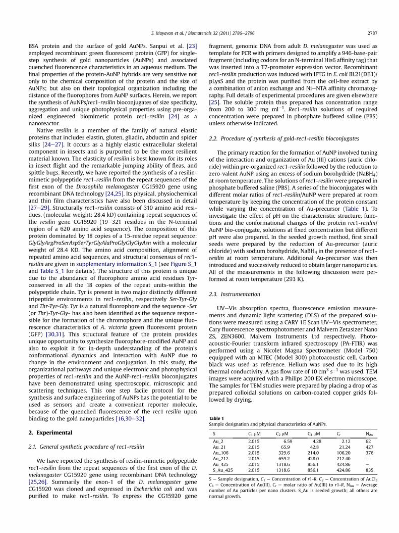
Table 1Sample designation and physical characteristics of AuNPs.
S C1 mM C2 mM C3 mM Cr NAu
Au_2 2.015 6.59 4.28 2.12 62Au_21 2.015 65.9 42.8 21.24 427Au_106 2.015 329.6 214.0 106.20 376Au_212 2.015 659.2 428.0 212.40 e
Au_425 2.015 1318.6 856.1 424.86 e
S_Au_425 2.015 1318.6 856.1 424.86 835
S ¼ Sample designation, C1 ¼ Concentration of r1-R, C2 ¼ Concentration of AuCl3C3 ¼ Concentration of Au(III), Cr ¼ molar ratio of Au(III) to r1-R, NAu ¼ Averagenumber of Au particles per nano clusters. S_Au is seeded growth; all others arenormal growth.
S. Mayavan et al. / Biomaterials 32 (2011) 2786e2796 2787
BSA protein and the surface of gold AuNPs. Sanpui et al. [23]employed recombinant green fluorescent protein (GFP) for single-step synthesis of gold nanoparticles (AuNPs) and associatedquenched fluorencence characteristics in an aqueous medium. Thefinal properties of the protein-AuNP hybrids are very sensitive notonly to the chemical composition of the protein and the size ofAuNPs; but also on their topological organization including thedistance of the fluorophores from AuNP surfaces. Herein, we reportthe synthesis of AuNPs/rec1-resilin bioconjugates of size specificity,aggregation and unique photophysical properties using pre-orga-nized engineered biomimetic protein rec1-resilin [24] as ananoreactor.
Native resilin is a member of the family of natural elasticproteins that includes elastin, gluten, gliadin, abductin and spidersilks [24e27]. It occurs as a highly elastic extracellular skeletalcomponent in insects and is purported to be the most resilientmaterial known. The elasticity of resilin is best known for its rolesin insect flight and the remarkable jumping ability of fleas, andspittle bugs. Recently, we have reported the synthesis of a resilin-mimetic polypeptide rec1-resilin from the repeat sequences of thefirst exon of the Drosophila melanogaster CG15920 gene usingrecombinant DNA technology [24,25]. Its physical, physiochemicaland thin film characteristics have also been discussed in detail[27e29]. Structurally rec1-resilin consists of 310 amino acid resi-dues, (molecular weight: 28.4 kD) containing repeat sequences ofthe resilin gene CG15920 (19e321 residues in the N-terminalregion of a 620 amino acid sequence). The composition of thisprotein dominated by 18 copies of a 15-residue repeat sequence:GlyGlyArgProSerAspSerTyrGlyAlaProGlyGlyGlyAsn with a molecularweight of 28.4 KD. The amino acid composition, alignment ofrepeated amino acid sequences, and structural consensus of rec1-resilin are given in supplementary information S_1 (see Figure S_1and Table S_1 for details). The structure of this protein is uniquedue to the abundance of fluorophore amino acid residues Tyr-conserved in all the 18 copies of the repeat units-within thepolypeptide chain. Tyr is present in two major distinctly differenttripeptide environments in rec1-resilin, respectively Ser-Tyr-Glyand Thr-Tyr-Gly. Tyr is a natural fluorophore and the sequence -Ser(or Thr)-Tyr-Gly- has also been identified as the sequence respon-sible for the formation of the chromophore and the unique fluo-rescence characteristics of A. victoria green fluorescent protein(GFP) [30,31]. This structural feature of the protein providesunique opportunity to synthesize fluorophore-modified AuNP andalso to exploit it for in-depth understanding of the protein’sconformational dynamics and interaction with AuNP due tochange in the environment and conjugation. In this study, theorganizational pathways and unique electronic and photophysicalproperties of rec1-resilin and the AuNP-rec1-resilin bioconjugateshave been demonstrated using spectroscopic, microscopic andscattering techniques. This one step facile protocol for thesynthesis and surface engineering of AuNPs has the potential to beused as sensors and create a convenient reporter molecule,because of the quenched fluorescence of the rec1-resilin uponbinding to the gold nanoparticles [16,30e32].
2. Experimental
2.1. General synthetic procedure of rec1-resilin
We have reported the synthesis of resilin-mimetic polypeptiderec1-resilin from the repeat sequences of the first exon of the D.melanogaster CG15920 gene using recombinant DNA technology[25,26]. Summarily the exon-1 of the D. melanogaster geneCG15920 was cloned and expressed in Escherichia coli and waspurified to make rec1-resilin. To express the CG15920 gene
fragment, genomic DNA from adult D. melanogaster was used astemplate for PCR with primers designed to amplify a 946-base-pairfragment (including codons for an N-terminal His6 affinity tag) thatwas inserted into a T7-promoter expression vector. Recombinantrec1-resilin production was induced with IPTG in E. coli BL21(DE3)/pLysS and the protein was purified from the cell-free extract bya combination of anion exchange and NieNTA affinity chromatog-raphy. Full details of experimental procedures are given elsewhere[25]. The soluble protein thus prepared has concentration rangefrom 200 to 300 mg ml�1. Rec1-resilin solutions of requiredconcentration were prepared in phosphate buffered saline (PBS)unless otherwise indicated.
2.2. Procedure of synthesis of gold-rec1-resilin bioconjugates
The primary reaction for the formation of AuNP involved tuningof the interaction and organization of Au (III) cations (auric chlo-ride) within pre-organized rec1-resilin followed by the reduction tozero-valent AuNP using an excess of sodium borohydride (NaBH4)at room temperature. The solutions of rec1-resilinwere prepared inphosphate buffered saline (PBS). A series of the bioconjugates withdifferent molar ratios of rec1-resilin/AuNP were prepared at roomtemperature by keeping the concentration of the protein constantwhile varying the concentration of Au-precursor (Table 1). Toinvestigate the effect of pH on the characteristic structure, func-tions and the conformational changes of the protein rec1-resilin/AuNP bio-conjugate, solutions at fixed concentration but differentpH were also prepared. In the seeded growth method, first smallseeds were prepared by the reduction of Au-precursor (auricchloride) with sodium borohydride, NaBH4 in the presence of rec1-resilin at room temperature. Additional Au-precursor was thenintroduced and successively reduced to obtain larger nanoparticles.All of the measurements in the following discussion were per-formed at room temperature (293 K).
2.3. Instrumentation
UVeVis absorption spectra, fluorescence emission measure-ments and dynamic light scattering (DLS) of the prepared solu-tions were measured using a CARY 1E Scan UVeVis spectrometer,Cary fluorescence spectrophotometer and Malvern Zetasizer NanoZS, ZEN3600, Malvern Instruments Ltd respectively. Photo-acoustic-Fourier transform infrared spectroscopy (PA-FTIR) wasperformed using a Nicolet Magna Spectrometer (Model 750)equipped with an MTEC (Model 300) photoacoustic cell. Carbonblack was used as reference. Helium was used due to its highthermal conductivity. A gas flow rate of 10 cm3 s�1 was used. TEMimages were acquired with a Philips 200 EX electron microscope.The samples for TEM studies were prepared by placing a drop of asprepared colloidal solutions on carbon-coated copper grids fol-lowed by drying.
S. Mayavan et al. / Biomaterials 32 (2011) 2786e27962788
3. Result and discussion
3.1. Size, shape and morphology of AuNP/rec1-resilin bioconjugates
Fig.1 displays the TEM images of AuNP/rec1-resilin bioconjugates(GrBCs) from colloidal sols with different AuCl3 concentrations in
Fig. 1. TEM images of rec1-resilin capped gold nanoparticles synthesized at different (Au cat(Scale bar 20 nm); and growth techniques: (e) normal growth, Au_425 (e); seeded growth
w2.01 mM solution of rec1-resilin in PBS (pH 7.4). It reveals that theparticle size and interparticle spacing are strongly dependent on (Aucation):(rec1-resilin) molar ratio (Cr) in the solution. At lower Cr(Au_2 Fig. 1(a) and Au_21, Fig. 1(b)), the AuNPs are dispersed andwell-separated. From the images, the GrBCs in the colloidal solsappear to be irregular and non directional. The particle size and its
ion):(rec1-resilin) molar ratios: 2.12 (a); 21.4 (b); 106.18 (c); and 212.36 (d) respectively(f) (scale bar 100 nm).
S. Mayavan et al. / Biomaterials 32 (2011) 2786e2796 2789
distribution along with the normal distribution fit of the experi-mental data are also shown as inset in the images. In the case of A_2,the particle size is <2 nm (m ¼ 1.26 nm) and distribution is narrow(s ¼ 0.37) and for A_21 increase in particle size (m ¼ 2.4 nm) alongwith broader distribution, (s ¼ 0.60) is identified. With increase inthe concentration of Au(III) from42.8 mMto0.214mM level (Au_106)in the sol, the interparticle spacing becomes smaller and exhibitsa spacing of 1e2 nm (Fig. 1(c)), without changing the particle size(m¼ 2.3 nm) anddistribution (s¼ 0.60). Atw0.43mM (Au_212) levelof precursor, the neighboring particles link together to form non-spherical particles (Fig. 1(d)).
In the case of spherical particles, the mean diameter, D (nm), ofthe AuNPs allows determination of themean number of gold atoms,NAu in the AuNPs (see supplementary information S_2 for details),and these values are shown in Table 1. The molar concentration ofAuNPs in Au_2, Au_21 and Au_106 were respectively 6.9 � 10�8,1.0� 10�7, 5.7� 10�7 mol L�1. Fig.1(e) and (f) show the TEM imagesfor AuNPs generated using solution containing 1.32 mM AuCl3 usingtwo different methods of preparation. The seeded growth method(S_Au_425) results in bigger size particles with relatively uniformspherical geometry; however, in the case of normal growth tech-nique (Au_425) theparticles are asymmetricwithboth spherical andnon-spherical shapes. The size distribution plot for S_Au_425 is alsoshown in the figure, which appears to be a bimodal normal distri-bution. A fit with single normal distribution is shown in the figureand the m and s values are 3.00 and 1.07 respectively.
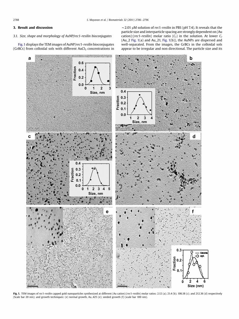
Rec1-resilin is observed to be an acidic peptide with the exper-imentally evaluated isoelectric point (PI) at w4.8, whereas thetheoretical PI value is 9.20 e considering all residues uniformlyexposed to water [29]. This discrepancy indicates the complexorganization of the protein in water, with the negatively chargedprotein surface exposed to water and positively charged residuesforming the core. The net surface charge of rec1-resilin is positive atpHs below w4.8 and negative above that level; and the nature ofenrichment of functional groups on the surface of rec1-resilin canbe controlled by varying the pH. This may provide further oppor-tunity for tuning the organization of the protein. Fig. 2(a) shows theTEM image (as prepared colloidal solution) of typical AuNPs (A_21)at low pH 2.8. The particles appear to be aggregated with a size of>20 nm. After reduction of the precursor AuCl3 during preparation,the colour of the colloidal sol changes from pale pink to black,indicating that the Au(0) spheres were formed first, followed byrapid aggregation. A decrease in solution pH below 4.8 (PI) leads tothe protonation of the functional residues, and the overall surfacecharge becomes positive, which in turn reduces the electrostaticinteraction of rec1-resilin surface with Au(III) cations and formsrandomly distributed aggregates instead of fractals. The hydrogen
Fig. 2. Effect of pH on self-organization of rec1-resilin and its effect on the stabilizationcation):(rec1-resilin) molar ratio: 21.4: (a) pH 2.8; (b) pH 11.8.
bonding of the carboxylic acid groups in protonated form is alsoresponsible for the interparticle interactions in the nanoparticleassembly. The TEM image (Fig. 2(b) reveals that AuNPs prepared atalkaline conditions (pH>10.8) are smaller (w1.8 nm), morespherical and uniformly distributed than that at pH 7. When thereaction is performed at pH > pKa (wpH 9.5) the amino acidresidue Tyr becomes the deprotonated “tyrosinate” form [29].Consequently, the capping efficiency of the protein increaseddramatically due to the electrostatic interaction. Tyrosinate can alsocause auto reduction of AuCl3 to AuNP. Therefore, nucleation andgrowth of AuNPs occur in a faster and more controlled environ-ment, yielding small uniform Au(0) nanospheres. This is to notethat dispersive and van der Waals forces must play some role in allof the above mechanisms as well. However, the steric stabilizationdoes not appear to provide colloidal stability of the sols as it fails tostabilize the colloid at the isoelectric point (pH ¼ w4.8).
3.2. Plasmon resonance characteristics of AuNP/rec1-resilinbioconjugates
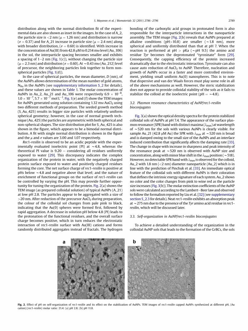
Fig. 3(a) shows theoptical density spectra for theprotein stabilizedcolloidal sols of AuNPs at pH 7.4. The appearance of the surface plas-mon resonance (SPR)bandwithdistinctmaxima (lmax) atwavelengthof w520 nm for the sols with various AuNPs is clearly visible. Forsample Au_21 (42.8 mM Au) the SPR with lmax at w520 nm is broadand is damped owing to the small size of the AuNPs and an interfaceinduced contribution that significantly affects the damping rate [33].The change in shapewith increase in sharpness and peak intensity ofthe resonance peak at w520 nm is observed with AuNP size andconcentration,alongwithminorblueshift in thelmaxposition (w518).However, nodetectable SPRbandwith lmax is observed for the colloid,Au_2 with 1.8 nm (<2 nm) diameter nanoparticle (Au_2) which is inline with the prediction of Pinchuk et al. [33]. An immediate opticalfeature of the colloidal sols with different AuNPs is their colorationthatdefines the intrinsic energy signature of eachsystem.Au_2 showsno color and the color changes from pink to wine red as the particlesize increases (Fig. 3(b)). Themolar extinction coefficientsof theAuNPsolswere calculatedaccording to the LamberteBeer lawandobservedto follow the formalism reported by Liu et al. [32] (see supplementarysection S_2.3 for details). Neat rec1-resilin exhibits an absorptionpeakatw275nmdue to the presence of the Tyr amino acid residue in rec1-resilin, which will be discussed later.
3.3. Self-organization in AuNP/rec1-resilin bioconjugates
To achieve a detailed understanding of the organization in thecolloidal AuNP sols that leads to the formation of the GrBCs, the sols
of AuNPs. TEM images of rec1-resilin capped AuNPs synthesized at different pH, (Au
Fig. 3. Photophysical properties of rec1-resilin stabilized aqueous colloidal sols containing different size and molar concentration of AuNPs; (a) UVeVis absorption cross section ofneat rec1-resilin and rec1-resilin stabilized aqueous colloidal sols containing different size and concentration of AuNPs. (b) optical photograph of the experimental sols.
S. Mayavan et al. / Biomaterials 32 (2011) 2786e27962790
were characterized using dynamic light scattering (DLS). In theabsence of rec1-resilin as a stabilizer, the Au(0) generated byreduction of AuCl3 with NaBH4 nucleate and grow into large par-ticle clusters and precipitate immediately. However, when rec1-resilin is introduced as a stabilizer the specific interactions betweenthe protein molecule and the AuNP surface greatly control thegrowth rate and lead to the formation of stable nanoparticles. Fig. 4illustrates the evolution of the Z average diameter Dh, of GrBCparticles and its distributions for colloidal sol with time for a typicalsystem (Au_425). It demonstrates that rec1-resilin interacts withthe Au nanoparticle precursor spontaneously and this interactionshifts the peak of the Dh distribution from 10.1 nm for neat proteintow79 nm (b_red). It can also be observed from the figure that thechemical reduction of the precursor using NaBH4 results in fasternucleation to create small Au(0) primary particles; and rec1-resilinis able to stabilize them efficiently without change in the distri-bution in Dh (t_10 min). This peptide protected system is stable formany months without aggregation, however with maturation ofthe sol marginal peak shift along with broadening of Dh is observedfrom DLS (t_mat).
The DLS data in Fig. 5 of different AuNP colloidal sols reveal thatthe self-assembly process and the GrBC particle size and sizedistribution in the final micelle-like structures of the sols arecontrolled by Cr. At lower Cr of 2 (Au_2), a bimodal distributioncentered around the peaks at 9 and 152 nm respectively is
Size (nm)
1 10 100 1000
In
ten
sity, %
0
4
8
12
16
20
b_red
t_10 min
t_mat
r1_R
Fig. 4. Effect of addition of metal precursor, its reduction to AuNP and maturation onhydrodynamic diameter, Dh of rec1-resilin.
apparent. The major component of the Dh of GrBC is at w152 nm(size of bare AuNP is w1.25 nm-Fig. 1) indicating a greater degreeof rec1-resilin association with the nanoparticle i.e., formation ofmultilayers (>7 layers considering average Dh of the protein of10.1 nm) or in multimeric form. The presence of some unconju-gated protein with Dh of w9 nm in the sol is also observed. Withincrease in Cr to 21 (Au_21) the major part of the bimodal distri-bution observed is the multilayer GrBC (Dh of w142 nm) with theminor component being monolayer particles (Dh of w28 nm). Noisolated polypeptide is evident in this system. On the contrary, inthe case of Au_106 the Dh of the GrBC exhibits a symmetricunimodal distribution with peak Dh centered around w32.6 nm,corresponding to particles with primarily a monolayer of adsorbedrec1-resilin. Increase in the AuNP concentration to 428 mM level(Au_212) results in the increase of dipoleedipole attractionbetween the AuNPs due to diminution of the interparticle distance,leading to the formation of larger interconnected particles witha multilayer of the protein capping the particles giving a peak of Dh
w91 nm. With further increase in the concentration of Au to856 mM (A_425) the peak position remains practically unchanged,however, the Dh distribution becomes substantially narrower.These observations confirm that the structure and morphology ofGrBC evolve with change of Cr in the sol and can be used to controlthe particle characteristics. Fig. 6 compares the distribution of Dh
Size, (nm)
10 100 1000
In
ten
sity %
0
4
8
12
16 Au_2
Au_21
Au_106
Au_212
Au_425
r1_R
Fig. 5. Distributions of hydrodynamic diameter, Dh of rec1-resilin/AuNP conjugateswith different levels of AuNPs in the sol.
Size (nm)
1 10 100 1000
In
ten
sity, %
0
4
8
12
16
S_Au_425
Au_425
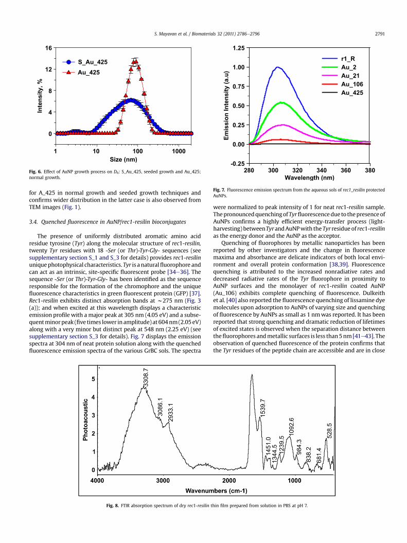
Fig. 6. Effect of AuNP growth process on Dh: S_Au_425, seeded growth and Au_425;normal growth. Wavelength (nm)
280 300 320 340 360 380
Em
is
sio
n In
te
ns
ity
(a
.u
)
-0.25
0.00
0.25
0.50
0.75
1.00
1.25
r1_R
Au_2
Au_21
Au_106
Au_425
Fig. 7. Fluorescence emission spectrum from the aqueous sols of rec1_resilin protectedAuNPs.
S. Mayavan et al. / Biomaterials 32 (2011) 2786e2796 2791
for A_425 in normal growth and seeded growth techniques andconfirms wider distribution in the latter case is also observed fromTEM images (Fig. 1).
3.4. Quenched fluorescence in AuNP/rec1-resilin bioconjugates
The presence of uniformly distributed aromatic amino acidresidue tyrosine (Tyr) along the molecular structure of rec1-resilin,twenty Tyr residues with 18 -Ser (or Thr)-Tyr-Gly- sequences (seesupplementary section S_1 and S_3 for details) provides rec1-resilinuniquephotophysical characteristics. Tyr is a naturalfluorophore andcan act as an intrinsic, site-specific fluorescent probe [34e36]. Thesequence -Ser (or Thr)-Tyr-Gly- has been identified as the sequenceresponsible for the formation of the chromophore and the uniquefluorescence characteristics in green fluorescent protein (GFP) [37].Rec1-resilin exhibits distinct absorption bands at w275 nm (Fig. 3(a)); and when excited at this wavelength displays a characteristicemission profile with a major peak at 305 nm (4.05 eV) and a subse-quentminorpeak (five times lower inamplitude) at 604nm(2.05eV)along with a very minor but distinct peak at 548 nm (2.25 eV) (seesupplementary section S_3 for details). Fig. 7 displays the emissionspectra at 304 nm of neat protein solution along with the quenchedfluorescence emission spectra of the various GrBC sols. The spectra
Fig. 8. FTIR absorption spectrum of dry rec1-resilin t
were normalized to peak intensity of 1 for neat rec1-resilin sample.ThepronouncedquenchingofTyrfluorescencedue to thepresenceofAuNPs confirms a highly efficient energy-transfer process (light-harvesting) betweenTyrandAuNPwith the Tyr residue of rec1-resilinas the energy donor and the AuNP as the acceptor.
Quenching of fluorophores by metallic nanoparticles has beenreported by other investigators and the change in fluorescencemaxima and absorbance are delicate indicators of both local envi-ronment and overall protein conformation [38,39]. Fluorescencequenching is attributed to the increased nonradiative rates anddecreased radiative rates of the Tyr fluorophore in proximity toAuNP surfaces and the monolayer of rec1-resilin coated AuNP(Au_106) exhibits complete quenching of fluorescence. Dulkeithet al. [40] also reported the fluorescence quenching of lissamine dyemolecules upon adsorption to AuNPs of varying size and quenchingof fluorescence by AuNPs as small as 1 nmwas reported. It has beenreported that strong quenching and dramatic reduction of lifetimesof excited states is observed when the separation distance betweenthefluorophores andmetallic surfaces is less than5nm[41e43]. Theobservation of quenched fluorescence of the protein confirms thatthe Tyr residues of the peptide chain are accessible and are in close
hin film prepared from solution in PBS at pH 7.
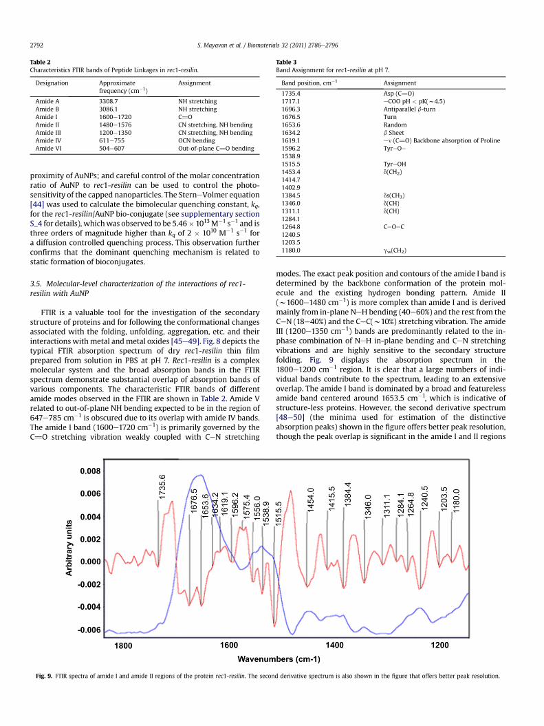
Table 2Characteristics FTIR bands of Peptide Linkages in rec1-resilin.
Designation Approximatefrequency (cm�1)
Assignment
Amide A 3308.7 NH stretchingAmide B 3086.1 NH stretchingAmide I 1600e1720 C]OAmide II 1480e1576 CN stretching, NH bendingAmide III 1200e1350 CN stretching, NH bendingAmide IV 611e755 OCN bendingAmide VI 504e607 Out-of-plane C]O bending
Table 3Band Assignment for rec1-resilin at pH 7.
Band position, cm�1 Assignment
1735.4 Asp (C]O)1717.1 eCOO pH < pK(w4.5)1696.3 Antiparallel b-turn1676.5 Turn1653.6 Random1634.2 b Sheet1619.1 en (C]O) Backbone absorption of Proline1596.2 TyreOe1538.91515.5 TyreOH1453.4 d(CH2)1414.71402.91384.5 ds(CH3)1346.0 d(CH)1311.1 d(CH)1284.11264.8 CeOeC1240.51203.51180.0 gw(CH2)
S. Mayavan et al. / Biomaterials 32 (2011) 2786e27962792
proximity of AuNPs; and careful control of the molar concentrationratio of AuNP to rec1-resilin can be used to control the photo-sensitivity of the capped nanoparticles. The SterneVolmer equation[44] was used to calculate the bimolecular quenching constant, kq,for the rec1-resilin/AuNP bio-conjugate (see supplementary sectionS_4 for details), whichwas observed to be 5.46�1013M�1 s�1 and isthree orders of magnitude higher than kq of 2 � 1010 M�1 s�1 fora diffusion controlled quenching process. This observation furtherconfirms that the dominant quenching mechanism is related tostatic formation of bioconjugates.
3.5. Molecular-level characterization of the interactions of rec1-resilin with AuNP
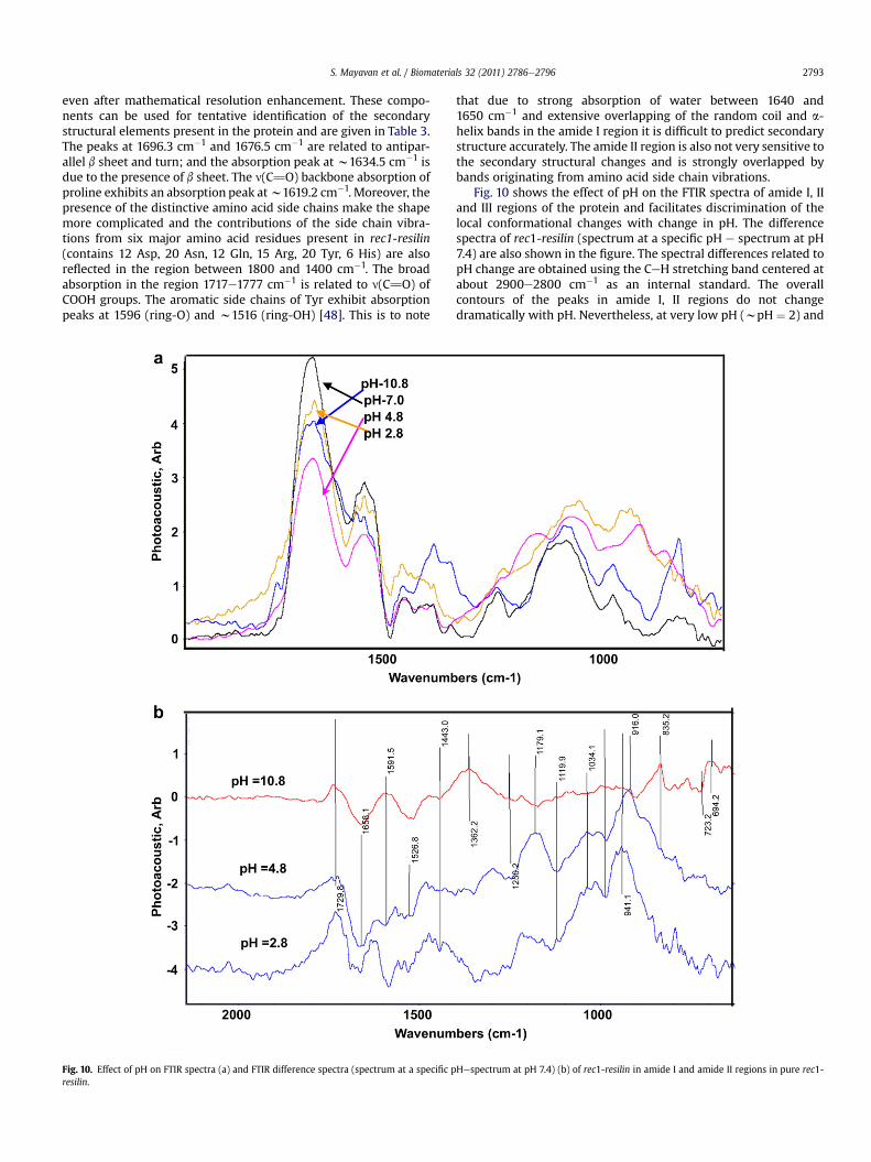
FTIR is a valuable tool for the investigation of the secondarystructure of proteins and for following the conformational changesassociated with the folding, unfolding, aggregation, etc. and theirinteractions withmetal andmetal oxides [45e49]. Fig. 8 depicts thetypical FTIR absorption spectrum of dry rec1-resilin thin filmprepared from solution in PBS at pH 7. Rec1-resilin is a complexmolecular system and the broad absorption bands in the FTIRspectrum demonstrate substantial overlap of absorption bands ofvarious components. The characteristic FTIR bands of differentamide modes observed in the FTIR are shown in Table 2. Amide Vrelated to out-of-plane NH bending expected to be in the region of647e785 cm�1 is obscured due to its overlap with amide IV bands.The amide I band (1600e1720 cm�1) is primarily governed by theC]O stretching vibration weakly coupled with CeN stretching
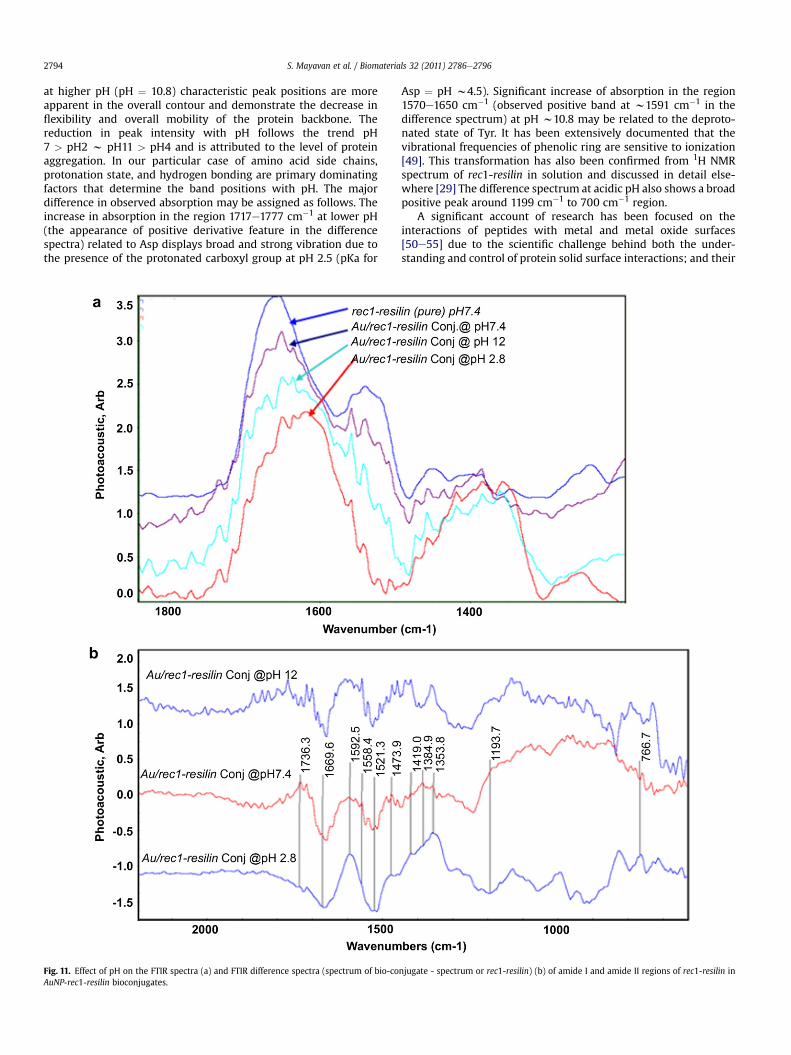
Fig. 9. FTIR spectra of amide I and amide II regions of the protein rec1-resilin. The secon
modes. The exact peak position and contours of the amide I band isdetermined by the backbone conformation of the protein mol-ecule and the existing hydrogen bonding pattern. Amide II(w1600e1480 cm�1) is more complex than amide I and is derivedmainly from in-plane NeH bending (40e60%) and the rest from theCeN (18e40%) and the CeC(w10%) stretching vibration. The amideIII (1200e1350 cm�1) bands are predominantly related to the in-phase combination of NeH in-plane bending and CeN stretchingvibrations and are highly sensitive to the secondary structurefolding. Fig. 9 displays the absorption spectrum in the1800e1200 cm�1 region. It is clear that a large numbers of indi-vidual bands contribute to the spectrum, leading to an extensiveoverlap. The amide I band is dominated by a broad and featurelessamide band centered around 1653.5 cm�1, which is indicative ofstructure-less proteins. However, the second derivative spectrum[48e50] (the minima used for estimation of the distinctiveabsorption peaks) shown in the figure offers better peak resolution,though the peak overlap is significant in the amide I and II regions
d derivative spectrum is also shown in the figure that offers better peak resolution.
S. Mayavan et al. / Biomaterials 32 (2011) 2786e2796 2793
even after mathematical resolution enhancement. These compo-nents can be used for tentative identification of the secondarystructural elements present in the protein and are given in Table 3.The peaks at 1696.3 cm�1 and 1676.5 cm�1 are related to antipar-allel b sheet and turn; and the absorption peak at w1634.5 cm�1 isdue to the presence of b sheet. The n(C]O) backbone absorption ofproline exhibits an absorption peak atw1619.2 cm�1. Moreover, thepresence of the distinctive amino acid side chains make the shapemore complicated and the contributions of the side chain vibra-tions from six major amino acid residues present in rec1-resilin(contains 12 Asp, 20 Asn, 12 Gln, 15 Arg, 20 Tyr, 6 His) are alsoreflected in the region between 1800 and 1400 cm�1. The broadabsorption in the region 1717e1777 cm�1 is related to n(C]O) ofCOOH groups. The aromatic side chains of Tyr exhibit absorptionpeaks at 1596 (ring-O) and w1516 (ring-OH) [48]. This is to note
Fig. 10. Effect of pH on FTIR spectra (a) and FTIR difference spectra (spectrum at a specific presilin.
that due to strong absorption of water between 1640 and1650 cm�1 and extensive overlapping of the random coil and a-helix bands in the amide I region it is difficult to predict secondarystructure accurately. The amide II region is also not very sensitive tothe secondary structural changes and is strongly overlapped bybands originating from amino acid side chain vibrations.
Fig. 10 shows the effect of pH on the FTIR spectra of amide I, IIand III regions of the protein and facilitates discrimination of thelocal conformational changes with change in pH. The differencespectra of rec1-resilin (spectrum at a specific pH e spectrum at pH7.4) are also shown in the figure. The spectral differences related topH change are obtained using the CeH stretching band centered atabout 2900e2800 cm�1 as an internal standard. The overallcontours of the peaks in amide I, II regions do not changedramatically with pH. Nevertheless, at very low pH (wpH ¼ 2) and
Hespectrum at pH 7.4) (b) of rec1-resilin in amide I and amide II regions in pure rec1-
S. Mayavan et al. / Biomaterials 32 (2011) 2786e27962794
at higher pH (pH ¼ 10.8) characteristic peak positions are moreapparent in the overall contour and demonstrate the decrease inflexibility and overall mobility of the protein backbone. Thereduction in peak intensity with pH follows the trend pH7 > pH2 w pH11 > pH4 and is attributed to the level of proteinaggregation. In our particular case of amino acid side chains,protonation state, and hydrogen bonding are primary dominatingfactors that determine the band positions with pH. The majordifference in observed absorption may be assigned as follows. Theincrease in absorption in the region 1717e1777 cm�1 at lower pH(the appearance of positive derivative feature in the differencespectra) related to Asp displays broad and strong vibration due tothe presence of the protonated carboxyl group at pH 2.5 (pKa for
Fig. 11. Effect of pH on the FTIR spectra (a) and FTIR difference spectra (spectrum of bio-coAuNP-rec1-resilin bioconjugates.
Asp ¼ pH w4.5). Significant increase of absorption in the region1570e1650 cm�1 (observed positive band at w1591 cm�1 in thedifference spectrum) at pH w10.8 may be related to the deproto-nated state of Tyr. It has been extensively documented that thevibrational frequencies of phenolic ring are sensitive to ionization[49]. This transformation has also been confirmed from 1H NMRspectrum of rec1-resilin in solution and discussed in detail else-where [29] The difference spectrum at acidic pH also shows a broadpositive peak around 1199 cm�1 to 700 cm�1 region.
A significant account of research has been focused on theinteractions of peptides with metal and metal oxide surfaces[50e55] due to the scientific challenge behind both the under-standing and control of protein solid surface interactions; and their
njugate - spectrum or rec1-resilin) (b) of amide I and amide II regions of rec1-resilin in
S. Mayavan et al. / Biomaterials 32 (2011) 2786e2796 2795
importance for medical, diagnostic and biotechnology applications.In general such interactions are reported to be the result of inter-play between electrostatic and hydrophobic interactions. Peelleet al. [51] examined homohexamers (X6) of the 20 natural aminoacids (X) for binding to five different inorganic materials includinggold nanoparticles; and reported that amino acid residues con-taining aromatic nitrogen (His or Try) or sulfur (Cys or Met) arecapable of binding to various types of NPs. Si et al. [52] employedsynthetic oligopeptides containing redox-active Tyr residues toprepare gold and silver nanoparticles. Kogot [53] and Johnson [54]reported control of protein orientation at interfaces using histidineresidue. Fig.11(a) illustrates the PA-FTIR of neat rec-1 resilin (pH 7.4)and Au-rec1-reslin composite films at different pH; and reflects themolecular level interactions (FTIR spectrum has been verticallyshifted for clarity and comparison). The FTIR difference spectra(rec1-resilin þ AuNp)-(rec1-resilin) of Au-protein complexes arealso shown in Fig. 11(b) that reveals the extent and nature ofstructural change in rec1-resilin upon interaction. At pH 7.4 thedifference spectrum of Au-protein complexes exhibits negativederivative features in amide I and amide II regions, which areindicative of no major interaction or coordination to peptide CeOand CeN groups. However, the appearance of positive features at1714e1770 cm�1 reveals direct interaction of Au with COOe of Aspresidue. In solution at pH 7.4, rec1-resilin is hydrophilic and nega-tively charged and the pH value is much higher than the pKa of 4.5for Asp that initiates interaction between the COOe groups on thepeptide side chain and the surface anchors to the gold surface. Thebroad but weak positive feature observed from 1240 to 740 cm�1
region at this pH may be related to the interaction of Au with Hisresidue [53]. In neutral and basic pH imidazole nitrogen in Hisresidues is in the deprotonated state and can interact with theprotein. The presence of several positive derivative features withweak intensities in the regions 1538e1608 cm�l is attributed to theparticipation of the tyrosine residue in metal-protein complexa-tion. The interaction of tyrosine with metal cations has beenreported through the spectral changes of the tyrosine band at1514 cm�1 in a series of metal-protein complexes by other inves-tigators [56]. At pH > pKa of Tyr (pH > 10) the amino acid residueTyr becomes the deprotonated “tyrosinate” form, consequently, thecapping efficiency of the protein increases dramatically due to theelectrostatic interaction and efficient electron transfer. The positivefeatures are clearly reflected in the difference spectrum of Au-rec1-resilin complexes at pH12. However, such interaction is notobserved in the spectra of pH 2.8. At pH 2.8 a significant change inthe amide I and amide II regions and the observed strong positiveintensity at w1592 may be attributed to conformational variationsof the protein in the presence of the Au ion and interaction of Auwith the polypeptide C]O and CeN groups in the Au-proteincomplexes. At acidic pH the presence of a negative feature at1700e1777 cm�1 in the difference spectra of the Au-proteincomplex is also indicative of no direct AueCOOe interaction in thismetal complex. This is to note that FTIR data provides only anoverall estimation and tentative identification of the interfacialinteractions between Au and rec1-resilin. Detailed investigation ofthe site-specific nature of the interaction is underway using solu-tion FTIR, XPS, 1D and 2D NMR and will be presented in future.
4. Conclusion
We have successfully used the environment induced tuning ofthe self-assembling of rec1-resilin for constrained synthesis andstabilization of nanostructured gold nanoparticles of controlled sizeand photophysical properties. The presence of uniformly distrib-uted chromophore sequences along themolecular structure of rec1-resilin provides a unique opportunity to synthesize fluorophore-
modified Au-nanoparticles in a facile one-step protocol. However,the particle size, photophysical properies of the AuNP-bio-conjugates and the exact nature of interfacial interaction betweenAu and rec1-resilin is critically dependent on pH. The calculation ofthe bimolecular quenching constant using the SterneVolmerequation confirms that the dominant mechanism involved forquenching of fluorescence of rec1-resilin in the presence of AuNP isstatic.
Acknowledgment
The authors acknowledge the financial support of the AustralianResearch Council (ARC) to carry out the research through theDiscovery Project funding (DP0451406) scheme. S. Mayavanexpresses thanks to University of South Australia for a President’sScholarships (UPS).
Appendix. Supplementary data
Supplementary data associated with the article can be found inonline version, at doi:10.1016/j.biomaterials.2010.12.030.
Appendix
Figures with essential color discrimination. Figs. 3e7, 9e11, inthis article are difficult to interpret in black and white. The fullcolor images can be found in the online version, at doi:10.1016/j.biomaterials.2010.12.030.
References
[1] Katz E, Willner I. Integrated nanoparticle-biomolecule hybrid systems:synthesis, properties, and applications. AngewChem Int Ed2004;43:6042e108.
[2] Shen Y, Kuang M, Shen Z, Nieberle J, Duan H, Frey H. Gold nanoparticlescoated with a thermosensitive hyperbranched polyelectrolyte: towards smarttemperature and pH nanosensors. Angew Chem Int Ed 2008;47:2227e30.
[3] Rosi NL, Mirkin CA. Nanostructures in biodiagnostics. Chem Rev 2005;105:1547e62.
[4] Daniel M-C, Astruc D. Gold nanoparticles: assembly, supramolecular chem-istry, quantum-size-related properties, and applications toward biology,catalysis, and nanotechnology. Chem Rev 2004;104:293e346.
[5] Lee J, Govorov AO, Kotov NA. Nanoparticle assemblies with molecular springs:a nanoscale thermometer. Angew Chem Int Ed 2005;44:7439e42.
[6] Chen Y-H, Hung H-H, Huang MH. Seed-mediated synthesis of palladiumnanorods and branched nanocrystals and their use as recyclable suzukicoupling reaction catalysts. J Am Chem Soc 2009;131:9114e21.
[7] Mayavan S, Choudhury NR, Dutta NK. Platinum catalyst nanoparticles fromdirected deposition in functional block copolymers. Adv Mater 2008;20(10):1819e24.
[8] Turner MG, Vaughan VB, Abdulkin OPH, Berenguer-Murcia P, Tikhov A,Johnson MS, et al. Selective oxidation with dioxygen by gold nanoparticlecatalysts derived from 55-atom clusters. Nature 2008;454:981e3.
[9] Herzing AA, Kiely CJ, Carley AF, Landon P, Hutchings GJ. Identification of activegold nanoclusters on iron oxide supports for CO oxidation. Science 2008;321:1331e5.
[10] Novo C, Funston AM, Mulvaney P. Direct observation of chemical reactions onsingle gold nanocrystals using surface plasmon spectroscopy. Nat Nano-technol 2008;3:598e602.
[11] Cai W, Gao HH, Sun J. Applications of gold nanoparticles in cancer nano-technology. Nanotech Sci Appl 2008;1:17e32.
[12] Schrinner M, Proch S, Mei Y, Kempe R, Miyajima N, Ballauff M. Stable bime-tallic gold-platinum nanoparticles immobilized on spherical polyelectrolytebrushes: synthesis, characterization, and application for the oxidation ofalcohols. Adv Mater 2008;20(10):1928e33.
[13] Kim C-K, Ghosh P, Rotello VM. Multimodal drug delivery using gold nano-particles. Nanoscale 2009;1:61e7.
[14] Niemeyer CM. Nanoparticles, proteins, and nucleic acids: biotechnologymeets materials science. Angew Chem Int Ed 2001;40:4128e58.
[15] Sau TK, Rogach AL, Jäckel F, Klar TA, Feldmann J. Properties and applications ofcolloidal nonspherical noble metal nanoparticles. Adv Mater 2010;22:1781e804.
[16] Hazarika P, Kukolka F, Niemeyer CM. Reversible binding of fluorescentproteins at DNA-gold nanoparticles. Angew Chem Int Ed 2006;45:6827e30.
S. Mayavan et al. / Biomaterials 32 (2011) 2786e27962796
[17] Aslan K, Perez-Luna VH. Nonradiative interactions between biotin-function-alized gold nanoparticles and fluorophore-labeled antibiotin. Plasmonics2006;1:111.
[18] Aslan K, Luhrs CC, Perez-Luna VH. Controlled and reversible aggregation ofbiotinylated gold nanoparticles with streptavidin. J Phys Chem B 2004;108:15631e9.
[19] Kato N, Caruso F. Homogeneous, competitive fluorescence quenchingimmunoassay based on gold nanoparticle/polyelectrolyte coated latex parti-cles. J Phys Chem B 2005;109:19604e12.
[20] Kukolka F, Niemeyer CM. Synthesis of fluorescent oligonucleotideeEYFPconjugate: towards supramolecular construction of semisynthetic biomolec-ular antennae. Org Biomol Chem 2004;2:2203e6.
[21] Kukolka F, Mueller BK, Paternoster S, Arndt SA, Niemeyer CM, Braeuchle C,et al. Single-molecule Förster resonance energy transfer analysis of fluores-cent DNA-protein conjugates for nanobiotechnology. Small 2006;2:1083e9.
[22] Iosin M, Toderas F, Baldeck PL, Astilean S. Study of proteinegold nanoparticleconjugates by fluorescence and surface-enhanced Raman scattering. J MolStruct 2009;924e926:196e200.
[23] Sanpui P, Pandey SB, Ghosh SS, Chattopadhyay A. Green fluorescent proteinfor in situ synthesis of highly uniform Au nanoparticles and monitoringprotein denaturation. J Colloid Interface Sci 2008;326:129e37.
[24] Lyons RE, Lesieur E, Kim M, Wong DCC, Huson MG, Nairn KM. Design andfacile production of recombinant resilin-like polypeptides: gene construc-tion and a rapid protein purification method. Protein Eng Des Sel 2007;20:25e32.
[25] Elvin CM, Huson AG, Maxwell MG, Pearson JM, Vuocolo RD, Liyou T, et al.Synthesis and properties of crosslinked recombinant pro-resilin. Nature2005;437:999e1002.
[26] Kim M, Elvin C, Brownlee A, Lyons R. High yield expression of recombinantpro-resilin: Lactose-induced fermentation in E. coli and facile purification.Protein Expr Purif 2007;52:230e6.
[27] Dutta NK, Choudhury NR, Truong MY, Kim M, Elvin CM, Hill AJ. Physicalapproaches for fabrication of organized nanostructure of resilin-mimeticelastic protein rec1-resilin. Biomaterials 2009;30:4868e76.
[28] Nairn KM, Lyons RE, Mulder RJ, Mudie ST, Cookson DJ, Lesieur E, et al.A synthetic resilin is largely unstructured. Biophys J 2008;95:3358e65.
[29] Dutta NK, ChoudhuryNR, TruongMY, KimM, Elvin CM, Hill AJ. A pH-responsiveinterface derived from resilin-mimetic protein Rec1-Resilin. Biomaterials2010;31:4434e46.
[30] Katju�sa B, Sixma TK, Kitts PA, Kain SR, Tsien RY, Ormö M, et al. Structural basisfor dual excitation and photoisomerization of the aequorea victoria green-fluorescent protein. Proc Nat Acad Sci 1997;94:2306e11.
[31] OrmöMC,CubittAB,KallioK,Gross LA,TsienRY,RemingtonSJ. Crystal structureofthe aequorea victoria green fluorescent protein. Science 1996;273:1392e5.
[32] Yun CS, Javier A, Jennings T, Fisher M, Hira S, Peterson S, et al. Nanometalsurface energy transfer in optical rulers, breaking the FRET barrier. J Am ChemSoc 2005;127:3115e9.
[33] Pinchuk A, Plessen GV, Kreibig U. Influence of interband electronic transitionson the optical absorption in metallic nanoparticles. J Phys D Appl Phys2004;37:3133e9.
[34] Liu X, Atwater M, Wang J, Huo Q. Extinction coefficient of gold nanoparticleswith different sizes and different capping ligands. Colloids Surf B Biointerfaces2007;58:3e7.
[35] Guzow K, Rzeska A, Mrozek J, Karolczak J, Majewski R, Szabelski M, et al.Photophysical properties of tyrosine and its simple derivatives in organic
solvents studied by time-resolved fluorescence spectroscopy and globalanalysis. Photochem Photobiol 2005;81:697e704.
[36] Noronha M, Lima JC, Lamosa P, Santos H, Maycock C, Ventura R, et al. Intra-molecular fluorescence quenching of tyrosine by the peptide a-carbonylgroup revisited. J Phys Chem A 2004;108:2155e66.
[37] Tsien RY. Constructing and exploiting the fluorescent protein paintbox.Angew Chem Int Ed 2009;48:5612e26.
[38] Giancotti V, Quadrifoglio F, Cowgill RW, Crane-Robinson C. Fluorescence ofburied tyrosine residues in proteins. Biochim Biophys Acta (BBA)-ProteinStruct 1980;624:60e5.
[39] Giancotti V, Fonda M, Crane-Robinson C. Tyrosine fluorescence of two tryp-tophan-free proteins: Histones H1 and H5. Biophys Chem 1977;6:379e83.
[40] Dulkeith E, Morteani AC, Niedereichholz T, Klar TA, Feldmann J, Levi SA.Fluorescence quenching of dye molecules near gold nanoparticles: radiativeand nonradiative effects. Phys Rev Lett 2002;89:203002e11.
[41] Lakowicz JR. Radiative decay engineering: Biophysical and Biomedical appli-cations. Anal Biochem 2001;298:1e24.
[42] Avouris P, Persson BNJ. Excited states at metal surfaces and their non-radia-tive relaxation. J Phys Chem 1984;88:837e48.
[43] Cnossen G, Drabe KE, Wiersma DA. Fluorescence properties of submonolayersof rhodamine 6G in front of a mirror. J Chem Phys 1993;98:5276e80.
[44] Shang L, Wang Y, Jiang J, Dong S. pH-Dependent protein conformationalchanges in albumin:gold nanoparticle bioconjugates: a spectroscopic study.Langmuir 2007;23:2714e21.
[45] Kong J, Yu S. Fourier transform infrared spectroscopic analysis of proteinsecondary structures. Acta Biochim Biophys Sin 2007;39(8):549e59.
[46] Mäntele FF. Infrared spectroscopy of proteins. In: Chalmers M, Griffiths PR,editors. Handbook of vibrational spectroscopy, vol. 5. Chichester: Wiley; 2002.p. 3399e425.
[47] Tremmel S, Beyermann M, Oschkinat H, Bienert M, Naumann D, Fabian H. 13C-labeled tyrosine residues as local IR probes for monitoring conformationalchanges in peptides and proteins. Angew Chem Int Ed 2005;44:4631e5.
[48] Venyaminov SY, Kalnin NN. Quantitative IR spectrophotometry of peptidecompounds in water (H2O) solutions. I. Spectral parameters of amino acidresidue absorption bands. Biopolymers 1990;30:1243e57.
[49] Cai S, Singh R. A distinct utility of the amide III infrared band for secondarystructure estimation of aqueous protein solutions using partial least squaresmethods. Biochemistry 2004;43:2541e9.
[50] Vallee A, Humblot V, Pradier C-M. Peptide interactions with metal and oxidesurfaces. Acc Chem Res 2010;43:1297e306.
[51] Peelle BR, Krauland EM, Wittrup KD, Belcher AM. Design criteria for engi-neering inorganic material-specific peptides. Langmuir 2005;21:6929e33.
[52] Si S, Bhattacharjee RR, Banerjee A, Mandal TK. A mechanistic and kinetic studyof the formation of metal nanoparticles by using synthetic tyrosine-basedoligopeptides. Chemistry e A European Journal 2006;12:1256e65.
[53] Kogot JM, England HJ, Strouse GF, Logan TM. Single Peptide Assembly ontoa 1.5 nm Au surface via a histidine tag. J Am Chem Soc 2008;130:16156e7.
[54] Johnson DL, Martin LL. Controlling protein orientation at interfaces usinghistidine tags: an alternative to Ni/NTA. J Am Chem Soc 2005;127:2018e9.
[55] Sinha S, Li Y, Williams D, Topp EM. Protein conformation in amorphous solidsby FTIR and by hydrogen/deuterium exchange with mass spectrometry. Bio-phys J 2008;95:5951e61.
[56] Nahar S, Tajmir-Riahi HA. Do metal ions alter the protein secondary structureof a light-harvesting complex of thylakoid membranes? J Inorg Biochem1995;58:223e34.