Selection pressure from neutralizing antibodies drives sequence evolution during acute infection...
22
Selection Pressure from Neutralizing Antibodies Drives Sequence Evolution during Acute Infection with Hepatitis C Virus Kimberly A. Dowd 1 , Dale M. Netski 1 , Xiao-Hong Wang 3 , Andrea L. Cox 1,2 , and Stuart C. Ray 1 1 Department of Medicine, Johns Hopkins Medical Institutions, Baltimore, MD, 21205, USA 2 Department of Oncology, Johns Hopkins Medical Institutions, Baltimore, MD, 21205, USA 3 Southwest Hospital, Third Military Medical University, Chongqing, Peoples Republic of China, 400038 Abstract Background & Aims—Despite recent characterization of hepatitis C virus-specific neutralizing antibodies, it is not clear to what extent immune pressure from neutralizing antibodies drives viral sequence evolution in vivo. This lack of understanding is particularly evident in acute infection, the phase when elimination or persistence of viral replication is determined and during which the importance of the humoral immune response has been largely discounted. Methods—We analyzed envelope glycoprotein sequence evolution, and neutralization of sequential autologous hepatitis C virus pseudoparticles in eight individuals throughout acute infection. Results—Amino acid substitutions occurred throughout the envelope genes, primarily within the hypervariable region 1 of E2. When individualized pseudoparticles expressing sequential envelope sequences were used to measure neutralization by autologous sera, antibodies neutralizing earlier sequence variants were detected at earlier time points than antibodies neutralizing later variants, indicating clearance and evolution of viral variants in response to pressure from neutralizing antibodies. To demonstrate the effects of amino acid substitution on neutralization, site-directed mutagenesis of a pseudoparticle envelope sequence revealed amino acid substitutions in hypervariable region 1 that were responsible for a dramatic decrease in neutralization sensitivity over time. In addition, high-titer neutralizing antibodies peaked at the time of viral clearance in all spontaneous resolvers, while chronically evolving subjects displayed low-titer or absent neutralizing antibodies throughout early acute infection. Conclusions—These findings indicate that during acute hepatitis C virus infection in vivo, virus- specific neutralizing antibodies drive sequence evolution and, in some individuals, play a role in determining the outcome of infection. Introduction The World Health Organization estimates that 170 million persons are infected with the hepatitis C virus (HCV) worldwide, of which four million are in the United States. 1, 2 While ~30% of acute HCV infections are spontaneously resolved, the majority progress to chronic infection. Persistent viremia can lead to complications such as cirrhosis and hepatocellular carcinoma, making HCV a major cause of liver disease worldwide. 3 , 4 Correspondence: Stuart C. Ray, M.D., Center for Viral Hepatitis Research, Johns Hopkins University School of Medicine, 855 North Wolfe St, Suite 530, Baltimore, MD 21205, phone: (410) 614-2891, fax: (410) 614-7564, [email protected]. The authors have no financial conflicts of interest to report. NIH Public Access Author Manuscript Gastroenterology. Author manuscript; available in PMC 2010 July 2. Published in final edited form as: Gastroenterology. 2009 June ; 136(7): 2377–2386. doi:10.1053/j.gastro.2009.02.080. NIH-PA Author Manuscript NIH-PA Author Manuscript NIH-PA Author Manuscript
Transcript of Selection pressure from neutralizing antibodies drives sequence evolution during acute infection...

Selection Pressure from Neutralizing Antibodies Drives SequenceEvolution during Acute Infection with Hepatitis C Virus
Kimberly A. Dowd1, Dale M. Netski1, Xiao-Hong Wang3, Andrea L. Cox1,2, and Stuart C.Ray11Department of Medicine, Johns Hopkins Medical Institutions, Baltimore, MD, 21205, USA2Department of Oncology, Johns Hopkins Medical Institutions, Baltimore, MD, 21205, USA3Southwest Hospital, Third Military Medical University, Chongqing, Peoples Republic of China,400038
AbstractBackground & Aims—Despite recent characterization of hepatitis C virus-specific neutralizingantibodies, it is not clear to what extent immune pressure from neutralizing antibodies drives viralsequence evolution in vivo. This lack of understanding is particularly evident in acute infection, thephase when elimination or persistence of viral replication is determined and during which theimportance of the humoral immune response has been largely discounted.
Methods—We analyzed envelope glycoprotein sequence evolution, and neutralization of sequentialautologous hepatitis C virus pseudoparticles in eight individuals throughout acute infection.
Results—Amino acid substitutions occurred throughout the envelope genes, primarily within thehypervariable region 1 of E2. When individualized pseudoparticles expressing sequential envelopesequences were used to measure neutralization by autologous sera, antibodies neutralizing earliersequence variants were detected at earlier time points than antibodies neutralizing later variants,indicating clearance and evolution of viral variants in response to pressure from neutralizingantibodies. To demonstrate the effects of amino acid substitution on neutralization, site-directedmutagenesis of a pseudoparticle envelope sequence revealed amino acid substitutions inhypervariable region 1 that were responsible for a dramatic decrease in neutralization sensitivity overtime. In addition, high-titer neutralizing antibodies peaked at the time of viral clearance in allspontaneous resolvers, while chronically evolving subjects displayed low-titer or absent neutralizingantibodies throughout early acute infection.
Conclusions—These findings indicate that during acute hepatitis C virus infection in vivo, virus-specific neutralizing antibodies drive sequence evolution and, in some individuals, play a role indetermining the outcome of infection.
IntroductionThe World Health Organization estimates that 170 million persons are infected with thehepatitis C virus (HCV) worldwide, of which four million are in the United States.1,2 While~30% of acute HCV infections are spontaneously resolved, the majority progress to chronicinfection. Persistent viremia can lead to complications such as cirrhosis and hepatocellularcarcinoma, making HCV a major cause of liver disease worldwide. 3,4
Correspondence: Stuart C. Ray, M.D., Center for Viral Hepatitis Research, Johns Hopkins University School of Medicine, 855 NorthWolfe St, Suite 530, Baltimore, MD 21205, phone: (410) 614-2891, fax: (410) 614-7564, [email protected] authors have no financial conflicts of interest to report.
NIH Public AccessAuthor ManuscriptGastroenterology. Author manuscript; available in PMC 2010 July 2.
Published in final edited form as:Gastroenterology. 2009 June ; 136(7): 2377–2386. doi:10.1053/j.gastro.2009.02.080.
NIH
-PA Author Manuscript
NIH
-PA Author Manuscript
NIH
-PA Author Manuscript
The HCV genome encodes a mutation prone polymerase, resulting in the existence of the virusas a quasispecies, defined as a collection of genetically related but distinct viral variants.5 Thecapacity of the virus to mutate continually most likely contributes to the establishment ofchronicity, as variants that escape immune responses have a survival advantage. Little is knownregarding the role of HCV-specific neutralizing antibodies (nAb) in modulating HCVpathogenesis or driving viral sequence evolution. Establishment of HCV glycoprotein-bearingretroviral pseudoparticles (pp) has only recently allowed for detailed studies of the nAbresponse,6,7 the majority of which used HCVpp expressing heterologous envelope sequences,usually the reference strain H77.8–13 The considerable heterogeneity of HCV, particularlywithin the envelope genes, may distort results from heterologous assays, resulting in an under-representation of nAb responses. To date, only a small number of neutralization studies haveused HCVpp expressing autologous, or person-specific, envelope sequences: two in the settingof single-source HCV outbreaks,14,15 and four studies of the nAb response in patient H, a well-studied individual from whom the H77 reference strain originated.11,13,16,17
During the acute phase, antibodies to heterologous HCV envelope proteins have been shownto appear later and at lower titers compared to antibodies directed against non-structuralproteins, suggesting that nAbs may play only a minor role in spontaneous resolution.10,13
However, the rapid evolution and greater variability of the envelope genes compared to therest of the HCV genome suggests that the circulating viral quasispecies is modulated byongoing humoral immune pressure. It is possible that the use of autologous HCVpp is necessaryto detect strain-specific antibodies appearing during acute infection. In support of thishypothesis, autologous HCVpp studies reported correlations between nAb responses in acuteHCV with both control of viremia14 and spontaneous resolution,15 associations not reportedwith heterologous antigen-based assays.
To assess the impact of immune pressure exerted by HCV nAb responses on viral sequenceevolution, we measured neutralization of subject-specific HCVpp in an autologous setting. Ourresults provide strong evidence that HCV nAb responses in acute infection have a direct impacton viral sequence evolution and that spontaneous resolution of the virus may be associatedwith the magnitude of the nAb response.
Materials and MethodsParticipants
Blood samples were obtained from consenting HCV-infected adults participating in aprospective study of young intravenous drug users as previously described.18 At each visit,participants were provided counseling to reduce the risks of drug use. Blood was drawn forisolation of serum, plasma, and PBMC in a protocol designed for monthly follow-up. Serumand plasma were stored at −80°C. The study protocol was approved by the institutional reviewboard of the Johns Hopkins School of Medicine
HCV envelope sequences and nAb responses were studied in eight subjects (Table 1). Allsubjects were initially infected with a genotype 1a virus, except s11, who was infected withgenotype 1b. Subjects s11 and s26 did not demonstrate clear outcomes of their primaryinfections (described in results), however their secondary infections resulted in persistence andclearance, respectively.
HCV testing protocolDetails of the HCV testing protocol have been described elsewhere.18 Briefly, serum or plasmasamples were tested for the presence of HCV-specific antibodies using the commerciallyavailable Ortho version 3.0 ELISA (Ortho Clinical Diagnostics) to identify seroconverters.
Dowd et al. Page 2
Gastroenterology. Author manuscript; available in PMC 2010 July 2.
NIH
-PA Author Manuscript
NIH
-PA Author Manuscript
NIH
-PA Author Manuscript

Then, HCV RNA testing was performed on samples collected prior to seroconversion todetermine the onset of viremia, and after seroconversion to evaluate the outcome of infection.Quantitative RNA detection was performed with the COBAS AMPLICOR HCV Monitorversion 2.0 and the COBAS TaqMan 48 assays (both from Roche Molecular Systems) whichhave limits of detection of 2.8 log10 IU/mL and 1.2 log10 IU/mL, respectively. QualitativeRNA detection was performed with the COBAS AMPLICOR Hepatitis C Virus Test version2.0 (Roche Molecular Systems), which has a limit of detection of 1.7 log10 IU/mL. Negativesamples were confirmed using either the Roche qualitative test or the Taqman assay. Strain-specific HCV clearance was defined as the presence of anti-HCV antibody with HCV RNAundetectable in serum or plasma specimens from at least two consecutive visits that wereobtained at least 300 days after the initial detection of viremia. Persistence was defined as thepersistent presence of anti-HCV with HCV RNA detectable in serum or plasma specimens thatwere obtained at least 300 days after initial viremia, and additionally confirmed by phylogeneticanalysis of viral sequences throughout follow-up.18 The date of initial viremia was estimatedas the midpoint between the last HCV RNA negative specimen and the first HCV RNA positivespecimen. For subjects who entered the study as HCV RNA positive, HCV antibody negative,that date was considered the initial infecting date. When applicable, the date of clearance wasestimated as the midpoint between the last HCV RNA positive specimen and the first HCVRNA negative specimen. Genotype was determined by performing phylogenetic analysis onCore-E1 region sequences as previously described.18,19
Cloning of HCV sequencesHemigenomic HCV clones—The 5.2-kb region from the 5’ UTR to the NS3/NS4a junctionwas amplified from serum or plasma, and archived as ~40 individual clones, as describedpreviously.20 For each test-visit, 20–34 clones were sequenced through a 450 nt regionspanning the E1E2 junction for hypervariable region 1 (HVR1) consensus determination andclonal sequence analysis.
E1E2 clones—A region including the last 27 aa of Core through the end of E2 was PCRamplified from hemigenomic clones with HCV-specific primers (Supplementary Table 1).PCR products were cloned into the expression vector pcDNA3.2/V5/Dest (Invitrogen) usingGateway technology. Sequences were assembled into contigs using Aligner (CodonCodeCorporation). All sequences have been deposited into Genbank/EMBL/DDBJ with accessionnumbers _pending_.
E1E2 Site directed mutagenesis clones—Mutagenesis clones were created with theQuikChange© II XL site-directed mutagenesis kit (Stratagene). Primers were designed usingthe Stratagene webtool [http://www.stratagene.com/qcprimerdesign]. PCR reactions wereperformed according to the manufacturer’s instructions.
Pseudoparticle production and neutralization assaysPseudoparticles containing the luciferase reporter gene were generated as described elsewhere.7,10,13 Pooled virus collected at 48 and/or 72 hours was either used immediately fortransduction of target cells or stored at −80°C. Neutralization assays were performed aspreviously described.21 Briefly, two-fold dilutions of heat-inactivated serum/plasma sampleswere incubated with pp for 1 hour at 37°C, added to Hep3B hepatoma cells in quadruplicatewells of a 96-well plate for 5 hours, followed by measurement of luciferase activity 72 hourspost-infection. Pseudoparticle infection resulting in luciferase activity was measured in termsof relative light units (RLUs) in the presence of test-plasma (RLUtest) versus average infectionin the presence of two or three separate HCV-negative specimens (RLUcontrol). Percentneutralization was calculated as 100 × [1−(RLUtest/RLUcontrol)]. Results are reported as %neutralization, or as 50% inhibitory dilution (ID50) values, the dilution of test-plasma that
Dowd et al. Page 3
Gastroenterology. Author manuscript; available in PMC 2010 July 2.
NIH
-PA Author Manuscript
NIH
-PA Author Manuscript
NIH
-PA Author Manuscript

resulted in ≥50% decrease in pp infectivity. As a control, all test-plasma neutralized murineleukemia virus (MLV) pp ≤30%.
Statistical AnalysisP values and R2 values were determined with SigmaStat (Systat Software, Inc.), using eitherlinear regression analysis or a Student t test. A P value of <.05 was considered significant.
ResultsStudy Subjects
We obtained E1E2 envelope sequence data and assessed autologous nAb titers in eight subjectsduring acute HCV infection (Table I). For each subject, sequence information was obtainedfor at least two test-visits separated by 2.1–25.2 months, with the first visit representing initialor early viremia, between 0.2–2.9 months after the estimated date of infection (Figure 1).Envelope sequences were used to create HCVpp, and the ability of autologous plasma samplesto neutralize the resulting HCVpp was assessed (Figures 2 and 3).
To verify if sequential sequences amplified from each subject represented ongoing evolutionfrom an initial infecting virus, genotyping and clonal sequencing were performed on Core-E1and E1–E2 amplicons, respectively, from test-visits and additional visits, one of whichrepresented first detectable viremia. Sequence analysis confirmed that for six of eight subjects,the viral quasispecies at initial HCV RNA detection was phylogenetically related to virusdetected throughout follow-up, confirming a single, distinct infection.
Subjects s11 and s26 demonstrated “unclear” clinical courses in which phylogentically distinctviral strains were detected at subsequent visits. Genotype 1b virus was detected in s11 frominitial viremia until 14.9 months, however at the following visit (16.0 months), genotype 1avirus was detected. Similarly, genotype 1a virus was detected in s26 from initial viremia until2.6 months, followed by detection of genotype 1b at the subsequent visit (3.4 months). Fors11, both test-visits occurred during the primary infection (0.2 and 9.3 months), therefore E1E2sequences represent genotype 1b variants. However, s26 test-visits correspond to time pointsduring both the primary and secondary infections (0.5 and 3.4 months), and therefore representgenotypes 1a and 1b, respectively. Because of the lack of an HCV RNA negative visitseparating the primary and secondary infections, both superinfection and clearance followedby reinfection remain possibilities. We therefore refrain from referring to s11 and s26 asspontaneous resolvers and omit them from any associated analyses.
Sequence changes accumulate in envelope genes during acute infectionAmino acid substitutions that occurred between test-visits are shown in Figure 1. For eachsubject, E1E2 sequences are depicted by horizontal lines, with substitutions represented byconnecting vertical lines. With the exception of s13 (1.3 month) test-visit, for which we wereunable to produce infectious HCVpp, each sequence represents the envelope clone used forassessing autologous neutralization. Sequences from s26, which represent genotype 1a and 1bvariants, are shown to illustrate the degree of heterogeneity between subtypes, but were omittedfrom analyses of sequence evolution. Substitutions were scattered throughout E1E2, howevermost were located within HVR1. The number of mutations that occurred between sequencedvisits ranged from 2 (s11) to 16 (s28), however infrequent sampling and different samplingintervals among the subjects precludes any detailed analyses of mutation rates.
Since the envelope clones used to create HCVpp are single viral isolates from each visit, weassessed how well the sequences represented the corresponding quasispecies. Since themajority of substitutions occurred within HVR1, E1E2 amplicons from ≥20 hemigenomic
Dowd et al. Page 4
Gastroenterology. Author manuscript; available in PMC 2010 July 2.
NIH
-PA Author Manuscript
NIH
-PA Author Manuscript
NIH
-PA Author Manuscript

clones were used to generate an HVR1 consensus for each test-visit. Sequence comparisonsdemonstrated that 12 of 16 HCVpp clones exactly matched their corresponding consensus andthat the remaining four only differed by ≤2 amino acid residues (Supplementary Figure 1). Insummary, multiple amino acid substitutions occurred throughout the envelope genes, primarilywithin HVR1, during acute infection with HCV. Sequence analysis confirmed that our HCVppenvelope clones represented the corresponding viral quasispecies.
Neutralization of autologous HCVpp shows ongoing escape from the humoral immuneresponse
HCVpp infectivity levels in Hep3B target cells are shown in Figure 2, compared to mock(empty vector) and MLV envelope controls. Because titrations of selected HCVpp, as well ascomparisons of freshly harvested versus freeze-thawed virus (resulting in ~50% decrease ininfectivity) gave equivalent results these levels of infectivity were sufficiently high to ensurethat the antibody concentration rather than the pp concentration in the assay was limiting.
Neutralization by acute-phase plasma specimens was measured using autologous HCVpp(Figure 3). For subjects with sequential HCVpp, neutralization was detected against the earlierHCVpp variant prior to, or at a greater magnitude than, the later HCVpp variant. In multiplesubjects (s18, s28, s29, and s110), neutralization of the later HCVpp variant corresponded withthe emergence of that sequence in the quasispecies. Clonal sequencing available for s29 (13.7month) revealed that the HVR1 consensus at this additional visit was identical to the 1.9 monthconsensus, demonstrating that the HVR1 remained unchanged for the first ~14 months ofinfection and implying that the initial changes must have occurred between 13.7 and 24.7months. These sequencing results help to explain the lack of detectable nAbs for the first 24months, and demonstrate an association in s29 between HVR1 evolution and the appearanceof nAbs. Overall, these results provide evidence of viral sequence evolution occurring as adirect response to immune pressure from nAbs.
Measurement of autologous neutralization is more sensitive than heterologousneutralization
We next assessed whether measuring neutralization of autologous antigens would result ingreater sensitivity compared to heterologous antigens. Neutralization by sequential plasmasamples from all eight subjects was measured against HCVpp expressing an H77 envelopesequence. Figure 4 compares ID50 neutralization titers against heterologous H77-HCVpp andautologous HCVpp (expressing the earliest sequence variant). With few exceptions,neutralization of autologous HCVpp resulted in higher titers when compared to heterologousH77-HCVpp. To quantitate the difference in neutralization sensitivity for each subject, wecalculated geometric mean titers (GMT) for heterologous (H77) and autologous (early variant)HCVpp neutralization results. Subtracting log10(GMTheterologous) from log10(GMTautologous)provided a measure of the difference in neutralization sensitivity between HCVpp variants. Atrend was detected between the difference in GMT values and the similarity of the autologousand heterologous HCVpp sequences, such that heterologous neutralization sensitivitydecreased as subject-specific E1E2 sequences diverged from the H77 sequence (R2= 0.33,P= .14) (Figure 5). When the analysis was limited to subjects infected with a genotype 1avariant (eliminating s11, for whom the H77 strain was a subtype mismatch), the correlationbecame significant (R2= 0.78, P=0.02). Subjects who displayed only modest differencesbetween heterologous and autologous neutralization results (s28, s29, s110), represented theautologous E1E2 sequences most closely related to H77 (see boxed points in Figure 5).
The association between neutralization sensitivity and sequence similarity, however, was onlyconsistent when comparing heterologous neutralization to neutralization of the earlierautologous HCVpp variant. Figure 6 shows all neutralization results for s110, demonstrating
Dowd et al. Page 5
Gastroenterology. Author manuscript; available in PMC 2010 July 2.
NIH
-PA Author Manuscript
NIH
-PA Author Manuscript
NIH
-PA Author Manuscript

that neutralization of the later autologous HCVpp variant (9.2 month) is actually less sensitivethan neutralization of heterologous H77-HCVpp. This phenomenon was also observed for s18,s28 and s29. These results suggest a hierarchy such that nAb responses against autologous(early) > heterologous-subtype-matched > autologous(late) HCVpp. Specifically, measuringneutralization of the initial infecting virus, before immune pressure has had a chance to drivesequence evolution, provides maximum detection of the nAb response in a given subject. Incomparison, heterologous neutralization provides an “average” representation, whileautologous (late) variants represent viruses that have specifically evaded the initial nAbresponse.
Mapping amino acid substitutions involved in nAb escapeFor s110, we attempted to identify the amino acid substitutions responsible for the differencein neutralization of early and late HCVpp variants. Neutralization by 9.2 month plasma wassignificantly higher against the earlier 2.9 month HCVpp variant (ID50 1:800) than thecontemporaneous 9.2 month HCVpp variant (ID50 undetectable at 1:50, reported as 1:25)(Figure 7A). Our results suggest that this decrease in neutralization sensitivity is due to aminoacid substitutions resulting in escape from the nAb response. Site directed mutagenesis wasused to create a panel of clones in which amino acids present in the earlier, 2.9 month envelopesequence were introduced into the later, 9.2 month envelope sequence in an effort to restoreneutralization sensitivity (Figure 7B). Mutant and wild-type HCVpp were then assessed forneutralization by 9.2 month serum. Mutants B and E separately showed significant increasesin neutralization sensitivity, and when these mutations were combined to create mutant F,neutralization reached 73%, almost restoring the level seen against the 2.9 month pp clone(89%). In terms of ID50 values, mutant F restored neutralization from undetectable (as seenagainst the 9.2 month pp) to a titer of 1:125. Although considerably less than the titer of 1:800seen against the 2.9 month variant, a significant amount of the change in neutralizationsensitivity over time could be attributed to the three HVR1 amino acid substitutions in mutantF (K384T, K408R, and S405P). In conclusion, we used sequential autologous HCVpp tosuccessfully map in vivo neutralization determinants.
Spontaneous resolving subjects display higher neutralization titers compared to chronicprogressors
Autologous neutralization profiles of the three subjects who spontaneously resolved theirviremia (s18, s110, s117) were strikingly different from the three subjects who progressed tochronic infection (s13, s28, s29) (Figure 3). The resolvers all possessed high-titer nAbs(>1:1000) which peaked at the time of viral clearance. In contrast, all chronically evolvingsubjects possessed low or absent nAb titers until well into acute infection, with a maximumtiter of 1:800 detected for s28 and s29 after more than 24 months into infection, well beyondthe acute phase. Taken together, these results suggest a role for nAbs in driving sequenceevolution and in a proportion of subjects, spontaneous resolution of viremia.
DiscussionIn this investigation, we measured neutralization against autologous HCVpp expressingenvelope variants cloned during acute infection. Producing HCVpp from sequential time pointsfor multiple subjects allowed us to assess the impact of nAbs on envelope sequence evolution,thereby demonstrating neutralization escape occurring in vivo during early infection. Severalaspects of the study are noteworthy: 1) earlier sequence variants were neutralized by autologousplasma prior to neutralization of later variants showing that nAbs are responsible for envelopesequence changes over time, 2) neutralization measured against heterologous H77-HCVpp wasless sensitive than neutralization measured against autologous HCVpp, and 3) for the sixsubjects in this study with clearly defined outcomes, spontaneous resolution was associated
Dowd et al. Page 6
Gastroenterology. Author manuscript; available in PMC 2010 July 2.
NIH
-PA Author Manuscript
NIH
-PA Author Manuscript
NIH
-PA Author Manuscript
with high-titer nAbs, while persistent viremia was associated with low-titer or absent nAbsduring the acute phase.
The role of the humoral response in controlling HCV infection remains controversial, with thepreponderance of evidence being negative based on delayed humoral responses, incompleteprotection in animal studies,22,23 and evidence of cell-to-cell spread of HCV in tissue culture.24 However, the extreme genetic diversity of HCV envelope genes25 coupled with thewidespread use of heterologous HCV antigens may disproportionately affect studies of anti-HCV antibody responses. This may explain why detectable humoral responses to HCV havebeen detected later than expected when compared to other infections,26 and why they appearlater than HCV-specific cellular immune responses in some studies.27 In a previous study, theuse of sequential autologous envelope antigens from a single individual showed continualneutralization escape during chronic infection, illustrating an effect of nAbs on viral sequenceevolution in vivo and demonstrating the advantages of using autologous antigens to study thehumoral response.16
Collectively, a sampled quasispecies will demonstrate preferred, or consensus, amino acidresidues at each position, while random sampling will produce viral variants with pointmutations differing from the consensus.28 Sequence evolution in response to nAb pressure isevident in substitutions that occur at the population sequence level, as opposed to substitutionsonly ever present in a minority of variants. A key aspect of this investigation was the abilityto control for this variable through sequence analysis. By extensive sequencing of the HVR1region at each test-visit, we were able to confirm that our HCVpp envelope sequences closelyor exactly represented the majority circulating variant at the sampled time point.
The use of autologous HCVpp to assess nAb titers is another important feature of thisinvestigation, since as previously mentioned, a majority of prior studies measuredneutralization in a heterologous setting. This report represents the largest collection ofautologous HCVpp clones analyzed to date. Importantly, for seven of the eight subjects studied,infectious HCVpp were generated for at least two sequential time points in acute infection.This provided a unique opportunity to assess escape from the nAb response over time, whichhas previously only been investigated in a single subject during chronic infection, and neverduring acute infection.16 Neutralization of HCVpp expressing earlier envelope variants wasdetected prior to, or at a greater magnitude than, neutralization of HCVpp expressing envelopevariants from later in infection. The results imply that pressure from nAbs drives sequenceevolution and effectively clears circulating viral variants, followed by replacement with escapevariants when viremia persists. This is not entirely surprising, since it is well known that HVR1mutates at a faster rate than the rest of the HCV genome and represents the major target ofnAbs.22 However, as previously discussed, a significant role for nAbs in acute HCV infectionhas been questioned. Our study also demonstrated that the use of autologous HCVpp provideda more sensitive measure of neutralization than heterologous HCVpp. Taken together, thesefindings suggest that continual neutralization of HCV occurs routinely during acute infection,and that this neutralization is not accurately measured with the use of heterologous antigens.
Since escape from nAbs can be detected with autologous HCVpp, we further attempted toidentify precise amino acid residues that were responsible. In one subject, neutralization ofmutants with autologous plasma identified three residues in HVR1 that were responsible for asignificant loss in neutralization sensitivity. It is unlikely that all substitutions in the envelopegenes are directly related to nAb escape; for example, T-cell epitopes identified in both E1 andE216,29–31 have been observed to mutate in a manner consistent with both immune escape32
and reversion to restore replicative fitness.33,34 Additionally, the structures of the HCVenvelope proteins are not known, making it difficult to determine if and how distal amino acidsmay interact upon protein folding. These considerations imply that mapping neutralization
Dowd et al. Page 7
Gastroenterology. Author manuscript; available in PMC 2010 July 2.
NIH
-PA Author Manuscript
NIH
-PA Author Manuscript
NIH
-PA Author Manuscript

determinants is a somewhat daunting task, however our results provide a proof-of-concept forthe use of autologous HCVpp. Current investigations are ongoing to identify invariantneutralizing epitopes for use in vaccine design. In particular, a specific domain of E2 has beenidentified as containing potentially useful neutralization epitopes.35
Our study subjects included three spontaneous resolvers and three chronic progressors. Allresolvers possessed high-titer nAb responses that peaked at the time of viral clearance, whileall chronic progressors possessed low-titer or absent nAb responses throughout the acute phase.It was surprising that 100% of the resolvers had such robust responses. Interestingly, the threeclearance subjects maintained viremia for lengthy periods before clearance (19, 11.9 and 15.3months for s18, s110, and s117, respectively), whereas spontaneous resolution is typicallyreported to occur in the first few months. It is possible that a subset of resolvers with “lateclearance” profiles are associated with high nAb titers. As previously mentioned, some formerstudies of more “classic” resolvers did not detect nAbs during the acute phase, albeit usingheterologous HCVpp. Also differing from previous studies is the detection of nAbs at veryearly visits (s11, s26, and s28), which then decline to undetectable levels before rising againlater in infection. We postulate that this may represent a brief response dominated by IgMisotype antibodies, and is supported by the finding that in s26, the initial visit at which nAbsare detected (0.5 months) was prior to HCV seroconversion, as assessed by a commercialELISA that is not specific for IgM.
In summary, our findings demonstrate a role for nAb responses in driving envelope sequenceevolution in acute HCV infection, and identified spontaneous resolving subjects characterizedby high-magnitude nAb responses. We show that autologous HCVpp provide a more sensitivemeasure of nAb responses as compared to heterologous HCVpp, and represent a unique toolfor mapping neutralization determinants in vivo. Studies with autologous HCVpp will furtherelucidate the role of nAbs in HCV infection and have the ability to enhance the ongoing searchfor neutralization epitopes suitable for vaccine development.
Supplementary MaterialRefer to Web version on PubMed Central for supplementary material.
AcknowledgmentsThe authors thank John Ticehurst and William Osburn for helpful discussions.
Grant support: R01 DA024565 (S.C.R.) and U19 AI040035 (A.L.C. and S.C.R.)
Abbreviations
GMT geometric mean titer
HVR1 hypervariable region 1
MLV murine leukemia virus
nAb neutralizing antibody
pp pseudoparticle
RLU relative light units
Reference List1. Hepatitis C. World Health Organization. global prevalence. Weekly Epidemiological Record
1997:341–348. [PubMed: 9385865]
Dowd et al. Page 8
Gastroenterology. Author manuscript; available in PMC 2010 July 2.
NIH
-PA Author Manuscript
NIH
-PA Author Manuscript
NIH
-PA Author Manuscript
2. Alter MJ, Kruszon-Moran D, Nainan OV, et al. The prevalence of hepatitis C virus infection in theUnited States, 1988 through 1994. N Engl J Med 1999;341:556–562. [PubMed: 10451460]
3. Perz JF, Armstrong GL, Farrington LA, et al. The contributions of hepatitis B virus and hepatitis Cvirus infections to cirrhosis and primary liver cancer worldwide. J Hepatol 2006;45:529–538.[PubMed: 16879891]
4. Alter MJ. Epidemiology of hepatitis C virus infection. World J Gastroenterol 2007;13:2436–2441.[PubMed: 17552026]
5. Martell M, Esteban JI, Quer J, et al. Hepatitis C virus (HCV) circulates as a population of different butclosely related genomes: quasispecies nature of HCV genome distribution. J Virol 1992;66:3225–3229. [PubMed: 1313927]
6. Bartosch B, Dubuisson J, Cosset FL. Infectious hepatitis C virus pseudo-particles containing functionalE1– E2 envelope protein complexes. J Exp Med 2003;197:633–642. [PubMed: 12615904]
7. Hsu M, Zhang J, Flint M, et al. Hepatitis C virus glycoproteins mediate pH-dependent cell entry ofpseudotyped retroviral particles. Proc Natl Acad Sci U S A 2003;100:7271–7276. [PubMed:12761383]
8. Kaplan DE, Sugimoto K, Newton K, et al. Discordant role of CD4 T-cell response relative toneutralizing antibody and CD8 T-cell responses in acute hepatitis C. Gastroenterology 2007;132:654–666. [PubMed: 17258733]
9. Bartosch B, Verney G, Dreux M, et al. An interplay between hypervariable region 1 of the hepatitis Cvirus E2 glycoprotein, the scavenger receptor BI, and high-density lipoprotein promotes bothenhancement of infection and protection against neutralizing antibodies. J Virol 2005;79:8217–8229.[PubMed: 15956567]
10. Netski DM, Mosbruger T, Depla E, et al. Humoral immune response in acute hepatitis C virusinfection. Clin Infect Dis 2005;41:667–675. [PubMed: 16080089]
11. Bartosch B, Bukh J, Meunier JC, et al. In vitro assay for neutralizing antibody to hepatitis C virus:evidence for broadly conserved neutralization epitopes. Proc Natl Acad Sci U S A 2003;100:14199–14204. [PubMed: 14617769]
12. Yu MY, Bartosch B, Zhang P, et al. Neutralizing antibodies to hepatitis C virus (HCV) in immuneglobulins derived from anti-HCV-positive plasma. Proc Natl Acad Sci U S A 2004;101:7705–7710.[PubMed: 15136748]
13. Logvinoff C, Major ME, Oldach D, et al. Neutralizing antibody response during acute and chronichepatitis C virus infection. Proc Natl Acad Sci U S A 2004;101:10149–10154. [PubMed: 15220475]
14. Lavillette D, Morice Y, Germanidis G, et al. Human serum facilitates hepatitis C virus infection, andneutralizing responses inversely correlate with viral replication kinetics at the acute phase of hepatitisC virus infection. J Virol 2005;79:6023–6034. [PubMed: 15857988]
15. Pestka JM, Zeisel MB, Blaser E, et al. Rapid induction of virus-neutralizing antibodies and viralclearance in a single-source outbreak of hepatitis C. Proc Natl Acad Sci U S A 2007;104:6025–6030.[PubMed: 17392433]
16. von Hahn T, Yoon JC, Alter H, et al. Hepatitis C virus continuously escapes from neutralizing antibodyand T-cell responses during chronic infection in vivo. Gastroenterology 2007;132:667–678.[PubMed: 17258731]
17. Meunier JC, Engle RE, Faulk K, et al. Evidence for cross-genotype neutralization of hepatitis C viruspseudo-particles and enhancement of infectivity by apolipoprotein C1. Proc Natl Acad Sci U S A2005;102:4560–4565. [PubMed: 15767578]
18. Cox AL, Netski DM, Mosbruger T, et al. Prospective evaluation of community-acquired acute-phasehepatitis C virus infection. Clin Infect Dis 2005;40:951–958. [PubMed: 15824985]
19. Ray SC, Arthur RR, Carella A, et al. Genetic Epidemiology of Hepatitis C Virus throughout Egypt.J Infect Dis 2000;182:698–707. [PubMed: 10950762]
20. Liu Z, Netski DM, Mao Q, et al. Accurate representation of the hepatitis C virus quasispecies in 5.2-kilobase amplicons. J Clin Microbiol 2004;42:4223–4229. [PubMed: 15365015]
21. Dowd KA, Hershow RC, Yawetz S, et al. Maternal Neutralizing Antibody and Transmission ofHepatitis C Virus to Infants. J Infect Dis 2008;198:1651–1655. [PubMed: 18928374]
Dowd et al. Page 9
Gastroenterology. Author manuscript; available in PMC 2010 July 2.
NIH
-PA Author Manuscript
NIH
-PA Author Manuscript
NIH
-PA Author Manuscript

22. Farci P, Shimoda A, Wong D, et al. Prevention of hepatitis C virus infection in chimpanzees byhyperimmune serum against the hypervariable region 1 of the envelope 2 protein. Proc Natl AcadSci USA 1996;93:15394–15399. [PubMed: 8986822]
23. Bukh J, Thimme R, Meunier JC, et al. Previously infected chimpanzees are not consistently protectedagainst reinfection or persistent infection after reexposure to the identical hepatitis C virus strain. JVirol 2008;82:8183–8195. [PubMed: 18550671]
24. Timpe JM, Stamataki Z, Jennings A, et al. Hepatitis C virus cell-cell transmission in hepatoma cellsin the presence of neutralizing antibodies. Hepatology 2008;47:17–24. [PubMed: 17941058]
25. Simmonds P. Genetic diversity and evolution of hepatitis C virus--15 years on. J Gen Virol2004;85:3173–3188. [PubMed: 15483230]
26. Chen M, Sallberg M, Sonnerborg A, et al. Limited humoral immunity in hepatitis C virus infection.Gastroenterology 1999;116:135–143. [PubMed: 9869611]
27. Lechner F, Wong DK, Dunbar PR, et al. Analysis of successful immune responses in persons infectedwith hepatitis C virus. J Exp Med 2000;191:1499–1512. [PubMed: 10790425]
28. Steinhauer DA, Holland JJ. Rapid evolution of RNA viruses. Annu Rev Microbiol 1987;41:409–433.[PubMed: 3318675]
29. Lauer GM, Barnes E, Lucas M, et al. High resolution analysis of cellular immune responses in resolvedand persistent hepatitis C virus infection. Gastroenterology 2004;127:924–936. [PubMed: 15362047]
30. Cox AL, Mosbruger T, Lauer GM, et al. Comprehensive analyses of CD8+ T cell responses duringlongitudinal study of acute human hepatitis C. Hepatology 2005;42:104–112. [PubMed: 15962289]
31. Ward S, Lauer G, Isba R, et al. Cellular immune responses against hepatitis C virus: the evidencebase 2002. Clin Exp Immunol 2002;128:195–203. [PubMed: 11985510]
32. Cox AL, Mosbruger T, Mao Q, et al. Cellular immune selection with hepatitis C virus persistence inhumans. J Exp Med 2005;201:1741–1752. [PubMed: 15939790]
33. Ray SC, Fanning L, Wang XH, et al. Divergent and convergent evolution after a common-sourceoutbreak of hepatitis C virus. J Exp Med 2005;201:1753–1759. [PubMed: 15939791]
34. Neumann-Haefelin C, Frick DN, Wang JJ, et al. Analysis of the evolutionary forces in animmunodominant CD8 epitope in hepatitis C virus at a population level. J Virol 2008;82:3438–3451.[PubMed: 18216107]
35. Keck ZY, Li TK, Xia J, et al. Definition of a conserved immunodominant domain on hepatitis C virusE2 glycoprotein by neutralizing human monoclonal antibodies. J Virol 2008;82:6061–6066.[PubMed: 18400849]
Dowd et al. Page 10
Gastroenterology. Author manuscript; available in PMC 2010 July 2.
NIH
-PA Author Manuscript
NIH
-PA Author Manuscript
NIH
-PA Author Manuscript

Figure 1.E1E2 amino acid substitutions occur during acute HCV infection. E1E2 envelope sequences,depicted as horizontal lines, are shown aligned to the block diagram representing thecorresponding region of the HCV polyprotein. Each horizontal line represents a sequence usedto generate HCVpp for autologous neutralization studies*, with connecting vertical linesrepresenting amino acid substitutions occurring between test-visits of a given subject.Substitutions were determined using the initial test-visit from each subject as the reference.For clarity, the amino acid sequences of the HVR1 region of E2 are shown on the far right.Outcome refers to the primary infection (s11 and s26 did not have clear outcomes as describedin the text). Sequences from s18 consistently maintained a 2 aa insertion at the beginning ofHVR1. Numbers refer to the HCV polyprotein location according to the reference strain H77.
Dowd et al. Page 11
Gastroenterology. Author manuscript; available in PMC 2010 July 2.
NIH
-PA Author Manuscript
NIH
-PA Author Manuscript
NIH
-PA Author Manuscript

*For s13 (1.3 months), an infectious HCVpp clone was not isolated and the depicted sequencerepresents a consensus of six envelope clones from that visit.
Dowd et al. Page 12
Gastroenterology. Author manuscript; available in PMC 2010 July 2.
NIH
-PA Author Manuscript
NIH
-PA Author Manuscript
NIH
-PA Author Manuscript

Figure 2.HCV pseudoparticle infectivity levels. HCVpp were produced in HEK-293T cells, harvested,and immediately used to infect target Hep3B cells. Infectivity levels, as determined bymeasuring luciferase activity, are reported in relative light units (RLU). Results of two separateHEK-293T cell transfections are reported as the mean + SD. For positive and negative controls,pp expressing the murine leukemia virus (MLV) envelope protein or no envelope protein(Mock) were used, respectively.
Dowd et al. Page 13
Gastroenterology. Author manuscript; available in PMC 2010 July 2.
NIH
-PA Author Manuscript
NIH
-PA Author Manuscript
NIH
-PA Author Manuscript
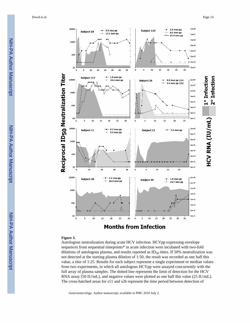
Figure 3.Autologous neutralization during acute HCV infection. HCVpp expressing envelopesequences from sequential timepoints* in acute infection were incubated with two-folddilutions of autologous plasma, and results reported as ID50 titers. If 50% neutralization wasnot detected at the starting plasma dilution of 1:50, the result was recorded as one half thisvalue, a titer of 1:25. Results for each subject represent a single experiment or median valuesfrom two experiments, in which all autologous HCVpp were assayed concurrently with thefull array of plasma samples. The dotted line represents the limit of detection for the HCVRNA assay (50 IU/mL), and negative values were plotted as one half this value (25 IU/mL).The cross-hatched areas for s11 and s26 represent the time period between detection of
Dowd et al. Page 14
Gastroenterology. Author manuscript; available in PMC 2010 July 2.
NIH
-PA Author Manuscript
NIH
-PA Author Manuscript
NIH
-PA Author Manuscript

phylogenetically distinct virus strains. *Only a single infectious HCVpp could be generatedfor s13.
Dowd et al. Page 15
Gastroenterology. Author manuscript; available in PMC 2010 July 2.
NIH
-PA Author Manuscript
NIH
-PA Author Manuscript
NIH
-PA Author Manuscript
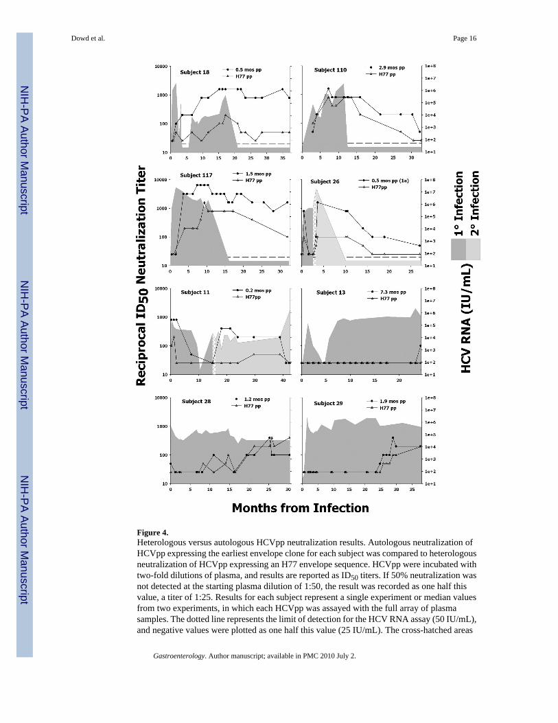
Figure 4.Heterologous versus autologous HCVpp neutralization results. Autologous neutralization ofHCVpp expressing the earliest envelope clone for each subject was compared to heterologousneutralization of HCVpp expressing an H77 envelope sequence. HCVpp were incubated withtwo-fold dilutions of plasma, and results are reported as ID50 titers. If 50% neutralization wasnot detected at the starting plasma dilution of 1:50, the result was recorded as one half thisvalue, a titer of 1:25. Results for each subject represent a single experiment or median valuesfrom two experiments, in which each HCVpp was assayed with the full array of plasmasamples. The dotted line represents the limit of detection for the HCV RNA assay (50 IU/mL),and negative values were plotted as one half this value (25 IU/mL). The cross-hatched areas
Dowd et al. Page 16
Gastroenterology. Author manuscript; available in PMC 2010 July 2.
NIH
-PA Author Manuscript
NIH
-PA Author Manuscript
NIH
-PA Author Manuscript

for s11 and s26 represent the time period between detection of phylogenetically distinct virusstrains.
Dowd et al. Page 17
Gastroenterology. Author manuscript; available in PMC 2010 July 2.
NIH
-PA Author Manuscript
NIH
-PA Author Manuscript
NIH
-PA Author Manuscript

Figure 5.Heterologous neutralization sensitivity decreases as the autologous E1E2 sequence divergesfrom H77. For each subject, over the entire time of follow-up, we calculated geometric meantiters (GMT) for heterologous (H77) and autologous (early variant) HCVpp neutralizationresults, including titers from all time points where both HCVpp variants were tested andneutralization was detectable against at least one of the two. The difference in GMTs[log10(GMTautologous) − log10(GMTheterologous)] was plotted against the % amino acidsimilarity of the subject-specific E1E2 sequence to the heterologous H77 E1E2 sequence.Boxed points represent s28, s29, and s110, who displayed only modest differences inheterologous versus autologous neutralization sensitivity and represent the autologoussequences most closely related to H77. The open circle denotes s11 (infected with a genotype1b virus), representing the only non-genotype 1a sequence.
Dowd et al. Page 18
Gastroenterology. Author manuscript; available in PMC 2010 July 2.
NIH
-PA Author Manuscript
NIH
-PA Author Manuscript
NIH
-PA Author Manuscript

Figure 6.Hierarchy of neutralization against autologous and heterologous HCVpp. Combining resultsshown in Figures 4 and 5, neutralization titers against autologous (early and late variants) andheterologous HCVpp are shown for s110. Taken together, the results demonstrate a hierarchyof neutralization, such that neutralization of autologous (2.9 month) > heterologous (H77) >autologous (9.2 month) HCVpp.
Dowd et al. Page 19
Gastroenterology. Author manuscript; available in PMC 2010 July 2.
NIH
-PA Author Manuscript
NIH
-PA Author Manuscript
NIH
-PA Author Manuscript

Figure 7.Mapping neutralization determinants using autologous HCVpp. A) For s110, neutralization of2.9 month pp (early variant) compared to 9.2 month pp (late variant) by plasma from 9.2 monthsinto infection showed a significant decrease in ID50 titer, as denoted by the arrow. B) Aminoacid substitutions in the 9.2 month pp sequence were back-mutated to express amino acids
Dowd et al. Page 20
Gastroenterology. Author manuscript; available in PMC 2010 July 2.
NIH
-PA Author Manuscript
NIH
-PA Author Manuscript
NIH
-PA Author Manuscript
present in the 2.9 month pp. Mutant and wild-type HCVpp were asessed for neutralization by9.2 month plasma. Results of 1–4 experiments are reported as both % neutralization (1:50dilution of serum, shown as mean + SD), and as median ID50 titers. When 50% neutralizationwas not detected at the starting plasma dilution of 1:50, ID50s were reported as 1:25, representedby the dotted line. Numbers refer to the HCV polyprotein location according to the referencestrain H77.
Dowd et al. Page 21
Gastroenterology. Author manuscript; available in PMC 2010 July 2.
NIH
-PA Author Manuscript
NIH
-PA Author Manuscript
NIH
-PA Author Manuscript

NIH
-PA Author Manuscript
NIH
-PA Author Manuscript
NIH
-PA Author Manuscript
Dowd et al. Page 22
Tabl
e I
Cha
ract
eris
tics o
f eig
ht st
udy
subj
ects
dur
ing
acut
e H
CV
infe
ctio
n.
IDG
ende
rA
geR
ace
Peak
Vir
emia
(IU
/mL
)Pe
ak A
LT
(IU
/L)
Dur
atio
n of
Vir
emia
(mon
ths)
Len
gth
ofFo
llow
-up
(mon
ths)
18M
ale
23W
hite
1 15
0 00
099
19.0
37.0
110
Mal
e28
Whi
te3
600
000
ND
11.9
32.3
117
Fem
ale
31W
hite
23 8
00 0
00N
D15
.332
.0
13Fe
mal
e25
Whi
te2
440
000
870
Pers
iste
nt24
.4
28M
ale
18W
hite
302
000
406
Pers
iste
nt30
.3
29Fe
mal
e25
Whi
te2
390
000
270
Pers
iste
nt37
.6
11a
Mal
e25
Bla
ck28
1 00
0N
D15
.642
.1
26a
Mal
e23
Whi
te16
700
000
ND
3.0
27.0
NO
TE. A
ll su
bjec
ts d
eclin
ed tr
eatm
ent.
Age
refe
rs to
age
at s
eroc
onve
rsio
n..IU
, int
erna
tiona
l uni
ts; A
LT, s
erum
ala
nine
am
inot
rans
fera
se; N
D, n
ot d
eter
min
ed.
a As n
oted
in th
e te
xt, s
11 a
nd s2
6 de
mon
stra
ted
evid
ence
of s
econ
dary
infe
ctio
ns. T
he re
porte
d pe
ak v
irem
ia a
nd d
urat
ion
of v
irem
ia re
fer t
o ch
arac
teris
tics o
f the
prim
ary
infe
ctio
n on
ly.
Gastroenterology. Author manuscript; available in PMC 2010 July 2.


![[Neutralizing antibodies to immunomodulatory therapies in MS-part II] Неутрализиращи антитела към имуномодулиращи терапии при множествена](https://static.fdokumen.com/doc/165x107/633338aaa290d455630a0a17/neutralizing-antibodies-to-immunomodulatory-therapies-in-ms-part-ii-neutralizirashchi.jpg)