Infiltrated Photonic Crystal Fibers for Sensing Applications
IAWA Journal, Vol. 26 (3), 2005: 325–338
S2 ORIENTATION OF MICROFIBRILS IN SOFTWOOD TRACHEIDS AND HARDWOOD FIBERS
Susan E. Anagnost1, Richard E. Mark2 & Robert B. Hanna1
SUMMARY
In this study the soft-rot method was applied to measuring the variation of microfibril angle (mfa) in loblolly pine, black cherry, sugar maple and canelo. For loblolly pine and black cherry, measurements of the radial wall indicated a gradual decrease in mfa across the earlywood portion of the growth ring, with an abrupt decrease at the latewood zone for pine, and in contrast only a slight decrease in microfibril angle across an annual ring of black cherry. In loblolly pine microfibril angle measure-ments indicated that the average microfibril angle in radial sections was very similar to the average for tangential sections of the same block. The average microfibril angles in the intermediate plane, or cell corner, were statistically similar to that of the tangential and radial plane, for pine, cherry, and maple. In canelo, microfibril angles in radial sections were significantly larger than in tangential and intermediate sections. In pine latewood the microfibril angles were less than the average mfa for the entire ring, and the earlywood microfibril angles were greater than the average mfa of the entire ring, thus the average mfa for the growth ring did not represent actual measured mfa values.
Key words: Microfibril angle, soft rot cavities, microfibril.
INTRODUCTION
In the wood cell wall, the orientation of the cellulose microfibrils within the S2 layer is a significant factor in determining wood mechanical properties. The microfibrils with the smallest angle to the longitudinal axis correlate with tensile and compres-sive strength and Youngʼs modulus maxima for wood cell walls (Butterfield 1998; Anagnost et al. 1999). The formation of cylindrical cavities by soft rot fungi along the microfibrils was shown to be an accurate method for determining the microfibril angle in the S2 layer, when compared to both the X-ray diffraction and iodine methods (Anagnost et al. 2000). Recent studies of microfibril angles of the S2 cell wall layer, within individual wood cells (softwood tracheids or hardwood fibers), have observed little variation of micro-
1) N.C. Brown Center for Ultrastructure Studies, Faculty of Wood Products Engineering, SUNY
College of Environmental Science and Forestry, Syracuse, NY, U.S.A.2) Empire State Paper Research Institute, Faculty of Paper Science and Engineering, SUNY
College of Environmental Science and Forestry, Syracuse, NY, U.S.A.
IAWA Journal, Vol. 26 (3), 2005326 327Anagnost, Mark & Hanna — Microfibril orientation
fibril angle (mfa) for southern pine (Anagnost et al. 2002). In contrast, it was reported for Scots pine (Pinus sylvestris) that the microfibril angle becomes steeper as the mi-crofibrils extend from the radial to the tangential plane (Khalili et al. 2001). These conflicting results may reflect species variability or possibly a difference in technique. Because fibers are tubular, measurement of soft rot cavities in longitudinal section must be made in the portion of the wall lying over the lumen midway between the two lateral walls (Anagnost et al. 2002). For example, when observing a radial sec-tion in a tracheid with a radial dimension of 30 μm, the cavity must pass through the cell wall 15 μm from both tangential walls to give an accurate measurement. Because of cell curvature, measuring microfibril angle around the cell longitudinal axis can only be achieved by turning the cell on its axis. Studies of microfibril angles observed within annual rings have indicated that micro-fibril angle can vary greatly in adjacent cells of Norway spruce (Bergander et al. 2002) and over 1 mm increments of annual rings of radiata pine (Donaldson 1998). In southern pine, the microfibril angle gradually decreased from earlywood to latewood, but greater variation was observed in the earlywood than in the latewood regions (Anagnost et al. 2002). When comparing radial and tangential measurements of the same annual rings, differ-ent results have been reported. For loblolly pine, the difference between X-ray diffraction measurements on radial and tangential faces varied between trees (Kretschmann et al. 1998). For one tree no difference was observed, while for a second tree, the microfibril angles measured on the tangential face were greater than those measured on the radial face. For Scots pine, larger angles were observed in radial walls than tangential walls in earlywood tracheids of the same annual rings, utilizing the soft-rot method (Khalili et al. 2001). The overall objective of this study was to observe the extent of variation of micro-fibril angle within the S2 layer as the cellulose microfibrils extend along and around the cell. Knowledge of microfibril angle variability between and within fibers provides evidence to support or disclaim the various theories on the basic cell wall structure and cellulose orientation. This study includes southern pine and three hardwoods to address variability between species. Several approaches were designed for this study. These approaches provided information on S2 microfibril angle orientation within an-nual rings, across annual rings, within fibers, and between species. First, soft rot cavities were measured in radial sections of southern pine and black cherry, to determine how microfibril angle changes within annual rings, and for south-ern pine, within earlywood and latewood zones. Second, a comparison was made of microfibril angle in the tangential walls and the corresponding radial walls of tracheids within one annual ring of southern pine. This approach provided a comprehensive analysis of microfibril angle within the annual ring, as well as within individual cells. A third approach was to examine the microfibril angle of the radial, tangential, and inter-mediate (cell corner) planes of several species of hardwoods (black cherry, sugar maple, canelo) and one softwood (southern pine). With this approach the degree of variation in microfibril orientation as the cellulose microfibrils extend around the cell could be determined.
IAWA Journal, Vol. 26 (3), 2005326 327Anagnost, Mark & Hanna — Microfibril orientation
MATERIALS AND METHODS
Microfibril angles were measured using the soft rot method (Anagnost et al. 2000). For exposure to soft rot, blocks were cut from disks of sugar maple (Acer saccharum), black cherry (Prunus serotina), canelo (Drimys winteri), a vessel-less dicotyledon, and Loblolly pine (Pinus taeda). All disks were cut from fresh logs, and stored at 4 °C until blocks were cut. Blocks were subjected to soft rot by exposure to either Phialocephala dimorphospora or Chaetomium globosum, according to the methods described in Wor-rall et al. (1991). The blocks were placed in glass jars containing reduced salts nutrient agar, on plastic mesh or maple feeder blocks. Exposures varied from 2 to 8 months.
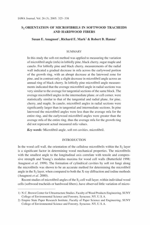
Fig. 1. A tracheid of southern pine containing soft rot cavities. This illustrates the effects of viewing a three-dimensional shape (a cylinder) in two dimensions. Microfibril angles appear to become smaller at the sides of the cell, if cell curvature is ignored. In this study, measurements of microfibril angles were made in the centers of the tracheids to remove any effects of cell curvature on microfibril angle. — Scale bar = 20 μm.
All blocks examined in this study were mature sapwood from the outer portion of each disk. Radial, tangential and inter-mediate sections (20 μm) were cut with a sliding microtome and observed with a Nikon Optiphot microscope equipped with Nomarski differential interference contrast light microscopy. Microfibril an-gles, the angle formed between the cell longitudinal axis and the central axis of the soft rot cavity, were measured on dig-ital images with image analysis software (Metamorph, Universal Imaging) (Anag-nost et al. 2000). To exclude any effect of cell wall curvature on the microfibril angle, only those cavities that extended across the lateral midpoint of the cells were measured as described previously (Fig. 1) (Anagnost et al. 2000).
1
Measurement of variation of mfa across annual rings on the radial walls of Loblolly pine and black cherry To determine the differences between microfibril angle of earlywood and latewood tracheids in loblolly pine, microfibril angles were measured beginning at the first early-wood tracheid measuring soft rot cavity orientation in each successive tracheid for four annual rings (rings A, B, C, and D). The soft-rot method was also used to determine if the variation was gradual or abrupt between earlywood and latewood zones within annual rings of pine, and to compare these results to measurements across two annual rings of black cherry. For pine, latewood was defined as those cells in which 2 times cell
IAWA Journal, Vol. 26 (3), 2005328 329Anagnost, Mark & Hanna — Microfibril orientation
wall thickness > the width of the cell lumen, or formula 2 according to Denne (1989). In most tracheids several measurements of mfa were obtained. The mfa for each tracheid was calculated. The average of earlywood tracheids, latewood tracheids and all tracheids were compared for each ring. For black cherry, microfibril angles were measured on fibers that contained soft rot cavities, beginning at the first fiber of each annual ring, measuring one cavity per fiber.
Comparison of radial wall mfa with tangential wall mfa within one annual ring of loblolly pine A radial section was obtained and mfa measurements within one annual ring were obtained as in the previous section. The ring contained 48 tracheids – 27 earlywood
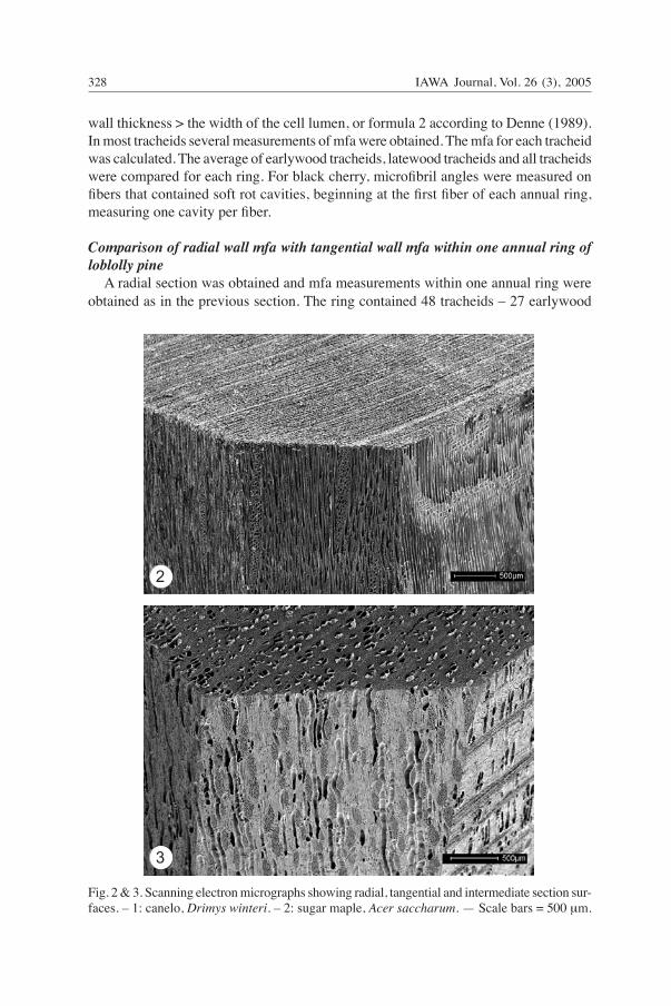
Fig. 2 & 3. Scanning electron micrographs showing radial, tangential and intermediate section sur-faces. – 1: canelo, Drimys winteri. – 2: sugar maple, Acer saccharum. — Scale bars = 500 μm.
2
3

IAWA Journal, Vol. 26 (3), 2005328 329Anagnost, Mark & Hanna — Microfibril orientation
tracheids and 21 latewood tracheids. Serial tangential sections of the same block were prepared, counting and preparing slides of each section. The average mfa for 46 tangen-tial sections were plotted against average mfa in each tracheid, with tangential sections corresponding to tracheids in the radial section.
Comparison of microfibril angle in the radial wall, tangential wall and intermediate wall (45° between radial and tangential wall) This method was developed in order to observe differences in microfibril orientation as the microfibrils extend around the cells from the radial wall to intermediate (cell corner) to the tangential wall. Radial sections were cut of loblolly pine, black cherry, sugar maple, and canelo, all previously subjected to soft rot. Corresponding tangential sections were cut from the same block. Each block was turned 45° between the radial and tangential surface, and intermediate sections were cut (Fig. 2 & 3). The average micro-fibril angles for each orientation (within a block) were subjected to a t-test at 99% confidence level (α = .01).
RESULTS AND DISCUSSION
Microfibril angles of southern pine, with an abrupt transition between earlywood and latewood, were compared with the pattern of diffuse porous hardwoods, i.e. maple and cherry. In addition, a comparison was made with a hardwood (canelo) that has an ana-tomical structure similar to softwoods, with the absence of vessels, and fiber tracheids that are similar in structure to softwood tracheids.
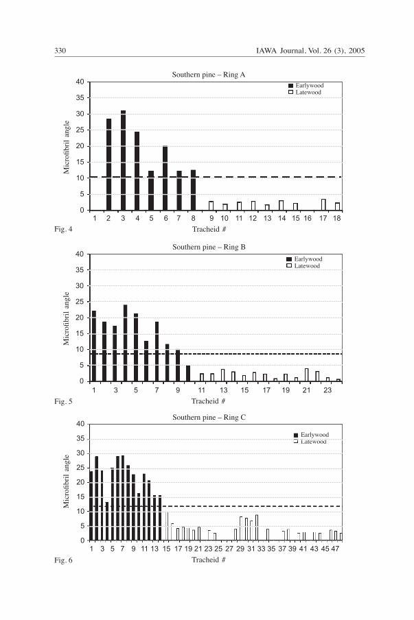
Annual ring variation of microfibril angle in southern pine and black cherry It is evident from Figures 4–7 that the microfibril angles in these specimens of loblolly pine changed abruptly between the earlywood and latewood. A slight decrease in mfa was evident within the earlywood zone, from the beginning of the ring towards the latewood zone (Fig. 4–7). In the latewood zone, the angles were more consistent. This is reflected in the greater range of mfa within earlywood tracheids than latewood tracheids of the same annual ring (Table 1). The ranges overlap considerably for rings
Table 1. Average microfibril angle (°) in radial sections of southern pine. Microfibril angles in earlywood tracheids exhibited a greater range of values than those in latewood tracheids.
Earlywood tracheids Latewood tracheids All tracheids _________________ __________________ _______________ mean std error mean std error mean std error
Ring A 20.0 (3.0) 2.5 (0.2) 10.2 (2.5) range 12.2–31.0 1.7–3.5 Ring B 16.1 (1.9) 2.3 (0.3) 8.0 (1.7) range 4.8–23.9 0.6–4.1 Ring C 22.3 (1.4) 4.5 (0.4) 11.1 (1.5) range 3.2–48.3 1.9–8.7 Ring D 24.7 (2.1) 4.4 (0.5) 14.8 (1.7) range 4.6–38.4 1.5–16.0
IAWA Journal, Vol. 26 (3), 2005330 331Anagnost, Mark & Hanna — Microfibril orientation
Fig. 4
Fig. 5
Fig. 6
40
35
30
25
20
15
10
5
0
40
35
30
25
20
15
10
5
0 1 2 3 4 5 6 7 8 9 10 11 12 13 14 15 16 17 18
Tracheid #
Southern pine – Ring AM
icro
fibril
ang
le
EarlywoodLatewood
Southern pine – Ring C
Mic
rofib
ril a
ngle
1 3 5 7 9 11 13 15 17 19 21 23 25 27 29 31 33 35 37 39 41 43 45 47
Tracheid #
EarlywoodLatewood
EarlywoodLatewood
40
35
30
25
20
15
10
5
0 1 3 5 7 9 11 13 15 17 19 21 23
Tracheid #
Mic
rofib
ril a
ngle
Southern pine – Ring B

IAWA Journal, Vol. 26 (3), 2005330 331Anagnost, Mark & Hanna — Microfibril orientation
C and D, with microfibril angles for the earlywood of ring C ranging from 3.2° to 48.3° and angles of 1.9° to 8.7° for latewood, indicating greater variability in the earlywood tracheids. Microfibril angles varied considerably from cell to adjacent cell, as was evident in Ring D, tracheids 8–11, that have angles of 36°, 27°, 38°, and 27°, respectively (Fig. 7). This tracheid to tracheid variation was less pronounced in the latewood. Similar variation of adjacent tracheids has been observed for Norway spruce (Bergander et al. 2002). Within annual rings of southern pine, the average value for microfibril angle was not representative of the majority of microfibril angles measured for individual tracheids (Table 1). Instead the average microfibril angle for an annual ring represented a value that lies between the average microfibril angles of latewood and earlywood tracheids. For example, for ring B (Table 1) the average mfa for all tracheids (8°) lies between the average for the earlywood zone (16°) and average of the latewood zone (2°). Upon closer examination within individual tracheids across the annual rings, it was clear that very few of the tracheids have average microfibril angles close to 8° (Fig. 5). This was true for the four annual rings of loblolly pine examined. It may be that for softwoods with abrupt transition zones between earlywood and latewood, the average values for mfa do not provide values that can be related to actual microfibril angles within indi-vidual tracheids. This implies that an assessment of wood properties that is based on the average microfibril angle is not likely to reflect the true properties of the latewood and earlywood zones. Previous studies have indicated agreement between the soft rot and other methods such as X-ray diffraction and iodine when the earlywood and latewood were examined separately (Anagnost et al. 2000; Bergander et al. 2002). The average
40
35
30
25
20
15
10
5
0 1 4 7 10 13 16 19 22 25 28 31 34 37 40 43 46 49 52 55
Tracheid #
Southern pine – Ring DM
icro
fibril
ang
le
EarlywoodLatewood
Fig. 7
Fig. 4–7. Measurements of microfibril angle on the radial wall of annual rings of Southern pine from the first earlywood to last latewood tracheid. The bold horizontal broken line indicates the average mfa for the ring. – 4: annual ring ̒ A̓ . – 5: annual ring ̒ Bʼ. – 6: annual ring ̒ Cʼ. – 7: annual ring ʻDʼ.
IAWA Journal, Vol. 26 (3), 2005332 333Anagnost, Mark & Hanna — Microfibril orientation
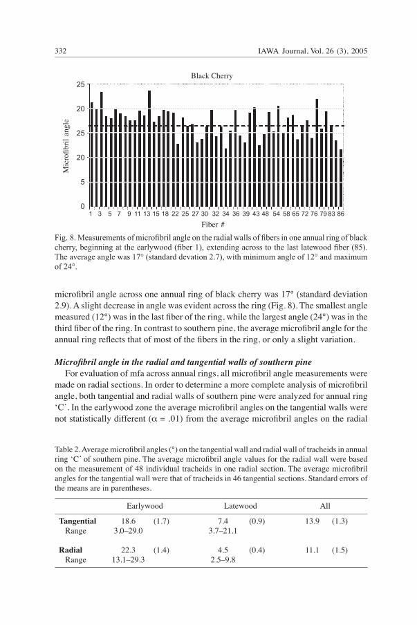
microfibril angle across one annual ring of black cherry was 17° (standard deviation 2.9). A slight decrease in angle was evident across the ring (Fig. 8). The smallest angle measured (12°) was in the last fiber of the ring, while the largest angle (24°) was in the third fiber of the ring. In contrast to southern pine, the average microfibril angle for the annual ring reflects that of most of the fibers in the ring, or only a slight variation.
Microfibril angle in the radial and tangential walls of southern pine For evaluation of mfa across annual rings, all microfibril angle measurements were made on radial sections. In order to determine a more complete analysis of microfibril angle, both tangential and radial walls of southern pine were analyzed for annual ring ʻCʼ. In the earlywood zone the average microfibril angles on the tangential walls were not statistically different (α = .01) from the average microfibril angles on the radial
25
20
25
20
5
0 1 3 5 7 9 11 13 15 18 22 25 27 30 32 34 36 39 43 48 54 58 65 72 76 79 83 86
Fiber #
Mic
rofib
ril a
ngle
Black Cherry
Fig. 8. Measurements of microfibril angle on the radial walls of fibers in one annual ring of black cherry, beginning at the earlywood (fiber 1), extending across to the last latewood fiber (85). The average angle was 17° (standard devation 2.7), with minimum angle of 12° and maximum of 24°.
Table 2. Average microfibril angles (°) on the tangential wall and radial wall of tracheids in annual ring ʻC ̓of southern pine. The average microfibril angle values for the radial wall were based on the measurement of 48 individual tracheids in one radial section. The average microfibril angles for the tangential wall were that of tracheids in 46 tangential sections. Standard errors of the means are in parentheses.
Earlywood Latewood All
Tangential 18.6 (1.7) 7.4 (0.9) 13.9 (1.3) Range 3.0–29.0 3.7–21.1
Radial 22.3 (1.4) 4.5 (0.4) 11.1 (1.5) Range 13.1–29.3 2.5–9.8

IAWA Journal, Vol. 26 (3), 2005332 333Anagnost, Mark & Hanna — Microfibril orientation
walls (Table 2, Fig. 9). The same was evident for latewood tracheids. In the tangential wall, the range of values and variability were greater in the earlywood than in the latewood, similar to that determined for the radial walls (Table 1). A wider range of values of measured microfibril angles was observed in the tangential wall than in the radial wall (Table 2).
Comparisons of microfibril angle in the radial wall, tangential wall, and intermediate wall of southern pine, black cherry, sugar maple and canelo Because of the difficulty in measuring microfibril angles around the cell axis of individual tracheids, microfibril angles were measured in radial, tangential, and inter-mediate sections of blocks (Fig. 10–18). Measurements of the intermediate sections correspond to measurements of the cell corner. For three of the four species examined (pine, maple and black cherry), there were no significant differences, at the 99% con-fidence level (α = .01), between the average microfibril angles of the radial, tangential, and intermediate cell walls (Table 3 & 4). For canelo, the microfibril angles measured in the radial wall were significantly larger, at the 99% confidence level, than those of the tangential and intermediate walls (Table 4). In this study, only canelo had results similar to those found previously for scots pine (Khalili et al. 2001) where the micro-fibril angle in the radial walls was greater than that found in the tangential wall and cell corners. Possible explanations are that there are species differences, or that there was a difference in the method used to measure microfibril angle. In measurements of Scots pine, the soft rot cavities in the intermediate and tangential walls were measured in radial sections, discounting cell curvature. In contrast, measurements were made on soft rot cavities that traverse the cell wall equidistant from the two lateral walls, thus accounting for cell wall curvature.
45
40
35
30
25
20
15
10
5
0
Mic
rofib
ril a
ngle
Ring C
1 3 5 7 9 11 13 15 17 19 21 23 25 27 29 31 33 35 37 39 41 43 45 47 49 51 53
Tracheid # (radial) or section # (tangential)
tangential earlywoodtangential latewoodradial earlywoodradial latewood
Fig. 9. The microfibril angle in ring ‘C’ of southern pine, comparing tangential sections and radial sections of the same block. Averages for 48 tracheids across a radial section were matched with averages of 46 serial tangential sections of the same block.

IAWA Journal, Vol. 26 (3), 2005334 335Anagnost, Mark & Hanna — Microfibril orientation
10
11
12
13
14
15
Fig. 10–12. Soft rot cavities in canelo. – 10: Radial section. The three soft rot cavities have micro-fibril angles of 13°, 19° and 14°. – 11: Intermediate section with soft rot cavities with an angle of 6° with respect to the longitudinal axis. – 12: A tangential section with soft rot cavities that measure 11° and 13°. — Scale bars = 20 μm. — Fig. 13–15. Soft rot cavities in black cherry. – 13: Radial section. Microfibril angle of the central soft rot cavity is 18°. – 14: Intermediate sec-tion. These soft rot cavities indicate a microfibril angle of 23°. – 15: A tangential section with soft rot cavities that measure 17°. — Scale bars = 20 μm.
With the exception of canelo, the frequency of microfibril angle values had only slight variations when considering orientation (Fig. 19–22). For southern pine (Fig. 19), maple (Fig. 20), and cherry (Fig. 21), the frequency distribution plots demonstrated little difference between microfibril angle values in the three orientations. For canelo (Fig. 22), the frequency distributions demonstrated that the microfibril angles of the radial walls were larger than those for the tangential and intermediate walls.
IAWA Journal, Vol. 26 (3), 2005334 335Anagnost, Mark & Hanna — Microfibril orientation
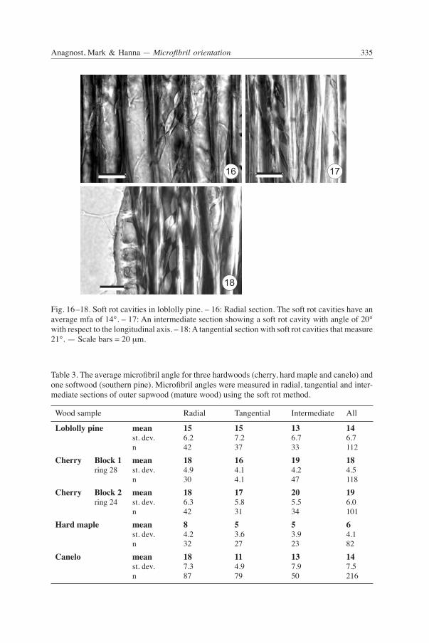
Table 3. The average microfibril angle for three hardwoods (cherry, hard maple and canelo) and one softwood (southern pine). Microfibril angles were measured in radial, tangential and inter-mediate sections of outer sapwood (mature wood) using the soft rot method.
Wood sample Radial Tangential Intermediate All
Loblolly pine mean 15 15 13 14 st. dev. 6.2 7.2 6.7 6.7 n 42 37 33 112 Cherry Block 1 mean 18 16 19 18 ring 28 st. dev. 4.9 4.1 4.2 4.5 n 30 4.1 47 118 Cherry Block 2 mean 18 17 20 19 ring 24 st. dev. 6.3 5.8 5.5 6.0 n 42 31 34 101 Hard maple mean 8 5 5 6 st. dev. 4.2 3.6 3.9 4.1 n 32 27 23 82 Canelo mean 18 11 13 14 st. dev. 7.3 4.9 7.9 7.5 n 87 79 50 216
Fig. 16–18. Soft rot cavities in loblolly pine. – 16: Radial section. The soft rot cavities have an average mfa of 14°. – 17: An intermediate section showing a soft rot cavity with angle of 20° with respect to the longitudinal axis. – 18: A tangential section with soft rot cavities that measure 21°. — Scale bars = 20 μm.
16 17
18

IAWA Journal, Vol. 26 (3), 2005336 337Anagnost, Mark & Hanna — Microfibril orientation
6
5
4
3
2
1
0
Freq
uenc
y
1 3 5 7 9 11 13 15 17 19 21 23 25 27 29 31 33
Microfibril angle
Loblolly pine
RadialTangentialIntermediate
Fig. 20. Frequency distributions of microfibril angle measured in radial, tangential and inter-mediate sections of maple.
RadialTangentialIntermediate
8
7
6
5
4
3
2
1
0
Freq
uenc
y
1 3 5 7 9 11 13 15 17 19 21 23 25 27 29 31 33 35
Microfibril angle
Cherry Block 1
Fig. 21. Frequency distributions of microfibril angle measured in radial, tangential and inter-mediate sections of cherry.

IAWA Journal, Vol. 26 (3), 2005336 337Anagnost, Mark & Hanna — Microfibril orientation
Table 4. T-test (α = .01) of the mean microfibril angle in three orientations, radial, tangential and intermediate cell wall S2 layer. Results indicate no significant differences between orientations at the 99% confidence level with the exception of canelo, which exhibited a significantly larger microfibril angle in the radial wall than in the tangential or intermediate cell wall.
Radial Tangential Intermediate
Loblolly pine 14.6 ± 2.3 15.5 ± 3.0 12.9 ± 3.0 Black cherry – 1 18.1 ± 2.1 15.6 ± 1.6 18.7 ± 1.5 Black cherry – 2 18.0 ± 2.8 17.4 ± 2.6 20.2 ± 2.4 Hard maple 7.8 ± 1.8 5.0 ± 1.6 4.7 ± 2.1 Canelo 18.3 ± 2.0 10.5 ± 1.4 13.0 ± 2.9
CONCLUSIONS
With the exception of canelo, microfibril angles in the S2 varied insignificantly around the cell axis, in the species examined in this study. For loblolly pine, black cherry and sugar maple, the helical path formed as soft-rot cavities grow around the cell within the S2 layer appeared on average, to follow a nearly constant angle. For canelo, the average microfibril angle in the radial wall was significantly larger than that of the tangential and intermediate walls. The average S2 microfibril angle for all tracheids across annual rings of southern pine did not reflect actual individual microfibril angles of the early- and latewood
RadialIntermediateTangential
20
18
16
14
12
10
8
6
4
2
0
Freq
uenc
y
2 4 6 8 10 12 14 16 18 20 22 24 26 28 30 32 34 36 38
Microfibril angle
Canelo
Fig. 22. Frequency distributions of microfibril angle measured in radial, tangential and intermedi-ate sections of canelo. For canelo, there was a greater frequency of smaller microfibril angles in the tangential and intermediate walls which was evident in the overall distribution.
IAWA Journal, Vol. 26 (3), 2005338
tracheids. Implications are that average microfibril angles for softwoods with abrupt early- to latewood transition, should not be considered valid for analyzing and predicting mechanical properties. Instead values for both early- and latewood microfibril angles need to be determined. Studies of the three orientations for other softwoods, such as spruce and hemlock would provide additional information for softwoods with a more gradual transition from earlywood to latewood.
ACKNOWLEDGEMENTS
The authors wish to acknowledge with gratitude the financial assistance of the Empire State Paper Research Institute (ESPRI) for support of this project. The authors greatly appreciate the technical assistance of Postdoctoral Associate, Dr. Iris Vazquez and graduate student, Eric Viskupic. Thanks to Prof. Claudio Donoso Z., of the Universidad Austral, Valdivia, Chile for collecting and forwarding specimen material of Canelo (Drimys).
REFERENCES
Anagnost, S.E., R.E. Mark & R.B. Hanna. 1999. Utilization of soft rot cavity formation as a tool for understanding the relation between microfibril angle and mechanical properties of cellulosic fibers. In: R. Perkins (ed.), Mechanics of Cellulosic Materials: 43–64. American Society of Mechanical Engineers, New York, NY.
Anagnost, S.E., R.E. Mark & R.B. Hanna. 2000. Utilization of soft-rot cavity orientation for the determination of microfibril angle. Wood and Fiber Sci. 32: 81–87.
Anagnost, S.E., R.E. Mark & R.B. Hanna. 2002. Variation of microfibril angle within individual fibers. Wood and Fiber Sci. 34: 337–349.
Bergander, A., J. Brändström, G. Daniel & L. Salmén. 2002. Fibril angle variability in earlywood of Norway spruce using soft rot cavities and polarization confocal microscopy. J. Wood Sci. 48: 255–263.
Butterfield, B.G. (ed.) 1998. Microfibril Angle in Wood. Proceedings of the IAWA/IUFRO International Workshop on the Significance of Microfibril Angle to Wood Quality. University of Canterbury, Private Bag 4800, Christchurch, New Zealand.
Denne, M.P. 1989. Definition of latewood according to Mork (1928). IAWA J. 10: 59–62.Donaldson, L.A. 1998. Between-tracheid variability of microfibril angles in radiata pine. In: B.G.
Butterfield (ed.), Microfibril Angle in Wood: 206–224. University of Canterbury, Christ-church, New Zealand.
Khalili, S., T. Nilsson & G. Daniel. 2001. The use of soft rot fungi for determining the orientation in the S2 layer of pine tracheids. Holz Roh- u.Werkstoff 58: 439–447.
Kretschmann, D.E., H.A. Alsen & S. Verrill. 1998. Variations of microfibril angle in loblolly pine: comparison of iodine crystallization and X-ray diffraction techniques. In: B.G. Butterfield (ed.), Microfibril Angle in Wood: 157–176. University of Canterbury, Christchurch, New Zealand.
Worrall J.J., S.E. Anagnost & C. J.K. Wang. 1991. Conditions for soft rot of wood. Can. J. Microbiol. 37: 869–874.
Copyright © 2022 FDOKUMEN