MR elastography for evaluating regeneration of tissue-engineered cartilage in an ectopic mouse model
Upload
khangminh22Category
view
0download
0
Original article
Role of Transient Elastography in Early Detection of
Hepatocellular Carcinoma in Cirrhotic Patients
Hanaa A. Baddour, Hesham E. El Sheikh, Ahmed E. shalan
Abstract
Background: Hepatocellular carcinoma is the most common primary
malignancy of the liver and one of the most frequent causes of death in
patients with liver cirrhosis. In Egypt it is one of the most challenging
tumors with high incidence, prevalence and mortality rates. The degree of
liver fibrosis is the strongest indicator of risk for HCC development that
is why liver stiffness measured by TE is useful in demarcating patients at
a high risk for HCC, who require frequent check-up by imaging
examinations. Aim of work: The aim of this study was to study the role
of US elastography (FibroScan) in early detection of HCC in cirrhotic
patients in comparison with triphasic CT. Patient and methods: This
study was conducted on 100 patients with liver cirrhosis, 50 of them had
HCC. The entire patients were diagnosed with liver cirrhosis by US
examination and underwent fibroscan to estimate the degree of fibrosis
and then triphasic CT to confirm or exclude the presence of HCC.
Results: There is a correlation between high TE results and the incidence
of HCC detection by CT, HCC detection was (100%) in the groups with LSM value (>30KPa)
and low (9.6%) in the group with LSM value (15-25KPa). Conclusion: The higher LSM, the
more liability for the patient to have HCC on CT scanning.
Keywords: Hepatocellular carcinoma, transient elastography, triphasic CT, liver stiffness
measurement.
Introduction
HCC is a primary malignancy of the
hepatocyte, the major cell type in the
liver (1) Ultrasonography is the least
expensive choice for screening, but it is
highly operator-dependent. A suspicious
lesion on a sonogram generally requires
additional imaging studies to confirm the
diagnosis and the stage of the tumor (2).
The elasticity of a body is defined as the
ability of the body to deform itself under
the action of a mechanical force.
Department of radiology,
Benha faculty of medicine,
Benha University, Egypt.
Correspondence to: Hanaa A. Baddour Department of radiology, Benha faculty of medicine,
Benha University, Egypt.
Email:
Received: 26 August 2019
Accepted: 17 May 2021
741

Role of TE in early detection of HCC in cirrhotic patients, 2021
The elasticity of a tissue can be estimated
on the basis of the speed of propagation
of wave. The higher the speed of
propagation of that wave, the higher the
stiffness of the tissue. Transient
elastography (TE) measures such speed
of propagation in relatively homogenous
organs such as the liver, by using
ultrasound (US) pulses to localize the
shear elastic wave at different times (3).
Patients and methods
This prospective study was conducted
between February 2018 to March 2019 on
100 patients with cirrhosis who visited
Gastrointestinal Surgery Center of
Mansoura University and gave their
informed consent after approval of the
institutional ethical committee. Fifty
patients had cirrhosis with HCC
diagnosed by US and confirmed by
triphasic CT study and patients 50without
evidence of HCC diagnosed by clinical,
laboratory, US and confirmed by
triphasic CT study.
The diagnosis of liver cirrhosis was done
by US examination, then we performed
fibroscan to these patients, either these
patients had cirrhosis on US examination
or not. Then triphasic CT was done to
confirm liver cirrhosis and the presence
of HCC.
Transient Elastography Liver stiffness
measrment (LSM) was performed using
Fibroscan, General Electric LOGIQ E9
(LE9) scanner with 2D shear wave
elastography on a conventional
ultrasound scanner. A time aligned
sequential tracking (TAST) technique
that enables high pulse repetition
frequency (PRF) shear wave tracking on
conventional ultrasound scanners was
implemented with the Comb-push
Ultrasound Shear Elastography (CUSE)
technique on the General Electric LOGIQ
E9 (LE9) scanner and combined to
realize large field-of-view (FOV) 2D
shear wave elastography. The elastic
wave propagates through the underlying
tissues the stiffer the tissue, the faster the
shear wave propagates.
The median value of ten successful
acquisitions expressed in kilopascal
(KPa) and was kept as representative of
liver stiffness measurement. The clinical
interpretation of TE depends on two
important parameters for results to be
considered reliable:
The success rate (the ratio of the
number of successful measurements to
the total number of acquisitions) should
be at least 60%.
IQR/Median (the coordination
between different measurements)
742

Benha medical journal , vol 38, issue 2, 2021
743
Results were expressed in
kiloPascals (KPa) and 10 validated
measurements were recorded for each
patient (4).
The degree of fibrosis measured by TE
correlated with the presence of HCC or
not by triphasic CT scan.
liver stiffness measured noninvasively by
TE has been reported to be well
correlated with histologically assessed
liver fibrosis stage (5).
Results
There was no statistical significant
difference between the groups as
regards the age, sex or smoking; however
HCC tends to be more common in males
than females. Also, HCC tends to be
more common in urban areas than rural
areas and more common in farmers than
non-farmers.
Hard liver was significantly common in
HCC group. There is no statistical
difference between the two groups as
regards the frequency of hepatomegaly or
splenomegaly.
Platelet count, RBCs count and albumin
were significantly lower in HCC group.
ALT, AST, ALP, INR and serum
bilirubin were significantly higher in
HCC group. Prothrombin time was
significantly prolonged in HCC group.
There was no statistical significant
difference between the groups as regard
fasting blood sugar, hemoglobin, WBCs
or s. creatinine; however s. creatinine
tends to be higher in HCC group.
All studied patients of both groups were
HCV Ab. positive and none of them were
HBsAg positive.
Alpha fetoprotein levels were
significantly higher in HCC group.
Most of patients of HCC group were
Child C rather than Child A & B.
Most of cases were Okuda stage III, CLIP
stage II, VISUM stage I and advanced
Tokyo staging.
There was no statistical significant
difference between the groups as regards
the severity of liver cirrhosis assessed by
MELD and uMELD scores however; they
tend to be higher in HCC group.
All patients were cirrhotic; most of them
had shrunken liver (42%) and
splenomegaly (94%). Ascites was found
in 22%, 34% had portal hypertension, 8%
had esophageal varices and only 14% had
portal vein thrombosis.
PVT was found in 10% of patients. Focal
lesions by CT tend to be single, more in
Role of TE in early detection of HCC in cirrhotic patients, 2021
Right lobe, ≥5cm in diameter. 10% of
patients had enlarged lymph nodes and
only 6% with hepatic vein involvement.
CT was superior in detection of both
lymph nodes and hepatic vein
involvement.
Liver stiffness and Inter Quartile Range
measured by Fibroscan were significantly
higher in HCC group. But, there was no
statistical significant difference between
the groups as regard the success rate
(Fig1).
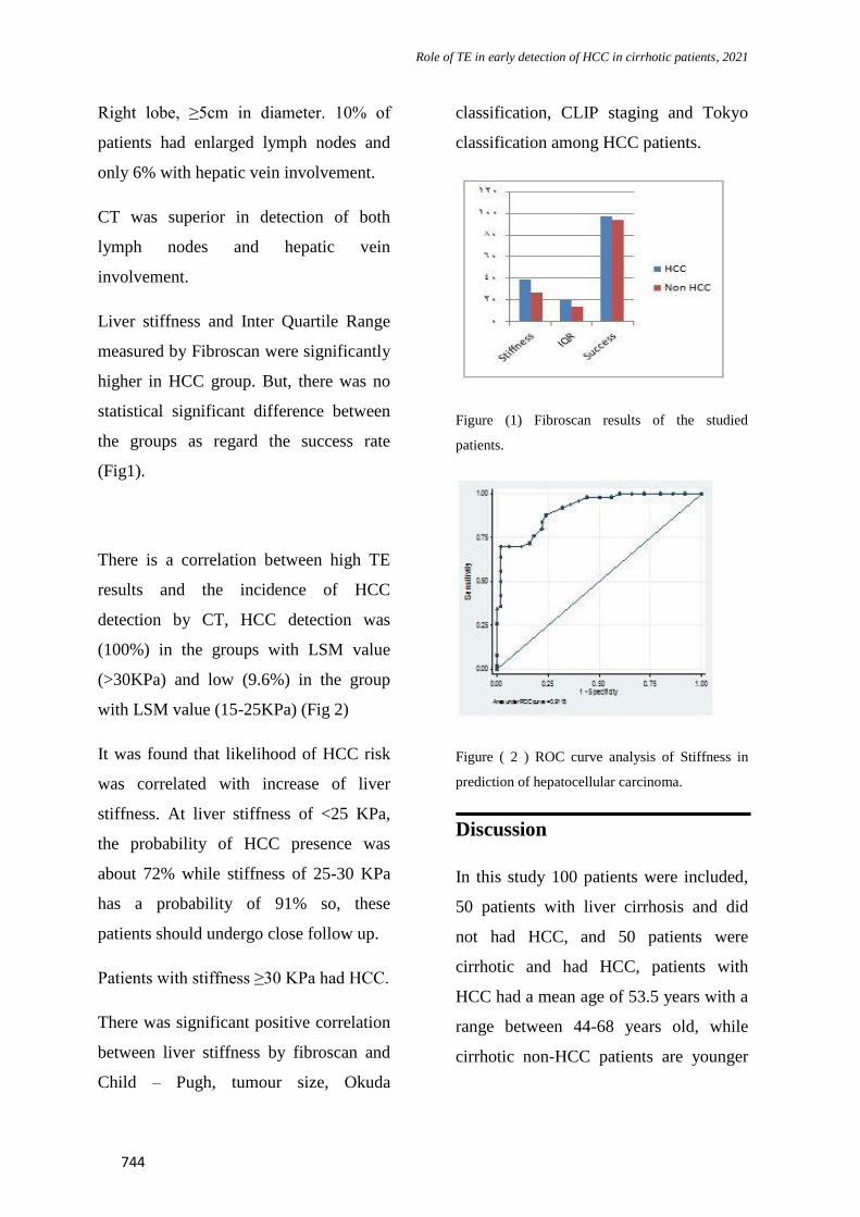
There is a correlation between high TE
results and the incidence of HCC
detection by CT, HCC detection was
(100%) in the groups with LSM value
(>30KPa) and low (9.6%) in the group
with LSM value (15-25KPa) (Fig 2)
It was found that likelihood of HCC risk
was correlated with increase of liver
stiffness. At liver stiffness of <25 KPa,
the probability of HCC presence was
about 72% while stiffness of 25-30 KPa
has a probability of 91% so, these
patients should undergo close follow up.
Patients with stiffness ≥30 KPa had HCC.
There was significant positive correlation
between liver stiffness by fibroscan and
Child – Pugh, tumour size, Okuda
classification, CLIP staging and Tokyo
classification among HCC patients.
Figure (1) Fibroscan results of the studied
patients.
Figure ( 2 ) ROC curve analysis of Stiffness in
prediction of hepatocellular carcinoma.
Discussion
In this study 100 patients were included,
50 patients with liver cirrhosis and did
not had HCC, and 50 patients were
cirrhotic and had HCC, patients with
HCC had a mean age of 53.5 years with a
range between 44-68 years old, while
cirrhotic non-HCC patients are younger
744

Benha medical journal , vol 38, issue 2, 2021
743
with a mean age of 51.3 years and
ranging between 39-67 years old (6).
HCC commonly presented in males more
than females with a male to female ratio
5.2:1(7) and 58% of HCC cases were
from urban areas (8)
The current study showed that 56% of
HCC cases had a history of smoking (9).
There was a traditional conflict about the
link between cigarette smoking and the
occurrence of HCC (10). But recent
evidence support that smoking is a clear
co-factor (11). In HCC patients,
hepatomegaly and hard liver were
observed in many patients (12).
Most of manifestations of decompensated
liver cirrhosis such as ascites, jaundice,
lower limb edema, hepatic
encephalopathy that are common findings
in chronic liver disease without HCC, are
not useful in early suspicion or diagnosis
of HCC comparing to cirrhosis (13).
AFP is the most commonly used
biomarker for patients at risk for HCC
(14). AFP in HCC cases had a mean
value of 627.6ng/ml which was
statistically higher than that of patients
with cirrhosis 11.7ng/ml.
Most of the patients with HCC (64%)
were Child C, followed by Child B (20%)
then Child A (16%) (9).
According to MELD and uMELD scores,
in this study the mean scores for patients
with HCC were 20.6 and 4.2 respectively,
while the mean scores for patients with
cirrhosis were 19.9 and 4.08
respectively(15).
US is very effective in early diagnosis of
HCC as it can detect about 76% of early
HCC patients (16). It is a good tool for
HCC detection with a sensitivity of about
80% and specificity of over 90%.
Abdominal US was done to evaluate the
liver status in the studied patients and all
of the patients (100%) with HCC had
sonographic evidence of liver cirrhosis.
By US examination, five patients (10%)
in this study were found to have portal
vein thrombosis.
Twenty six patients, representing 52% of
HCC cases had single focal hepatic
lesion, while two focal lesions and
multiple hepatic focal lesions were
present in eight (16%) and sixteen (32%)
of cases respectively (17).
In our study we observed that 18 (36%)
of HCC patients had their tumour size
less than 5cm while 32 (64%) had their
tumour size more than 5 cm (15), while
the right lobe was predominantly more
affected by tumour (54%) than left lobe
(10%) and both lobes (36%) (18). Five
745

Role of TE in early detection of HCC in cirrhotic patients, 2021
patients (10%) of HCC cases were found
to have lymph node enlargement (19).
As regard Okuda staging, most of HCC
cases were in the late stage; stage III
(46%), followed by stage II (38%) then
stage I (16%) (20).
According to CLIP staging, most of HCC
cases were presented at the intermediate
stage II (62%) comparing to the patients
presented at the early stage (10%) and the
advanced stage (28%) respectively (20).
According to Tokyo staging, most of
HCC patients were presented at advanced
stage (58%) comparing to the patients
presented at early stage (42%) (21).
The important observation in our study is
that most of patients are diagnosed at
advanced stages so; there must be a
screening program for early detection of
the patients with HCC.
When we used ROC curve, the cutoff
value for HCC was 30.4 KPa with
sensitivity of about 72% , specificity
about 84% , positive predictive value
about 81.82% and negative predictive
value about 75%(22).
There was a positive correlation between
the liver stiffness & the presence of HCC.
Also, prediction of occurrence of HCC by
fibroscan may be of useful value. SSLR
for HCC presence by liver stiffness was
0.7272 in <25 KPa, 0.9167 in 25.1 to
30kPa, 1.00 in 30.1 to 35 KPa, 1.1428 in
35.1 to 40kPa and 1.3333 in >40 KPa, so
there is a direct correlation between high
degree of LSM by TE and detection of
HCC in cirrhotic patient by triphasic CT.
Patients who had high LSM> 30 KPa was
proved (100%) to had HCC by CT, while
patients with LSM <25 KPa was found to
had HCC on CT scanning with a
percentage 72% and almost all the
patients in our study that did not had
HCC on CT scanning had LSM between
15-25 (23).
Also there was direct correlation between
the size of the tumor and stiffness
measured by fibroscan. The bigger the
size of the tumor; the higher the stiffness
(24).
We noticed that there is a direct
correlation between Okuda classification,
CLIP staging and Tokyo classification
with the stiffness measured by fibroscan.
The more advanced liver disease
(according to each classification); the
higher stiffness of fibroscan.
In spite that liver biopsy is the gold
standard diagnostic tool to compare with
in our study for detection of hepatic focal
lesions and incertitude about their nature
rather than triphasic CT, its' invasive
painful interventional nature was a
746

Benha medical journal , vol 38, issue 2, 2021
745
limitation that stimulated the search for
noninvasive approaches but we
recommend using biopsy as a gold
standard modality in spite of triphasic CT
to confirm the diagnosis of HCC for its
certainty.
Our study was conducted on a small
sample volume, so it needs to be applied
on a large sample volume to be more
reliable.
The final diagnosis was done using
triphasic CT not biopsy as a gold standard
diagnostic tool, which is known to be the
most confirmative for diagnosis.
Conclusion
The results show that the higher LSM the
more liability for the patient to have HCC
on CT scanning.
Also there was direct correlation between
the size of the tumor and stiffness
measured by fibroscan. The bigger the
size of the tumor; the higher the stiffness.
References:
1. Motola-Kuba, D.; Zamora-Valdés, D.; Uribe,
M. (2006). Hepatocellular carcinoma.An
overview. Ann. Hepatol.; 5 (1): 16-24.
2. B. Saar, F. Kellner-Weldon (2008)
Radiological diagnosis of hepatocellular
carcinoma. Liver Int. 2008 Feb; 28(2): 189–
199. doi: 10.1111/j.1478-3231.2007.01655.x
3. Sandrin, L.;Fourquet,
B.;Hasquenoph,J.M.,S.Yon, C.Fournieret al. (
2003): Transient elastography: a new
noninvasive method for assessment of hepatic
fibrosis. Ultrasound Med Biol.;29(12):1705-
13.
4. Mohamed S Elzawawy, Shaimaa A
Hassanein, Rasha M El Nomrosy (2018): The
role of fibroscan in assessment of liver
cirrhosis in patients with chronic liver disease.
Menoufia medical journal, vol31,issue2
5. Castera, L.; Vergniol, J.; Foucher, J, BrigitteLe
Bail, Chanteloup E., HaaserM., Darriet,P.
Couzigou,V. Lédinghen et al.(2005):
Prospective comparison of transient
elastography, Fibrotest, APRI, and liver biopsy
for the assessment of fibrosis in chronic
hepatitis. C. Gastroenterology. 2005;128:343–
50.
6. El-Awady MK, Mostafa L, Tabll AA,
Abdelhafez TH, Bader El Din NG, Zayed N,
Shenawy RE, El Abd Y, Hasan RM, Zaghlol
H, El Khayat H, Abdel Aziz AO. et al. (2012):
Association of IL28B SNP With Progression
of Egyptian HCV Genotype 4 Patients to End
Stage Liver Disease. Hepat Mon. 2012
Apr;12(4):271-7. doi: 10.5812/hepatmon.835.
Epub 2012 Apr 30. PubMed PMID: 22690235;
PubMed Central PMCID: PMC3360937.
7. Alsina, A.E.; Beharry, A.; Beharry,
N.Kemmer,E.Franco,H.Rojas,Guy W. Neff et
al. (2012): Epidemiology of Hepatocellular
Carcinoma in Florida – Part I: A Statewide
Report. Florida Public Health Review; 9: 18-
23.
8. Attalla, M.S.; El-Azab, M.S.; El-Bakary, A.A.
et al. (2009): IS AFLATOXIN B1 A
COMMON RISK FACTOR FOR
HEPATOCELLULAR CARCINOMA?
747

Role of TE in early detection of HCC in cirrhotic patients, 2021
Mansoura J. Forensic Med. Clin.Toxicol. Vol.
XVII, No. 2.15-25
9. Abu El Makarem, M.A.; Abdel-Aleem, A.;
Ali, A. Rafet Saber, M.Shatat. Reham DA.et
al. (2011): Diagnostic significance of plasma
osteopontin in hepatitis C virus related
hepatocellular carcinoma. Ann Hepatol.; 10:
296–305.
10. El-Serag, H.B,; Richardson, P.A. and
Everhart, J.E. ( 2001):The role of diabetes in
hepatocellularcarcinoma: a case–control study
among United States Veterans. Am. J.
Gastroenterol.;96:2462–2467.
11. Trichopoulos, Christina Bamia (2011)
Hepatocellular Carcinoma Risk Factors and
Disease Burden in a European J Natl Cancer
Inst.2011 Nov 16; 103(22): 1686-1695. doi:
10.1093ljncildjr395
12. Di Bisceglie, A.M. (2002): Epidemiology and
clinical presentation of hepatocellular
carcinoma. J Vasc Intery Radiol. 2002 Sep;
13(9 Pt): S169-71. PMID: 12354833
13. Mohamad, N.H.; Heba, M. E.; Nadia, M. M. et
al. (2000): Review of epidemiologic and
clinicopathologic features of 403
hepatocellular carcinoma (HCC) patients.
Journal of the Egyptian Nat. Cancer Inst.;
12(2): 87-93.
14. T. Behne and M. S. Copur (2012): Biomarkers
for Hepatocellular Carcinoma. Hindawi
Publishing Corporation International Journal
of Hepatology Volume 2012, Article ID
859076, 7 pages doi:10.1155/2012/859076.
15. Mohamed, A.A.; El-Toukhy , N.; Atta, M.M.
(2013): Glypican-3 as a tumor marker for
hepatocellular carcinoma. Journal of Applied
Pharmaceutical Science;3 (06):083-087.
16. Zoli, M.; Magalotti, D.; Bianchi, G., Emilio
Pisi, Cristina Gueli et al. (2007): Efficacy of
asurveillance program for early detection of
hepatocellular Carcinoma.Cancer; 78:977-85.
17. Abdelgawad IA, Mossallam GI, Radwan NH,
ElZawahry HM, Elhifnawy NM (2013): Can
Glypican3 be Diagnostic for Early
Hepatocellular Carcinoma among Egyptian
Patients? APJCP.;14(12):7345-7349.
18. Salem, R.; Gilbertsen, M.; Butt, Z. Memon;K.
Vouche;M. Cella;D. et al. (2013 ):Increased
quality of life among hepatocellular carcinoma
patients treated with radioembolization,
compared with
chemoembolization.ClinGastroenterolHepatol.
2013 Oct;11(10):1358-1365
19. Gomaa, A.I.; Hashim, M.S. and Waked, I.
(2014): Survival in Patients with
Hepatocellular Carcinoma in Egypt. PLOS
ONE | www.plosone.org . | Volume 9 | Issue 3
| e90929.
20. El-Zayadi, A.R.; Badran, H.M. Shawky, S.,
Emara;s, El-Bareedy;A. Sobhi;M.et al. (2010):
Effect of surveillance for hepatocellular
carcinoma on tumor staging and treatment
decisions in Egyptian patients. Hepatol. Int.;
4(2):500-506.
21. Sarma, S.; Sharma, B.; Chawla, Y. K.,
Kapil;s., Singla; B., Kalra;N et al. (2010):
Comparison of 7 staging systems in north
Indian cohort of hepatocellular
carcinoma.Tropical
Gastroenterology;31(4):271–278.
22. Foucher, J.; Castéra, L.; Bernard, P.H.
Adhoute, Xavier,et al. (2006 )Prevalence and
factors associated with failure of liver stiffness
measurement using FibroScan in a
prospective study of 2114 examinations. Eur J
Gastroenterol Hepatol.;18(4):411–412.
23. Masuzaki, R.; Tateishi, R.; Yoshida
(2009):Prospective risk assessment for
hepatocellular carcinoma development in
748

Benha medical journal , vol 38, issue 2, 2021
747
patients with chronic hepatitisC by transient
elastography. Hepatology; 49: 1954-1961
24. Liana Gheorghe, SperantaIacob,
CarmenGhidu, Iacob;R., Cerban;R.,
Croitoru;A., et al.,(2012):Liver Stiffness Value
Assessed by Transient Elastography Correlates
With Nodule Size in Patients With
Hepatocellular Carcinoma. Gastroenterology
142(5):S-979-S-980.
To cite this article: Hanaa A. Baddour, Hesham E. El Sheikh, Ahmed E. shalan. Role of
Transient Elastography in Early Detection of Hepatocellular Carcinoma in Cirrhotic
Patients. BMFJ 2021; 38(2):741-749. DOI: 10.21608/bmfj.2021.16278.1041
749

Role of TE in early detection of HCC in cirrhotic patients, 2021
Copyright © 2022 FDOKUMEN