Profiling of mitochondrial associated proteins from rat colon
Upload
independentCategory
view
4download
0
BioMed CentralBMC Cancer
ss
Open AcceResearch articleRole of the p53/p21 system in the response of human colon carcinoma cells to DoxorubicinRaffaella Ravizza, Marzia B Gariboldi, Laura Passarelli and Elena Monti*Address: Dept. of Structural and Functional Biology, Section of Pharmacology, University of Insubria, Via A. da Giussano 10, 21052 Busto Arsizio (VA), Italy
Email: Raffaella Ravizza - [email protected]; Marzia B Gariboldi - [email protected]; Laura Passarelli - [email protected]; Elena Monti* - [email protected]
* Corresponding author
AbstractBackground: Colon adenocarcinomas are refractory to a number of widely used anticanceragents. Multifactorial mechanisms have been implicated in this intrinsically resistant phenotype,including deregulation of cell death pathways. In this regard, the p53 protein has a well establishedrole in the control of tumor cell response to DNA damaging agents; however, the relationshipbetween p53-driven genes and drug sensitivity remains controversial. The present studyinvestigates the role of the p53/p21 system in the response of human colon carcinoma cells totreatment with the cytotoxic agent doxorubicin (DOX) and the possibility to modify thetherapeutic index of DOX by modulation of p53 and/or p21 protein levels.
Methods: The relationship between p53 and p21 protein levels and the cytotoxic effect of DOXwas investigated, by MTT assay and western blot analysis, in HCT116 (p53-positive) and HT29(p53-negative) colon cancer cells. We then assessed the effects of DOX in two isogenic cell linesderived from HCT116 by abrogating the expression and/or function of p53 and p21 (HCT116-E6and HCT116 p21-/-, respectively). Finally, we evaluated the effect of pre-treatment with thepiperidine nitroxide Tempol (TPL), an agent that was reported to induce p21 expressionirrespective of p53 status, on the cytotoxicity of DOX in the four cell lines. Comparisons of IC50values and apoptotic cell percentages were performed by ANOVA and Bonferroni's test forindependent samples. C.I. calculations were performed by the combination Index method.
Results: Our results indicate that, in the colon carcinoma cell lines tested, sensitivity to DOX isassociated with p21 upregulation upon drug exposure, and DOX cytotoxicity is potentiated by pre-treatment with TPL, but only in those cell lines in which p21 can be upregulated.
Conclusions: p21 induction may significantly contribute to the response of colonadenocarcinomas cells to DOX treatment; and small molecules that can exploit p53-independentpathways for p21 induction, such as TPL, may find a place in chemotherapeutic protocols for theclinical management of colorectal cancer, where p53 function is often lost, due to genetic orepigenetic defects or to post-transcriptional inactivating mechanisms.
Published: 15 December 2004
BMC Cancer 2004, 4:92 doi:10.1186/1471-2407-4-92
Received: 15 September 2004Accepted: 15 December 2004
This article is available from: http://www.biomedcentral.com/1471-2407/4/92
© 2004 Ravizza et al; licensee BioMed Central Ltd. This is an Open Access article distributed under the terms of the Creative Commons Attribution License (http://creativecommons.org/licenses/by/2.0), which permits unrestricted use, distribution, and reproduction in any medium, provided the original work is properly cited.
Page 1 of 10(page number not for citation purposes)

BMC Cancer 2004, 4:92 http://www.biomedcentral.com/1471-2407/4/92
BackgroundColorectal cancer is the second most common cause ofcancer-related mortality in Western countries, with about1 million new cases every year diagnosed world-wide and500,000 patients dying from the disease [1]. Of thepatients, 30% have advanced disease at presentation,either locally or at distant sites; in this setting, chemother-apy remains the only viable therapeutic option. However,even this option is severely hindered by the inherentresistance of metastatic colon cancer to many currentlyused anticancer agents. A variety of mechanisms by whichcancer cells resist chemotherapy have been described,including enhanced export of drugs from cancer cells andalterations in drug metabolism and/or in drug-targetinteractions [2]. In addition, the response of cancer cellsto genotoxic therapies may be critically impaired bydefects in the response mechanisms to DNA damage [3]or in cell cycle regulatory pathways [4].
Over the past decade, induction of apoptosis has emergedas a major event in tumor cell response to cytotoxic agents(for a recent review see [5]). This view, although recentlychallenged by some Authors [6], has attracted considera-ble attention on deregulation of cell death pathways as akey determinant of drug resistance.
Two separable, although extensively cross-talking, path-ways leading to apoptosis have been characterized [7,8].The extrinsic pathway is initiated by ligation of transmem-brane receptors to activate membrane proximal "activa-tor" caspases, which in turn cleave and activatedownstream "effector" caspases. The intrinsic pathwayrequires disruption of the mitochondrial membrane andthe release of mitochondrial proteins, two events that areregulated by the opposing actions of pro- and antiapop-totic Bcl-2 family members. "Intrinsic stresses", such asthose produced by DNA-damaging agents, activate theintrinsic apoptotic pathway; the multifunctional tran-scription factor p53 is thought to be part of a "fast track"connection between nuclear DNA damage and the intrin-sic pathway machinery [9]. p53 regulates multipleresponses to genotoxic stress by transcriptional activationor repression of a number of genes encoding proteinsinvolved in cell cycle control (p21WAF1/Cip1), DNA repair(gadd45), and apoptosis (e.g. Bax, Bcl2 and survivin)[10]. Mutations in p53 and in the p53 pathway can pro-duce multidrug resistance in vitro and in vivo, and reintro-duction of wildtype p53 into p53 null tumor cells can re-establish chemosensitivity [11]. p53 status is not a univer-sal predictor of treatment response, in part because not alldrugs absolutely require p53 for their apoptotic function[12] and in some settings, p53 loss can enhance drug-induced apoptotic cell death [13]. Still, loss of p53 func-tion correlates with multidrug resistance in many tumortypes [11] and the observation that this is a common
defect in human tumors has spurred an active search forstrategies aimed at directly activating cell death pathwaysdownstream of p53. In this scenario, the role played bythe cyclin-dependent kinase inhibitor p21 is particularlyintriguing, as this protein can be activated by both p53-dependent and p53-independent mechanisms and canassume pro- or anti-apoptotic functions, depending onthe cellular context (for a review see [14]).
The present research focuses on the role of p21 in tumorcell response to treatment with cytotoxic agents, and onthe possibility to improve the therapeutic index of suchagents by modulating p21 status by p53-dependent andindependent pathways. The following issues have beenaddressed: (a) analysis of the relationship between p21status and sensitivity to treatment with the cytotoxic anti-cancer agent doxorubicin (DOX) in p53-positive and -negative colon cancer cell lines; (b) design of treatmentstrategies based on the use of small molecules able tomodulate p21 status; for the present study we have used alow molecular weight, stable nitroxide radical, 4-hydroxy-2,2,6,6,tetramethylpiperidne-N-oxyl (also known as Tem-pol, TPL; figure 1) that was shown to exert an antiprolifer-ative effect against different cancer cell lines [15] and toincrease p21 levels in a p53-null human leukemic cell line[16]. Our data indicate that p21 modulation may
Structure of 4-hydroxy-2,2,6,6,tetramethylpiperidne-N-oxyl (also known as Tempol, TPL)Figure 1Structure of 4-hydroxy-2,2,6,6,tetramethylpiperidne-N-oxyl (also known as Tempol, TPL)
Page 2 of 10(page number not for citation purposes)
BMC Cancer 2004, 4:92 http://www.biomedcentral.com/1471-2407/4/92
significantly affect cell response to DOX treatment in thecolon cancer cell lines tested.
MethodsReagentsStandard chemicals, including 4-hydroxy-2,2,6,6-tetrame-thylipiperidine-N-oxyl (Tempol, TPL) and cell culture rea-gents were purchased from Sigma-Aldrich srl. (Milan,Italy), unless otherwise indicated; doxorubicin (DOX)was kindly provided by Dr. A Suarato (Pfizer-Pharmacia,Milan, Italy).
Cell linesThe human colon carcinoma cell lines HCT116, HT29(obtained from ATCC, Rockville, MD) and HCT116 p21-negative cells (HCT116 p21-/-), kindly provided by Dr B.Vogelstein (Johns Hopkins University, Baltimore, MD,USA), were maintained in DMEM medium supplementedwith 10% FBS (Mascia Brunelli); the HCT116-E6 cell line,obtained from HCT116 cells by transfection with pCMV-neo-E6 plasmid (provided by Dr B. Vogelstein) contain-ing the HPV16-E6 human gene, was maintained inISCOVE medium supplemented with 10% FBS andgeneticin (500 µg/ml). All the cell lines were cultured at37°C, in an atmosphere of 5% CO2 and 95% humidity.
Cytotoxicity assaysThe effects of DOX and/or TPL on cell growth wereassessed by the MTT assay [17]. Briefly, cells were seededonto 96-well plates and allowed to grow for 24 h prior totreatment. Three different treatment schedules were used:a) 24 h medium, 1 h DOX (0.05 – 10 µM) followed by 72h incubation in drug-free medium; b) 24 h TPL (0.05 – 10mM) followed by 72 h incubation in drug-free medium;c) 24 h TPL followed by 1 h DOX and by 72 h incubationin drug-free medium. For combination experiments, thewhole range of DOX concentrations (0.05 – 10 µM) wastested following pretreatment with fixed TPL concentra-tions, corresponding to the IC25 or IC50values obtained foreach cell line according to schedule (b). At the end of thetreatment period, 50 µl of MTT (2 mg/ml in PBS) wereadded to each well at 37°C for 3 h and the reduction ofMTT by viable cells was measured colorimetrically at 570nm, using a Universal Microplate Reader EL800 (Bio-TekInstruments). IC25 and IC50 values (i.e. the concentrationsyielding 75% and 50% cell survival fractions, respectively)were calculated according to the median effect equationand analysis of the interaction between DOX and TPL wasperformed as described by Chou & Talalay [18].
Transfection of HCT116 cellsTransfection of HCT116 cells with the pCMVneo-E6 plas-mid was performed by electroporation as described byYanez and Porter [19], using a Bio-Rad Gene Pulser unit atthe following conditions: 280 V, 960 µF. 48 h post-trans-
fection the cells were selected by adding 500 µg/ml ofgeneticin to the culture medium. The efficiency and stabil-ity of transfection were checked by Western blot analysisof whole cell lysates. Control cells were mock-transfectedwith the pCMVneo plasmid.
Preparation of cell extracts and immunoblottingThe expression of p53 and p21, before and after 1 h expo-sure to DOX (1.0 and 10 µM) followed by 23 h incubationin drug-free medium, or after 24 h exposure to TPL (1.0and 2.5 mM), was evaluated by Western blot analysis oftotal protein extracts (lysis buffer: NP40 1%, leupeptin 10µg/ml and aprotinin 10 µg/ml in TBS). Protein concentra-tion in the cellular lysates was determined by the BCAassay (Pierce, Rockford, IL, USA). 15 µg of protein extract/lane were loaded onto 11% polyacrylamide gels and sep-arated under denaturing conditions. Protein samples werethen transferred onto nitrocellulose membrane and West-ern blot analysis was performed by standard techniques.using a mouse anti-p53 monoclonal antibody (DO-1;Santa Cruz Biotechnology, Inc., Santa Cruz, CA, USA) anda rabbit anti-p21 polyclonal antibody (C-19; Santa CruzBiotechnology). Proteins were visualized using peroxi-dase-conjugated anti-mouse and anti-rabbit secondaryantibodies (Amersham Pharmacia Biotech) and the ECLPlus Western Blotting Detection Reagents (AmershamPharmacia Biotech).
Densitometric analysis was performed using the ScionImage software (Scion Corporation, Frederick, MD).
Flow cytometric analysis of apoptotic cellsThe presence of apoptotic cells in HCT116, HT29,HCT116-E6 and HCT116 p21 -/-, before and after 1 hexposure to DOX (1.0 and 10 µM) followed by 23 h incu-bation in drug-free medium, was evaluated by flow cyto-metric analysis, using a Becton Dickinson FACScaliburflow cytometer. Cells were detached by trypsinization,washed in phosphate-buffered saline (PBS) and fixed inice-cold 70% ethanol for 20 min at -20°C. After an addi-tional wash in PBS, DNA was stained with 50 mg/ml pro-pidium iodide in PBS in the presence of RNAse A (30 U/ml) at 37°C for 30 minutes. 5 × 10⁵ cell samples were ana-lyzed and data were processed using the CellQuest soft-ware (Becton Dickinson). The percentage of apoptoticcells in each sample was determined based on the sub-G1peaks detected in monoparametric histograms.
Statistical analysisComparisons of IC50 values and apoptotic cell percentagesin the four different cell lines were performed by ANOVAand Bonferroni's test for independent samples. C.I. calcu-lations an relative statistical analysis were performed asdescribed by Chou and Talalay [18]. According to thismethod, a combination index (C.I.) can be calculated from
Page 3 of 10(page number not for citation purposes)
BMC Cancer 2004, 4:92 http://www.biomedcentral.com/1471-2407/4/92
dose-response curves obtained following exposure toDOX and/or TPL as single agents and in combination. C.I.values approximating 1.0 indicate additive interactionsbetween the two agents; C.I. < 1.0 indicate synergy and,conversely, C.I. > 1.0 indicate antagonism.
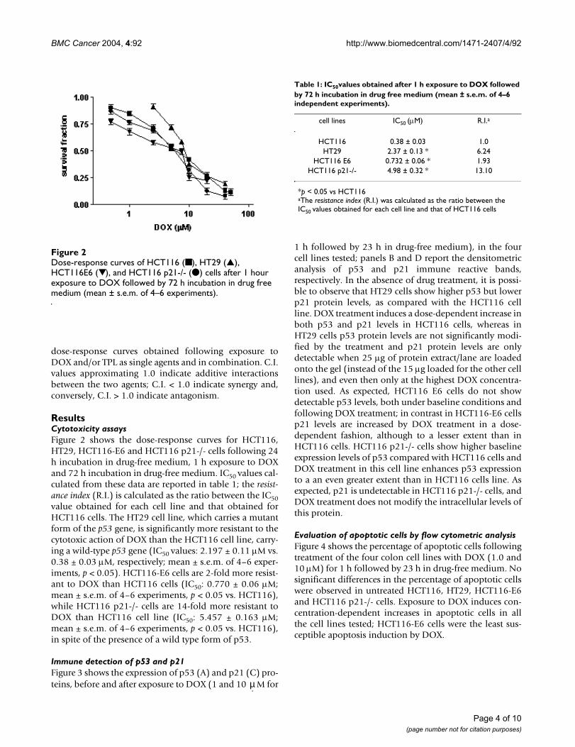
ResultsCytotoxicity assaysFigure 2 shows the dose-response curves for HCT116,HT29, HCT116-E6 and HCT116 p21-/- cells following 24h incubation in drug-free medium, 1 h exposure to DOXand 72 h incubation in drug-free medium. IC50 values cal-culated from these data are reported in table 1; the resist-ance index (R.I.) is calculated as the ratio between the IC50value obtained for each cell line and that obtained forHCT116 cells. The HT29 cell line, which carries a mutantform of the p53 gene, is significantly more resistant to thecytotoxic action of DOX than the HCT116 cell line, carry-ing a wild-type p53 gene (IC50 values: 2.197 ± 0.11 µM vs.0.38 ± 0.03 µM, respectively; mean ± s.e.m. of 4–6 exper-iments, p < 0.05). HCT116-E6 cells are 2-fold more resist-ant to DOX than HCT116 cells (IC50: 0.770 ± 0.06 µM;mean ± s.e.m. of 4–6 experiments, p < 0.05 vs. HCT116),while HCT116 p21-/- cells are 14-fold more resistant toDOX than HCT116 cell line (IC50: 5.457 ± 0.163 µM;mean ± s.e.m. of 4–6 experiments, p < 0.05 vs. HCT116),in spite of the presence of a wild type form of p53.
Immune detection of p53 and p21Figure 3 shows the expression of p53 (A) and p21 (C) pro-teins, before and after exposure to DOX (1 and 10 M for
1 h followed by 23 h in drug-free medium), in the fourcell lines tested; panels B and D report the densitometricanalysis of p53 and p21 immune reactive bands,respectively. In the absence of drug treatment, it is possi-ble to observe that HT29 cells show higher p53 but lowerp21 protein levels, as compared with the HCT116 cellline. DOX treatment induces a dose-dependent increase inboth p53 and p21 levels in HCT116 cells, whereas inHT29 cells p53 protein levels are not significantly modi-fied by the treatment and p21 protein levels are onlydetectable when 25 µg of protein extract/lane are loadedonto the gel (instead of the 15 µg loaded for the other celllines), and even then only at the highest DOX concentra-tion used. As expected, HCT116 E6 cells do not showdetectable p53 levels, both under baseline conditions andfollowing DOX treatment; in contrast in HCT116-E6 cellsp21 levels are increased by DOX treatment in a dose-dependent fashion, although to a lesser extent than inHCT116 cells. HCT116 p21-/- cells show higher baselineexpression levels of p53 compared with HCT116 cells andDOX treatment in this cell line enhances p53 expressionto a an even greater extent than in HCT116 cells line. Asexpected, p21 is undetectable in HCT116 p21-/- cells, andDOX treatment does not modify the intracellular levels ofthis protein.
Evaluation of apoptotic cells by flow cytometric analysisFigure 4 shows the percentage of apoptotic cells followingtreatment of the four colon cell lines with DOX (1.0 and10 µM) for 1 h followed by 23 h in drug-free medium. Nosignificant differences in the percentage of apoptotic cellswere observed in untreated HCT116, HT29, HCT116-E6and HCT116 p21-/- cells. Exposure to DOX induces con-centration-dependent increases in apoptotic cells in allthe cell lines tested; HCT116-E6 cells were the least sus-ceptible apoptosis induction by DOX.
Dose-response curves of HCT116 (■ ), HT29 (▲), HCT116E6 (▼), and HCT116 p21-/- (● ) cells after 1 hour exposure to DOX followed by 72 h incubation in drug free medium (mean ± s.e.m. of 4–6 experiments)Figure 2Dose-response curves of HCT116 (■ ), HT29 (▲), HCT116E6 (▼), and HCT116 p21-/- (● ) cells after 1 hour exposure to DOX followed by 72 h incubation in drug free medium (mean ± s.e.m. of 4–6 experiments).
µ
Table 1: IC50values obtained after 1 h exposure to DOX followed by 72 h incubation in drug free medium (mean ± s.e.m. of 4–6 independent experiments).
cell lines IC50 (µM) R.I.a
HCT116 0.38 ± 0.03 1.0HT29 2.37 ± 0.13 * 6.24
HCT116 E6 0.732 ± 0.06 * 1.93HCT116 p21-/- 4.98 ± 0.32 * 13.10
*p < 0.05 vs HCT116aThe resistance index (R.I.) was calculated as the ratio between the IC50 values obtained for each cell line and that of HCT116 cells
Page 4 of 10(page number not for citation purposes)

BMC Cancer 2004, 4:92 http://www.biomedcentral.com/1471-2407/4/92
p53 (A) and p21 (C) protein levels in HCT116, HT29, HCT116 E6 and HCT116 p21-/- cells following 1 h exposure to DOX and 23 h incubation in drug-free mediumFigure 3p53 (A) and p21 (C) protein levels in HCT116, HT29, HCT116 E6 and HCT116 p21-/- cells following 1 h exposure to DOX and 23 h incubation in drug-free medium. Panels B and D: densitometric analysis of p53 and p21-immune reactive bands, respectively (white bars: untreated; light grey bars: DOX 1 µM; dark grey bars: DOX 10 µM).
Page 5 of 10(page number not for citation purposes)

BMC Cancer 2004, 4:92 http://www.biomedcentral.com/1471-2407/4/92
Effects of TPL on cell survival and p53/p21 levelsTable 2 reports the IC25 and IC50 values obtained for thefour cell lines after 24 h of continuous TPL exposure fol-lowed by 72 h in drug-free medium. TPL can be observedto inhibit cell growth in all four cell lines; although no sig-nificant differences can be detected among IC50 values,HCT116 p21-/- cells appear to be less responsive than theother three cell lines. Figure 5 shows that 24 h exposure toTPL (1 and 2.5 mM) induces a dose-dependent increase inboth p53 and in p21 levels in HCT116 cells, whereas inHT29 cell line TPL treatment only induces a dose-depend-ent increase in p21 expression. Exposure of HCT116-E6cells to TPL (1 and 2.5 mM) for 24 h does not induce anyvariations in p53 expression, while a dose-dependentincrease in p21 expression can be observed followingtreatment with the nitroxide. In HCT116 p21-/- cells TPL
induce a slight increase in p53 protein levels but, asexpected, p21 levels were unaffected by TPL treatment.
Effects of TPL pretreatment on DOX-induced cytotoxicityFigure 6 shows the effect of 24 h pretreatment with TPL, atfixed concentrations corresponding to the IC25 and IC50values obtained for each cell line, on DOX cytotoxicity.The cells' response to DOX is expressed as the IC50 valuesderived from dose/response curves obtained after 1 hexposure to DOX with or without pretreatment with TPL(24 h), followed by 72 h in drug-free medium. Analysis ofcytotoxicity data shows a synergistic interaction (C.I.<1)between DOX and TPL for both TPL concentrations inHCT116 cells and in HCT116-E6 and HT29 cells at thelower concentration; only additive effects (C.I. ≈ 1) can beobserved in HCT116 p21 -/- cells. IC50 values for DOX and
Percentage of apoptotic cells in HCT116, HT29, HCT116 E6 and HCT116 p21-/- cells before and after 1 h exposure to DOX followed by 23 h incubation in drug-free medium (white bars: untreated; light grey bars: DOX 1 µM; dark grey bars: DOX 10 µM)Figure 4Percentage of apoptotic cells in HCT116, HT29, HCT116 E6 and HCT116 p21-/- cells before and after 1 h exposure to DOX followed by 23 h incubation in drug-free medium (white bars: untreated; light grey bars: DOX 1 µM; dark grey bars: DOX 10 µM). Mean ± s.e.m. of 3 independent experiments.
Table 2: IC50values obtained after 24 h exposure to TPL followed by 72 h incubation in drug free medium. (mean ± s.e.m. of 4–6 independent experiments).
Cell lines IC25 (µM) IC50 (µM) R.I.a
HCT116 280.00 ± 39.21 560.00 ± 77.78 1.0HT29 320.40 ± 50.03 665.63 ± 102.91 1.19
HCT116 E6 400.06 ± 60.00 837.3 ± 103.99 1.49HCT116 p21-/- 700.00 ± 99.14 1114.55 ± 182.2 1.99
a The resistance index (R.I.) was calculated as the ratio between the IC50 values obtained for each cell line and that of HCT116 cells
Page 6 of 10(page number not for citation purposes)

BMC Cancer 2004, 4:92 http://www.biomedcentral.com/1471-2407/4/92
p53 (A) and p21 (C) protein levels in HCT116, HT29, HCT116 E6 and HCT116 p21-/- cells following 24 h exposure to TPLFigure 5p53 (A) and p21 (C) protein levels in HCT116, HT29, HCT116 E6 and HCT116 p21-/- cells following 24 h exposure to TPL. Panels B and D: densitometric analysis of p53 and p21-immune reactive bands, respectively (white bars: untreated; light grey bars: TPL 1.0 mM; dark grey bars: TPL 2.5 mM).
Page 7 of 10(page number not for citation purposes)

BMC Cancer 2004, 4:92 http://www.biomedcentral.com/1471-2407/4/92
TPL according to the three different schedules are reportedin table 3.
DiscussionResistance of colorectal cancer to established treatmentregimens remains a major concern in oncology; thusattempts at improving the survival of patients affected bythis disease depend largely on strategies targeting tumorcell resistance, which cannot be rationally planned with-out a detailed knowledge of the mechanisms underlyingthis phenomenon. A current paradigm regarding cancerchemotherapy indicates disabling of the intrinsic apop-totic pathway as a key factor in the response of tumor cellsto anticancer drugs [3,5,12]. Therefore, strategies aimingat re-establishing the cell's capability to activate a celldeath program are an area of active research. The presentstudy was performed in order to define the role of the
p53/p21 pathway in the response of colorectal carcinomacells to DOX, a cytotoxic agent that is typically devoid ofeffects in this tumor type. The results obtained in our cyto-toxicity studies indicate that in the cell lines examined p53status is not unequivocally related to the response toDOX: in fact, while p53-deficient cells (HT-29, HCT116-E6) are indeed less responsive than the p53/wt parentalHCT116 cell line, the highest resistance index wasobtained for HCT116 p21-/- cells, harboring two wildtypep53 alleles. As expected, treatment with DOX leads to p53upregulation in the cell lines expressing wildtype p53; thiseffect has been thoroughly documented in colon cancercells as well as in tumor cell lines derived from other tis-sues, and has been attributed to phosphorylation and sub-sequent stabilization of p53, possibly through activationof DNA-dependent protein kinase or ATM (ataxia-teleangectasia mutated) kinase (see e.g. [20-22]). InHCT116 cells, p21 expression parallels p53 activation;however, data obtained in HT29 and HCT116-E6 cellsclearly indicate the existence of p53-independent path-ways for p21 induction, that have been extensively charac-terized (for a review see [23]) and can be activated tovariable extents (HCT116 E6 > HT29) upon exposure toDOX. Interestingly, the extent of the cytotoxic effectsobserved in the small panel of colon cell lines testedrather seems to parallel the cells' ability to upregulate p21(HCT116 > HCT116-E6 > HT29 > HCT116 p21-/-). Thisresult is somewhat unexpected: in fact, whereas the func-tion of p21 in cell growth arrest following DNA damagehas been established for a long time [24], the role playedby this protein in the ultimate fate of tumor cells exposedto cytotoxic agents is far from clear-cut [14,25]. In anumber of studies, p21 has actually been reported to pro-tect tumor cells against cell death induced by enforcedp53 expression [26] or by low doses of cytotoxic agents[13,27-30]. However, in other experimental settings, p53-dependent or -independent induction of p21 expressionseems to be a prerequisite for apoptosis [31-34] and tosensitize tumor cells to the action of different agents [35-37]. The putative mechanisms by which p21 might actu-
Table 3: IC50values obtained after 24 h exposure to TPL followed by 1 h exposure to DOX and 72 h incubation in drug free medium (mean ± s.e.m. of 4–6 independent experiments).
DOX + TPL (IC25) + TPL (IC50) C.I.a
HCT116 0.38 ± 0.03 0.102 ± 0.04 0.053 ± 0.003 0.4–0.6HT29 2.20 ± 0.11 1.06 ± 0.12 0.49 ± 0.015 0.6–0.8
HCT116 E6 0.77 ± 0.06 0.54 ± 0.6 0.24 ± 0.07 0.6–0.8HCT116 p21-/- 5.457 ± 0.16 4.98 ± 0.15 2.549 ± 0.21 0.8–1.1
aCombination index (C.I.) ≈ 1.0: additivity; C.I.<1.0: synergy; C.I.>1.0: antagonism
Effect of 24 h pre-treatment with TPL on DOX IC50 values obtained in HCT116, HT29, HCT116 E6 and HCT116 p21-/- cells following 1 h exposure to DOX and 72 h in drug-free mediumFigure 6Effect of 24 h pre-treatment with TPL on DOX IC50 values obtained in HCT116, HT29, HCT116 E6 and HCT116 p21-/- cells following 1 h exposure to DOX and 72 h in drug-free medium. For each cell line, two TPL concentrations corre-sponding to IC25 and IC50 (see table 2), were used. Mean ± s.e.m. of 4–6 experiments.
Page 8 of 10(page number not for citation purposes)

BMC Cancer 2004, 4:92 http://www.biomedcentral.com/1471-2407/4/92
ally induce apoptosis have recently been reviewed [38],but still await full elucidation.
Interestingly, the situation outlined by our results doesnot seem to conform to either view: in fact, while induc-tion of p21 in p53-proficient and -deficient cell lines isassociated with increased response to drug treatment, thiswas not accompanied by a parallel increase in apoptoticcells, as no significant differences in apoptosis wereobserved between HCT116 cells and the 14-fold resistantHCT-116 p21-/- cell line (figure 4). This suggests thatmodes of cell death other than apoptosis may operate intumor cells following exposure to DOX, or, more gener-ally, to DNA-damaging agents, a concept that is beginningto be proposed by a number of Authors [39,40]; of note,recent experimental evidence indicates p21 as one of themajor determinants of terminal growth arrest induced bycytotoxic agents [41-43]. Therefore, although issuesrelated to terminal growth arrest and senescence have notbeen specifically addressed in the present study, the possi-bility that these phenomena might play a role linking celldeath to the observed increases in p21 levels should notbe disregarded.
The hypothesis that the cytotoxic response of the tumorcell lines tested in the present study may depend on p21induction is further corroborated by data obtainedfollowing pre-treatment with the piperidine nitroxideTPL. The choice of this compound was dictated byprevious findings indicating that TPL induces cell death ina number of tumor cell lines irrespective of their p53 sta-tus [15], and that it increases p21 levels in p53-null cells[16]. The results of the present study show that TPL affectsthe four colon cell lines to similar extents, thus confirmingthat its growth inhibitory effect is independent of p53function. HCT-116 p21-/- cells are actually slightly lessresponsive than the other cell lines [even though the dif-ference does not attain statistical significance), which sug-gests the possibility that the effects of TPL are due in partto its ability to increase p21 levels. Interestingly, thenitroxide also induces p21 expression even in p53-defi-cient cell lines; this observation suggests that TPL can acti-vate p53-independent pathways for p21 induction, asalready noted following exposure of HT29 and HCT116E6 cells to DOX. Moreover, activation of such pathways byTPL appears to sensitize tumor cells to the action of DOX:in fact, synergistic potentiation of DOX cytotoxicity isachieved by TPL in those cell lines where p21 expressioncan be induced, but only additive effects between TPL andDOX are observed in HCT116 p21-/-, where p21 expres-sion is constitutively absent.
ConclusionsIn summary, the results of the present study strongly sug-gest that 1) p21 induction may significantly contribute to
the response of colon adenocarcinoma cells to DOX treat-ment; and 2) small molecules that can exploit p53-inde-pendent pathways for p21 induction, such as TPL, mayfind a place in chemotherapeutic protocols for the clinicalmanagement of colorectal cancer, where p53 function isoften lost, due to genetic or epigenetic defects or to post-transcriptional inactivating mechanisms.
Competing interestsThe author(s) declare that they have no competinginterests
AcknowledgmentsSupported by a grant from the Italian Ministry for Education, University and Research (MIUR), Projects with Relevant National Impact (PRIN), 2003 (to E.M.).
References1. Ferlay J, Bray F, Pisani P, Parkin DM: GLOBOCAN 2000: Cancer
Incidence, Mortality and Prevalence Worldwide. Volume 5.IARC CancerBase Lyon, IARCPress; 2001.
2. Gottesman MM: Mechanisms of cancer drug resistance. AnnuRev Med 2002, 53:615-627.
3. Shah AM, Schwartz GK: Cell cycle-mediated drug resistance: anemerging concept in cancer therapy. Clin Cancer Res 2001,7:2168-2181.
4. Ferreira CG, Epping M, Kruyt FA, Giaccone G: Apoptosis: targetof cancer therapy. Clin Cancer Res 2002, 8:2024-2034.
5. Brown JM, Wilson G: Apoptosis genes and resistance to cancertherapy: what does the experimental and clinical data tell us?Cancer Biol Ther 2003, 2:477-490.
6. Green DR: Apoptotic pathways: paper wraps stone bluntsscissors. Cell 2000, 102:1-4.
7. Johnstone RW, Ruefli AA, Lowe SW: Apoptosis: a link betweencancer genetics and chemotherapy. Cell 2002, 108:153-164.
8. Wang X: The expanding role of mitochondria in apoptosis.Genes Dev 2001, 15:2922-2933.
9. Lowe SW, Lin AW: Apoptosis in cancer. Carcinogenesis 2000,21:485-495.
10. Vousden K, Lu X: Live or let die: the cell's response to p53.Nature Rev Cancer 2002, 2:594-604.
11. Wallace-Brodeur RR, Lowe SW: Clinical implications of p53mutations. Cell Mol Life Sci 1999, 55:64-75.
12. Herr I, Debatin MK: Cellular stress response and apoptosis incancer therapy. Blood 2001, 98:2603-2614.
13. Bunz F, Hwang PM, Torrance C, Waldman T, Zhang Y, Dillehay L,Williams J, Lengauer C, Kinzler KW, Vogelstein B: Disruption ofp53 in human cancer cells alters the responses to therapeu-tic agents. J Clin Invest 1999, 104:263-269.
14. Liu X, Bishop WR, Liu M: Differential effects of cell cycle regu-latory protein p21WAF1/Cip1 on apoptosis and sensitivity tocancer chemotherapy. Drug Resist Updates 2003, 6:183-195.
15. Gariboldi MB, Lucchi S, Caserini C, Supino R, Oliva C, Monti E: Anti-proliferative effect of the piperidine nitroxide TEMPOL onneoplastic and nonneoplastic mammalian cell lines. Free RadBiol Med 1998, 24:913-923.
16. Gariboldi MB, Rimoldi V, Supino R, Favini E, Monti E: The nitroxideTEMPOL induces oxidative stress, p21 WAF1/CIP1, and celldeath in HL60 cells. Free Rad Biol Med 2000, 29:633-641.
17. Supino R: MTT assays. In In Methods in molecular biology: in vitro tox-icity testing protocols Volume 43. Edited by: O'Hare SM, Atterwill CK.Totowa, NJ: Humana Press; 1998:137-149.
18. Chou T-C, Talalay P: Quantitative analysis of dose-effect rela-tionships: The combined effects of multiple drugs or enzymeinhibitors. Adv Enzyme Regul 1984, 22:27-55.
19. Yanez RJ, Porter AC: Influence of DNA delivery method ongene targeting frequencies in human cells. Somat Cell Mol Genet1999, 25:27-31.
Page 9 of 10(page number not for citation purposes)

BMC Cancer 2004, 4:92 http://www.biomedcentral.com/1471-2407/4/92
Publish with BioMed Central and every scientist can read your work free of charge
"BioMed Central will be the most significant development for disseminating the results of biomedical research in our lifetime."
Sir Paul Nurse, Cancer Research UK
Your research papers will be:
available free of charge to the entire biomedical community
peer reviewed and published immediately upon acceptance
cited in PubMed and archived on PubMed Central
yours — you keep the copyright
Submit your manuscript here:http://www.biomedcentral.com/info/publishing_adv.asp
BioMedcentral
20. Kaeser MD, Pebernard S, Iggo RD: Regulation of p53 stability andfunction in HCT116 colon cancer cells. J Biol Chem 2004,279:7598-7605.
21. Kurz EU, Douglas P, Lees-Miller SP: Doxorubicin activates ATM-dependent phosphorylation of multiple downstream targetsin part through the generation of reactive oxygen species. JBiol Chem in press. 2004, Oct 14
22. Take Y, Kumano M, Teraoka H, Nishimura S, Okuyama A: DNA-dependent protein kinase inhibitor (OK-1035) suppressesp21 expression in HCT116 cells containing wild-type p53induced by adriamycin. Biochem Biophys Res Commun 1996,221:207-212.
23. Gartel AL, Tyner AL: Transcriptional regulation of thep21((WAF1/CIP1)) gene. Exp Cell Res 1999, 246:280-289.
24. Dotto GP: p21(WAF1/Cip1): more than a break to the cellcycle? Biochim Biophys Acta 2000, 1471:M43-M56.
25. Gartel AL, Tyner AL: The role of the cyclin-dependent kinaseinhibitor p21 in apoptosis. Mol Cancer Ther 2002, 1:639-649.
26. Gorospe M, Cirielli C, Wang X, Seth P, Capogrossi MC, Holbrook NJ:p21(Waf1/Cip1) protects against p53-mediated apoptosis ofhuman melanoma cells. Oncogene 1997, 14:929-935.
27. Javelaud D, Besancon F: Inactivation of p21WAF1 sensitizescells to apoptosis via an increase of both p14ARF and p53 lev-els and an alteration of the Bax/Bcl-2 ratio. J Biol Chem 2002,277:37949-37954.
28. Martinez LA, Yang J, Vazquez ES, Rodriguez-Vargas M, del C, Olive M,Hsieh JT, Logothetis CJ, Navone NM: p21 modulates threshold ofapoptosis induced by DNA-damage and growth factor with-drawal in prostate cancer cells. Carcinogenesis 2002,23:1289-1296.
29. Mahyar-Roemer M, Roemer K: p21 Waf1/Cip1 can protecthuman colon carcinoma cells against p53-dependent andp53-independent apoptosis induced by natural chemopre-ventive and therapeutic agents. Oncogene 2001, 20:3387-3398.
30. Blagosklonny MV, Robey R, Bates S, Fojo T: Pretreatment withDNA-damaging agents permits selective killing of check-point-deficient cells by microtubule-active drugs. J Clin Invest2000, 105:533-539.
31. Chopin V, Toillon RA, Jouy N, Le Bourhis X: p21(WAF1/CIP1) isdispensable for G1 arrest, but indispensable for apoptosisinduced by sodium butyrate in MCF-7 breast cancer cells.Oncogene 2004, 23:21-29.
32. Agrawal S, Agarwal ML, Chatterjee-Kishore M, Stark GR, ChisolmGM: Stat1-dependent, p53-independent expression ofp21(waf1) modulates oxysterol-induced apoptosis. Mol CellBiol 2002, 22:1981-1992.
33. Wu Q, Kirschmeier P, Hockenberry T, Yang TY, Brassard DL, WangL, McClanahan T, Black S, Rizzi G, Musco ML, Mirza A, Liu S: Tran-scriptional regulation during p21WAF1/CIP1-induced apop-tosis in human ovarian cancer cells. J Biol Chem 2002,277:36329-36337.
34. Ahmad N, Adhami VM, Afaq F, Feyes DK, Mukhtar H: Resveratrolcauses WAF-1/p21-mediated G(1)-phase arrest of cell cycleand induction of apoptosis in human epidermoid carcinomaA431 cells. Clin Cancer Res 2001, 7:1466-73.
35. Fulda S, Debatin KM: Sensitization for tumor necrosis factor-related apoptosis-inducing ligand-induced apoptosis by thechemopreventive agent resveratrol. Cancer Res 2004,64:337-346.
36. Satou M, Aizawa S, Hayakari M, Ookawa K, Tsuchida S: Enhancedsensitivity to cis-diamminedichloroplatinum(II) of a humancarcinoma cell line with mutated p53 gene by cyclin-depend-ent kinase inhibitor p21(WAF1) expression. Cancer Sci 2003,94:286-291.
37. Chinery R, Brockman JA, Peeler MO, Shyr Y, Beauchamp RD, CoffeyRJ: Antioxidants enhance the cytotoxicity of chemothera-peutic agents in colorectal cancer: a p53-independent induc-tion of p21WAF1/CIP1 via C/EBPbeta. Nature Med 1997,3:1233-1241.
38. Liu S, Bishop WR, Liu M: Differential effects of cell cycle regula-tory protein p21(WAF1/Cip1) on apoptosis and sensitivity tocancer chemotherapy. Drug Resist Updat 2003, 6:183-195.
39. Roninson IB: Tumor cell senescence in cancer treatment. Can-cer Res 2003, 63:2705-2715.
40. te Poele RH, Okorokov AL, Jardine L, Cummings J, Joel SP: DNAdamage is able to induce senescence in tumor cells in vitroand in vivo. Cancer Res 2002, 62:1876-1883.
41. Chang BD, Swift ME, Shen M, Fang J, Broude EV, Roninson IB: Molec-ular determinants of terminal growth arrest induced intumor cells by a chemotherapeutic agent. Proc Natl Acad Sci US A 2002, 99:389-394.
42. Chang BD, Broude EV, Fang J, Kalinichenko TV, Abdryashitov R,Poole JC, Roninson IB: p21Waf1/Cip1/Sdi1-induced growtharrest is associated with depletion of mitosis-control pro-teins and leads to abnormal mitosis and endoreduplication inrecovering cells. Oncogene 2000, 19:2165-2170.
43. Chang BD, Xuan Y, Broude EV, Zhu H, Schott B, Fang J, Roninson IB:Role of p53 and p21waf1/cip1 in senescence-like terminalproliferation arrest induced in human tumor cells by chem-otherapeutic drugs. Oncogene 1999, 18:4808-4818.
Pre-publication historyThe pre-publication history for this paper can be accessedhere:
http://www.biomedcentral.com/1471-2407/4/92/prepub
Page 10 of 10(page number not for citation purposes)
Copyright © 2022 FDOKUMEN