Role of nitric oxide and prostanoids in the regulation of leg blood flow and blood pressure in...
14
J Physiol 590.6 (2012) pp 1481–1494 1481 The Journal of Physiology Role of nitric oxide and prostanoids in the regulation of leg blood flow and blood pressure in humans with essential hypertension: effect of high-intensity aerobic training Michael Nyberg 1 , Lasse G. Jensen 1 , Pia Thaning 2 , Ylva Hellsten 1 and Stefan P. Mortensen 2 1 Department of Exercise and Sport Sciences, University of Copenhagen, Copenhagen, Denmark 2 Copenhagen Muscle Research Centre, Rigshospitalet, Copenhagen, Denmark Key points • Nitric oxide and prostanoids are substances that dilate the blood vessels. We examined the role of these vasodilators in the regulation of blood flow to contracting muscle and systemic blood pressure before and after a training intervention in subjects with essential hypertension and in healthy controls. • We show that blood flow to the exercising leg is lower in essential hypertension. • Surprisingly, this effect on blood flow is not the result of a reduced capacity of the nitric oxide and prostanoid systems to dilate the blood vessels; however, these systems do appear to play a role in the training induced reduction in blood pressure. • These findings advance our understanding of vascular dysfunction associated with essential hypertension and the mechanisms underlying the blood pressure reducing effect of exercise. Abstract We examined the role of nitric oxide (NO) and prostanoids in the regulation of leg blood flow and systemic blood pressure before and after 8 weeks of aerobic high-intensity training in individuals with essential hypertension (n = 10) and matched healthy control subjects (n = 11). Hypertensive subjects were found to have a lower (P < 0.05) blood flow to the exercising leg than normotensive subjects (30 W: 2.92 ± 0.16 vs . 3.39 ± 0.37 l min −1 ). Despite the lower exercise hyperaemia, pharmacological inhibition of the NO and prostanoid systems reduced leg blood flow to a similar extent during exercise in the two groups and vascular relaxation to the NO-dependent vasodilator acetylcholine was also similar between groups. High-intensity aerobic training lowered (P < 0.05) resting systolic (∼9 mmHg) and diastolic (∼12 mmHg) blood pressure in subjects with essential hypertension, but this effect of training was abolished when the NO and prostanoid systems were inhibited. Skeletal muscle vascular endothelial NO synthase uncoupling, expression and phosphorylation status were similar in the two groups before and after training. These data demonstrate that a reduction in exercise hyperaemia in hypertensive subjects is not associated with a reduced capacity of the NO and prostanoid systems to induce vasodilatation or with altered acetylcholine-induced response. However, our data suggest that the observed reduction in blood pressure is related to a training-induced change in the tonic effect of NO and/or prostanoids on vascular tone. (Resubmitted 24 November 2011; accepted after revision 19 January 2012; first published online 23 January 2012) Corresponding author M. Nyberg: Department of Exercise and Sport Sciences, Section of Human Physiology, the August Krogh Building, Universitetsparken 13, DK-2100 Copenhagen Ø, Denmark. Email: mnyberg@ifi.ku.dk Abbreviations LBF, leg blood flow; LVC, leg vascular conductance; eNOS, endothelial nitric oxide synthase; NOx, nitrite and nitrate; L-NMMA, N G -monomethyl-L-arginine. C 2012 The Authors. Journal of Physiology C 2012 The Physiological Society DOI: 10.1113/jphysiol.2011.225136
-
Upload
independent -
Category
Documents
-
view
0 -
download
0
Transcript of Role of nitric oxide and prostanoids in the regulation of leg blood flow and blood pressure in...
J Physiol 590.6 (2012) pp 1481–1494 1481
The
Jou
rnal
of
Phys
iolo
gy
Role of nitric oxide and prostanoids in the regulation of legblood flow and blood pressure in humans with essentialhypertension: effect of high-intensity aerobic training
Michael Nyberg1, Lasse G. Jensen1, Pia Thaning2, Ylva Hellsten1 and Stefan P. Mortensen2
1Department of Exercise and Sport Sciences, University of Copenhagen, Copenhagen, Denmark2Copenhagen Muscle Research Centre, Rigshospitalet, Copenhagen, Denmark
Key points
• Nitric oxide and prostanoids are substances that dilate the blood vessels. We examined the roleof these vasodilators in the regulation of blood flow to contracting muscle and systemic bloodpressure before and after a training intervention in subjects with essential hypertension and inhealthy controls.
• We show that blood flow to the exercising leg is lower in essential hypertension.• Surprisingly, this effect on blood flow is not the result of a reduced capacity of the nitric oxide
and prostanoid systems to dilate the blood vessels; however, these systems do appear to play arole in the training induced reduction in blood pressure.
• These findings advance our understanding of vascular dysfunction associated with essentialhypertension and the mechanisms underlying the blood pressure reducing effect of exercise.
Abstract We examined the role of nitric oxide (NO) and prostanoids in the regulation of leg bloodflow and systemic blood pressure before and after 8 weeks of aerobic high-intensity training inindividuals with essential hypertension (n = 10) and matched healthy control subjects (n = 11).Hypertensive subjects were found to have a lower (P < 0.05) blood flow to the exercising legthan normotensive subjects (30 W: 2.92 ± 0.16 vs. 3.39 ± 0.37 l min−1). Despite the lower exercisehyperaemia, pharmacological inhibition of the NO and prostanoid systems reduced leg blood flowto a similar extent during exercise in the two groups and vascular relaxation to the NO-dependentvasodilator acetylcholine was also similar between groups. High-intensity aerobic training lowered(P < 0.05) resting systolic (∼9 mmHg) and diastolic (∼12 mmHg) blood pressure in subjectswith essential hypertension, but this effect of training was abolished when the NO and prostanoidsystems were inhibited. Skeletal muscle vascular endothelial NO synthase uncoupling, expressionand phosphorylation status were similar in the two groups before and after training. These datademonstrate that a reduction in exercise hyperaemia in hypertensive subjects is not associatedwith a reduced capacity of the NO and prostanoid systems to induce vasodilatation or with alteredacetylcholine-induced response. However, our data suggest that the observed reduction in bloodpressure is related to a training-induced change in the tonic effect of NO and/or prostanoids onvascular tone.
(Resubmitted 24 November 2011; accepted after revision 19 January 2012; first published online 23 January 2012)Corresponding author M. Nyberg: Department of Exercise and Sport Sciences, Section of Human Physiology, theAugust Krogh Building, Universitetsparken 13, DK-2100 Copenhagen Ø, Denmark. Email: [email protected]
Abbreviations LBF, leg blood flow; LVC, leg vascular conductance; eNOS, endothelial nitric oxide synthase; NOx,nitrite and nitrate; L-NMMA, NG-monomethyl-L-arginine.
C© 2012 The Authors. Journal of Physiology C© 2012 The Physiological Society DOI: 10.1113/jphysiol.2011.225136

1482 M. Nyberg and others J Physiol 590.6
Introduction
Skeletal muscle blood flow is tightly regulated to matchtissue oxygen demands (Andersen & Saltin, 1985).Increased peripheral resistance and decreased maximalvasodilatation are, however, well known characteristicfeatures of chronic hypertension (Taddei et al. 1997) andas a consequence this disease state may be associated withreductions in blood flow. This potential impairment inoxygen delivery to contracting muscle could lead to tissueischaemia and impaired tolerance to physical activity.
Within the vascular system, nitric oxide (NO) andvasodilator prostanoids relax smooth muscle cells, thusresulting in vasodilatation, and although pharmacologicalinhibition of the synthesis of either does not affect themagnitude of exercise hyperaemia, the ∼30% reduction inblood flow observed during combined blockade (Saunderset al. 2005; Mortensen et al. 2007; Nyberg et al. 2010)suggest that these two compounds act in synergy tocontribute to muscle blood flow control during exercise.As the functions of both the NO and prostanoid systemsare severely affected in essential hypertension (Taddei et al.1997), a reduced blood flow to contracting muscle couldbe linked to an inadequacy of these systems to inducevasodilatation. Exercise training has been shown to lowerleg blood flow at the same absolute workload (Kiens et al.1993; Proctor et al. 2001; Krustrup et al. 2004), but towhat extent this is related to alterations in the NO andprostanoid system and whether this adaptation is also pre-sent in hypertensive subjects remain unknown.
Training interventions have proved successful inlowering blood pressure in individuals with hypertension.Although training characteristics have been shown not tobe predictive of the blood pressure response to aerobictraining, the magnitude of blood pressure reduction isassociated with the gain in VO2max (Cornelissen & Fagard,2005). This relationship is intriguing as improvementsin VO2max to a large extent are an effect of adaptationsin central haemodynamics (Saltin et al. 1968), whereasimproved peripheral vascular function (Taddei et al.1997a) and remodelling (Heerkens et al. 2007) and/ordecreases in muscle sympathetic nerve activity (MSNA;Laterza et al. 2007) are likely to explain the reduction inblood pressure. Nevertheless, this association does suggestthat high-intensity aerobic training is a powerful stimulusfor reducing blood pressure due to its large effect onimprovements in VO2max (Helgerud et al. 2007).
NO plays an important role in whole body bloodpressure regulation in humans (Joyner & Casey, 2009)and the training-induced effect on blood pressure maybe related to an improved function of endothelial nitricoxide synthase (eNOS), as evidenced by an augmentedvascular relaxation to the NO-dependent vasodilatoracetylcholine (ACh) after training (Higashi et al. 1999).However, direct evidence demonstrating a role of NO
in the training-induced lowering of blood pressure inhumans is lacking.
Putative mechanisms underlying the effect of trainingon vascular NO function include changes in eNOS proteinexpression, phosphorylation status of eNOS, and eNOSuncoupling. The latter is linked to an increased mono-merization of the homodimeric enzyme and leads toproduction of superoxide instead of NO (Forstermann& Munzel, 2006). In an animal model of hypertension,uncoupled eNOS has been shown to increase super-oxide formation and decrease vascular NO bioavailability(Mollnau et al. 2002), but the extent to which eNOSuncoupling is present in essential hypertension andwhether training can affect the degree of uncouplingremain to be determined.
To address these issues, we examined the effect ofinhibiting the formation of NO and prostanoids on restingand exercising haemodynamics before and after an 8 weekaerobic high-intensity training period in normotensivesubjects and individuals diagnosed with essential hyper-tension. In addition, NO metabolites formed within thevascular network at rest and during acute exercise andmuscle enzymes involved in the synthesis of NO were alsomeasured before and after training.
Methods
Ethical approval
Ten hypertensive subjects (systolic and diastolic bloodpressure of 183 ± 7 and 101 ± 5 mmHg (± SEM),respectively) and 11 age, weight and height matchednormotensive controls (142 ± 5 and 74 ± 3 mmHg) werestudied (Table 1). The reported blood pressure values inTable 1 represent an average of readings obtained after30 min of supine rest at three different time points duringexperimental day 1. The subjects were also instructedto perform home blood pressure monitoring, andmeasurements from the normotensive subjects showedvalues of 119 ± 4 and 70 ± 2 mmHg (average of 2 dailymeasurements performed for 1 week), thereby confirmingthat these individuals were normotensive. The studywas approved by the Ethics Committee of Copenhagenand Frederiksberg communities (H-2-2009-096) andconducted in accordance with the guidelines of theDeclaration of Helsinki. Written informed consent wasobtained from all subjects before enrolment into the study.
Nine out of 10 hypertensive subjects were undertreatment with angiotensin-converting enzyme (ACE)inhibitors (n = 4), calcium channel blockers (n = 6)and/or diuretics (n = 4). Potential subjects takingβ-blockers were excluded from the study. Two weeks priorto the first experimental day the hypertensive subjects wereinstructed to stop their medication. Two subjects were sub-sequently allowed to take a low dose of diuretic medication
C© 2012 The Authors. Journal of Physiology C© 2012 The Physiological Society
J Physiol 590.6 Role of NO and prostanoids in hypertension 1483
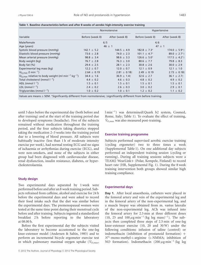
Table 1. Baseline characteristics before and after 8 weeks of aerobic high-intensity exercise training
Normotensive Hypertensive
Variable Before (week 0) After (week 8) Before (week 0) After (week 8)
Male/female 6/5 4/6Age (years) 46 ± 1 47 ± 1Systolic blood pressure (mmHg) 142.1 ± 5.2 144.5 ± 4.9 182.8 ± 7.3∗ 174.0 ± 5.9∗†Diastolic blood pressure (mmHg) 73.6 ± 2.8 74.0 ± 2.3 101.1 ± 4.7∗ 89.0 ± 2.7∗†Mean arterial pressure (mmHg) 98.8 ± 3.4 98.6 ± 3.3 126.6 ± 5.0∗ 117.5 ± 4.2∗†Body weight (kg) 76.7 ± 2.8 76.3 ± 3.0 80.6 ± 7.7 79.8 ± 8.3Body fat (%) 29.4 ± 2.5 28.1 ± 2.3 30.8 ± 2.6 30.0 ± 2.9Experimental leg mass (kg) 12.2 ± 0.7 12.0 ± 0.7 12.1 ± 0.9 12.1 ± 1.0VO2max (l min−1) 2.64 ± 0.19 2.81 ± 0.18† 2.49 ± 0.19 2.73 ± 0.18†VO2max relative to body weight (ml min−1 kg−1) 34.4 ± 1.6 36.9 ± 1.4† 32.6 ± 2.7 36.1 ± 2.7†Total cholesterol (mmol l−1) 4.4 ± 0.2 4.6 ± 0.3 4.8 ± 0.2 4.9 ± 0.2HDL (mmol l−1) 1.5 ± 0.1 1.5 ± 0.1 1.5 ± 0.1 1.5 ± 0.1LDL (mmol l−1) 2.4 ± 0.2 2.6 ± 0.3 3.0 ± 0.2 2.9 ± 0.1Triglycerides (mmol l−1) 1.0 ± 0.2 1.0 ± 0.1 1.2 ± 0.2 1.1 ± 0.2
Values are means ± SEM. ∗Significantly different from normotensive; †significantly different from before training.
until 3 days before the experimental day (both before andafter training) and at the start of the training period dueto developed symptoms (headache). Five of the subjectsremained without medication throughout the trainingperiod, and the four subjects taking diuretics stoppedtaking the medication 2–3 weeks into the training perioddue to a lowering of blood pressure. All subjects werehabitually inactive (less than 1 h of moderate intensityexercise per week), had normal resting ECG and no signsof ischaemia or arrhythmias during exercise (ECG), andwere non-smokers, and none of the subjects in eithergroup had been diagnosed with cardiovascular disease,renal dysfunction, insulin resistance, diabetes, or hyper-cholesterolaemia.
Study design
Two experimental days separated by 1 week wereperformed before and after an 8 week training period. Sub-jects refrained from caffeine, alcohol and exercise for 24 hbefore the experimental days and were asked to recordtheir food intake such that the diet was similar beforethe experimental days. The premenopausal women weretested at the same time point during their menstrual cyclebefore and after training. Subjects ingested a standardizedbreakfast 2 h before reporting to the laboratoryat 08.00 h.
Before the first experimental day the subjects visitedthe laboratory to become accustomed to the one-legknee-extensor model (Andersen & Saltin, 1985) and toperform an incremental bicycle ergometer exercise testin which pulmonary maximal oxygen uptake (VO2max,
l min−1) was determined(Quark b2 system, Cosmed,Rome, Italy; Table 1). To evaluate the effect of training,VO2max was also measured post-training.
Exercise training programme
Subjects performed supervised aerobic exercise training(cycling ergometer) two to three times a week(Supplemental Table 1). On one additional day subjectsperformed an independent training session (cycling orrunning). During all training sessions subjects wore aTEAM2 WearLink+ (Polar, Kempele, Finland) to recordheart rate (HR, Supplemental Fig. 1). During the 8 weektraining intervention both groups showed similar hightraining compliance.
Experimental days
Day 1. After local anaesthesia, catheters were placed inthe femoral artery and vein of the experimental leg andin the femoral artery of the non-experimental leg, anda muscle biopsy was obtained from m. vastus lateralisof the non-experimental leg. ACh was infused intothe femoral artery for 2.5 min at three different doses(10, 25 and 100 μg min−1 (kg leg mass)−1). The sub-jects then completed three steps of 2.5 min of one-legknee-extensor exercise (10, 20 and 30 W) under thefollowing conditions: infusion of saline (control) orindomethacin (inhibition of prostanoid formation) +N G-mono-methyl-L-arginine (L-NMMA; inhibition ofNO formation). Indomethacin (200 μg min−1 (kg leg
C© 2012 The Authors. Journal of Physiology C© 2012 The Physiological Society
1484 M. Nyberg and others J Physiol 590.6
mass)−1; Confortid, Actavis, Copenhagen, Denmark)and L-NMMA (2.1 mg min−1 (kg leg mass)−1; Clinalfa,Bachem, Bubendorf, Switzerland) were infused into thefemoral artery for 5 min before start of exercise andthroughout exercise.
Day 2. Catheters for intravascular microdialysis probeswere placed in the femoral artery and vein of theexperimental leg 4–5 cm below the inguinal ligament andadvanced 10 cm in the proximal direction. A microdialysisprobe (CMA 70 bolt, CMA microdialysis, Stockholm)with a 10 mm membrane (20 kDa cut-off) was insertedinto these two catheters. Correct placement of the probeswas verified by ultrasound. Additionally, one catheter(20 Ga) for measurements of intra-arterial pressure wasplaced into the femoral artery of the non-experimentalleg. Microdialysate was collected for 10 min during restingconditions and during one-leg knee-extensor exercise(20 W). The microdialysis probes were perfused at a rate of5 μl min−1 with Ringer–acetate solution and to determinethe relative exchange of the stable metabolites of NO,nitrate and nitrite (NOx), a small amount (2.7 nM) of[2-3H]ATP (<0.1μCi ml−1) was added to the perfusatefor calculation of probe recovery. Dalteparin (25 IE ml−1;Fragmin, Pfizer) was added to the perfusate to avoid bloodclotting on the membrane. The molecular probe recovery(PR) was calculated as:
PR = (dpminfusate – dpmdialysate)/dpminfusate,
where dpm denotes disintegrations per minute (Scheller& Kolb, 1991; Jansson et al. 1994). The [3H]ATP activity(in dpm) was measured on a liquid scintillation counter(Tri-Carb 2000; Copenhagen, Denmark) after additionof the perfusate to 3 ml of Ultima Gold scintillationliquid (Perkin Elmer). After collection of samples, themicrodialysate was weighed, and the actual flow ratewas calculated to estimate any loss of fluid or abnormaldecrease in perfusion rate.
Femoral arterial blood flow (LBF) was measured withultrasound Doppler (Logic E9, GE Healthcare) equippedwith a linear probe operating an imaging frequency of9 MHz and Doppler frequency of 4.2–5.0 MHz. The siteof blood velocity measurements in the common femoralartery was distal to the inguinal ligament but above thebifurcation into the superficial and profound femoralbranch to avoid turbulence from the bifurcation. Allrecordings were obtained at the lowest possible insonationangle and always below 60 deg. The sample volume wasmaximized according to the width of the vessel, and keptclear of the vessel walls. A low-velocity filter (velocities<1.8 m s−1) rejected noises caused by turbulence at thevascular wall. Doppler tracings and B-mode imageswere recorded continuously and Doppler tracings wereaveraged over 16 heart cycles at the time of blood sampling.
Vessel diameter was determined after each Dopplerrecording. Arterial diameter measures were assessedduring the systole from arterial B-mode images with thevessel parallel to the transducer. Intra-arterial pressurewas monitored with transducers (Pressure MonitoringKit, Baxter Deerfield, IL, USA) positioned at the levelof the heart. Blood samples were drawn after 2 minof exercise/infusion and blood gases, haemoglobin andlactate were measured using an ABL725 analyser (Radio-meter Copenhagen, Denmark). Leg mass was calculatedfrom whole-body dual-energy X-ray absorptiometryscanning (Prodigy, GE Healthcare).
Analysis of nitrate and nitrite
The stable metabolites of NO, nitrite and nitratewere measured using a fluorometric assay kit (CaymanChemical Co., Ann Harbor, MI, USA).
Quantification of protein expression
Freeze dried tissue samples of m. vastus lateralis werehomogenized in lysis buffer and Western blot analysiswas performed as previously described (Bangsbo et al.2009). Low temperature SDS-PAGE (LT-PAGE) wasperformed for detection of eNOS monomers usingreported procedures (Klatt et al. 1995). Briefly, totalproteins were incubated in 1 × Laemmli buffer withoutdithiothreitol (DTT) and then subjected to SDS-PAGEwith 5% gel. Gels and buffers were equilibrated at 4◦Cbefore electrophoresis, and the buffer tank was placedin cooling conditions to maintain temperature of the gel<15◦C. Subsequent to LT-PAGE, the gels were transferredand the blots probed as routine Western blot.
Antibodies used were: eNOS, eNOS-PThr495, nNOS(BD Transduction Laboratories, USA), and eNOS-PSer1177
(Calbiochem Millipore, Billerica, MA, USA). To controlfor loading differences the blots were also probed with anantibody against GAPDH (Abcam, Cambridge, UK).
Statistical analysis
A two-way ANOVA was performed to test significancebetween the normotensive and hypertensive subjects anda two-way repeated measures ANOVA was performed todetect training-induced changes within each group. Aftera significant F test, pairwise differences were identifiedusing the Student–Newman–Keuls post hoc procedure.Differences in baseline characteristics were assessed withStudent’s t test for unpaired and paired data whencomparing between groups and within each group,respectively. The significance level was set at P < 0.05 anddata are means ± SEM.
C© 2012 The Authors. Journal of Physiology C© 2012 The Physiological Society
J Physiol 590.6 Role of NO and prostanoids in hypertension 1485
Results
Subject characteristics
The 8 week training period lowered systolic (P < 0.05)and diastolic (P < 0.05) blood pressure in the hypertensivegroup, whereas VO2max was increased in both normotensive(P < 0.001) and hypertensive (P < 0.05; Table 1) sub-jects. In the hypertensive group, the improvement inVO2max tended (P = 0.084, r2 = 0.57) to correlate with thereduction in blood pressure.
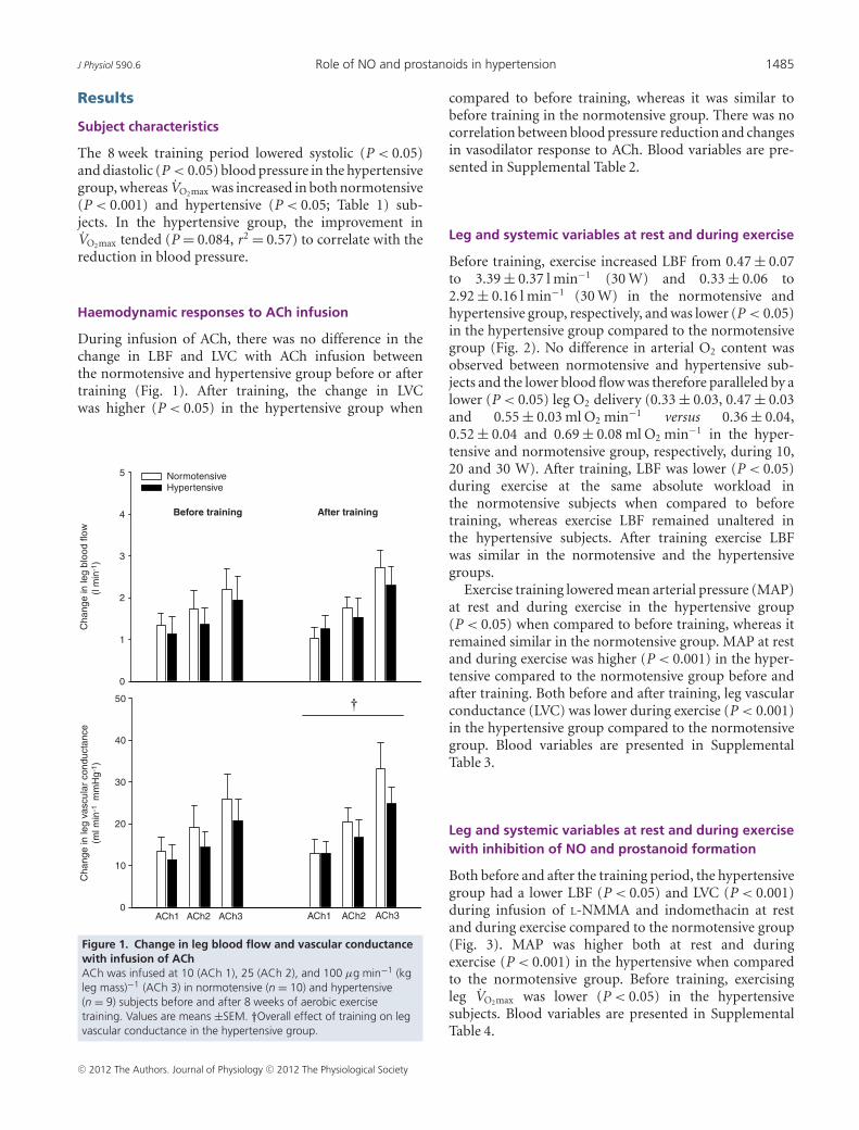
Haemodynamic responses to ACh infusion
During infusion of ACh, there was no difference in thechange in LBF and LVC with ACh infusion betweenthe normotensive and hypertensive group before or aftertraining (Fig. 1). After training, the change in LVCwas higher (P < 0.05) in the hypertensive group when
Cha
nge
in le
g bl
ood
flow
(l m
in-1
)
0
1
2
3
4
5 NormotensiveHypertensive
Before training After training
Cha
nge
in le
g va
scul
ar c
ondu
ctan
ce(m
l min
-1 m
mH
g-1 )
0
10
20
30
40
50
ACh1 ACh2 ACh3 ACh2ACh1 ACh3
Figure 1. Change in leg blood flow and vascular conductancewith infusion of AChACh was infused at 10 (ACh 1), 25 (ACh 2), and 100 μg min−1 (kgleg mass)−1 (ACh 3) in normotensive (n = 10) and hypertensive(n = 9) subjects before and after 8 weeks of aerobic exercisetraining. Values are means ±SEM. †Overall effect of training on legvascular conductance in the hypertensive group.
compared to before training, whereas it was similar tobefore training in the normotensive group. There was nocorrelation between blood pressure reduction and changesin vasodilator response to ACh. Blood variables are pre-sented in Supplemental Table 2.
Leg and systemic variables at rest and during exercise
Before training, exercise increased LBF from 0.47 ± 0.07to 3.39 ± 0.37 l min−1 (30 W) and 0.33 ± 0.06 to2.92 ± 0.16 l min−1 (30 W) in the normotensive andhypertensive group, respectively, and was lower (P < 0.05)in the hypertensive group compared to the normotensivegroup (Fig. 2). No difference in arterial O2 content wasobserved between normotensive and hypertensive sub-jects and the lower blood flow was therefore paralleled by alower (P < 0.05) leg O2 delivery (0.33 ± 0.03, 0.47 ± 0.03and 0.55 ± 0.03 ml O2 min−1 versus 0.36 ± 0.04,0.52 ± 0.04 and 0.69 ± 0.08 ml O2 min−1 in the hyper-tensive and normotensive group, respectively, during 10,20 and 30 W). After training, LBF was lower (P < 0.05)during exercise at the same absolute workload inthe normotensive subjects when compared to beforetraining, whereas exercise LBF remained unaltered inthe hypertensive subjects. After training exercise LBFwas similar in the normotensive and the hypertensivegroups.
Exercise training lowered mean arterial pressure (MAP)at rest and during exercise in the hypertensive group(P < 0.05) when compared to before training, whereas itremained similar in the normotensive group. MAP at restand during exercise was higher (P < 0.001) in the hyper-tensive compared to the normotensive group before andafter training. Both before and after training, leg vascularconductance (LVC) was lower during exercise (P < 0.001)in the hypertensive group compared to the normotensivegroup. Blood variables are presented in SupplementalTable 3.
Leg and systemic variables at rest and during exercisewith inhibition of NO and prostanoid formation
Both before and after the training period, the hypertensivegroup had a lower LBF (P < 0.05) and LVC (P < 0.001)during infusion of L-NMMA and indomethacin at restand during exercise compared to the normotensive group(Fig. 3). MAP was higher both at rest and duringexercise (P < 0.001) in the hypertensive when comparedto the normotensive group. Before training, exercisingleg VO2max was lower (P < 0.05) in the hypertensivesubjects. Blood variables are presented in SupplementalTable 4.
C© 2012 The Authors. Journal of Physiology C© 2012 The Physiological Society

1486 M. Nyberg and others J Physiol 590.6
The effect of training on leg and systemic variables atrest and during exercise during inhibition of NO andprostanoid formation
Taking into account the change in baseline bloodflow with infusion of L-NMMA and indomethacin,
Leg
bloo
d flo
w (
l min
)
0
1
2
3
4
5
6 NormotensiveHypertensive
Before training After training
*
Mea
n ar
teria
l pre
ssur
e (m
mH
g)
80
100
120
140
160
180
* *
0
10
20
30
40
Rest 10 W 20 W 30 W
* *
Rest 10 W 20 W 30 W
Figure 2. Leg blood flow, mean arterial pressure, and legvascular conductance during rest and one-leg knee-extensorexerciseExercise was performed at 10, 20 and 30 W in normotensive (n = 9)and hypertensive (n = 9) subjects before and after 8 weeks ofaerobic high-intensity training. Values are means ± SEM. ∗Overalldifference between groups; †overall effect of training on meanarterial pressure in the hypertensive group and overall effect oftraining on leg blood flow in the normotensive group.
the normotensive group had a lower (P < 0.05)change in LBF during exercise at 10 W (0.34 ± 0.08versus 0.73 ± 0.09 l min−1) and 20 W (0.49 ± 0.08 versus0.70 ± 0.13 l min−1) after training, when compared tobefore training (Fig. 4). There was no effect of training
Leg
bloo
d flo
w (
l min
-1)
0
1
2
3
4
5 NormotensiveHypertensive
Before training After training
*
Mea
n ar
teria
l pre
ssur
e (m
mH
g)
80
100
120
140
160
180
200 * *Le
g va
scul
ar c
ondu
ctan
ce (
ml m
in-1
mm
Hg-
1 )
0
5
10
15
20
25
30
Rest 10 W 20 W 30 W
* *
Rest 10 W 20 W 30 W
*
Figure 3. Leg blood flow, mean arterial pressure, and legvascular conductance during rest and one-leg knee-extensorexercise with infusion of L-NMMA and indomethacinExercise was performed at 10, 20, and 30 W in normotensive (n = 9)and hypertensive (n = 8) subjects before and after 8 weeks ofaerobic high-intensity training. Values are means ± SEM. ∗Overalldifference between groups.
C© 2012 The Authors. Journal of Physiology C© 2012 The Physiological Society
J Physiol 590.6 Role of NO and prostanoids in hypertension 1487
Cha
nge
in le
g bl
ood
flow
(l m
in-1
)
-1.2
-1.0
-0.8
-0.6
-0.4
-0.2
0.0
Before trainingAfter training
*
*
Normotensive Hypertensive
10 W 20 W 30 W 10 W 20 W 30 W
Before training
After training
Mea
n ar
teria
l pre
ssur
e (m
mH
g)
100
110
120
130
140
150
160ControlL-NMMA+indomethacin
Hypertensive
Normotensive
Before training
After training
*
A
B
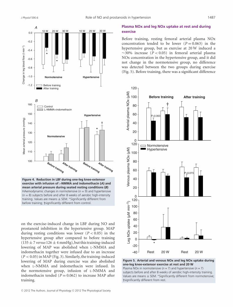
Figure 4. Reduction in LBF during one-leg knee-extensorexercise with infusion of L-NMMA and indomethacin (A) andmean arterial pressure during seated resting conditions (B)HAemodynamic changes in normotensive (n = 9) and hypertensive(n = 8) subjects before and after 8 weeks of aerobic high-intensitytraining. Values are means ± SEM. ∗Significantly different frombefore training; †significantly different from control.
on the exercise-induced change in LBF during NO andprostanoid inhibition in the hypertensive group. MAPduring resting conditions was lower (P < 0.05) in thehypertensive group after compared to before training(135 ± 7 versus 126 ± 4 mmHg), but this training-inducedlowering of MAP was abolished when L-NMMA andindomethacin together were infused due to an increase(P < 0.05) in MAP (Fig. 3). Similarly, the training-inducedlowering of MAP during exercise was also abolishedwhen L-NMMA and indomethacin were infused. Inthe normotensive group, infusion of L-NMMA andindomethacin tended (P = 0.062) to increase MAP aftertraining.
Plasma NOx and leg NOx uptake at rest and duringexercise
Before training, resting femoral arterial plasma NOxconcentration tended to be lower (P = 0.063) in thehypertensive group, but as exercise at 20 W induced a∼30% increase (P < 0.05) in femoral arterial plasmaNOx concentration in the hypertensive group, and it didnot change in the normotensive group, no differencewas detected between the two groups during exercise(Fig. 5). Before training, there was a significant difference
Art
eria
l pla
sma
NO
x (
M)
0
20
40
60
80
100
120
Ven
ous
plas
ma
NO
x (
M)
0
20
40
60
80
100
120
Leg
NO
x up
take
(M
min
-1)
-40
-20
0
20
40
60
80
100
120
Before training After training
*
Rest 20 W Rest 20 W
P=0.063
P=0.073
NormotensiveHypertensive
Figure 5. Arterial and venous NOx and leg NOx uptake duringone-leg knee-extensor exercise at rest and 20 WPlasma NOx in normotensive (n = 7) and hypertensive (n = 7)subjects before and after 8 weeks of aerobic high-intensity training.Values are means ± SEM. ∗Significantly different from normotensive;†significantly different from rest.
C© 2012 The Authors. Journal of Physiology C© 2012 The Physiological Society

1488 M. Nyberg and others J Physiol 590.6
(P < 0.05) between the two groups in leg NOx uptake.After training, venous NOx tended (P = 0.073) to belower during resting conditions in the hypertensives whencompared to the normotensives, whereas no difference inresting NOx uptake was detected.
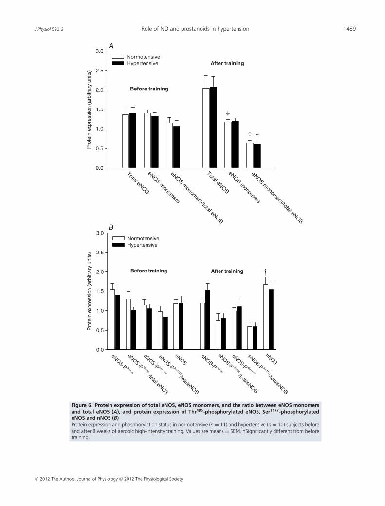
Skeletal muscle eNOS and nNOS protein, eNOSuncoupling and eNOS phosphorylation in muscletissue
No group differences in skeletal muscle protein expressionand phosphorylation status in any of the enzymesinvestigated was observed before or after the trainingperiod (Fig. 6). Training induced a decrease (P < 0.05)in the level of eNOS monomers in the normotensive butnot in the hypertensive subjects. Skeletal muscle eNOSuncoupling (the ratio between eNOS monomers and totaleNOS) in both the normotensive (P < 0.05) and thehypertensive (P < 0.05) group was lower after training.This reduction in eNOS uncoupling was associatedwith a tendency towards an increased expression oftotal eNOS in both the normotensive (P = 0.067) andhypertensive (P = 0.052) subjects. When accounting forchanges in total eNOS, training tended (P = 0.062)to promote dephosphorylation of eNOS at threonineresidue 495 (eNOS-PThr495; phosphorylation here inhibitsenzyme activity) in the normotensive group whereas nochange was detected in the phosphorylation of eNOS atserine residue 1177 (eNOS-PSer1177; phosphorylation hereincreases enzyme activity) in either group. There was anincrease (P < 0.05) in the protein level of nNOS in thenormotensive group after training. Representative blotsare presented in Supplemental Fig. 2.
Effect of sex and menopausal status
Careful examination of individual data for vascularresponses, response to double blockade and NO synthaselevels and properties provided no indication that eitherpre- or post-menopausal women differed from the malesin this regard.
Discussion
In the current study, we show for the first time that bloodflow to exercising skeletal muscle is lower in humanswith essential hypertension and that this blunted exerciseresponse is not associated with a reduced capacity ofthe NO and prostanoid systems to induce vasodilatationduring muscle contraction or an attenuated vasodilatorresponse to ACh. In the normotensive, but not the hyper-tensive, group, the period of training lowered leg bloodflow during exercise at the same absolute workload, aneffect which also was paralleled by a reduced dependency
of the NO and prostanoid system in this subject group,as evidenced by a lower reduction in leg blood flowduring infusion of L-NMMA and indomethacin aftertraining. In contrast to the unaltered role of NO andprostanoids on the regulation of exercise hyperaemia atthe local muscular level, the mechanism responsible forthe training-induced lowering of systemic blood pressureappears to be associated with changes in the tonic effect ofNO and/or prostanoids on vascular tone, as the effect oftraining was abolished when these systems were inhibited.Furthermore, despite indications of a lower resting arterialNO formation before training in the hypertensive group,skeletal muscle eNOS content, degree of uncoupling andphosphorylation status were similar between the twogroups. However, in both groups, training induced areduction in eNOS uncoupling.
Vascular physiology of essential hypertension
Regulation of blood flow to the exercising leg. The lowerblood flow to contracting leg muscles in hypertensivesubjects is an important novel finding that suggests thathypertension is associated with not only an elevation insystemic blood pressure, but also an inadequate bloodflow to skeletal muscle which is essential for exercisecapacity and which, with time and severity of the disease,could lead to tissue ischaemia. The mechanisms under-lying the reduction in exercise induced blood flow arenot completely clear. However, the function of boththe NO and the prostanoid systems have been shown tobe affected in the forearm vasculature of individuals withessential hypertension (Taddei et al. 1997), indicating thatan inadequacy in the formation of NO and vasodilatorprostanoids could potentially explain the lower hyper-aemic response to exercise in these subjects. We examinedthis possibility in the present study by pharmacologicalinhibition of the synthesis of NO and prostanoids. Theinhibition reduced leg blood flow during exercise toa similar extent in both groups, suggesting that thecapacity to produce NO and vasodilator prostanoidsduring leg exercise is not affected in essential hypertension.In accordance with this finding, the hypertensive sub-jects showed a similar vascular relaxation to the mainlyNO-dependent vasodilator ACh (Mortensen et al. 2009)and the two groups also had similar muscle protein levelsand phosphorylation status of enzymes catalysing NOformation. The similar vasodilator response to ACh foundin the current study is in accordance with a study on sub-jects with type 2 diabetes (Thaning et al. 2011), a diseasestate known to be associated with endothelial dysfunction,but in contrast to studies on forearm vasculature ofhypertensive subjects showing reduced vascular relaxation(Taddei et al. 1997; Higashi et al. 1999). This discrepancymay reflect differences in the vasculature of the upper and
C© 2012 The Authors. Journal of Physiology C© 2012 The Physiological Society
J Physiol 590.6 Role of NO and prostanoids in hypertension 1489
Pro
tein
exp
ress
ion
(arb
itrar
y un
its)
0.0
0.5
1.0
1.5
2.0
2.5
3.0NormotensiveHypertensive After training
Before training
Total eNOS
Total eNOS
eNOS monom
ers/total eNOS
eNOS monom
ers/total eNOS
eNOS monom
ers
eNOS monom
ers
Pro
tein
exp
ress
ion
(arb
itrar
y un
its)
0.0
0.5
1.0
1.5
2.0
2.5
3.0NormotensiveHypertensive
After trainingBefore training
nNOS
nNOSeNOS-P Ser1177/totaleNOS
eNOS-P Ser1177/totaleNOS
eNOS-P Ser1177
eNOS-P Ser1177
eNOS-P Thr495
eNOS-P Thr495
eNOS-P Thr495 /totaleNOS
eNOS-P Thr495 /total eNOS
A
B
Figure 6. Protein expression of total eNOS, eNOS monomers, and the ratio between eNOS monomersand total eNOS (A), and protein expression of Thr495-phosphorylated eNOS, Ser1177-phosphorylatedeNOS and nNOS (B)Protein expression and phosphorylation status in normotensive (n = 11) and hypertensive (n = 10) subjects beforeand after 8 weeks of aerobic high-intensity training. Values are means ± SEM. †Significantly different from beforetraining.
C© 2012 The Authors. Journal of Physiology C© 2012 The Physiological Society

1490 M. Nyberg and others J Physiol 590.6
lower extremities and/or the severity and hence effect ofhypertension on endothelial function.
Combined, the above findings suggest that the lowerblood flow during exercise is not associated with a reducedcapacity to form NO or prostanoids. Alternatively, it ispossible that the observed lower blood flow to contractingskeletal muscle was related to alterations in the bluntingof sympathetic outflow normally observed in contractingmuscles of healthy animals and humans (Rosenmeieret al. 2004), since this mechanism has been shown to beimpaired in rat models of hypertension (Zhao et al. 2006)as well as in the forearm of human hypertensive subjects(Vongpatanasin et al. 2011).
Shear-stress induced NO formation. The likely stimulusfor endothelial NO production has been identified asincreased flow through the vessel lumen (Pohl et al. 1986).In animal models, this shear stress induced dilatation isreduced in hypertension due to impaired NO function(Koller, 2002). In accordance, a tendency towards lowerresting femoral arterial plasma NOx concentration inthe hypertensive subjects was observed before training,indicating that the shear stress induced formation of NOwithin the arterial system was impaired in these subjects.Interestingly, acute exercise at 20 W increased the femoralarterial NOx concentration in the hypertensive group tolevels similar to that of the normotensive group, suggestingthat a higher arterial shear stress was needed to stimulateNO production in the hypertensive subjects.
eNOS uncoupling. Uncoupling of eNOS has beensuggested to be present in animal models of hyper-tension (Cosentino & Luscher, 1998; Kerr et al. 1999;Mollnau et al. 2002), as well as in essential hypertension(Higashi et al. 2002), and this alteration has been shown toincrease superoxide formation and decrease vascular NObioavailability (Mollnau et al. 2002). In the present studyno difference in eNOS uncoupling was detected betweenthe two groups. This discrepancy may be related to thevessel type investigated as the level of eNOS uncouplingwas determined in the microvasculature of skeletal muscle,whereas earlier studies have used larger upstream vessels(Cosentino & Luscher, 1998; Kerr et al. 1999; Mollnauet al. 2002). Interestingly, in the study by Higashi andcoworkers (2002), infusion of tetrahydrobiopterin, anessential cofactor for coupling of eNOS (Forstermann & Li,2010), augmented endothelium-dependent vasodilatationin both normotensive and hypertensive individuals,suggesting that eNOS uncoupling was present in bothgroups. The lack of difference in eNOS coupling at theprotein level between the two groups in the current studymay, therefore, reflect that the extent of eNOS uncouplingat the microvascular level is related to age and/or inactivityand not to disease state.
Adaptive responses to exercise trainingin hypertensive individuals
Regulation of blood flow to the exercising leg.In the normotensive subjects, blood flow duringexercise performed at the same absolute workload was∼0.4 l min−1 lower after training, which is in congruencewith findings on young healthy individuals (Kiens et al.1993; Proctor et al. 2001; Krustrup et al. 2004). Thelower blood flow to the trained leg during exercisein the normotensive group is thought to be due totraining adaptations within the skeletal muscle that resultin an optimized blood flow distribution and improvedconditions for oxygen diffusion (Saltin et al. 1976;Kalliokoski et al. 2001; Proctor et al. 2001). In contrast,the ∼0.35 l min−1 lower blood flow to the working limb inthe hypertensive subjects before training is likely to reflecta pathological reduction due to an imbalance betweenvasoconstriction and vasodilatation (Taddei et al. 1997b;Hansen et al. 2011), structural alterations of the bloodvessels (Heerkens et al. 2007) and/or increased MSNA(Laterza et al. 2007). The ability of the hypertensivesubjects to perform the same workload with less bloodflow and oxygen delivery suggests that the reduction inblood flow was compensated for by an increased oxygenextraction, greater anaerobic energy production and/orbetter mechanical efficiency. We did not, however, detectany significant differences in leg oxygen extraction, oxygenuptake, or lactate release, indicating that more than oneadaptation was responsible for the compensation. Theunaltered leg blood flow after training in the hypertensivesubjects may reflect that an improvement in vascularfunction occurred in parallel with some of the changesleading to the training induced lowering of blood flow.
In the normotensive subjects, the lower leg bloodflow after training was paralleled by less reductionin exercise-induced blood flow with pharmacologicalinhibition of the NO and prostanoid system in thenormotensive subjects. This novel observation suggeststhat a potential mechanism responsible for the lower legblood flow after training was a reduced role of the NOand prostanoid systems for skeletal muscle blood flowregulation. Conversely, in the hypertensive subjects nodifference in the effect of pharmacological inhibition wasobserved after training, suggesting an unaltered formationof NO and prostanoids during muscle contraction.
Blood pressure regulation in essential hypertension.From a meta-analysis including 30 hypertensive studies,it has been found that aerobic training reduces systolicand diastolic blood pressure by ∼7 and ∼5 mmHg,respectively (Cornelissen & Fagard, 2005). Consequently,the reductions of ∼9 and ∼12 mmHg found in the currentstudy are well above the expected outcome and of highclinical relevance as a reduction of either 10 or 5 mmHg
C© 2012 The Authors. Journal of Physiology C© 2012 The Physiological Society
J Physiol 590.6 Role of NO and prostanoids in hypertension 1491
in systolic and diastolic blood pressure, respectively, willin the long term be associated with a 40% lower risk ofstroke death and 30% lower risk of death from ischaemicheart disease or other vascular causes throughout middleage (Lewington et al. 2002).
An association between the training induced magnitudeof blood pressure reduction and the gain in VO2max has pre-viously been reported (Cornelissen & Fagard, 2005), andin congruence with this observation we did detect largereductions in blood pressure, which tended (P = 0.084)to correlate with the improvement in VO2max in thehypertensive group. These observations emphasize thathigh-intensity aerobic training, which is known for itslarge effect on improvements in VO2max (Helgerud et al.2007), is a powerful stimulus for reducing blood pressure.The mechanisms underlying this association are intriguingas central haemodynamic adaptations are likely to explainthe improvement in VO2max (Saltin et al. 1968), whereasthe lowering of blood pressure is thought to be due tochanges in peripheral vascular function (Taddei et al.1997a), vascular remodelling (Heerkens et al. 2007) and/ora decrease in MSNA (Laterza et al. 2007).
Role of NO and prostanoids in the regulation of bloodpressure. A novel observation was that L-NMMA andindomethacin increased blood pressure after training inthe hypertensive subjects. The unaltered blood pressurein normotensive subjects and hypertensive subjects beforetraining is in agreement with previous studies (Mortensenet al. 2007, 2009) using a similar dose of these inhibitorsin young healthy subjects. The blood pressure raisingeffect of combined inhibition after training is likely tobe related to an increased formation and bioavailability ofNO and possibly also improved balance between vaso-constrictor and vasodilator prostanoids after training.With regard to the latter possibility, the endotheliumin hypertensive subjects has been suggested to producecyclooxygenase-derived vasoconstrictor prostanoids aswell as superoxide anions which reduce NO bioavailability(Taddei et al. 1997b, 1998).
The effect of L-NMMA and indomethacin on bloodpressure could have contributed to activation of thearterial baroreflex and subsequent inhibition of centralsympathetic outflow (Hansen et al. 1994), but the lackof change in heart rate makes this possibility unlikely.The increased perfusion pressure in the leg could be aconfounding factor for the observed change in leg bloodflow and vascular conductance. However, infusion ofN G-nitro-L-arginine methyl ester and indomethacin hasbeen shown to increase blood pressure in healthy subjects(Boushel et al. 2002), but despite this effect on centralhaemodynamics, exercise hyperaemia was reduced to asimilar extent as in studies where no changes in bloodpressure have been observed (Mortensen et al. 2007, 2009).
In addition, Frandsen et al. (2001) reported no influenceon leg blood flow during exercise with systemic NOSinhibition, despite an increase in blood pressure. Theincreased blood pressure during infusion of L-NMMAand indomethacin in trained hypertensive subjects is,therefore, unlikely to have altered the leg blood flowresponse to exercise to an extent that would have affectedour conclusions.
Although it is well established that aerobic traininglowers blood pressure in hypertensive subjects the under-lying mechanisms remain unidentified. NO is importantfor whole body blood pressure regulation in humans(Joyner & Casey, 2009) and training has been shown toimprove the function of eNOS, but in congruence with thefindings from the current study, no correlation betweenblood pressure reduction and improved endothelialfunction was observed (Higashi et al. 1999). Despite theunaltered function of the NO and prostanoid system onlocal blood flow regulation during exercise, inhibitionof these systems did fully abolish the training-inducedreduction in resting and exercising blood pressureat the systemic level. This discrepancy between localand systemic effects suggests that vascular resistance isregulated upstream from the microvasculature, but moreevidence is needed to confirm this proposition.
Shear-stress induced NO formation. Exercise is known tobe associated with acute changes in endothelial shear stress(Tinken et al. 2010), and it has been shown that increasesin intraluminal shear stress improve NO-mediatedendothelium-dependent dilatation in animals (Woodmanet al. 2005) and humans (Tinken et al. 2010).High-intensity exercise eliciting very high blood flows andhence shear stress may, therefore, be a potent exercisemodality to reduce blood pressure in hypertensive sub-jects due to its effect on endothelial NO function,which is in accordance with the normalization of restingarterial NOx after training observed in the current study.Additional studies investigating the effect of differentexercise intensities on shear stress induced alterations inendothelial NO function and blood pressure changes arewarranted.
eNOS phosphorylation. Exercise training has beenshown to increase phosphorylation of eNOS on serineresidue 1177 (Hambrecht et al. 2003) and to decreasephosphorylation of eNOS on threonine residue 495(Calvert et al. 2011), both leading to increasedenzyme activity. In the current study no difference inphosphorylation status was detected between the groupswith only a tendency towards a lower phosphorylation onthreonine residue 495 in the normotensive subjects aftertraining. What underlines this discrepancy is unclear, but itmay reflect differences in the vessel (Hambrecht et al. 2003)
C© 2012 The Authors. Journal of Physiology C© 2012 The Physiological Society
1492 M. Nyberg and others J Physiol 590.6
and species (Calvert et al. 2011) investigated as well asdisease state (Hambrecht et al. 2003; Calvert et al. 2011).
Exercise hyperaemia as an evaluation of vascularfunction
In the current study we demonstrate that althoughthe vasodilator response to ACh was unaffected inthe leg of hypertensive subjects, blood flow to contra-cting muscle was lower. This finding underscores that,whereas ACh infusion specifically tests the capacityof a few endothelium-dependent vasoactive systems,including NO synthesis, vasodilatation in responseto exercise includes an integrated response involvingsympathetic activity, and numerous endothelium- andnon-endothelium-dependent vasoactive systems. Withinthe present conditions exercise hyperaemia appears to bea better indicator of vascular function than the vasodilatorresponse to ACh.
Conclusion
The current study provides evidence for a lower bloodflow to the exercising leg in hypertensive subjects thatis not associated with a reduced capacity of the NO andprostanoid systems to induce vasodilatation or with alteredACh-induced response. In contrast to the unaffectedfunction of these systems in the peripheral circulation,our data suggest that the observed reduction in systemicblood pressure is related to a training-induced change inthe tonic effect of NO and/or prostanoids on vasculartone.
References
Andersen P & Saltin B (1985). Maximal perfusion of skeletalmuscle in man. J Physiol 366, 233–249.
Bangsbo J, Gunnarson TP, Wendell J, Nybo L & Thomassen M(2009). Reduced volume and increased training intensityelevate muscle Na+-K+ pump α-2-subunit expression as wellas short- and long-term work capacity in humans. J ApplPhysiol 107, 1771–1780.
Boushel R, Langberg H, Gemmer C, Olesen J, Crameri R,Scheede C, Sander M & Kjaer M (2002). Combinedinhibition of nitric oxide and prostaglandins reduces humanskeletal muscle blood flow during exercise. J Physiol543, 691–698.
Calvert JW, Condit ME, Aragon JP, Nicholson CK, Moody BF,Hood RL, Sindler AL, Gundewar S, Seals DR, Barouch LA &Lefer DJ (2011). Exercise protects against myocardialischemia-reperfusion injury via stimulation of β3-adrenergicreceptors and increased nitric oxide signaling: role of nitriteand nitrosothiols. Circ Res 108, 1448–1458.
Cornelissen VA & Fagard RH (2005). Effects of endurancetraining on blood pressure, blood pressure-regulatingmechanisms, and cardiovascular risk factors. Hypertension46, 667–675.
Cosentino F & Luscher TF (1998). Tetrahydrobiopterin andendothelial function. Eur Heart J 19, G3–G8.
Forstermann U & Munzel T (2006). Endothelial nitric oxidesynthase in vascular disease: from marvel to menace.Circulation 113, 1708–1714.
Forstermann U & Li H (2010). Therapeutic effect of enhancingendothelial nitric oxide synthase (eNOS) expression andpreventing eNOS uncoupling. Br J Pharmacol164, 213–223.
Hambrecht R, Adams V, Erbs S, Linke A, Krankel N, Shu Y,Baither Y, Gielen S, Thiele H, Gummert JF, Mohr FW &Schuler G (2003). Regular physical activity improvesendothelial function in patients with coronary artery diseaseby increasing phosphorylation of endothelial nitric oxidesynthase. Circulation 107, 3152–3158.
Frandsen U, Bangsbo J, Sander M, Hofner L, Betak A, Saltin B& Hellsten Y (2001). Exercise-induced hyperaemia and legoxygen uptake are not altered during effective inhibition ofnitric oxide synthase with N G-nitro-L-arginine methyl esterin humans. J Physiol 531, 257–264.
Hansen J, Jacobsen TN & Victor RG (1994). Is nitric oxideinvolved in the tonic inhibition of central sympatheticoutflow in humans? Hypertension 24, 439–444.
Hansen AH, Nyberg M, Bangsbo J, Saltin B & Hellsten Y(2011). Exercise alters the balance between vasoactivecompounds in skeletal muscle of individuals with essentialhypertension. Hypertension 58, 943–949.
Helgerud J, Høydal K, Wang E, Karlsen T, Berg P, Bjerkaas M,Simonsen T, Helgesen C, Hjorth N, Back R & Hoff J (2007).Aerobic high-intensity intervals improve VO2max morethan moderate training. Med Sci Sports Exerc39, 665–671.
Heerkens EHJ, Izzard AS & Heagerty AM (2007). Integrins,vascular remodeling, and hypertension. Hypertension 49,1–4.
Higashi Y, Sasaki S, Kurisu S, Yoshimizu A, Sasaki N, MatsuuraH, Kajiyama G & Oshima G (1999). Regular aerobic exerciseaugments endothelium-dependent vascular relaxation innormotensive as well as hypertensive subjects: role ofendothelium-derived nitric oxide. Circulation 100,1194–1202.
Higashi Y, Sasaki S, Nakagawa K, Fukuda Y, Matsuura H,Oshima T & Chayama K (2002). Tetrahydrobiopterinenhances forearm vascular response to acetylcholine in bothnormotensive and hypertensive individuals. Am J Hypertens15, 326–332.
Jansson PA, Veneman T, Nurjhan N & Gerich J (1994). Animproved method to calculate adipose tissue interstitialsubstrate recovery for microdialysis studies. Life Sci 54,1621–1624.
Joyner MJ & Casey DP (2009). The catacholamines strike back– what NO does not. Circ J 73, 1783–1792.
Kalliokoski KK, Iokonen V, Takala TO, Sipila H, Knuuti J &Nuutila P (2001). Enhanced oxygen extraction and reducedflow heterogeneity in exercising muscle in endurance-trainedmen. Am J Physiol Endocrinol Metab 280, 1015–1021.
Kiens B, Essen-Gustavsson B, Christensen NJ & Saltin B (1993).Skeletal muscle substrate utilization during submaximalexercise in man: effect of endurance training. J Physiol 469,459–478.
C© 2012 The Authors. Journal of Physiology C© 2012 The Physiological Society
J Physiol 590.6 Role of NO and prostanoids in hypertension 1493
Klatt P, Schmidt K, Lehner D, Glatter O, Bachinger HP &Mayer B (1995). Structural analysis of porcine brain nitricoxide synthase reveals a role for tetrahydrobiopterin andL-arginine in the formation of an SDS-resistant dimer.EMBO J 14, 3687–3695.
Kerr S, Brosnan MJ, McIntyre M, Reid JL, Dominiczak AF &Hamilton CA (1999). Superoxide anion production isincreased in a model of genetic hypertension: role of theendothelium. Hypertension 33, 1353–1358.
Koller A (2002). Signaling pathways of mechanotransductionin arteriolar endothelium and smooth muscle cells inhypertension. Microcirculation 9, 277–294.
Krustrup P, Hellsten Y & Bangsbo J (2004). Intense intervaltraining enhances human skeletal muscle oxygen uptake inthe initial phase of dynamic exercise at high but not at lowintensities. J Physiol 559, 335–345.
Laterza MC, de Matos LD, Trombetta IC, Braga AM, Roveda F,Alves MJ, Krieger EM, Negrau CE & Rondon MU (2007).Exercise training restores baroreflex sensitivity in nevertreated hypertensive patients. Hypertension49, 1298–1306.
Lewington S, Clarke R, Qizbash N, Peto R & Collins R (2002).Age-specific relevance of usual blood pressure to vascularmortality: a meta analysis of individual data for one millionadults in 61 prospective studies. Lancet360, 1903–1913.
Mollnau H, Wendt M, Szocs K, Lasseque B, Schulz E, Oelze M,Li H, Bodenschatz M, August M, Kieschow AL, TsilimingasN, Walter U, Forstermann U, Meinertz T, Griendling K &Munzel T (2002). Effects of angiotensin II infusion on theexpression and function of NAD(P)H oxidase andcomponents of the nitric oxide/cGMP signaling. Circ Res90, 58–65.
Mortensen SP, Gonzalez-Alonso J, Damsgaard R, Hellsten Y &Saltin B (2007). Inhibition of nitric oxide andprostaglandins, but not endothelial derived hyperpolarizingfactors, reduces blood flow and aerobic energy turnover inthe exercising human leg. J Physiol 581, 853–861.
Mortensen SP, Nyberg M, Thaning P, Saltin B & Hellsten Y(2009). Adenosine contributes to blood flow regulation inthe exercising human leg by increasing prostaglandin andnitric oxide formation. Hypertension 53, 993–999.
Nyberg M, Mortensen SP, Saltin B, Hellsten Y & Bangsbo J(2010). Low blood flow at onset of moderate-intensityexercise does not limit muscle oxygen uptake. Am J PhysiolRegul Integr Comp Physiol 298, 843–848.
Pohl U, Holtz J, Busse R & Bassenge E (1986). Crucial role ofendothelium in the vasodilator response to increased flow invivo. Hypertension 8, 37–44.
Proctor DN, Miller JD, Dietz NM, Minson CT & Joyner MJ(2001). Reduced submaximal leg blood flow afterhigh-intensity aerobic training. J Appl Physiol 91, 2619–2627.
Rosenmeier JB, Hansen J & Gonzales-Alonso J (2004).Circulating ATP-induced vasodilatation overridessympathetic vasoconstrictor activity in human skeletalmuscle. J Physiol 558, 351–365.
Saltin B, Blomqvist G, Mitchell JH, Johnson RL Jr, WildenthalK & Chapman CB (1968). Response to exercise after bed restand after training. Circulation 38, 1–78.
Saltin B, Nazar K, Costill DL, Stein E, Jansson E, Essen B &Gollnick D (1976). The nature of the training response;peripheral and central adaptations of one-legged exercise.Acta Physiol Scand 96, 289–305.
Saunders NR, Dinenno FA, Pyke KE, Rogers AM &Tschakovsky ME (2005). Impact of combined NO and PGblockade on rapid vasodilation in a forearmmild-to-moderate exercise transition in humans. Am JPhysiol Heart Circ Physiol 288, 214–220.
Scheller D & Kolb J (1991). The internal reference technique inmicrodialysis: a practical approach to monitoring dialysisefficiency and calculating tissue concentration from dialysatesamples. J Neurosci Methods 40, 31–38.
Thaning P, Bune LT, Zaar M, Saltin B & Rosenmeier JB (2011).Functional sympatholysis during exercise in patients withtype 2 diabetes with intact response to acetylcholine.Diabetes Care 34, 1–6.
Taddei S, Virdis A, Mattei P, Ghiadoni L, Fasolo CB, Sudano I& Salvetti A (1997a). Hypertension causes premature agingof endothelial function in humans. Hypertension 29, 736–743.
Taddei S, Virdis A, Ghiadoni L, Magagna A & Salvetti A(1997b). Cyclooxygenase inhibition restores nitric oxideactivity in essential hypertension. Hypertension 29, 274–279.
Taddei S, Virdis A, Ghiadoni L, Magagna A & Salvetti A (1998).Vitamin C improves endothelium-dependent vasodilationby restoring nitric oxide activity in essential hypertension.Circulation 97, 2222–2229.
Tinken TM, Thijssen DHJ, Hopkins N, Dawson EA, Cable NT& Green DJ (2010). Shear stress mediates endothelialadaptations to exercise training in humans. Hypertension 55,312–318.
Vongpatanasin W, Wang Z, Arbique D, Arbique G,Adams-Huet B, Mitchell JH, Victor RG & Thomas GD(2011). Functional sympatholysis is impaired in hypertensivehumans. J Physiol 589, 1209–1220.
Woodman CR, Price EM & Laughlin MH (2005). Shear stressinduces eNOS mRNA expression and improvesendothelium-dependent dilation in senescent soleus musclefeed arteries. J Appl Physiol 98, 940–946.
Zhao W, Swanson SA, Ye J, Li X, Shelton JM, Zhang W &Thomas GD (2006). Reactive oxygen species impairsympathetic vasoregulation in skeletal muscle in angiotensinII-dependent hypertension. Hypertension48, 637–643.
Author contributions
The experiments were conducted at the CopenhagenMuscle Research Centre, Rigshospitalet, Denmark. Thecontributions of the authors were as follows: conceptionand design of the study: M.N., Y.H. and S.P.M;collection, analysis and interpretation of data: M.N.,L.G.J., P.T., Y.H. and S.P.M.; drafting the article orrevising it critically for important intellectual content:M.N., Y.H. and S.P.M. All authors approved the finalversion.
C© 2012 The Authors. Journal of Physiology C© 2012 The Physiological Society

1494 M. Nyberg and others J Physiol 590.6
Acknowledgements
Assia A. Bada and Morten Overgaard are gratefullyacknowledged for their excellent medical assistance. This workwas supported by a grant from the Lundbeck foundation and
the Danish Medical Research Council. M.N. was supported by agrant from the P. Carl Petersen foundation. S.P.M. was supportedby a grant from the Danish Heart Foundation and the DanishCouncil for Independent Research – Medical Sciences.
C© 2012 The Authors. Journal of Physiology C© 2012 The Physiological Society