THE LUMBAR AND SACRUM MOVEMENT PATTERN DURING THE BACK SQUAT EXERCISE
Upload
khangminh22Category
view
0download
0
University of ConnecticutOpenCommons@UConn
Master's Theses University of Connecticut Graduate School
5-8-2014
A Comparison of Single Leg Squat and Side StepCut Kinematics in Healthy and ACLReconstructed PopulationsJarrett JE [email protected]
This work is brought to you for free and open access by the University of Connecticut Graduate School at OpenCommons@UConn. It has beenaccepted for inclusion in Master's Theses by an authorized administrator of OpenCommons@UConn. For more information, please [email protected].
Recommended CitationSorge, Jarrett JE, "A Comparison of Single Leg Squat and Side Step Cut Kinematics in Healthy and ACL Reconstructed Populations"(2014). Master's Theses. 587.https://opencommons.uconn.edu/gs_theses/587

A Comparison of Single Leg Squat and Side Step Cut Kinematics in Healthy and ACL Reconstructed Populations
Jarrett JE Sorge, ATC, CES
B.S., Bridgewater State University, 2009
A Thesis
Submitted in Partial Fulfillment of the
Requirements for the Degree of
Master of Science
At the
University of Connecticut
2014
ii
APPROVAL PAGE
Masters of Science Thesis
A Comparison of Single Leg Squat and Side Step Cut Kinematics in
Healthy and ACL Reconstructed Populations
Presented by
Jarrett JE Sorge, B.S. Major Advisor________________________________________________________________
Lindsay J. DiStefano Associate Advisor_____________________________________________________________
Douglas J. Casa Associate Advisor_____________________________________________________________
Craig R. Denegar Associate Advisor_____________________________________________________________
Thomas H. Trojian
University of Connecticut
2014

iii
Acknowledgements Dr. Lindsay DiStefano: I cannot thank you enough for your guidance through this process. I struggled through my first semester at UConn without you; never thinking that I would be able to catch up. It’s amazing that this process has finally come to end. I have learned an incredible amount from you. You have instilled a passion for injury prevention that I will have forever. Thank you for allowing me to learn, teach, research, and grow under you. Dr. Douglas Casa: First, thank you for accepting me into the program. I cannot describe how much better of an athletic trainer I have become since I joined the program. Your passion and enthusiasm for the profession as well as the appropriate care for our athletes will stay with me forever. Thank you for serving on my committee and for being a mentor for me. I am truly grateful. Dr. Thomas Trojian: Thank you so much for taking the time out of your busy schedule to come to our data collection to clear our participants. I appreciate your passion for injury prevention and all of the work that you have invested into our department. Dr. Craig Denegar: Thank you for being apart of my thesis committee. I have truly enjoyed having you as a professor and a mentor. You have challenged me to take evidence-based practice to the next level, and really analyze research. Your advice has shaped this thesis project into what it is today. Jessica Martinez: This project would never have finished without you. Although we often went about things in interesting ways, they always got done. I cannot thank you enough for the late night answers to questions, the mid-day reassurances, or the mid morning panic. Thank you for being a mentor, a colleague, and most importantly a friend. To my family and friends: Thank you for supporting me on this opportunity to go back to school. Your love, support, and encouragement have made this process much easier. And Katie LeClair, thank you for driving the 75 miles round trip each way to make this happen. Thank you for your adaptability, assurance, and lack of knowledge of athletic training. I love you.

iv
TABLE OF CONTENTS
I. Review of the Literature……………........……………………………………………6
Cost Association……………........………………………………………………..6
Return to Play and Quality of Life……………........……………………………...7
ACL Injury Mechanism……………........………………………………………...8
Risk Factors of Non-Contact ACL Injury……………........……………………..10
Incidence of ACL Reinjury……………........……………………………………13
Rehabilitation Phases……………........………………………………………….15
Return to Play Criteria……………........………………………………………...18
Movement Components of the Side Step Cut……………........…………………20
Movement Components of the Single Leg Squat……………........…………..…23
Recognition of Risk Factors……………........…………………………………...27
Conclusion……………........………………………………………………..…...28
II. Introduction…………………………………………………………………………..36
III. Methods………………………………………………………………………………39
IV. Results………………………………………………………………………………..44
V. Discussion……………………………………………………………………………45
VI. Appendix…………………………………………………………………………..…52

v
ABSTRACT
A Comparison of Single Leg Squat and Side Step Cut Kinematics in Healthy and ACL Reconstructed Populations Jarrett JE Sorge, University of Connecticut STUDY DESIGN: Case Control Study OBJECTIVE: To compare single leg squat (SLS) kinematics to side step cut (SSC) kinematics and vertical ground reaction force in individuals with a history of ACL reconstruction and healthy controls. BACKGROUND: There are currently no objective criteria to progress athletes into cutting activities during rehabilitation. The single leg squat possesses similar three-planar neuromuscular control as a cutting task. Assessing movement dysfunction during the single leg squat could limit injury risk during cutting activities. METHODS: 44 individuals active in cutting, jumping, or landing activities participated in this study. 22 athletes had a history of ACL reconstruction (14 male, 8 female) Age, 21.7± 3.8 years; Height, 174.5± 7.2 cm; Mass, 76.2± 9.9 kg). 22 healthy athletes (14 male, 8 female) with no history of ACL reconstruction or any other lower extremity surgery (Age: 21.6± 3.6 years; height: 173.8± 9.2 cm; mass: 75.0± 10.5) served as a matched control group based on sex, height, mass, age, and activity level. Kinematic data was collected during both tasks; participants completed five single leg squats and two side step cutting tasks on each leg. The means across were determined and correlated between tasks. Independent sample t-tests were used to determine any significance between groups. RESULTS: Individuals with a history of ACL reconstruction squatted and cut with significantly less sagittal plane motion compared to healthy controls. Healthy controls also cut with more trunk rotation towards the direction of travel and higher VGRF compared to individuals with a history of ACLR. Numerous correlations were seen between tasks. CONCLUSION: Sagittal, frontal, and transverse plane motion during the SLS were predictive of motion during the SSC. Lack of frontal and transverse plane trunk, hip, and knee control during the SLS resulted in positions of increased lateral trunk flexion, hip adduction, and medial knee displacement during a cutting task. The SLS can be used as a clinical predictor of SSC in athletes during injury prevention or return to play rehabilitation. Key Words: single leg squat, cutting, side step cut, anterior cruciate ligament, injury prevention

6
Review of the Literature
Anterior cruciate ligament (ACL) injuries are one of the most debilitating
musculoskeletal injuries suffered in sport. The ACL is a primary stabilizer of the knee;
therefore, rupture can lead to functional instability. An estimated 75,000-250,000 ACL injuries
occur annually in the United States; this estimate has risen through the years due to the dramatic
increase in sport participation from a pediatric age through adult life. 1-3 Since the passing of
Title IX in 1972, male athletic participation has increased 3% while female participation has
increased more than 9-fold (0.3 million to 2.8 million). 4 ACL injuries have been reported at a
frequency 2-9 times greater in females compared to males in the same cutting and jumping
sports. 5,6 Although the United States has no national injury tracking system, Marshall et al.3
reported through survey that 1 in 90 hospital or emergency room visits involved a cruciate
ligament injury. These injuries require long-term health care, treatment, and rehabilitation. The
root of ACL injury prevention involves undertaking a comprehensive understanding of the
etiology of ACL injury, identifying and modifying risk factors that predispose athletes to ACL
injury, and following evidence based return to play guidelines that minimize the risk of re-injury.
Cost Association
ACL-Reconstruction (ACLR) is the standard treatment for ACL rupture, designed to
limit long-term intra-articular damage and restore stability and function. 7 Not all ACL deficient
patients require surgical reconstruction; the decision is based on their ability to maintain
adequate knee function. 5 An estimated two-thirds of patients opt for reconstruction, which
rapidly becomes costly with surgery and rehabilitation estimated at $17,000-25,000 per
incidence. 2,8 However, long term cost analysis shows that ACL reconstruction is not

7
significantly more expensive than conservative treatment due to the associated meniscal injuries
and early development of osteoarthritis in those who elect not to have reconstructive surgery. 9
Return to Play and Quality of Life
ACL injuries are not only costly, but can also have dramatic effects on a patient’s return
to participation, activity level, and long term quality of life. ACL injuries can result in the loss of
entire seasons of participation or the loss of scholarship for the high school athlete. 10 In a study
concerning return to play and future ACL risk, Brophy et al. 11 determined that 72% of soccer
athletes returned to their sport at an average of 12-14 months after surgery, with 85% of those
returning to soccer at the same or higher level of play. At a long term follow up of 7-8 years post
ACLR, only 35% were still playing their sport. Of those still playing, only 46% were still
playing at the same or higher level of play as before their injury. 11 Similarly, Ardern et al. 12
surveyed 314 ACLR individuals 2-7 years after reconstruction. The investigators found that only
41% of their participants had attempted competitive sport at follow-up, and only 29% were
actively participating at their pre-injury competitive level. 12 More than one-half of the studied
individuals who did not return to their pre-injury level of competition cited function of their knee
as their reasoning. 12
Lohmander et al. 13 found that radiographic patellofemoral or tibiofemoral osteoarthritis
was present in 51% of the ACL injured female soccer athletes studied 12 years after injury. Of
the 84 women who answered the questionnaires, 75% reported having symptoms affecting their
knee related quality of life (Figure 1).

8
Figure 1. Knee Injury and Osteoarthritis Score (KOOS) results reported by Lohmander et al13 The ACL injured female soccer players (mean age 31) are represented by solid circles, the reference group with no symptoms of knee OA (mean age 55) are represented by triangles, and the reference group of uninjured female soccer players (mean age 20) are represented by diamonds
Ahlden et al. 14 conducted the largest known study reporting results in over 16,000 patients with
a history of ACL reconstruction through the Swedish National ACL Register. The study
collected results KOOS scores from registry respondents at 1, 2, and 5 years postoperatively.
Ahlden found that patients who underwent a second surgery had significantly poorer knee related
quality of life compared to those who had had their first reconstruction. Participants with an
additional ACL reconstruction also displayed no significant improvement in symptoms, pain,
and activities of daily living at 5 years post surgery compared with their preoperative values. 15
ACL Injury Mechanism
ACL injuries are characterized by a contact or non-contact mechanism. A non-contact
mechanism involves no contact with an opposing player, equipment, or ground at the time of
injury. Mechanisms of non-contact ACL injury normally involve multi-planar knee loading
events. 1,16 The most common mechanism reported by the athlete is planting and pivoting. 6 An
reference group. The ACL-injured soccer players alsoscored significantly worse (P ! 0.001) in all dimensionsof the KOOS, except the ADL subscale, in comparisonwith the other reference group (without radiographicknee OA, mean age 55 years, 27% women). The largestdifferences were again found in Sports/Rec function andknee-related QOL. As reported in the KOOS question-naire, the knee problems resulted in a modified lifestylein 50% of the injured players, and a lack of confidence inthe index knee was reported by 70% of those with anACL injury.
The women defined as having radiographic kneeOA scored lower on all of the KOOS subscales com-pared with individuals without radiographic OA. How-ever, the differences were only statistically significant forthe subscales on pain (P " 0.03) and other symptoms(P " 0.004) (Table 3). There were no significant differ-ences in the KOOS dimensions in terms of whether ornot the patient had undergone surgical reconstruction ofthe ACL (P ! 0.2).
SF-36 results. The injured players scored signif-icantly worse than the reference group of Swedishwomen ages 25–34 years in the physical functioning
subscale of the SF-36 questionnaire. However, the play-ers scored better than the reference group in the sub-scale on social functioning. In the other 6 subscales ofthe SF-36 there were no significant differences betweenthe groups (Table 4).
Multivariate analyses. Using multivariate mod-els, we evaluated the influence of reconstructive ACLsurgery on the radiographic outcome and on the likeli-hood of being symptomatic as assessed by the KOOS.Reconstruction of the ACL did not significantly influ-ence the prevalence of definite radiographic knee OA,nor did it influence whether the subject was symptom-atic. However, there was a tendency toward an increasedlikelihood to have radiographic patellofemoral OA de-
Figure 1. The Knee Injury and Osteoarthritis Score (KOOS) profile.Results are the mean scores and 95% confidence intervals for theKOOS subscales pain, other symptoms, activities of daily living (ADL),function in sports and recreation (Sport/Rec), and knee-related qualityof life (QOL), reported as an outcome profile for the anterior cruciateligament injury group (solid circles; n " 84, mean age 31 years), thereference group without radiographic knee osteoarthritis (open re-verse triangles; n " 55, mean age 55 years), and the reference group ofuninjured female soccer players (open diamonds; n " 108, mean age20 years). For the latter reference group, the 95% confidence intervalswere too small to be visualized.
Table 4. Short Form 36-item health survey subscale scores in women12 years after injury to the anterior cruciate ligament during soccerplay*
Study group(n " 84)
Swedish femalepopulation
ages 25–34 years(n " 896)
Physical functioning 82 (80–85) 93 (92–94)Role-physical 80 (72–87) 87 (85–89)Bodily pain 73 (68–78) 77 (76–79)General health 81 (77–84) 80 (79–82)Vitality 66 (62–70) 66 (65–68)Social functioning 94 (91–96) 89 (88–90)Role-emotional 86 (80–92) 87 (85–89)Mental health 82 (79–85) 80 (79–81)
* Values are mean (95% confidence interval).
Table 5. Influence of surgical reconstruction of the anterior cruciateligament (ACL) on the radiographic outcome and the self-reportedoutcome in female soccer players 12 years after the ACL injury*
Outcome
ACL reconstruction
Crude ORAdjusted OR
(95% CI)†
Radiographic knee OA‡ 1.7 1.7 (0.6–5.0)Radiographic tibiofemoral OA 1.4 1.3 (0.5–3.9)Radiographic patellofemoral OA 5.8 14 (0.9–224)
Symptomatic§ 1.0 0.8 (0.3–2.6)Symptomatic radiographic knee OA§ 1.5 1.7 (0.5–5.3)
* For radiographic outcome, n " 67, of whom 41 were ACL recon-structed. For self-reported outcome, n " 84, of whom 52 were ACLreconstructed. Odds ratios (OR) with 95% confidence intervals (95%CIs) are from logistic regression, using subjects without reconstructiveACL surgery as the reference category.† Adjusted for age at assessment, body mass index, surgically treatedmeniscal injury, occupational workload, and spare-time activity level.‡ Either tibiofemoral or patellofemoral radiographic osteoarthritis(OA) (see Patients and Methods for definition).§ Symptomatic as assessed by the Knee Injury and OsteoarthritisOutcome Score (see Patients and Methods for definition).
HIGH OA PREVALENCE IN ACL-INJURED FEMALE SOCCER PLAYERS 3149
9
ACL injury normally involves a change of direction or cut, combined with deceleration, the knee
near full extension, and the foot fixed on the playing surface. 1,16,17 In one of the first studies to
retrospectively analyze mechanisms of ACL injury, Boden et al. 16 surveyed 132 patients (143
knees) after sustaining an ACL injury. The study found that a noncontact mechanism was the
cause of 72% sustained injuries. 16 Additionally, the National Collegiate Athletic Association
(NCAA) studied ACL injuries prospectively in men’s and women’s basketball and men’s and
women’s soccer. Each sport had high incidences of non-contact ACL injury, men’s and
women’s basketball athletes experiencing the highest incidence rate at 80% each. The non-
contact rate in men’s and women’s soccer was slightly lower; women’s soccer suffered a 63%
rate and men’s soccer a 48% rate of non-contact ACL injury. 6 In an even higher estimate,
Myklebust et al. 18 followed 212 teams in the three upper level men’s and women’s Norwegian
handball divisions through two full seasons (estimated 3392 players) and found that 95% of ACL
injuries occurred without contact from another player.
Ireland described the “position of no return” (Figure 2) as the reported vulnerable cause
of noncontact ACL injury. The position of no return is described as including a forward flexed
back, adducted and internally rotated hips, the knee in a less flexed and valgus position, tibia
rotated, and landing on one foot with the weight forward. 19 Hewett et al. 8 described four
positions that seemed to occur during many ACL injuries, especially in women: as the athlete
lands, the knee buckles inward, the knee is relatively straight, most of the athlete’s weight is on
the single limb, and the trunk tends to be flexed laterally, causing the athlete’s center of mass to
be shifted outside the base of support. These events also occur in men, but seem to be more
exaggerated in women. 8 The results from observational studies generally agree that valgus
motion, often accompanied with transverse plane knee rotation motions, were contributing

10
factors to the ACL injury mechanism. 1,4,16,17 Hewett et al. 20 also demonstrated that ACL injuries
demonstrated both lateral trunk motion and knee abduction. 20 Boden et al. 16 added, through the
use of video analysis, the position of the leg after a non-contact injury was near foot-strike with
the knee close to full extension.
Figure 2. The “Position of No Return” for ACL Injury compared with the “Safe Position” as described by Ireland et al.19
Risk Factors of Non-Contact ACL Injury
Identification of risk factors that predispose athletes to non-contact ACL rupture has
become a crucial aspect of injury prevention. There have many studies that have looked at
identifying non-modifiable and modifiable risk factors and explaining their roles in ACL injury.
Many of the risk factors aim to explain the greater risk of ACL injury in female athletes incident
compared to men participating in the same activities.
Non-Modifiable Risk Factors
There are several anatomical risk factors that have been proposed to explain the risk of
ACL injury, especially in female athletes. Joint laxity, narrow intercondylar notch width,
SEX DIFFERENCES AND CONTRIBUTING FACTORSIn order to reduce the rate of ACL injuries in the female
athlete, we must focus on those factors that can be modified.These factors include playing style, preparation, and skillacquisition from a very young age. Contributing factors areintrinsic (not controllable), extrinsic (controllable), or both(partially controllable) (Table 2).
INJURY MECHANISM: NONCONTACT ACL,POSITION OF NO RETURNBy understanding the mechanisms of injury in sport, we can
design intervention programs to reduce the risk of injury.Observations of ACL injury mechanisms in basketball showthe athlete coming down in an uncontrolled landing, eithercatching the ball or trying not to go out at the baseline. Awhiplike snap of the lower extremity is seen as the ACL tears.In visualizing this high-risk "position of no return," we
comprehend the importance of a "get-down," knee-flexed,2-footed balanced position. Figure 1 diagrammatically showsthe position of no return and the safe position, from the jointpositions of the back, hips, knee, and foot. In the no-returnposition, the hip abductors and extensors have shut down, andthe pelvis and hip are uncontrolled. Muscle groups that wouldnormally upright the individual are unable to perform thisfunction due to their mechanical disadvantages and the length-ening of the muscle groups.
Noncontact injury patterns are similar in males and females.Figure 2 includes still photographs and line drawings of thismechanism of injury. Athletes injure their knees as they comedown from a shot. Note the relatively extended knee initially;by the second frame, the ACL has failed. Hip and trunk-pelvis-hip control were previously lost, and lower extremity align-ment was hip internal rotation and adduction, knee valgus, andtibial external rotation on a pronated, externally rotated foot.Figure 3 shows a left knee from the left and the back. Theinitially abducted hip goes into relative internal rotation andadduction on a pronated, externally rotated foot. At first, thereis relatively little knee flexion; then the body weight goesforward as the body flexes over the legs, and, again, extremevalgus stress occurs after the ACL has failed. The hip and kneepositions of rotation and less flexion are observed as the ACL
Table 2. Factors Contributing to ACL InjuriesCombined
Intrinsic Extrinsic (partially controllable)Alignment Strength Proprioception (positionHyperextension Conditioning sense/balance)Physiologic rotatory Shoes Neuromuscular
laxity Motivation activation patternsACL size Order of firingNotch size and shape Acquired skillsHormonal influencesInherited skills and
coordination
Figure 1. The position-of-no-return mechanism for ACL injury andthe safe position.
Figure 2. Sequence of body and lower extremity positions as thisathlete tears his left ACL. By the second frame, the ACL has mostlikely torn. Note the planting of the foot, the adducted hip, thevalgus knee, the extemally rotated tibia, and the body fallingforward to the opposite side.
fails. The gluteus maximus and hamstrings are unable toprotect the ACL.
PREVENTION PROGRAMSThe role of neuromuscular training in reducing the risk of
serious knee injuries was studied in high school volleyball andbasketball players.26 A 6-week preseason training program toreduce landing forces and increase hamstring power using plyo-metrics was instituted.26 After 1 season of tracking 1263 athletes,untrained females demonstrated a knee injury rate 3.7 timeshigher than that for trained females and 4.6 times higher than thatfor males. Based on the results of this study, neuromusculartraining appears to reduce the risk of injury in female volleyballand basketball players.A prospective, controlled study of proprioceptive training
was conducted in 40 Italian semiprofessional and amateursoccer teams, which included 600 male players.28 Over 3
152 Volume 34 * Number 2 * June 1999

11
posterior tibial plateau slope, and static alignment, have all been extensively researched. 10,17,21,22
Ligamentous laxity at the ACL can be objectively reported by measuring the anterior translation
of the tibia, mostly commonly using a KT-1000 ligament arthrometer. 22 Several studies have
reported that a combination of risk factors increases the risk of ACL injury. Uhorchak et al. 23
reported that the combination of body weight, BMI, intercondylar notch width, as well as joint
laxity were all significant risk factors for ACL injuries. Evans et al, 24 concluded that an elevated
BMI as well as a narrow notch width may predispose young military athletes to ACL injury.
Gender differences have been extensively researched in terms of ACL injury risk. Long-
term NCAA injury data investigations have proven to show that there is a much higher ACL
injury incident in women when compared to men participating in the same sports. 6,25 Hormonal
influences have been a proposed reasoning for the higher rate of ACL injury in females. 26,27
Females may have increased anterior-posterior knee laxity during the preovulatory phase of their
menstrual cycle, subsequently causing greater ACL injury risk. Another gender specific risk
factor associated with ACL injury is quadriceps angle (Q-Angle). Q-Angle is the angle drawn
from the ASIS to the midpoint in the patella and then from the midpoint of the patella to the
tibial tuberosity. A high Q-Angle is reported to possibly alter biomechanics at the lower limb and
place the knee in positions of valgus stress. However, Myer et al. 28 reported that increases in
static Q-Angle measurements were not predictive of ACL injury risk during dynamic movement.
Q-Angle has also been shown to not be a significant factor in peak knee valgus during a single
leg squat task. 29
Modifiable Risk Factors
12
Modifiable risk factors have also been extensively studied in relation to ACL injury
prevention. Lack of active neuromuscular control may destabilize the knee and increase the ACL
injury risk in athletes. The term “neuromuscular control” refers to the unconscious dynamic
stabilization at a particular joint in response to sensory stimuli. 30 Dynamic stabilization at the
knee is extremely important for the prevention of ACL injury; without proper dynamic
stabilizers, the ACL would fail with forces sustained during everyday activities. 22 Co-activation
of the hamstrings, quadriceps, and gastrocnemius muscles at the knee are all important in the
dynamic stabilization at the knee. 10,22 ACL injury occurs during moments of high load at the
knee when muscular control is not adequate enough to prevent translation at the knee.
Considering the gender bias seen with noncontact ACL injury, several studies have
looked at comparing movement patterns between men and women. Women have also been
shown to move, land, and absorb forces differently from men. In a systematic review, Dai et al. 21
summarized that females tend to restrict sagittal plane motion and increase motion in the frontal
and transverse planes when performing athletic tasks. This combination of motion results in
increased loading at the knee and specifically the ACL. Hewett et al. 4 screened 205 female
athletes who were participants in high-risk sports and followed them to determine risk factors of
ACL injury in female athletes. Of the 205 athletes screened, 9 had a confirmed ACL rupture. All
9 displayed eight degrees greater knee abduction angle, a 2.5 times greater knee abduction
moment, and 20% higher ground reaction force when compared with the 196 uninjured. They
concluded that knee abduction moments and angles during landing tasks were predictors of ACL
risk in female athletes.4
Myer et al. 31 prospectively studied the hamstring and quadriceps strength and ratio of
male and female athletes prior to injury. Female athletes who subsequently suffered ACL injury

13
had less hamstring strength but not quadriceps strength. Conversely, female athletes who did not
suffer ACL injury had lessquadriceps strength without decreased hamstring strength compared to
males. 31 Griffin et al. 30 reported on the consensus statement made at the Hunt Valley Consensus
Conference that neuromuscular factors are significant contributors to ACL injury rate in females.
Several studies have reported that during cutting tasks, females exhibit much greater peak valgus
moments and frontal plane motion compared to males given the same tasks. 32,33 Although
evidence is becoming increasingly abundant, further research needs to be conducted to prove that
risk factors vary between males and females.
Incidence of ACL Re-Injury
The most significant risk factor for ACL injury is a previous history of ACL rupture. 34,35
Risk of a second ACL injury is greatest with the return to cutting and pivoting sport-specific
activities, especially in the first 12 months following ACL reconstruction. 35,36 Paterno et al. 36
reported that within the first 12 months of activity after return to sport, subjects with ACLR were
15 times more likely to sustain an ACL injury compared with subjects with no history of ACLR.
Rate of injury to the graft as well as the contralateral knee during return to play has been studied
extensively. In a large cohort study, Shelbourne et al. 37 prospectively followed 1415 people for
five years who underwent ACL reconstruction. Of the 1415 people, 136 (9.6%) suffered a
subsequent injury to either knee at follow up. 45% of subsequent tears were on the ACLR side,
and 55% of tears were on the contralateral side. 37 No significant difference between men and
women for subsequent ACL tear in the ACL reconstructed knee was reported; however, women
had a significantly higher incidence of ACL injury to the contralateral knee. 37

14
Two other studies reported similar results with respect to rate of contralateral knee injury.
Salmon et al. 35 followed up with 612 ACLR patients five years after reconstruction, and 71 had
suffered an additional ACL injury. ACL graft rupture occurred in 39 patients (6%) and
contralateral ACL rupture occurred in 35 patients (6%). 3 patients suffered both a graft rupture
and a contralateral ACL injury. 35 Wright et al. 34 prospectively followed 235 patients after
reconstruction and reported 14 (6%) subsequent ACL ruptures. Seven ruptures were graft
ruptures and 7 ruptures were of the contralateral knee. In a smaller prospective study that only
includes ACLR individuals who suffered a non-contact mechanism of injury, Paterno et al. 36
found a much higher incidence of re-injury. Of the 63 subjects that met the inclusion criteria, 16
suffered a subsequent noncontact ACL injury, 12 to the contralateral knee. ACL injury rates
(reinjury or contralateral injury) after ACL reconstruction range from 1 in 4 (25%) to 1 in 17
(6%) after return to sport participation. 34-36 Identification of biomechanical risk factors is
necessary to effectively reduce the high rate of re-injury.
Return to Play Timetable
In addition to limiting intra-articular damage, restoring function and stability, the goal of
ACL reconstruction is to return the patient to his or her previous level of activity as quickly and
safely as possible. Failure to restore adequate range of motion, strength, and normal gait during
rehabilitation often results in long-term deficits and a poorer quality of life. ACLR patients have
been shown to seek treatment for symptoms of osteoarthritis 15-20 years before patients without
a history of ACL reconstruction. 5 Benyonn et al. 38 and Shelbourne et al. 39 have both
demonstrated that accelerated ACLR rehabilitation (19 weeks) produces the same effects (knee
laxity, clinical outcome, patient satisfaction, patient function, and proprioception) compared with
a group of non-accelerated (32 weeks) rehabilitation.

15
Rehabilitation Phases
Preoperative Phase
There is no consensus on the correct or ideal timing of ACL reconstruction. 5 Many
patients have difficulty regaining range of motion prior to surgery; therefore, many surgeons
suggest preoperative rehabilitation prior to surgery that will accelerate postoperative
rehabilitation. The main goals of the preoperative phase are to reduce swelling, pain, restore full
range of motion, regain neuromuscular control, and normalize gait prior to surgery. 5,7,40,41
Another critical aspect of the preoperative phase is patient education. Informing athletes on
surgeon selection, the surgical procedure, as well as the rehabilitative process are all necessary
components of the preoperative phase.
Early Postoperative Phase: Day 1 – Week 4
The early postoperative phase begins during the first hours after surgery and extends to 2-
4 weeks after surgery. The two main goals of early rehabilitation are achievement of full
extension and regaining neuromuscular control. 40 One of the most common complications after
ACL reconstruction is loss of range of motion. Restoration of motion, especially terminal knee
extension is the primary goal during the first days of rehabilitation after ACL reconstruction. 41
Rehabilitation that incorporates early joint motion has been found to be beneficial for reducing
pain and decreasing scar tissue formation. 42 Failure to extend the knee fully results in abnormal
joint arthrokinematics and quadriceps inhibition. 41 Rehabilitation and restoration of motion
begin immediately after surgery with the use of a continuous passive motion machine, designed
to minimize the effects of immobilization43 and continues with active range of motion protocols.
16
In addition, an aggressive approach to controlling pain and inflammation prevents quadriceps
inhibition, maintains knee extension, and allows for a quicker return to weight bearing. 7
The trend in ACL rehabilitation is toward earlier weight bearing. Investigations have
shown that immediate weight bearing does not compromise the ACL graft and may be beneficial
at reducing the incidence of anterior knee pain. 42 Patients are partial weight bearing immediately
after surgery, aided by crutches and are gradually progressed to full weight bearing between days
4-14 post-operation, as leg strength improves, gait normalizes, and the patient gains confidence.
7,40,41 Patellar mobilization as well as a combination of safe isometric and isotonic closed and
open kinetic chain strengthening exercises are initiated during the first two weeks after surgery.
7,41 Strengthening is often assisted by electrical neuromuscular stimulation to facilitate
quadriceps contraction, to minimize atrophy, and to reeducate the muscle.43
Criteria used to progress patients to the second phase of rehabilitation include: quadriceps
control, full passive knee extension, passive range of motion 00 to 900, normal patellar mobility
compared contralaterally, minimal joint effusion, and independent ambulation, with or without
crutches. 7,40,41
Intermediate Postoperative Phase: Week 4-12
The intermediate postoperative phase begins once patients have sufficiently completed
the goals defined during the early rehabilitative phase. The primary goals during the
intermediate phase are to regain full flexion and hyperextension, increase strength and
neuromuscular control, improve proprioception, and achieve normal gait. 40 The intermediate
postoperative phase is a critical time period because the processes of graft healing and tunnel
formation are at their most vulnerable stages. 7,40,42 Rehabilitation exercises during this phase

17
should be prescribed with maximal graft protection in mind.43 Rehabilitation is continued
from stage one, with progressions in open and closed kinetic chain exercises, as well as
incorporating active motion and cardiovascular endurance through the stationary bicycle and
aquatic therapy. 39,41 Cryotherapy should be continued to address pain control and joint effusion. 7
Flexion can be gradually increased while normal extension and patellar mobility should be
maintained. 7 Incorporation of proprioceptive drills as well as neuromuscular control exercises
attempt to facilitate joint stiffness and co-contraction of the quadriceps, hamstrings, and gastroc-
soleus complex at the knee. Muscular co-contraction at the knee protects the graft from anterior
translation forces that could disrupt the maturation and incorporation of the graft. 40 Gait training
on a treadmill to identify and correct any gait pattern impairments is essential once the patient
begins full weight bearing ambulation. 7 Deficits present in the early stages of rehabilitation will
most likely persist through the late stages if not addressed.
Criteria to progress athletes to the late stage of postoperative rehabilitation include:
minimal joint line or patellofemoral pain, minimal joint effusion, full extension, at least 1250 of
flexion, normal gait pattern, and quadriceps and hamstring strength 60% compared to
contralateral side.7,41
Late Postoperative Phase: Week 12 – Return to Play
Early stage ACL rehabilitation often follows strict criteria based guidelines for range of
motion and exercise selection and progression. In contrast, late stage rehabilitation is typically
broader with generalized categories of exercise selection and limited objective progressions. 44
During late stage rehabilitation, running and sport specific drills are initiated and functional
strength and proprioception is normalized. 40 Graft fixation and maturation continues to be a

18
primary concern; controlled loading enhances ligament healing, while excessive loading can
potentially damage the graft. 44 Shelbourne et al. 39 reported positive objective and subjective
results in one of the first accelerated (6 month) rehabilitation and return to play studies after ACL
reconstruction. Since then, the majority of ACL studies show a return to sport using an
accelerated rehabilitation at 6 to 12 months.40
Functional training and sport specific drills incorporate exercises that are relevant the
athlete’s sport into the rehabilitation program. 40 Neuromuscular training becomes the main focus
of rehabilitation, with emphasis placed on static and dynamic stability. Patients must be trained
to allow them to possess sufficient functional stability to prevent the knee from positions that are
risk factors of subsequent tears or graft elongation. 7,44 Plyometric exercises are also emphasized
during late stage rehabilitation, designed to recruit the neuromuscular system and elastic
properties of the muscles and joints surrounding the knee. 40 Straight line running is normally
incorporated by three to four months, with duration and speed minimized to allow for
neuromuscular adaptation. 7,40 Speed and duration are increased gradually over the course of the
late stage, with patient education and compliance to ensure the patient does not do too much too
soon. 40 Once straight ahead running is performed successfully and without setback, variations in
running, cutting, and agility activities are introduced as well as dynamic movements in the
frontal and transverse planes. 7,40
Return to Play Criteria
There has been no specific measurable outcome criterion shown to correlate with
successful return to sports in the ACL reconstructed athlete. Most clinicians use a combination
of functional, clinical, and subjective testing. 40 In a systematic review, Barber-Westin et al. 45

19
found that 60% of studies reported time postoperatively as criteria for return to play following
ACL reconstruction. Myer et al. 46 studied the deficits in strength, control, and performance of
limbs in athletes cleared for return to play following ACLR. The study found that there was
significant asymmetry between limbs of the ACLR group with respect to force generation
(vertical jump height) and force absorption (VGRF) when compared to a control group. These
results indicate that up to 11 months after surgery and after release to sport, there are still
significant deficits between the reconstructed limb and the non-injured limb that are independent
of time after surgery. 46 Time from surgery is a counterintuitive criterion that does not address the
neuromuscular and biomechanical deficiencies that an athlete might possess. 46 There is a need
for individualized objective and subjective guidelines to safely progress the ACLR athlete into
their return to sport participation.
Myer et al. 44 created a 5-phase rehabilitation protocol with individual goals and criteria
for progression through each phase. This criteria for return to sport included: (1) drop vertical
jump landing force bilateral symmetry (within 15%), (2) T-test time (within 10%), (3) single
limb average peak power test for 10 seconds (bilateral symmetry within 15%), (4) reassessment
of tuck jump (20 percentage points of improvement from initial test score or perfect 80 point
score). 44 Van Grinsven7 listed a similar 4 phase rehabilitation progression with criteria to
progress to each phase. Return to sport criteria included: (1) (VAS score), no pain or swelling
(2) full flexion and extension, (3) quadriceps and hamstring strength >85% compared to
contralateral side, (4) Hop tests (one-legged timed hop test, single leg hop for distance, tripe hop
for distance) >85% compared to the contralateral side. 7 The general consensus is that once a
patient has gained full range of motion, his or her hop tests are over 85% compared to the

20
healthy side, his or her strength ratio of the quadriceps and hamstrings are over 85% compared to
the healthy side, and the physician has cleared the athlete, they can now return to sport.7,40,44,47
Although isokinetic testing and single leg hop tests are more objective measurements
compared to time postoperatively, they fail to address the multi-planar motion that is
characterized in a cutting task. Cutting involves frontal, sagittal, and transverse plane motion at
the trunk, hip, knee, and ankle; whereas, isokinetic tests and single leg hop tests generally only
assess sagittal plane motions. Initiating cutting is a very important phase of rehabilitation;
however, there fails to be objective criteria that address the multi-planar movement involved in
cutting in the literature. The single leg squat has been used as a valid and reliable assessment tool
for the analysis of faulty movement patterns especially in regard to preventing injury at the trunk,
hip, and knee. 48-50 Previous research has shown kinematic and biomechanical landing
differences between genders and ACLR history during cutting tasks. 32,51-55 These differences,
especially increased frontal and transverse plane motion and limited sagittal plane motion, have
been shown to occur in both single leg squat and cutting tasks in these populations. 52,54-59
Determining a correlation between the movements would help clinicians make rehabilitation
progression decisions that limit injury risk.
Movement Components of the Side Step Cut
The initiation of cutting is an important part of rehabilitation and must be done safely and
with objective criteria for the progression to cutting. Cutting has been one of the proposed
causes of noncontact injury to the ACL. 1,16,17 Allowing an ACLR athlete to initiate cutting
prematurely can increase the risk of re-injury. Cutting in the ACLR athlete has been extensively
studied. Many studies have reported that the side step cutting maneuver places a much greater

21
strain on the knee because of the higher moments of frontal plane motion, especially in
women.20,33,52,54
Sagittal Plane Motion
Malinzak et al. 52 conducted one of the initial studies that compared knee motion during
various athletic tasks between men and women. The investigators found that female subjects
were demonstrated less knee flexion during cutting tasks. Females also displayed greater
normalized quadriceps activation and less hamstring activation. 52 Blackburn et al. 60 found that
active trunk flexion during landing promoted more knee and hip flexion compared to a more
erect trunk posture. Others have reported that during deceleration, female athletes exhibit less hip
flexion. 54 Miranda et al. 53 compared side step cutting kinematics between males and females
with ACLR to a control group. They reported that females with no history of ACL
reconstruction appeared to perform the jump cut maneuver with greater landing stiffness than
males with or without a history of ACLR and females with a history of ACLR. 53 Males and
females with a history of ACLR performed the jump-cut maneuver with less energy than the
control group, resulting in lower peak vertical GRF. 53 Coats-Thomas compared ACL intact
males and females to ACL reconstructed males and females during a side step cutting task. They
reported that there was a delayed peak activation of the quadriceps, hamstring, and
gastrocnemius muscles after landing in ACL reconstructed men and women compared to the
healthy control group. ACL reconstructed men and women also had a higher quadriceps
activation compared to hamstring activation during the load phase when compared to healthy
controls. 61 Hanson et al. 62 also studied muscle activation during a cutting task, comparing
Division I male and female soccer players. They found that females displayed significantly
greater vastus lateralis activation compared to males during the preparatory and load phase of a

22
cutting task, reaffirming that females cut with great quadriceps activation than males. 62 Without
co-activation of the hamstrings during cutting tasks, increased quadriceps activation puts greater
load on the ACL.
Frontal Plane Motion
Several studies have reported that women have had a much greater tendency to cut with
high knee abduction moments compared to men during a side step cutting task. 32,52,55
Kristianslund et al. 63 compared the differences between a drop jump and a side step cut in 184
handball players. Knee abduction moments were shown to be 6 times higher in a side step
cutting task compared to a drop vertical jump task. They also reported that athletes had lower
knee flexion angles and higher knee valgus and internal rotation angles both at initial contact and
at maximum flexion. 63 Jamison et al. 64 found a positive association between knee abduction
moment and lateral trunk deviation during a side step cut. As the torso moves away from the
cutting direction, the knee abduction moment increases. Imwalle et al. 51 found that the most
significant predictor of knee abduction was hip adduction during a 450 and 900 cutting task in
healthy female soccer players. Females have been shown to have greater hip adductor moments
during deceleration and have exhibited decreased hip extensor moments compared with male
athletes, possibly attributing to their higher incidence of ACL injury. 54 Hewett et al. 20 found that
ACLR female subjects showed greater lateral trunk and knee abduction moments at landing
compared with uninjured control groups. This lack of neuromuscular trunk control and trunk
stability has been reported as leading to uncontrolled knee abduction and ACL strain.
Transverse Plane Motion
23
Pollard et al. 54 found that when compared with male athletes, female athletes
demonstrated significatly greater hip internal rotation during the early phase of the side step
cutting maneuver. McLean et al.30 also reported that peak knee valgus loading was directly
associated with higher initial hip flexion and hip internal rotation positions. Frank et al. 65 found
that internal trunk rotation displacement was the greatest predictor for knee varus moment during
a cutting task in healthy athletes. Less trunk rotation displacement toward the direction of cutting
and hip adduction moment were associated with greater in knee varus moment during cutting. 65
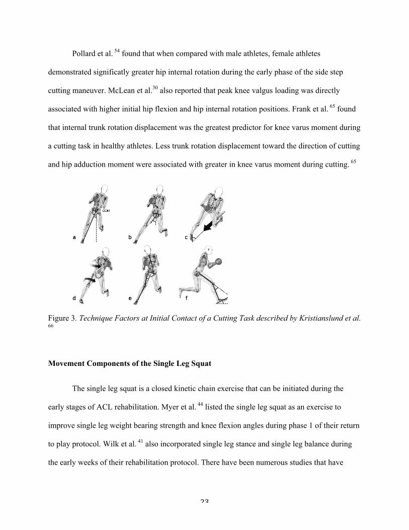
Figure 3. Technique Factors at Initial Contact of a Cutting Task described by Kristianslund et al. 66
Movement Components of the Single Leg Squat
The single leg squat is a closed kinetic chain exercise that can be initiated during the
early stages of ACL rehabilitation. Myer et al. 44 listed the single leg squat as an exercise to
improve single leg weight bearing strength and knee flexion angles during phase 1 of their return
to play protocol. Wilk et al. 41 also incorporated single leg stance and single leg balance during
the early weeks of their rehabilitation protocol. There have been numerous studies that have

24
focused on the differences between men and women and various kinematic chain recruitment
strategies for single leg squat completion. The single leg squat has been previously used as a
screening tool for injury risk. 50,58 Single leg stance exercises have been used as strengthening
exercises for the hip, especially in regards to gluteus medius activation. The gluteus medius is
the primary hip stabilizer in both the frontal and transverse planes. Inactivation or weakness of
the gluteus medius results in femoral adduction and internal rotation, and an increase in medial
knee displacement; risk factors for ACL injury. 19 Single leg squat exercises have been shown to
produce higher peak gluteus maximus and medius activation when compared to double limb
stance exercises. 67,68 Utilizing single leg squat as an assessment for functional dynamic position
in athletes is important because of the ability to incorporate it into the earlier stages of ACLR
rehabilitation.
Single Leg Squat as an Injury Assessment
Biomechanical analysis utilizing validated and reliable clinical screening assessment
have been utilized to determine modifiable injury risk in athletes and to attempt to screen athletes
for potential injury. Several studies have attempted to validate the single leg squat as an
observational movement dysfunction screening assessment tool. Chmielewski et al. 69 estimated
the intra and interrater reliability of the single leg squat as a movement assessment. Graders
rating the movement using an ordinal grading system (poor, fair, good) resulted in good
reliability between graders. However, when asked to specifically rate the movement using
different body segments, reliability decreased. 69 These findings were determined to be better
than chance but not high enough reliability to be used clinically. Poulsen et al. 70 also used an
ordinal scale to grade single leg squat motion. Poulsen had intrarater reliability ranging from
(0.38 – 0.94) and the generalized weighted kappa score for interrater reliability (0.68). Like

25
Chmielewski, Poulsen et al.’s results did not create enough reliability to exceed a minimal
clinical standard. Both studies attributed their lack of reliability due to the subjective nature of
the scoring and the lack of education given to the graders before evaluation. Crossley et al. 49
also used an ordinal scale to grade single leg squat performance, but had a much more developed
determination of criterion to determine a good, fair, or poor squat. Using this method of
assessment, Crossley et al. 49 had acceptable reliability for clinical use. Intrarater reliability
ranged from (k=0.61 – 0.80) and interrater relialibity ranged from (k= 0.60 – 0.80). Stensrud et
al. 50 evaluated subjective assessment of subjective single leg squat and compared it to re-
assessment through 2-d video analysis. Receiver operating characteristic (ROC) showed strong
reliability (AUC = 0.88 – 0.89). Weeks et al. 48 compared 2 dimensional SLS analysis to 3
dimensional motion analysis to determine reliability between experienced clinicians and
students. Intra-class correlation coefficients were calculated to estimate the reliability between
the groups. Interrater reliability was good for experienced clinicians (ICC = 0.71) and students
(ICC = 0.60). Intrarater reliability was excellent for experienced clinicians (ICC = 0.81) and
good for students (ICC = 0.71). 48 These previous studies show that the single leg squat can be
used as clinical screening tool, but there is a need for more standardized and reliable criterion for
clinicians to use during grading and assessment.
Sagittal Plane Motion
Compared to males, females use significantly less trunk flexion during the descent phase
of the single leg squat (Figure 3). 57 Increased trunk flexion during the single leg squat has been
shown to reduce strain on the ACL by increasing hamstring force output by 35% during the
single leg squat. 71 Females have also shown to demonstrate more ankle dorsiflexion and hip

26
flexion compared to men during a single leg squat. 59 Limited dorsiflexion range of motion
during the single leg squat has been shown to produce positions of medial knee displacement. 72
Figure 4. Observed gender differences during the decent phase of a single leg squat task in the sagittal plane. 57
Frontal Plane Motion During the Single Leg Squat
Crossley et al. 49 were the first researchers to study that functional performance of the
single leg squat could indicate hip muscle function. They found that people who had poor
performance in the single leg squat task (balance, trunk, pelvis, hip, and knee positioning) were
shown to have delay in hip abductor activation. This was an important finding because of the
tendency for the hip to move into adduction with decreased activity of the gluteus medius, a
component of the “position of no return” as described by Ireland. 19 Graci et al. 57 found that
females experience greater hip adduction and knee abduction at both 450 and peak knee flexion
of a single leg squat task (Figure 4). Zeller et al. 59 also found that females started in a more knee
valgus position and remained in a valgus position throughout the single leg squat in comparison
to men. Conversely, Pantano et al. 29 reported that static knee valgus, especially in relation to an
increased Q-Angle, did not relate to the amount of knee valgus seen during a single leg squat,
In the sagittal, frontal and transverse planes, the following joint angles werecalculated at 45KF and PKF: trunk (trunk relative to pelvis), hip (femur relative topelvis), and knee (tibia relative to femur). The pelvis angle relative to the lab (globalcoordinate system) was calculated to shed light on the contribution of the pelvissegment to the net trunk and net hip angles. For the hip and knee, angles wereexpressed in the reference frame of the proximal segment and positive valuesrepresent flexion, adduction and medial rotation. For the trunk and pelvis, positivevalues represent flexion, lateral flexion toward the non-weight-bearing limb andtransverse rotation toward the non-weight-bearing limb. The time spent to performthe squat was also calculated as the difference between the knee joint EOM and theSOM time points. Dependent measures were averaged across repetitions for eachsubject. A two-tailed, independent samples T-test was performed on the trunk,pelvis, hip and knee angles in the three planes of motion (x, y, z) at 45KF and PKF.The alpha level was set at p ! 0.05.
2.5. Reliability testing
Between-day intra-rater reliability of the dependent measures was calculatedusing the intraclass correlation coefficient (ICC(3,3)). Data on 19 subjects werecollected on two occasions, approximately 4.52 " 1.89 days apart. The ICCs wascalculated so that the standard error of measurement (SEM) could be also estimated.
3. Results
Males were taller and heavier than females but body mass indexwas not different between genders (Table 1). Dependent measures
showed good to excellent reliability [16] in each of the three planesof motion (Supplementary Table). Females flexed their trunk lessthan males at both the 45KF (p = 0.013) and PKF (p = 0.006)position (Fig. 2). At 45KF females laterally flexed their trunktoward the weight-bearing limb while males laterally flexed theirtrunk toward their non-weight-bearing limb, although thisdifference was not statistically significant (p = 0.095). Femalesrotated their trunk in the transverse plane toward the weight-bearing limb less than males (p = 0.039, Fig. 3a). In the transverseplane at 45KF females rotated the pelvis toward the weight-bearing limb (in the same direction of the trunk) while malestoward the non-weight-bearing limb (opposite direction of thetrunk) (p = 0.004, Fig. 3b).
Compared to males, females presented greater hip adduction atboth 45KF (p = 0.035) and PKF (p = 0.013) and presented greaterknee abduction at both 45KF (p = 0.009) and PKF (p = 0.0008)(Fig. 3c and d). Females also performed the squat in less time(2.36 s " 0.79 s) than males 3.18 s " 0.83; p = 0.041). All the statisti-cally significant differences were greater than the SEM (Supplemen-tary Table 1). Means, standard deviations (SD) and effect sizes areprovided in Table 2. In Supplementary Table 2 the means and SD oftwo females and males with similar height and weight are reported.
Fig. 2. (a) Time series curves of the trunk angle in the sagittal plane normalized as % squat cycle and averaged across subjects for each gender. 45KF and PKF represent the timepoints where 45 knee flexion and peak knee flexion occurred. Thick lines represent the means. Error bars represent the SD at each time point. Asterisks refer to significantdifferences (p < 0.05). (b) Descent squat phase in one female and one male subject.
Table 2Mean (SD), p-value and effect size for each kinematic variable.
45KF PKF
Females Males p-Value Effect size Females Males p-Value Effect size
TrunkSagittal #19.12 (8.87) #11.49 (6.58) 0.013* 0.98 #19.28 (9.24) #7.04 (7.91) 0.006* 1.42Frontal #0.74 (3.24) 1.64 (2.61) 0.095 0.81 #4.12 (5.22) #4.75 (3.68) 0.76 0.14Transverse #0.96 (2.27) #3.56 (2.74) 0.039* 1.03 #0.34 (3.10) #2.21 (3.39) 0.23 0.57
PelvisSagittal 22.71 (9.98) 20.70 (6.13) 0.59 0.24 26.77 (11.71) 30.19 (11.31) 0.53 0.29Frontal 0.49 (2.40) #0.58 (2.58) 0.39 0.40 3.02 (2.33) 3.05 (3.52) 0.98 0.01Transverse #1.49 (1.46) 1.17 (1.96) 0.004* 1.54 #4.23 (3.79) #4.05 (3.09) 0.91 0.05
HipSagittal 41.15 (11.98) 40.74 (9.85) 0.94 0.04 59.09 (15.47) 72.39(21.88) 0.15 0.70Frontal 9.69 (3.50) 6.15 (3.24) 0.035* 1.05 17.28 (2.62) 13.53 (3.22) 0.013* 1.28Transverse 0.35 (4.23) 1.21 (4.39) 0.68 0.19 #1.04 (4.40) #0.70 (3.87) 0.86 0.08
KneeSagittal 45.39 (0.17) 45.31 (0.09) 0.24 0.55 69.77 (7.27) 76.43 (10.15) 0.12 0.75Frontal #0.89 (3.95) 3.34 (2.14) 0.009* 1.33 #1.25 (4.77) 7.004 (4.11) 0.0008* 1.85Transverse 6.72 (3.12) 6.44 (5.34) 0.89 0.06 4.10 (4.89) 7.76 (6.06) 0.17 0.66
For the hip and knee, angles were expressed in the reference frame of the proximal segment and positive values represent flexion, adduction and medial rotation. For the trunkand pelvis, positive values represent flexion, lateral flexion toward the non-weight bearing limb and transverse rotation toward the non-weight bearing limb.
* Significant differences (p < 0.05).
V. Graci et al. / Gait & Posture 36 (2012) 461–466 463

27
and should not be used to predict knee valgus during the task. Mauntel et al. 72 indicated that
during a single leg squat, healthy subjects who displayed medial knee displacement had a higher
reliance on their hip adductors rather than an inadequate activation of the hip abductors.
Figure 5. Observed gender differences during the decent phase of a single leg squat task in the frontal plane.57
Transverse Plane Motion
Limited research into transverse plane motion during the single leg squat has been
reported. Graci et al. 57 reported that females rotated their trunk toward the weight-bearing limb
less than males. Women have also shown significantly more ankle pronation and hip external
rotation during the single leg squat. 59
Recognition of Risk Factors
The gold standard for recognizing ACL injury risk factors is in a laboratory setting using
3D motion analysis software and forceplate data. However, this method is very expensive,
costing thousands of dollars to acquire all necessary equipment, and requires specialty training
for clinicians. Although the gold standard, motion analysis in a laboratory lacks the feasible
The data reported in Supplementary Table 2 shows a similar trend inmovement pattern to the data from the entire female and malesample (except for trunk flexion at 45KF). Thus, the kinematicdifferences across groups are unlikely to be due simply to thedifferences in height and weight between females and males.
4. Discussion
The primary new finding of the present study was that femalesand males used different movement strategies at all the levels ofthe kinematic chain (i.e. trunk, pelvis, hip and knee) to complete asquat on a single leg. During the descent phase of the squat,females showed a more erect posture (less trunk flexion) thanmales. It has been argued that this posture may expose females tothe risk of ACL injuries by increasing the demand on the quadricepsto maintain the control of the center of mass [12]. In drop landing,for example, the vertical ground reaction force vector falls betweenthe hip and the knee in the sagittal plane resulting in flexionmoments at the hip and knee joints [17]. Bending the trunkforward moves the vertical ground reaction force vector fartherfrom the hip joint center, thereby increasing the demand on the hipextensors and decreasing the demand on the knee extensors [17].One reason females in our study maintained a more erect posturethan men may have been because they lacked the hip extensorstrength to control the forward displacement of the center of massin the descent phase. As a result females had to rely on thequadriceps, a strategy that could place the ACL at risk for injury[17]. Speculatively, our finding that females perform the task in
less time than males could be explained by the fact that femalesmight have had less hip muscles strength than males.
Previous authors found that by asking the subjects to flex theirtrunk forward, hip and knee flexion also increased in drop landing[13]. These findings suggest that trunk flexion is a primary strategythat, if employed, contributes to safer hip and knee kinematics in thesagittal plane for energy absorption in drop landing. In our study wedid not find significant differences in hip and knee flexion betweengenders, perhaps due to the use of a different task (single leg squat).A single leg squat is not a high acceleration task and likely does notrequire the same degree of hip and knee flexion as a drop landing. Onthe other hand, a unique finding in the current study is that femalesmaintained a more erect posture and displayed greater hipadduction and knee abduction than males [13]. Previous authorsfailed to find an association between hip and knee frontal planeangles and trunk flexion [13], likely because they did not comparekinematics between genders. Our finding is important becausehigher knee abduction occurring together with decreased trunkflexion has been proposed to be a risk factor for ACL injury [12]. Theassociation between hip and knee frontal plane angles and trunkflexion found in our study is new. The causal relationship betweentrunk sagittal plane motion and hip and knee frontal plane motion,however, needs further investigation.
In the transverse plane, females rotated their trunk toward theweight-bearing limb to a lesser degree than males. Trunk rotationin the females also occurred in the direction of pelvis rotationwhile males rotated their pelvis toward the non-weight-bearinglimb. During gait, the trunk and pelvis move in phase in opposite
Fig. 3. Time series curves of the trunk angle (a) and pelvis angle (b) in the transverse plane and of the hip angle (c) and knee angle (d) in the frontal plane normalized as % squatcycle and averaged across subjects for each gender. 45KF and PKF represent the time points where 45 knee flexion and peak knee flexion occurred. Thick lines represent themeans. Error bars represent the SD at each time point. Asterisks refer to significant differences (p < 0.05). (e) Descent squat phase in one female and one male subject.
V. Graci et al. / Gait & Posture 36 (2012) 461–466464

28
practical application that clinicians are looking for when attempting to evaluate athletes with
injury risk factors. There is a need for an evaluation tool that can be reliable, easily used, and
practical in a real time setting.
Video analysis has become a more efficient evaluative tool popularly used in sport
medicine settings to evaluate movement dysfunction and injury risk. Video analysis requires less
expensive equipment is much more feasible for transportation. Padua et al. 73,74 published the
Landing Error Scoring System (LESS), which has been shown to be a valid and reliable
assessment tool for recognizing ACL risk factors in athletes performing a jump-landing task
during video analysis as well as real time assessment. Several other studies have also shown
good inter and intra-rater reliability during single leg squat video analysis. 48-50 Video analysis
provides clinicians the ability to view a dynamic task in a more controlled environment and
allows clinicians the ability to slow down movement and find peak positions of various joint
segments when looking for injury risk that they may not see during a real time assessment.
Conclusion
Objective return to play criteria has been demonstrated as an extremely important
component of ACL rehabilitation. There is very little research to determine objective
requirements to initiate cutting during the rehabilitation program. Cutting involves movement in
the sagittal, frontal and transverse planes that can cause much higher loads to the ACL than
straight ahead running. It has also been shown that females experience these loads more than
men, possibly a contributing factor to the dramatic increase in ACL injury incidence in women.
The single leg squat has similar multi-plane movement components as the side step cut. Single
leg squat assessment has been shown to be consistent with video analysis in analyzing frontal
29
plane knee motion deficits during single leg squat performance, providing a time and cost
effective screening tool. 48,50 Attempting to screen athletes through a cost and time effective
screening tool assessment could help reduce risk of re-injury during return to sport.

30
REFERENCES
1. Shimokochi Y, Shultz SJ. Mechanisms of noncontact anterior cruciate ligament injury. J Athl Train. 2008;43(4):396-408.
2. Brophy RH, Wright RW, Matava MJ. Cost analysis of converting from single-bundle to double-bundle anterior cruciate ligament reconstruction. Am J Sports Med. 2009;37(4):683-687.
3. Marshall, S.W., D.A. Padua, and M.L. McGrath, Incidence of ACL Injury, in Understanding and Preventing Noncontact ACL Injuries / American Orthopaedic Society for Sports Medicine, T.E. Hewett, S.J. Shultz, and L.Y. Griffin, Editors. 2007, Human Kinetics: Champaign, IL.
4. Hewett TE, Myer GD, Ford KR, et al. Biomechanical measures of neuromuscular control and valgus loading of the knee predict anterior cruciate ligament injury risk in female athletes: A prospective study. Am J Sports Med. 2005;33(4):492-501.
5. Beynnon BD, Johnson RJ, Abate JA, Fleming BC, Nichols CE. Treatment of anterior cruciate ligament injuries, part I. Am J Sports Med. 2005;33(10):1579-1602.
6. Arendt EA, Agel J, Dick R. Anterior cruciate ligament injury patterns among collegiate men and women. J Athl Train. 1999;34(2):86-92.
7. van Grinsven S, van Cingel RE, Holla CJ, van Loon CJ. Evidence-based rehabilitation following anterior cruciate ligament reconstruction. Knee Surg Sports Traumatol Arthrosc. 2010;18(8):1128-1144.
8. Hewett TE, Ford KR, Hoogenboom BJ, Myer GD. Understanding and preventing acl injuries: Current biomechanical and epidemiologic considerations - update 2010. N Am J Sports Phys Ther. 2010;5(4):234-251.
9. Farshad M, Gerber C, Meyer DC, Schwab A, Blank PR, Szucs T. Reconstruction versus conservative treatment after rupture of the anterior cruciate ligament: Cost effectiveness analysis. BMC Health Serv Res. 2011;11:317-6963-11-317.
10. Hewett TE, Myer GD, Ford KR. Anterior cruciate ligament injuries in female athletes: Part 1, mechanisms and risk factors. Am J Sports Med. 2006;34(2):299-311.
11. Brophy RH, Schmitz L, Wright RW, et al. Return to play and future ACL injury risk after ACL reconstruction in soccer athletes from the multicenter orthopaedic outcomes network (MOON) group. Am J Sports Med. 2012;40(11):2517-2522.
12. Ardern CL, Taylor NF, Feller JA, Webster KE. Return-to-sport outcomes at 2 to 7 years after anterior cruciate ligament reconstruction surgery. Am J Sports Med. 2012;40(1):41-48.

31
13. Lohmander LS, Ostenberg A, Englund M, Roos H. High prevalence of knee osteoarthritis, pain, and functional limitations in female soccer players twelve years after anterior cruciate ligament injury. Arthritis Rheum. 2004;50(10):3145-3152.
14. Ahlden M, Samuelsson K, Sernert N, Forssblad M, Karlsson J, Kartus J. The swedish national anterior cruciate ligament register: A report on baseline variables and outcomes of surgery for almost 18,000 patients. Am J Sports Med. 2012;40(10):2230-2235.
15. Ahlden M, Samuelsson K, Sernert N, Forssblad M, Karlsson J, Kartus J. The swedish national anterior cruciate ligament register: A report on baseline variables and outcomes of surgery for almost 18,000 patients. Am J Sports Med. 2012;40(10):2230-2235.
16. Boden BP, Dean GS, Feagin JA,Jr, Garrett WE,Jr. Mechanisms of anterior cruciate ligament injury. Orthopedics. 2000;23(6):573-578.
17. Alentorn-Geli E, Myer GD, Silvers HJ, et al. Prevention of non-contact anterior cruciate ligament injuries in soccer players. part 1: Mechanisms of injury and underlying risk factors. Knee Surg Sports Traumatol Arthrosc. 2009;17(7):705-729.
18. Myklebust G, Maehlum S, Engebretsen L, Strand T, Solheim E. Registration of cruciate ligament injuries in norwegian top level team handball. A prospective study covering two seasons. Scand J Med Sci Sports. 1997;7(5):289-292.
19. Ireland ML. Anterior cruciate ligament injury in female athletes: Epidemiology. J Athl Train. 1999;34(2):150-154.
20. Hewett TE, Torg JS, Boden BP. Video analysis of trunk and knee motion during non-contact anterior cruciate ligament injury in female athletes: Lateral trunk and knee abduction motion are combined components of the injury mechanism. Br J Sports Med. 2009;43(6):417-422.
21. Dai B, Herman D, Liu H, Garrett WE, Yu B. Prevention of ACL injury, part I: Injury characteristics, risk factors, and loading mechanism. Res Sports Med. 2012;20(3-4):180-197.
22. Harmon KG, Ireland ML. Gender differences in noncontact anterior cruciate ligament injuries. Clin Sports Med. 2000;19(2):287-302.
23. Uhorchak JM, Scoville CR, Williams GN, Arciero RA, St Pierre P, Taylor DC. Risk factors associated with noncontact injury of the anterior cruciate ligament: A prospective four-year evaluation of 859 west point cadets. Am J Sports Med. 2003;31(6):831-842.
24. Evans KN, Kilcoyne KG, Dickens JF, et al. Predisposing risk factors for non-contact ACL injuries in military subjects. Knee Surg Sports Traumatol Arthrosc. 2012;20(8):1554-1559.
25. Agel J, Arendt EA, Bershadsky B. Anterior cruciate ligament injury in national collegiate athletic association basketball and soccer: A 13-year review. Am J Sports Med. 2005;33(4):524-530.

32
26. Hewett TE, Zazulak BT, Myer GD. Effects of the menstrual cycle on anterior cruciate ligament injury risk: A systematic review. Am J Sports Med. 2007;35(4):659-668.
27. Zazulak BT, Paterno M, Myer GD, Romani WA, Hewett TE. The effects of the menstrual cycle on anterior knee laxity: A systematic review. Sports Med. 2006;36(10):847-862.
28. Myer GD, Ford KR, Hewett TE. The effects of gender on quadriceps muscle activation strategies during a maneuver that mimics a high ACL injury risk position. J Electromyogr Kinesiol. 2005;15(2):181-189.
29. Pantano KJ, White SC, Gilchrist LA, Leddy J. Differences in peak knee valgus angles between individuals with high and low Q-angles during a single limb squat. Clin Biomech (Bristol, Avon). 2005;20(9):966-972.
30. Griffin LY, Agel J, Albohm MJ, et al. Noncontact anterior cruciate ligament injuries: Risk factors and prevention strategies. J Am Acad Orthop Surg. 2000;8(3):141-150.
31. Myer GD, Ford KR, Barber Foss KD, Liu C, Nick TG, Hewett TE. The relationship of hamstrings and quadriceps strength to anterior cruciate ligament injury in female athletes. Clin J Sport Med. 2009;19(1):3-8.
32. McLean SG, Huang X, van den Bogert AJ. Association between lower extremity posture at contact and peak knee valgus moment during sidestepping: Implications for ACL injury. Clin Biomech (Bristol, Avon). 2005;20(8):863-870.
33. Stearns KM, Pollard CD. Abnormal frontal plane knee mechanics during sidestep cutting in female soccer athletes after anterior cruciate ligament reconstruction and return to sport. Am J Sports Med. 2013.
34. Wright RW, Dunn WR, Amendola A, et al. Risk of tearing the intact anterior cruciate ligament in the contralateral knee and rupturing the anterior cruciate ligament graft during the first 2 years after anterior cruciate ligament reconstruction: A prospective MOON cohort study. Am J Sports Med. 2007;35(7):1131-1134.
35. Salmon L, Russell V, Musgrove T, Pinczewski L, Refshauge K. Incidence and risk factors for graft rupture and contralateral rupture after anterior cruciate ligament reconstruction. Arthroscopy. 2005;21(8):948-957.
36. Paterno MV, Rauh MJ, Schmitt LC, Ford KR, Hewett TE. Incidence of contralateral and ipsilateral anterior cruciate ligament (ACL) injury after primary ACL reconstruction and return to sport. Clin J Sport Med. 2012;22(2):116-121.
37. Shelbourne KD, Gray T, Haro M. Incidence of subsequent injury to either knee within 5 years after anterior cruciate ligament reconstruction with patellar tendon autograft. Am J Sports Med. 2009;37(2):246-251.
33
38. Beynnon BD, Johnson RJ, Naud S, et al. Accelerated versus nonaccelerated rehabilitation after anterior cruciate ligament reconstruction: A prospective, randomized, double-blind investigation evaluating knee joint laxity using roentgen stereophotogrammetric analysis. Am J Sports Med. 2011;39(12):2536-2548.
39. Shelbourne KD, Nitz P. Accelerated rehabilitation after anterior cruciate ligament reconstruction. J Orthop Sports Phys Ther. 1992;15(6):256-264.
40. Cascio BM, Culp L, Cosgarea AJ. Return to play after anterior cruciate ligament reconstruction. Clin Sports Med. 2004;23(3):395-408, ix.
41. Wilk KE, Reinold MM, Hooks TR. Recent advances in the rehabilitation of isolated and combined anterior cruciate ligament injuries. Orthop Clin North Am. 2003;34(1):107-137.
42. Beynnon BD, Johnson RJ, Abate JA, Fleming BC, Nichols CE. Treatment of anterior cruciate ligament injuries, part 2. Am J Sports Med. 2005;33(11):1751-1767.
43. Wilk KE, Andrews JR. Current concepts in the treatment of anterior cruciate ligament disruption. J Orthop Sports Phys Ther. 1992;15(6):279-293.
44. Myer GD, Paterno MV, Ford KR, Quatman CE, Hewett TE. Rehabilitation after anterior cruciate ligament reconstruction: Criteria-based progression through the return-to-sport phase. J Orthop Sports Phys Ther. 2006;36(6):385-402.
45. Barber-Westin SD, Noyes FR. Factors used to determine return to unrestricted sports activities after anterior cruciate ligament reconstruction. Arthroscopy. 2011;27(12):1697-1705.
46. Myer GD, Martin L,Jr, Ford KR, et al. No association of time from surgery with functional deficits in athletes after anterior cruciate ligament reconstruction: Evidence for objective return-to-sport criteria. Am J Sports Med. 2012;40(10):2256-2263.
47. Hartigan EH, Axe MJ, Snyder-Mackler L. Time line for noncopers to pass return-to-sports criteria after anterior cruciate ligament reconstruction. J Orthop Sports Phys Ther. 2010;40(3):141-154.
48. Weeks BK, Carty CP, Horan SA. Kinematic predictors of single-leg squat performance: A comparison of experienced physiotherapists and student physiotherapists. BMC Musculoskelet Disord. 2012;13:207-2474-13-207.
49. Crossley KM, Zhang WJ, Schache AG, Bryant A, Cowan SM. Performance on the single-leg squat task indicates hip abductor muscle function. Am J Sports Med. 2011;39(4):866-873.
50. Stensrud S, Myklebust G, Kristianslund E, Bahr R, Krosshaug T. Correlation between two-dimensional video analysis and subjective assessment in evaluating knee control among elite female team handball players. Br J Sports Med. 2011;45(7):589-595.

34
51. Imwalle LE, Myer GD, Ford KR, Hewett TE. Relationship between hip and knee kinematics in athletic women during cutting maneuvers: A possible link to noncontact anterior cruciate ligament injury and prevention. J Strength Cond Res. 2009;23(8):2223-2230.
52. Malinzak RA, Colby SM, Kirkendall DT, Yu B, Garrett WE. A comparison of knee joint motion patterns between men and women in selected athletic tasks. Clin Biomech (Bristol, Avon). 2001;16(5):438-445.
53. Miranda DL, Fadale PD, Hulstyn MJ, Shalvoy RM, Machan JT, Fleming BC. Knee biomechanics during a jump-cut maneuver: Effects of gender and ACL surgery. Med Sci Sports Exerc. 2012.
54. Pollard CD, Sigward SM, Powers CM. Gender differences in hip joint kinematics and kinetics during side-step cutting maneuver. Clin J Sport Med. 2007;17(1):38-42.
55. Stearns KM, Pollard CD. Abnormal frontal plane knee mechanics during sidestep cutting in female soccer athletes after anterior cruciate ligament reconstruction and return to sport. Am J Sports Med. 2013.
56. Kristianslund E, Krosshaug T. Comparison of drop jumps and sport-specific sidestep cutting: Implications for anterior cruciate ligament injury risk screening. Am J Sports Med. 2013;41(3):684-688.
57. Graci V, Van Dillen LR, Salsich GB. Gender differences in trunk, pelvis and lower limb kinematics during a single leg squat. Gait Posture. 2012;36(3):461-466.
58. Willson JD, Ireland ML, Davis I. Core strength and lower extremity alignment during single leg squats. Med Sci Sports Exerc. 2006;38(5):945-952.
59. Zeller BL, McCrory JL, Kibler WB, Uhl TL. Differences in kinematics and electromyographic activity between men and women during the single-legged squat. Am J Sports Med. 2003;31(3):449-456.
60. Blackburn JT, Padua DA. Influence of trunk flexion on hip and knee joint kinematics during a controlled drop landing. Clin Biomech (Bristol, Avon). 2008;23(3):313-319.
61. Coats-Thomas MS, Miranda DL, Badger GJ, Fleming BC. Effects of ACL reconstruction surgery on muscle activity of the lower limb during a jump-cut maneuver in males and females. J Orthop Res. 2013.
62. Hanson AM, Padua DA, Troy Blackburn J, Prentice WE, Hirth CJ. Muscle activation during side-step cutting maneuvers in male and female soccer athletes. J Athl Train. 2008;43(2):133-143.

35
63. Kristianslund E, Krosshaug T. Comparison of drop jumps and sport-specific sidestep cutting: Implications for anterior cruciate ligament injury risk screening. Am J Sports Med. 2013;41(3):684-688.
64. Jamison ST, Pan X, Chaudhari AM. Knee moments during run-to-cut maneuvers are associated with lateral trunk positioning. J Biomech. 2012;45(11):1881-1885.
65. Frank B, Bell DR, Norcross MF, Blackburn JT, Goerger BM, Padua DA. Trunk and hip biomechanics influence anterior cruciate loading mechanisms in physically active participants. Am J Sports Med. 2013;41(11):2676-2683.
66. Kristianslund E, Faul O, Bahr R, Myklebust G, Krosshaug T. Sidestep cutting technique and knee abduction loading: Implications for ACL prevention exercises. Br J Sports Med. 2012.
67. Krause DA, Jacobs RS, Pilger KE, Sather BR, Sibunka SP, Hollman JH. Electromyographic analysis of the gluteus medius in five weight-bearing exercises. J Strength Cond Res. 2009;23(9):2689-2694.
68. Lubahn AJ, Kernozek TW, Tyson TL, Merkitch KW, Reutemann P, Chestnut JM. Hip muscle activation and knee frontal plane motion during weight bearing therapeutic exercises. Int J Sports Phys Ther. 2011;6(2):92-103.
69. Chmielewski TL, Hodges MJ, Horodyski M, Bishop MD, Conrad BP, Tillman SM. Investigation of clinician agreement in evaluating movement quality during unilateral lower extremity functional tasks: A comparison of 2 rating methods. J Orthop Sports Phys Ther. 2007;37(3):122-129.
70. Poulsen DR, James CR. Concurrent validity and reliability of clinical evaluation of the single leg squat. Physiother Theory Pract. 2011;27(8):586-594.
71. Kulas AS, Hortobagyi T, DeVita P. Trunk position modulates anterior cruciate ligament forces and strains during a single-leg squat. Clin Biomech (Bristol, Avon). 2012;27(1):16-21.
72. Mauntel TC, Begalle RL, Cram TR, et al. The effects of lower extremity muscle activation and passive range of motion on single leg squat performance. J Strength Cond Res. 2013;27(7):1813-1823.
73. Padua DA, Marshall SW, Boling MC, Thigpen CA, Garrett WE,Jr, Beutler AI. The landing error scoring system (LESS) is a valid and reliable clinical assessment tool of jump-landing biomechanics: The JUMP-ACL study. Am J Sports Med. 2009;37(10):1996-2002.
74. Padua DA, Boling MC, Distefano LJ, Onate JA, Beutler AI, Marshall SW. Reliability of the landing error scoring system-real time, a clinical assessment tool of jump-landing biomechanics. J Sport Rehabil. 2011;20(2):145-156.

36
Introduction
Anterior cruciate ligament (ACL) injuries are one of the most debilitating injuries
suffered in sport. An estimated 200,000 ACL injuries occur annually in the United States1, a
number that is likely to rise due to an increase in sport participation at young ages. 2,3 ACL-
Reconstruction (ACLR) has become the standard treatment for ACL rupture. Two-thirds of
patients opt for reconstruction after ACL rupture, which rapidly becomes costly with surgery and
rehabilitation estimates between $17,000-25,000 per incidence. 3,4 Following ACLR, athletes
who are able to return to sport participation are at a high risk for ACL re-injury to both the
ipsilateral and contralateral knee. 5-9 More over, 12-year follow-up shows that the majority of
ACLR patients present with early symptoms of osteoarthritis, pain, and decreased knee-related
quality of life; 10 reinjury increases these risks further. 11 Return-to-play criteria guidelines must
address the reinjury risk.
The high rate of ACL re-injury, especially following return to cutting and pivoting
activities, and the poor long-term health outcomes after ACLR, require better understanding of
prevention strategies. Previous investigators have reported that ACL rupture is most commonly a
result of a non-contact mechanism (70% of the time); other studies have reported it as high as
95%.12-14 Joint kinematics contribute to ACL injury incidence. Video analysis has described key
trunk, hip, and knee moments that increase risk of ACL injury. 15-17 In addition, frontal plane
knee loading and high peak vertical ground reaction forces during landing tasks have been shown
to be predictors of ACL injury, especially in female athletes. 15-17 Landing with high peak ground
reaction force, as well as lateral trunk flexion, excessive frontal plane motion, and limited lower
extremity flexion increases excessive frontal plane motion and injury risk. 18-20 These modifiable
risk factors have been shown to predict subsequent ACL injuries in athletes with previous
37
ACLR; thus, athletes may not have corrected the risk factors that put them at risk for the original
injury. 9 Therefore, it is critical to find ways to screen athletes for these specific movement
characteristics that may influence injury risk.
Post-operative rehabilitation plays a critical role in returning ACLR patients to their
athletic or occupational activities as soon as possible. However, to our knowledge, there is no
consensus on a specific rehabilitation protocol advancing athletes back to play. Although the
early phase of ACLR rehabilitation has specific and criteria-based guidelines for range of
motion, exercise selection progressions and guidelines for the later stages of rehabilitation
become more vague. 21 Late stage rehabilitation movements, such as running, cutting, and
jumping, expose ACLR patients to higher loads and motions that pose the greatest risk of re-
injury. There is limited evidence to appropriately progress through these late stages of
rehabilitation while also limiting injury risk. There have been several proposed subjective and
objective return to play and rehabilitative criteria-based progression assessments. 21-23 The
introduction of cutting is an important phase of the rehabilitation progression and should be
initiated carefully. Both clinician and athlete should feel comfortable and confident with the
strength and neuromuscular control needed to safely perform the motion in all three planes. The
single leg squat has been used as a valid and reliable assessment tool for the analysis of faulty
movement patterns at the trunk, hip, and knee. 24-26 Kinematic and biomechanical differences in
landing exist between genders and athletes with a history of ACLR history. 16-18,27-29 These
differences, especially greater frontal and transverse plane motion and limited sagittal plane
motion, have been shown to occur in both single leg squat and cutting tasks in these populations.
16,18,28,30-33 However, currently there is no valid clinical assessment tool for cutting tasks.
Therefore, the purpose of this study was to determine if there is a correlation between single leg

38
squat kinematics and side step cut kinematics in both healthy adults and adults with a history of
ACL reconstruction.

39
Methods
Participants
Forty-four individuals active in cutting, jumping, or landing activities participated in this
study. Twenty-two athletes had a history of ACL reconstruction (14 male, 8 female) Age,
21.7± 3.8 years; Height, 174.5± 7.2 cm; Mass, 76.2± 9.9 kg). 22 healthy athletes (14 male, 8
female) with no history of ACL reconstruction or any other lower extremity surgery
(Age: 21.6± 3.6 years; height: 173.8± 9.2 cm; mass: 75.0± 10.5) served as a matched
control group (TABLE 1). Participants were included if they met the following criteria: 1) had a
history of ACL reconstruction (TABLE 2), 2) active in jumping, cutting, and/or landing
activities, 3) and were between the ages of 13-40 years old. Twenty-two healthy participants,
with no history of ACL reconstruction or any other lower extremity surgery were then recruited
and matched based on their age, sex, height, mass, and activity level to the ACLR group.
Participants in the HC group were excluded based on asymmetrical knee laxity, measured by the
study physician using a KT-2000 during a pretesting knee assessment. Participants were
recruited by verbal invitation, the UCONN LISTSERV, flyers posted on bulletin boards, and
flyers distributed by email. Prior to testing sessions, informational meetings were held with all
participants in which they provided informed consent to participate in the study. Forms were
approved by the University of Connecticut Institutional Review Board.
The study began in October 2013 and was completed in February 2014. Participants
attended a single test session, lasting 90-120 minutes in the Human Performance Laboratory at
the University of Connecticut. Prior to the testing session, participants were asked to complete
eight questionnaires to determine injury history, activity level, and subjective knee function
ratings. Questionnaires included: 1) baseline questionnaire, 2) the International Knee
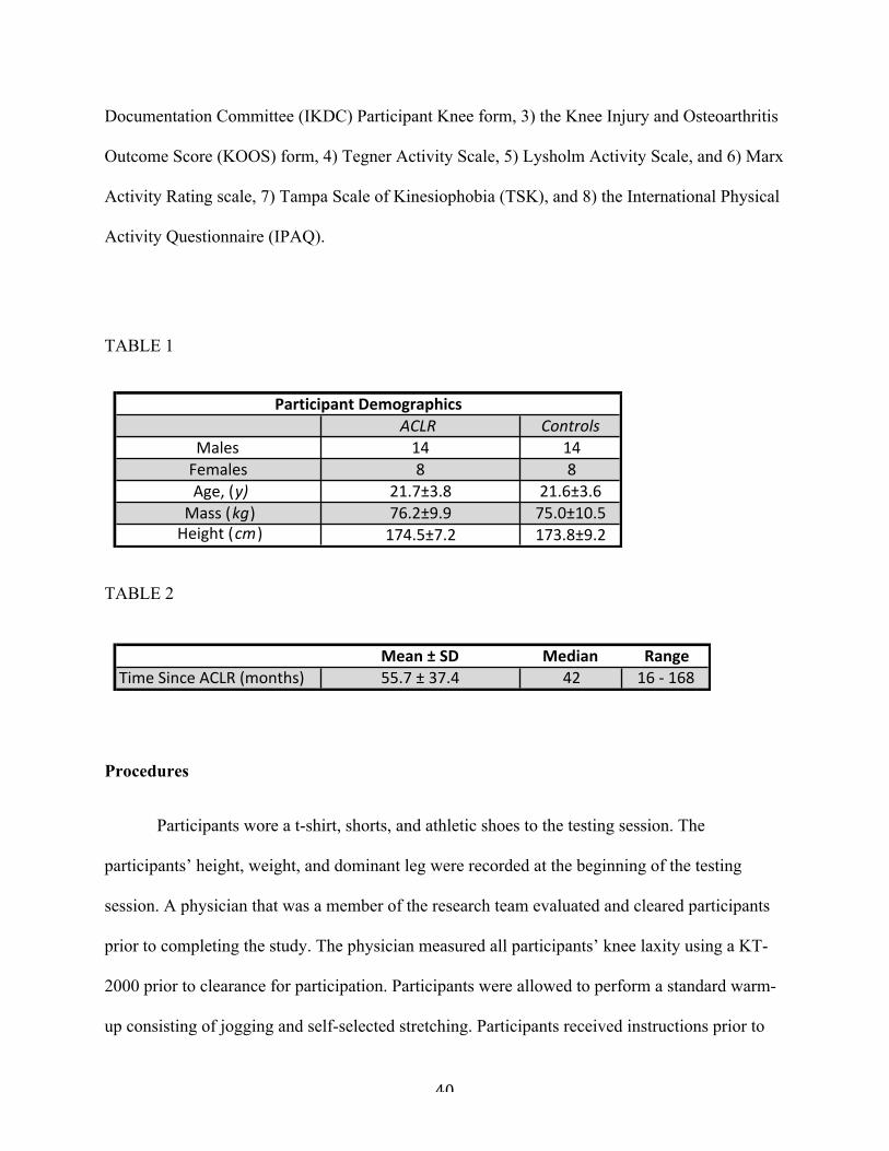
40
Documentation Committee (IKDC) Participant Knee form, 3) the Knee Injury and Osteoarthritis
Outcome Score (KOOS) form, 4) Tegner Activity Scale, 5) Lysholm Activity Scale, and 6) Marx
Activity Rating scale, 7) Tampa Scale of Kinesiophobia (TSK), and 8) the International Physical
Activity Questionnaire (IPAQ).
TABLE 1
TABLE 2
Procedures
Participants wore a t-shirt, shorts, and athletic shoes to the testing session. The
participants’ height, weight, and dominant leg were recorded at the beginning of the testing
session. A physician that was a member of the research team evaluated and cleared participants
prior to completing the study. The physician measured all participants’ knee laxity using a KT-
2000 prior to clearance for participation. Participants were allowed to perform a standard warm-
up consisting of jogging and self-selected stretching. Participants received instructions prior to
ACLR% ControlsMales 14 14Females 8 8Age,.(y) 21.7±3.8 21.6±3.6Mass.(kg) 76.2±9.9 75.0±10.5Height.(cm) 174.5±7.2 173.8±9.2
Participant)Demographics
Mean%±%SD Median RangeTime%Since%ACLR%(months) 55.7%±%37.4 42 16%<%168
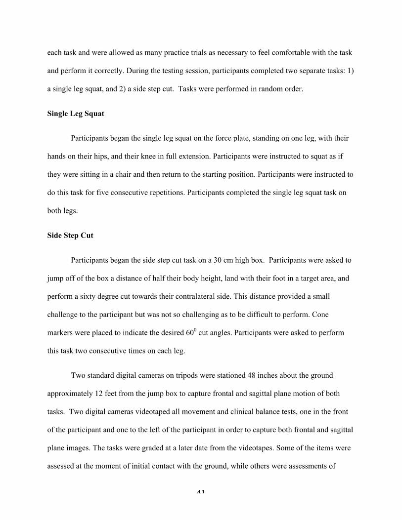
41
each task and were allowed as many practice trials as necessary to feel comfortable with the task
and perform it correctly. During the testing session, participants completed two separate tasks: 1)
a single leg squat, and 2) a side step cut. Tasks were performed in random order.
Single Leg Squat
Participants began the single leg squat on the force plate, standing on one leg, with their
hands on their hips, and their knee in full extension. Participants were instructed to squat as if
they were sitting in a chair and then return to the starting position. Participants were instructed to
do this task for five consecutive repetitions. Participants completed the single leg squat task on
both legs.
Side Step Cut
Participants began the side step cut task on a 30 cm high box. Participants were asked to
jump off of the box a distance of half their body height, land with their foot in a target area, and
perform a sixty degree cut towards their contralateral side. This distance provided a small
challenge to the participant but was not so challenging as to be difficult to perform. Cone
markers were placed to indicate the desired 600 cut angles. Participants were asked to perform
this task two consecutive times on each leg.
Two standard digital cameras on tripods were stationed 48 inches about the ground
approximately 12 feet from the jump box to capture frontal and sagittal plane motion of both
tasks. Two digital cameras videotaped all movement and clinical balance tests, one in the front
of the participant and one to the left of the participant in order to capture both frontal and sagittal
plane images. The tasks were graded at a later date from the videotapes. Some of the items were
assessed at the moment of initial contact with the ground, while others were assessments of

42
motion in the few seconds following initial ground contact. These included: 1) trunk, hip, and
knee flexion sagittal plane motion (peak angle and displacement) 2) trunk, hip, and knee frontal
plane motion (peak angle and displacement) 3) trunk, hip, knee, and foot transverse plane motion
(peak angle and displacement) and overall impression of squat and cutting “quality.” All of
these items can be reasonably expected to be associated with an increased force on the ACL.
Instrumentation
An electromagnetic motion analysis system Trackstar; Ascension Technologies, Inc.,
Burlington, VT) synchronized with a non-conductive force plate (model 4060-NC; Bertec
Corporation, Columbus, OH) collected three-dimensional lower extremity kinematics and
kinetics at sampling frequencies of 144 Hz and 1440 Hz, respectively, during the movement
tests. Six electromagnetic sensors were placed and secured with double-sided tape bilaterally on
the anteromedial tibia, lateral thigh, and on the sacrum and thorax. Eight bony landmarks were
digitized to determine joint centers1 using a stylus with a seventh sensor attached: medial and
lateral malleoli, medial and lateral femoral epicondyles, bilateral anterior superior iliac spines,
T12/L1 spinous process, and C7 spinous process. Three-dimensional coordinates of the lower
extremity and trunk were estimated using MotionMonitor software (Innovative Sports
Technology, Chicago, IL). Euler angles calculated joint angles of the knee, hip and trunk. All
kinematic data was filtered with a fourth-order low-pass Butterworth filter at 14.5 Hz. Kinematic
and kinetic data were reduced using a customized software program to determine joint angles at
initial contact, peak and minimum joint angles, and peak and minimum joint kinetics.
Data Reduction and Analyses

43
The averages across the trials were calculated for peak, minimum, and displacement for
kinematic and kinetic variables associated with the movement tests. Three dimensional peak
trunk, hip, and knee angles were determined during the stance phase of the sidestep cutting task.
The stance phase was defined as the time period between initial contact with the force plate until
takeoff for the rebound jump. Initial contact was defined as when vertical ground reaction force
exceeds 10 N. Takeoff was identified as the time when vertical ground reaction force drops
below 10 N. Peak vertical ground reaction force and joint moments for trunk flexion-extension,
trunk lateral flexion, trunk rotation, hip flexion-extension, hip abduction-adduction, hip internal-
external rotation, knee flexion-extension, knee valgus-varus, and knee internal-external rotation,
and medial knee displacement were also determined during the stance phase. The peak vertical
ground reaction force was normalized to body weight (N) for each participant’s (% body
weight).
The limb with the most recent ACL reconstruction was used as the testing limb in the
ACLR group. Healthy control limbs were then determined based on the limb dominance of the
ACLR limb. Limb dominance was determined by what foot the participant felt they could kick a
ball farthest with. The association between tasks was analyzed using Pearson correlation
coefficients. P level was set at a priori of < 0.05 for all variables. Independent sample T-tests
were then used to evaluate any significance between groups. All data were analyzed using SPSS
(version 21.0, SPSS Inc, Chicago, Illinois).
1. Bell AL, Pedersen DR, Brand RA. A comparison of the accuracy of several hip center
location prediction methods. J Biomech. 1990;23(6):617–621.

44
RESULTS
We observed numerous correlations for dependent variables during the single leg squat
and cutting task (P<0.05) (TABLE 10-11). We also observed several significant differences
between the healthy and ACLR groups during both tasks (TABLE 3-8). During the single leg
squat, the ACLR group displayed less sagittal plane motion, specifically: peak hip flexion
(P=0.02), hip flexion displacement (P=0.04), knee flexion displacement (P=0.01), and trunk
flexion displacement (P=0.03). During the cutting task, participants in the ACLR group
possessed lower VGRF (P<0.001), less knee (P=0.03) and hip (P=0.04) flexion at initial contact,
as well as less peak hip (P=0.03) and trunk (P=0.04) flexion (FIGURE 6-8).
45
DISCUSSION
The findings of this study indicate that single leg squat (SLS) kinematics are highly
correlated with movements during side step cutting (SSC) in both healthy individuals and
individuals with a history of ACLR, which support our hypotheses (TABLE 9). Evaluating
movement patterns during a SLS may enable clinicians to predict movement pattern dysfunctions
during more dynamic, sport-specific movements. Utilizing the SLS as a clinical assessment tool
to evaluate injury risk may enhance injury prevention and rehabilitation efforts.
Medial motion of the knee joint, as measured by two-dimensional medial knee
displacement (MKD) and three-dimensional knee valgus are often implicated during lower
extremity injury mechanisms7,34,35 and, consequently, are a focus of clinical evaluations. Knee
valgus motion and torque also predict future ACL injury risk. 36 This motion may be a movement
compensation that leads to abnormal loading throughout the hip and knee joints as it likely
results from a combination of hip adduction, hip internal rotation, tibial abduction, tibial internal
rotation, and ankle eversion. 35 Our findings demonstrate that participants who perform a cutting
task with excessive MKD, or knee valgus, also demonstrate these motions during a single leg
squat. This combination of uncontrolled movement at the hip, knee, and ankle may increase
future injury risk. 7,12 Mauntel et al. 37 found that participants who had MKD during a SLS
utilized the hip adductor muscles to a greater extent and possessed limited passive ankle
dorsiflexion compared to those who did not display MKD. 37 Padua et al. 34 reported similar
findings during a double leg squat. Inactivation or weakness of the gluteus medius results in
adduction and internal rotation at the hip and increased MKD. 26 Addressing the gluteus/adductor
activation ratio through gluteal strengthening and adductor inactivation as well as increasing
dorsiflexion range of motion should be emphasized during rehabilitation and injury prevention.

46
Focusing on these issues may allow for reduced MKD during SLS, which may translate to
biomechanics during dynamic cutting tasks and reduce injury risk. 37
In addition to MKD, other movements during the SLS (ie. hip adduction, lateral trunk
flexion, and trunk, hip, and knee rotation displacement) also predicted MKD during the cutting
task. These variables were also correlated between the SLS and SSC. Poor hip and trunk
neuromuscular control may result in excessive knee joint loading. Hip adduction may be an
important clinical variable to evaluate during a SLS as it has been shown during a jump landing
to predict future ACL injury risk. 38 Trunk rotation away from the direction of the cut, hip
internal rotation, and knee internal rotation have also been shown to be predictors of ACL
loading in previous research. 18,20,27,29,39 Trunk control has recently been more extensively studied
on its role in ACL injury. 20,40,41 Limiting rotation at the trunk may inhibit the “turn-key”
mechanism described by Frank et al. 40 that exaggerates knee transverse plane motion seen
during higher intensity movements. Limiting trunk rotation and lateral flexion, as well as
emphasizing moderate trunk flexion during the squat may be a critical aspect of ACL injury
prevention. This movement pattern should then be conveyed into more dynamic movement
cutting progressions with an emphasis placed on teaching athletes to cut with their trunk facing
their new direction of travel.
Hip and knee sagittal plane motion during the SLS was only moderately correlated with
motion during the cutting task in the ACLR group, but not in the HC group. These findings were
in contrast to our hypotheses, and we believe may be a result of the generally limited amount of
sagittal plane motion utilized in a cutting task compared to the SLS. On average, our participants
performed the SSC task with approximately 50-60 degrees less knee flexion and 20-30 degrees
less hip flexion compared to the SLS. We may have only observed the moderate correlation for

47
the ACLR group because these participants completed the SLS with significantly less hip and
knee flexion compared to the healthy group with relatively more variability. This inherent
variability within the ACLR group may positively influence the ability to see the relationship
between flexion utilized between the two tasks. We believe evaluating sagittal plane motion
during a SLS may still be clinically important for both healthy and ACLR populations since it
may represent limitations in other neuromuscular factors, such as quadriceps strength and ankle
dorsiflexion motion37,42, but this motion appears unrelated to motion that occurs during a cutting
task. In contrast, to our knowledge, no research has implicated sagittal plane motion or forces
during a cutting task with injury risk. Instead of absorbing force through flexion, individuals may
rely on frontal and transverse plane motion to change direction during cutting tasks and future
research should further evaluate the role of sagittal plane motion during sidestep cutting on
injury risk.
Our ACLR population was very diverse; participants ranging from 16 months to 14 years
since their reconstruction. Though our ACLR group had all been cleared and were active in
sports requiring landing and change of direction, we observed significant group difference
between the ACLR and HC groups in each task. These group differences may be influential
factors that contribute to the high rate of ACL reinjury. 6 The ACLR group displayed
significantly less peak sagittal plane motion and sagittal plane displacement in both tasks and at
initial contact during the cutting task. Previous research has shown that increased landing
stiffness, characterized by limited trunk, hip, and knee flexion place individuals at risk for ACL
injury. 12,43-45 Lack of sagittal plane motion in the ACLR group shows that the ACLR particpants
were not absorbing force during their cutting task. However, we found that the ACLR group had
less normalized VGRF during the cut compared to the HC group. This is especially alarming

48
because the ACLR group chose to cut with less effort and still were unable absorb this reduced
force using sagittal plane motion compared to the healthy controls. Our VGRF results are
similar to those published by Miranda et al. 17 and Paterno et al. 46 Miranda et al. reported
reduced VGRFs during a cutting task in their ACLR group compared to healthy controls and
Paterno et al. reported reduced VGRFs in an ACLR participants’ injured limb compared to their
uninjured limb. History of ACLR seems to be continuously showing that athletes, regardless of
time from surgery, are unable to cut and absorb forces through sagittal plane motion on their
injured limb. ACLR athletes may be choosing to absorb force in other planes of motion, which
we saw in our results. Quadriceps strength impairment is a consequence after ACL injury, 47,48
most likely due to arthogenic muscular inhibition seen after ACLR. 49 Clinicians need to assess
side-to-side asymmetrical force production and absorption differences in their athletes before
making return to play decisions. We also observed trunk rotational differences between groups.
Participants with a history of ACLR rotated their trunk away from the direction of the cut
compared to the healthy controls. Trunk rotation away from the direction of the cut has been
shown to further increase frontal plane knee loading, which may increase ACL injury risk. 40
These group differences reiterate previous research showing that individuals with ACLR history
continue to possess modifiable differences regardless of time from surgery. 50
Currently, there are few objective criteria to determine when ACLR patients can safely
advance to cutting and pivoting progressions during rehabilitation and return to play. 23 Examples
of objective measures used are: circumferential thigh girth measurement, goniometry, functional
performance hop tests, and isokinetic tests. There has also been a push in sports medicine for
patient oriented outcomes to capture their health-related quality of life after injury. The Visual
Analog Scale (VAS) and the International Knee Documentation Committee Subjective Form
49
(IKDC) have been used to determine subjective outcomes after knee injury. 21-23 The consensus
is a patient can return to sport once they have gained full knee range of motion, their hop tests
are over 85% compared to the healthy side, their strength ratio of the quadriceps and hamstrings
are over 85% compared to the healthy side, and the athlete has been cleared by their physician.21-
23
The aforementioned assessments address knee pain, function, force generation, and
sagittal plane strength; however, they fail to address the multi-planar motion involved in cutting,
one of the proposed mechanisms of ACL (re)injury. 2,12,51 Allowing patients to progress to late
stage rehabilitative movements based on a time criteria and not neuromuscular strength and
postural control, fails to take into account the individuality of ACL rehabilitation. Each patient
rehabilitates and gains functional control at a different rate; therefore patients need an
individualized strength and control assessment during their return to play progression. Our
findings have determined that there is no longer a need for a cutting screening assessment.
Clinicians can use real time or two-dimensional video analysis of a SLS and can accurately
predict movement biomechanics during a cut.
Understanding that athletes who perform a SLS with increased MKD, lateral trunk
flexion, hip adduction/abduction, and trunk/hip rotation are at risk for increased ACL loading
can allow clinicians to screen athletes who do not possess the neuromuscular control to correctly
perform a cutting task, reducing the high rate of ACL reinjury. 6,7 Reducing injury risk during
rehabilitation of ACL reconstruction or correcting neuromuscular deficiencies through injury
prevention is a goal that all clinicians, coaches, and athletes share. The SLS should be utilized as
an assessment as well as a strengthening, balance, and control exercise during injury prevention,
especially in athletes rehabilitating from ACLR. Several studies have found that neuromuscular

50
and strength training have improved performance as well as landing biomechanics. 45,52,53 The
three planar control that the SLS requires properly prepares athletes for a cutting return to sport
progression.
Our study simply shows that the way an individual, with or without a history of lower
extremity surgery, controls their movement during a single leg squat indicates their control
during a cutting task. Our study does not indicate whether improvement in single leg squat
control would result in improvements in cutting mechanics. Further research should therefore
evaluate if improvement in dynamic control and range of motion during the single leg squat also
improves control and motion during cutting tasks in both a healthy and ACLR population. This
study’s implications to sport participation are also limited because we used an anticipated cutting
task, which allowed us to see how our participants’ chose to move and allowed us to better
standardize the task. Houck et al. found that frontal plane knee moments were significantly
higher during an unanticipated cutting task when compared to an anticipated task. 54 The loads
placed on our participants were therefore most likely less than the loads required during an
unanticipated or spontaneous cut that they face during normal sport participation because they
were able to plan their movement. There is also limited research comparing movement strategies
between individuals with a history of ACLR and matched controls during a single leg squat.
Further studies need to continue exploring differences in SLS, cutting, and landing strategies
between healthy and ACLR groups.
Conclusion
Readiness to return to cutting during return to sport participation is a clinical decision that
needs to made with objective criteria. Currently, there are insufficient guidelines for screening

51
whether ACLR patients have the proper neuromuscular control needed to safely return to cutting
progressions. Increased dynamic movement such as cutting and pivoting puts athletes at a higher
risk for injury or reinjury6; evaluating and predicting movement can enable clinicians to limit
injury risk. Our results suggest that lack of frontal and transverse plane trunk, hip, and knee
control during the SLS resulted in positions of increased lateral trunk flexion, hip adduction, and
MKD during a cutting task; which have all been shown to be significant predictors of ACL
loading during dynamic cutting tasks. 19,20,36,54 Utilizing the SLS as a screening tool for injury
risk can help clinicians make rehabilitative progression decisions based on objective criteria.
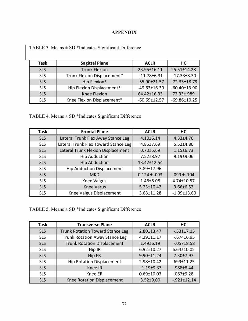
52
APPENDIX
TABLE 3. Means ± SD *Indicates Significant Difference
TABLE 4. Means ± SD *Indicates Significant Difference
TABLE 5. Means ± SD *Indicates Significant Difference
Task Sagittal*Plane ACLR HCSLS Trunk(Flexion 23.95±16.11 25.51±14.28SLS Trunk(Flexion(Displacement* (A11.78±6.31 (A17.33±8.30SLS Hip(Flexion* (A55.90±21.57 (A72.33±18.79SLS Hip(Flexion(Displacement* (A49.63±16.30 (A60.40±13.90SLS Knee(Flexion 64.42±16.33 72.33±.989SLS Knee(Flexion(Displacement* (A60.69±12.57 (A69.86±10.25
Task Frontal+Plane ACLR HCSLS Lateral(Trunk(Flex(Away(Stance(Leg 4.10±6.14 4.33±4.76SLS Lateral(Trunk(Flex(Toward(Stance(Leg 4.85±7.69 5.52±4.80SLS Lateral(Trunk(Flexion(Displacement 0.70±5.69 1.15±6.73SLS Hip(Adduction 7.52±8.97 9.19±9.06SLS Hip(Abduction 13.42±12.54SLS Hip(Adduction(Displacement 5.89±17.96SLS MKD 0.124(±(.093 .099(±(.104SLS Knee(Valgus 1.46±8.08 4.74±10.57SLS Knee(Varus 5.23±10.42 3.66±6.52SLS Knee(Valgus(Displacement 3.68±11.28 (L1.09±13.60
Task Transverse)Plane ACLR HCSLS Trunk(Rotation(Toward(Stance(Leg 2.80±13.47 (<.531±7.15SLS Trunk(Rotation(Away(Stance(Leg 4.29±11.17 (<.674±6.95SLS Trunk(Rotation(Displacement 1.49±6.19 (<.057±8.58SLS Hip(IR 6.92±10.27 6.64±10.05SLS Hip(ER 9.90±11.24 7.30±7.97SLS Hip(Rotation(Displacement 2.98±10.42 .699±11.25SLS Knee(IR (<1.19±9.33 .988±8.44SLS Knee(ER 0.69±10.03 .067±9.28SLS Knee(Rotation(Displacement 3.52±9.00 (<.921±12.14

53
TABLE 6. Means ± SD *Indicates Significant Difference
TABLE 7. Means ± SD *Indicates Significant Difference
Task Sagittal*Plane ACLR HCCut VGRF* 2.23±.34 2.88±0.54Cut Trunk5Flexion 23.27±7.50 25.97±12.73Cut Trunk5Flexion5Disp 11.71±5.00 11.82±5.67Cut Trunk5Flexion5IC 12.73±7.77 15.96±10.02Cut HipFlexion5IC* 5D24.96±13.23 5D32.61±10.22Cut Hip5Flexion5Disp 10.20±4.70 12.92±6.23Cut Hip5Flexion* 5D29.38±16.39 5D39.30±11.58Cut Knee5Flexion5IC* 11.45±11.52 17.70±8.70Cut Knee5Flexion 43.64±14.55 47.78±11.61Cut Knee5Flexion5Disp 32.95±8.78 33.07±11.18
Task Frontal+Plane ACLR HCCut Lateral)Trunk)Flex)Away)Stance)Leg 3.85±7.20 5.09±8.10Cut Lateral)Trunk)Flexion)Displacement 9.02±5.83 11.37±4.86Cut Lateral)Trunk)Flex)Toward)Stance)Leg 6.88±8.71 5.59±9.68Cut Lateral)Trunk)Flexion)IC 1.09±7.24 .484±6.79Cut Hip)Adduction)IC )J5.81±7.57 )J9.71±7.82Cut Hip)Adduction )J.2.91±8.70 )J8.69±8.60Cut Hip)Adduction)Displacement 11.87±4.65 10.59±6.08Cut Hip)Abduction 0.10±11.03 )J6.05±9.78Cut MKD .122)±).117 .100)±).109Cut Knee)Valgus)IC )J2.30±5.93 )J1.32±5.10Cut Knee)Valgus )J1.44±8.30 )J1.44±7.38Cut Knee)Valgus)Displacement 6.75±5.35 7.06±3.21Cut Knee)Varus )J1.64±7.74 )J1.34±7.26

54
TABLE 8. Means ± SD *Indicates Significant Difference
Task Sagittal*Plane ACLR HCCut VGRF* 2.23±.34 2.88±0.54Cut Trunk5Flexion 23.27±7.50 25.97±12.73Cut Trunk5Flexion5Disp 11.71±5.00 11.82±5.67Cut Trunk5Flexion5IC 12.73±7.77 15.96±10.02Cut HipFlexion5IC* 5D24.96±13.23 5D32.61±10.22Cut Hip5Flexion5Disp 10.20±4.70 12.92±6.23Cut Hip5Flexion* 5D29.38±16.39 5D39.30±11.58Cut Knee5Flexion5IC* 11.45±11.52 17.70±8.70Cut Knee5Flexion 43.64±14.55 47.78±11.61Cut Knee5Flexion5Disp 32.95±8.78 33.07±11.18
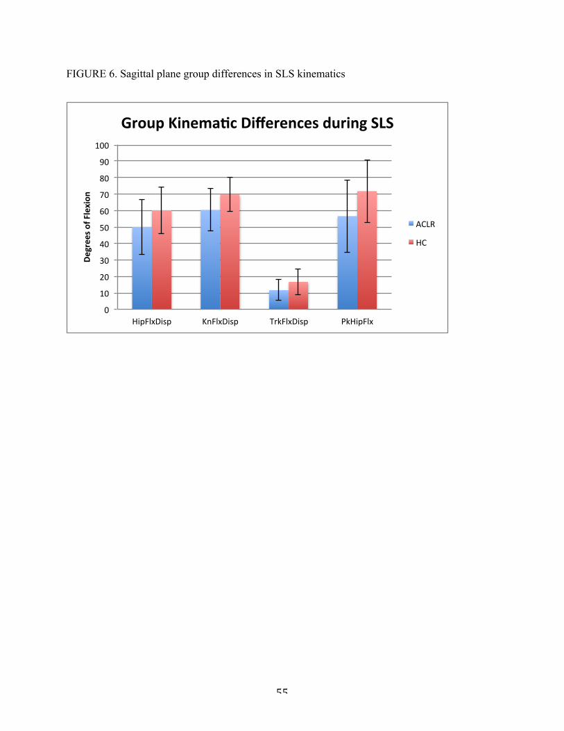
55
FIGURE 6. Sagittal plane group differences in SLS kinematics
0"
10"
20"
30"
40"
50"
60"
70"
80"
90"
100"
HipFlxDisp" KnFlxDisp" TrkFlxDisp" PkHipFlx"
Degrees&o
f&Flexion
&
Group&Kinema4c&Differences&during&SLS&
ACLR"
HC"

56
FIGURE 7. Sagittal and transverse plane group differences in SSC kinematics
!30$
!20$
!10$
0$
10$
20$
30$
40$
50$
60$
70$
PkTrkRot$ PkHipFlx$ KnFlxIC$ HipFlxIC$
Degrees&o
f&Mo*
on&
Group&Kinema*c&Differences&during&SSC&
ACLR$
HC$
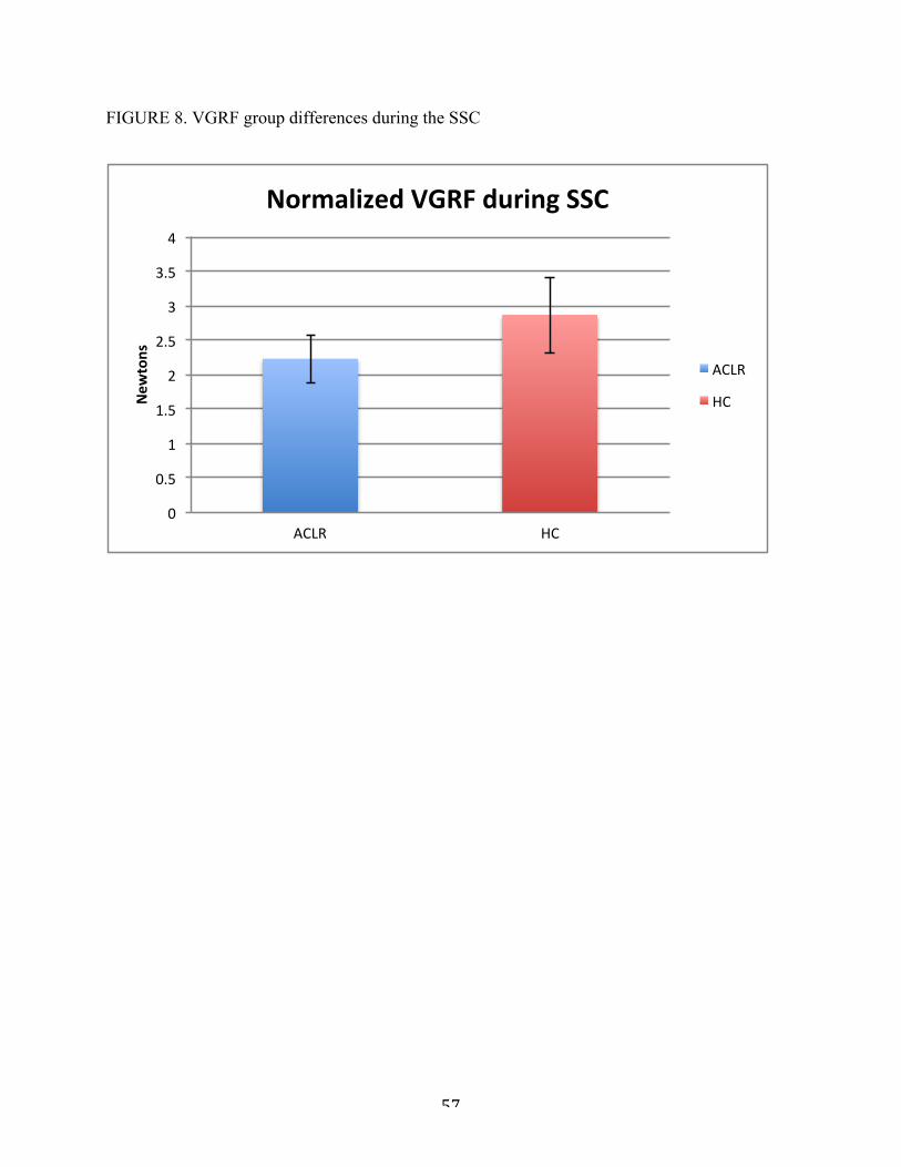
57
FIGURE 8. VGRF group differences during the SSC
0"
0.5"
1"
1.5"
2"
2.5"
3"
3.5"
4"
ACLR" HC"
New
tons(
Normalized(VGRF(during(SSC(
ACLR"
HC"

58
TABLE 9. Significant research question correlations between tasks
Cut$Variable SLS$Variable R/value P/value Cut$Variable SLS$Variable R/value P/value
2g MKD MKD 0.89 <.001 1g MKD MKD 0.89 <.0012i Hip-ER Hip-ER 0.80 <.001 1j Knee-ER Knee-ER 0.79 <.001
2i Hip-IR Hip-IR 0.71 <.001 1j Knee-IR Knee-IR 0.75 <.001
2d Knee-Flexion Knee-Flexion 0.63 0.002 1i Hip-IR Hip-IR 0.61 0.002
2hTrunk-Rot-
Away-Stance-Leg
Trunk-Rot-Away-Stance-
Leg0.60 0.005 1h
Trunk-Rot-Away-Stance-
Leg
Trunk-Rot-Away-Stance-
Leg0.58 0.004
2bTrunk-Flexion
Trunk-Flexion 0.60 0.007 1h
Trunk-Rot-Toward-
Stance-Leg
Trunk-Rot-Toward-
Stance-Leg0.51 0.014
2j Knee-ER Knee-ER 0.59 0.006 1f Hip-Adduction
Hip-Adduction
0.59 0.003
2f Hip-Adduction
Hip-Adduction
0.56 0.01 1i Hip-ER Hip-ER 0.53 0.011
2j Knee-IR Knee-IR 0.52 0.02 1g Knee-Valgus Knee-Valgus 0.66 0.001
2c Hip-Flexion Hip-Flexion 0.52 0.02
2a VGRF Knee-Flexion 0.44 0.04 1eLat-Trunk-Flex-Away-Stance-Leg
Lat-Trunk-Flex-Away-Stance-Leg
0.46 0.027
Weak/Correlations
Research$Question
Research$Question
ACLR Healthy,Controls
Strong/Correlations
Moderate/Correlations

59
TABLE 10.
SLS Cut R'Value P'Value
Hip$Rotation$Disp Trunk$Rot$Toward$Stance$Leg 0.795 <.001Knee$ER Knee$ER 0.79 <.001
Knee$Rotation$Disp Trunk$Rot$Toward$Stance$Leg 0.766 <.001Knee$IR Knee$IR 0.745 <.001Hip$IR Knee$ER C0.739 <.001
MKD MKD 0.89 <.001Knee$Valgus$Disp MKD 0.877 <.001Hip$Adduction$Disp MKD 0.875 <.001
Trunk$Lateral$Flexion$Disp MKD 0.856 <.001Hip$Abduction MKD 0.816 <.001Knee$Valgus MKD C0.717 <.001
Hip$Rotation$Disp MKD 0.918 <.001Knee$Rotation$Disp MKD 0.882 <.001Trunk$Rotation$Disp Trunk$Rot$Toward$Stance$Leg$Disp 0.831 <.001Trunk$Rotation$Disp MKD 0.823 <.001Hip$Adduction$Disp Trunk$Rot$Toward$Stance$Leg 0.82 <.001
MKD Trunk$Rot$Toward$Stance$Leg 0.804 <.001Trunk$Lateral$Flexion$Disp Trunk$Rot$Toward$Stance$Leg 0.787 <.001
Knee$Valgus$Disp Trunk$Rot$Toward$Stance$Leg 0.774 <.001Hip$Abduction Trunk$Rot$Toward$Stance$Leg 0.759 <.001
Hip$IR Knee$Valgus 0.702 <.001Hip$Rotation$Disp Hip$Adduction C0.716 <.001
Strong'Correlations'of'SLS'to'SSC'Kinematics'in'Healthy'Controls
Strong'Transverse'Plane'Correlations
Strong'Frontal'Plane'Correlations
Strong'Multiplanar'Correlations

60
TABLE 11.
SLS Cut R'Value P'Value
Hip$ER Hip$ER 0.8 <.001Trunk$Rot$Toward$Stance$Leg Trunk$Rotation$IC 0.77 <.001
Hip$IR Hip$IR 0.71 <.001Hip$Rotation$Disp Trunk$Rot$Away$Stance$Leg B0.72 0.001Trunk$Rotation$Disp Trunk$Rot$Away$Stance$Leg B0.79 <.001
MKD MKD 0.894 <.001Hip$Abduction Hip$Abduction 0.72 <.001Knee$Varus Knee$Varus 0.71 <.001
MKD Hip$Abduction 0.7 0.001Lateral$Trunk$Flex$Toward$Stance$Leg Lateral$Trunk$Flex$Toward$Stance$Leg 0.7 <.001
Hip$Adduction Lateral$Trunk$Flex$Toward$Stance$Leg B0.702 0.001
Hip$Abduction Trunk$Rot$Away$Stance$Leg B0.747 <.001Hip$Adduction$Disp Trunk$Rot$Away$Stance$Leg B0.766 <.001
MKD Trunk$Rot$Away$Stance$Leg B0.794 <.001
Strong'Transverse'Plane'Correlations
Strong'Frontal'Plane'Correlations
Strong'Multiplanar'Correlations
Strong'Correlations'of'SLS'to'SSC'Kinematics'in'Individuals'with'a'History'of'ACLR

6
REFERENCES
1. Marshall, S.W., D.A. Padua, and M.L. McGrath, Incidence of ACL Injury, in Understanding and Preventing Noncontact ACL Injuries / American Orthopaedic Society for Sports Medicine, T.E. Hewett, S.J. Shultz, and L.Y. Griffin, Editors. 2007, Human Kinetics: Champaign, IL.
2. Shimokochi Y, Shultz SJ. Mechanisms of noncontact anterior cruciate ligament injury. J Athl Train. 2008;43(4):396-408.
3. Brophy RH, Wright RW, Matava MJ. Cost analysis of converting from single-bundle to double-bundle anterior cruciate ligament reconstruction. Am J Sports Med. 2009;37(4):683-687.
4. Hewett TE, Ford KR, Hoogenboom BJ, Myer GD. Understanding and preventing acl injuries: Current biomechanical and epidemiologic considerations - update 2010. N Am J Sports Phys Ther. 2010;5(4):234-251.
5. Wright RW, Dunn WR, Amendola A, et al. Risk of tearing the intact anterior cruciate ligament in the contralateral knee and rupturing the anterior cruciate ligament graft during the first 2 years after anterior cruciate ligament reconstruction: A prospective MOON cohort study. Am J Sports Med. 2007;35(7):1131-1134.
6. Paterno MV, Rauh MJ, Schmitt LC, Ford KR, Hewett TE. Incidence of contralateral and ipsilateral anterior cruciate ligament (ACL) injury after primary ACL reconstruction and return to sport. Clin J Sport Med. 2012;22(2):116-121.
7. Salmon L, Russell V, Musgrove T, Pinczewski L, Refshauge K. Incidence and risk factors for graft rupture and contralateral rupture after anterior cruciate ligament reconstruction. Arthroscopy. 2005;21(8):948-957.
8. Leys T, Salmon L, Waller A, Linklater J, Pinczewski L. Clinical results and risk factors for reinjury 15 years after anterior cruciate ligament reconstruction: A prospective study of hamstring and patellar tendon grafts. Am J Sports Med. 2012;40(3):595-605.
9. Paterno MV, Schmitt LC, Ford KR, et al. Biomechanical measures during landing and postural stability predict second anterior cruciate ligament injury after anterior cruciate ligament reconstruction and return to sport. Am J Sports Med. 2010;38(10):1968-1978.
10. Lohmander LS, Ostenberg A, Englund M, Roos H. High prevalence of knee osteoarthritis, pain, and functional limitations in female soccer players twelve years after anterior cruciate ligament injury. Arthritis Rheum. 2004;50(10):3145-3152.
11. Ahlden M, Samuelsson K, Sernert N, Forssblad M, Karlsson J, Kartus J. The swedish national anterior cruciate ligament register: A report on baseline variables and outcomes of surgery for almost 18,000 patients. Am J Sports Med. 2012;40(10):2230-2235.

7
12. Boden BP, Dean GS, Feagin JA,Jr, Garrett WE,Jr. Mechanisms of anterior cruciate ligament injury. Orthopedics. 2000;23(6):573-578.
13. Arendt EA, Agel J, Dick R. Anterior cruciate ligament injury patterns among collegiate men and women. J Athl Train. 1999;34(2):86-92.
14. Myklebust G, Maehlum S, Engebretsen L, Strand T, Solheim E. Registration of cruciate ligament injuries in norwegian top level team handball. A prospective study covering two seasons. Scand J Med Sci Sports. 1997;7(5):289-292.
15. Hewett TE, Torg JS, Boden BP. Video analysis of trunk and knee motion during non-contact anterior cruciate ligament injury in female athletes: Lateral trunk and knee abduction motion are combined components of the injury mechanism. Br J Sports Med. 2009;43(6):417-422.
16. Stearns KM, Pollard CD. Abnormal frontal plane knee mechanics during sidestep cutting in female soccer athletes after anterior cruciate ligament reconstruction and return to sport. Am J Sports Med. 2013.
17. Miranda DL, Fadale PD, Hulstyn MJ, Shalvoy RM, Machan JT, Fleming BC. Knee biomechanics during a jump-cut maneuver: Effects of gender and ACL surgery. Med Sci Sports Exerc. 2012.
18. Pollard CD, Sigward SM, Powers CM. Gender differences in hip joint kinematics and kinetics during side-step cutting maneuver. Clin J Sport Med. 2007;17(1):38-42.
19. Kristianslund E, Faul O, Bahr R, Myklebust G, Krosshaug T. Sidestep cutting technique and knee abduction loading: Implications for ACL prevention exercises. Br J Sports Med. 2012.
20. Jamison ST, Pan X, Chaudhari AM. Knee moments during run-to-cut maneuvers are associated with lateral trunk positioning. J Biomech. 2012;45(11):1881-1885.
21. Myer GD, Paterno MV, Ford KR, Quatman CE, Hewett TE. Rehabilitation after anterior cruciate ligament reconstruction: Criteria-based progression through the return-to-sport phase. J Orthop Sports Phys Ther. 2006;36(6):385-402.
22. van Grinsven S, van Cingel RE, Holla CJ, van Loon CJ. Evidence-based rehabilitation following anterior cruciate ligament reconstruction. Knee Surg Sports Traumatol Arthrosc. 2010;18(8):1128-1144.
23. Barber-Westin SD, Noyes FR. Factors used to determine return to unrestricted sports activities after anterior cruciate ligament reconstruction. Arthroscopy. 2011;27(12):1697-1705.
24. Weeks BK, Carty CP, Horan SA. Kinematic predictors of single-leg squat performance: A comparison of experienced physiotherapists and student physiotherapists. BMC Musculoskelet Disord. 2012;13:207-2474-13-207.

8
25. Stensrud S, Myklebust G, Kristianslund E, Bahr R, Krosshaug T. Correlation between two-dimensional video analysis and subjective assessment in evaluating knee control among elite female team handball players. Br J Sports Med. 2011;45(7):589-595.
26. Crossley KM, Zhang WJ, Schache AG, Bryant A, Cowan SM. Performance on the single-leg squat task indicates hip abductor muscle function. Am J Sports Med. 2011;39(4):866-873.
27. Imwalle LE, Myer GD, Ford KR, Hewett TE. Relationship between hip and knee kinematics in athletic women during cutting maneuvers: A possible link to noncontact anterior cruciate ligament injury and prevention. J Strength Cond Res. 2009;23(8):2223-2230.
28. Malinzak RA, Colby SM, Kirkendall DT, Yu B, Garrett WE. A comparison of knee joint motion patterns between men and women in selected athletic tasks. Clin Biomech (Bristol, Avon). 2001;16(5):438-445.
29. McLean SG, Huang X, van den Bogert AJ. Association between lower extremity posture at contact and peak knee valgus moment during sidestepping: Implications for ACL injury. Clin Biomech (Bristol, Avon). 2005;20(8):863-870.
30. Kristianslund E, Krosshaug T. Comparison of drop jumps and sport-specific sidestep cutting: Implications for anterior cruciate ligament injury risk screening. Am J Sports Med. 2013;41(3):684-688.
31. Graci V, Van Dillen LR, Salsich GB. Gender differences in trunk, pelvis and lower limb kinematics during a single leg squat. Gait Posture. 2012;36(3):461-466.
32. Willson JD, Ireland ML, Davis I. Core strength and lower extremity alignment during single leg squats. Med Sci Sports Exerc. 2006;38(5):945-952.
33. Zeller BL, McCrory JL, Kibler WB, Uhl TL. Differences in kinematics and electromyographic activity between men and women during the single-legged squat. Am J Sports Med. 2003;31(3):449-456.
34. Padua DA, Bell DR, Clark MA. Neuromuscular characteristics of individuals displaying excessive medial knee displacement. J Athl Train. 2012;47(5):525-536.
35. Hewett TE, Myer GD, Ford KR. Anterior cruciate ligament injuries in female athletes: Part 1, mechanisms and risk factors. Am J Sports Med. 2006;34(2):299-311.
36. Hewett TE, Myer GD, Ford KR, et al. Biomechanical measures of neuromuscular control and valgus loading of the knee predict anterior cruciate ligament injury risk in female athletes: A prospective study. Am J Sports Med. 2005;33(4):492-501.
37. Mauntel TC, Begalle RL, Cram TR, et al. The effects of lower extremity muscle activation and passive range of motion on single leg squat performance. J Strength Cond Res. 2013;27(7):1813-1823.
9
38. Shultz SJ, Schmitz RJ, Benjaminse A, Chaudhari AM, Collins M, Padua DA. ACL research retreat VI: An update on ACL injury risk and prevention. J Athl Train. 2012;47(5):591-603.
39. Frank B, Bell DR, Norcross MF, Blackburn JT, Goerger BM, Padua DA. Trunk and hip biomechanics influence anterior cruciate loading mechanisms in physically active participants. Am J Sports Med. 2013;41(11):2676-2683.
40. Frank B, Bell DR, Norcross MF, Blackburn JT, Goerger BM, Padua DA. Trunk and hip biomechanics influence anterior cruciate loading mechanisms in physically active participants. Am J Sports Med. 2013;41(11):2676-2683.
41. Kulas AS, Hortobagyi T, DeVita P. Trunk position modulates anterior cruciate ligament forces and strains during a single-leg squat. Clin Biomech (Bristol, Avon). 2012;27(1):16-21.
42. Chmielewski TL, Stackhouse S, Axe MJ, Snyder-Mackler L. A prospective analysis of incidence and severity of quadriceps inhibition in a consecutive sample of 100 patients with complete acute anterior cruciate ligament rupture. J Orthop Res. 2004;22(5):925-930.
43. Dai B, Herman D, Liu H, Garrett WE, Yu B. Prevention of ACL injury, part I: Injury characteristics, risk factors, and loading mechanism. Res Sports Med. 2012;20(3-4):180-197.
44. Ireland ML. Anterior cruciate ligament injury in female athletes: Epidemiology. J Athl Train. 1999;34(2):150-154.
45. Myer GD, Ford KR, Palumbo JP, Hewett TE. Neuromuscular training improves performance and lower-extremity biomechanics in female athletes. J Strength Cond Res. 2005;19(1):51-60.
46. Paterno MV, Ford KR, Myer GD, Heyl R, Hewett TE. Limb asymmetries in landing and jumping 2 years following anterior cruciate ligament reconstruction. Clin J Sport Med. 2007;17(4):258-262.
47. Thomas AC, Villwock M, Wojtys EM, Palmieri-Smith RM. Lower extremity muscle strength after anterior cruciate ligament injury and reconstruction. J Athl Train. 2013.
48. Coats-Thomas MS, Miranda DL, Badger GJ, Fleming BC. Effects of ACL reconstruction surgery on muscle activity of the lower limb during a jump-cut maneuver in males and females. J Orthop Res. 2013.
49. Palmieri-Smith RM, Thomas AC. A neuromuscular mechanism of posttraumatic osteoarthritis associated with ACL injury. Exerc Sport Sci Rev. 2009;37(3):147-153.
50. Myer GD, Martin L,Jr, Ford KR, et al. No association of time from surgery with functional deficits in athletes after anterior cruciate ligament reconstruction: Evidence for objective return-to-sport criteria. Am J Sports Med. 2012;40(10):2256-2263.

10
51. Alentorn-Geli E, Myer GD, Silvers HJ, et al. Prevention of non-contact anterior cruciate ligament injuries in soccer players. part 1: Mechanisms of injury and underlying risk factors. Knee Surg Sports Traumatol Arthrosc. 2009;17(7):705-729.
52. Myer GD, Paterno MV, Ford KR, Hewett TE. Neuromuscular training techniques to target deficits before return to sport after anterior cruciate ligament reconstruction. J Strength Cond Res. 2008;22(3):987-1014.
53. Padua DA, Distefano LJ. Sagittal plane knee biomechanics and vertical ground reaction forces are modified following ACL injury prevention programs: A systematic review. - Sports Health.2009 Mar;1(2):165-73. (1941-7381 (Print); 1941-0921 (Linking)).
54. Houck JR, Duncan A, De Haven KE. Comparison of frontal plane trunk kinematics and hip and knee moments during anticipated and unanticipated walking and side step cutting tasks. Gait Posture. 2006;24(3):314-322.
Copyright © 2022 FDOKUMEN