Role of Lipid Peroxidation in Cellular Responses to d , l -Sulforaphane, a Promising Cancer...
30
Role of Lipid Peroxidation in Cellular Responses to D, L- Sulforaphane, A Promising Cancer Chemopreventive Agent † Rajendra Sharma ‡,* , Abha Sharma ‡ , Pankaj Chaudhary ‡ , Virginia Pearce § , Rit Vatsyayan ‡ , Shivendra V. Singh ∥ , Sanjay Awasthi ‡ , and Yogesh C. Awasthi ‡ ‡ Department of Molecular Biology and Immunology, University of North Texas Health Science Center, Fort Worth, TX § Department of Pharmacology and Neuroscience, University of North Texas Health Science Center, Fort Worth, TX ∥ Department of Pharmacology and Chemical Biology, University of Pittsburgh, Pittsburgh, PA Abstract D, L-Sulforaphane (SFN), a synthetic analogue of broccoli-derived L-isomer, is a highly promising cancer chemopreventive agent substantiated by inhibition of chemically-induced cancer in rodents and prevention of cancer development and distant site metastasis in transgenic mouse models of cancer. SFN is also known to inhibit growth of human cancer cells in association with cell cycle arrest and reactive oxygen species-dependent apoptosis, but the mechanism of these cellular responses to SFN exposure is not fully understood. Because 4-hydroxynonenal (4-HNE), a product of lipid peroxidation (LPO), the formation of which is regulated by hGSTA1-1, assumes a pivotal role in oxidative stress-induced signal transduction, the present study investigated its contribution in growth arrest and apoptosis induction by SFN using HL60 and K562 human leukemic cell lines as model. The SFN-induced formation of 4-HNE was suppressed in hGSTA1-1 over expressing cells, which also acquired resistance to SFN-induced cytotoxicity, cell cycle arrest, and apoptosis. While resistance to SFN-induced cell cycle arrest by ectopic expression of hGSTA1-1 was associated with changes in levels of G2/M regulatory proteins, resistance to apoptosis correlated with increased Bcl- xL/Bax ratio, inhibition of nuclear translocation of AIF, and attenuated cytochrome c release in cytosol. The hGSTA1-1 over expressing cells showed enhanced cytoplasmic export of Daxx, nuclear accumulation of transcription factors Nrf2 and HSF1, and up regulation of their respective client proteins, γ-GCS and HSP70. These findings not only reveal a central role of 4-HNE in cellular responses to SFN but also reaffirm that 4-HNE contributes to oxidative stress mediated signaling. D, L-Sulforaphane (SFN), a synthetic analogue of naturally-occurring L-isomer abundant in several cruciferous vegetables (e.g., broccoli), is a potent inhibitor of chemically-induced cancer in experimental rodents (1–5). It has been shown to modulate inflammation, induce apoptosis, cause cell cycle arrest, and inhibit several Phase 1 enzymes that may activate chemical carcinogens (1). Even though the mechanisms of the chemo preventive activities of SFN are not completely understood, it has been suggested that besides inhibiting Phase I enzymes, SFN can also induce Phase 2 detoxification enzymes such as glutathione transferases (GSTs) through transcriptional activation of antioxidant response element (ARE) driven genes regulated by nuclear factor E2-related factor-2 or Nrf2 (1,6–8). Being an electrophile, SFN † Supported in part by NIH Grants ES012171 EY 04396 (YCA), CA77495 (SA, and Supplement to this grant to AS) and CA115498 (SVS). * Address correspondence to: Rajendra Sharma, Ph.D, Research Associate Professor, Department of Molecular Biology and Immunology, University of North Texas Health, Science Center, Fort Worth, TX 76107, Tel: 817-735-2140, Fax: 817-735-2118, [email protected]. NIH Public Access Author Manuscript Biochemistry. Author manuscript; available in PMC 2011 April 13. Published in final edited form as: Biochemistry. 2010 April 13; 49(14): 3191–3202. doi:10.1021/bi100104e. NIH-PA Author Manuscript NIH-PA Author Manuscript NIH-PA Author Manuscript
-
Upload
independent -
Category
Documents
-
view
0 -
download
0
Transcript of Role of Lipid Peroxidation in Cellular Responses to d , l -Sulforaphane, a Promising Cancer...

Role of Lipid Peroxidation in Cellular Responses to D, L-Sulforaphane, A Promising Cancer Chemopreventive Agent†
Rajendra Sharma‡,*, Abha Sharma‡, Pankaj Chaudhary‡, Virginia Pearce§, Rit Vatsyayan‡,Shivendra V. Singh∥, Sanjay Awasthi‡, and Yogesh C. Awasthi‡‡Department of Molecular Biology and Immunology, University of North Texas Health ScienceCenter, Fort Worth, TX§Department of Pharmacology and Neuroscience, University of North Texas Health Science Center,Fort Worth, TX∥Department of Pharmacology and Chemical Biology, University of Pittsburgh, Pittsburgh, PA
AbstractD, L-Sulforaphane (SFN), a synthetic analogue of broccoli-derived L-isomer, is a highly promisingcancer chemopreventive agent substantiated by inhibition of chemically-induced cancer in rodentsand prevention of cancer development and distant site metastasis in transgenic mouse models ofcancer. SFN is also known to inhibit growth of human cancer cells in association with cell cyclearrest and reactive oxygen species-dependent apoptosis, but the mechanism of these cellularresponses to SFN exposure is not fully understood. Because 4-hydroxynonenal (4-HNE), a productof lipid peroxidation (LPO), the formation of which is regulated by hGSTA1-1, assumes a pivotalrole in oxidative stress-induced signal transduction, the present study investigated its contribution ingrowth arrest and apoptosis induction by SFN using HL60 and K562 human leukemic cell lines asmodel. The SFN-induced formation of 4-HNE was suppressed in hGSTA1-1 over expressing cells,which also acquired resistance to SFN-induced cytotoxicity, cell cycle arrest, and apoptosis. Whileresistance to SFN-induced cell cycle arrest by ectopic expression of hGSTA1-1 was associated withchanges in levels of G2/M regulatory proteins, resistance to apoptosis correlated with increased Bcl-xL/Bax ratio, inhibition of nuclear translocation of AIF, and attenuated cytochrome c release incytosol. The hGSTA1-1 over expressing cells showed enhanced cytoplasmic export of Daxx, nuclearaccumulation of transcription factors Nrf2 and HSF1, and up regulation of their respective clientproteins, γ-GCS and HSP70. These findings not only reveal a central role of 4-HNE in cellularresponses to SFN but also reaffirm that 4-HNE contributes to oxidative stress mediated signaling.
D, L-Sulforaphane (SFN), a synthetic analogue of naturally-occurring L-isomer abundant inseveral cruciferous vegetables (e.g., broccoli), is a potent inhibitor of chemically-inducedcancer in experimental rodents (1–5). It has been shown to modulate inflammation, induceapoptosis, cause cell cycle arrest, and inhibit several Phase 1 enzymes that may activatechemical carcinogens (1). Even though the mechanisms of the chemo preventive activities ofSFN are not completely understood, it has been suggested that besides inhibiting Phase Ienzymes, SFN can also induce Phase 2 detoxification enzymes such as glutathione transferases(GSTs) through transcriptional activation of antioxidant response element (ARE) driven genesregulated by nuclear factor E2-related factor-2 or Nrf2 (1,6–8). Being an electrophile, SFN
†Supported in part by NIH Grants ES012171 EY 04396 (YCA), CA77495 (SA, and Supplement to this grant to AS) and CA115498(SVS).*Address correspondence to: Rajendra Sharma, Ph.D, Research Associate Professor, Department of Molecular Biology and Immunology,University of North Texas Health, Science Center, Fort Worth, TX 76107, Tel: 817-735-2140, Fax: 817-735-2118, [email protected].
NIH Public AccessAuthor ManuscriptBiochemistry. Author manuscript; available in PMC 2011 April 13.
Published in final edited form as:Biochemistry. 2010 April 13; 49(14): 3191–3202. doi:10.1021/bi100104e.
NIH
-PA Author Manuscript
NIH
-PA Author Manuscript
NIH
-PA Author Manuscript

causes oxidative stress by generating reactive oxygen species (ROS) which are believed tocontribute to its biological properties (1). ROS mediated signaling for cell cycle arrest andapoptosis along with DNA damage and depleted cellular glutathione (GSH) levels are alsoimplicated in the mechanisms of its chemopreventive activity (1–8).
Membrane lipid peroxidation (LPO) is an inevitable consequence of ROS induced oxidativestress and there is ample evidence that the electrophilic LPO products including lipidhydroperoxides and α β-unsaturated carbonyls particularly, 4-hydroxynonenal (4-HNE) playa crucial role in ROS induced signaling (9–17). In recent years, 4-HNE has emerged as animportant second messenger molecule involved in signaling for cell proliferation, cell cyclearrest, differentiation, apoptosis, and regulation of the expression of a multitude of genes incells of diverse origin (9–13). 4-HNE has also been shown to modulate survival and deathsignaling pathways in a concentration dependent manner by interacting with several signalingproteins involved in both, the extrinsic and the intrinsic pathways of apoptosis (18,19).Furthermore, 4-HNE has been shown to modulate the expression and functions of stress-responsive transcription factors, Nrf2 (20) and heat shock factor1 (HSF1), and the transcriptionrepressor, Daxx or death associated Fas interacting protein (19,21,22).
Since ROS are implicated in the biological activities of SFN we reasoned that ROS inducedLPO products, particularly 4-HNE could contribute to these activities. We have tested thispostulate by studying the effects of SFN in an in vitro model in which ROS induced LPO hasbeen suppressed by over expression of the alpha class GSTA1-1 isozyme. Apart fromcatalyzing the conjugation of toxic electrophiles to GSH, GST A1-1 also catalyzes the GSH-dependent reduction of phospholipids hydroperoxides (PL-OOH) and fatty acidhydroperoxides (FA-OOH) through its Se-independent glutathione peroxidase (GPx) activitythereby terminating the autocatalytic chain of LPO reactions resulting in decreased intracellular4-HNE levels (14,15,23). Previous studies conducted on various GSTA1-1 transfected celltypes in culture have shown that these cells have reduced 4-HNE levels and acquire resistanceto ROS induced apoptosis (9,10,23). Present studies were designed to elucidate the putativecontributions of 4-HNE in the mechanisms of SFN-induced cytotoxicity and signaling in twohuman leukemic cell lines viz. HL60 derived from a promyelocytic leukemia patient, and K562,originated from chronic myelogenous leukemia having specific chromosome abnormalitydesignated as Philadelphia chromosome(24). Specifically, we have addressed the question asto whether or not the over expression of hGSTA1-1 can affect SFN-induced cytotoxicity andsignaling events by modulating intracellular levels of 4-HNE in these cells. We have alsostudied the effects of SFN on cell cycle arrest and apoptosis related proteins, stress responsivetranscription factors Nrf2, HSF1, and the transcription repressor Daxx. Furthermore, we haveexamined whether or not these effects of SFN can be abrogated by the over expression ofGSTA1-1 in these cells. Results of these studies strongly indicate that 4-HNE plays a crucialrole in the mechanisms of SFN-induced signaling. Our data also suggests that the α-class GSTscan modulate cell survival and death signaling by regulating the intracellular concentrationsof 4-HNE which reinforces our previous assertion that these enzymes play an important rolein the over all regulation of ROS induced signaling.
Materials and MethodsMaterials
Epoxy-activated Sepharose 6B, glutathione (GSH), 1-chloro-2, 4-dinitrobenzene (CDNB),cumene hydroperoxide (CU-OOH), and 3-(4, 5-dimethylthiazol-2-yl)-2, 5-diphenyltetrazolium bromide (MTT) were obtained from Sigma (St. Louis, MO). RPMI 1640medium, fetal bovine serum, phosphate-buffered saline (PBS), and penicillin/streptomycinwere purchased from GIBCO, Inc (Grand Island, NY). All reagents for SDS polyacrylamide
Sharma et al. Page 2
Biochemistry. Author manuscript; available in PMC 2011 April 13.
NIH
-PA Author Manuscript
NIH
-PA Author Manuscript
NIH
-PA Author Manuscript

gel electrophoresis (SDS-PAGE) and Western transfer were purchased from Invitrogen(Carlsbad, CA). Sulforaphane (SFN) was obtained from Sigma (St. Louis, MO).
AntibodiesAntibodies against Bax (N-20, sc-493, polyclonal), caspase3 (sc-7148, polyclonal), Bcl-xL(sc-23958, monoclonal), Nrf2 (sc-722, polyclonal), Daxx (sc-7152, polyclonal), and HSF1(sc-9144, polyclonal), Hsp 70, cyclin B1 (sc-595), cdk1/cdk2 (sc-53219), histone 3(sc-H0164,polyclonal), and VDAC1 (sc-8829, polyclonal) were obtained from Santa Cruz Biotechnology(Santa Cruz, CA) while AIF (# 4642, polyclonal) was obtained from Cell SignalingTechnology, Inc. Horseradish peroxidase (HRP)-conjugated secondary antibodies as wellthose against GAPDH were purchased from Southern Biotech (Birmingham, AL). Polyclonalantibodies raised against the human α class GSTs and the GPx were the same as those used inour previous studies (15). Antibodies against 4-HNE-protein adduct (4-HNE 11S) werepurchased from Alpha Diagnostics (San Antonio, TX).
Cell CulturesHL60, and K562 cells were grown as suspension cultures in RPMI 1640 medium containingL-glutamine supplemented with 10% (v/v) fetal bovine serum and 1% penicillin/streptomycin(v/v) at 37°C in a 5% CO2 humidified atmosphere. Cells were passaged by mechanical de-segregation and transfer of small number of cells to new flasks.
Stable transfection with hGSTA1Based on the previously reported cDNA sequence for hGSTA1 (23), PCR primers weredesigned to amplify the complete coding sequence (656 base pair) of hGSTA1 from pET-30a(+)/hGSTA1 vector. The amplified cDNA was sub cloned into the pTarget-T mammalianexpression vector (Promega). Cells were then transfected with pTarget-T/hGSTA1, or with thevector alone, using the Lipofectamine Plus reagent (Invitrogen, San Diego, CA) according tothe manufacturer’s instructions. Stable transfectants were isolated by selection on 300µg/mlG418 for ~2 weeks. Single clones of stably transfected cells were obtained by limiting dilution.Further characterization of the selected G418- resistant stable clones expressing hGSTA1-1was achieved by Western blotting and enzyme assays.
Enzyme assaysFor determination of the activity of the antioxidant enzymes and the enzymes involved in GSHhomeostasis in the pTarget-T/hGSTA1 or the empty vector transfected cells, the respective cellswere homogenized in buffer A containing 1.4mM β-mercaptoethanol (β-Me) by sonication.The sonicated cells were centrifuged for 45 min at 28,000g at 4°C, and the 28,000g supernatantwas then subjected to GSH-affinity chromatography (25). The purified hGSTA1-1 was elutedfrom the GSH-affinity resin in 50mM Tris-HCl, pH 9.6 containing 10mM GSH and 1.4 mMβ-Me. Enzyme activities were assayed both in the crude 28,000g fraction as well as the purifiedhGSTA1-1.
GST activity towards CDNB (26) and SFN (27), GPx activity towards cumene hydroperoxide(CU-OOH) (28), and the activities of glutathione reductase (GR) and γ-glutamylcysteinesynthetase (γ-GCS) were determined by the previously reported methods (29,30).
Western Blot AnalysisFor the detection of the expressed hGSTA1-1 protein, stably transfected HL60 and K562 cellswere collected by centrifuging for 5 min at 500g, washed twice with PBS, and resuspended inhypotonic lysis buffer (buffer A). After a brief sonication for 15 sec, three times each, the celllysate was centrifuged at 28,000g and the supernatants were measured for protein concentration
Sharma et al. Page 3
Biochemistry. Author manuscript; available in PMC 2011 April 13.
NIH
-PA Author Manuscript
NIH
-PA Author Manuscript
NIH
-PA Author Manuscript

by the Bradford method (31). Cell lysates containing 50µg protein were separated with 12%polyacrylamide gels according to the method of Laemmli (32), and subjected to Western blotanalysis essentially according to the method of Towbin et al. (33) using the polyclonalantibodies against the human alpha class GSTs. Chemiluminescence reagents (Invitrogen)were used to develop the immunoblots by following the manufacturer’s instructions.
For detection of other proteins, treated cells were collected, washed with PBS and resuspendedin radio-immunoprecipitation assay (RIPA) lysis buffer, containing 1x PBS, (pH 7.4), 1%Nonidet-P-40 (NP-40), 0.5% sodium deoxycholate, 0.1% sodium dodecyl sulphate, 1mMphenylmethanesulfonyl fluoride (PMSF), and 2µg/ml of pepstatin. After sonicating three timesfor 5s, the lysates were centrifuged at 14,000 rpm for 15 min and the supernatants collected.Sub-cellular fractionated extracts containing cytoplasmic, mitochondrial and nuclear proteinswere prepared by a modification of the published procedure used previously (34). Proteinconcentrations in various extracts were determined by the Bradford assay (31). Separation ofproteins in the various extracts was ascertained by Western blot analysis using markerantibodies for different proteins as described above.
Cytotoxicity AssaysThe sensitivity of the cells to SFN was measured by the MTT assay as described by Mosmann(35) with slight modifications. Briefly, 2× 10 4 cells in 190 µl of medium were plated to eachwell in 96-well flat bottomed micro titer plates. 10µl of PBS containing various amounts ofSFN were added. Eight replicate wells for each concentration of SFN were used in theseexperiments. Following incubation of the plates at 37°C for 24h, 10µl of MTT solution (5mg/ml in PBS) was added to each well, and the plates were incubated for additional 4h at 37°C.The plates were centrifuged at 2000g for 10 min. The medium within the wells was aspiratedand 100µl of dimethylsulfoxide (DMSO) was added to each well. The intracellular formazandye crystals were dissolved by shaking the plates at room temperature for 2h. The absorbanceof formazan at 562nm was measured using a microplate reader (Elx808 BioTek Instruments,Inc). The concentration of SFN resulting in a 50% decrease in formazan formation (IC50) wasobtained by plotting a dose-response curve.
Determination of cellular GSH levelsIntracellular GSH content was determined in cellular homogenates prepared in Buffer Awithout β-mercaptoethanol by the method of Beutler et al (36). Briefly, cells were resuspendedin buffer A and sonicated. To 200µl of the lysate, 300 µl of precipitating solution (0.2 M glacialmeta-phosphoric acid, 5 M NaCl, 5 mM EDTA) was added. The acid-precipitated proteinswere pelleted by centrifugation at 4°C for 10 min at 20, 000 × g. To determine the GSH content,200 µl of the acid-soluble supernatants were mixed with 800 µl of 0.3 M Na2HPO4, mixed andthe initial OD read at 412nm. 100 µl of 0.6 mM 5, 5-dithiobis (2-nitrobenzoic acid) (DTNB in1% sodium citrate) was added to a final volume of 1 ml. The increase in absorption at 412 nmwas monitored and used to determine the amount of GSH in the samples.
Determination of the intracellular 4-HNE levelsIntracellular levels of 4-HNE in the control and SFN (20µM) treated cells (2×10⁷) weredetermined spectrophotometrically by using the LPO 586 (Oxis International) measurementkit as per the manufacturer’s instructions.
Immunofluorescence detection of 4-HNE-protein adducts and localization of Nrf2Control and SFN-treated HL60 cells were washed 2x with ice-cold phosphate buffered saline(PBS) (pH 7.4). These cells were spread onto clean glass slides, using a cytospin (Cytospin 2,Shandon, USA), fixed with 4% paraformaldehyde for 30 min at room temperature and
Sharma et al. Page 4
Biochemistry. Author manuscript; available in PMC 2011 April 13.
NIH
-PA Author Manuscript
NIH
-PA Author Manuscript
NIH
-PA Author Manuscript

permeabilized with 0.1% Triton X-100 for 30 min. Slides were once again washed with PBSand blocked with 3% goat serum in PBS for 2h. For the immunofluorescence detection of 4-HNE adducts, cells were incubated with primary antibodies (1: 250) against the 4-HNE-proteinadduct overnight at 4°C in a humidified chamber. After washing them 3× 10 min with PBS,cells were incubated with TRITC conjugated anti-mouse secondary antibodies (1:1000) for 2h.Slides were mounted in a medium containing 1.5 µg/ml DAPI(Vector Laboratories, Inc., USA)and observed under the fluorescence microscope (Olympus, Japan). For theimmunofluorescence detection of Nrf2, control and SFN-treated cells were processed in asimilar manner as described above except that these cells were incubated with anti-Nrf2antibody diluted 1:50 in PBS containing 1% bovine serum albumin overnight at 4 °C. Afterwashing with ice-cold PBS, the slides were incubated with FITC-labeled goat anti-rabbitimmunoglobulin G (1:100; Southern Biotech, USA) for 2h at room temperature. The slideswere viewed under a Nikon E600 microscope with 40X objective using filters for DAPI, FITCand TRITC stains.
Flow cytometric analysisThe effect of SFN on cell cycle distribution was determined by FACS analysis according tothe protocol provided by Flow Cytometry & Laser Capture Microdissection Core Facility,UNTHSC. Cells (2×10⁵ in a 100mm dish) were treated with 20µM SFN for 5 and 24h at 37°C. Appropriate controls were also set up. Care was taken to assure that the cells were no morethan 50% confluent on the day of the treatment. After treatment, floating and adherent cellswere collected, washed with PBS two times by centrifugation at 300g for 5 min at 4°C, andfixed with 70% ethanol. On the day of flow analysis, cell suspensions were centrifuged, countedand approximately 600,000 cells were resuspended in 500 µl PBS in flow cytometry tubes.Cells were incubated with 2.5 µl RNAse (20mg/ml) and incubated at 37°C for 30 min afterwhich they were treated with 5 µl of propidium iodide (1mg/ml) solution and incubated at roomtemperature for 30 min in the dark. The stained cells were analysed using the Beckman CoulterCytomics FC500, Flow Cytometry Analyzer. CXP2.2 analysis software from Beckman Coulterwas used to deconvolute the cellular DNA content histograms to obtain quantitation of thepercentage of cells in the respective phases (G1, S and G2/M) of the cell cycle. Appearance ofthe sub-G0/G1 peak indicates the cells undergoing apoptosis.
Statistical analysisData presented are mean ±SD of triplicate samples from three different experiments. Statisticalsignificance between different groups was determined using Students t test. A p value of <0.05was considered significant.
ResultsStable transfection of hGSTA1 in HL60 and K562 cells
The cells were separately transfected with pTarget empty vector, and the pTarget-T/hGSTA1cDNA. Subsequent selection of stably transfected clones in the presence of G418 resulted inhigh expression of hGSTA1-1 as indicated by the results of Western blot analysis shown inFig 1A and B where the hGSTA1 transfected cells showed a robust expression of hGSTA1-1while the wild type and empty vector-transfected cells did not show any detectable expressionof hGSTA1-1. There were no noticeable differences in the morphology or growth kinetics ofthe empty vector, and hGSTA1-transfected cells.
GST activity towards 1-chloro-2, 4-dinitrobenzene (CDNB) was found to be increased byalmost 2-fold in hGSTA1-transfected cells whereas selenium-independent GPx activitiestowards cumene hydroperoxide (CU-OOH) in hGSTA1-transfected cells was increased byabout 4.5-fold as compared to that in empty vector-transfected cells. Lesser fold increase in
Sharma et al. Page 5
Biochemistry. Author manuscript; available in PMC 2011 April 13.
NIH
-PA Author Manuscript
NIH
-PA Author Manuscript
NIH
-PA Author Manuscript

the conjugating activity against CDNB is perhaps because of the high constitutive level of thisactivity in cells (Table 1). Together, these results confirmed the presence of a functionalhGSTA1-1 protein in the transfected cells. Similar to that observed in our previous studies(16,17), the activities of catalase, superoxide dismutase, and GPx activity towards H2O2 werecomparable in the vector and GSTA1 transfected cells. Since GSTA1-1 has no GPx activitytowards H2O2, these results indicated that the capacities of the empty vector or the GSTA1transfected cells to detoxify H2O2 and superoxide were similar. There was a 2-fold increase inthe activity of gamma glutamyl cysteine synthase (γ-GCS), the rate limiting GSH synthesizingenzyme. There was a slight but statistically insignificant increase in the GSH levels ofhGSTA1-1 expressing cells. However, depletion of GSH levels upon SFN treatment was foundto be comparable in the vector and hGSTA1 transfected cells(data not presented) indicatingthat intracellular alterations in GSH levels did not contribute to the effects of GSTA1-transfection on SFN cytotoxicity, and SFN-mediated signaling described later in thiscommunication.
Overexpression of hGSTA1-1 inhibits SFN-induced LPOIn order to assess the effect of GSTA1 transfection on SFN-induced LPO, we compared thelevels of 4-HNE in empty vector and hGSTA1 transfected cells spectrophotometrically as wellas by comparing the immunofluorescence using antibodies against 4-HNE. Results of thesestudies showed that SFN treatment caused a dose dependent increase in cellular 4-HNE levelsand that the expression of hGSTA1-1 attenuated both, the basal and SFN-induced 4-HNE levelsin these cells (Fig. 1C and 1D). Overall, our studies indicated that the over expression ofhGSTA1-1 in cells significantly suppressed the generation of 4-HNE and the accumulation of4-HNE-protein adducts.
Overexpression of hGSTA1-1 attenuates SFN cytotoxicityTo investigate the protective role of hGSTA1-1 against SFN cytotoxicity in HL60 and K562cells, we compared the effects of SFN at different concentrations in the empty vector, andhGSTA1-transfected HL60 cells for a period of 24h by MTT assay. The IC50 values of SFN asdetermined in three independent experiments were found to be 19± 3.4, and 42 ±2.6 µM forthe empty vector and hGSTA1-1 overexpressing HL60 cells, respectively whereas the IC50 ofSFN for empty vector and hGSTA1-1 overexpressing K562 cells were found to be 16.4 ± 1.64and 39.5 ± 2.23 µM respectively. The cell viability data presented in Fig. 2A and 2B indicatedthat the expression of hGSTA1-1 in the transfected cells conferred about 2-fold resistance toSFN-induced cytotoxicity.
Over expression of hGSTA1-1 attenuates SFN-induced cell cycle arrestA number of studies have shown that plant derived isothiocyanates such as SFN exert theircytotoxicity on cancer cells through cell cycle arrest in the G2/M phase and apoptosis (2–5).These effects have been attributed to ROS induced signaling. Since transfection withhGSTA1 was observed to attenuate intracellular 4-HNE levels (Fig. 1C), we examined ifhGSTA1-1 over expressing cells were resistant to SFN-induced cell cycle arrest. For theseexperiments we treated the empty vector and hGSTA1-transfected cells with 20µM SFN forvarious time points and quantified percent cells in the different phases of cell cycle in thecontrol and SFN-treated cells by flow cytometry. Microscopic observations supported by theFACS analysis data (Fig 3 A–E) demonstrated that SFN (20µM) caused a gradual time-dependent accumulation of the empty vector and hGSTA1-transfected cells in the G2/M phaseof the cell cycle. Vector transfected cells however were significantly more susceptible to theSFN-induced cell cycle arrest in the G2/M phase and subsequent apoptosis (subG0/G1 cells)as compared to those expressing hGSTA1-1 (Fig. 3, C–E). A significant population of cellsunderwent apoptosis upon exposure to SFN (Fig. 3B–E) and this population of apoptotic cells
Sharma et al. Page 6
Biochemistry. Author manuscript; available in PMC 2011 April 13.
NIH
-PA Author Manuscript
NIH
-PA Author Manuscript
NIH
-PA Author Manuscript
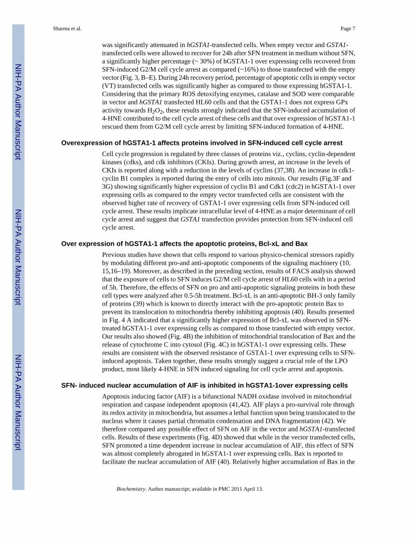
was significantly attenuated in hGSTA1-transfected cells. When empty vector and GSTA1-transfected cells were allowed to recover for 24h after SFN treatment in medium without SFN,a significantly higher percentage (~ 30%) of hGSTA1-1 over expressing cells recovered fromSFN-induced G2/M cell cycle arrest as compared (~16%) to those transfected with the emptyvector (Fig. 3, B–E). During 24h recovery period, percentage of apoptotic cells in empty vector(VT) transfected cells was significantly higher as compared to those expressing hGSTA1-1.Considering that the primary ROS detoxifying enzymes, catalase and SOD were comparablein vector and hGSTA1 transfected HL60 cells and that the GSTA1-1 does not express GPxactivity towards H2O2, these results strongly indicated that the SFN-induced accumulation of4-HNE contributed to the cell cycle arrest of these cells and that over expression of hGSTA1-1rescued them from G2/M cell cycle arrest by limiting SFN-induced formation of 4-HNE.
Overexpression of hGSTA1-1 affects proteins involved in SFN-induced cell cycle arrestCell cycle progression is regulated by three classes of proteins viz., cyclins, cyclin-dependentkinases (cdks), and cdk inhibitors (CKIs). During growth arrest, an increase in the levels ofCKIs is reported along with a reduction in the levels of cyclins (37,38). An increase in cdk1-cyclin B1 complex is reported during the entry of cells into mitosis. Our results (Fig.3F and3G) showing significantly higher expression of cyclin B1 and Cdk1 (cdc2) in hGSTA1-1 overexpressing cells as compared to the empty vector transfected cells are consistent with theobserved higher rate of recovery of GSTA1-1 over expressing cells from SFN-induced cellcycle arrest. These results implicate intracellular level of 4-HNE as a major determinant of cellcycle arrest and suggest that GSTA1 transfection provides protection from SFN-induced cellcycle arrest.
Over expression of hGSTA1-1 affects the apoptotic proteins, Bcl-xL and BaxPrevious studies have shown that cells respond to various physico-chemical stressors rapidlyby modulating different pro-and anti-apoptotic components of the signaling machinery (10,15,16–19). Moreover, as described in the preceding section, results of FACS analysis showedthat the exposure of cells to SFN induces G2/M cell cycle arrest of HL60 cells with in a periodof 5h. Therefore, the effects of SFN on pro and anti-apoptotic signaling proteins in both thesecell types were analyzed after 0.5-5h treatment. Bcl-xL is an anti-apoptotic BH-3 only familyof proteins (39) which is known to directly interact with the pro-apoptotic protein Bax toprevent its translocation to mitochondria thereby inhibiting apoptosis (40). Results presentedin Fig. 4 A indicated that a significantly higher expression of Bcl-xL was observed in SFN-treated hGSTA1-1 over expressing cells as compared to those transfected with empty vector.Our results also showed (Fig. 4B) the inhibition of mitochondrial translocation of Bax and therelease of cytochrome C into cytosol (Fig. 4C) in hGSTA1-1 over expressing cells. Theseresults are consistent with the observed resistance of GSTA1-1 over expressing cells to SFN-induced apoptosis. Taken together, these results strongly suggest a crucial role of the LPOproduct, most likely 4-HNE in SFN induced signaling for cell cycle arrest and apoptosis.
SFN- induced nuclear accumulation of AIF is inhibited in hGSTA1-1over expressing cellsApoptosis inducing factor (AIF) is a bifunctional NADH oxidase involved in mitochondrialrespiration and caspase independent apoptosis (41,42). AIF plays a pro-survival role throughits redox activity in mitochondria, but assumes a lethal function upon being translocated to thenucleus where it causes partial chromatin condensation and DNA fragmentation (42). Wetherefore compared any possible effect of SFN on AIF in the vector and hGSTA1-transfectedcells. Results of these experiments (Fig. 4D) showed that while in the vector transfected cells,SFN promoted a time dependent increase in nuclear accumulation of AIF, this effect of SFNwas almost completely abrogated in hGSTA1-1 over expressing cells. Bax is reported tofacilitate the nuclear accumulation of AIF (40). Relatively higher accumulation of Bax in the
Sharma et al. Page 7
Biochemistry. Author manuscript; available in PMC 2011 April 13.
NIH
-PA Author Manuscript
NIH
-PA Author Manuscript
NIH
-PA Author Manuscript

mitochondria of vector transfected cells upon SFN treatment was consistent with the observedhigher nuclear accumulation of AIF in these cells (Fig. 4 B and D). This would also beconsistent with the observed higher sensitivity of the vector transfected cells to SFN-inducedapoptosis as compared to the hGSTA1-1 over expressing cells. Strikingly, lack of anysignificant induction of caspase3 during SFN-induced apoptosis suggested that caspase3independent apoptosis in these cells may be mediated by AIF which was attenuated inhGSTA1-transfected cells with ameliorated 4-HNE levels.
SFN- induced nuclear accumulation of Nrf2 is exacerbated in hGSTA1-1 over expressingcells
The transcription factor Nrf2 interacts with ARE and regulates the transcription of genesencoding antioxidant proteins (6,7,43) involved in protection against oxidative stress. Wecompared the levels of Nrf2 in the cytosolic and nuclear fractions of SFN treated empty vectorand hGSTA1 transfected cells to study the possible role of 4-HNE in the nuclear translocationof Nrf2 upon SFN treatment. Results of these studies (Fig. 5A–C) indicated that exposure toSFN led to an enhanced accumulation of Nrf2 in the nuclear extracts of both, the empty vectorand hGSTA1 transfected cells in a time- dependent manner. Western blot analyses data (Fig.5A) indicated a rapid nuclear accumulation of Nrf2 within 30 min of SFN treatment whichpersisted for at least up to 5h thereby suggesting this to be an early event for the increasedtranscription of antioxidant enzymes to protect cells from the SFN-induced oxidative stress.The results of Western blot analysis and immunofluorescence studies clearly showed that thenuclear accumulation of Nrf2 was significantly higher in hGSTA1 transfected cells (Fig.5A–C). Nuclear translocation of Nrf2 has been suggested to be initiated by its dissociation fromKeap1 during oxidative stress (44). Our results showing relatively higher nuclear accumulationof Nrf2 in 4-HNE depleted, hGSTA1-1 over expressing cells suggested that SFN-induced upregulation of Nrf2 is independent of 4-HNE or lipid peroxidation and may result from the directinteraction of electrophilic SFN with the cysteine residues of Keap1.
Overexpression of hGSTA1-1 exacerbates SFN- induced activation of HSF1 by promotingtranslocation of Daxx to the cytosol
The role of transcription factor HSF1 has been suggested in the mechanisms of defense againstoxidative stress (45). Heat shock proteins (Hsps), the client proteins of HSF1, play an importantrole in the recovery of cells from the toxic effects of oxidative stress caused by heat, radiationand chemicals (19–21,45,46). Western blot data presented in Fig.6A and 6B indicatedsignificantly more induction and enhanced nuclear accumulation of HSF1 in SFN-treatedhGSTA1-1 over expressing HL60 and K562 cells respectively as compared to that in vectortransfected cells. This correlated with significantly higher expression of Hsp70 (Fig. 6C) inthe hGSTA1 transfected cells as compared to the vector transfected cells. Death associated Fasbinding protein, Daxx has been shown to be a transcription repressor which translocates tocytoplasm upon exposure of cells to oxidative stress (19,47). The translocation of Daxx fromnucleus to cytoplasm has been shown to induce nuclear accumulation of HSF1 and inductionof Hsp70 (19). Results presented in Fig. 6D and 6E show that a time dependent accumulationof Daxx in the cytoplasm of hGSTA1-transfected cells is accompanied with HSF1 activationThese effects of SFN on HSF1 and Daxx appear to be independent of 4-HNE. Taken together,our results suggest that GSTA1-1 may also protect cells from SFN-induced toxicity throughthe activation of stress responsive HSF1 and Daxx.
DiscussionIt has already been well demonstrated that exposure to SFN causes the generation of ROS incells and tissues. Furthermore, ROS-induced cellular signaling is believed to contribute to atleast some of its known chemo-preventive properties (1–5). The results of present studies
Sharma et al. Page 8
Biochemistry. Author manuscript; available in PMC 2011 April 13.
NIH
-PA Author Manuscript
NIH
-PA Author Manuscript
NIH
-PA Author Manuscript

indicate that at least some of the biological activities associated with the chemo-protectiveproperties of SFN may be mediated through LPO-products, particularly 4-HNE generatedduring SFN-induced oxidative stress and that these activities can be modulated by GSTs. Overexpression of GSTA1-1 in HL60 and K562 cells was observed to suppress 4-HNE levels bylimiting SFN- induced LPO and confer a remarkable resistance to cells against SFN-mediatedcytotoxicity, cell-cycle arrest, and apoptosis. These findings are consistent with recent studies(9) showing that 4-HNE is involved in the mechanisms of ROS mediated signaling and thatGSTs play an important regulatory role in oxidative stress-induced signaling.
The attenuation of 4-HNE levels in hGSTA1-1 over expressing cells after SFN treatmentunderscores the role of GSTA1-1 in regulating the levels of 4-HNE. Since 4-HNE is known tocause cell cycle arrest, apoptosis, and cytotoxicity to cells via necrosis (48–50), the observedeffects of GSTA1-1 over expression on these biological activities of SFN may be attributed tothe LPO suppression capability of GSTA1-1. While it is possible that increased SFN-conjugating activity of GSTA1-1 over expressing cells may lower the actual concentration ofSFN by its accelerated conjugation with GSH, our results showing no significant alteration inthe GSH levels of SFN treated empty vector, or hGSTA1-transfected cells suggest that theobserved protective effects of GSTA1-1 against SFN toxicity may be imparted preferentiallythrough the inhibition of SFN induced LPO and consequent lowering of 4-HNE levels, ratherthan GST-GSH mediated detoxification of SFN.
The observed effects of SFN in HL60 and K562 cells are essentially similar to those reportedin previous studies with colon and prostate cancer cells (2,3). Exposure to SFN caused cellcycle arrest in the G2/M phase in both these cell types. hGSTA1-1 over expression causedapproximately two-fold resistance to SFN-induced cell cycle arrest as well as apoptosis. Cellcycle arrest at the G2/M phase by SFN has been suggested to be regulated by cell cycle relatedproteins, cyclin B1 and Cdk1, and /or disruption of normal mitotic microtubule polymerizationand histone acetylation (51,52). Cyclins and cyclin-dependent kinase complexes play animportant role in the G2-M transition mechanisms. By binding to Cdk1/2, cyclin B1 canactivate Cdk1/2 (cdc2) to facilitate its nuclear accumulation for mitotic initiation in the late G2phase of mammalian cells (4,37,38). A time dependent induction of Cdk1 and cyclin B1 inSFN-treated hGSTA1-1 over expressing cells indicate that the inhibition of SFN-inducedaccumulation of 4-HNE by hGSTA1-1 rescued them from the G2/M arrest through an up-regulated expression of Cdk1 and cyclin B1. Inhibition of SFN induced apoptosis in GSTA1-1over expressing cells could also be attributed to the effect of suppression of 4-HNE levels andsubsequent modulation of mitochondrial apoptotic pathways. This is indicated by the activationof Bcl-xL and attenuated translocation of Bax to mitochondria in GSTA1-transfected cells.Furthermore, in GSTA1-transfected cells, SFN-induced cytochrome C release to cytosol andnuclear accumulation of AIF are also inhibited. These findings would indicate that SFN caninduce apoptosis in human erythroleukemic cells through mitochondria mediated caspaseindependent pathway (53) and that this can be modulated by suppression of LPO by GSTs.
Transcriptional regulation of many genes encoding antioxidant proteins under stressfulconditions for maintenance of cellular redox homeostasis involves an important role for Nrf2,a short half life protein, sequestered in the cytoplasm through its binding with Keap1. Nrf2 istargeted for ubiquitination when bound to its inhibitory protein Keap1, but is activated byoxidative stress when it dissociates from Keap1, translocates into the nucleus, andtransactivates the antioxidant response element (ARE) to induce the phase 2 detoxification,and antioxidant enzymes (1,6–8). Our results of Western analysis and immunfluorescencemicroscopy showed nuclear accumulation of Nrf2 upon SFN exposure, in the nuclei of both,the empty vector, and hGSTA1-transfected cells. However, relatively higher nuclearaccumulation of Nrf2 observed in hGSTA1-1 over expressing cells as compared to the emptyvector transfected cells may be a contributory factor to the resistance of GSTA1-1 over
Sharma et al. Page 9
Biochemistry. Author manuscript; available in PMC 2011 April 13.
NIH
-PA Author Manuscript
NIH
-PA Author Manuscript
NIH
-PA Author Manuscript

expressing cells to SFN toxicity. Enhanced nuclear accumulation of Nrf2 in GSTA1-1 overexpressing cells upon treatment with SFN may also suggest a reprogramming of protectivemachinery of the cells to alleviate electrophilic stress. Our results demonstrate that SFNexposure leads to nuclear translocation of HSF1 and up regulated expression of a representativeHSF1-client protein, Hsp70 which correlates with the observed inhibition of SFN- inducednuclear accumulation of AIF in GSTA1-1 over expressing cells and their resistance toapoptosis. Our results also suggest that SFN-induced up regulation of heat shock proteins mostlikely results from the translocation of HSF1 transcription repressor protein- Daxx (19) fromthe nucleus to cytoplasm. We demonstrate that SFN-treatment promotes the translocation ofDaxx from nucleus to cytoplasm which is accompanied with the nuclear translocation of HSF1and up regulation of the expression of HSF1 related genes. These combined effects of GSTA1-1should collectively contribute to the protective mechanisms against oxidative stress caused bySFN. Interestingly, these results suggest that similar to 4-HNE (19), SFN also inducesmechanisms to self limit its own toxicity. Since GSTA1-1 over expressing cells with sub-constitutive 4-HNE levels show higher degree of activation of Nrf2 and HSF1, it appearsunlikely that 4-HNE is involved in up regulation of these parameters. Instead, SFN being astrong electrophilic compound can directly interact with Keap1 and Daxx to affect theactivation of Nrf2 and HSF1, respectively.
In summary, the results of the present studies indicate that SFN-induced LPO contributes toits cytotoxicity to cells which can be inhibited by the over expression of GSTA1-1 and that theprotective effects of GSTA1-1 are associated with its ability to regulate 4-HNE levels in cells.GSTA1-1 and possibly other Alpha class GSTs seem to play a crucial role in protecting cellsfrom the cell-cycle arrest and apoptotic effects of SFN through a well orchestrated interplayof several pro-survival signaling pathways as presented in a model shown in Fig. 7. Perhapsthis model can be extrapolated to the overall protective role of GSTs during oxidative/electrophilic stress and the role of 4-HNE in regulation of signaling. Further studies are neededto validate this model, particularly for deciphering the mechanisms of interplay between Nrf2and HSF1 and associated genes upon exposure to agents that promote LPO.
Abbreviations
SFN D, L sulforaphane
4-HNE 4-hydroxynonenal
GST glutathione transferase
hGSTA1-1 human glutathione transferase isozyme A1-1
LPO lipid peroxidation
ROS reactive oxygen species
GSH glutathione
Daxx death associated Fas binding protein
Nrf2 Nuclear factor-E2-related factor-2
HSF1 heat shock factor 1
CDNB 1-chloro-2.4-dinitrobenzene
FACS Fluorescence activated cell sorting
Sharma et al. Page 10
Biochemistry. Author manuscript; available in PMC 2011 April 13.
NIH
-PA Author Manuscript
NIH
-PA Author Manuscript
NIH
-PA Author Manuscript

AcknowledgmentsWe thank Xiangle Sun (Core Facility at the University Of North Texas Health Science Center, Fort Worth, TX) forhelping with flow cytometry and laser capture micro-dissection (supported by National Institutes of Health GrantISIORR018999-01A1).
References1. Juge N, Mithen RF, Traka M. Molecular basis for chemoprevention by sulforaphane: a comprehensive
review. Cell Mol. Life Sci 2007;64:1105–1127. [PubMed: 17396224]2. Payrastre GL, Li P, Lumeau S, Cassar G, Dupont MA, Chevolleau S, Gasc N, Tulliez J, Tercé F.
Sulforaphane, a naturally occurring isothiocyanate, induces cell cycle arrest and apoptosis in HT29human colon cancer cells. Cancer Res 2000;60:1426–1433. [PubMed: 10728709]
3. Singh SV, Herman-Antosiewicz A, Singh AV, Lew KL, Srivastava SK, Kamath R, Brown KD, ZhangL, Baskaran R. Sulforaphane-induced G2/M phase cell cycle arrest involves checkpoint kinase 2-mediated phosphorylation of cell division cycle 25C. J Biol Chem 2004;279:25813–25822. [PubMed:15073169]
4. Singh SV, Srivastava SK, Choi S, Lew KL, Antosiewicz J, Xiao D, Zeng Y, Watkins SC, Johnson CS,Trump DL, Lee YJ, Xiao H, Herman-Antosiewicz A. Sulforaphane-induced cell death in humanprostate cancer cells is initiated by reactive oxygen species. J. Biol. Chem 2005;280:19911–19924.[PubMed: 15764812]
5. Xiao D, Srivastava SK, Lew KL, Zeng Y, Hershberger P, Johnson CS, Trump DL, Singh SV. Allylisothiocyanate, a constituent of cruciferous vegetables, inhibits proliferation of human prostate cancercells by causing G2/M arrest and inducing apoptosis. Carcinogenesis 2003;24:891–897. [PubMed:12771033]
6. McWalter GK, Higgins LG, McLellan LI, Henderson CJ, Song L, Thornalley PJ, Itoh K, YamamotoM, Hayes JD. Transcription factor Nrf2 is essential for induction of NAD(P)H:quinone oxidoreductase1, glutathione S-transferases, and glutamate cysteine ligase by broccoli seeds and isothiocyanates. J.Nutr 2004;134:3499S–3506S. [PubMed: 15570060]
7. Zhang Y. Cancer-preventive isothiocyanates: measurement of human exposure and mechanism ofaction. Mutat. Res 2004;555:173–190. [PubMed: 15476859]
8. Thimmulappa RK, Mai KH, Srisuma S, Kensler TW, Yamamoto M, Biswal S. Identification of Nrf2-regulated genes induced by the chemopreventive agent sulforaphane by oligonucleotide microarray.Cancer Res 2002;62:5196–5203. [PubMed: 12234984]
9. Yang Y, Sharma R, Sharma A, Awasthi S, Awasthi YC. Lipid peroxidation and cell cycle signaling:4-hydroxynonenal, a key molecule in stress mediated signaling. Acta Biochim Pol 2003;50:319–336.[PubMed: 12833161]
10. Cheng JZ, Singhal SS, Sharma A, Saini M, Yang Y, Awasthi S, Zimniak P, Awasthi YC. Transfectionof mGSTA4 in HL-60 cells protects against 4-hydroxynonenal-induced apoptosis by inhibiting JNK-mediated signaling. Arch Biochem Biophys 2001;392:197–207. [PubMed: 11488593]
11. Cheng JZ, Singhal SS, Saini M, Singhal J, Piper JT, Van Kuijk FJ, Zimniak P, Awasthi YC, AwasthiS. Effects of mGST A4 transfection on 4-hydroxynonenal-mediated apoptosis and differentiation ofK562 human erythroleukemia cells. Arch Biochem Biophys 1999;372:29–36. [PubMed: 10562413]
12. Sharma R, Brown D, Awasthi S, Yang Y, Sharma A, Patrick B, Saini MK, Singh SP, Zimniak P,Singh SV, Awasthi YC. Transfection with 4-hydroxynonenal-metabolizing glutathione S-transferaseisozymes leads to phenotypic transformation and immortalization of adherent cells. Eur. J. Biochem2004;271:1690–1701. [PubMed: 15096208]
13. Awasthi YC, Sharma R, Sharma A, Yadav S, Singhal SS, Chaudhary P, Awasthi S. Self-regulatoryrole of 4-hydroxynonenal in signaling for stress-induced programmed cell death. Free Radic BiolMed 2008;45:111–118. [PubMed: 18456001]
14. Yang Y, Sharma R, Cheng JZ, Saini MK, Ansari NH, Andley UP, Awasthi S, Awasthi YC. Protectionof HLE B-3 cells against hydrogen peroxide- and naphthalene-induced lipid peroxidation andapoptosis by transfection with hGSTA1 and hGSTA2. Invest. Ophthalmol. Vis. Sci 2002;43:434–445. [PubMed: 11818388]
Sharma et al. Page 11
Biochemistry. Author manuscript; available in PMC 2011 April 13.
NIH
-PA Author Manuscript
NIH
-PA Author Manuscript
NIH
-PA Author Manuscript

15. Yang Y, Cheng JZ, Singhal SS, Saini M, Pandya U, Awasthi S, Awasthi YC. Role of glutathione S-transferases in protection against lipid peroxidation: overexpression of hGSTA2-2 in K562 cellsprotects against hydrogen peroxide induced apoptosis and inhibits JNK and caspase 3 activation. J.Biol. Chem 2001;276:19220–19230. [PubMed: 11279091]
16. Yang Y, Sharma A, Sharma R, Patrick B, Singhal SS, Zimniak P, Awasthi S, Awasthi YC. Cellspreconditioned with mild, transient UVA irradiation acquire resistance to oxidative stress and UVA-induced apoptosis: role of 4-hydroxynonenal in UVA-mediated signaling for apoptosis. J. Biol. Chem2003;278:41380–41388. [PubMed: 12888579]
17. Sharma A, Patrick B, Li J, Sharma R, Jeyabal PVS, Reddy, Prasada MRV, Awasthi S, Awasthi YC.Glutathione S-transferases as antioxidant enzymes: Small cell lung cancer (H69) cells transfectedwith hGSTA1 resist doxorubicin-induced apoptosis. Arch Biochem. Biophys 2006;452:165–173.[PubMed: 16890185]
18. Li J, Sharma R, Patrick B, Sharma A, Jeyabal PV, Reddy PM, Saini MK, Dwivedi S, Dhanani S,Ansari NH, Zimniak P, Awasthi S, Awasthi YC. Regulation of CD95 (Fas) expression and Fas-mediated apoptotic signaling in HLE B-3 cells by 4-hydroxynonenal. Biochemistry 2006;45:12253–12264. [PubMed: 17014078]
19. Sharma R, Sharma A, Dwivedi S, Zimniak P, Awasthi S, Awasthi YC. 4-Hydroxynonenal self-limitsFas-mediated DISC-independent apoptosis by promoting export of Daxx from the nucleus to thecytosol and its binding to Fas. Biochemistry 2008;47:143–156. [PubMed: 18069800]
20. Chen ZH, Saito Y, Yoshida Y, Sekine A, Noguchi N, Niki E. 4-Hydroxynonenal induces adaptiveresponse and enhances PC12 cell tolerance primarily through induction of thioredoxin reductase 1via activation of Nrf2. J. Biol. Chem 2005;280:41921–41927. [PubMed: 16219762]
21. Jacobs AT, Marnett LJ. HSF1-mediated BAG3 expression attenuates apoptosis in 4-hydroxynonenal-treated colon cancer cells via stabilization of anti-apoptotic Bcl-2 proteins. J. Biol Chem2009;284:9176–9183. [PubMed: 19179333]
22. Jacobs AT, Marnett LJ. Heat shock factor 1 attenuates 4-Hydroxynonenal-mediated apoptosis: criticalrole for heat shock protein 70 induction and stabilization of Bcl-xL. J. Biol. Chem 2007;282:33412–33420. [PubMed: 17873279]
23. Zhao T, Singhal SS, Piper JT, Cheng J, Pandya U, Clark-Wronski J, Awasthi S, Awasthi YC. Therole of human glutathione S-transferases hGSTA1-1 and hGSTA2-2 in protection against oxidativestress. Arch Biochem. Biophys 1999;367:216–224. [PubMed: 10395737]
24. Koeffler HP, Golde DW. Human myeloid leukemic cell lines: A Review. Blood 1980;56:344–350.[PubMed: 6996765]
25. Singhal SS, Saxena M, Ahmad H, Awasthi S, Haque AK, Awasthi YC. Glutathione S-transferasesof human lung: characterization and evaluation of the protective role of the alpha-class isozymesagainst lipid peroxidation. Arch. Biochem. Biophys 1992;299:232–241. [PubMed: 1444461]
26. Habig WH, Pabst MJ, Jakoby WB. Glutathione S-transferases. The first enzymatic step in mercapturicacid formation. J. Biol. Chem 1974;249:7130–7139. [PubMed: 4436300]
27. Kolm RH, Danielson UH, Zhang Y, Talalay P, Mannervik B. Isothiocyanates as substrates for humanglutathione transferases: structure-activity studies. Biochem. J 1995;311:453–459. [PubMed:7487881]
28. Awasthi YC, Beutler E, Srivastava SK. Purification and properties of human erythrocyte glutathioneperoxidase. J.Biol Chem 1975;250:5144–5149. [PubMed: 807573]
29. Carlberg I, Mannervik B. Glutathione reductase. Methods Enzymol 1985;113:484–490. [PubMed:3003504]
30. Seelig GF, Meister A. Gamma-glutamylcysteine synthetase. Interactions of an essential sulfhydrylgroup. J. Biol. Chem 1984;259:3534–3538. [PubMed: 6142890]
31. Bradford MM. A rapid and sensitive method for the quantitation of microgram quantities of proteinutilizing the principle of protein-dye binding. Anal. Biochem 1976;72:248–254. [PubMed: 942051]
32. Laemmli UK. Cleavage of structural proteins during the assembly of the head of bacteriophage T4.Nature 1970;227:680–685. [PubMed: 5432063]
33. Towbin H, Staehelin T, Gordon J. Electrophoretic transfer of proteins from polyacrylamide gels tonitrocellulose sheets: procedure and some applications. Proc. Natl. Acad. Sci. U S A 1979;76:4350–4354. [PubMed: 388439]
Sharma et al. Page 12
Biochemistry. Author manuscript; available in PMC 2011 April 13.
NIH
-PA Author Manuscript
NIH
-PA Author Manuscript
NIH
-PA Author Manuscript

34. Englander EW, Hu Z, Sharma A, Lee HM, Wu ZH, Greeley GH. Rat MYH, a glycosylase for repairof oxidatively damaged DNA, has brain-specific isoforms that localize to neuronal mitochondria. J.Neurochem 2002;83:1471–1480. [PubMed: 12472901]
35. Mosmann T. Rapid colorimetric assay for cellular growth and survival: application to proliferationand cytotoxicity assays. J. Immunol Methods 1983;65:55–63. [PubMed: 6606682]
36. Beutler, E. Red Cell Metabolism. In: Grune; Stratton, editors. A Manual of Biochemical Methods.Orlando, FL: 1984.
37. Takizawa CG, Morgan DO. Control of mitosis by changes in the subcellular location of cyclin-B1-Cdk1 and Cdc25C. Current Opinion in Cell Biology 2000;12:658–665. [PubMed: 11063929]
38. Sánchez I, Dynlacht BD. New insights into cyclins, CDKs, and cell cycle control. Semin. Cell DevBiol 2005;16:311–321. [PubMed: 15840440]
39. Kaufmann T, Schinzel A, Borner C. Bcl-w(edding) with mitochondria. Trends Cell Biol 2004;14:8–12. [PubMed: 14729175]
40. Ganju N, Eastman A. Bcl-X(L) and calyculin A prevent translocation of Bax to mitochondria duringapoptosis. Biochem. Biophys. Res. Commun 2002;291:1258–1264. [PubMed: 11883953]
41. Candé C, Cohen I, Daugas E, Ravagnan L, Larochette N, Zamzami N, Kroemer G. Apoptosis-inducingfactor (AIF): a novel caspase-independent death effector released from mitochondria. Biochimie2002;84:215–222. [PubMed: 12022952]
42. Lorenzo HK, Susin SA, Penninger J, Kroemer G. Apoptosis inducing factor (AIF): a phylogeneticallyold, caspase-independent effector of cell death. Cell Death and Differ 1999;6:516–524.
43. Kwak MK, Itoh K, Yamamoto M, Sutter TR, Kensler TW. Role of transcription factor Nrf2 in theinduction of hepatic phase 2 and antioxidative enzymes in vivo by the cancer chemoprotective agent,3H-1, 2-dimethiole-3-thione. Mol Med 2001;7:135–145. [PubMed: 11471548]
44. Dinkova-Kostova AT, Holtzclaw WD, Cole RN, Itoh K, Wakabayashi N, Katoh Y, Yamamoto M,Talalay P. Direct evidence that sulfhydryl groups of Keap1 are the sensors regulating induction ofphase 2 enzymes that protect against carcinogens and oxidants. Proc Natl Acad Sci U S A2002;99:11908–11913. [PubMed: 12193649]
45. Nollen EA, Morimoto RI. Chaperoning signaling pathways: molecular chaperones as stress-sensing'heat shock' proteins. J. Cell Sci 2002;115:2809–2816. [PubMed: 12082142]
46. Dai C, Whitesell L, Rogers AB, Lindquist S. Heat shock factor 1 is a powerful multifaceted modifierof carcinogenesis. Cell 2007;130:1005–1118. [PubMed: 17889646]
47. Salomoni P, Khelifi AF. Daxx: death or survival protein? Trends Cell Biol 2006;16:97–104. [PubMed:16406523]
48. Esterbauer H, Schaur RJ, Zollner H. Chemistry and biochemistry of 4-hydroxynonenal,malonaldehyde and related aldehydes. Free Radic. Biol. Med 1991;11:81–128. [PubMed: 1937131]
49. Barrera G, Pizzimenti S, Muraca R, Barbiero G, Bonelli G, Baccino FM, Fazio VM, Dianzani MU.Effect of 4-Hydroxynonenal on cell cycle progression and expression of differentiation-associatedantigens in HL-60 cells. Free Radic Biol Med 1996;20:455–462. [PubMed: 8720918]
50. Awasthi YC, Sharma R, Cheng JZ, Yang Y, Sharma A, Singhal SS, Awasthi S. Role of 4-hydroxynonenal in stress-mediated apoptosis signaling. Mol Aspects Med 2003;24:219–230.[PubMed: 12893000]
51. Jackson SJT, Singletary KW. Sulforaphane inhibits human MCF-7 mammary cancer cell mitoticprogression and tubulin polymerization. J. Nutr 2004;134:2229–2236. [PubMed: 15333709]
52. Dashwood RH, Ho E. Dietary histone deacetylase inhibitors: from cells to mice to man. Semin CancerBiol 2007;17:363–369. [PubMed: 17555985]
53. Joza N, Susin SA, Daugas E, Stanford WL, Cho SK, Li CY, Sasaki T, Elia AJ, Cheng HY, RavagnanL, Ferri KF, Zamzami N, Wakeham A, Hakem R, Yoshida H, Kong YY, Mak TW, Zúñiga-PflückerJC, Kroemer G, Penninger JM. Essential role of the mitochondrial apoptosis-inducing factor inprogrammed cell death. Nature 2001;410:549–554. [PubMed: 11279485]
Sharma et al. Page 13
Biochemistry. Author manuscript; available in PMC 2011 April 13.
NIH
-PA Author Manuscript
NIH
-PA Author Manuscript
NIH
-PA Author Manuscript

Sharma et al. Page 14
Biochemistry. Author manuscript; available in PMC 2011 April 13.
NIH
-PA Author Manuscript
NIH
-PA Author Manuscript
NIH
-PA Author Manuscript

Fig. 1. Expression of hGSTA1-1 in hGSTA1 transfected cellsHL60 (A) and K562 (B) cells were transfected with the cDNA of hGSTA1 cloned in thepTarget-T mammalian expression vector or the vector alone as described in the Methodssection. The supernatant fraction (28,000g) of homogenates of the wild-type (WT), vector (VT)and hGSTA1-transfected (hGSTA1-Tr) HL60 cells containing 30µg of protein was subjectedto SDS-PAGE in 12% gel. Expression of hGSTA1-1 in the stable-transfected clone selectedin G418 (300µg/ml) was analyzed by Western blot analysis using polyclonal primaryantibodies against human α-class GSTs raised in rabbits and peroxidase-conjugated goat anti-rabbit secondary antibodies. The blot was developed using chemiluminescence (SupersignalWest Pico, Pierce) reagents. Lanes have been appropriately marked on the immunoblots.Representative immunoblot from one of the several hGSTA1-1expressing clones selected isshown. C. Levels of 4-HNE in SFN treated VT and hGSTA1-1 expressing HL60 and K562cells: VT and hGSTA1-1 expressing cells (2×10⁷) were incubated with RPMI completemedium containing SFN (0–40µM) for 5h at 37°C. After pelleting them by centrifugation, cells
Sharma et al. Page 15
Biochemistry. Author manuscript; available in PMC 2011 April 13.
NIH
-PA Author Manuscript
NIH
-PA Author Manuscript
NIH
-PA Author Manuscript

were washed and sonicated (3× 10s, 30W) in PBS containing BHT (5mM final concentration)on ice. 4-HNE levels in the cell pellets were measured by using LPO-586 kit as per themanufacturer’s instructions. Data presented are Mean ± SD (n=3, * and ** represent asignificant difference in 4-HNE levels of VT and hGSTA1-1 expressing HL60 and K562 cellsrespectively; p< 0.05). D. Immunofluorescence analysis of 4-HNE adducts in SFN treatedVT and hGSTA1-1 expressing HL60 cells: VT and hGSTA1-1 expressing HL60 cells(1×10⁶) were treated with SFN (20µM) for 2h at 37°C in RPMI complete medium. Afterpelleting them by centrifugation at 1000rpm (5min), cells were washed and resuspended inPBS. Aliquots of cell suspensions were cytospun at 500rpm for 5 min onto the superfrost Fisherbrand slides and fixed in 4% paraformaldehyde for 20min. The cells were incubated with theprimary antibodies against 4-HNE-protein adduct (1:500) prepared in the blocking buffer (1%BSA +1% goat serum in PBS) overnight at 4°C in a humidified chamber. After three washingswith PBS (5min each), cells were incubated with TRITC conjugated secondary antibodies(1:500) for 2h at room temperature. Subsequently, this was once again followed by threewashings with PBS (10min each), mounted with Vecta Shield containing DAPI and observedunder the Nikon Eclipse E800 fluorescence microscope with a 40x objective. Different panelsof photographs with DAPI and TRITC stains have been appropriately marked in the figure.
Sharma et al. Page 16
Biochemistry. Author manuscript; available in PMC 2011 April 13.
NIH
-PA Author Manuscript
NIH
-PA Author Manuscript
NIH
-PA Author Manuscript

Fig. 2. Cytotoxic effects of SFN on VT and hGSTA1-1 expressing cellsVT and hGSTA1-1 expressing HL60 (A) and K562 (B) cells (2×10⁴) were plated into replicatewells of a 96 well plate in RPMI complete growth medium. After incubating the cells overnight,cells were treated with different concentrations of SFN (0–60µM) prepared in DMSO (finalconcentration; 0.02%) with appropriate controls and were incubated for 24h at 37 °C afterwhich the MTT assay was performed as described in the Materials and Methods section. TheOD580 values of samples subtracted from those of respective blanks (no cells) were normalizedwith control values. The values shown are Mean ± SD (n=3 done in quadruplets, p<0.01).
Sharma et al. Page 17
Biochemistry. Author manuscript; available in PMC 2011 April 13.
NIH
-PA Author Manuscript
NIH
-PA Author Manuscript
NIH
-PA Author Manuscript

Sharma et al. Page 18
Biochemistry. Author manuscript; available in PMC 2011 April 13.
NIH
-PA Author Manuscript
NIH
-PA Author Manuscript
NIH
-PA Author Manuscript

Sharma et al. Page 19
Biochemistry. Author manuscript; available in PMC 2011 April 13.
NIH
-PA Author Manuscript
NIH
-PA Author Manuscript
NIH
-PA Author Manuscript
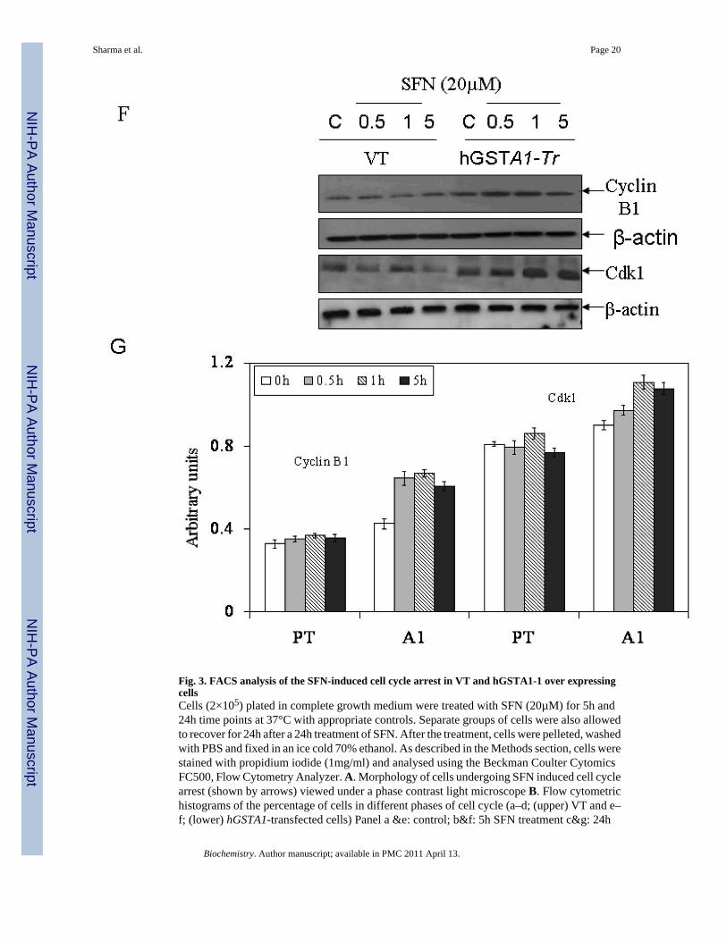
Fig. 3. FACS analysis of the SFN-induced cell cycle arrest in VT and hGSTA1-1 over expressingcellsCells (2×10⁵) plated in complete growth medium were treated with SFN (20µM) for 5h and24h time points at 37°C with appropriate controls. Separate groups of cells were also allowedto recover for 24h after a 24h treatment of SFN. After the treatment, cells were pelleted, washedwith PBS and fixed in an ice cold 70% ethanol. As described in the Methods section, cells werestained with propidium iodide (1mg/ml) and analysed using the Beckman Coulter CytomicsFC500, Flow Cytometry Analyzer. A. Morphology of cells undergoing SFN induced cell cyclearrest (shown by arrows) viewed under a phase contrast light microscope B. Flow cytometrichistograms of the percentage of cells in different phases of cell cycle (a–d; (upper) VT and e–f; (lower) hGSTA1-transfected cells) Panel a &e: control; b&f: 5h SFN treatment c&g: 24h
Sharma et al. Page 20
Biochemistry. Author manuscript; available in PMC 2011 April 13.
NIH
-PA Author Manuscript
NIH
-PA Author Manuscript
NIH
-PA Author Manuscript

SFN treatment d&h: 24h recovered cells after 24h of SFN treatment. C and D, Bar chartsshowing the percentage of cells in the respective phases (sub G0/G1, G0/G1, S and G2/M) ofthe cell cycle (Mean ± SD) from three independent experiments. E. Bar chart showing theeffect of SFN on the cell cycle of K562 (VT and hGSTA1 transfected) cells. F Western blotanalysis of cell cycle related proteins Cdk1 and cyclin B1expression respectively in controland SFN treated VT and hGSTA1-1 expressing HL60 cells. G. Densitometric analysis of bandsobtained for cyclin B1 and cdk1 on immunoblots.* and **represent significant differences inthe percentages of cells in apoptosis (sub G0/G1) and G2/M Phase respectively in VT andhGSTA1-1.
Sharma et al. Page 21
Biochemistry. Author manuscript; available in PMC 2011 April 13.
NIH
-PA Author Manuscript
NIH
-PA Author Manuscript
NIH
-PA Author Manuscript

Sharma et al. Page 22
Biochemistry. Author manuscript; available in PMC 2011 April 13.
NIH
-PA Author Manuscript
NIH
-PA Author Manuscript
NIH
-PA Author Manuscript

Fig. 4. Effects of SFN on the expression of apoptosis related proteins (Bcl-xL, Bax, AIF andcytochrome C) in VT and hGSTA1-1 expressing HL60 cellsPlated cells (5×10⁶) were incubated with SFN (20µM) in complete RPMI growth medium fordifferent time points (0.5h, 1h and 5h) at 37°C. After treatment, they were pelleted bycentrifugation and washed 2X with PBS. Whole cell extracts were prepared in RIPA lysisbuffer while sub cellular (cytoplasmic, nuclear and mitochondrial) fractionation was performedas described in Materials and Methods and by the Imgenex Kit as per the manufacturer’sinstructions. Western blot analyses of these extracts were carried out by using antibodiesagainst Bcl-xL (panel A), Bax (panel B); cytochrome C (panel C) and AIF (panel D).Immunoblots were also probed with β-actin (total and cytoplasmic extracts to ascertain equal
Sharma et al. Page 23
Biochemistry. Author manuscript; available in PMC 2011 April 13.
NIH
-PA Author Manuscript
NIH
-PA Author Manuscript
NIH
-PA Author Manuscript

loading of proteins. The blot was developed using chemiluminescence (Supersignal West Pico,Pierce) reagents to detect the bands on immunoblots.
Sharma et al. Page 24
Biochemistry. Author manuscript; available in PMC 2011 April 13.
NIH
-PA Author Manuscript
NIH
-PA Author Manuscript
NIH
-PA Author Manuscript

Sharma et al. Page 25
Biochemistry. Author manuscript; available in PMC 2011 April 13.
NIH
-PA Author Manuscript
NIH
-PA Author Manuscript
NIH
-PA Author Manuscript

Fig. 5. A. Effect of SFN on the expression and localization of Nrf2 in VT and hGSTA1-1 expressingcellsCells were treated with SFN (20µM) for different time points (0.5, 1 and 5h). Cytoplasmic andnuclear extracts of control and treated cells were prepared and subjected to Western blotanalyses as described in the legend of Fig. 4. A representative Western blot showing theexpression of Nrf2 in cytoplasmic and nuclear protein fractions obtained from control and SFNtreated VT and hGSTA1-1 expressing HL60 cells. B. Western blot of nuclear extract of VTand hGSTA1-1 expressing K562 cells. C. Immunofluorescence localization of Nrf2 incontrol and SFN treated VT and hGSTA1-1 expressing HL60 cells. Control and SFNtreated cells (2×10⁵) for immunolocalization of Nrf2 were essentially processed as describedin the legend for Fig. 2B except that antibodies used were those against Nrf2 (diluted 1:200 inblocking buffer) and FITC conjugated secondary antibodies.
Sharma et al. Page 26
Biochemistry. Author manuscript; available in PMC 2011 April 13.
NIH
-PA Author Manuscript
NIH
-PA Author Manuscript
NIH
-PA Author Manuscript

Sharma et al. Page 27
Biochemistry. Author manuscript; available in PMC 2011 April 13.
NIH
-PA Author Manuscript
NIH
-PA Author Manuscript
NIH
-PA Author Manuscript

Fig. 6. Effect of SFN on the expression and localization of HSF1 (A and B), Hsp70 (C) and Daxx(D and E) in VT and hGSTA1-1 expressing cellsCells (2×10⁶) were treated with SFN (20µM) in complete growth medium at 37°C for differenttime points as shown in the figure. Total cell extracts in RIPA buffer, cytoplasmic and nuclearfractions of the cells were prepared as described in the Method section. Western blot analysesof the SDS-PAGE resolved proteins (50–60µg protein in each lane) were carried out by usingantibodies against Daxx, HSF1, and Hsp70 as described in the Methods section.
Sharma et al. Page 28
Biochemistry. Author manuscript; available in PMC 2011 April 13.
NIH
-PA Author Manuscript
NIH
-PA Author Manuscript
NIH
-PA Author Manuscript

Fig. 7. A theoretical model for the mechanisms by which SFN-induced generation and accumulationof 4-HNE affects signaling for cell cycle arrest and apoptosis and inhibition of these effects of SFNby GSTA1-1The model illustrates that treatment of cells with SFN causes enhanced LPO-inducedaccumulation of 4-HNE which contributes to SFN-induced cell cycle arrest in G2/M phasethrough inhibition of cyclin B1 and cdk1, and apoptosis via down regulation of anti-apoptoticBcl-xL, increased translocation of proapototic Bax to mitochondria, increased accumulationof AIF to nucleus and cytoplasmic release of cytochrome C. These SFN-induced effects areinhibited by the enforced expression of GSTA1-1 in cells. The over expression of GSTA1-1limits the formation of 4-HNE by reducing upstream LPO products, which leads to the upregulation of Bcl-xL, facilitated cytoplasmic export of the transcription repressor Daxxaccompanied by enhanced nuclear accumulation of the transcription factors Nrf2 and HSF1,and activation of the associated stress responsive antioxidant and heat shock proteins.
Sharma et al. Page 29
Biochemistry. Author manuscript; available in PMC 2011 April 13.
NIH
-PA Author Manuscript
NIH
-PA Author Manuscript
NIH
-PA Author Manuscript

NIH
-PA Author Manuscript
NIH
-PA Author Manuscript
NIH
-PA Author Manuscript
Sharma et al. Page 30
Table 1
Specific activities of antioxidant enzymes in empty vector (VT) and hGSTA1-transfected HL60 cells
S.No.Enzyme
Specific activity(nmol/min/mg protein, Mean ± SD, n=3)
VT hGSTA1-Tr
1 GSTs
*CDNB 66.38 ± 2.7 121.7 ± 4.6
*SFN 92.0 ± 6.4 170 ± 11
2 GPx
**CU-OOH 0.175 ± 0.034 0.788 ± 0.12
3 GR 36 ± 2.4 43 ± 1.82
4 γGCS 44.4 ± 3.67 80.13 ± 4.48
*Substrates of GSTs
**Substrate of GPx
The activities of GSTs against 1-chloro-2, 4-dinitrobenzene (CDNB) (26) and sulforaphane (SFN) (27) were measured in the crude cytosolic extracts(28000g supernatants) prepared from the empty vector and hGSTA1-transfected cells. GPx activity against cumene hydroperoxide (CU-OOH) (27),activities of GR (28) and γGCS (29) were also estimated in the crude extracts.
Biochemistry. Author manuscript; available in PMC 2011 April 13.