‘Flower’ of Magnolia grandiflora is not flower and what about ‘basal angiosperms’
Upload
independentCategory
view
1download
0
Introduction
Ethnobotanical knowledge coupled with rationale-driven scientificresearch may help in identifying potent new drugs against cancer.Here we report the results of a complex protein isolated from S.grandiflora, a member of the family Papilionaceae. S. grandiflorais a small tree commonly known as ‘sesbania’ and ‘agathi’ and isan important source of dietary nutrients in Southeast Asian coun-tries [1]. Some pharmacognostic studies have been reported on
the bark of this plant [2]. In the Siddha system of Indian traditionalmedicine, different parts of this plant have been accredited for alleviating a spectrum of ailments including inflammation, leprosy,gout and rheumatism [3]. The flowers and leaves are enriched withvitamins and minerals and have been reportedly associated withanti-inflammatory, analgesic and antipyretic effects [4, 5].Additionally, the leaves have demonstrated anxiolytic and anticon-vulsive activity in experimental animals [6] and have proven effec-tive against hepatitis [7] and in inhibiting HIV-1 protease activity[8], thus enabling significant protection against such maladies.Recent reports indicate that an aqueous suspension of S. grandifloraleaves exhibit protective effects against cigarette smoke-inducedoxidative damage in rats [9], whereas alcoholic extracts of the leaveshave been documented to provide significant protective effectsagainst hepatotoxicity [10]. Additionally, the anti-urolithiatic and
A novel protein fraction from Sesbania grandiflora shows
potential anticancer and chemopreventive efficacy,
in vitro and in vivo
Krishna P. Laladhas a, Vino T. Cheriyan b, Vineshkumar T. Puliappadamba b, Smitha V. Bava b, Rajesh G. Unnithan c, Parvathy L. Vijayammal c, Ruby John Anto b, *
a Department of Zoology, St. Stephan’s College, Pathanapuram, Kerala, Indiab Molecular Carcinogenesis and Chemoprevention Laboratory, Division of Cancer Research,
Rajiv Gandhi Centre for Biotechnology, Thiruvananthapuram, Kerala, Indiac Department of Biochemistry, University of Kerala, Thiruvananthapuram, Kerala, India
Received: July 23, 2008; Accepted: December 10, 2008
Abstract
We report mechanism-based evidence for the anticancer efficacy of a protein fraction, SF2 (Sesbania fraction 2) isolated from the flowerof the medicinal plant, Sesbania grandiflora (S. grandiflora). The fraction was evaluated in two murine ascites tumour cell lines andhuman cancer cell lines of different origin for its anticancer effect. SF2 inhibited cell proliferation and induced apoptosis as demonstratedby DNA fragmentation and externalization of phosphatidyl serine in Daltons lymphoma ascites (DLA) and colon cancer cells (SW-480).Sensitivity to SF2 in these cells was associated with activation of caspases 3, 8 and 9, poly (ADP-ribose) polymerase cleavage andcytochrome C release which attests apoptosis induced cell death. Mechanistically, SF2 down-regulated phorbol myristate acetate (PMA)induced NF-�B, a transcription factor which controls the expression of genes encoding proteins involved in cell regulation and growthcontrol. Additionally, SF2 also down-regulated anti-apoptotic factors such as Bcl-2, p-Akt and cyclooxygenase-2 induced by the tumourpromoter PMA suggestive of a possible explanation for its anticancer effect. In vivo studies using ascites and solid tumour modelsstrongly support in vitro findings as SF2 administration increased the life span and decreased the tumour volume in mice bearing tumour.In vivo toxicological evaluation revealed the pharmacological safety of SF2 and may serve as a potential anticancer drug candidate.
Keywords: Sesbania grandiflora • apoptosis • chemoprevention • anticancer
J. Cell. Mol. Med. Vol 14, No 3, 2010 pp. 636-646
*Correspondence to: Ruby John ANTO,Molecular Carcinogenesis and Chemoprevention Laboratory, Division of Cancer Research, Rajiv Gandhi Centre for Biotechnology,Thiruvananthapuram, Kerala – 695014, India.Tel.: 91-471- 2347975Fax: 91-471-2348096E-mail: [email protected]
© 2009 The AuthorsJournal compilation © 2010 Foundation for Cellular and Molecular Medicine/Blackwell Publishing Ltd
doi:10.1111/j.1582-4934.2008.00648.x
J. Cell. Mol. Med. Vol 14, No 3, 2010
637© 2009 The AuthorsJournal compilation © 2010 Foundation for Cellular and Molecular Medicine/Blackwell Publishing Ltd
antioxidant properties of this plant has been well documented [11, 12]. The flowers of this plant are edible and are consumed asa popular traditional remedy for night blindness, bronchitis, nasalcatarrh, headache and frontal sinus pain alleviation [13]. Recently,two proteins, namely SGF60 and SGF90 isolated from the flowersof this plant have been shown to inhibit �-glucosidase, and hencehas been speculated as a potential drug against type 2 diabetes[14]. Even though Ayurvedic literatures [15] mention the anti-tumour effect of S. grandiflora fruit, there is no mechanism-basedevidence in the literature on the anticancer therapeutic potential ofS. grandiflora. In this study, we report some promising resultsobtained from a complex protein fraction of the flower againstascites and colon cancer cells characterizing apoptosis by mor-phological features such as caspase activation, cytochrome Crelease and poly (ADP-ribose) polymerase (PARP) degradation.We also assessed the potential molecular mechanism in supportof its anticancer effect, and additionally present evidence for thefirst time in line with our in vitro observations, the anti-tumoureffect of S. grandiflora in ascites and solid tumour models.
Materials and methods
Reagents and antibodies
Annexin V apoptosis detection kit and antibodies against cytochrome C,Bcl-2, p65, p50 and �-actin were obtained from Santa Cruz Biotechnology(Santa Cruz, CA, USA). Antibodies against phospho-Akt, caspases 3, 8 and9 and PARP were purchased from Cell Signaling Technology (Beverly, MA,USA) and the fluorimetric substrates for the caspases were obtained fromCalbiochem (La Jolla, CA, USA). The rest of the chemicals were obtainedfrom Sigma Chemicals (St. Louis, MO, USA).
Drug extraction
Fresh flowers of S. grandiflora were ground in phosphate buffered saline(PBS) in ice and the extract was filtered using cheese cloth and was furthersubjected to centrifugation at 2000 rpm for 10 min. at 4�C. The clear super-natant was subjected to protein precipitation using ammonium sulphatesaturation and the three fractions (0–40% [SF1], 40–70% [SF2] and70–100% [SF3]) obtained were centrifuged, dialysed, passed throughSephadex G-25 column and lyophilized as described earlier [16].
In vitro studies
The murine ascites tumour cell lines – DLA and Ehrlich ascites (EAC), thenormal murine cell line – murine embryonic fibroblast (MEF), the humanlung cancer cell lines – A549 and H1299, the human cervical cancer celllines – HeLa and ME-180 and the human colon cancer cell lines – SW-480,SW-620 and HT-55 were used for the in vitro studies. The cytotoxicity wasmeasured by MTT assay [17]. Inhibition of DNA synthesis and DNA damage was assessed by [3H]thymidine incorporation assay and DNA
fragmentation assay, respectively [18]. Annexin V–FITC staining was doneaccording to manufacturer’s protocol [17]. The cleavage of caspases 3, 8and 9 induced was detected by Western blot and assayed spectrofluorimet-rically as described earlier [19]. Cleavage of PARP, the inhibition of Akt phosphorylation and down-regulation of Bcl-2 and COX-2 expressionwere detected by Western blot [19]. Mitochondria-free cytosol was iso-lated as described earlier [17] and immunoblotted against anti-cytochrome C. NF-�B down-regulation was studied by electrophoretic mobility shiftassay (EMSA) [18].
In vivo studies
Ascites tumour modelFive randomized groups (eight per group) of inbred male Swiss albinomice of 9–11 weeks age were used for the study. While group I was usedas negative control (PBS injection), group II was initially injected with thedrug from second day onwards for 10 alternate days (100 mg/kg bodyweight in sterile saline, intraperitoneally [i.p.]) and thereafter twice a week for 6 months. These mice were later evaluated to studyparameters related to chronic toxicity. Groups III, IV and V received 1 � 106 DLA cells in 0.1 ml sterile saline i.p. (day 1). Group III animalswere designated as positive control. Mice belonging to group IV weretreated with the drug from second day onwards for 10 alternate days (50mg/kg body weight in sterile saline, i.p.) whereas, mice of group V received the same mode of drug treatment, but 7 days later after thetumour cell implantation.
Solid tumour modelSwiss albino mice (male, eight per group) of 9–11 weeks were used for thesolid tumour reduction study. Groups II, III and IV were injected with 1 � 106 DLA cells in 100 �l PBS subcutaneously on the lower hind flank.For group I (control) and III animals, SF2 (50 mg/kg body weight in 100 �lPBS) was injected subcutaneously on the following day and continued for10 alternate days. A similar course of treatment was given to group IV, butafter 7 days of cell injection and was continued for 10 alternate days. GroupII animals were kept as the positive control (PBS injection). The solidtumours were measured from day 6 onwards and the volume was calcu-lated as reported previously [20].
Toxicological analysis
Chronic toxicitySwiss albino mice (n = 8) were injected (i.p.) every alternate days for thefirst 10 days with SF2 (100 mg/kg body weight) followed by twice weeklyregimen for 6 months. Equal number of control mice received only PBSinjection. After 6 months, animals were killed by cervical dislocation andthe liver tissue was collected. One portion of the liver was preserved in10% buffered formalin. Sections were stained with haematoxylin and eosin[21]. Blood collected from the ocular vein was studied for the total and dif-ferential counts of white blood cell (WBC) and the haemoglobin content[22]. The activity of serum alkaline phosphatase (ALP) was determined bythe method of King and Armstrong [23, 24] and the level of blood ureanitrogen (BUN) was evaluated according to standard protocol [25]. Theactivity of serum and liver glutamate oxaloacetic transaminase (GOT) andglutamate pyruvic transaminase (GPT) was determined by the Reitman andFrankel method [26].

638 © 2009 The AuthorsJournal compilation © 2010 Foundation for Cellular and Molecular Medicine/Blackwell Publishing Ltd
Acute toxicityTo determine acute toxicity, doses of 0 (control), 100, 200, 500 and 700 mg/kg of SF2 was given to groups of six mice each. The mice wereobserved continuously for 1 hr for any gross behavioural changes anddeath, and intermittently for the next 6 and 24 hrs after dosing. Thebehaviour parameters observed were convulsion, hyperactivity, seda-tion, grooming, food and water intake, etc.
Statistical analysis
Statistical significance was calculated using one-way ANOVA followed byTukey post hoc analysis (P � 0.0001 for in vitro studies and P � 0.001 forin vivo studies).
Results
A complex protein fraction, SF2 isolated from S. grandiflora induces cytotoxicity, nuclear membrane damage and externalization of phosphatidyl serine in mouse ascites cells
Three protein fractions from S. grandiflora flower extract (SF1,SF2 and SF3) were prepared as described under the section‘Materials and methods’. The ascites tumour cell lines, DLA andEAC were exposed to each of the three fractions of S. grandifloraand the cell viability was determined by MTT assay after 72 hrs oftreatment. Among the three fractions investigated, only SF2showed growth inhibitory effect, whereas both SF1 and SF3 didnot show any effect up to 200 �g/ml in both the cells studied (Fig. 1A). We observed a dose-dependent growth inhibition inboth cell lines, when exposed to the SF2 fraction in the range,1–100 �g /ml. As DLA turned out to be the most sensitive cell line(IC50–8.5 �g/ml), it was selected for further studies (Fig. 1A). In[3H] thymidine incorporation assay, SF2 also inhibited DNA syn-thesis in DLA cells (Fig. 1B). However, SF2 was non-toxic up to 100 �g/ml towards the normal murine fibroblasts (Fig. 1C).Evidence supporting SF2-induced apoptosis was obtained bystaining with annexin V which binds to the externalized phos-phatidyl serine which is an early marker of apoptosis. As shown inFig. 1D, in DLA cells, 22% and 51% of the cells were annexin pos-itive after treatment with 5 and 10 �g/ml of SF2, respectively, for16 hrs. Some cells also showed typical PI staining (yellowish red)suggesting the appearance of late apoptotic or necrotic cells (Fig. 1D).
SF2 induces caspase activation, PARP cleavage,DNA fragmentation and cytochrome C release in DLA cells
SF2-induced activation of caspases 3, 8 and 9 in DLA cells wasassessed both spectrofluorimetrically and by Western blot
(Fig. 2A). Treatment with 10 �g/ml SF2 in DLA cells for 24 hrsproduced 4.4-, 2.5- and 2.8-fold increases in enzymatic activity ofcaspases 3, 8 and 9, respectively, compared to untreated control(Fig. 2A).
DNA fragmentation representing the extent of DNA damage isa hallmark of apoptosis. Figure 2B clearly indicates that 5 and 10 �g/ml SF2 induce DNA fragmentation of DLA cells. Next, weexamined cleavage of the DNA repairing protein, PARP, which is asubstrate of caspase 3. When DLA cells were treated with 5 or 10 �g/ml SF2, the 116-kD form of PARP was cleaved into the 85-kD forms (Fig. 2C). We also observed a dose-dependentrelease of cytochrome C from the mitochondria to the cytoplasm,strongly indicating that the apoptosis induced by SF2 is throughthe mitochondrial pathway (Fig. 2D).
SF2 reduces Ascites as well as solid tumourdevelopment in Swiss albino mice
As the in vitro studies showed promising results, we conducted anin vivo study of SF2 using ascites and solid tumour models asdescribed in the section ‘Materials and methods’. In the ascitestumour model, the average lifespan of the positive control mice(group III) was 25 days and developed a visible tumour after 14 days, whereas the animals that were treated with SF2 24 hrsafter tumour transplantation (group IV) did not develop any visibletumours for the first 25 days. These animals had an increase inlifespan of 105% compared to the positive control. In the case ofgroup V animals, which were treated with the drug 7 days aftertumour transplantation, even though the growth of tumour wasdelayed, the increase in lifespan was 49% indicating that chemo-preventive efficacy of SF2 is much higher than its anti-tumoureffect (Fig. 3B). There was no tumour development in the negativecontrol groups (group I) which were kept under observation andmonitored for 6 months.
In the solid tumour model, to our surprise, the group III ani-mals, which received the drug 1 day after tumour transplantation,did not develop any tumour during the observation period.However, group IV animals that received the drug, 7 days aftertumour transplantation, developed tumours initially, but furthergrowth of tumour was arrested, again indicating the chemopre-ventive efficacy of the drug (Fig. 3C). A substantial increase in thelifespan (54%) was also noted.
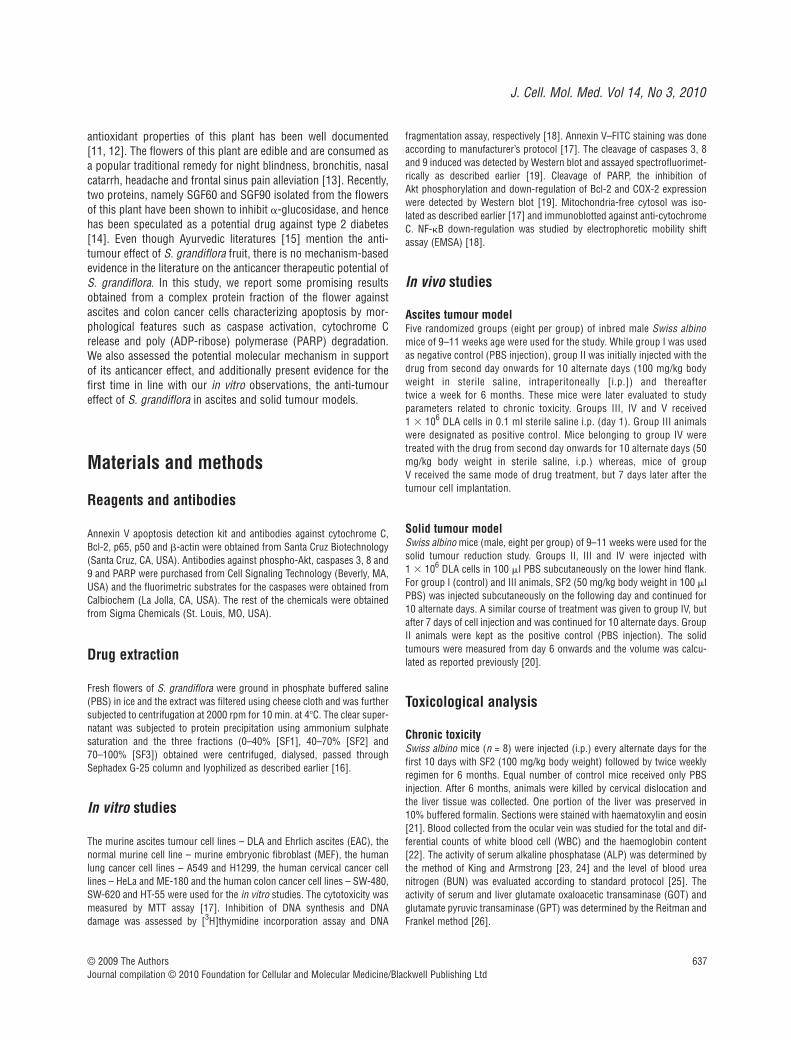
SF2 does not produce any toxicity in the blood,liver or kidney of Swiss albino mice
To rule out the possibility of any adverse side effects of SF2 weconducted a detailed toxicity study, both chronic and acute. In thechronic toxicity study, we evaluated the haematotoxicity, nephro-toxicity and hepatotoxicity due to the SF2 fraction by analysing thelevel of the total and differential WBC count (Fig. 4A), serum ALP(Fig. 4B), level of BUN (Fig. 4C) and GOT and GPT profiles in

J. Cell. Mol. Med. Vol 14, No 3, 2010
639© 2009 The AuthorsJournal compilation © 2010 Foundation for Cellular and Molecular Medicine/Blackwell Publishing Ltd
serum and hepatic tissues (Fig. 4D). Results do not reveal any sig-nificant changes in any of the parameters studied proving that thedrug is non-toxic and pharmacologically safe, in vivo. Thehistopathology data also supported these results (data not shown).
In the acute toxicity study, when the mice were observed forbehavioural changes after i.p. administration of a single dose ofSF2, the groups treated with 100 and 200 mg/kg did not exhibitany abnormal behavioural responses. However, mice whichreceived 500 and 700 mg/kg of SF2 showed toxic symptoms.These include hyperactivity, water intake and sedation. Thesesymptoms appeared almost within 1 hr after the administration of
SF2. All the animals at the dose level of 700 mg/kg and 60% of theanimals at the dose level of 500 mg/kg died within 2 days. The LD50 value for a single i.p. dose was found to be 416 mg/kg.
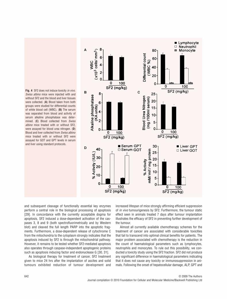
SF2 induces cytotoxicity to human cancer cellsand induces apoptosis in the colon cancer cellline, SW-480
The promising data obtained using the murine ascites cell linesboth in the in vitro and in vivo conditions encouraged us to extend
Fig. 1 SF2 induces cytotoxicity to ascitescells and brings about membrane flip-flopin DLA cell lines. (A) Murine ascites celllines, DLA and EAC (5 � 103) were platedin 96-well plates, treated with indicatedconcentrations of Sesbania protein frac-tions – SF1, SF2 and SF3 for 72 hrs, MTTwas added and viability was determined.(B) DLA cells were seeded as in MTTassay, treated with SF2 for 24 hrs, [3H]thymidine was added during last 6 hrsand thymidine incorporation was meas-ured. For both (A) and (B) the results areexpressed as the mean percentage ofcontrol of quadruplicate determinationsfrom three independent experiments. (C)MEF cells were seeded as mentioned inFig. 1A, treated with indicated concentra-tions of SF2 for 72 hrs, MTT was addedand viability was determined asdescribed. (D) Cells were treated withSF2 for 16 hrs, washed with PBS andstained with annexin V–FITC/propidiumiodide mixture, and photographed (20�).Annexin V positive cells were countedand expressed graphically.

640 © 2009 The AuthorsJournal compilation © 2010 Foundation for Cellular and Molecular Medicine/Blackwell Publishing Ltd
the study in human cancer cell lines. We screened human cancercell lines of various origins. Even though all the investigated cellsshowed cytotoxicity on exposure to the SF2 fraction, in the rangefrom 1 to 25 �g/ml, colon cancer cells were more sensitive (Fig. 5A).Among the colon cancer cell lines studied, SW-480 was the mostsensitive (Fig. 5A) and the IC50 was found to be 10 �g/ml. SF2also inhibited DNA synthesis in all the three colon cancer cell lineswith maximum inhibition in SW-480 cells (Fig. 5B). Hence wefocused on this cell line for further investigations.
Spectrofluorimetric analysis using fluorogenic substrates spe-cific for caspases showed that SF2 activates caspases 3, 8 and 9in a dose-dependent manner in SW-480 cells (Fig. 5C). Additionalevidence supporting SF2-induced apoptosis was obtained byannexin V–propidium iodide staining in which 16% and 37% of the
cells were annexin V positive after treatment with 5 and 10 �g/mlof SF2, respectively, for 16 hrs. Some cells also showed typical PIstaining, especially with 10 �g/ml of SF2 treatment (Fig. 5D). Ourresults support the notion that SF2 treatment initiates signallingpathways leading to apoptosis.
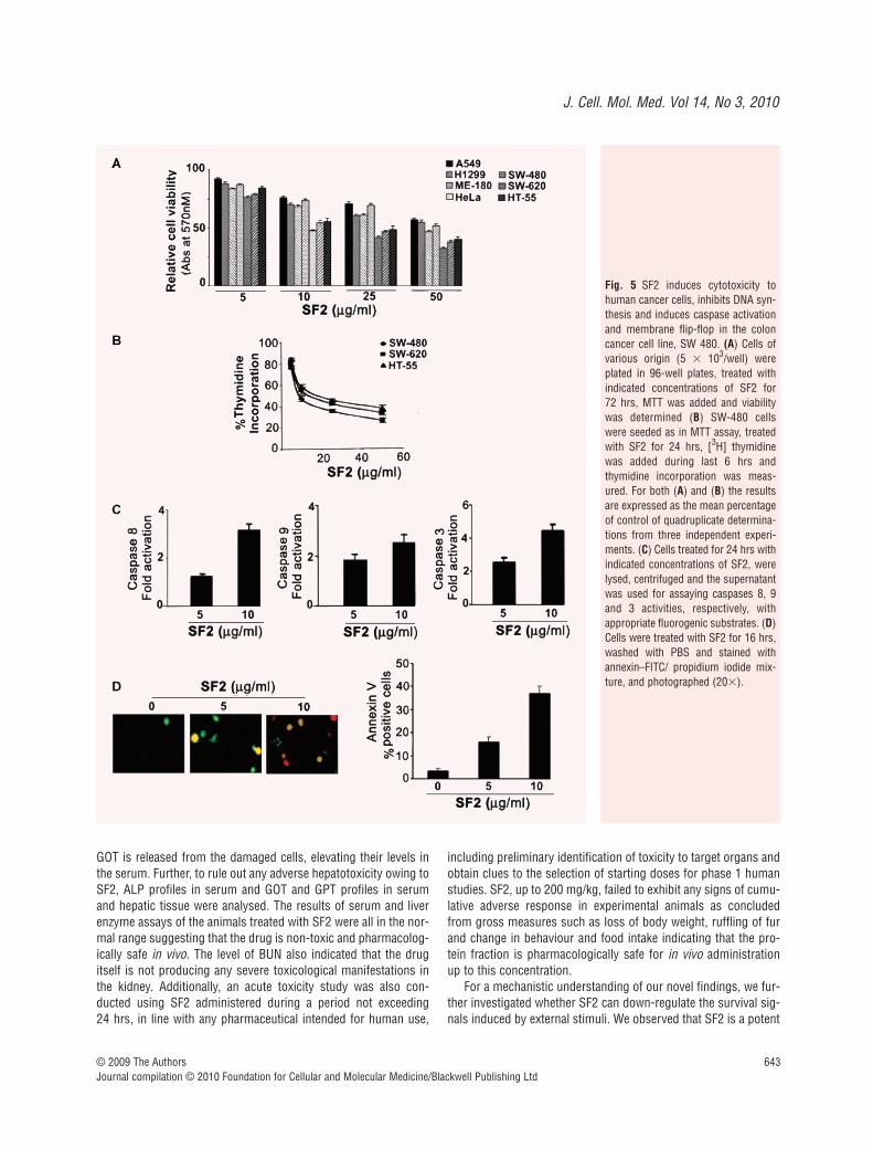
SF2 down-regulates the activation of anti-apoptotic factors, NF-�B, Bcl-2, Akt and COX-2 induced by PMA
We also explored whether SF2 can down-regulate various survivalsignals which are the key regulators of apoptosis. We observed adose-dependent inhibition of NF-�B by SF2, in SW-480 cells
Fig. 2 SF2 induces activation of caspases,DNA fragmentation, PARP cleavage andcytochrome-C release in DLA cells. (A)Cells treated for 24 hrs with indicated con-centrations of SF2 were lysed, centrifugedand the supernatant was used for assayingthe caspases 8, 9 and 3 activities, respec-tively, using appropriate fluorogenic sub-strates. Cell lysates were blotted againstspecific antibodies for the respective cas-pases. (B) The cells were treated with indi-cated concentrations of SF2 for 24 hrs;DNA was extracted and subjected to elec-trophoresis. (C) Cells treated for 24 hrswith indicated concentrations of SF2, werelysed and blotted against anti-PARP anti-body. (D) Cells were treated with SF2 for24 hrs, and mitochondria-free cytosol wasprepared and blotted against anti-cytochrome-C and �-actin antibodies.

J. Cell. Mol. Med. Vol 14, No 3, 2010
641© 2009 The AuthorsJournal compilation © 2010 Foundation for Cellular and Molecular Medicine/Blackwell Publishing Ltd
treated with PMA, a well-known inducer of NF-�B (Fig. 6A).Antibodies against both p50 and p65 subunits of NF-�B shifted theactive NF-�B complex producing super shifts whereas incubationwith excess unlabelled oligonucleotide containing the NF-�B bind-ing site completely removed the active complex confirming thespecificity of the bands (Fig. 6B). Since SF2 induced the release ofcytochrome C from the mitochondria, the involvement of mito-chondrial pathway is evident in SF2-induced apoptosis. Hence weinvestigated the role of SF2 in regulating Bcl-2, the mitochondrialmembrane bound anti-apoptotic factor. We observed a strong anddose-dependent down-regulation of Bcl-2 by SF2 (Fig. 6C). SF2also down-regulated the expression of COX-2 which also providessurvival advantages to cancer cells (Fig. 6D). Phosphorylation ofAkt, another survival signal that in many cases is regulated by NF-�B [27, 28], was also inhibited by SF2 (Fig. 6E).
Taken together, the above noted novel mechanistic observa-tions strongly attest the potential anticancer efficacy of SF2, inboth in vitro and in vivo model systems.
DiscussionIn this study we have documented findings pertinent to theprospect for therapeutic applications of S. grandiflora. An activefraction (SF2) derived from the flower of S. grandiflora contains apharmacologically safe pro-apoptotic compound. Despite existinganecdotal information on anticancer properties associated withthe fruit of this plant in the Indian system of medicine (Ayurveda),no information exemplifying anti-tumour efficacy of flowers of thisplant exists in the literature. Our studies revealed that flowers ofS. grandiflora are associated with more potent anticancer efficacycompared to the leaves and fruit (data not shown). We elucidatedthe mechanistic pathway through which an active fraction of theflower, SF2, induced apoptosis in murine, as well as in human can-cer cell lines, and confirmed its anticancer effect in mice bearingascites and solid tumours.
Although the mechanism by which SF2 achieves these effectsremains unknown, several studies document that caspase activation
Fig. 3 SF2 inhibits the development of ascites and solid tumours in Swiss Albino mice. (A) Schematic representation of different experimental groupsand treatment schedule for Ascites and solid tumour model. (B) The average survival time of each group of mice in Ascites tumour model. The valuesare mean of eight mice per group S.D. Groups IV and V were compared with group III (P � 0.001). (C) Mean of tumour volume in mice treated withSesbania fraction 2 subcutaneously. The solid tumour size was measured from day 6 onwards and volume was calculated. The values are mean of 8 mice per group S.D. Groups III and IV were compared with group II (P � 0.001).
642 © 2009 The AuthorsJournal compilation © 2010 Foundation for Cellular and Molecular Medicine/Blackwell Publishing Ltd
and subsequent cleavage of functionally essential key enzymesperform a central role in the biological processing of apoptosis[29]. In concordance with the currently acceptable dogma forapoptosis, SF2 induced a dose-dependent activation of the cas-pases 3, 8 and 9 (both spectrofluorimetrically and by Westernblot) and cleaved the full length PARP into the apoptotic frag-ments. Furthermore, a dose-dependent release of cytochrome Cfrom the mitochondria to the cytoplasm strongly indicates that theapoptosis induced by SF2 is through the mitochondrial pathway.However, it remains to be tested whether SF2-mediated apoptosisalso operates through caspase-independent apoptogenic proteinssuch as apoptosis inducing factor and endonuclease G [30, 31].
As biological therapy for treatment of cancer, SF2 treatmentgiven to mice 24 hrs after the implantation of ascites and solidtumours exhibited reduction of tumour development and
increased lifespan of mice strongly affirming efficient suppressionof in vivo tumourigenesis by SF2. Furthermore, the tumour staticeffect seen in animals treated 7 days after tumour implantationillustrates the efficacy of SF2 in preventing further development ofthe tumour.
Almost all currently available chemotherapy schemes for thetreatment of cancer are associated with considerable toxicitiesthat fail to transcend into optimal clinical benefits for patients. Themajor problem associated with chemotherapy is the reduction inthe count of haematological parameters such as lymphocytes,neutrophils and monocytes. To rule out this possibility, we con-ducted a toxicity study using the SF2 fraction. SF2 did not produceany significant difference in haematological parameters indicatingthat it does not cause any toxicity or immunosuppression in ani-mals. Following the onset of hepatocellular damage, ALP, GPT and
Fig. 4 SF2 does not induce toxicity in vivo.Swiss albino mice were injected with andwithout SF2 and the blood and liver tissueswere collected. (A) Blood taken from bothgroups were studied for differential countsof white blood cell (WBC). (B) The serumwas separated from blood and activity ofserum alkaline phosphatase was deter-mined. (C) Blood collected from Swissalbino mice treated with or without SF2,were assayed for blood urea nitrogen. (D)Blood and liver collected from Swiss albinomice treated with or without SF2 wereassayed for GOT and GPT levels in serumand liver using standard protocols.
J. Cell. Mol. Med. Vol 14, No 3, 2010
643© 2009 The AuthorsJournal compilation © 2010 Foundation for Cellular and Molecular Medicine/Blackwell Publishing Ltd
GOT is released from the damaged cells, elevating their levels inthe serum. Further, to rule out any adverse hepatotoxicity owing toSF2, ALP profiles in serum and GOT and GPT profiles in serumand hepatic tissue were analysed. The results of serum and liverenzyme assays of the animals treated with SF2 were all in the nor-mal range suggesting that the drug is non-toxic and pharmacolog-ically safe in vivo. The level of BUN also indicated that the drugitself is not producing any severe toxicological manifestations inthe kidney. Additionally, an acute toxicity study was also con-ducted using SF2 administered during a period not exceeding 24 hrs, in line with any pharmaceutical intended for human use,
including preliminary identification of toxicity to target organs andobtain clues to the selection of starting doses for phase 1 humanstudies. SF2, up to 200 mg/kg, failed to exhibit any signs of cumu-lative adverse response in experimental animals as concludedfrom gross measures such as loss of body weight, ruffling of furand change in behaviour and food intake indicating that the pro-tein fraction is pharmacologically safe for in vivo administrationup to this concentration.
For a mechanistic understanding of our novel findings, we fur-ther investigated whether SF2 can down-regulate the survival sig-nals induced by external stimuli. We observed that SF2 is a potent
Fig. 5 SF2 induces cytotoxicity tohuman cancer cells, inhibits DNA syn-thesis and induces caspase activationand membrane flip-flop in the coloncancer cell line, SW 480. (A) Cells ofvarious origin (5 � 103/well) wereplated in 96-well plates, treated withindicated concentrations of SF2 for 72 hrs, MTT was added and viabilitywas determined (B) SW-480 cellswere seeded as in MTT assay, treatedwith SF2 for 24 hrs, [3H] thymidinewas added during last 6 hrs andthymidine incorporation was meas-ured. For both (A) and (B) the resultsare expressed as the mean percentageof control of quadruplicate determina-tions from three independent experi-ments. (C) Cells treated for 24 hrs withindicated concentrations of SF2, werelysed, centrifuged and the supernatantwas used for assaying caspases 8, 9and 3 activities, respectively, withappropriate fluorogenic substrates. (D)Cells were treated with SF2 for 16 hrs,washed with PBS and stained withannexin–FITC/ propidium iodide mix-ture, and photographed (20�).
644 © 2009 The AuthorsJournal compilation © 2010 Foundation for Cellular and Molecular Medicine/Blackwell Publishing Ltd
inhibitor of NF-�B, a transcription factor which is currently envis-aged to be therapeutically beneficial in cancer treatment armento-rium. In recent years, naturally occurring compounds have gainedconsiderable attention because of their alleged therapeuticallybeneficial effects attributable to multimodal actions including inac-tivation of NF-�B and other survival signalling cascades and fewerassociated toxicities. Nuclear translocation of NF-�B and its bind-ing to DNA at specific �B-sites, rapidly induce a variety of genesincluding COX-2 [32]. Furthermore, NF-�B induction up-regulatesgenes that antagonizes pro-apoptotic signals, including Akt,thereby suppressing cell death pathways [33]. It was also veryinteresting to note that inhibition of NF-�B by SF2 was accompa-nied by rapid down-regulation of the anti-apoptotic protein moi-eties – COX-2 and Akt. Akt/PKB has been shown to regulate IKKactivity in both direct and indirect manners [34]. It is known thatAkt may act through the NF-�B pathway and that the COX-2 geneis regulated at the promoter level by NF-�B [35]. It has also beenreported that inhibition of COX-2 leads to inhibition of Akt, whichin turn promotes apoptosis [36]. Recent studies indicate that con-stitutive COX-2 expression via constitutive NF-�B expression may
be the principal mechanism for gastric carcinogenesis andtumourigenesis [37]. Epidemiological, animal and human dataindicate that inhibitors of cyclooxygenase are chemopreventive forcolon cancer in which COX-2 is overexpressed in 80–85% ofcases [38]. In our study, we observed a significant down-regula-tion of COX-2 by SF2 in colon cancer cells, which strongly indi-cates a possible role for SF2 as a putative chemopreventive agentagainst colon carcinogenesis.
Several types of cancer cells constitutively express NF-�B, whichcontribute to their resistance to apoptosis induced by chemothera-peutic drugs [39]. Extensive studies reported in literature indicatethat the anti-apoptotic protein Bcl-2 inhibits the release of apopto-genic cytochrome C from mitochondria into the cytosol [40, 41],suggesting that Bcl-2 may participate in stabilizing mitochondria[42]. Aberrant overexpression of Bcl-2 rescues precancerous cellclones from apoptotic elimination. Existing data suggest a potentialrole for Bcl-2 in altering NF-�B activity [43, 44]. It has been sug-gested that signalling pathways that lead to NF-�B down-regulationare mediated by one of the Bcl-2 homology domains that are con-served among members of the Bcl-2 gene family [45].
Fig. 6 SF2 down-regulates the activation of NF-�B, phosphorylation of Akt and up-regulation of Bcl-2 and COX-2 induced by PMA. (A) SW-480 cellswere treated with or without PMA for 1 hr and then with SF2 for 2 hrs as indicated, nuclear extracts were prepared, and EMSA was done. (B) The nuclearextracts from PMA-stimulated cells were incubated with either p65 or p50 antibody or unlabeled oligo. The arrowhead indicates the positions of theactive DNA binding complex of NF-�B. (C) SW-480 cells were treated with or without PMA for 6 hrs and then with SF2 for 24 hrs, as indicated and thewhole cell lysate was resolved on 15% gel and blotted against Bcl-2 antibody. (D) SW-480 cells were treated with or without PMA for 1 hr and then withSF2 for 24 hrs and the whole cell lysate was resolved on 8% gel and blotted against COX-2 antibody. (E) SW-480 cells were treated with or withoutPMA for 1 hr and then with SF2 for 2 hrs and the whole cell lysate was resolved on 10% gel and blotted against phospho-Akt serine 473.

J. Cell. Mol. Med. Vol 14, No 3, 2010
645© 2009 The AuthorsJournal compilation © 2010 Foundation for Cellular and Molecular Medicine/Blackwell Publishing Ltd
Several studies including ours have shown that antioxidantsand vitamins significantly down-regulate anti-apoptotic factorscontributing to anticancer effect [46, 47]. S. grandiflora is a richsource of amino acids, essential minerals, and vitamins such asvitamin A, vitamin C, thiamine, riboflavin and nicotinic acid, andrecent studies demonstrate the hypolipidemic as well as antioxi-dant efficacy of Sesbania [12, 48–50]. Taken together, theseobservations indicate additional mechanisms by which Sesbaniaprovides protection to DNA from oxidative damage induced byvarious carcinogens leading to the ‘initiation’ of cancer.
As further corollary to our study, we also separated the SF2fraction by column chromatography and cytotoxicity of the frac-tions obtained were assessed by MTT assay. Unfortunately, noneof the components induced significant cytotoxicity (data notshown) which indicates that various components in the SF2 frac-tion may afford a synergistic cytotoxic effect. Further studies areunderway to isolate active compounds and to explore molecularpathways associated with chemoprotection and chemoprevention.Additionally, multiple signal transduction pathways regulating the
apoptotic program induced by SF2 are also being actively pursuedso as to make the formulation an interesting candidate which canbe used in the treatment of colon cancer and other malignancies.
In conclusion, the results of this study demonstrate that apotent protein fraction isolated from flower of the medicinal plantS. grandiflora without any undesired toxicity potentiates apoptosis,reduces tumour cell viability and interferes in abrogating prolifera-tive signals which are otherwise conducive for tumour growth.
Acknowledgements
We acknowledge L. T. Gayathri, C. N. Sreekanth and B. S. Vinod for tech-nical help, and Kristin Dominiak for editorial assistance and proof read-ing. This work was supported in part by a program support from theDepartment of Biotechnology, Government of India (to R.J.A.) andgrants from the Department of Science and Technology, Government ofIndia (to K.P.L.).
References
1. Ferantinos L. Human consumption ofSesbania grandiflora. In: Macklin B, EvansDO, editors. Perennial Sesbania species inagroforestry systems. Hawaii: NFTA; 1990.p. 105.
2. Chaudhari RH. Comparative pharmacolo-gistic studies on the stem bark of Sesbaniasesban (L) and S.grandiflora (L). Bull Bot.Surv Ind. 1996; 8: 216–20.
3. Joshy SG. Leguminoceae. Text book ofmedicinal plants. New Delhi: Oxford andTBH Publishing Co. Pvt. Ltd.; 2000. p. 130.
4. Thamboli S. Antiinflammatory activity of S. grandiflora. Indian Drugs. 1996; 33:504–6.
5. Thamboli SA. Analgesic and antipyreticactivity of S. grandiflora. Indian Drugs.2000; 37: 95–8.
6. Kasture VS, Deshmukh VK, Chopde CT.Anxiolytic and anticonvulsive activity ofSesbania grandiflora leaves in experimentalanimals. Phytother Res. 2002; 16: 455–60.
7. Vijayakumar G. Preliminary studies on theeffect of indigenous plant S. grandiflora incarbon tetrachloride induced hepatitis incalves. Indian J. Veterenery Med. 1997;17: 17–21.
8. Tewtrakal SS, Rattanasuwan P. HIV-1 pro-tease inhibitory effects of some selectedplants in Caesalpiniaceae and Papilionaceaefamilies. J. Sci. Technol. 2003; 25: 509–14.
9. Ramesh T, Begum V. Protective effect ofSesbania grandiflora against cigarettesmoke-induced oxidative damage in rats.J Med Food. 2008 11: 369–75.
10. Pari LUA. Protective effect of Sesbaniagrandiflora against erythromycin estolate-induced hepatotoxicity. Therapie. 2003; 5:439–43.
11. Doddola SPH, Koganti B, Prasad KV.Evaluation of Sesbania grandiflora forantiurolithiatic and antioxidant properties.Nat Med. 2008; 3: 300–7.
12. Ramesh TMR, Hazeena Begum V. Effectof Sesbania grandiflora on lung antioxidantdefense system in cigarette smokeexposed rats. Int J Biol Chem. 2007; 1:141–8.
13. Sivarajan VV, Indira B. Ayurvedic drugsources. New Delhi: Oxford & IBHPublishing Co., New Delhi, India; 1995.
14. Boonmee A, Reynolds CD, Sangvanich P.Alpha-glucosidase inhibitor proteins fromSesbania grandiflora flowers. Planta Med.2007; 73: 1197–201.
15. Kirithikar KR, Basu BD. Indian medicinalplants. Vol. 1. New Delhi: InternationalBook Distributors; 1967. pp. 735–6.
16. D’Silva I, Heath MC. Purification andcharacterization of two novel hypersensi-tive response-inducing specific elicitorsproduced by the cowpea rust fungus. JBiol Chem. 1997; 272: 3924–7.
17. Anto RJ, Mukhopadhyay A, Denning K, et al. Curcumin (diferuloylmethane)induces apoptosis through activation ofcaspase-8, BID cleavage and cytochrome crelease: its suppression by ectopic expres-sion of Bcl-2 and Bcl-xl. Carcinogenesis.2002; 23: 143–50.
18. Anto RJ, Maliekal TT, Karunagaran D. L-929 cells harboring ectopically expressedRelA resist curcumin-induced apoptosis.J Biol Chem. 2000; 275: 15601–4.
19. Anto RJ, Venkatraman M, KarunagaranD. Inhibition of NF-�B sensitizes A431cells to epidermal growth factor-inducedapoptosis, whereas its activation byectopic expression of RelA confers resist-ance. J Biol Chem. 2003; 278: 25490–8.
20. Ruby AJ, Kuttan G, Babu KD, et al. Anti-tumour and antioxidant activity of naturalcurcuminoids. Cancer Lett. 1995; 94: 79–83.
21. Prasad SB, Giri A. Antitumor effect ofcisplatin against murine ascites Dalton’slymphoma. Indian J Exp Biol. 1994; 32:155–62.
22. Mukherjee KL. Medical laboratory tech-nology. A procedure manual for routinediagnostic tests. Vol. 2. New Delhi: TataMcGraw Hill Publishers; 1988.
23. Moss DW, Baron DN, Walker PG, et al.The King-Armstrong method. J ClinPathol. 1972; 25: 368–9.
24. Narayanan S. Alkaline phosphatase astumor marker. Ann Clin Lab Sci. 1983; 13:133–6.
25. Wootton I. Micro analysis in medical bio-chemistry. 4th ed. A and J Churchill LondonLtd.; 1964.
26. Reitman S, Frankel S. A colorimetricmethod for the determination of serumglutamic oxalacetic and glutamic pyruvictransaminases. Am J Clin Pathol. 1957;28: 56–63.
646 © 2009 The AuthorsJournal compilation © 2010 Foundation for Cellular and Molecular Medicine/Blackwell Publishing Ltd
27. Pianetti S, Arsura M, Romieu-Mourez R,et al. Her-2/neu overexpression inducesNF-�B via a PI3-kinase/Akt pathwayinvolving calpain-mediated degradation ofIkappaB-alpha that can be inhibited by thetumor suppressor PTEN. Oncogene. 2001;20: 1287–99.
28. Ozes ON, Mayo LD, Gustin JA, et al. NF-�B activation by tumour necrosis fac-tor requires the Akt serine-threoninekinase. Nature. 1999; 401: 82–5.
29. Miller DK. The role of the Caspase familyof cysteine proteases in apoptosis. Semin Immunol. 1997; 9: 35–49.
30. Du J, Suzuki H, Nagase F, et al.Methylglyoxal induces apoptosis in Jurkatleukemia T cells by activating c-Jun N-terminalkinase. J Cell Biochem. 2000; 77: 333–44.
31. Li H, Zhu H, Xu CJ, et al. Cleavage of BIDby caspase 8 mediates the mitochondrialdamage in the Fas pathway of apoptosis.Cell. 1998; 94: 491–501.
32. Santoro MG, Rossi A, Amici C. NF-�B andvirus infection: who controls whom. EMBOJ. 2003; 22: 2552–60.
33. Meng F, Liu L, Chin PC, et al. Akt is adownstream target of NF-�B. J Biol Chem.2002; 277: 29674–80.
34. Kane LP, Shapiro VS, Stokoe D, et al.Induction of NF-�B by the Akt/PKB kinase.Curr Biol. 1999; 9: 601–4.
35. St-Germain ME, Gagnon V, Parent S, et al. Regulation of COX-2 protein expres-sion by Akt in endometrial cancer cells is
mediated through NF-�B /I-�B pathway.Mol Cancer. 2004; 3: 1–11.
36. Fan XM, Jiang XH, Gu Q, et al. Inhibitionof Akt/PKB by a COX-2 inhibitor inducesapoptosis in gastric cancer cells.Digestion. 2006; 73: 75–83.
37. Joo Weon Lim, Hyeyoung Kim, KyungHwan Kim. Expressions of Ku70 and Ku80mediated by NF-�B and cyclooxygenase-2are related to cell proliferation in humangastric cancer cells J Biol Chem. 2002;277: 46093–100.
38. Williams CS, Mann M, DuBois RN. Therole of cyclooxygenases in inflammation,cancer, and development. Oncogene.1999; 18: 7908–16.
39. Cusack JC, Liu R, Baldwin AS. NF-�B andchemoresistance: potentiation of cancerdrugs via inhibition of NF-�B. Drug ResistUpdat. 1999; 2: 271–3.
40. Kluck RM, Bossy-Wetzel E, Green DR, et al. The release of cytochrome C frommitochondria: a primary site for Bcl-2 reg-ulation of apoptosis. Science. 1997; 275:1132–6.
41. Yang JLX, Bhalla K. Prevention of apopto-sis by Bcl-2: release of cytochrome c frommitochondria blocked. Science. 1997; 275:1129–32.
42. Green DR, Reed JC. Mitochondria andapoptosis. Science. 1998; 281: 1309–12.
43. Tamatani M, Che YH, Matsuzaki H, et al.Tumor necrosis factor induces Bcl-2 andBcl-x expression through NF-�B activation
in primary hippocampal neurons. J. Biol.Chem. 1999; 274: 8531–8.
44. Zong WX, Edelstein LC, Chen C, et al.The prosurvival Bcl-2 homolog Bfl-1/A1 isa direct transcriptional target of NF-�B thatblocks TNF-alpha induced apoptosis.Genes Dev. 1999; 13: 382–7.
45. Stefan G, Patrick AB, Klaus S-O. Bcl-2Down-regulates the activity of transcrip-tion factor NF-�B induced upon apoptosis.J Cell Biol. 1996; 134: 13–23.
46. Bava SV, Puliappadamba VT, Deepti A,et al. Sensitization of taxol-induced apop-tosis by curcumin involves down-regula-tion of NF-�B and the serine/threoninekinase Akt and is independent of tubulinpolymerization. J Biol Chem. 2005; 280:6301–8.
47. Kenichi Satake ET, Akiko I, Yasumasa K,et al. Anti-tumor effect of vitamin A and Don head and neck squamous cell carci-noma. Auris Nasus Larynx. 2003; 30;403–12.
48. The wealth of India: raw materials. NewDelhi: Council of Scientific IndustrialResearch; 1972: 9. pp. 295–8.
49. Ramesh T, Hazeena Begum V.Hypolipidemic effect of Sesbania grandi-flora on cigarette smoke exposed rats.Pharmacologyonline. 2006; 3: 309–23.
50. Ramesh T, Mahesh R, Hazeena Begum V.Effect of Sesbania grandiflora on membrane-bound ATPases in cigarette smoke exposedrats. J Pharmacol Toxicol. 2007; 2: 559–66.
Copyright © 2022 FDOKUMEN