Exogenous metallothionein-IIA promotes accelerated healing after a burn wound
This article was downloaded by: [Universitaetsbibliothek Bayreuth], [Mr Hartmut Frank]On: 08 June 2012, At: 07:28Publisher: Taylor & FrancisInforma Ltd Registered in England and Wales Registered Number: 1072954 Registeredoffice: Mortimer House, 37-41 Mortimer Street, London W1T 3JH, UK
Toxicological & EnvironmentalChemistryPublication details, including instructions for authors andsubscription information:http://www.tandfonline.com/loi/gtec20
Effects of copper on lipid peroxidation,glutathione, metallothionein,and antioxidative enzymes in thefreshwater mussel Anodonta anatinaAndhika Puspito Nugroho a b & Hartmut Frank aa Environmental Chemistry and Ecotoxicology, University ofBayreuth, D-95440 Bayreuth, Germanyb Laboratory of Ecology, Faculty of Biology, Gadjah MadaUniversity, Yogyakarta 55281, Indonesia
Available online: 13 Mar 2012
To cite this article: Andhika Puspito Nugroho & Hartmut Frank (2012): Effects of copper on lipidperoxidation, glutathione, metallothionein, and antioxidative enzymes in the freshwater musselAnodonta anatina , Toxicological & Environmental Chemistry, 94:5, 918-929
To link to this article: http://dx.doi.org/10.1080/02772248.2012.675156
PLEASE SCROLL DOWN FOR ARTICLE
Full terms and conditions of use: http://www.tandfonline.com/page/terms-and-conditions
This article may be used for research, teaching, and private study purposes. Anysubstantial or systematic reproduction, redistribution, reselling, loan, sub-licensing,systematic supply, or distribution in any form to anyone is expressly forbidden.
The publisher does not give any warranty express or implied or make any representationthat the contents will be complete or accurate or up to date. The accuracy of anyinstructions, formulae, and drug doses should be independently verified with primarysources. The publisher shall not be liable for any loss, actions, claims, proceedings,demand, or costs or damages whatsoever or howsoever caused arising directly orindirectly in connection with or arising out of the use of this material.
Toxicological & Environmental ChemistryVol. 94, No. 5, May 2012, 918–929
Effects of copper on lipid peroxidation, glutathione, metallothionein,
and antioxidative enzymes in the freshwater mussel Anodonta anatina
Andhika Puspito Nugrohoab and Hartmut Franka*
aEnvironmental Chemistry and Ecotoxicology, University of Bayreuth, D-95440 Bayreuth,Germany; bLaboratory of Ecology, Faculty of Biology, Gadjah Mada University,Yogyakarta 55281, Indonesia
(Received 20 August 2011; final version received 8 March 2012)
Copper is an essential element to all animals. At elevated concentrations, it istoxic and can participate in the formation of reactive oxygen species, leading tocellular damage. In this study, the ecotoxicological relevance of copper wasinvestigated with freshwater mussels, Anodonta anatina. When the mussels wereexposed to copper at environmentally realistic concentrations, either via the water(0.3 mmolL�1 Cu) or fed with Cu-loaded algae (equivalent to 0.06mmolL�1 Cu),the level of thiobarbituric acid-reactive substances rose and glutathionedecreased. This was associated with the induction of metallothionein and,relative to total protein, of glutathione reductase and the antioxidative enzymessuperoxide dismutase, catalase, and glutathione peroxidase. But, since the overallprotein-synthetic capacity was hampered by the copper insult, the activities of theenzymes relative to tissue weight and copper concentrations were depressed.During depuration, most parameters started to normalize although not returningto control values within 12 days.
Keywords: copper; Anodonta anatina; thiobarbituric acid-reactive substances;glutathione; metallothionein; antioxidative enzymes
Introduction
Metals are brought to the earth’s surface by mining for a multitude of agricultural,industrial, and technological applications. One of the technologically important metals iscopper (Cu), used for electrical power installations and in the building sector, as animalfeed additive or fungicide, as part of machineries, vehicles, electric appliances, and in manyother consumer products. During its use, it is released by corrosion and/or abrasion,mobilized as particulate matter and dry or wet deposited, to some extent ending up in thesediments of freshwater ecosystems (Smolders et al. 2003). In non-contaminatedfreshwater ecosystems, its concentrations range from 0.02 to 0.3 mmolL�1 (1–20 mgL�1)(Momcilovic 2004). Close to mining activities, aquatic copper pollution can reach levels ofup to 30 mmolL�1 (1.7mgL�1) (Smolders et al. 2003).
Mussels live at the interface of free-flowing waters and sediments and may bechronically exposed to copper for long time periods or intermittently at fluctuating levels,depending upon temporary hydrological conditions and extent of sediment oxygenation
*Corresponding author. Email: [email protected]
ISSN 0277–2248 print/ISSN 1029–0486 online
� 2012 Taylor & Francis
http://dx.doi.org/10.1080/02772248.2012.675156
http://www.tandfonline.com
Dow
nloa
ded
by [
Uni
vers
itaet
sbib
lioth
ek B
ayre
uth]
, [M
r H
artm
ut F
rank
] at
07:
28 0
8 Ju
ne 2
012
(Bhaduri et al. 2000; Poot, Gillissen, and Koelmans 2007). Copper is an essential elementof their circulatory oxygen carrier hemocyanin (Momcilovic 2004) and plays a role ascofactor of a number of enzymes such as cytochrome oxidase, superoxide dismutase(SOD), alcohol dehydrogenase, dopamine hydroxylase, tyrosinase, and lysyl oxidase(Serafim and Bebianno 2009). However, at excessive concentrations copper can participatein the formation of reactive oxygen species (ROS) through a Haber–Weiss cycle,producing hydroxyl radicals (�OH) from hydrogen peroxide (H2O2) and superoxide (O�2 )(Bigot et al. 2011; Company et al. 2008). ROS may cause cellular damage by lipidperoxidation when the antioxidative defense systems of aquatic animals are overwhelmed,leading to inactivation of membrane enzymes, destruction of proteins (Remmer et al.1989), and changes in the DNA structure (Company et al. 2008; Lackner 1998; Serafimand Bebianno 2009).
Mussels can cope with moderately elevated copper in various ways (Serafim andBebianno 2009). In the cytosol, glutathione (GSH) and metallothionein (MT), the latter afamily of cysteine-rich proteins (Ivankovic et al. 2010), provide protection againstincreased concentrations through binding the copper ions to the thiol groups of theircysteine residues (Company et al 2008; Freedman, Ciriolo, and Peisach 1989). Otherstrategies against copper-induced oxidative toxicity is the induction of enzymes such asSOD, catalase (CAT), glutathione peroxidase (GPX), and glutathione reductase (GR)(Isani et al. 2003).
Copper has been observed in high concentrations in the tissue of freshwater pearlmussels Margaritifera margaritifera (Frank and Gerstmann 2007) and other Europeanfreshwater mussel species (Tallandini et al. 1986). Their populations are strongly affectedEurope-wide and some are threatened with extinction (Cuttelod, Seddon, and Neubert2011). Understanding the potential involvement of Cu in this phenomenon is the majormotivation for this study.
In this work, Anodonta anatina is used as model species. In previous publications,it has been shown that A. anatina can accumulate copper from the water or by feedingon copper-containing algae (Nugroho and Frank 2011b). This article focuses on theeffects of copper on MT and GSH and on antioxidative enzymes as response tooxidative stress, signaled by increased levels of thiobarbituric acid-reactive substances(TBARS).
Materials and methods
Chemicals
Isotopically enriched (99%) 63Cu oxide (Euriso-top, Saarbrucken, Germany) was used.Concentrated HNO3 (69%) and concentrated HCl (30%) were of suprapure grade (Merck,Darmstadt, Germany); other chemicals (Carl Roth, Karlsruhe, Germany; Sigma-Aldrich,Munich, Germany) were of analytical grade. Cleaning of labware and preparation ofthe Cu2þ stock solution are described in a previous publication (Nugroho andFrank 2011b).
Animals and experimental design
Seventy duck mussels (A. anatina) (ZOO-Erlebnis Online Shop, Grossefehn, Germany)with shell lengths of 10–12 cm and weights between 100 and 200 g were brought to thelaboratory in pond water. Mussel handling, acclimatization, and experimental design
Toxicological & Environmental Chemistry 919
Dow
nloa
ded
by [
Uni
vers
itaet
sbib
lioth
ek B
ayre
uth]
, [M
r H
artm
ut F
rank
] at
07:
28 0
8 Ju
ne 2
012
(Nugroho and Frank 2011b) as well as the preparation of normal and Cu-loaded algaehave been described earlier (Nugroho and Frank 2011a). The mussels were divided intothree groups consisting of 21 mussels each. The first group was kept in artificial pondwater (APW); the second one was exposed to 0.3 mmolL�1 (20mgL�1) 63Cu2þ in the water;the third group received daily 1.5mgL�1 freeze-dried 63Cu-loaded algae (40mmol 63Cuper kg dry weight) for 24 days, equivalent to a nominal concentration of 0.06 mmol(3.6 mgL�1) 63Cu per liter APW.
For sampling, three mussels of each group were taken for analysis at days 0, 6, 12, 18,and 24 (exposure), and at days 30 and 36 (depuration). The mussels’ soft bodies weredissected on ice into gills, mantle, kidney, and digestive gland. Two aliquots of every tissuefraction, about 5–10mg each, were placed in separate 2-mL microtubes of known weight.The first aliquot was used for the determination of MT and the second one for thedetermination of TBARS, GSH, enzyme activities, and proteins. All microtubes were keptin a freezer at �80�C until further analysis. The remainders of the tissues were placed in15-mL polypropylene (PP) tubes of known weights and were lyophilized for copperdetermination.
Analytical methods
Sample preparation
Frozen tissue samples in microtubes were thawed and immediately mixed with 500 mLsucrose (0.5mol L�1)/Tris-HCl (20mmol L�1; pH 8.6) buffer, to which leupeptine(6 mmolL�1) and phenylmethanesulfonylfluoride (PMSF) (0.5mmol L�1) were added asanti proteolytic agents and �-mercaptoethanol (0.01%) as reducing agent. The mixtureswere sonicated in an ice bath with 12 strokes of a sonicator (Labsonic U tip sonicator, B.Braun Biotech International, Melsungen, Germany) at 20 kHz, acoustic power 50W. Thehomogenates were centrifuged at 4�C for 30min at 10,000� g (Heraeus Multifuge 1L-R,Thermo Scientific, Osterode, Germany). Supernatants were used for MT determination.
For the determination of TBARS, GSH, enzyme activities, and proteins, frozen tissuesamples of 5–10mg were thawed and immediately mixed with 500 mL phosphate buffer(50mmol L�1; pH 7.4) containing 150mmol L�1 KCl, 1mmol L�1 ethylenediaminetetra-acetic acid (EDTA), 1mmol L�1 dithiothreitol (DTT), and 0.01% (w/v) PMSF. Thesamples were homogenized in an ice bath with 12 strokes of a sonicator at 20 kHz, acousticpower 50W, and centrifuged at 4�C for 30min at 10,000� g. The supernatants were usedfor analysis.
Total copper
Total copper in lyophilized tissues and freeze-dried algal food, and – every second day –the actual copper concentrations in APW were determined by inductively-coupled plasmamass spectrometry. Details have been described previously (Nugroho and Frank 2011b).
Lipid peroxidation
Lipid peroxidation was determined following the method of Buege and Aust (1978) bymeasuring TBARS, expressed as malondialdehyde (MDA) equivalents. Absorbances ofsamples were read at 535 nm with a microplate reader (Biotek Synergy HT, BadFriedrichshall, Germany). TBARS levels were estimated using a standard curve obtained
920 A.P. Nugroho and H. Frank
Dow
nloa
ded
by [
Uni
vers
itaet
sbib
lioth
ek B
ayre
uth]
, [M
r H
artm
ut F
rank
] at
07:
28 0
8 Ju
ne 2
012
with 1,1,3,3-tetramethoxypropane (99%; VWR, Darmstadt, Germany) as stable precursor
of MDA and expressed as mmol kg�1 tissue wet weight (tww).
Glutathione
GSH was determined according to Anderson (1985). Absorbances of samples weremeasured at 412 nm with a microplate reader. The GSH content was estimated using a
standard curve obtained with reduced GSH and expressed as mmol kg�1 tww.
Metallothioneins
MT concentrations were determined by the spectrophotometric method of Viarengo et al.
(1997) modified by Verlecar, Jena, and Chainy (2008). Absorbances of samples were readat 412 nm with a microplate reader. The MT content was determined using GSH (Carl
Roth, Karlsruhe, Germany) as standard, assuming that 1 mmol GSH is equivalent to
0.055 mmol MT. Concentrations of MT were expressed as mmol kg�1 tww.
Enzyme activities
SOD activities were determined by the procedure of Beauchamp and Fridovich (1971),based on the inhibition of nitrotetrazolium blue reduction and measuring sample
absorbances at 560 nm. CAT activities were assayed spectrophotometrically according to
Rao, Paliyath, and Ormrod (1996) by monitoring the decrease in the absorbance of H2O2
at 240 nm. GPX activities were determined according to Paglia and Valentine (1967) and
GR activities according to Massey and William (1965) in the presence of GSSG, in both
cases following the rate of NADPH oxidation at 340 nm. Absorbances were measured witha microplate reader; enzyme activities were calculated in units per milligram protein and
per gram tww.
Proteins
Proteins were determined by the dye-binding assay (Kruger 1994). Absorbances of thesamples were read at 595 nm with a microplate reader. The concentrations were
determined using BSA (�96%; Sigma-Aldrich, Munich, Germany) for calibration.
Statistical data analyses
The variability of the observed parameters and of total Cu concentration in the different
organs were tested by two-way analysis of variance (ANOVA) considering exposure time
and copper exposure pathways as independent variables, followed by the Duncan multiplecomparison tests ( p< 0.05) if significant differences were found. Data were transformed to
log(Xþ 1) units before statistical analysis for the homogeneity of variance and normality.
Linear regression analysis was performed for evaluating the relationship between Cuconcentration and the observed parameters, followed by Pearson correlation analysis for
testing the strength of linear relationship.
Toxicological & Environmental Chemistry 921
Dow
nloa
ded
by [
Uni
vers
itaet
sbib
lioth
ek B
ayre
uth]
, [M
r H
artm
ut F
rank
] at
07:
28 0
8 Ju
ne 2
012
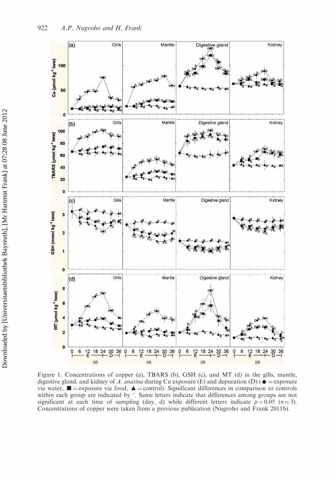
Figure 1. Concentrations of copper (a), TBARS (b), GSH (c), and MT (d) in the gills, mantle,digestive gland, and kidney of A. anatina during Cu exposure (E) and depuration (D) (^¼ exposurevia water, g¼ exposure via food, m¼ control). Significant differences in comparison to controlswithin each group are indicated by �. Same letters indicate that differences among groups are notsignificant at each time of sampling (day, d) while different letters indicate p< 0.05 (n¼ 3).Concentrations of copper were taken from a previous publication (Nugroho and Frank 2011b).
922 A.P. Nugroho and H. Frank
Dow
nloa
ded
by [
Uni
vers
itaet
sbib
lioth
ek B
ayre
uth]
, [M
r H
artm
ut F
rank
] at
07:
28 0
8 Ju
ne 2
012
Results
Copper concentrations in all organs increased during exposure (Figure 1a, taken from the
previous publication Nugroho and Frank 2011b). When Cu was administered via the
water, the levels started to rise instantaneously, reaching a maximum of up to 6.5-fold of
controls at day 24. From the food, Cu increased in the digestive gland to a similarly high
level, but in the gills and mantle it was much lower; for the kidney uptake was moderate.
Upon depuration, Cu was quickly but not completely eliminated, especially not from the
mantle and the digestive gland.Initial TBARS levels (Figure 1b) were highest in the gills and the digestive gland,
i.e., about 60 mmol kg�1 tww; in the mantle and the kidney they were between 25 and
40 mmol kg�1 tww. Upon Cu exposure via the water, TBARS rose significantly within the
first six days, except for the kidney. At day 24, highest levels in relative terms were reached
in the mantle (about double of control), in absolute terms in the gills and digestive gland
(100mmol kg�1 tww); in the kidney, TBARS were elevated by about 60%. Upon Cu
exposure via the food, the effects on the digestive gland and the kidney were almost equally
strong as for exposure via the water, while in the gills and the mantle the slightly increased
levels were not significantly different from controls ( p> 0.05). Correlation analyses
between copper and TBARS confirmed strong relationships in all organs (r> 0.6;
p< 0.05), except for the kidney. During depuration, the concentrations decreased slowly,
the levels after 12 days of depuration being only slightly lower than in the beginning of
depuration.Initial levels of GSH (Figure 1c) were highest in the gills and the kidney, i.e., about
3.0mmol kg�1 tww, somewhat lower in the mantle, i.e., about 2.5mmol kg�1 tww, and
lowest in the digestive gland, i.e., 1.6mmol kg�1 tww. Upon Cu uptake, GSH started to
decrease within the first 6 days at similar relative rates in all organs, reaching lowest levels
at day 24, especially when Cu-exposure took place via the water. In the mantle GSH
decreased by about 40%, in the other organs between 20 (digestive gland and kidney) and
30% (gills) at day 24. Upon depuration, GSH-levels tended to increase but remained lower
than in controls, even after 12 days.Initial MT levels (Figure 1d) ranged from 1.5 (kidney) to 3.0 (gills) mmol kg�1 tww.
Upon Cu exposure via the water, MT increased in parallel to Cu, being significantly
different from control on day 12 and later. Highest MT levels were reached on day 24,
i.e., MT was increased by 300% from control in the digestive gland, 200% in the gills and
mantle, and 100% in the kidney. Upon Cu-exposure via the food, MT increased in the
digestive gland and the kidney almost to the same degree, while in the gills and the mantle
MT levels were only slightly elevated. Correlation analyses between copper and MT
confirmed their strong relation in all organs (r> 0.6; p< 0.05). Upon depuration and
simultaneous with Cu elimination, MT decreased strongly in the gills, the digestive gland,
and the kidney within the first 6 days; in the mantle, MT remained high, parallel to the
slow elimination of Cu.The antioxidative enzymes are presented in Figure 2 in two ways, i.e., relative to
protein contents (open symbols, left ordinate) and to tissue wet weight (tww) (filled
symbols, right ordinate). SOD (Figure 2a) had the highest initial activity in the kidney,
i.e., about 9 units (U) per mg protein (120 U per g tww), for the other organs ranging from
3 to 5 U per mg protein (20–40 U per g tww). For CAT (Figure 2b), the initial levels were
similar for all four organs at about 5 U per mg protein (65 U per g tww). GPX (Figure 2c)
was initially highest in the kidney, i.e., about 0.04 U per mg protein (0.5 U per g tww), in
Toxicological & Environmental Chemistry 923
Dow
nloa
ded
by [
Uni
vers
itaet
sbib
lioth
ek B
ayre
uth]
, [M
r H
artm
ut F
rank
] at
07:
28 0
8 Ju
ne 2
012

Figure 2. SOD (a), CAT (b), GPX (c), and GR (d) activities in the gills, mantle, digestive gland, andkidney of A. anatina during Cu exposure (E) and depuration (D) in units (U) per mg protein (leftordinate;S¼ upon exposure via water, h¼ upon exposure via food, 4¼ control) and in units per gtissue wet weight (tww) (right ordinate; ^¼ exposure via water, g¼ exposure via food,m¼ control). Significant differences in comparison to controls within each group are indicated by�. Same letters indicate that differences among groups are not significant at each time of sampling(day, d) while different letters indicate p< 0.05 (n¼ 3).
924 A.P. Nugroho and H. Frank
Dow
nloa
ded
by [
Uni
vers
itaet
sbib
lioth
ek B
ayre
uth]
, [M
r H
artm
ut F
rank
] at
07:
28 0
8 Ju
ne 2
012
the other organs at about 0.03 U per mg protein (0.25 U per g tww), taking the differencesin protein contents among the organs into account (Nugroho and Frank 2012).
Upon Cu exposure via the water, the activities of the three enzymes relative to proteincontents increased in all organs. CAT and GPX reached highest levels at day 24 in thedigestive gland, the former being increased by 250% from control, the latter about double;SOD behaved similarly. Upon Cu-uptake from the food, the activities of all three enzymesin the digestive gland and the kidney increased almost equally as when exposed via thewater, while in the gills and mantle only small effects were seen. Correlation analysisshowed highly significant correlations (r> 0.6; p< 0.05) between the enzymes and copperin the gills and mantle (water pathway), and the digestive gland and kidney (water andfood pathways). During depuration, the activities decreased, especially of SOD and CATin the digestive gland, although not returning to control values until the end of depuration.
Relative to tissue wet weight, however, SOD and GPX activities decreased. This wasstrongest for SOD (Figure 2a) in the kidney, i.e., to about 50% of control at day 24, andfor GPX in the mantle (40 % of control) and the digestive gland (50 % of control). Uptakeof Cu from the food resulted in decreases in the digestive gland and kidney similar touptake via the water, while in the gills and the mantle SOD and GPX remained at controllevels. CAT activities were largely unchanged, except for the kidney in which it wasdoubled. During depuration, SOD and GPX activities increased in all organs, but not fullyback to control values.
Initial GR levels (Figure 2d) relative to protein contents were highest in the digestivegland (0.1 U per mg protein), in the other organs being only a third (0.03 U per mgprotein). Upon aqueous Cu exposure, increases were strong for the digestive gland and thegills; for the mantle and kidney they were moderate (�50%), but in all cases correlated tocopper (r> 0.6; p< 0.05). Relative to wet weight, the GR activities declined upon Cuexposure via the water, strongest in the digestive gland to reach about 30% of control atday 24, in the other organs about 70%. Upon Cu exposure via the food, GR activities inthe digestive gland were similarly depressed as via the water, in the gills and mantle theyremained at control levels. During depuration, GR began to normalize although not fullyback to control levels.
Discussion
As shown previously (Nugroho and Frank 2011b), uptake of Cu from the water(Figure 1a) led to a general and fast rise of its concentrations in all tissues and organs,while upon Cu-uptake with the food primarily the digestive gland and the intestines (notshown) were burdened.
In respect to lipid peroxidation, increased TBARS reflect the damage to biologicalmembranes by ROS as a consequence of excess Cu (Company et al. 2008). The initialTBARS levels found in the digestive gland of A. anatina (7.0 nmolmg�1 protein) wereabout 7–10-fold higher than those reported by Sabatini et al. (2011) for the freshwatermussel Diplodon chilensis (0.7 nmolmg�1 protein) and by Bouskill et al. (2006) for thezebra mussel Dreissena polymorpha (1.0 nmolmg�1 protein). Parallel to the accumulationof copper, TBARS rose by 60–70% in all organs when the metal was taken up by the waterpathway, while by the food pathway the effect was focused on the digestive gland; thelatter has also been reported by Sabatini et al. (2011), although they used algae with a Cu-contamination level about 800 times higher than in our case and a different feedingschedule. Other researchers reported an increase in TBARS by about 100% in the whole
Toxicological & Environmental Chemistry 925
Dow
nloa
ded
by [
Uni
vers
itaet
sbib
lioth
ek B
ayre
uth]
, [M
r H
artm
ut F
rank
] at
07:
28 0
8 Ju
ne 2
012
tissue (2.0 nmolmg�1 protein) of D. polymorpha upon exposure to water-dissolved Cu at1.6 mmolL�1 (100mgL�1) for 7 days (Bouskill et al. 2006), i.e., at a Cu-concentrationabout 5-fold higher than in this study. For A. anatina, increases by about 50%, e.g., in thedigestive gland, were observed within the same period of exposure.
Consumption of GSH can occur due to its role as a metal-complexing agent, and as anantioxidant and scavenger of reactive intermediates of lipid peroxidation (Canesi et al.1999; Lackner 1998). The initial GSH-levels in the gills (300 nmolmg�1 protein) and thedigestive gland (170 nmolmg�1 protein) of A. anatina were about 2–4-fold lower than inthe same organs of the swollen river mussel Unio tumidus (500 and 600 nmolmg�1 protein)(Doyote et al. 1997). Copper exposure was associated with the depletion of GSH by about20–40% in all organs of A. anatina, irrespective of exposure pathway. Although uponcopper uptake via the food TBARS were predominantly increased in the digestive gland,GSH was lowered in all organs. This suggests a generalized, systemic mobilization andexchange of GSH between organs. For comparison, when U. tumidus was exposed towater-dissolved Cu (Doyotte et al. 1997) at about twice the concentration (0.5 mmolL�1,30 mgL�1) used in our study, GSH in the gills and the digestive gland was lowered byabout 15% within 3 days, comparable to the 20–25% we have observed with A. anatinaafter 6 days of exposure. The same authors have emphasized that the depletion wasassociated with decreased GR activities, similarly as we have observed with A. anatina(Figure 2d).
Binding of copper to MT is a detoxification process and meant to control itsintracellular levels (Company et al. 2008; Serafim and Bebianno 2009). Initial MT levels inthe gills and mantle of A. anatina (2–3mmol kg�1 tww) were about 6–9-fold lower than thatreported for the whole soft tissue of D. polymorpha (about 18 mmol kg�1 tww; Ivankovicet al. 2010). In parallel to Cu uptake, MT levels were induced in all organs via bothexposure pathways, showing dose–response relationships. The increase in molar concen-trations of MT in the gills, mantle, and kidney of A. anatina (Figure 1d) were largelysufficient to complex the extra copper, considering a binding ratio of 12 (Adam et al. 2010;Eisler 1993); for the digestive gland, however, this ratio of extra Cu versus newlysynthesized MT tended to be higher, especially upon Cu-exposure via the food pathway. Inview of the fact that MT may additionally serve as an antioxidant, the induction of MT israther limited and may constitute a considerable physiological challenge. Copper exposureinduced an increase of MT by 10% in the whole soft tissue of D. polymorpha (20mmol kg�1
ww; Ivankovic et al. 2010) upon exposure to water-dissolved Cu at 0.5 mmolL�1
(30 mgL�1) for 7 days, i.e., at a concentration about 1.5-fold higher than our study. Withinthe same period of exposure, the increases were slight lower than all organs of exposedA. anatina (20–30%). Bouskill et al. (2006) also reported a 10% increase of MT in thewhole tissue (500 mgmg�1 protein) of D. polymorpha but exposed for 7 days to1.6 mmolL�1 (100 mgL�1) Cu in water, i.e., more than 5-fold higher than in this study.In field studies, MT in the gills of the giant floater Pyganodon grandis correlated positivelywith elevated Cu concentrations (Bonneris et al. 2005; Perceval et al. 2006). A similar studyalso showed a positive relationship between Cu and MT in the soft tissue of D. polymorphafound in the St Lawrence River, Canada (de Lafontaine et al. 2000).
Relative to protein contents, the major antioxidative enzymes were induced duringcopper exposure (Figure 2), SOD, and CAT in the digestive gland by 60% and 240%,respectively. Sabatini et al. (2011) reported the induction of SOD and CAT activities byonly about 50% relative to protein in the digestive gland of D. chilensis after 4–5 weeks ofexposure via food to much higher Cu concentration (equivalent to 50 mmolL�1). However,relative to tissue wet weight – and thus relative to copper – the activities of SOD, GPX,
926 A.P. Nugroho and H. Frank
Dow
nloa
ded
by [
Uni
vers
itaet
sbib
lioth
ek B
ayre
uth]
, [M
r H
artm
ut F
rank
] at
07:
28 0
8 Ju
ne 2
012
and GR were depressed, especially in the digestive gland (GPX by 30%, GR by 45 % after6 days of exposure), since the overall protein-synthetic capacity was hampered (Nugrohoand Frank 2012). Doyotte et al. (1997) also reported the decrease of GPX andGR activities in the digestive gland (by 15 and 30%) of U. tumidus exposed for 3 days towater-dissolved Cu at 0.5mmolL�1 (30mgL�1), twice the concentration used in this study.
Overall, Cu-exposure of A. anatina obviously entailed considerable oxidative stress,depression of GSH, induction of MT, and perturbations in the activities of GR and theantioxidative enzymes. Exposure to Cu via the water affected all organs including the gillsand mantle, by the food being mainly the digestive gland. Taking into account that in theseexperiments the nominal concentration of copper contained in the food was fairly lowrelative to the copper concentration in the water, exposure via the food and the impact onthe digestive gland was particularly strong, the centrally important organ for digestion,catabolism, and uptake of nutrients and electrolytes such as Ca2þ. In conjunction with theeffects on the kidney and the mantle lasting beyond the actual exposure, and together withthe derangements of calcium homeostasis and of carbohydrate and protein metabolism(Nugroho and Frank 2012), the exposure to copper at moderately elevated environmen-tally realistic levels has profound pathobiochemical consequences.
Conclusions
Overall, mussels are under considerable oxidative stress when exposed to Cu-concentra-tions moderately elevated above natural conditions. Utilization of GSH and induction ofMT constitute a first line of defense. The activation of a second line by induction ofantioxidative enzymes is rather inefficient as protein-synthetic capacities are stronglyaffected. All this evidence suggests that copper at fairly low levels can affect the vitality ofmussels at all life stages, making it likely to contribute to the population declines ofEuropean freshwater bivalves.
Acknowledgments
Financial support by Directorate General of Higher Education, Ministry of National Education ofthe Republic of Indonesia, is highly appreciated.
References
Adam, V., I. Fabrik, T. Eckschlager, M. Stiborova, L. Trnkova, and R. Kizek. 2010. Vertebrate
metallothioneins as target molecules for analytical techniques. Trends in Analytical Chemistry 29:
409–18.
Anderson, M.E. 1985. Determination of glutathione and glutathione disulfide in biological samples.
Methods in Enzymology 113: 543–53.
Beauchamp, C., and I. Fridovich. 1971. Superoxide dismutase: Improved assays and an assay
applicable to polyacrylamide gels. Analytical Biochemistry 44: 276–86.Bhaduri, B., J. Harbor, B. Engel, and M. Grove. 2000. Assessing watershed-scale, long-term
hydrologic impacts of land-use change using a GIS-NPS model. Environmental Management 26:
643–58.Bigot, A., L. Minguez, L. Giamberini, and F. Rodius. 2011. Early defense responses in the
freshwater bivalve Corbicula fluminea exposed to copper and cadmium: Transcriptional and
histochemical studies. Environmental Toxicology 26: 623–32.
Toxicological & Environmental Chemistry 927
Dow
nloa
ded
by [
Uni
vers
itaet
sbib
lioth
ek B
ayre
uth]
, [M
r H
artm
ut F
rank
] at
07:
28 0
8 Ju
ne 2
012
Bonneris, E., O. Perceval, S. Masson, L. Hare, and P.G.C. Campbell. 2005. Sub-cellular partitioning
of Cd, Cu and Zn in tissues of indigenous unionid bivalves living along a metal exposure gradient
and links to metal-induced effects. Environmental Pollution 135: 195–208.Bouskill, N.J., R.D. Handy, T.E. Ford, and T.S. Galloway. 2006. Differentiating copper and arsenic
toxicity using biochemical biomarkers in Asellus aquaticus and Dreissena polymorpha.
Ecotoxicology and Environmental Safety 65: 342–9.Buege, J.A., and S.D. Aust. 1978. Microsomal lipid peroxidation. Methods in Enzymology 52:
302–10.
Canesi, L., A. Viarengo, C. Leonzio, M. Filippelli, and G. Gallo. 1999. Heavy metals and
glutathione metabolism in mussel tissues. Aquatic Toxicology 46: 67–76.Company, R., A. Serafim, R.P. Cosson, A. Fiala-Medioni, L. Camus, A. Colaco, R. Serrao-Santos,
and M.J. Bebianno. 2008. Antioxidant biochemical responses to long-term copper exposure in
Bathymodiolus azoricus from Menez-Gwen hydrothermal vent. Science of the Total Environment
389: 407–17.Cuttelod, A., M. Seddon, and E. Neubert. 2011. European red list of non-marine molluscs.
Luxembourg: Publications Office of the European Union.de Lafontaine, Y., F. Gagne, C. Blaise, G. Costan, P. Gagnon, and H.M. Chan. 2000. Biomarkers in
zebra mussels (Dreissena polymorpha) for the assessment and monitoring of water quality of the St
Lawrence River (Canada). Aquatic Toxicology 50: 51–71.Doyotte, A., C. Cossu, M. Jacquin, M. Babut, and P. Vasseur. 1997. Antioxidant enzymes,
glutathione and lipid peroxidation as relevant biomarkers of experimental or field exposure in the
gills and the digestive gland of the freshwater bivalve Unio tumidus. Aquatic Toxicology 39:
93–110.
Eisler, R. 1993. Zinc hazards to fish, wildlife, and invertebrates: A synoptic review. Laurel, MD: U.S.
Department of the Interior, Fish, and Wildlife Service, Patuxent Wildlife Research Center.Frank, H., and S. Gerstmann. 2007. Declining populations of freshwater pearl mussels
(Margaritifera margaritifera) are burdened with heavy metals and DDT/DDE. Ambio 36: 571–4.Freedman, J.H., M.R. Ciriolo, and J. Peisach. 1989. The role of glutathione in copper metabolism
and toxicity. The Journal of Biological Chemistry 264: 5598–605.
Isani, G., M. Monari, G. Andreani, M. Fabbri, and E. Carpene. 2003. Effect of copper exposure on
the antioxidant enzymes in bivalve mollusk Scapharca inaequivalvis. Veterinary Research
Communications 27: 691–3.Ivankovic, D., J. Pavicic, V. Beatovic, R.S. Klobucar, and G.I.V. Klobucar. 2010. Inducibility of
metallothionein biosynthesis in the whole soft tissue of zebra mussels Dreissena polymorpha
exposed to cadmium, copper, and pentachlorophenol. Environmental Toxicology 25: 198–211.Kruger, N.J. 1994. The Bradford method for protein quantitation. In Methods in molecular biology:
Basic protein and peptide protocols, Vol. 32, ed. J.M. Walker, 9–15. New Jersey: Humana Press.Lackner, R. 1998. ‘‘Oxidative stress’’ in fish by environmental pollutants. In Fish ecotoxicology, eds.
T. Braunbeck, D.E. Hinton and B. Streit, 203–24. Switzerland: Birkhauser Verlag.Massey, V., and C.H. Williams. 1965. On the reaction mechanism of yeast glutathione reductase.
The Journal of Biological Chemistry 240: 4470–81.
Momcilovic, B. 2004. Copper. In Elements and their compounds in the environment: Metals and their
compounds, Vol. 2, eds. E. Merian, M. Anke, M. Ihnat and M. Stoeppler, 731–50. Weinheim:
Wiley-VCH Verlag.Nugroho, A.P., and H. Frank. 2011a. Producing Cu-loaded algae for feeding experiments: Effects of
copper on Parachlorella kessleri. Toxicological and Environmental Chemistry 93: 537–48.Nugroho, A.P., and H. Frank. 2011b. Uptake, distribution, and bioaccumulation of copper in the
freshwater mussel Anodonta anatina. Toxicological and Environmental Chemistry 93: 1838–50.
Nugroho, A.P., and H. Frank. 2012. Effects of copper exposure on calcium, carbohydrate, and
protein levels in the freshwater mussel Anodonta anatina. Toxicological and Environmental
Chemistry 94: 99–108.Paglia, D.E., and W.N. Valentine. 1967. Studies on quantitative and qualitative characterization of
erythrocyte glutathione peroxidase. Journal of Laboratory and Clinical Medicine 70: 158–69.
928 A.P. Nugroho and H. Frank
Dow
nloa
ded
by [
Uni
vers
itaet
sbib
lioth
ek B
ayre
uth]
, [M
r H
artm
ut F
rank
] at
07:
28 0
8 Ju
ne 2
012
Perceval, O., Y. Couillard, B. Pinel-Alloul, E. Bonneris, and P.G.C. Campbell. 2006. Long-termtrends in accumulated metals (Cd, Cu and Zn) and metallothionein in bivalves from lakes within asmelter-impacted region. Science of the Total Environmental 369: 403–18.
Poot, A., F. Gillissen, and A.A. Koelmans. 2007. Effects of flow regime and flooding on heavy
metal availability in sediment and soil of a dynamic river system. Environmental Pollution 148:779–87.
Rao, M.V., G. Paliyath, and D.P. Ormrod. 1996. Ultraviolet-B and ozone-induced biochemical
changes in antioxidant enzymes of Arabidopsis thaliana. Plant Physiology 110: 125–36.Remmer, H., W. Kessler, H. Einsele, Th. Hintze, G. Diaz de Toranzo, A.M. Gharaibeh, andH. Frank. 1989. Ethanol promotes oxygen-radical attack on proteins but not on lipids. Drug
Metabolism Reviews 20: 219–32.Sabatini, S.E., I. Rocchetta, D.E. Nahabedian, C.M. Luquet, M.R. Eppis, L. Bianchi, M. del,and C. Rıos de Molina. 2011. Oxidative stress and histological alterations produced by
dietary copper in the freshwater bivalve Diplodon chilensis. Comparative Biochemistry andPhysiology C 154: 391–8.
Serafim, A., and M.J. Bebianno. 2009. Metallothionein role in the kinetic model of copperaccumulation and elimination in the clam Ruditapes decussatus. Environmental Research 109:
390–9.Smolders, A.J.P., R.A.C. Lock, G. Van der Velde, R.I.M. Hoyos, and J.G.M. Roelofs. 2003. Effectsof mining activities on heavy metal concentrations in water, sediment, and macroinvertebrates in
different reaches of the Pilcomayo River, South America. Archives of EnvironmentalContamination and Toxicology 44: 314–23.
Tallandini, L., A. Cassini, N. Favero, and V. Albergoni. 1986. Regulation and subcellular
distribution of copper in the freshwater mussel Anodonta cygnea (L.) and Unio elongatulus (Pf.).Comparative Biochemistry and Physiology C 84: 43–9.
Verlecar, X.N., K.B. Jena, and G.B.N. Chainy. 2008. Modulation of antioxidant defences indigestive gland of Perna viridis (L.), on mercury exposures. Chemosphere 71: 1977–85.
Viarengo, A., E. Ponzano, F. Dondero, and R. Fabbri. 1997. A simple spectrophotometric methodfor metallothionein evaluation in marine organisms: An application to Mediterranean andAntarctic mollusks. Marine Environmental Research 44: 69–84.
Toxicological & Environmental Chemistry 929
Dow
nloa
ded
by [
Uni
vers
itaet
sbib
lioth
ek B
ayre
uth]
, [M
r H
artm
ut F
rank
] at
07:
28 0
8 Ju
ne 2
012
Copyright © 2022 FDOKUMEN