Sulfur mustard induces expression of metallothionein-1A in human airway epithelial cells
7
© 2011 Nourani et al, publisher and licensee Dove Medical Press Ltd. This is an Open Access article which permits unrestricted noncommercial use, provided the original work is properly cited. International Journal of General Medicine 2011:4 413–419 International Journal of General Medicine Dovepress submit your manuscript | www.dovepress.com Dovepress 413 ORIGINAL RESEARCH open access to scientific and medical research Open Access Full Text Article DOI: 10.2147/IJGM.S17916 Sulfur mustard induces expression of metallothionein-1A in human airway epithelial cells Mohammad Reza Nourani 1 Majid Ebrahimi 1 Mehryar Habibi Roudkenar 3 Ensieh Vahedi 1 Mostafa Ghanei 1 Abbas Ali Imani Fooladi 2 1 Chemical Injury Research Center; 2 Microbial Product Research Center, Baqiyatallah University of Medical Sciences; 3 Research Center, Iranian Blood Transfusion Organization, Tehran, Iran Correspondence: Abbas Ali Imani Fooladi Microbial Product Research Center, Baqiyatallah University of Medical Sciences, Tehran 14359-44711, Iran Tel/fax +98 21 8821 1523 Email [email protected] Background: Sulfur mustard can cause several long-term complications in the organs of individuals exposed to this toxic gas, and among these, pulmonary sequelae are the most important. More than 25 years after the Iran–Iraq war, thousands of Iranians are suffering from the chronic respiratory complications of sulfur mustard. Currently, based on several clinical findings, bronchiolitis obliterans is confirmed as the major diagnosis in these patients. Numerous studies have revealed that this disorder is strongly associated with oxidative stress due to excessive production of harmful reactive substances and decreased levels of endogenous antioxidants. Metallothioneins (MTs) are a group of low molecular weight sulfhydryl-rich intra- cellular proteins, and several isoforms have been identified in humans. MT-1A is an inducible and important MT isoform, which is transcriptionally activated by a variety of stress stimuli, such as free radicals. Methods: MT-1 mRNA expression and protein levels in endobronchial biopsy samples from 24 sulfur mustard-exposed patients and 15 unexposed control cases were evaluated by semi- quantitative reverse transcriptase polymerase chain reaction, real-time reverse transcriptase polymerase chain reaction, and immunohistochemistry. Results: mRNA- MT-1A expression levels in sulfur mustard-exposed patients were upregulated compared with normal samples. Protein expression was also markedly higher in controls than in sulfur mustard-exposed patients. Conclusion: Upregulation of MT-1A mRNA in patients who have been exposed to sulfur mustard seems to be due to oxidative stress, which is induced in an attempt to ameliorate this harmful situation by reestablishment of homeostasis, but depletion of its protein might be due to secondary consequences of sulfur mustard toxicity, which are as yet not understood. Keywords: sulfur mustard, metallothionein-1A, airway, epithelial cells Introduction Respiratory system sequelae are the most important long-term complications in patients exposed to sulfur mustard. Thousands of Iranian civilians and veterans are still suffer- ing from delayed respiratory difficulties caused by inhalation of this toxic gas during the Iraq conflict with Iran more than 25 years ago. 1,2 Cough, dyspnea, excessive sputum production, and hemoptysis are among the most prominent chronic pulmonary symptoms in sulfur mustard-injured patients. 3,4 Nowadays, bronchiolitis obliterans is part of the diagnosis in these patients. 5 However, a number of differences have been found between this kind of bronchiolitis obliterans and other kinds, such as that following lung transplantation, and there is no progres- sion pattern, rare fibrosis, differing severity, and less obliteration. 6 Investigations have
Transcript of Sulfur mustard induces expression of metallothionein-1A in human airway epithelial cells

© 2011 Nourani et al, publisher and licensee Dove Medical Press Ltd. This is an Open Access article which permits unrestricted noncommercial use, provided the original work is properly cited.
International Journal of General Medicine 2011:4 413–419
International Journal of General Medicine Dovepress
submit your manuscript | www.dovepress.com
Dovepress 413
O r I G I N A L r e s e A r c h
open access to scientific and medical research
Open Access Full Text Article
DOI: 10.2147/IJGM.S17916
sulfur mustard induces expression of metallothionein-1A in human airway epithelial cells
Mohammad reza Nourani1
Majid ebrahimi1
Mehryar habibi roudkenar3
ensieh Vahedi1
Mostafa Ghanei1
Abbas Ali Imani Fooladi2
1chemical Injury research center; 2Microbial Product research center, Baqiyatallah University of Medical sciences; 3research center, Iranian Blood Transfusion Organization, Tehran, Iran
correspondence: Abbas Ali Imani Fooladi Microbial Product research center, Baqiyatallah University of Medical sciences, Tehran 14359-44711, Iran Tel/fax +98 21 8821 1523 email [email protected]
Background: Sulfur mustard can cause several long-term complications in the organs of
individuals exposed to this toxic gas, and among these, pulmonary sequelae are the most
important. More than 25 years after the Iran–Iraq war, thousands of Iranians are suffering
from the chronic respiratory complications of sulfur mustard. Currently, based on several
clinical findings, bronchiolitis obliterans is confirmed as the major diagnosis in these patients.
Numerous studies have revealed that this disorder is strongly associated with oxidative stress
due to excessive production of harmful reactive substances and decreased levels of endogenous
antioxidants. Metallothioneins (MTs) are a group of low molecular weight sulfhydryl-rich intra-
cellular proteins, and several isoforms have been identified in humans. MT-1A is an inducible
and important MT isoform, which is transcriptionally activated by a variety of stress stimuli,
such as free radicals.
Methods: MT-1 mRNA expression and protein levels in endobronchial biopsy samples from
24 sulfur mustard-exposed patients and 15 unexposed control cases were evaluated by semi-
quantitative reverse transcriptase polymerase chain reaction, real-time reverse transcriptase
polymerase chain reaction, and immunohistochemistry.
Results: mRNA- MT-1A expression levels in sulfur mustard-exposed patients were upregulated
compared with normal samples. Protein expression was also markedly higher in controls than
in sulfur mustard-exposed patients.
Conclusion: Upregulation of MT-1A mRNA in patients who have been exposed to sulfur
mustard seems to be due to oxidative stress, which is induced in an attempt to ameliorate this
harmful situation by reestablishment of homeostasis, but depletion of its protein might be due
to secondary consequences of sulfur mustard toxicity, which are as yet not understood.
Keywords: sulfur mustard, metallothionein-1A, airway, epithelial cells
IntroductionRespiratory system sequelae are the most important long-term complications in patients
exposed to sulfur mustard. Thousands of Iranian civilians and veterans are still suffer-
ing from delayed respiratory difficulties caused by inhalation of this toxic gas during
the Iraq conflict with Iran more than 25 years ago.1,2
Cough, dyspnea, excessive sputum production, and hemoptysis are among the
most prominent chronic pulmonary symptoms in sulfur mustard-injured patients.3,4
Nowadays, bronchiolitis obliterans is part of the diagnosis in these patients.5 However,
a number of differences have been found between this kind of bronchiolitis obliterans
and other kinds, such as that following lung transplantation, and there is no progres-
sion pattern, rare fibrosis, differing severity, and less obliteration.6 Investigations have

International Journal of General Medicine 2011:4submit your manuscript | www.dovepress.com
Dovepress
Dovepress
414
Nourani et al
shown that the pulmonary disorder in patients with chronic
sulfur mustard exposure is strongly associated with oxidative
stress, caused by an imbalance between excessive creation
of reactive oxygen and/or nitrogen species and depletion of
endogenous antioxidants,7 along with the release of several
kinds of inflammatory mediators from a number of cell
types.8,9 A recent study has shown that serum glutathione
levels are significantly lower in patients exposed to sulfur
mustard than in controls, but the level of malondialdehyde
is significantly higher in sulfur mustard victims.10 More
recently, in a study of airway biopsy samples, we demon-
strated that expression of lipocalin-2 at the mRNA level was
significantly higher in sulfur mustard-injured patients in
comparison with controls, but there was no significant differ-
ence in the expression of lipocalin-2 protein between patients
and controls.11 These data confirm that oxidative stress due
to excessive production of harmful reactive substances and
decreased levels of endogenous protective antioxidants is a
phenomenon in these patients that plays a pivotal role in the
pathology of late pulmonary complications of sulfur mustard
exposure. However, the exact mechanisms still need to be
elucidated.
Metallothioneins (MTs) are a group of low molecular
weight (6–7 kDa), sulfhydryl-rich intracellular proteins
with 61 to 68 amino acid residues, initially recognized by
Margoshes and Vallee in 1957 as cadmium-binding pro-
teins in equine kidney tissue. In human beings, several MT
isoforms, from MT-1 through MT-4, have been identified.12
MT-1 is an inducible MT isoform encoded by a group of
more than 10 functional genes, amongst which MT-1A is
a broadly disseminated MT isoform in the human body. Its
transcription is activated by a variety of stress stimuli, includ-
ing metals, glucocorticoids, a number of proinflammatory
cytokines, and reactive oxygen species.13,14 Intracellular metal
homeostasis, heavy metal detoxification, and scavenging of
a wide variety of compounds, including hydroxyl radicals,
superoxide, hydrogen peroxide, and nitric oxide, are among
the accepted roles of MT-1, although the exact molecular
mechanisms involved are not fully understood.15,16
A number of studies have demonstrated coexpression of
MT-1 and lipocalin-2 in some illnesses, including oxidative
stress-mediated lung injuries.17 The present study aimed to
assess the expression of MT-1A at the mRNA and protein
levels in bronchial biopsy samples of sulfur mustard-injured
patients in comparison with controls. Our results may
pave the way to new treatments for the chronic respiratory
complications encountered in these patients.
Material and methodsstudy designTwenty-four sufferers of chronic respiratory sequelae from
exposure to sulfur mustard during the 1980–1988 Iran–Iraq
war were enrolled as the sulfur mustard-exposed group
and 15 unexposed individuals as the control group. All of the
subjects were male. Contact with sulfur mustard was confirmed
by documents from the Iranian military health services at the
time of exposure. These victims all developed pulmonary
symptoms immediately after contact with sulfur mustard,
without any symptom-free periods. The study was approved
by the ethics committee of the Baqiyatallah University of
Medical Sciences. The procedures conformed to the guiding
principles of the Declaration of Helsinki, and all subjects
signed informed consent forms for participation in the study.
Potential subjects having other interactive criteria, as well as
those involving other chronic pulmonary diseases (eg, asthma),
autoimmune disease (eg, rheumatoid arthritis), lung cancer,
diabetes mellitus, acute infective bronchitis, or pneumonia,
were excluded. Drug addicts, elderly people (>65 years old),
smokers, organ transplant recipients, and patients with a his-
tory of occupational pulmonary exposure to other toxic agents
were also excluded. Age, gender, and pulmonary function
test data for the two groups are shown in Table 1.
All subjects were anesthetized by inhalation of 2%
aerosolized lidocaine and intravenous midazolam, and slept
Table 1 characteristics and pulmonary function test results of sM-injured patient and control group
Control group subjects n = 15
SM-injured patients n = 24
P value
Age range 22–57 30–58 –Age (mean ± sD) 43.6 ± 10.92 42.9 ± 5.48 0.83
FVc (mean ± sD) 3.34 ± 0.79 2.87 ± 0.89 0.11
FeV1 (mean ± sD) 2.71 ± 0.81 1.92 ± 0.87 0.007*
FeV1/FVc (mean ± sD) 79.85 ± 6.15 67.88 ± 15.81 0.001*
rV (mean ± sD) 2.38 ± 0.97 3.75 ± 1.75 0.04*
Note: *P , 0.05.Abbreviations: FVc, forced vital capacity; FeV1, forced expiratory volume in 1 second; rV, residual volume; sD, standard deviation.

International Journal of General Medicine 2011:4 submit your manuscript | www.dovepress.com
Dovepress
Dovepress
415
Metallothionein-1A in human airway epithelium
lightly throughout the procedure. Bronchoscopy was car-
ried out using a flexible fiberoptic bronchoscope (BF1T;
Olympus, Tokyo, Japan) passed through the airway to reach
the segmental and subsegmental carinae, and endobronchial
biopsy specimens were taken from these regions using
bronchoscopic forceps (Olympus). Supplemental oxygen
was given throughout the procedure, and oxygen saturation
was checked at regular intervals by a pulse oximeter until
the subjects regained consciousness.
Two biopsy samples were taken from each patient, and
were immediately and separately immersed in Tripure iso-
lation reagent (Roche, Mannheim, Germany) and formalin
(Merck, Darmstadt, Germany). The samples in Tripure were
stored at −80°C until RNA extraction, and the formalin
samples were kept at 4°C for immunohistochemistry.
reverse transcriptase polymerase chain reaction analysis of MT-1A gene expressionWe have already described the reverse transcriptase poly-
merase chain reaction procedure used in this study.11 In brief,
all the RNA contained in the airway biopsy specimens was
harvested in Tripure isolation reagent in accordance with
the manufacturer’s protocol and kept at −80°C during the
procedure. The RNA extracted was evaluated by Nanodrop
spectrophotometer (ND-1000; Wilmington, DE), and its
quality was confirmed by electrophoresis in 1% agarose gel
(Cinnagen, Tehran, Iran). Aliquots of 500 ng of isolated
RNA were utilized as templates for cDNA synthesis by
SuperScript III reverse transcriptase (Invitrogen, Carlsbad,
CA) following the manufacturer’s instructions.
Semiquantitative reverse transcriptase polymerase
chain reaction for the MT-1A gene was carried out using
equal amounts of synthesized cDNA, in a final reaction
volume of 25 µL. All reagents and recombinant Taq DNA
polymerase were obtained from Cinnagen, and the reactions
were done in a master cycler thermal cycler. Specific prim-
ers for MT-1A and β-actin (as a housekeeping gene) were
designed using primer3 software (http://frodo.wi.mit.
edu/) and ordered from Bioneer (Daejeon, South Korea,
see Table 2). The polymerase chain reaction conditions
comprised primary denaturation at 94°C for 5 minutes, fol-
lowed by 30 polymerase chain reaction cycles comprising
denaturation at 94°C for 30 seconds, annealing at 59°C (both
genes at the same temperature) for 30 seconds, extension
at 72°C for 60 seconds, followed by 5 minutes of terminal
extension at 72°C. Finally, the polymerase chain reaction
products were electrophoretically separated in 2% agarose
gel and dyed with ethidium bromide (Cinnagen). Bands
were visualized under ultraviolet light in gel documentation
(Bio-RadLaboratories, Hercules, CA).
Quantitative real-time reverse transcriptase polymerase
chain reaction was then performed in a Rotor-Gene RG 3000
(Corbett Research, Mortlake, Australia). The amplification
procedure, run in triplicate for each sample, used SYBR
Green Premix (Takara Holdings, Inc, Shiga, Japan) according
to the manufacturer’s instructions. Quantitative polymerase
chain reaction criteria comprised initial denaturation at 94°C
for 1 minute, followed by 40 amplification cycles, including
denaturation at 94°C for 20 seconds, annealing at 59°C for
30 seconds, and extension at 72°C for 30 seconds. β-actin
gene expression was used to normalize threshold cycle values
(Ct) of the target gene, MT-1A, and provided us with a con-
trol for relative quantitative evaluation of the abundance of
transcripts using the 2−∆∆CT method.
ImmunohistochemistryWe have described the immunohistochemistry procedures
used in this study in detail elsewhere.18 Briefly, 10 airway
biopsy samples from sulfur mustard-injured patients and
10 specimens from unexposed controls were examined.
All samples were fixed in 4% formalin (Merck) and then
immersed in phosphate-buffered saline (Takara Holdings,
Inc) containing 30% sucrose (Wako, Osaka, Japan).
Water-embedded sections 15 µm thick were prepared by
cryostat (Histo-line, Milan, Italy), and incubated at 4°C with
primary antibody at a dilution of 1:200 in phosphate-buffered
saline for 12 hours. The primary antibody was a mouse
monoclonal antibody raised against the human MT-1 isoform
Table 2 sequence and features of Pcr
Gene (gene bank ID) Primer sequence (5′ to 3′) Annealing Tm (°C) Product size (bp)
MT-1A (NM_005946) Forward: ATGGAccccAAcTGcTccTGc 59 177 reverse: cAGcTGcAcTTcTcTGATGcccβ-actin (NM_001101) Forward: TTcTAcAATGAGcTGcGTGTGG 59 119
reverse: GTGTTGAAGGTcTcAAAcATGAT

International Journal of General Medicine 2011:4submit your manuscript | www.dovepress.com
Dovepress
Dovepress
416
Nourani et al
(Abcam, Cambridge, UK). Next, the sections were incubated
with biotinylated antimouse secondary antibody (Santa Cruz
Biotechnology, Santa Cruz, CA), and diluted to 1:200 for
immunostaining. The sections were visualized using the
mouse ABC staining system (Santa Cruz Biotechnology),
and using 3,3’-diaminobenzidine as the substrate.
statistical analysisData were calculated as mean ± the standard deviation
of fold-changes in MT-1A gene expression in three inde-
pendent experiments. SPSS software (v 15.0; SPSS, Inc,
Chicago, IL) was used for the statistical analyses. Student’s
t-test was used for the evaluation of differences in gene
expression between the sulfur mustard-injured group and
the unexposed group, and P , 0.05 was considered to be
statistically significant.
ResultsIn total, 39 subjects participated in this study, comprising
24 sulfur mustard-injured patients and 15 normal unex-
posed control individuals. The average age of the sulfur
mustard-injured patients and the unexposed controls was not
significantly different (42.9 versus 43.6 years, respectively,
P = 0.83, see Table 1).
The results of pulmonary function testing are shown in
Table 1. Although forced vital capacity in the control group
was higher than in sulfur mustard-injured cases, the dif-
ference was not statistically significant (P = 0.11). On the
other hand, forced expiratory volume in 1 second (FEV1) in
the sulfur mustard group was significantly lower than in the
controls (P = 0.007). Moreover, FEV1/forced vital capacity
also differed between the two groups, being significantly
higher in the controls (P = 0.001). Residual volume was
significantly elevated in sulfur mustard-injured patients in
comparison with controls (P = 0.43).
We initially used a semiquantitative reverse transcriptase
polymerase chain reaction to elucidate whether there were
any variations in MT-1A gene expression among the control
samples, and our results revealed no significant differences
(data not shown).
Next, we examined the expression of MT-1A in the sulfur
mustard-injured patients. Because the controls had expressed
identical levels of the gene, all of them were used for com-
parison with the results in the sulfur mustard-injured group.
Our data revealed that MT-1A mRNA is upregulated in sulfur
mustard-injured patients (Figure 1). The expression of MT-1A
was also quantitatively evaluated by real-time reverse tran-
scriptase polymerase chain reaction. The results showed that
expression of this gene at the mRNA level is 4.0 ± 2.60 times
higher in sulfur mustard-injured patients in comparison with
control samples (P = 0.001, see Table 3).
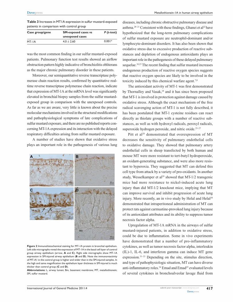
Immunohistochemistry was employed for evaluation and
localization of MT-1 protein expression in airway biopsy
samples from the two groups. It was immediately observed
that the thickness of the bronchial epithelium layer in sulfur
mustard-exposed patients was about twice that in normal
control biopsies. In the control group, strong MT-1 immuno-
reactivity was seen in airway epithelium cells, especially in
the basal (germinal) layer (Figures 2A and 2C). In contrast,
in sulfur mustard-injured specimens, very weak MT-1 protein
expression was seen in the bronchial epithelial cells, espe-
cially at the luminal side of the brush border cells, indicating
very low MT-1 protein expression (Figures 2B and 2D).
DiscussionBronchiolitis obliterans is the major long-term pulmonary
complication in Iranian victims of sulfur mustard exposure,5
but the exact molecular mechanisms involved in the symp-
toms observed need to be clarified. Air trapping in expiratory
high-resolution computed tomography scans (data not shown)
200 bp
100 bp
MT-1A (177 bp)
β-actin (119 bp)
M 1 2 3 4 5 6 7 8 9 10 11 12
Figure 1 MT-1A gene expression in sM-injured patients and in unexposed control cases. Gene expressions were measured by semiquantitative reverse transcriptase polymerase chain reaction, and were upregulated in sM-injured patients (lanes 3–12). Normal samples (lanes 1 and 2), sM-injured patients samples (lanes 3–13).Abbreviations: sM, sulfur mustard; M, 100 bp marker; MT, metallothionein.
International Journal of General Medicine 2011:4 submit your manuscript | www.dovepress.com
Dovepress
Dovepress
417
Metallothionein-1A in human airway epithelium
was the most common finding in our sulfur mustard-exposed
patients. Pulmonary function test results showed an airflow
obstruction pattern highly indicative of bronchiolitis obliterans
as the major chronic pulmonary disorder in these patients.
Moreover, our semiquantitative reverse transcriptase poly-
merase chain reaction results, confirmed by quantitative real-
time reverse transcriptase polymerase chain reaction, indicate
that expression of MT-1A at the mRNA level was significantly
elevated in bronchial biopsy samples from the sulfur mustard-
exposed group in comparison with the unexposed controls.
As far as we are aware, very little is known about the precise
molecular mechanisms involved in the structural modifications
and pathophysiological symptoms of late complications of
sulfur mustard exposure, and there are no published reports con-
cerning MT-1A expression and its interaction with the delayed
respiratory difficulties arising from sulfur mustard exposure.
A number of studies have shown that oxidative stress
plays an important role in the pathogenesis of various lung
diseases, including chronic obstructive pulmonary disease and
asthma.20–21 Consistent with these findings, Ghanei et al22 have
hypothesized that the long-term pulmonary complications
of sulfur mustard exposure are neutrophil-dominant and/or
lymphocyte-dominant disorders. It has also been shown that
oxidative stress due to excessive production of reactive sub-
stances and depletion of endogenous antioxidants plays an
important role in the pathogenesis of these delayed pulmonary
sequelae.10,23 The recent finding that sulfur mustard increases
endogenous production of reactive oxygen species suggests
that reactive oxygen species are likely to be involved in the
toxicity induced by this chemical warfare agent.7,8
The antioxidant activity of MT-1 was first demonstrated
by Thornalley and Vasak,24 and it has since been proposed
that MT-1 is involved in protection against damage caused by
oxidative stress. Although the exact mechanism of the free
radical scavenging action of MT-1 is not fully described, it
has been postulated that MT-1 cysteine residues can react
directly as thiolate groups with a number of reactive sub-
stances, as well as with hydroxyl radicals, peroxyl radicals,
superoxide hydrogen peroxide, and nitric oxide.25–27
Pitt et al28 demonstrated that overexpression of MT
decreases the sensitivity of pulmonary endothelial cells
to oxidative damage. They showed that pulmonary artery
endothelial cells in sheep transfected by both human and
mouse MT were more resistant to tert-butyl hydroperoxide,
an oxidant-generating substance, and were also more resis-
tant to hyperoxia. They suggested that MT can defend this
cell type from attack by a variety of pro-oxidants. In another
study, Wesselkamper et al29 showed that MT-1/2 transgenic
mice had more resistance to nickel-induced acute lung
injury than did MT-1/2 knockout mice, implying that MT
can improve survival and inhibit progression of acute lung
injury. More recently, an in vivo study by Helal and Helal30
demonstrated that intraperitoneal administration of MT can
protect rats against carmustine-provoked lung injury because
of its antioxidant attributes and its ability to suppress tumor
necrosis factor alpha.
Upregulation of MT-1A mRNA in the airways of sulfur
mustard-injured patients, in addition to oxidative stress,
could be due to inflammation. Some in vivo experiments
have demonstrated that a number of pro-inflammatory
cytokines, as well as tumor necrosis factor alpha, interleukin
(IL)-1, IL-6, and interferon gamma can induce MT gene
expression.31–33 Depending on the site, stimulus direction,
and type of pathophysiologic situation, MT can have diverse
anti-inflammatory roles.34 Emad and Emad35 evaluated levels
of several cytokines in bronchoalveolar lavage fluid from
Table 3 Increases in MT1A expression in sulfur mustard-exposed patients in comparison with control group
Case group/gene SM-exposed cases vs unexposed cases
P (t-test)
MT-1A 4.0 ± 2.60 0.001*
A BL L
Bm
Bm
10 µ
C D
20 µ
10 µ
20 µ
Figure 2 Immunohistochemical staining for MT-1A protein in bronchial epithelium. Left side micrographs reveal the expression of MT-1A in the basal cell layer of control group airway epithelium (arrow, A and C). right side micrographs show MT-1A expression in sM-injured airway epithelium (B and D). Note the immunoreactivity of MT-A1 in the control group is higher and wider than in the sM-injured samples. In the high and same magnification the epithelium layer thickness in SM-injured is much thicker than control group (C and D).Abbreviations: L, airway lumen; Bm, basement membrane; MT, metallothionein; sM, sulfur mustard.

International Journal of General Medicine 2011:4submit your manuscript | www.dovepress.com
Dovepress
Dovepress
418
Nourani et al
sulfur mustard-exposed patients and demonstrated that IL-8,
IL-1β, IL-6, IL-12, and tumor necrosis factor alpha levels in
sulfur mustard-injured patients were all significantly higher
than in controls, and suggested that neutrophilic pulmo-
nary inflammation is possibly the mechanism underlying
the long-term pulmonary complications of sulfur mustard
exposure. Molecular level investigation of the existence
of inflammation in the airways of sulfur mustard-injured
patients is underway in our laboratory, and preliminary, as
yet unpublished data show that tumor necrosis factor alpha
expression at the mRNA level is significantly upregulated in
sulfur mustard-exposed patients in comparison with controls,
suggesting that inflammatory processes are involved in this
chronic pulmonary complication. Consistent with these
findings, Takano et al36 have shown that MT-1/2 knockout
mice are more vulnerable to acute pulmonary inflammatory
injury mediated by intratracheal instillation of lipopolysac-
charide than wild-type mice. Additionally, in line with these
findings, a complementary DNA microarray study based
on acute inflammatory murine lung damage induced by
lipopolysaccharide and diesel exhaust particles revealed that
MT-1 gene expression at the mRNA level is significantly
upregulated in mice exposed to these irritants.17 Similarly, a
more recent study showed that MT plays an important role
in ozone-induced pulmonary inflammation, and showed that
lung inflammation is significantly greater in MT-1/2 knockout
than wild-type mice.37 However, despite numerous investiga-
tions of the possible contribution of MT-1 to inflammatory
lung injuries, its exact role still needs to be elucidated.
Paradoxically, our immunohistochemistry results
revealed that, in contrast with the upregulation of MT-1A
mRNA expression in airway biopsy samples from sulfur
mustard-injured patients, its protein level was higher in
control subjects. Several MT-1 immunoreactive cells were
seen in the bronchial epithelia of our control cases, and
immunoreactivity was more intense in cells adjacent to the
basement membrane. This finding is in accordance with that
of Courtade et al,38 who evaluated the expression of MT in
the normal human lung, and observed positively stained
pleural endothelial cells and basal cells from the bronchial
epithelium. In contrast, in our chemically-injured cases, only
a very weak expression of MT-1 was observed on the luminal
side of the bronchial epithelium. We have also already shown
an inconsistency between mRNA and protein expression of
lipocalin-2 and heme oxygenase in the bronchial epithelium
of sulfur mustard-exposed patients compared with unexposed
cases.11,39,40 We hypothesize that this discrepancy between
mRNA and protein expression of MT-1A may be caused by
translational inefficiency and/or posttranslational regulation.
Interestingly, it has been revealed that, despite a huge amount
of evidence strongly suggesting that the expression of MT
genes is transcriptionally regulated, new data have also
indicated that posttranslational modulations also have effects
on MT gene expression.14 This discrepancy may be due to a
change in the expression of microRNAs, which are posttran-
scriptional regulators that bind to complementary sequences
on target messenger RNA transcripts, usually resulting in
translational repression and gene silencing.41
Intriguingly, it has been shown that administration of
N-acetylcysteine can significantly improve clinical symptoms
in sulfur mustard-exposed patients,19 and it has also been
demonstrated that N-acetylcysteine, as an antioxidant, pro-
vides thiol groups which are essential for serum glutathione
production.39 In this way, N-acetylcysteine can compensate
for decreased expression of MT-1 protein, but, in spite of
this improvement, no cure has as yet been achieved in sulfur
mustard-injured patients.
ConclusionThis study shows that although the expression of MT-1 at
the mRNA level was significantly increased in the bronchial
epithelium of sulfur mustard-exposed patients, expression of
MT-1 protein was significantly higher in the airways of our
controls. This upregulation of MT-1 mRNA seems to be due
to oxidative stress and depletion of the endogenous antioxi-
dants which exist in the lungs of patients who have inhaled
sulfur mustard, and may reflect an attempt to ameliorate this
harmful situation by reestablishment of homeostasis. The
contrasting depletion of its protein may be a result of sec-
ondary effects of sulfur mustard toxicity. Therefore, further
studies clarifying the mechanisms involved in the long-term
outcome of sulfur mustard exposure are warranted.
AcknowledgmentWe thank the members of our laboratory at the Chemical
Injury Research Center, Baqiyatallah University of Medical
Sciences for their contributions to this research.
DisclosureThe authors report no conflicts of interest in this work.
References1. Ghanei M, Aslani J, Khateri S, Hamadanizadeh K. Public health status of the
civil population of Sardasht 15 years following large-scale war time expo-sure to sulfur mustard, 2003. J Burns Surg Wound Care. 2003;2:7–18.
2. Khateri S, Ghanei M, Soroush M, Haines D. Incidence of lung, eye and skin lesions as late complications in 34,000 Iranians with wartime expo-sure to mustard agent. J Occup Environ Med. 2003;452:1136–1143.

International Journal of General Medicine
Publish your work in this journal
Submit your manuscript here: http://www.dovepress.com/international-journal-of-general-medicine-journal
The International Journal of General Medicine is an international, peer-reviewed open-access journal that focuses on general and internal medicine, pathogenesis, epidemiology, diagnosis, monitoring and treat-ment protocols. The journal is characterized by the rapid reporting of reviews, original research and clinical studies across all disease areas.
A key focus is the elucidation of disease processes and management protocols resulting in improved outcomes for the patient.The manu-script management system is completely online and includes a very quick and fair peer-review system. Visit http://www.dovepress.com/ testimonials.php to read real quotes from published authors.
International Journal of General Medicine 2011:4 submit your manuscript | www.dovepress.com
Dovepress
Dovepress
Dovepress
419
Metallothionein-1A in human airway epithelium
3. Emad A, Rezaian GR. The diversity of the effects of sulfur mustard gas inhalation on respiratory system 10 years after a single, heavy exposure. Chest. 1997;112:734–730.
4. Balali-Mood M, Hefazi M. Comparison of early and late toxic effects of sulfur mustard in Iranian veterans. Basic Clin Pharmacol Toxicol. 2006;99:273–282.
5. Ghanei M, Mokhtari M, Mohammad MM, Aslani J. Bronchiolitis obliterans following exposure to sulfur mustard: chest high resolution computed tomography. Eur J Radiol. 2004;52:164–169.
6. Ghanei M, Tazelaar HD, Chilosi M, et al. An international collaborative pathologic study of surgical lung biopsies from mustard gas exposed patients. Respir Med. 2008;102:825–830.
7. Han S, Espinoza LA, Liao H, Boulares AH, Smulson ME. Protection by antioxidants against toxicity and apoptosis induced by the sulphur mustard analog 2-chloroethylethyl sulphide (CEES) in Jurkat T cells and normal human lymphocytes. Br J Pharmacol. 2004;141:795–802.
8. Korkmaz A, Yaren Y, Topal T, Oter S. Molecular targets against mustard toxicity: Implication of cell surface receptors, peroxynitrite production, and PARP activation. Arch Toxicol. 2006;80:662–670.
9. Toews GB. Impact of bacterial infections on airway diseases. Eur Respir Rev. 2005;14:62–68.
10. Shohrati M, Ghanei M, Shamspour N, et al. Glutathione and malon-dialdehyde levels in late pulmonary complications of sulfur mustard intoxication. Lung. 2010;188:77–83.
11. Ebrahimi M, Roudkenar MH, Imani Fooladi AA, et al. Discrepancy between mRNA and protein expression of neutrophil gelatinase-associated lipocalin in bronchial epithelium induced by sulfur mustard. J Biomed Biotechnol. 2010;2010:823131.
12. Thirumoorthy N, Manisenthil Kumar KT, Sundar SA, et al. Metallothionein: An overview. World J Gastroenterol. 2007;13:993–996.
13. Lu H, Hunt DM, Ganti R, et al. Metallothionein protects human retinal pigment epithelial cells aganist apoptosis and oxidative stress. Exp Eye Res. 2002;74:83–92.
14. Haq F, Mahoney M, Koropatnick J. Signaling events for metallothionein induction. Mutat Res. 2003;533:211–226.
15. Davis SR, Cousins RJ. Metallothionein expression in animals: A physiological perspective on function. J Nutr. 2000;130:1085–1088.
16. Sato M, Kondoh M. Recent studies on metallothionein: Protection against toxicity of heavy metals and oxygen free radicals. Tohoku J Exp Med. 2002;196:9–22.
17. Yanagisawa R, Takano H, Inoue K, et al. Complementary DNA microar-ray analysis in acute lung injury induced by lipopolysaccharide and diesel exhaust particles. Exp Biol Med (Maywood). 2004;229:1081–1087.
18. Nourani MR, Owada Y, Kitanaka N, et al. Localization of epidermal-type fatty acid binding protein in macrophages in advanced atretic follicles of adult mice. J Mol Histol. 2005;36:391–400.
19. Ghanei M, Aslani J, Khateri S, Hamadanizadeh K. Public health status of the civil population of Sardasht 15 years following large-scale war time exposure to sulfur mustard. J Burns Wounds.2003;2:7.
20. Greene L. Asthma, oxidant stress and diet. Nutrition. 1999;15:899–907. 21. Rahman I, Adcock IM. Oxidative stress and redox regulation of lung
inflammation in COPD. Eur Respir J. 2006;28:219–242. 22. Ghanei M, Shohrati M, Jafari M, Ghaderi S, Alaeddini F, Aslani J.
N-acetylcysteine improves the clinical conditions of mustard gas-exposed patients with normal pulmonary function tests. Basic Clin Pharmacol Toxicol. 2008;10:428–432.
23. Naghii MR. Sulfur mustard intoxication, oxidative stress, and antioxidants. Mil Med. 2002;167:573–575.
24. Thornalley PJ, Vasak M. Possible role for metallothionein in protection against radiation-induced oxidative stress. Kinetics and mechanism of its reaction with superoxide and hydroxyl radicals. Biochim Biophys Acta. 1985;827:36–44.
25. Kumari MV, Hiramatsu M, Ebadi M. Free radical scavenging actions of metallothionein isoforms I and II. Free Radic Res. 1998;29:93–101.
26. Min KS, Nishida K, Onosaka S. Protective effect of metallothionein to ras DNA damage induced by hydrogen peroxide and ferric ion-nitrilotriacetic acid. Chem Biol Interact. 1999;122:137–152.
27. Zangger K, Shen G, Oz G, Otvos JD, Armitage IM. Oxidative dimerization in metallothionein is a result of intermolecular disulphide bonds between cysteines in the alpha-domain. Biochem J. 2001;359: 353–360.
28. Pitt BR, Schwarz M, Woo ES, et al. Overexpression of metallothionein decreases sensitivity of pulmonary endothelial cells to oxidant injury. Am J Physiol. 1997;273:L856–L865.
29. Wesselkamper SC, McDowell SA, Medvedovic M, et al. The role of metallothionein in the pathogenesis of acute lung injury. Am J Respir Cell Mol Biol. 2006;34:73–82.
30. Helal GK, Helal OK. Metallothionein attenuates carmustine-induced oxidative stress and protects against pulmonary fibrosis in rats. Arch Toxicol. 2009;83:87–94.
31. De SK, McMaster MT, Andrews GK. Endotoxin induction of murine metallothionein gene expression. J Biol Chem. 1990;265: 15267–15274.
32. Sato M, Sasaki M, Hojo H. Tissue specific induction of metallothionein synthesis by tumor necrosis factor-alpha. Res Commun Chem Pathol Pharmacol. 1992;75:159–172.
33. Waelput W, Broekaert D, Vandekerckhove J, Brouckaert P, Tavernier J, Libert C. A mediator role for metallothionein in tumor necrosis factor-induced lethal shock. J Exp Med. 2001;194:1617–1624.
34. Inoue K, Takano H, Shimada A, Satoh M. Role of metallothionein in inflammatory lung diseases. Curr Respir Med Rev. 2009;5:6–11.
35. Emad A, Emad Y. CD4/CD8 ratio and cytokine levels of the BAL fluid in patients with bronchiectasis caused by sulfur mustard gas inhalation. J Inflamm (Lond). 2007;4:2.
36. Takano H, Inoue K, Yanagisawa R, et al. Protective role of metallo-thionein in acute lung injury induced by bacterial endotoxin. Thorax. 2004;59:1057–1062.
37. Inoue K, Takano H, Kaewamatawong T, et al. Role of metallothionein in lung inflammation induced by ozone exposure in mice. Free Radic Biol Med. 2008;45:1714–1722.
38. Courtade M, Carrera G, Paternain JL, et al. Metallothionein expression in human lung and its varying levels after lung transplantation. Chest. 1998;113:371–378.
39. Dekhuijzen PNR. Antioxidant properties of N-acetylcysteine: Their relevance in relation to chronic obstructive pulmonary disease. Eur Respir J. 2004;23:629–636.
40. Nourani MR, Yazdani S, Roudkenar MH, et al. HO1 mRNA and protein do not change in parallel in bronchial biopsies of patients after long term exposure to sulfur mustard, gene regulation and systems biology. Gene Regul Syst Bio. 2009;4:83–90.
41. Bartel DP. MicroRNAs: Target recognition and regulatory functions. Cell. 2009;136:215–233.