A Fast Microfluidic Temperature Control Device for Studying Microtubule Dynamics in Fission Yeast

Molecular Biology of the CellVol. 13, 2977–2989, September 2002
Role of Fission Yeast Tup1-like Repressors and Prr1Transcription Factor in Response to Salt StressAmanda Greenall,* Andrew P. Hadcroft,*† Panagiota Malakasi,* Nic Jones,‡Brian A. Morgan,* Charles S. Hoffman,§ and Simon K. Whitehall*�
*School of Biochemistry and Genetics, University of Newcastle upon Tyne, Newcastle NE2 4HH,United Kingdom; ‡Paterson Institute for Cancer Research, Christie Hospital NHS Trust, ManchesterM20 4BX, United Kingdom; and §Biology Department, Boston College, Chestnut Hill,Massachusetts 02467
Submitted December 4, 2001; Revised April 29, 2002; Accepted June 13, 2002Monitoring Editor: Elizabeth Craig
In Schizosaccharomyces pombe, the Sty1 mitogen-activated protein kinase and the Atf1 transcriptionfactor control transcriptional induction in response to elevated salt concentrations. Herein, wedemonstrate that two repressors, Tup11 and Tup12, and the Prr1 transcription factor also functionin the response to salt shock. We find that deletion of both tup genes together results inhypersensitivity to elevated cation concentrations (K� and Ca2�) and we identify cta3�, whichencodes an intracellular cation transporter, as a novel stress gene whose expression is positivelycontrolled by the Sty1 pathway and negatively regulated by Tup repressors. The expression ofcta3� is maintained at low levels by the Tup repressors, and relief from repression requires theSty1, Atf1, and Prr1. Prr1 is also required for KCl-mediated induction of several other Sty1-dependent genes such as gpx1� and ctt1�. Surprisingly, the KCl-mediated induction of cta3�
expression occurs independently of Sty1 in a tup11� tup12� mutant and so the Tup repressors linkinduction to the Sty1 pathway. We also report that in contrast to a number of other Sty1- andAtf1-dependent genes, the expression of cta3� is induced only by high salt concentrations.However, in the absence of the Tup repressors this specificity is lost and a range of stressesinduces cta3� expression.
INTRODUCTION
Exposure of cells to environmental stress triggers a rapidincrease in the transcription of genes whose products haveprotective functions (Toone and Jones, 1998). Key to thisresponse are stress-activated protein kinase (SAPK) path-ways that transmit the signal from stress sensors to thetranscription factors that regulate gene expression. Thesepathways are evolutionarily conserved, and homologs of themammalian SAP kinases, p38/RK/CSBP (Marshall, 1994),are present in both Saccharomyces cerevisiae (Hog1) (Brewsteret al., 1993) and Schizosaccharomyces pombe (Sty1/Spc1) (Mill-er et al., 1995; Shiozaki and Russell, 1995). The Hog1 path-way in S. cerevisiae is activated essentially by hyperosmolar-ity (Brewster et al., 1993), whereas the S. pombe Sty1
pathway, like mammalian p38, is activated by a range ofadverse conditions (Millar et al., 1995; Shiozaki and Russell,1996; Degols and Russell, 1997; Buck et al., 2001).
Models of SAPK-dependent regulation of transcriptionhave been almost exclusively based upon the positive con-trol of activators. However, recent analysis of S. cerevisiae hasdemonstrated that the Sko1 repressor regulates the expres-sion of Hog1-dependent osmostress genes, such as ENA1and GRE2, via recruitment of the Ssn6(Cyc8)-Tup1 globalcorepressor complex (Marquez et al., 1998; Proft and Ser-rano, 1999; Garcia-Gimeno and Struhl, 2000; Proft et al.,2001). Ssn6-Tup1 mediates its function via the organizationof repressive chromatin structures (Cooper et al., 1994; Ed-mondson et al., 1996; Watson et al., 2000; Bone and Roth,2001; Wu et al., 2001) and by inhibition of the basal tran-scription machinery (Redd et al., 1997; Papamichos-Chronakis et al., 2000; Zaman et al., 2001). This global repres-sor controls the expression of numerous genes throughinteraction with a variety of site-specific DNA binding pro-teins (Smith and Johnson, 2000). Relief from this repressionis achieved by control of the proteins that serve to tether thecomplex to DNA; for example, Sko1 is phosphorylated byHog1 at three sites in its N-terminal region, disrupting the
Article published online ahead of print. Mol. Biol. Cell 10.1091/mbc.01–12–0568. Article and publication date are at www.molbiol-cell.org/cgi/doi/10.1091/mbc.01–12–0568.
† Present address: Sir William Dunn School of Pathology, Univer-sity of Oxford, South Parks Rd., Oxford OX1 3RE, United King-dom.
� Corresponding author. E-mail address: [email protected].
© 2002 by The American Society for Cell Biology 2977

interaction with Ssn6-Tup1 (Proft et al., 2001). Therefore, acomponent of the osmotic induction of some genes occursvia derepression rather than by activation.
In fission yeast, Sty1 operates via the transcriptional acti-vators Atf1/Gad7 (Takeda et al., 1995; Kanoh et al., 1996) andPap1 (Toda et al., 1991). Atf1 is phosphorylated in a Sty1-dependent manner and loss of Atf1 results in hypersensitiv-ity to osmotic stress, high levels of calcium, and an inabilityto respond to deteriorating nutritional conditions (Takeda etal., 1995; Kanoh et al., 1996; Ohmiya et al., 1999b). In addition,Atf1 forms a heterodimeric complex with Pcr1, a relatedATF/CREB factor, which is also required for transcriptionalinduction of some stress genes (Watanabe and Yamamoto,1996). Pap1 activates transcription in response to oxidativestress, and its subcellular localization is regulated in a Sty1-dependent manner (Toone et al., 1998). Recently, Prr1, ahomolog of Skn7 in S. cerevisiae (Brown et al., 1993), has alsobeen implicated in the transcriptional response to oxidativestress (Ohmiya et al., 1999a). Skn7 and Prr1 have heat-shockfactor-like DNA binding domains and also share homologywith bacterial “two-component” response regulators thatare controlled by histidine-to-aspartate phosphorelay sys-tems (Appleby et al., 1996).
Herein, we have addressed the roles of the Tup-like re-pressors Tup11 and Tup12 (Mukai et al., 1999; Janoo et al.,2001) in the response to stress in S. pombe. We find thatdeletion of both tup genes in combination results in hyper-sensitivity to KCl and CaCl2, and we also identify cta3� as anovel stress gene that is negatively regulated by Tup11-Tup12. The expression of cta3� is rapidly and specificallyinduced in response to salt shock in a Sty1- and Atf1-depen-dent manner, but the dependence on the Sty1 pathway forinduction is lost in a tup11� tup12� mutant. Furthermore,Tup11 and Tup12 proteins function as specificity factors by
preventing induction of cta3� in response to inappropriatestresses such as heat and oxidative stress. We also reveal anew role for the “response regulator” protein Prr1 and dem-onstrate that it is required for proper KCl-mediated tran-scriptional induction of Sty1-dependent genes such as cta3�,ctt1�, and gpx1�.
MATERIALS AND METHODS
StrainsRoutine culture of S. pombe and general genetic methods wereperformed as described in Moreno et al. (1991). The strains used inthis study are described in Table 1. The cta3� gene was disruptedusing a polymerase chain reaction (PCR)-based approach as de-scribed by Bahler et al. (1998). Oligonucleotides 5� KO (5�-TTTGA-TTTTACTTATATTTCTCCCCTTCTACTCATCCCGATATATTCT-TACTTCCTTGATTCAATCTCAAATATTGTTCAGCTTAGC-TACAAATCCCACT-3�) and KO 3� (5�-ATAAATCCTTTACGATT-TGTCGGTTCTGTGAAAACGATACACTCACGCATATTCAT-ATACATATTCATGGCAAGAAAACATCTGACATAAAACG-CCTAGG-3�) were used to amplify a 1.6-kb ura4�-containing frag-ment from pRep42. The amplified fragment was used to transformstrain NT5 strain to Ura�, creating strain SW95. Integration at thecorrect locus was confirmed by PCR analysis.
PlasmidsThe tup11� coding sequence was amplified by PCR from a cDNAlibrary by using the following primers: 5�-GCACGGATCCCATG-GCGTCAGTGGAGGATGC-3� and 5�-CTAGGGATCCAATTCAA-GGAGATGCAGGGTC-3�. The tup12� coding sequence was ampli-fied using primers 5�-GCACGGATCCCATGATTACTGTCCGC-CAATC-3� and 5�-CTGCTAGGCATATGGCGCTCATGAAA-CAAACCG-3�. Fragments were cleaved with BamHI and clonedinto the BamHI site of derivatives of pRep41 and pRep42 vectors
Table 1. Strains used in this study
Strain Genotype Reference
NT4 h� ade6-M210 leu1-32 ura4-D18 Zhu et al. (1994)NT5 h� ade6-M216 leu1-32 ura4-D18 Zhu et al. (1994)SW54 h� ade6-M210 leu1-32 ura4-D18 tup11�ura4� Janoo et al. (2001)BSP03 h� ade6� leu1-32 ura4-D18 tup12�ura4� Janoo et al. (2001)RJP12 h� ura4�fbp1-lacZ tup11�ura4� tup12�ura4� Janoo et al. (2001)SW76 h� ade6� tup11�ura4� tup12�ura4� ura4-D18 leu1-32 This studyHAI003 h� ade6�M216 leu1-32 ura4-D18 cta3-lacZ�ura4� Nishikawa et al. (1999)SW107 h� ade6� leu1-32 ura4-D18 tup11�ura4� tup12�ura4� cta3-lacZ�ura4� This studyKS14709 h� atf1HA6H�ura4� leu1-32 ura4-D18 Shiozaki & Russell (1996)JM1144 h� sty1-1 leu1-32 ura4-D18 Millar et al. (1995)JM1160 h� ade6-M216 leu1-32 ura4-D18 sty1�ura4� Millar et al. (1995)NT147 h90 leu1-32 ura4-D18 atf1�ura4� Takeda et al. (1995)JX125 h90 ade6� leu1-32 ura4-D18 pcr1�ura4� Kanoh et al. (1996)RJP59 h� his7-366 ura4�fbp1-lacZ pcr1�ura4� tup11�ura4� tup12�ura4� Janoo et al. (2001)SW89 h� ade6� leu1-32 ura4-D18 tup11�ura4� sty1-1 This studySW88 h� leu1-32 ura4-D18 tup12�ura4� sty1-1 This studySW90 h� ade6� leu1-32 ura4-D18 tup11�ura4� tup12�ura4� sty1-1 This studySW91 h� leu1-32 ura4-D18 tup11�ura4� tup12�ura4� sty1�ura4� This studySW95 h� ade6-M216 leu1-32 ura4-D18 cta3�ura4� This studySW93 h� ade6� leu1-32 ura4-D18 cta3�ura4� tup11�ura4� tup12�ura4� This studySW92 h� leu1-32 ura4-D18 atf1�ura4� tup11�ura4� tup12�ura4� This studyRJP59 h� ura4�fbp1-lacZ pcr1�ura4� tup11�ura4� tup12�ura4� his7-336 Janoo et al. (2001)SW97 h� ade6-M216 ura4-D18 his7-336 leu1-32 prr1�ura4� This studySW96 h� ade6� ura4-D18 leu1-32 prr1�ura4� tup11�ura4� tup12�ura4� his7-336 This study
A. Greenall et al.
Molecular Biology of the Cell2978

that allow the expression of proteins as N-terminal HA or 6HisMycfusions (Craven et al., 1998).
RNA AnalysisRNA samples were prepared from 0.25 to 0.5 � 109 cells. Pelletswere washed in H2O and resuspended in 200 �l of RNA buffer (50mM Tris-HCl pH 8.0, 100 mM NaCl, 50 mM EDTA pH 8.0, and0.25% SDS) with 200 �l of phenol/chloroform. Cells were disruptedwith 0.75 ml of glass beads (0.5 mm; Biospec Products, Bartlesville,OK) in a Ribolyser (Hybaid, Middlesex, United Kingdom). A further0.75 ml of RNA buffer was added followed by spinning in a mi-crofuge for 10 min. The aqueous layer was subjected to two furtherphenol/chloroform extractions before the RNA was precipitatedwith 0.1 volume of sodium acetate, pH 5.2, and 0.6 volume ofisopropanol. RNA pellets were washed in 70% ethanol and resus-pended in H2O. RNA analysis was as described by White et al.(1986). Briefly, a 10–15-�g sample of total RNA was denatured withglyoxal, separated on a 1.2% agarose gel prepared in 15 mM sodiumphosphate, pH 6.5, and transferred to a GeneScreen hybridizationmembrane (PerkinElmer Life Sciences, Boston, MA). The his3�
probe has been described previously (Baum et al., 1997). Othergene-specific probes were produced by PCR amplification fromgenomic DNA by using the appropriate primers. All probes werelabeled with [�-32P]dCTP by using a Prime-a-Gene labeling kit(Promega, Madison, WI). Transcript levels were quantified relativeto the loading control using a PhosphorImager BAS-1500 (Fuji PhotoFilm, Tokyo, Japan).
�-Galactosidase AssaysAssays were performed as described previously (Takeda et al.,1995).
CoprecipitationsWhole cell extracts were prepared as described by Whitehall et al.(1999) with some modification. Cultures were grown to mid-logphase (OD595 � 0.25–0.5) in EMM medium. Cells were harvestedwashed once and snap frozen. Pellets were washed in 1 ml of lysisbuffer (50 mM Tris-HCl pH 7.4, 150 mM NaCl, 0.5% NP-40, 10 mMimidazole, 2 �g/ml pepstatin, 2 �g/ml leupeptin, 2 �g/ml aproti-nin, and 100 �g/ml phenylmethylsulfonyl fluoride). Cells weredisrupted with 2 ml of glass beads by vortexing twice for 45 s with1-min incubation on ice in between. Protein extracts were recovered
and centrifuged at 13,000 rpm for 10 min at 4°C. Protein precipita-tions were performed by adding 25 �l of nickel-agarose (50% slurryin lysis buffer) to 1 mg of whole protein extract and incubating at4°C for 1 h with gentle agitation. Precipitates were recovered bycentrifugation and washed four times with lysis buffer containing200 mM NaCl and 20 mM imidazole. Samples were analyzed bySDS-PAGE and proteins were transferred to nitrocellulose mem-brane and subjected to Western blotting by using monoclonal hem-agglutinin (HA) (12CA5) antibody (Babco, Berkeley, CA).
Electrophoretic Mobility Shift Assays (EMSAs)Whole cell extracts were prepared as described above except thatcells were grown in YE5S medium and extracts were prepared inbuffer containing 25 mM HEPES pH 7.6, 0.1 mM EDTA, 150 mMKCl, 0.1% Triton X-100, 25% glycerol, 1 mM dithiothreitol, 0.5 mMphenylmethylsulfonyl fluoride 2 �g/ml pepstatin, 2 �g/ml leupep-tin, and 2 �g/ml aprotinin. Radiolabeled DNA fragments wereprepared using PCR amplification as described in Zhu et al. (1994).The oligonucleotides used for amplification of probe 1 were 5�-TA-AAACACCGACATGTAGCC-3� and 5�-TTGAGAGAAACTAAC-CAAGG-3�. The oligonucleotides for probe 2 were 5�-CTCTGTCATG-GAAATCCACAC-3� and 5�-ATAAGCAGCAAAGCTTGCCTG-3�.Binding reactions were performed by adding 15 �g of whole cellextract to 20-�l reactions containing 25 mM HEPES pH 7.6, 34 mM KCl,5 mM MgCl2, and 2 �g of poly[d(I-C)]. Reactions were incubated for 10min at room temperature before the addition of �0.5 ng of radiola-beled probe DNA followed by a further 20-min incubation. Sampleswere analyzed by electrophoresis through 4% polyacrylamide gels runin 0.5� Tris-borate-EDTA buffer. Antibody supershift was performedby adding 0.2 �g of monoclonal HA antibody (12CA5) (Babco) 10 minafter the addition of probe DNA.
RESULTS
To address the role of repressors in the transcriptional re-sponse to stress in fission yeast we examined whether cellslacking the tup genes tup11� and tup12� exhibited anystress-related phenotypes. We found that single and doubletup mutant strains exhibited an increased tolerance to cad-mium but that the tup11� tup12� mutant strain had de-creased tolerance to elevated levels of Ca2� and K� ions. Thedegree of sensitivity to these salt stresses was similar to that
Figure 1. Stress-related phenotypes of cells lacking Tup repressors. (A) Exponentially growing cultures (�0.4 � 107 cells/ml) were dilutedserially, spotted onto YE5S agar, and incubated for 2–3 d at 30°C or spotted onto YE5S agar supplemented with CaCl2, KCl, or CdSO4 at theindicated concentration and incubated for 3–4 d at 30°C. (B) Exponentially growing cultures of wild type (w.t.) (NT4), atf1� (NT147), andtup11� tup12� (SW76) strains were treated with H2O2 (to a final concentration of 50 mM), and viable cell numbers were determined by platingonto YE5S agar.
Transcriptional Response to Salt Stress
Vol. 13, September 2002 2979

associated with loss of either the Atf1 transcription factor orthe Sty1 MAP kinase that are known to control the inductionof genes in response to elevated cation concentrations(Shiozaki and Russell, 1996; Wilkinson et al., 1996). Thetup11� tup12� double mutant strain was only slightly lesssensitive to KCl than the sty1-1 and atf1� strains and thetup11� tup12� strain was actually more sensitive to CaCl2than strain lacking Sty1 (Figure 1A). In contrast, the doubletup mutant strain had wild-type levels of tolerance to highsorbitol concentrations, indicating that although they areK�- and Ca2�-intolerant they are not osmosensitive (ourunpublished data).
The similarity in the sensitivities of tup11� tup12� andatf1� strains to elevated K�/Ca2� ions was unexpected be-cause Tup11 and Tup12 have been previously demonstratedto be repressors (Mukai et al., 1999; Janoo et al., 2001),whereas Atf1 is primarily a transcriptional activator. Wetherefore investigated whether tup11� tup12� cells sharedany other phenotypes with atf1� cells. It has recently beendemonstrated that atf1� cells are sensitive to an acute oxi-dative stress (Nguyen et al., 2000; Quinn et al., 2002). Whenchallenged with a high dose of H2O2 (50 mM) atf1� cellsrapidly lose viability (Figure 1B). In contrast, tup11� tup12�
cells were only slightly more sensitive than wild-type cells inthis assay. Furthermore, although atf1� cells conjugatepoorly (Takeda et al., 1995), a tup11� tup12� strain conju-gates in nutrient-rich media (Janoo et al., 2001). Hence,tup11� tup12� cells and atf1� cells share only a subset ofphenotypes. Taken together, these findings are consistentwith Tup11 and Tup12 having overlapping functions andindicate that Tup11 and Tup12 play roles in the cellularresponse to stress.
Tup11 and Tup12 Negatively Regulate Expression ofSalt-Stress gene cta3�
Cells lacking atf1� and both tup genes have similar sensitiv-ities to salt stress and so we examined whether the expres-sion of genes known to be induced by exposure to salt stress,via Atf1, were also regulated by the Tup repressors (Figure2, A and B). We found that expression of cta3�, whichencodes a cation-transporting P-type ATPase (Ghislain et al.,1990; Halachmi et al., 1992; Benito et al., 2002), was markedlyinfluenced by loss of the Tup proteins; deletion of both tupgenes together resulted in a large increase in the basal levelof expression (Figure 2, A and B). Loss of both Tup proteins
Figure 2. Tup11 and Tup12 repress the transcription of the salt stress gene cta3�. (A) Strains used are indicated above the lane and werewild type (w.t.) (NT4), tup12� (BSP03), tup11� (SW53), tup11� tup12� (SW76), atf1� (78 �147), and sty1-1 (JM1144). Log phase culturesgrowing at 30°C in YE5S (lanes 1, 4, 7, 10, 13, and 16) were incubated with CaCl2 (to final concentration of 0.3 M) for 15 min (lanes 2, 5, 8,11, 14, and 17) and 30 min (lanes 3, 6, 9, 12, 15, and 18). Total RNA was extracted, separated by electrophoresis, and Northern blots wereanalyzed with the indicated probes. The level of his3� mRNA was used as a loading control. (B) Quantification of cta3� mRNA levels in A.(C) Influence of Tup proteins on the activity of a cta3-lacZ reporter. �-Galactosidase assays were performed on extracts derived fromexponentially growing cells (open bars) and cells treated with KCl to 0.6 M for 60 min (black bars). The strains used were wild type (w.t.)(HAI003) and tup11� tup12� (SW107). Data is the mean of three independent cultures. (D) Deletion of cta3� does not rescue the salt sensitivityof tup11� tup12� cells. Exponentially growing cultures (�0.4 � 107 cells/ml) were diluted serially, spotted onto YE5S agar and incubated for2–3 d at 30°C or spotted onto YE5S agar supplemented with CaCl2 or KCl (at the indicated concentration) and incubated for 3–4 d at 30°C.Strains used were wild type (w.t.) (NT4), tup11� tup12� (SW76), cta3� (SW95), and cta3� tup11� tup12� (SW93).
A. Greenall et al.
Molecular Biology of the Cell2980
also resulted in a large increase in expression after exposureto CaCl2 (Figure 2, A and B) and KCl (Figures 5, 7, and 8).Thus, Tup11 and Tup12 function in a partially redundantmanner to repress cta3� expression and limit the level ofinduction. As previously observed (Nishikawa et al., 1999),the induction of cta3� expression is completely dependentupon both the Sty1 MAP kinase and the Atf1 transcriptionfactor, and thus cta3� displays a novel pattern of stressregulation that is positively controlled by Sty1 and Atf1 butnegatively regulated by the Tup repressors. Indeed, deletionof the tup genes either singly or in combination had onlyminor effects on the expression levels of other Sty1-dependentgenes such as gpd1�, ctt1�, and gpx1� in unstressed cells and incells subjected to a CaCl2 shock (Figure 2, A and B).
To confirm that increased level of cta3� transcripts asso-ciated with deletion of tup genes was due to an effect ontranscription and not mRNA stability, we measured theexpression of an integrated cta3� promoter—lacZ reporter(Nishikawa et al., 1999). It is highly unlikely that Tup repres-sors would specifically influence the stability of lacZ tran-scripts. Consistent with the Northern analysis, deletion ofboth the tup genes resulted in 14-fold increase in expressionof the lacZ reporter relative to the wild-type control (Figure2C). Furthermore, exposure of cells to high KCl concentra-tions (0.6 M for 1 h) increased the level of expression sev-enfold in wild-type cells and threefold in the cells lackingthe Tup repressors (Figure 2C). These results suggest that S.pombe Tup proteins exert their effects at the level of tran-scription.
To determine whether the high level of cta3� expressionobserved in the tup11� tup12� double mutant confers theincreased sensitivity of this strain to elevated K� and Ca2�
concentrations we examined the effect of deleting the cta3�
gene in the presence or absence of the tup genes. Loss of Cta3function has previously been reported to result in increasedsensitivity to elevated Ca2� concentrations (Ghislain et al.,1990). In contrast, Nishikawa et al. (1999) found that cta3 nullcells did not exhibit any detectable change in resistance toK�, Ca2�, or Na� ions. In agreement with the latter studyour cta3� mutant exhibited wild-type levels of resistance toboth Ca2� and K� (Figure 2D). Moreover, deletion of thecta3� gene in a tup11� tup12� strain did not rescue thesalt-sensitive phenotype associated with the loss of the tupgenes (Figure 2D), and plasmid-mediated overexpression ofcta3� in wild-type cells did not result in any increasedsensitivity to KCl or CaCl2 (our unpublished data). Theseresults indicate that the salt sensitivity of tup� cells is notsimply due to the elevated expression of the cta3� gene.
To date, the fbp1� gene encoding fructose 1,6-bisphos-phatase is the only gene that has been identified as a targetgene for Tup11-Tup12–mediated repression (Mukai et al.,1999; Janoo et al., 2001). The expression of fbp1� is alsopositively regulated by the Sty1 pathway (Takeda et al., 1995;Kanoh et al., 1996; Stettler et al., 1996), but its expression isinduced by carbon limitation (Hoffman and Winston, 1991)and not by other acute stresses that activate Sty1, such asheat shock, oxidative stress, and osmotic shock. Further-more, the cAMP pathway negatively regulates the expres-sion of fbp1�, and mutations that disrupt this pathway resultin increased expression under repressing (glucose-rich) con-ditions (Hoffman and Winston, 1991). In contrast, growingcells under carbon-limiting conditions did not induce the
expression of cta3� nor were mRNA levels influenced by adeletion of git2� that encodes adenylate cyclase (our unpub-lished data).
Formation of Protein Complexes on the cta3�
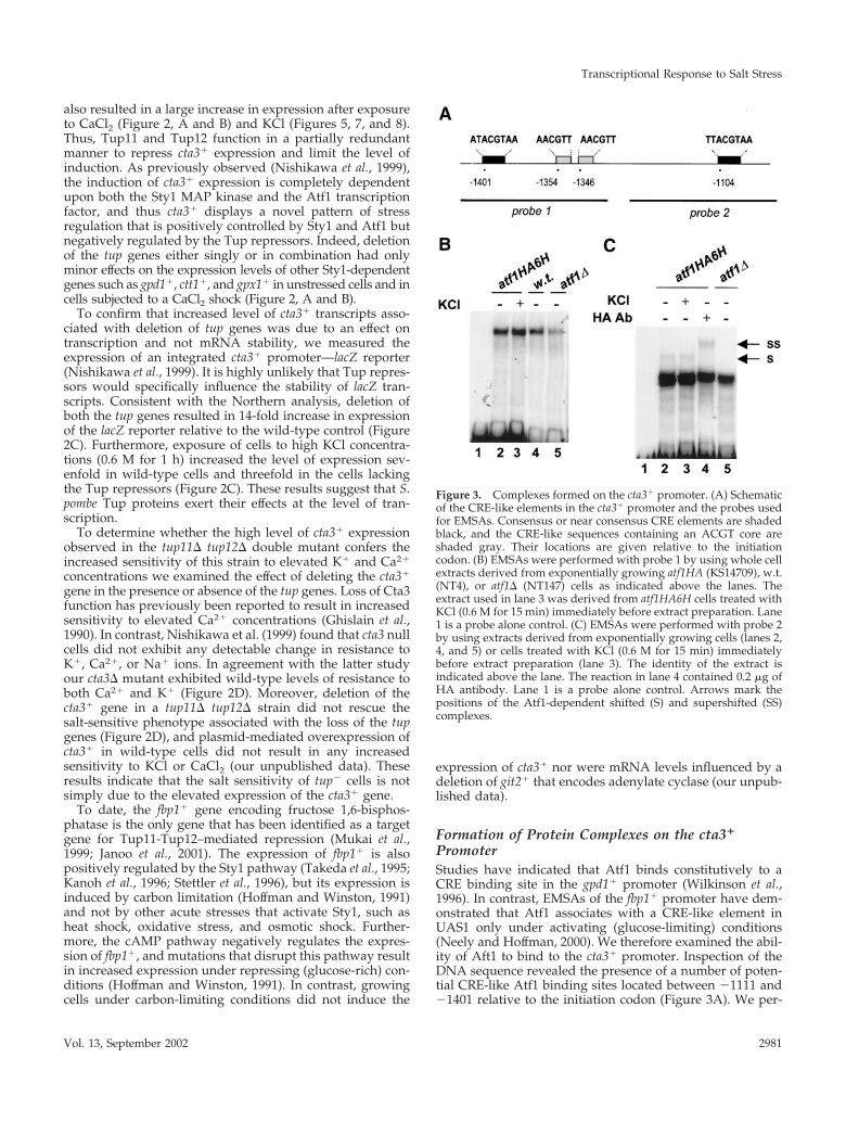
PromoterStudies have indicated that Atf1 binds constitutively to aCRE binding site in the gpd1� promoter (Wilkinson et al.,1996). In contrast, EMSAs of the fbp1� promoter have dem-onstrated that Atf1 associates with a CRE-like element inUAS1 only under activating (glucose-limiting) conditions(Neely and Hoffman, 2000). We therefore examined the abil-ity of Aft1 to bind to the cta3� promoter. Inspection of theDNA sequence revealed the presence of a number of poten-tial CRE-like Atf1 binding sites located between �1111 and�1401 relative to the initiation codon (Figure 3A). We per-
Figure 3. Complexes formed on the cta3� promoter. (A) Schematicof the CRE-like elements in the cta3� promoter and the probes usedfor EMSAs. Consensus or near consensus CRE elements are shadedblack, and the CRE-like sequences containing an ACGT core areshaded gray. Their locations are given relative to the initiationcodon. (B) EMSAs were performed with probe 1 by using whole cellextracts derived from exponentially growing atf1HA (KS14709), w.t.(NT4), or atf1� (NT147) cells as indicated above the lanes. Theextract used in lane 3 was derived from atf1HA6H cells treated withKCl (0.6 M for 15 min) immediately before extract preparation. Lane1 is a probe alone control. (C) EMSAs were performed with probe 2by using extracts derived from exponentially growing cells (lanes 2,4, and 5) or cells treated with KCl (0.6 M for 15 min) immediatelybefore extract preparation (lane 3). The identity of the extract isindicated above the lane. The reaction in lane 4 contained 0.2 �g ofHA antibody. Lane 1 is a probe alone control. Arrows mark thepositions of the Atf1-dependent shifted (S) and supershifted (SS)complexes.
Transcriptional Response to Salt Stress
Vol. 13, September 2002 2981

formed EMSAs by using whole cell extracts and a DNAfragment corresponding to the �1477 to �1297 region of thepromoter. This region includes a near consensus CRE ele-ment and two CRE-like sequences containing the highlyconserved ACGT core sequence. A major slow-migratingcomplex was formed on this probe (Figure 3B). This bindingactivity was not changed by subjecting cells to stress (KCl 0.6M for 15 min) before extract preparation. The complex wasalso present in extracts derived from atf1� cells, indicatingthat it does not require Atf1. Furthermore, the mobility ofthe complex was unchanged when HA antibody was in-cluded in reactions containing HA epitope-tagged Atf1 (ourunpublished data). Next, we examined the ability of com-plexes to form on a probe corresponding to the �1249 and�1058 region of the promoter that contains a single CREelement. In this case, we also observed a binding activitythat was Atf1-independent (Figure 3C). However, we alsodetected a slow-migrating complex that was absent in reac-tions lacking Atf1. Also, the mobility of this complex wasreduced by the addition of the HA antibody to reactionscontaining HA epitope-tagged Atf1. This Atf1-dependentcomplex was present in reactions using extracts derivedfrom unstressed and stressed cells, indicating that at leastunder these experimental conditions Atf1 binds constitu-tively to this region of the cta3� promoter. We were unableto properly assess the role of Tup proteins on DNA bindingactivity; when tup� extracts were used a marked reduction inthe level of complex formation on both probes was observed.However, this seemed to be due to difficulties in preparingextracts from these cells rather than a specific effect because wefound that extracts lacking Tup proteins also showed a re-duced ability to form complexes on a DNA probe unrelated tothe cta3� promoter (our unpublished data).
Tup11 and Tup12 InteractOur data indicate that Tup11 is capable of repressing cta3�
expression in the absence of Tup12 and vice versa. It is alsoknown that S. cerevisiae Tup1 tetramerizes through its N-terminal domain (Varanasi et al., 1996; Jabet et al., 2000) andbased on homology it is very likely that the S. pombe Tupproteins form homotetramers. However, it is possible that inaddition to functioning in homomeric complexes Tup11 andTup12 may also function in a heteromeric complex. There-fore, we investigated the ability of Tup11 and Tup12 tointeract using a coprecipitation assay. Whole cell extractswere prepared from wild-type cells that expressed 6His-tagged Tup11 (or Tup12) and coexpressed HA-taggedTup11 (or Tup12). Ni2�-agarose was then used to precipitateHis-tagged Tup proteins, and the presence of HA-taggedTup proteins was examined by Western blotting (Figure 4).In these experiments, Tup11 copurified with Tup12 and viceversa, indicating that Tup11 and Tup12 physically interact.The specificity of this interaction was demonstrated by theabsence of HA-tagged Tup proteins in control precipitatesderived from cells extracts expressing the empty 6His vector(Figure 4, lanes 7 and 8). Thus, Tup11 and Tup12 have thepotential to regulate gene expression in the same proteincomplex.
Tup Repressors Link Transcriptional Induction tothe Sty1 PathwayTo test whether the high level of cta3� expression observedin the absence of the Tup repressors was dependent uponthe Sty1 MAP kinase cta3� mRNA levels were examined ina strain that lacks both Sty1 and Tup function (sty1-1 tup11�tup12�). In this mutant the level of cta3� transcripts wassimilar to that observed in tup11� tup12� cells, indicatingthat Sty1 is not required for basal levels of expression (Fig-ure 5, A and B). Surprisingly, exposure of this strain to aKCl-mediated shock resulted in induction of cta3� expres-sion, indicating that in the absence of the Tup proteins theSty1 MAP kinase is not required for the stress-mediatedinduction of cta3�. Consistent with these observations theexpression of cta3� was also induced by salt shock (0.6 MKCl) in a sty1� tup11� tup12� strain (our unpublished data).The expression of other genes such as pyp2� and gpd1� wasnot induced in the sty1-1 tup11� tup12� triple mutant, al-though deletion of the tup genes did restore the basal level ofexpression in sty1� cells (Figure 5A). The kinetics of KCl-mediated induction of cta3� were similar in wild-type andtup11� tup12� cells, with mRNA levels peaking at 20 minbut elevated mRNA levels persisted for a greater length oftime in cells lacking the Tup proteins (Figure 5, C and D). Inthe sty1-1 tup11� tup12� triple mutant strain induction wasdelayed and peak mRNA levels were not observed until 30min after the addition of KCl.
We next determined whether removal of Tup11 andTup12 rescued any of the other phenotypes associated withloss of Sty1. We examined the ability of cells to grow onmedium supplemented with cadmium. Deletion of the tupgenes in a sty1� background increases resistance to cad-mium (Figure 1A) but unexpectedly deletion of tup11� andtup12� in a sty1-1 background reduced cadmium tolerance(Figure 6A). Thus, the resistance of tup� cells to cadmiumdepends on Sty1 function and in its absence they becomehypersensitive. The elongated cell morphology of sty1-1 cellsthat is indicative of a G2 cell cycle delay was slightly exac-erbated by deletion of the tup genes (Figure 6B). Further-more, deletion of the tup genes in an aft1� or a sty1-1background resulted in a small increase in sensitivity to KCl
Figure 4. Tup11 and Tup12 coprecipitate. Whole cell extracts wereprepared from wild-type cells containing plasmids expressingepitope-tagged Tup proteins: pRep42-HisMycTup12 (lanes 1–3),pRep42-HisMycTup11 (lanes 4–6), pRep42-HisMyc empty vector(lanes 7 and 8), pRep41-HATup12 (lanes 1, 4, and 7), and pRep41-HATup11 (lanes 2, 5, and 8). Extracts were precipitated with Ni2�-agarose analyzed on 8% SDS polyacrylamide gels and subjected toWestern blotting by using HA monoclonal antibody.
A. Greenall et al.
Molecular Biology of the Cell2982
and the tup11� tup12� atf1� triple mutant strain was slightlyless tolerant to CaCl2 than the parental strains (Figure 6C).
We next addressed whether removal of Tup11-Tup12 re-pression rendered the induction of cta3� independent ofAtf1. Deletion of atf1� in a tup11� tup12� mutant strainresulted in a further increase in cta3� transcript levels inunstressed cells, suggesting that nonactivated Atf1 mayhave a repressive effect on transcription that is independentof Tup11 and Tup12 (Figure 7, A and B). A similar effect hasbeen observed previously; the decrease in the basal level ofctt1� mRNA associated with loss of Sty1 function is sup-pressed by deletion of atf1� (Degols and Russell, 1997). Inthe atf1� tup11� tup12� background, cta3� mRNA levels didnot increase after exposure to KCl (0.6 M), indicating thatAtf1 is absolutely required for induction of cta3� in responseto a salt shock.
The bZIP transcription factor Pcr1 that can heterodimerizewith Atf1 (Kanoh et al., 1996) is also required for stress-mediated induction of cta3� expression (Figure 7, C and D).Examination of cta3� mRNA levels in a pcr1� tup11� tup12�strain revealed that Pcr1 is not required for the high level ofbasal expression, and furthermore in this strain expression
of cta3� was partially induced in response to a salt shock.Thus, the Tup repressors ensure that induction of cta3�
remains dependent upon the Sty1 MAP kinase and to alesser extent, the activator Pcr1.
Prr1 Is Involved in Regulation of Gene Expressionin Response to Elevated K� IonsOur analysis suggests that another factor may regulate tran-scription of cta3� independently of Sty1. The Prr1 transcrip-tion factor is known to regulate oxidative stress responsivegenes (Ohmiya et al., 1999a), but there is no evidence thatSty1 regulates its activity directly. Therefore, we analyzedmRNA levels in a prr1� strain and found that the level ofcta3� transcripts after exposure to KCl was significantlyreduced in comparison with the wild-type strain (Figure 8,A and B). Furthermore, the influence of Prr1 was not con-fined to the cta3� gene because KCl-mediated induction ofboth ctt1� and gpx1� expression was also significantly re-duced in the prr1� mutant strain. This was surprising be-cause Prr1 has previously been reported not to be involvedin the transcriptional response to high salt (Ohmiya et al.,
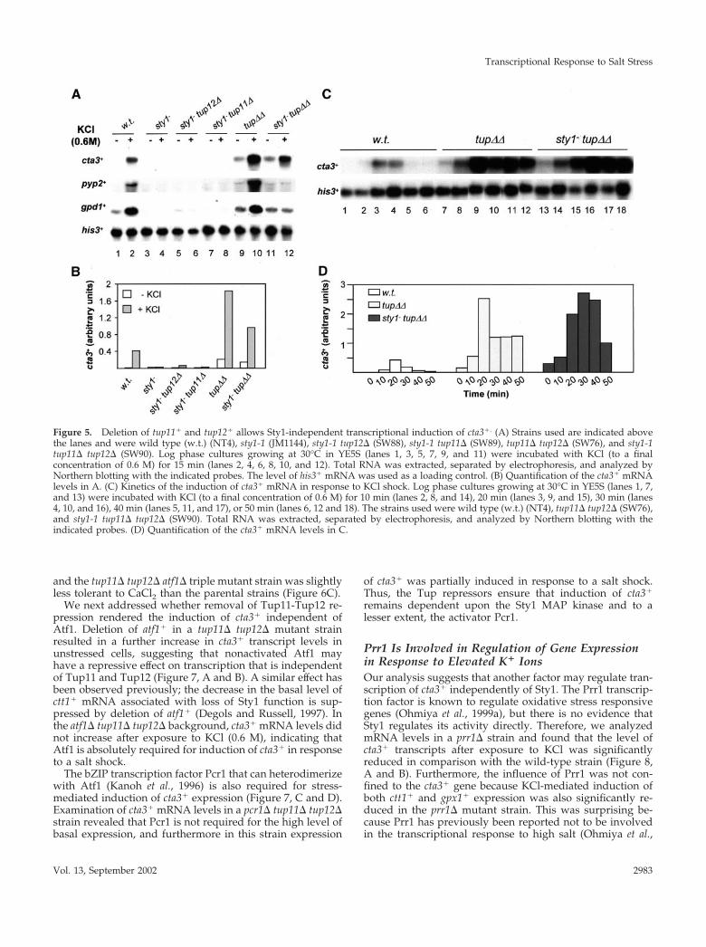
Figure 5. Deletion of tup11� and tup12� allows Sty1-independent transcriptional induction of cta3�. (A) Strains used are indicated abovethe lanes and were wild type (w.t.) (NT4), sty1-1 (JM1144), sty1-1 tup12� (SW88), sty1-1 tup11� (SW89), tup11� tup12� (SW76), and sty1-1tup11� tup12� (SW90). Log phase cultures growing at 30°C in YE5S (lanes 1, 3, 5, 7, 9, and 11) were incubated with KCl (to a finalconcentration of 0.6 M) for 15 min (lanes 2, 4, 6, 8, 10, and 12). Total RNA was extracted, separated by electrophoresis, and analyzed byNorthern blotting with the indicated probes. The level of his3� mRNA was used as a loading control. (B) Quantification of the cta3� mRNAlevels in A. (C) Kinetics of the induction of cta3� mRNA in response to KCl shock. Log phase cultures growing at 30°C in YE5S (lanes 1, 7,and 13) were incubated with KCl (to a final concentration of 0.6 M) for 10 min (lanes 2, 8, and 14), 20 min (lanes 3, 9, and 15), 30 min (lanes4, 10, and 16), 40 min (lanes 5, 11, and 17), or 50 min (lanes 6, 12 and 18). The strains used were wild type (w.t.) (NT4), tup11� tup12� (SW76),and sty1-1 tup11� tup12� (SW90). Total RNA was extracted, separated by electrophoresis, and analyzed by Northern blotting with theindicated probes. (D) Quantification of the cta3� mRNA levels in C.
Transcriptional Response to Salt Stress
Vol. 13, September 2002 2983

1999a) and indeed KCl-mediated induction of some genessuch as gpd1� occurs independently of Prr1 (Ohmiya et al.,1999a; Figure 8, A and B). To determine the role that Prr1plays in control of cta3� expression, we measured cta3�
mRNA levels in a tup11� tup12� prr1� triple mutant strain.In this background the expression of cta3� was induced byexposure to high concentrations of KCl (0.6 M) (Figure 8, Cand D). However, the deletion of prr1� in a tup11� tup12�background resulted in a decrease in the basal level of cta3�
mRNA (Figure 8, C and D). Thus, Prr1 activity contributes tothe high basal level of expression that is associated with lossof the Tup repressors.
In vitro experiments have demonstrated that recombinantPrr1 binds to a heat shock-like element in the ste11� pro-moter (Ohmiya et al., 1999b). Analysis of the cta3� promoterrevealed the presence of such an element (GGAAAATTC)located at �2068 relative to the initiation codon. However, inassays using this region of the promoter and whole cellextracts we were unable to detect a Prr1-dependent bindingactivity (our unpublished data). Therefore, we cannot ex-clude the possibility that the role of Prr1 in regulation ofcta3� expression is indirect.
Tup11 and Tup12 Prevent Induction in Response toInappropriate StressesSty1, and thus in turn Atf1-Pcr1, is activated in response toa number of environmental insults. Accordingly, the expres-sion of Atf1- and Pcr1-dependent genes such as ctt1�, pyp2�,and gpx1� are induced in response to a variety of stressessuch as UV irradiation, heat shock, and hyperosmolarity andan oxidative stress elicited by exposure to high concentra-
tions of H2O2 (Shiozaki and Russell, 1996; Wilkinson et al.,1996; Degols and Russell, 1997; Nguyen et al., 2000; Quinn etal., 2002). However, some Atf1-Pcr1 target genes are inducedonly by a subset of these stresses. For example, we foundthat cta3� expression was induced specifically in response tosalt shock but not by oxidative stress (6 mM H2O2) or byheat shock (15 min at 42°C) (Figure 9, A and D). In contrast,ctt1� and gpx1� mRNA levels were both induced by thesetreatments, indicating that Atf1 (and Pcr1) were active underthese conditions. This indicates that activation of the Sty1pathway per se is not sufficient to induce the expression ofcta3� and mechanisms must exist to prevent induction ofgene expression in response to such “inappropriate stress-es.” We wanted to examine the possibility that Tup11 andTup12 play a role in this process. Therefore, we measuredthe levels of cta3� mRNA after exposing a tup11� tup12�strain to an oxidative stress (6 mM H2O2) and a heat shock(15 min at 42°C). In contrast to the wild-type strain, theexpression of cta3� was significantly induced by heat stressand by exposure to high levels of H2O2. In addition, the levelof cta3� transcripts was induced by hypotonic conditions ina tup11� tup12� strain but not in a wild-type strain (ourunpublished data). We also examined cta3� transcript levelsin a sty1� tup11� tup12� triple mutant strain (Figure 9, Band D). This revealed that the induction in expression inresponse to heat shock was partly independent of the MAPkinase. In contrast, the induction of cta3� expression inresponse to oxidative stress mediated by H2O2 was com-pletely dependent upon Sty1, suggesting a difference in themechanism of induction. Further analysis indicated that theresponse to heat shock was independent of Prr1 (Figure 9, Cand D) but dependent upon Atf1 (our unpublished data).
Figure 6. Genetic interactions. (A) Deletion of the tupgenes does not rescue phenotypes associated with thesty1-1 mutation. The indicated strains were subcul-tured onto YE5S agar (control) or subcultured ontoYE5S agar supplemented with CdSO4 (to the indicatedconcentration) and incubated at 30°C for 3–4 d. (B)Comparison of the morphology of wild type (w.t.)(NT4), sty1-1 (JM1144), tup11� tup12� (SW76), andsty1-1 tup11� tup12� (SW90) cells. (C) Exponentiallygrowing cultures were diluted serially, spotted ontoYE5S agar or YE5S agar supplemented with CaCl2 orKCl (at the indicated concentration), and incubated for2 d at 30°C. The strains used were wild type (w.t.)(NT4), tup11� tup12� (SW76), sty1-1 (JM1144), sty1-1tup11� tup12� (SW90) atf1� (NT147), and atf1� tup11�tup12� (SW92).
A. Greenall et al.
Molecular Biology of the Cell2984

Taken together, these findings indicate that the Tup repres-sors function as part of the mechanism that ensures thespecificity of stress-mediated transcriptional induction at thecta3� promoter.
DISCUSSION
In this study, we reveal roles for S. pombe Tup11 and Tup12in the cellular response to elevated K� and Ca2� levels. Weidentify cta3� as a novel stress-induced gene whose tran-scription is coregulated by the Sty1 MAP kinase pathwayand the Tup repressors. Our results indicate that the Tuprepressors fulfill a number of functions in the control ofcta3� expression. First, they maintain low levels of basalexpression and limit the level of induction. Second, theyensure that induction of expression is linked to the Sty1pathway. And third, they maintain the specificity of induc-tion. We also reveal a new role for the response regulatorPrr1 and demonstrate that it functions to regulate geneexpression in response to elevated salt concentrations. Prr1is known to contribute to the regulation of several geneswhose expression is induced by oxidative stress via the Pap1transcription factor (Ohmiya et al., 1999a), and so Prr1 reg-ulates gene expression in response to a number of stresses.
Tup11–Tup12 InteractionOur data and that of others (Mukai et al., 1999; Janoo et al.,2001) suggest that Tup11 and Tup12 can function in homo-
meric complexes. In addition, we demonstrate that Tup11and Tup12 have the potential to form a heteromeric com-plex. This is significant because full function requires bothrepressors; some derepression of a fbp1::lacZ reporter is ob-served upon deletion of a single tup gene (Janoo et al., 2001).Furthermore, single tup mutants have demonstrable pheno-types such as increased resistance to cadmium. Thus, regu-lation of some genes may depend upon both repressors andthe formation of heteromeric Tup complexes.
Relief of Tup11-Tup12–mediated RepressionThe Hog1 MAP kinase in S. cerevisiae plays a direct role inrelieving Ssn6-Tup1–mediated repression at osmostressgenes; Hog1 phosphorylates Sko1, reducing its affinity forthe corepressor complex (Proft et al., 2001). It is possible thatthe Sty1 MAP kinase may similarly antagonize the action ofTup11-Tup12; however, our results demonstrate that theAtf1, Pcr1, and Prr1 transcription factors are required forrelief from Tup-mediated repression at the cta3� promoter.It is probable that the S. pombe Tup proteins function, at leastin part, through the organization of repressive chromatinstructures (Mukai et al., 1999), and therefore it is possiblethat Atf1-Pcr1 and Prr1 overcome this repression by recruit-ing positive-acting chromatin remodeling complexes such asSwi-Snf or histone acetylase complexes (HATs). In supportof this, DNA binding by the Atf1-Pcr1 heterodimer is knownto alter local nucleosome positioning at the ade6-M26 hot-spot and thereby promote meiotic recombination (Mizuno et
Figure 7. Transcription induc-tion of cta3� in cells lackingTup11 and Tup12 is Atf1 depen-dent. (A) Strains used are indi-cated above the lanes and werewild type (w.t.) (NT4), atf1�(NT147), tup11� tup12� (SW76),and atf1� tup11� tup12� (SW92).Log phase cultures growing at30°C in YE5S (lanes 1, 3, 5, and 7)were incubated with KCl (to afinal concentration of 0.6 M) for15 min (lanes 2, 4, 6, and 8). TotalRNA was extracted, separated byelectrophoresis and subjected toNorthern analysis with the indi-cated probes. The level of cdc2�
mRNA was used as a loadingcontrol. (B) Quantification ofcta3� mRNA levels in A. (C) Asfor A, except strains used werewild type (w.t.) (NT4), pcr1�(JX25), tup11� tup12� (SW76),and pcr1� tup11� tup12� (RJP59).(D) Quantification of cta3�
mRNA levels in C.
Transcriptional Response to Salt Stress
Vol. 13, September 2002 2985

al., 1997; Kon et al., 1998). Moreover, genes such as SUC2 inS. cerevisiae are regulated by the interplay between Ssn6-Tup1 repression and Swi-Snf–mediated activation (Gavinand Simpson, 1997).
In S. cerevisiae, Hog1-dependent transcriptional induc-tion of HAL1 requires the Gcn4 activator that relievesTup1-Ssn6 –mediated repression by competing with Sko1for the occupancy of a single CRE binding site (Pascual-Ahuir et al., 2001). This CRE element functions as a dualcontrol element and integrates both positive and negativeregulatory signals. Furthermore, analysis of the S. pombefbp1� promoter, which is regulated by both Atf1-Pcr1 andTup11-Tup12, has demonstrated the presence of a controlelement (called UAS2) that contains a CRE-like sequenceand is bound by multiple activators and repressors (Neelyand Hoffman, 2000). Interestingly, the Atf1-Pcr1 transcrip-tion factor does not bind to UAS2 directly, but it doesinfluence the protein complexes that assemble on it (Neelyand Hoffman 2000). The cta3� promoter contains a num-ber of CRE-like elements at least one of which mediates
Atf1 binding (Figure 3C). It will be interesting to deter-mine the contributions of these elements to activation andrepression of cta3� transcription.
Tup Repressors Maintain Specificity of InductionThe advent of stressful conditions results in the rapid andSty1-dependent phosphorylation of Atf1 (Shiozaki and Rus-sell, 1996; Wilkinson et al., 1996). Although the precise role ofAtf1 phosphorylation remains obscure, it is evident thattranscriptional activation by Atf1 is dependent upon Sty1.However, deletion of the tup genes allows transcriptionalinduction of cta3� to occur in sty1� cells. Furthermore, in atup11� tup12� mutant induction of cta3� expression doesnot require Pcr1 or Prr1. Thus, the Tup repressors functionto “wire” induction to the Sty1 pathway, insulating it from
Figure 8. Prr1 is involved in the regulation of gene expression inresponse to high salt. (A) Strains used are indicated above thelanes and were wild type (w.t.) (NT4) and prr1� (SW97). Logphase cultures growing at 30°C in YE5S (lanes 1 and 3) wereincubated with KCl (to a final concentration of 0.6 M) for 15 min(lanes 2 and 4). Total RNA was prepared and subjected to North-ern analysis with the indicated probes. The level of his3� mRNAwas used as a loading control. (B) Quantification of the mRNAlevels in A. (C) Strains used are indicated above the lanes andwere tup11� tup12� (SW76) and prr1� tup11� tup12� (SW96), andthe treatment was as described in A. (D) Quantification of themRNA levels in C.
Figure 9. Tup11 and Tup12 prevent induction in response to in-appropriate stresses. (A) Strains used were wild type (w.t.) (NT4)and tup11� tup12� (SW76). Mid log cultures growing at 30°C inYE5S (lanes 1 and 4) were incubated with H2O2 (final concentration6 mM) for 15 min (lanes 2 and 5) or shifted to 42°C for 15 min (lanes3 and 6). Total RNA was prepared and subjected to Northernanalysis with the indicated probes. The level of cdc2� mRNA wasused as a loading control. (B) Strain used was sty1� tup11� tup12�(SW91) and the treatments were as described in A. (C) Strain usedwas prr1� tup11� tup12� (SW91) and the treatments were as de-scribed in A. (D) Quantification of the cta3� mRNA levels in A, B,and C. The strains are indicated above the graphs and the treat-ments are indicated below.
A. Greenall et al.
Molecular Biology of the Cell2986

interfering signals. These results also suggest that Atf1 ac-tivity can be “uncoupled” from Sty1 in this specific case andthat an additional mechanism for activating transcriptionthat requires Atf1 exists. The finding that Prr1 also controlsexpression of cta3� suggests that it may function as part ofthis mechanism.
The Sty1 pathway in S. pombe is fundamentally different tothe Hog1 pathway in S. cerevisiae because it is triggered byexposure to a wide range of adverse environmental condi-tions. As a consequence, a large number of Sty1 target genesare up-regulated by multiple stresses. The products of suchgenes may comprise a set of “general stress response pro-teins” that are necessary because a single environmentalinsult may result in multiple classes of intracellular stress(Rep et al., 2001). Nonetheless, discrete stimuli also producedistinct transcriptional outputs, because there are subsets ofSty1-dependent genes, such as cta3�, that are induced onlyby specific stresses. A major question to be addressed is themechanism by which Sty1 signaling is integrated into theregulation of such genes. Expression of cta3� is not inducedby oxidative stress, heat shock, carbon limitation, or sexualdifferentiation (Figure 6; our unpublished data), and further-more cta3� is only poorly induced by an osmotic shockmediated by high sorbitol concentrations (Nishikawa et al.,1999). Thus, the transcriptional response is triggered essen-tially by elevated intracellular cation concentrations ratherthan by an osmotic effect (i.e., decrease in turgor pressureacross the plasma membrane). The cta3� gene encodes aputative intracellular P-type ATPase transporter that is in-
volved in cation extrusion or sequestration into intracellularcompartments. Loss of function leads to an accumulation ofcytoplasmic Ca2� levels (Ghislain et al., 1990; Halachmi et al.,1992), although recent evidence suggests that Cta3 is primar-ily a K� ion pump (Benito et al., 2002). It is thus consistentthat it is salt stress that specifically that triggers its transcrip-tional induction. However, removal of the constraints im-posed by Tup repressors allows cta3� to be induced inresponse to other stresses such as elevated temperature andoxidative stress. Thus, the Tup repressors function as a partof a mechanism that adds specificity to Sty1-dependent tran-scriptional induction.
Our results also indicate that activation of the Sty1 path-way alone is insufficient to induce cta3� expression andimplies that an elevated cation concentration triggers anadditional pathway that is required to circumvent repres-sion (Figure 10). In this respect it may be significant that Prr1is involved in the regulation of cta3� expression because itsstructure suggests that it may be one part the target of ahistidine-aspartate phosphorelay pathway. Recent work hasidentified several of these pathways in fission yeast (Nguyenet al., 2000; Buck et al., 2001), and current experiments areaddressing its contribution to the regulation of Prr1 in theresponse to stress.
ACKNOWLEDGMENTS
We thank Janet Quinn, Mark Toone, and Burk Braun for advice onthe manuscript. We also thank Burk Braun, Hirofumi Aiba, andJonathan Millar for providing strains. This work was supported bya National Institutes of Health grant GM-46226 (to C.S.H.), by aMedical Research Council Ph.D. research studentship (to P.M.), aMedical Research Council Career Establishment Grant (to B.A.M.),and by a Biotechnology and Biological Sciences Research Councilproject grant (13/P11981) (to S.K.W.).
REFERENCES
Appleby, J.L., Parkinson, J.S., and Bourret, R.B. (1996). Signal trans-duction via the multi-step phosphorelay: not necessarily a road lesstraveled. Cell 86, 845–848.
Bahler, J., Wu, J.Q., Longtine, M.S., Shah, N.G., McKenzie, A.,Steever, A.B., Wach, A., Philippsen, P., and Pringle, J.R. (1998).Heterologous modules for efficient and versatile PCR-based genetargeting in Schizosaccharomyces pombe. Yeast 14, 943–951.
Baum, B., Wuarin, J., and Nurse, P. (1997). Control of S-phaseperiodic transcription in the fission yeast mitotic cycle. EMBO J. 16,4676–4688.
Benito, B., Garciadeblas, B., and Rodriguez-Navarro, A. (2002). Po-tassium- or sodium-efflux ATPase, a key enzyme in the evolution offungi. Microbiology 148, 933–941.
Bone, J.R., and Roth, S.Y. (2001). Recruitment of the yeast Tup1p-Ssn6p repressor is associated with localized decreases in histoneacetylation. J. Biol. Chem. 276, 1808–1813.
Brewster, J.L. de Valoir, T., Dwyer, N.D., Winter, E., and Gustin,M.C. (1993). An osmosensing signal transduction pathway in yeast.Science 259, 1760–1763.
Brown, J.L., North, S., and Bussey, H. (1993). SKN7, a yeast multi-copy suppressor of a mutation affecting cell wall beta-glucan assem-bly, encodes a product with domains homologous to prokaryotictwo-component regulators and to heat shock transcription factors. J.Bacteriol. 175, 6908–6915.
Figure 10. Model for the regulation of cta3� expression. Undernonstress conditions, cta3� expression is repressed by Tup11and/or Tup12 that are tethered to the promoter through interactionwith a site-specific DNA binding protein “X.” Activation of the Sty1pathway alone is insufficient to induce expression, and the Tuprepressors prevent activation by Atf1-Pcr1 and Prr1. Elevated Ca2�
or K� concentrations trigger the activity of other pathways (indi-cated by dashed lines) that interfere with Tup repression and/orfacilitate activation via the response regulator Prr1 and Atf1-Pcr1. Incells lacking the Tup repressors, specificity is lost and expression isinduced in response to a range of stresses.
Transcriptional Response to Salt Stress
Vol. 13, September 2002 2987

Buck, V., Quinn, J., Soto Pino, T., Martin, H., Saldanha, J., Makino,K., Morgan, B.A., and Millar, J.B. (2001). Peroxide sensors for thefission yeast stress-activated mitogen-activated protein kinase path-way. Mol. Biol. Cell 12, 407–419.
Cooper, J.P., Roth, S.Y., and Simpson, R.T. (1994). The global tran-scriptional regulators, SSN6 and TUP1, play distinct roles in theestablishment of a repressive chromatin structure. Genes Dev. 8,1400–1410.
Craven, R.A., Griffiths, D.J.F., Sheldrick, K.S., Randall, R.E., Hagan,I.M., and Carr, A.M. (1998). Vectors for the expression of taggedproteins in Schizosaccharomyces pombe. Gene 221, 59–68.
Degols, G., and Russell, P. (1997). Discrete roles of the Spc1 kinaseand the Atf1 transcription factor in the UV response of Schizosac-charomyces pombe. Mol. Cell. Biol. 17, 3356–3363.
Edmondson, D.G., Smith, M.M., and Roth, S.Y. (1996). Repressiondomain of the yeast global repressor Tup1 interacts directly withhistones H3 and H4. Genes Dev. 10, 1247–1259.
Garcia-Gimeno, M.A., and Struhl, K. (2000). Aca1, and Aca2, ATF/CREB activators in Saccharomyces cerevisiae, are important for carbonsource utilization but not the response to stress. Mol. Cell. Biol. 20,4340–4349.
Gavin, I.M., and Simpson, R.T. (1997). Interplay of yeast globaltranscriptional regulators Ssn6p-Tup1p and Swi-Snf and their effecton chromatin structure. EMBO J. 16, 6263–6271.
Ghislain, M., Goffeau, A., Halachmi, D., and Eilam, Y. (1990). Cal-cium homeostasis and transport are affected by disruption of cta3, anovel gene encoding Ca2�-ATPase in Schizosaccharomyces pombeJ. Biol. Chem. 265, 18400–18407.
Halachmi, D. Ghislain, M., and Eilam, Y. (1992). An intracellularATP-dependent calcium pump within the yeast Schizosaccharomycespombe, encoded by the gene cta3. Eur. J. Biochem. 207, 1003–1008.
Hoffman, C.S., and Winston, F. (1991). Glucose repression of tran-scription of the Schizosaccharomyces pombe fbp1 gene occurs by acAMP signaling pathway. Genes Dev. 5, 561–571.
Jabet, C., Sprague, E.R., van Denmark, A.P., and Wolberger, C.(2000). Characterization of the N-terminal domain of the yeast tran-scriptional repressor Tup1. Proposal for an association model of therepressor complex Tup1 x Ssn6. J. Biol. Chem. 275, 9011–9018.
Janoo, R.T.K., Neely, L.A., Braun, B.R., Whitehall, S.K., and Hoff-man, C.S. (2001). Transcriptional regulators of the Schizosaccharomy-ces pombe fbp1 gene include two redundant Tup1p-like corepressors,and the CCAAT binding factor activation complex. Genetics 157,1205–1215.
Kanoh, J., Watanabe, Y., Ohsugi, M., Iino, Y., and Yamamoto, M.(1996). Schizosaccharomyces pombe gad7� encodes a phosphoproteinwith a bZIP domain which is required for proper G1 arrest and geneexpression under nitrogen starvation. Genes Cells 1, 391–408.
Kon, N., Schroeder, S.C., Krawchuk, M.D., and Wahls, W.P. (1998).Regulation of the Mts1-Mts2-dependent ade6–M26 meiotic recom-bination hot spot and developmental decisions by the Spc1 mitogen-activated protein kinase of fission yeast. Mol. Cell. Biol. 18, 7575–7583.
Marquez, J.A., Pascual-Ahuir, A., Proft, M., and Serrano, R. (1998).The Ssn6-Tup1 repressor complex of Saccharomyces cerevisiae is in-volved in the osmotic induction of HOG-dependent and -indepen-dent genes EMBO J. 17, 2543–2553.
Marshall, C.J. (1994). MAP kinase kinase kinase, MAP kinase kinaseand MAP kinase. Curr. Opin. Genet. Dev. 4, 82–89.
Millar, J.B.A., Buck, V., and Wilkinson, M.G. (1995). Pyp1 and Pyp2PTPases dephosphorylate an osmosensing MAP kinase controllingcell size at division in fission yeast. Genes Dev. 9, 2117–2130.
Mizuno, K., Emura, Y., Baur, M., Kohli, J., Ohta, K., and Shibata, T.(1997). The meiotic recombination hot spot created by the single-base substitution ade6–M26 results in remodeling of chromatinstructure in fission yeast. Genes Dev. 11, 876–886.
Moreno, S., Klar, A., and Nurse, P. (1991). Molecular genetic anal-ysis of fission yeast Schizosaccharomyces pombe. Methods Enzymol.194, 795–823.
Mukai, Y., Matsuo, E., Roth, S.Y., and Harashima, S. (1999). Con-servation of histone binding and transcriptional repressor functionsin a Schizosaccharomyces pombe Tup1p homolog. Mol. Cell. Biol. 19,8461–8468.
Neely, L.A., and Hoffman, C.S. (2000). Protein kinase A, and mito-gen-activated protein kinase pathways antagonistically regulate fis-sion yeast fbp1 transcription by employing different modes of actionat two upstream activation sites. Mol. Cell. Biol. 20, 6426–6434.
Nguyen, A.N., Lee, A., Place, W., and Shiozaki, K. (2000). Multistepphosphorelay proteins transmit oxidative stress signals to the fis-sion yeast stress-activated protein kinase. Mol. Biol. Cell 11, 1169–1181.
Nishikawa, T., Aiba, H., and Mizuno, T. (1999). The cta3� gene thatencodes a cation-transporting P-type ATPase is induced by saltstress under control of the Wis-Sty1 MAPKK-MAPK cascade infission yeast. FEBS Lett. 455, 183–187.
Ohmiya, R., Kato, C., Yamada, H., Aiba, H., and Mizuno, T. (1999a).A fission yeast gene (prr1�) that encodes a response regulatorimplicated in oxidative stress response. J. Biochem. 125, 1061–1066.
Ohmiya, R., Kato, C., Yamada, H., Aiba, H., and Mizuno, T. (1999b).Isolation of multicopy suppressors of the calcium sensitivity of amutant lacking the bZIP transcription factor Atf1 in fission yeast.Mol. Gen. Genet. 261, 297–306.
Papamichos-Chronakis, M., Conlan, R.S., Gounalaki, N., Copf, T.,and Tzamarias, D. (2000). Hrs1/Med3 is a Cyc8-Tup1 corepressortarget in the RNA polymerase II holoenzyme. J. Biol. Chem. 275,8397–8403.
Pascual-Ahuir, A., Serrano, R., and Proft, M. (2001). The Sko1 re-pressor, and Gcn4 activator antagonistically modulate stress-regu-lated transcription in Saccharomyces cerevisiae. Mol. Cell. Biol. 21,16–25.
Proft, M., Pascual-Ahuir, A., de Nadal, E., Arino, J., Serrano, R., andPosas, F. (2001). Regulation of the Sko1 transcription repressor bythe Hog1 MAP kinase in response to osmotic stress. EMBO J. 20,1123–1133.
Proft, M., and Serrano, R. (1999). Repressors and upstream repress-ing sequences of the stress regulated ENA1 gene in Saccharomycescerevisiae: bZIP protein Sko1p confers HOG-dependent osmotic reg-ulation. Mol. Cell. Biol. 19, 537–546.
Quinn, J., Findlay, V.J., Dawson, K., Millar, J.B.A., Jones, N., Mor-gan, B.A., and Toone, W.M. (2002) Distinct regulatory proteinscontrol the graded transcriptional response to increasing H2O2 lev-els in the fission yeast S. pombe. Mol. Biol. Cell 13, 804–816.
Redd, M.J., Arnaud, M.B., and Johnson, A.D. (1997). A complexcomposed of Tup1 and Ssn6 represses transcription in vitro. J. Biol.Chem. 272, 11193–11197.
Rep, M., Proft, M., Remize, F., Tamas, M., Serrano, R., Thevelein,J.M., and Hohmann, S. (2001). The Saccharomyces cerevisiae Sko1ptranscription factor mediates HOG pathway-dependent osmoticregulation of a set of genes encoding enzymes implicated in protec-tion from oxidative damage. Mol. Microbiol. 40, 1067–1083.
Stettler, S., Warbrick, E., Prochnik, S., Mackie, S., and Fantes, P.(1996). The wis1 signal transduction pathway is required for expres-sion of cAMP-repressed genes in fission yeast. J. Cell Sci. 109,1927–1935.
A. Greenall et al.
Molecular Biology of the Cell2988

Shiozaki, K., and Russell, P. (1995). Cell cycle control linked toextracellular environment by MAP kinase pathway in fission yeast.Nature 378, 739–743.
Shiozaki, K., and Russell, P. (1996). Conjugation, meiosis, and theosmotic stress response are regulated by Spc1 kinase through Atf1transcription factor in fission yeast. Genes Dev. 10, 2276–2288.
Smith, R., and Johnson, A.D. (2000). Turning genes off by Ssn6-Tup1: a conserved system of transcriptional repression in eu-karyotes. Trends Biochem. Sci. 25, 325–330.
Takeda, T., Toda, T., Kominami, K., Kohnosu, A., Yanagida, M., andJones, N. (1995). Schizosaccharomyces pombe atf1� encodes a transcrip-tion factor required for sexual development and entry into station-ary phase. EMBO J. 14, 6193–6208.
Toda, T., Shimanuki, M., and Yanagida, M. (1991). Fission yeastgenes that confer resistance to staurosporine encode an AP-1-liket-ranscription factor and a protein kinase related to the mammalianERK1/MAP2 and budding yeast FUS3 and KSS1 kinases. GenesDev. 5, 60–73.
Toone, W.M., and Jones, N. (1998). Stress-activated signaling path-ways in yeast. Genes Cells 3, 485–498.
Toone, W.M., Kuge, S., Samuels, M., Morgan, B.A., Toda, T., andJones, N. (1998). Regulation of the fission yeast transcription factorPap1 by oxidative stress: requirement for the nuclear export factorCrm1 (Exportin) and the stress-activated MAP kinase. StyI/Spc1.Genes Dev. 12, 1453–1463.
Varanasi, U.S., Klis, M., Mikesell, P.B., and Trumbly, R.J. (1996). TheCyc8 (Ssn6)-Tup1 co-repressor is composed of one Cyc8 and fourTup1 subunits. Mol. Cell. Biol. 16, 6707–6714.
Watanabe, Y., and Yamamoto, M. (1996). Schizosaccharomyces pombepcr1� encodes a CREB/ATF protein involved in regulation of geneexpression for sexual development. Mol. Cell. Biol. 16, 704–711.
Watson, A.D., Edmondson, D.G., Bone, J.R., Mukai, Y., Yu, Y.,Stillman, D.J., and Roth, S.Y. (2000). Ssn6-Tup1 interacts with classI histone deacetylases required for repression. Genes Dev. 14, 2737–2744.
White, J.H., Barker, D.G., Nurse, P., and Johnston, L.H. (1986).Periodic transcription as a means of regulatory gene expressionduring the cell cycle, contrasting modes of expression of DNA ligasein budding and fission yeast. EMBO J. 5, 1705–1709.
Whitehall, S., Stacey, P., Dawson, K., and Jones, N. (1999). Cell cycleregulated transcription in fission yeast: Cdc10-Res protein interac-tions during the cell cycle and domains required for regulatedtranscription. Mol. Biol. Cell. 10, 3705–3715.
Wilkinson, M.G., Samuels, M., Takeda, T., Toone, W.M. Shieh, J.-C.,Toda, T. Millar, J.B.A., and Jones, N. (1996). The Atf1 transcriptionfactor is a target for the Sty1 stress-activated MAP kinase in fissionyeast. Genes Dev. 10, 2289–2301.
Wu, J., Suka, N. Carlson, M., and Grunstein, M. (2001) TUP1 utilizeshistone H3/H2B-specific HDA1 deacetylase to repress gene activityin yeast. Mol. Cell 7, 117–126.
Zaman, Z., Ansari, A.Z., Koh, S.S., Young, R., and Ptashne, M.(2001). Interaction of a transcriptional repressor with the RNA poly-merase II holoenzyme plays a crucial role in repression. Proc. Natl.Acad. Sci. USA 98, 2550–2554.
Zhu, Y., Takeda, T., Nasmyth, K., and Jones, N. (1994). pct1�, whichencodes a new DNA-binding partner of p85cdc10, is required formeiosis in the fission yeast Schizosaccharomyces pombe. Genes Dev. 8,885–898.
Transcriptional Response to Salt Stress
Vol. 13, September 2002 2989
Copyright © 2022 FDOKUMEN