Rituximab-Mediated Cell Signaling and Chemo/Immuno-sensitization of Drug-Resistant B-NHL Is...
13
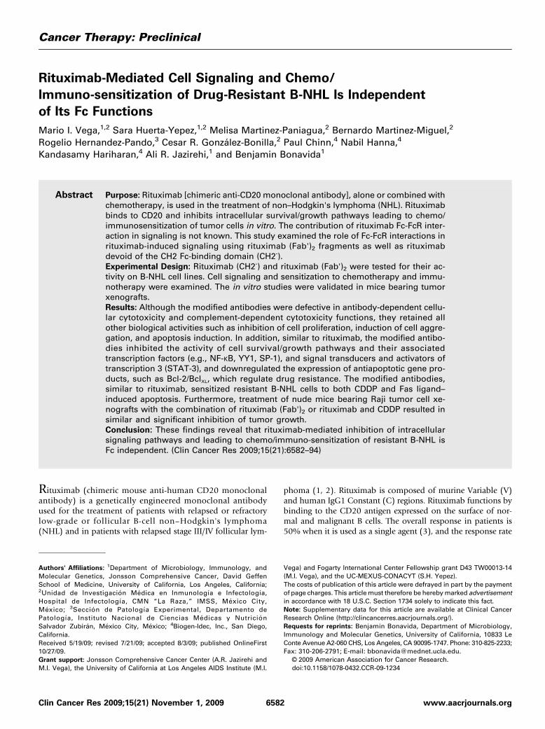
Cancer Therapy: Preclinical Rituximab-Mediated Cell Signaling and Chemo/ Immuno-sensitization of Drug-Resistant B-NHL Is Independent of Its Fc Functions Mario I. Vega, 1,2 Sara Huerta-Yepez, 1,2 Melisa Martinez-Paniagua, 2 Bernardo Martinez-Miguel, 2 Rogelio Hernandez-Pando, 3 Cesar R. González-Bonilla, 2 Paul Chinn, 4 Nabil Hanna, 4 Kandasamy Hariharan, 4 Ali R. Jazirehi, 1 and Benjamin Bonavida 1 Abstract Purpose: Rituximab [chimeric anti-CD20 monoclonal antibody], alone or combined with chemotherapy, is used in the treatment of non–Hodgkin's lymphoma (NHL). Rituximab binds to CD20 and inhibits intracellular survival/growth pathways leading to chemo/ immunosensitization of tumor cells in vitro. The contribution of rituximab Fc-FcR inter- action in signaling is not known. This study examined the role of Fc-FcR interactions in rituximab-induced signaling using rituximab (Fab') 2 fragments as well as rituximab devoid of the CH2 Fc-binding domain (CH2 - ). Experimental Design: Rituximab (CH2 - ) and rituximab (Fab') 2 were tested for their ac- tivity on B-NHL cell lines. Cell signaling and sensitization to chemotherapy and immu- notherapy were examined. The in vitro studies were validated in mice bearing tumor xenografts. Results: Although the modified antibodies were defective in antibody-dependent cellu- lar cytotoxicity and complement-dependent cytotoxicity functions, they retained all other biological activities such as inhibition of cell proliferation, induction of cell aggre- gation, and apoptosis induction. In addition, similar to rituximab, the modified antibo- dies inhibited the activity of cell survival/growth pathways and their associated transcription factors (e.g., NF-κB, YY1, SP-1), and signal transducers and activators of transcription 3 (STAT-3), and downregulated the expression of antiapoptotic gene pro- ducts, such as Bcl-2/Bcl XL , which regulate drug resistance. The modified antibodies, similar to rituximab, sensitized resistant B-NHL cells to both CDDP and Fas ligand– induced apoptosis. Furthermore, treatment of nude mice bearing Raji tumor cell xe- nografts with the combination of rituximab (Fab') 2 or rituximab and CDDP resulted in similar and significant inhibition of tumor growth. Conclusion: These findings reveal that rituximab-mediated inhibition of intracellular signaling pathways and leading to chemo/immuno-sensitization of resistant B-NHL is Fc independent. (Clin Cancer Res 2009;15(21):6582–94) Rituximab (chimeric mouse anti-human CD20 monoclonal antibody) is a genetically engineered monoclonal antibody used for the treatment of patients with relapsed or refractory low-grade or follicular B-cell non–Hodgkin's lymphoma (NHL) and in patients with relapsed stage III/IV follicular lym- phoma (1, 2). Rituximab is composed of murine Variable (V) and human IgG1 Constant (C) regions. Rituximab functions by binding to the CD20 antigen expressed on the surface of nor- mal and malignant B cells. The overall response in patients is 50% when it is used as a single agent (3), and the response rate Authors' Affiliations: 1 Department of Microbiology, Immunology, and Molecular Genetics, Jonsson Comprehensive Cancer, David Geffen School of Medicine, University of California, Los Angeles, California; 2 Unidad de Investigación Médica en Inmunología e Infectología, Hospital de Infectología, CMN “La Raza, ” IMSS, México City, México; 3 Sección de Patología Experimental, Departamento de Patología, Instituto Nacional de Ciencias Médicas y Nutrición Salvador Zubirán, México City, México; 4 Biogen-Idec, Inc., San Diego, California. Received 5/19/09; revised 7/21/09; accepted 8/3/09; published OnlineFirst 10/27/09. Grant support: Jonsson Comprehensive Cancer Center (A.R. Jazirehi and M.I. Vega), the University of California at Los Angeles AIDS Institute (M.I. Vega) and Fogarty International Center Fellowship grant D43 TW00013-14 (M.I. Vega), and the UC-MEXUS-CONACYT (S.H. Yepez). The costs of publication of this article were defrayed in part by the payment of page charges. This article must therefore be hereby marked advertisement in accordance with 18 U.S.C. Section 1734 solely to indicate this fact. Note: Supplementary data for this article are available at Clinical Cancer Research Online (http://clincancerres.aacrjournals.org/). Requests for reprints: Benjamin Bonavida, Department of Microbiology, Immunology and Molecular Genetics, University of California, 10833 Le Conte Avenue A2-060 CHS, Los Angeles, CA 90095-1747. Phone: 310-825-2233; Fax: 310-206-2791; E-mail: [email protected]. F 2009 American Association for Cancer Research. doi:10.1158/1078-0432.CCR-09-1234 6582 Clin Cancer Res 2009;15(21) November 1, 2009 www.aacrjournals.org
-
Upload
independent -
Category
Documents
-
view
1 -
download
0
Transcript of Rituximab-Mediated Cell Signaling and Chemo/Immuno-sensitization of Drug-Resistant B-NHL Is...
Cancer Therapy: Preclinical
Rituximab-Mediated Cell Signaling and Chemo/
Immuno-sensitization of Drug-Resistant B-NHL Is Independent
of Its Fc Functions
Mario I. Vega,1,2 Sara Huerta-Yepez,1,2 Melisa Martinez-Paniagua,2 Bernardo Martinez-Miguel,2
Rogelio Hernandez-Pando,3 Cesar R. González-Bonilla,2 Paul Chinn,4 Nabil Hanna,4
Kandasamy Hariharan,4 Ali R. Jazirehi,1 and Benjamin Bonavida1
Abstract Purpose: Rituximab [chimeric anti-CD20 monoclonal antibody], alone or combined with
chemotherapy, is used in the treatment of non–Hodgkin's lymphoma (NHL). Rituximab
binds to CD20 and inhibits intracellular survival/growth pathways leading to chemo/
immunosensitization of tumor cells in vitro. The contribution of rituximab Fc-FcR inter-
action in signaling is not known. This study examined the role of Fc-FcR interactions in
rituximab-induced signaling using rituximab (Fab')2 fragments as well as rituximab
devoid of the CH2 Fc-binding domain (CH2-).
Experimental Design: Rituximab (CH2-) and rituximab (Fab')2 were tested for their ac-
tivity on B-NHL cell lines. Cell signaling and sensitization to chemotherapy and immu-
notherapy were examined. The in vitro studies were validated in mice bearing tumor
xenografts.
Results: Although the modified antibodies were defective in antibody-dependent cellu-
lar cytotoxicity and complement-dependent cytotoxicity functions, they retained all
other biological activities such as inhibition of cell proliferation, induction of cell aggre-
gation, and apoptosis induction. In addition, similar to rituximab, the modified antibo-
dies inhibited the activity of cell survival/growth pathways and their associated
transcription factors (e.g., NF-κB, YY1, SP-1), and signal transducers and activators of
transcription 3 (STAT-3), and downregulated the expression of antiapoptotic gene pro-
ducts, such as Bcl-2/BclXL, which regulate drug resistance. The modified antibodies,
similar to rituximab, sensitized resistant B-NHL cells to both CDDP and Fas ligand–
induced apoptosis. Furthermore, treatment of nude mice bearing Raji tumor cell xe-
nografts with the combination of rituximab (Fab')2 or rituximab and CDDP resulted in
similar and significant inhibition of tumor growth.
Conclusion: These findings reveal that rituximab-mediated inhibition of intracellular
signaling pathways and leading to chemo/immuno-sensitization of resistant B-NHL is
Fc independent. (Clin Cancer Res 2009;15(21):6582–94)
Rituximab (chimeric mouse anti-human CD20 monoclonalantibody) is a genetically engineered monoclonal antibodyused for the treatment of patients with relapsed or refractorylow-grade or follicular B-cell non–Hodgkin's lymphoma(NHL) and in patients with relapsed stage III/IV follicular lym-
phoma (1, 2). Rituximab is composed of murine Variable (V)and human IgG1 Constant (C) regions. Rituximab functions bybinding to the CD20 antigen expressed on the surface of nor-mal and malignant B cells. The overall response in patients is50% when it is used as a single agent (3), and the response rate
Authors' Affiliations: 1Department of Microbiology, Immunology, and
Molecular Genetics, Jonsson Comprehensive Cancer, David Geffen
School of Medicine, University of California, Los Angeles, California;2Unidad de Investigación Médica en Inmunología e Infectología,
Hospital de Infectología, CMN “La Raza,” IMSS, México City,
México; 3Sección de Patología Experimental, Departamento de
Patología, Instituto Nacional de Ciencias Médicas y Nutrición
Salvador Zubirán, México City, México; 4Biogen-Idec, Inc., San Diego,
California.
Received 5/19/09; revised 7/21/09; accepted 8/3/09; published OnlineFirst
10/27/09.
Grant support: Jonsson Comprehensive Cancer Center (A.R. Jazirehi and
M.I. Vega), the University of California at Los Angeles AIDS Institute (M.I.
Vega) and Fogarty International Center Fellowship grant D43 TW00013-14
(M.I. Vega), and the UC-MEXUS-CONACYT (S.H. Yepez).
The costs of publication of this article were defrayed in part by the payment
of page charges. This articlemust therefore be herebymarked advertisementin accordance with 18 U.S.C. Section 1734 solely to indicate this fact.
Note: Supplementary data for this article are available at Clinical Cancer
Research Online (http://clincancerres.aacrjournals.org/).
Requests for reprints: Benjamin Bonavida, Department of Microbiology,
Immunology and Molecular Genetics, University of California, 10833 Le
Conte Avenue A2-060 CHS, Los Angeles, CA 90095-1747. Phone: 310-825-2233;
Fax: 310-206-2791; E-mail: [email protected].
F 2009 American Association for Cancer Research.
doi:10.1158/1078-0432.CCR-09-1234
6582Clin Cancer Res 2009;15(21) November 1, 2009 www.aacrjournals.org
is significantly higher when rituximab is used in combinationwith chemotherapy (4). The antitumor effect of rituximabdepends, in part, on FcγR-dependent immune activation (5).Also, it has been reported that human FcγR polymorphism cor-relates with the efficacy of B-tumor cell depletion and clinicalresponse (6, 7). It has been shown that rituximab can induceapoptosis in some systems but not others (8, 9). In additionto the above mechanisms, we have reported that rituximabcan also sensitize resistant tumor cells to apoptosis by chemo-therapeutic drugs and Fas ligand. Sensitization was the result ofrituximab-mediated inhibition of cell survival signaling path-ways and downregulation of antiapoptotic resistant factors(10–13).Modulation of the signaling pathways upon treatment of B-
NHL cells with rituximab might be the direct result of triggeringthe CD20 receptor by rituximab (Supplementary Fig. S1) or,conceivably, through cross-linking of the Fc fragment of rituxi-mab with FcRs expressed on the tumor cells or on host-effectorcells. Cross-linking of Fc on target cells by rituximab may eitheractivate or inhibit intracellular signaling pathways and the bal-ance can lead to the observed rituximab-mediated inhibition ofsurvival pathways. Four classes of Fc receptors have been iden-tified, namely, FcγRI, FcγRII, FcγRIII, and FcγRIV. The activat-ing and inhibitory receptors (FcγRIIB) transmit their signal viaimmune receptor tyrosine-based activation motif and inhibito-ry immune receptor tyrosine-based inhibitory motif (ITIM),respectively (14, 15). The FcR receptors show significant dif-ferences in their affinity for individual Fc isotypes (16, 17).Thus, it is conceivable to postulate that rituximab-mediatedcell signaling may be dependent, in part, on the rituximabFc fragments triggering FcR-bearing cells.To address the role of Fc-FcR interactions in rituximab-
mediated cell signaling and sensitization in vitro, we have pre-pared (Fab')2 fragments from rituximab and have geneticallyconstructed rituximab molecules devoid of the CH2 domainthat is responsible for both antibody-dependent cellular cyto-toxicity (ADCC) and complement-dependent cytotoxicity(CDC) effector functions (18, 19). In the preparation of mono-meric CH2-deleted antibodies, a dimeric form was also gener-
ated following strong noncovalent interactions. The presentstudy investigated the functional role of the Fc fragment ofrituximab in cell signaling and chemo/immunosensitization.By comparison with rituximab, the following objectives wereinvestigated using B-NHL cell lines as models: (a) Do themodified antibodies [rituximab (Fab')2 and rituximab (CH2-)]inhibit tumor cell proliferation and require cross-linking witha secondary anti-Ig antibody for the induction of apoptosis?(b) Do the modified antibodies trigger the cells and inhibitthe activity of the constitutively activated p38 mitogen-activatedprotein kinase (MAPK) cell survival pathway (and associatedtranscription factors) and, consequently, inhibition of the ex-pression of the antiapoptotic gene products Bcl-2/Bcl-xL thatregulate resistance? (c) Do the modified antibodies sensitizeresistant B-NHL cell lines to apoptosis induced by chemothera-peutic drugs and Fas ligand? and (d) Does rituximab (Fab)'2mimic rituximab in the inhibition of tumor growth xenograftwhen used in combination with CDDP? The findings showthe Fc independence of rituximab-mediated biological effects.
Materials and Methods
Cell lines
The Ramos, Raji, and Daudi cell lines were purchased from AmericanType Culture Collection. The AIDS-NHL B-cell line 2F7 was kindly pro-vided by Dr. Otoniel Martinez-Maza (Jonsson Comprehensive CancerCenter, Los Angeles, CA; ref. 10). Cells were grown in RPMI 1640 (Med-iatech, Cellgro) supplemented with 10% heat-inactivated fetal bovineserum (Life Technologies, Invitogen Co.) and 1% bacteriofungicide so-lution containing 10,000 U/mL penicillin G, 10 mg/mL streptomycin,and 25 μg/mL fungizone (Cellgro). Cells were cultured in 5% CO2 at37°C.
Antibodies and reagents
Rituximab was commercially obtained. The genetically engineered ri-tuximab with deleted CH2 domain (monomeric and dimeric CH2
-) wasdeveloped at Biogen-IDEC, Inc. Human IgG (Sigma) antibody was usedas control. The entire light chain, heavy variable, CH1 and CH3 do-mains were not changed. In addition, the first residue of the hinge re-gion was changed from alanine to valine. The expression construct wastransfected into Chinese hamster ovary DG44 cells secreting a mixtureof antibodies differentiated by disulfide formation in the hinge region.One form has properly formed disulfide and is the “Monomeric” anti-body, rituximab (CH2-). The second form has intrachain disulfides en-abling noncovalent interactions between two antibodies to form a“Dimeric” antibody, rituximab (CH2-) Dimer. This Dimer antibody isextremely stable as evidenced by the fact that SDS is required to breakthe interaction. The Monomeric and Dimeric forms were separated byhigh performance liquid chromatography. The rituximab (Fab')2 frag-ment was also obtained from Biogen-IDEC. It was prepared by pepsindigestion and purified to homogeneity as determined by SDS-PAGEanalysis (Supplementary Fig. S2). The anti-Fas and anti–Bcl-2 antibo-dies used for Western were purchased from Dako, the agonist antibodyagainst Fas (CH-11) and anti-Fas antibody (UB2) used for flow werepurchased from MBL. The anti-phospho Lyn, anti-Lyn, anti-p38 MAPKantibodies, and anti–Bcl-XL antibody were purchased from Cell Signal-ing and Santa Cruz, respectively.
Antibody-dependent cellular cytotoxicity
Raji cells (CCL-86, American Type Culture Collection) were grown inRPMI 1640 (Irvine Scientific) supplemented with 10% heat-inactivatedfetal bovine serum (Hyclone), 2 mmol/L L-glutamine, 100 units/mLof penicillin, and 100 μg/mL of streptomycin. Activated human periph-eral blood mononuclear cells (PBMC) were used as effector cells and
Translational Relevance
Rituximab has been used for the treatment of
CD20-expressing B-cell malignancies. The in vivomechanism of action has been proposed to be
through antibody-dependent cellular cytotoxicity,
complement-dependent cytotoxicity, and apoptosis.
A subset of patients exhibits a poor clinical response
due to Fc polymorphism. Reported studies showed
that rituximab mediates cell signaling and inhibits
several survival pathways, and sensitizes resistant
tumors to apoptosis by chemotherapeutic drugs. In
addition, the combination of rituximab and chemo-
therapy results in a significant clinical response.
The role of the Fc fragment of rituximab in mediating
cell signaling and chemosensitization was examined
and was found to be Fc independent. These studies
suggest that the chemosensitizing effect of rituximab
should also occur in patients with Fc polymorphism.
6583 Clin Cancer Res 2009;15(21) November 1, 2009www.aacrjournals.org
Cell Signaling and Sensitization by Rituximab (Fab')2

51Cr-labeled Raji as target. PBMC were isolated from whole blood ofhealthy donors using Histopaque (Sigma-Aldrich Corp.) and were cul-tured at a concentration of 5 × 10⁶ cells/mL in complete medium with20 U/mL recombinant human interleukin 2 (Invitrogen) in 75-cm2 tis-sue culture flasks at 37°C and 5% CO2. After overnight culture, 10
6 Rajitarget cells were labeled with 150 μCi of 51Cr (Amersham PharmaciaBiotech) for 1 h at 37°C and 5% CO2. The cells were washed four timesand resuspended in 5 mL of complete medium; 50 μL of cell suspen-sion was dispensed into each well containing equal volume of test orcontrol antibodies. All wells were plated in triplicate into a 96-well,round-bottomed tissue culture plate. The effector cells were harvested,washed once with complete medium, and added at 1 × 10⁶ cells in100 μL volume per well to obtain a 50:1 effector to target ratio. Thefollowing control wells were also included in triplicate: target cellsincubated with 100 μL complete medium to determine spontaneousrelease and target cells incubated with 100 μL 0.5% Triton X-100(Sigma-Aldrich Corp.) to determine maximum release. The culturewas incubated for 4 h at 37°C and 5% CO2, and the 51Cr released inthe culture supernatant due to cell lysis was determined on a γ counter(ISODATA). The cytotoxicity was expressed as the percentage of specificlysis and calculated as follows:
%Specific lysis
¼51Cr release of test samples� spontaneous 51Cr release
Maximum 51Cr release� spontaneous 51Cr release� 100
Complement-dependent cytotoxicity
The CDC activity of antibodies was determined using B-cell lines andrabbit complement. Dilutions of antibodies were made and 50 μL weredispensed into each 96-well plates in triplicates. The Raji cells were la-beled with 51Cr (150 μCi/106 cells) for 1 h at 37°C and 5% CO2. Thecells were washed four times and resuspended in complete medium,and 1 × 10⁴ cells in 50 μL were dispensed into each well. Normal rabbitcomplement (100 μL; ICN/Cappel Pharmaceuticals, Inc.) diluted 1:5 incomplete medium was added. Materials and Methods for the spontane-ous and maximum release control and set up were calculated as de-scribed above for ADCC. The cultures were incubated 4 h at 37°Cand 5% CO2. The radioactivity released into the culture supernatantwas determined on a γ counter. The formula for calculating the percent-age of specific cell lysis is described above in ADCC. Also, CDC wasachieved with human serum as source of complement and cytotoxicityassessed by flow as described (10).
Isolation of natural killer cells from human PBMCs andcytotoxic assays
PBMC were isolated from healthy donors by Lymphoprep (Axis-Shield PoC) gradient centrifugation. The subpopulations of natural kill-er (NK) cells were purified from isolated PBMC using negative selectionand Dynabeads (Dynal, Invitrogen) according to the manufacturer's in-structions. The purity, checked by flow cytometry (CD56+, CD16+), was>95% (data not shown). The purified NK cells were used as effectorscells for the cytotoxic assays described below.
Cell-mediated cytotoxicity was measured by CytoTox 96 Non-Radio-active Cytotoxicity Assay (Promega Co.) based on assessment of the totalrelease of cytoplasmic lactate dehydrogenase into the cultured mediumas a consequence of damaged cell membranes. Raji cells were incubatedin 5% CO2 at 37°C for 18 h in the absence or presence of IgG control,rituximab, or (Fab')2 rituximab. Raji cells were mixed with the effectorscells at the effector to target ratio of 40:1 and incubated at 37°C for 6 h inthe presence or absence of CDDP (5 μg/mL). The assay was done accord-ing to themanufacturer's instructions. The specific lysis was calculated bya standard formula according to the manufacturer's instructions.
Flow cytometric assays
Fas expression. An anti-human IgG-FITC antibody (Immunotech,Beckman Coulter Co.) was used to determine the cell binding of the
various rituximab antibodies. Cells were suspended at 106/mL in mediumand were treated with an optimal concentration (137 nmol/L) antibody for60 min, washed twice with PBS, and incubated with 3 μL of anti–IgG human-FITC. Immunofluorescence was carried out as described(11, 12). Extracellular detection was determined in cells treated withanti-Fas antibody or isotype control. The cells were washed twice andincubated with anti-mouse antibody phycoerythrin conjugated (Pharmin-gen) and analyzed by flow cytometry with a FACS Epics-XL MCL flowcytometer (Coulter, Inc.).
Active caspase-3. Active caspase-3 was analyzed by flow cytometryusing an FITC-conjugated anti–active caspase-3 monoclonal antibody(BD Pharmigen) as was described previously (11, 12). Briefly, 2 × 10⁶ cellsper sample were preincubated with rituximab (137 nmol/L) for 26 h andCH-11 antibody was added for an additional 18 h. For cross-linking, thecells were incubated with rituximab (137 nmol/L) and anti-human IgG an-tibody (342 nmol/L; ICN Pharmaceuticals) for 24 h. The cells were stainedintracellularly with anti-active caspase 3 antibody and the samples wereanalyzed by flow cytometry. Population data were acquired on a Flow cen-tre EPICSR XL-MCL (Coulter, Co.) with the System II Software and thepercent-positive cells were recorded.
Microscopic analysis
2F7 (106) cells were grown in six-well plates and treated with differ-ent antibodies [rituximab, rituximab (Fab')2, rituximab (CH2
-), rituxi-mab (CH2
-) Dimer] at 137 nmol/L for 6 h. The cells were visualized onan inverted microscope. Images were captured using Adobe Photoshop3.0.5 (Adobe System, Inc.) and a Sony DKC-5000 digital camera.
Measurement of cell growth
2F7 cells (2 × 10⁵/mL) were incubated at 37°C for different time per-iods in the presence and absence of different concentrations of variousrituximab antibodies (6, 9, 68.5, 137, and 274 nmol/L). The irrelevanthuman IgG isotype antibody (342 nmol/L) was used as control. Cellswere stained with trypan blue and viable cells were counted using lightmicroscopy.
Western blot analysis for protein expression
This was done as described previously (10). 2F7 and Ramos cells wereincubated at 37°C for 24 h. Cells extracts for protein analysis were pre-pared by lysing 2 × 10⁶ cells on ice with cold 200 μL of radioimmuno-precipitation assay buffer [1% NP40, 0.1% SDS, 0.5% deoxycholic acid,complete protease inhibitor cocktail tablets (Roche Diagnostic Co.), and1× PBS]. Lysates were transferred to microcentrifuge tubes and sonicatedin SONICATOR, model W-220F (Heat-System Ultrasonic, Inc.), for 10 s.Lysates were centrifuged at 12,000 × g at 4°C for 5 min. Protein concen-tration was quantified using the Bio-Rad protein assay (Bio-Rad Labora-tories). Gel loading buffer Bio-Rad (Bio-Rad Laboratories) was addedto the cell lysates, at a 1:1 volume. Samples were boiled for 5 min andwere separated on 12% SDS-polyacrilamide minigels and transferredto nitrocellulose membrane Hybond enhanced chemiluminescence(Amersham Pharmacia Biotech) in Trans-Blot SD semidry Transfer cellSystem (Bio-Rad).
Gel mobility shift assays
This was done as described (10). Briefly, cells (106) were harvestedafter treatment and washed twice with cold Dulbeco PBS (Cellgro). Af-ter washing, the cells were lysed in 1mL of NP40 lysis buffer [10mmol/LTris-HCl (pH 7.5), 10 mmol/L NaCl, 3 mmol/L MgCl2, and 0.5%NP40] on ice for 5 min. Samples were centrifuged at 300 × g at 4°Cfor 5 min. The pellet was washed twice in NP40 buffer. Nuclei werethen lysed in nuclear extraction buffer [20 mmol/L HEPES (pH 7.9),25% glycerol, 0.42 mmol/L NaCl, 1.5 mmol/L MgCl2, 0.2 mmol/LEDTA, 0.5 mmol/L phenylmethylsulfonyl fluoride, and 0.5 mmol/LDTT] and sonicated 10 s at 4°C. The protein concentration was deter-mined using the Bio-Rad protein assay. The nuclear proteins were fro-zen at -80°C. Both buffers contained the complete protease inhibitor
6584Clin Cancer Res 2009;15(21) November 1, 2009 www.aacrjournals.org
Cancer Therapy: Preclinical
cocktail tablets from Roche. Nuclear protein (5 μg) was mixed for 30min at room temperature with Biotin-labeled oligonucleotide probeNF-κB, Sp1, YY1, and signal transducers and activators of transcription3 (STAT-3), using the electrophoretic mobility shift assay kit purchasedfrom Panomics, Inc., following the manufacturer's instructions. Ten mi-croliters was subjected to 5% PAGE for 90 min in Tris-borate EDTAbuffer (Bio-Rad Laboratories), and transferred to Nylon transfer mem-brane Hybond-N+ (Amersham) using the Trans-Blot SD semidry Trans-fer cell System (Bio-Rad). The membranes were transferred to a UVCross-linker FB-UVXL-1000 Fisher technology (Fisher Scientific) for3 min. The detection was made as per the manufacturer's instructions,after the membranes were exposed using Hyperfilm enhanced chemi-luminescence (Amersham Pharmacia Biotech).
In vivo inhibition of Raji tumor xenograft in nude mice
The Raji ascites model was adapted from Zhu et al. (20) and Xionget al. (21). Briefly, Raji cells (6.5 × 10⁶) were inoculated i.p. into 6- to8-wk-old female athymic Nude-FoxN (Hardan). All mice were primed i.p. with 200 μL of pristine (Sigma) to induce inflammation on the weekbefore cell inoculation. Treatment started 1 wk after tumor cell inocu-lation by i.p. injection of rituximab or (Fab)'2 rituximab (100 μg/200μL; five animals per group). One and 2 d after antibody treatment,CDDP (4 mg/Kg in 200 μL) was administrated (i.p.) for two cyclesof 10-dintervals. At 60 d after tumor inoculation, the animals weresacrificed, and various organs were recovered for histologic and patho-logic analyses. Organs were analyzed by immunohistologic stainingwith a series of antibodies to confirm human tumor cells of B-cell or-igin. The animal experiments were done in accordance with allowedinstitutional and international regulations, and research protocols wereapproved by them.
Spleen histology and morphometric analysis. After sacrifice, thespleens were removed from the mice. Spleen tissues were fixed with4% (v/v) neutral buffered formalin. The specimens were dehydratedand embedded in paraffin. For histologic examination, 4-μm sectionsof fixed embedded tissues were cut on a Leica model 2165 rotary mi-crotome (Leica), placed on glass slides, deparaffinized, and stained se-quentially with H&E to assess the tumor cells infiltrates. The area (μm2)was calculated from the stained slides, selecting the follicles. Eight fol-licles were analyzed for each slide, and the experimental group and theresults were expressed as average and SDs of infiltrated tumor cells area(μm2). Analyses were done by a pathologist and a hematopathologistand then averaged.
Immunohistochemistry. Deparaffinized 4-μm spleen sections wereincubated sequentially in accordance with the instructions of the LSABkit from DAKO Corporation. Antigen retrieval was done by immersingthe slides in 0.01% sodium citrate (pH 6.0) incubated for 5 min in boil-ing water. The endogenous peroxidase activity was inhibited by im-mersing the slides in 3% H2O2-methanol for 25 min, and thebackground nonspecific binding was reduced by incubating with 1%bovine serum albumin in PBS for 60 min. The slides were incubatedovernight at room temperature with the human-specific antibodyagainst CD20 (1:100; Santa Cruz). Finally, the slides were washed fivetimes in 0.1 mol/L PBS (pH 7.4) for 8 min. To reduce the variability, allsamples from each group were processed at the same time in a singleexperiment using a single batch of antibody diluted in PBS-bovine se-rum albumin. After washing, the slides were incubated with a biotiny-lated secondary antibody container in the LSAB kit from DAKOcorporation for 30 min at room temperature followed by an incubationwith a streptavidin-horseradish peroxidase conjugate container in theLSAB kit for 30 min at room temperature, and then with 3,3′-diamino-benzidine tetra-hydrochloride (liquid 3,3′-diaminobenzidine, DAKOCorporation) for 1 to 6 min in the dark. The reaction was arrested withdistilled water and the slides were counterstained with hematoxilyn.Thereafter, the tissues were washed in tap water for 5 min., dehydratedthrough ethanol baths (70%, 90%, and 100%) in xylene, and mountedwith E-2 Mount medium (Shandon Lab). Finally, the slides were ana-lyzed under light microscopy (Olympus BX-40).
In situ detection of DNA fragmentation (terminal deoxynucleotidyltransferase-mediated dUTP nick end labeling). Tissue tumor sampleswere embedded in paraffin, and 3- to 5-μm slices were cut with a mi-crotome and placed on coated slides, paraffin was removed from thetissue sections with xylene, and the sections were rehydrated in gradedethanol solutions. DNA fragmentation was detected by the terminaldeoxynucleotidyl transferase-mediated dUTP nick end labeling (TU-NEL) method. After the tissue sections were deparaffinized, the slideswere immersed in 0.01% sodium citrate (pH 6.0) incubating for 20min in boiling water. The endogenous peroxidase activity was inhibitedby immersing the slides in 3% H2O2-methanol for 25 min. The slideswere incubated for 1 h in 37°C with the TdT enzyme of the TUNEL kit(Roche). After three washes in PBS 0.1 mol/L (pH 7.4) for 5 min, theslides were incubated with POD solution for 30 min in 37°C afterwashing, and the color was developed with 3,3′-diaminobenzidinetetra-hydrochloride (liquid DAB, DAKO Corporation) for 1 to 5 minin the dark. The reaction was arrested with distilled water and the slideswere counterstained with hematoxilyn. Thereafter, the tissues werewashed in tap water for 5 min, dehydrated through ethanol baths(70%, 90%, and 100%) in xylene and mounted with E-2 Mount medi-um (Shandon Lab). Finally, the slides were analyzed under light mi-croscopy (Olympus BX-40).
Statistical analysis .
All results were expressed as the mean ± SD of data obtained fromthree to four separate experiments. The statistical significance of the dif-ferences between group means was determined using one-way ANOVAto compare variance. Significant differences were considered for proba-bilities of <5% (P < 0.05).
Results
Rituximab-mediated biological activities (except CDC andADCC) are Fc independent. We have reported that rituximabtriggers CD20+ B-NHL cells and results in the inhibition of sev-eral intracellular survival pathways leading to chemo/immuno-sensitization (11–13). Cell signaling by rituximab might haveresulted from both the direct triggering of the CD20 receptoron tumor cells and by coengagement of the Fc receptor on tu-mor cells through the Fc-binding region of rituximab. The im-pact of Fc binding on rituximab-mediated cell signaling wasexamined using CH2- (the Fc-binding domain) deleted rituxi-mab and by rituximab (Fab')2 fragments. To investigate thesepossibilities, the role of Fc was examined by abolishing theproperties of both genetically engineered (CH2-) rituximaband rituximab (Fab')2 fragments.Cell surface binding of the various antibodies to B-NHL cell
lines was examined by flow cytometry. Treatment of the four B-NHL cell lines (2F7, Raji, Ramos, and Daudi) with rituximabshowed significant binding to all cell lines and the mean fluo-rescence intensity was significantly augmented compared withthe control IgG used. By comparison, using the same molarconcentrations, rituximab (CH2-), rituximab (CH2-) Dimer,and rituximab (Fab')2 stained all four cell lines and the meanfluorescence intensity was in the same range as those observedwith the native rituximab molecule (Supplementary Table S1).The activities of rituximab (Fab')2 and genetically engineered
CH2-deleted rituximab molecules [monomeric rituximab(CH2-) and rituximab (CH2-) Dimer] were compared with thoseof unmodified native rituximab. The CH2 domain has beenshown to exert several of the effector functions of the Fc fragmentof antibodymolecules such as its involvement inmediating both
6585 Clin Cancer Res 2009;15(21) November 1, 2009www.aacrjournals.org
Cell Signaling and Sensitization by Rituximab (Fab')2
CDC and ADCC (14). Rituximab mediated a very significantCDC activity, which was dependent on the antibody concentra-tion used and reached a maximum cytotoxicity of ∼80% (Sup-plementary Fig. S3A). As expected, both rituximab (CH2-) andrituximab (CH2-) Dimer showed no CDC activity at any of theantibody concentrations used, which was similar to the baselineobserved with the IgG isotype control (Supplementary Fig. S3A).In addition, lack of CDC activity by rituximab (Fab')2 was alsoconfirmed in an independent experiment (SupplementaryFig. S3B). Further, ADCC activity was examined by using humanperipheral blood as effector cells and radiolabeled target cells asdescribed in Materials and Methods. Rituximab exerted signifi-cant ADCC activity, and the percent-specific lysis was a functionof the antibody concentration used. In contrast, both rituximab(CH2-) and rituximab (CH2-) Dimer did not show any ADCCactivity above the control IgG levels (Supplementary Fig. S3C).These findings corroborate the CDC and ADCC functional prop-erties of the CH2 domain and show that the Fc effector functionsare lost by the rituximab-modified molecules.We next examined the ability of the various rituximab mole-
cules to induce apoptosis when used alone or following cross-linking with secondary anti-human IgG. Treatment with variousrituximab antibodies alone did not induce any significant apo-ptosis in all of the four cell lines (2F7, Raji, Ramos, and Daudi)tested. Previous studies have shown that cross-linking of mono-meric rituximab results in induction of apoptosis in several celllines (22). Thus, we examined the ability of the rituximab mo-lecules to induce apoptosis following cross-linking with anti-human IgG antibody as described in methods. The modifiedrituximab exerted significant apoptosis in all of the four celllines tested compared with cells treated with anti-IgG alone.The extent of apoptosis of both modified and unmodified ritux-imab molecules was comparable for all four lines tested (Fig.1A). We selected to use 2F7 B-cell line as a representative forsubsequent experiments. Treatment of B-NHL cell lines with ri-tuximab results in significant homotypic aggregation of the tu-mor target cells (23). Like rituximab, the modified rituximabantibodies induced significant aggregation of 2F7 cells (Fig.1B) and other B-NHL cell lines tested (data not shown). Fur-ther, treatment of the 2F7 B-NHL cell line with rituximab orwith modified rituximab antibodies resulted in significant in-hibition of cell growth and the extent of inhibition was ap-proximately equal for all four antibody molecules (Fig. 1C).Altogether, the above findings show that although, as ex-
pected, the modified rituximab molecules have lost their Fc-me-diated ADCC and CDC effector functions, they retained otherantibody-mediated effects such as binding to CD20, inductionof apoptosis following cross-linking, cell aggregation, and inhi-bition of cell growth.Independence of Fc for rituximab-mediated inhibition of cell
survival signaling pathways. We have reported that rituximabtriggers B-NHL cell lines and modifies several intracellularsignaling pathways such as the p38 MAPK and the NF-κB path-ways in both AIDS NHL (10, 24) and non-AIDS NHL (24).These molecular signaling pathways were inhibited by ritu-ximab and resulted in the selective downregulation of anti-apoptotic resistant factors such as Bcl-2 and Bcl-xL andconsequently led to sensitization of the resistant tumor cellsto drug-mediated apoptosis. Because the B-NHL cell lines ex-press Fc receptors and in vivo rituximab-treated cells interact
Fig. 1. Induction of apoptosis, cell aggregation, and inhibition ofproliferation by rituximab (CH2-), rituximab (CH2-) Dimer, and rituximab(Fab')2. A, apoptosis. Tumor cells (2 × 10⁶/mL; 2F7, Raji, Ramos, and Daudi)were incubated overnight at 37°C with rituximab, rituximab (CH2-),rituximab (CH2-) Dimer, and rituximab (Fab')2 at the concentration of 137nmol/L alone or cross-linked with goat anti-human IgG (342.5 nmol/L), andthe cells were then evaluated for apoptosis for the presence of activecaspase-3 (*, P < 0.001). B, cell aggregation. The 2F7 tumor cells (1 × 10⁶)were treated with 137 nmol/L of the different rituximab molecules. The cellswere then visualized on an inverted microscope and the images werecaptured and analyzed using Adobe Photoshop 3.0.5 and a Sony DKC-5000digital camera. C, proliferation. The 2F7 cells (1 × 10⁶/mL) were cultured inthe absence or the presence of rituximab molecules at concentrations of68.5 and 137 nmol/L for 24 h at 37°C. The cells were recovered andviability was determined microscopically by trypan blue dye exclusion.The number of viable cells was recorded and the data were analyzed bydetermining percent of control of cells cultured in medium alone(*, P < 0.001).
6586Clin Cancer Res 2009;15(21) November 1, 2009 www.aacrjournals.org
Cancer Therapy: Preclinical
with host FcR–bearing cells, it was plausible that rituximab-me-diated signaling in B-NHL cells lines may have resulted not onlyfrom direct triggering of CD20 but by the cooperation of the Fcfragments of rituximab interacting with tumor surface Fc recep-tors. These possibilities were addressed by comparing rituximabwith (Fab')2 antibodies in the inhibition of cell signaling path-ways. We examined representative gene products in the survivalsignaling pathways that are modified by rituximab as well asseveral transcription factors that are regulated by such survivalpathways. Rituximab inhibited the Src-kinase p-Lyn, but notLyn, and a similar inhibition was also observed by the mod-ified antibodies (Fig. 2A). Likewise, the p-p38 MAPK activity,but not p38 MAPK, was also significantly inhibited by themodified rituximab antibodies (Fig. 2B). Inhibition of thep38 MAPK activity by the modified antibodies, such as ritux-imab, resulted in the inhibition of Bcl-2 in the 2F7 cell line(10) and Bcl-xL expression in Ramos (Bcl-2 deficient; refs. 13,22), and the extent of the inhibition was the same for ritux-
imab and the modified antibodies (Fig. 2C and D). Inhibitionby rituximab of the p38 MAPK pathway also results in the inhi-bition of the NF-κB pathway as well as STAT-3–mediated activity(10). Thus, we examined the transcription factors regulated bythe activation of p38 MAPK pathway and found that both nativeand modified antibodies significantly inhibited the activities ofthe transcription factors NF-κB (Fig. 3A), YY1 (the transcriptionfactor Yin Yang 1; Fig. 3B), SP-1 (Fig. 3C), and STAT-3 (Fig. 3D)as assessed by electrophoretic mobility shift assay. The extent ofinhibition was the same for all of the antibodymolecules used atequivalent concentrations. The specificity of rituximab-mediatedinhibition of the transcription factors was shown by failure of ri-tuximab to inhibit the transcription factor AP-2 (SupplementaryFig. S3D).Altogether, the above findings show that rituximab signals
the cells directly via its interaction with the CD20 receptorand there was no detectable contribution from the Fc fragmentof rituximab.
Fig. 2. Inhibition of the p38 MAPKpathway and downregulation of Bcl-2or Bcl-XL expression by rituximab,rituximab (CH2-), and rituximab (Fab')2.The 2F7 and Ramos tumor cells werecultured with 137 nmol/L of the differentrituximab molecules for 24 h at 37°Cand lysates were prepared as describedin Materials and Methods. The lysatesfrom 2F7-treated cells were thenanalyzed for the presence of Lyn andp-Lyn (A), p38 MAPK, p-p38 MAPK (B),and for Bcl-2 (C). Lysates fromBcl-2–deficient Ramos were analyzedfor BclXL (D). The Western blots wereanalyzed by densitometry to showthe relative levels of the proteinscompared with levels in untreated cells.The data are representative of twosimilar experiments.
6587 Clin Cancer Res 2009;15(21) November 1, 2009www.aacrjournals.org
Cell Signaling and Sensitization by Rituximab (Fab')2
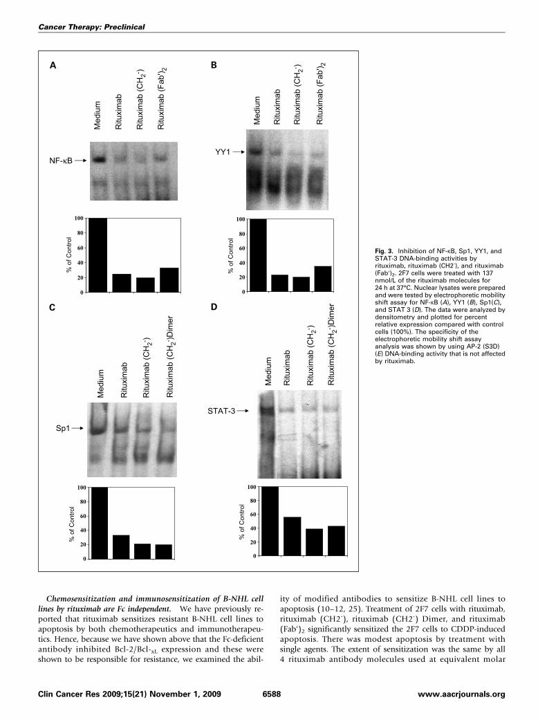
Chemosensitization and immunosensitization of B-NHL celllines by rituximab are Fc independent. We have previously re-ported that rituximab sensitizes resistant B-NHL cell lines toapoptosis by both chemotherapeutics and immunotherapeu-tics. Hence, because we have shown above that the Fc-deficientantibody inhibited Bcl-2/Bcl-xL expression and these wereshown to be responsible for resistance, we examined the abil-
ity of modified antibodies to sensitize B-NHL cell lines toapoptosis (10–12, 25). Treatment of 2F7 cells with rituximab,rituximab (CH2-), rituximab (CH2-) Dimer, and rituximab(Fab')2 significantly sensitized the 2F7 cells to CDDP-inducedapoptosis. There was modest apoptosis by treatment withsingle agents. The extent of sensitization was the same by all4 rituximab antibody molecules used at equivalent molar
Fig. 3. Inhibition of NF-κB, Sp1, YY1, andSTAT-3 DNA-binding activities byrituximab, rituximab (CH2-), and rituximab(Fab')2. 2F7 cells were treated with 137nmol/L of the rituximab molecules for24 h at 37°C. Nuclear lysates were preparedand were tested by electrophoretic mobilityshift assay for NF-κB (A), YY1 (B), Sp1(C),and STAT 3 (D). The data were analyzed bydensitometry and plotted for percentrelative expression compared with controlcells (100%). The specificity of theelectrophoretic mobility shift assayanalysis was shown by using AP-2 (S3D)(E) DNA-binding activity that is not affectedby rituximab.
6588Clin Cancer Res 2009;15(21) November 1, 2009 www.aacrjournals.org
Cancer Therapy: Preclinical
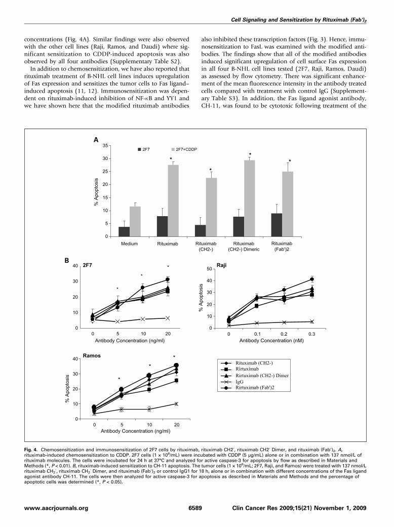
concentrations (Fig. 4A). Similar findings were also observedwith the other cell lines (Raji, Ramos, and Daudi) where sig-nificant sensitization to CDDP-induced apoptosis was alsoobserved by all four antibodies (Supplementary Table S2).In addition to chemosensitization, we have also reported that
rituximab treatment of B-NHL cell lines induces upregulationof Fas expression and sensitizes the tumor cells to Fas ligand–induced apoptosis (11, 12). Immunosensitization was depen-dent on rituximab-induced inhibition of NF-κB and YY1 andwe have shown here that the modified rituximab antibodies
also inhibited these transcription factors (Fig. 3). Hence, immu-nosensitization to FasL was examined with the modified anti-bodies. The findings show that all of the modified antibodiesinduced significant upregulation of cell surface Fas expressionin all four B-NHL cell lines tested (2F7, Raji, Ramos, Daudi)as assessed by flow cytometry. There was significant enhance-ment of the mean fluorescence intensity in the antibody treatedcells compared with treatment with control IgG (Supplement-ary Table S3). In addition, the Fas ligand agonist antibody,CH-11, was found to be cytotoxic following treatment of the
Fig. 4. Chemosensitization and immunosensitization of 2F7 cells by rituximab, rituximab CH2-, rituximab CH2- Dimer, and rituximab (Fab')2. A,rituximab-induced chemosensitization to CDDP. 2F7 cells (1 × 10⁶/mL) were incubated with CDDP (5 μg/mL) alone or in combination with 137 nmol/L ofrituximab molecules. The cells were incubated for 24 h at 37°C and analyzed for active caspase-3 for apoptosis by flow as described in Materials andMethods (*, P < 0.01). B, rituximab-induced sensitization to CH-11 apoptosis. The tumor cells (1 × 10⁶/mL; 2F7, Raji, and Ramos) were treated with 137 nmol/Lrituximab CH2
-, rituximab CH2- Dimer, and rituximab (Fab')2 or control IgG1 for 18 h, alone or in combination with different concentrations of the Fas ligand
agonist antibody CH-11. The cells were then analyzed for active caspase-3 for apoptosis as described in Materials and Methods and the percentage ofapoptotic cells was determined (*, P < 0.05).
6589 Clin Cancer Res 2009;15(21) November 1, 2009www.aacrjournals.org
Cell Signaling and Sensitization by Rituximab (Fab')2
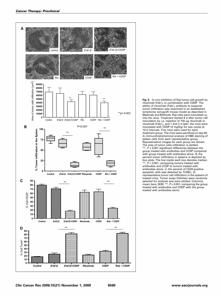
Fig. 5. In vivo inhibition of Raji tumor cell growth byrituximab (Fab')2 in combination with CDDP. Theability of rituximab (Fab)'2 antibody to suppresstumor infiltration was examined in an establishedlymphoma xenograft mouse model as described inMaterials and Methods. Raji cells were inoculated i.p.into the mice. Treatment started 8 d after tumor cellinoculation by i.p. injection of 100 μg rituximab orrituximab (Fab)'2, and 1 and 2 d later, the mice wereinoculated with CDDP (4 mg/Kg) for two cycles at10-d intervals. Five mice were used for eachtreatment group. The mice were sacrificed on day 60.A, immunohistochemical analysis of H&E staining ofspleen cells from each representative group.Representative images for each group are shown.The area of tumor cells infiltration is plotted.**, P < 0.001 significant differences between thegroup treated with antibodies and CCDP comparedwith group treated with antibodies alone. B, thepercent tumor infiltration in spleens is depicted bybox plots. The line inside each box denotes median.**, P < 0.001, comparing tumors treated withantibodies and CCDP to tumors treated withantibodies alone. C, the percent of CD20-positiveapoptotic cells was detected by TUNEL. D,representative tumor cell infiltration in the spleens oftreated mice. Tumor areas (follicles) were randomlyselected for analysis and were plotted. Columns,mean; bars, SEM. **, P < 0.001, comparing the grouptreated with antibodies and CDDP with the grouptreated with antibodies alone.
6590Clin Cancer Res 2009;15(21) November 1, 2009 www.aacrjournals.org
Cancer Therapy: Preclinical
tumor cells (2F7, Raji, and Ramos) with the modified anti-bodies, and the level of sensitization was a function of theCH-11 antibody concentration used (Fig. 4B).Altogether, the above findings show that, such as rituximab,
rituximab CH2- or rituximab (Fab')2 sensitize B-NHL celllines to both the CDDP chemotherapeutic drug and to theFas ligand–induced apoptosis. These results also suggest thatchemosensitization and immunosensitization by rituximabin vivo can be Fc-independent.In vivo therapeutic effect of rituximab (Fab')2 in combination
with CDDP on Raji tumor xenografts. Nude mice were inocu-lated i.p. with Raji cells and treated with rituximab, rituximab(Fab)'2, CDDP, and combinations as described in Materials andMethods. The mice were sacrificed at day 60 and their spleenswere removed and analyzed. A representative immunohisto-chemical analysis of tumor cell infiltrates in the spleen of micefollowing various treatments is shown in Fig. 5A. Clearly, incontrast to single agent treatment, the combination treatmentof rituximab plus CDDP or rituximab (Fab')2 plus CDDP re-duced significantly the area of tumor cell infiltrates. The datain Fig. 5B show that untreated mice had 75 ± 8.6% tumor in-filtrates. Treatment with single-agent rituximab, rituximab(Fab')2, or CDDP did not have any significant effect on the per-cent of tumor infiltrate compared with untreated mice. In con-trast, treatment with the combination of rituximab plus CDDPor rituximab (Fab')2 plus CDDP resulted in significant inhibi-tion of tumor cell infiltrates (P < 0.001). The combination treat-ment reduced tumor infiltrates from a mean of 75% to a meanof 18% (four fold reduction). These findings show the thera-peutic effect of the combined treatment on tumor growth.The tumor infiltrates were further examined by staining
with a specific human anti-CD20 antibody to directly examineRaji tumor cell infiltrates. The findings corroborated thoseobtained above using the immunohistochemical staining. Con-trol, untreated mice had a mean of 80% CD20-positive cells,
and treatment with single agents rituximab, rituximab (Fab')2,or CDDP did not have any significant inhibition on tumorinfiltrates. However, the combination treatment of bothrituximab (Fab')2 plus CDDP or rituximab plus CDDP reducedthe frequency of CD20-positive cells to a mean of 20%, a 4-foldreduction (Fig. 5C).The in vitro analysis shown in Fig. 4A showed that the com-
bination treatment of rituximab or rituximab (Fab')2 plusCDDP resulted in significant induction of apoptosis. Thus, weexamined if the combination treatments in vivo in mice alsoresulted in the induction of apoptosis in the tumor cells. Thespleen cells derived from the mice were examined for apoptosisby the TUNEL assay as described in Materials and Methods. Thefindings in Fig. 5D show clearly that antibody treatment did notshow any apoptosis compared with control. Treatment withCDDP induced a slight increase (∼15%) of apoptotic cells abovecontrol and antibody treatment. However, the combinationtreatment of rituximab plus CDDP or rituximab (Fab')2 plusCDDP resulted in significant potentiation in the frequency ofapoptotic cells (80%). Here again, there was 4-fold increaseabove controls following the combination treatment. Altogeth-er, these findings show that the combination treatment resultsin significant inhibition of tumor cell infiltrates in the spleen aswell as the induction of apoptosis. Further, there were no differ-ences between treatments with rituximab or rituximab (Fab')2.
Discussion
We have reported that rituximab can trigger B-NHL cell linesin vitro and modify several intracellular signaling pathways thatresult in reversing the chemoresistant and immunoresistantphenotypes to sensitive phenotypes. These studies did not ad-dress whether rituximab directly triggers the cells via the CD20receptor in the absence of any additional costimulation via the
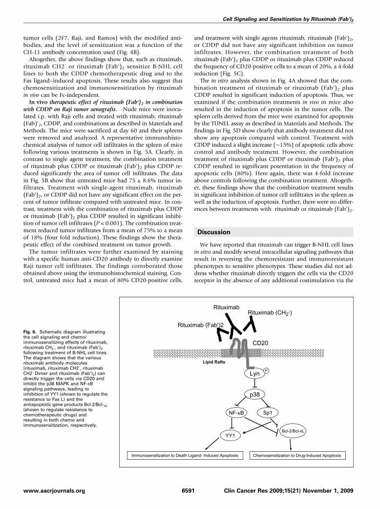
Fig. 6. Schematic diagram illustratingthe cell signaling and chemo/immunosensitizing effects of rituximab,rituximab CH2
-, and rituximab (Fab')2following treatment of B-NHL cell lines.The diagram shows that the variousrituximab antibody molecules[rituximab, rituximab CH2-, rituximabCH2- Dimer and rituximab (Fab')2] candirectly trigger the cells via CD20 andinhibit the p38 MAPK and NF-κBsignaling pathways, leading toinhibition of YY1 (shown to regulate theresistance to Fas L) and theantiapoptotic gene products Bcl-2/Bcl-xL(shown to regulate resistance tochemotherapeutic drugs) andresulting in both chemo andimmunosensitization, respectively.
6591 Clin Cancer Res 2009;15(21) November 1, 2009www.aacrjournals.org
Cell Signaling and Sensitization by Rituximab (Fab')2
Fc fragment of rituximab and Fc receptors expressed on the tu-mor cells. The present findings provide evidence that rituximab-mediated biological activities (except ADCC and CDC) and cellsignaling are Fc independent as corroborated by the use ofrituximab (Fab')2 and genetically engineered CH2-depleted ri-tuximab. The various functions examined with the modifiedantibodies were conserved and were comparable with unmod-ified rituximab such as the ability to inhibit cell proliferation,induce cell aggregation, induce apoptosis following cross-link-ing with a secondary anti-IgG antibody, and to inhibit the con-stitutively activated p38 MAPK and NF-κB signaling pathways.In addition, the various antibodies were comparable in theirability to sensitize resistant tumor cells to both chemotherapeu-tic drugs and Fas ligand–induced apoptosis. In vivo studies inmice showed the therapeutic efficacy of rituximab (Fab')2 incombinationwithCDDP, andwas similar to rituximabplusCDDP,as determined by inhibition of tumor growth. These findingsshow that cell signaling observed in vitro by rituximab resultsprimarily from the direct trigger of the CD20 receptor in the ab-sence of any contribution from the Fc region of anti-CD20 anti-body and can also take place in vivo in an Fc-independentmanner. The findings suggest that the in vivo observed chemo-sensitization and immunosensitization following rituximabtreatment may still take place in individuals who have reducedFc-dependent effector functions such as ADCC and CDC activ-ities (26) and in patients expressing low affinity variant of Fc-receptors due to polymorphism (6, 27).Several studies have identified the CH2 domain of the IgG an-
tibody molecule to mediate effectors functions such as its role inADCC and CDC (28, 29). This was corroborated with the genet-ically engineered monomeric rituximab (CH2-) and rituximab(CH2-) Dimer that were found to be devoid of any ADCC andCDC activities. These modified antibodies bind the corre-sponding CD20 receptor on the surface of B-NHL cell lines sim-ilar to rituximab. In contrast to their deficiencies in CDC andADCC, the CH2-deleted and (Fab')2 antibodies significantly in-hibited cell growth and induced apoptosis following cross-linkingwith anti-IgG antibodies. These various effects, therefore, are in-dependent of the Fc fragment and do not require cross-linking ofthe Fc fragment of rituximab with Fc receptor–bearing B-NHLcells. The present findings are in agreement with those of Carder-elli et al. (30) who have reported that an (Fab')2 fragment of anti-CD20 B1 antibody can induce apoptosis.We have reported previously that rituximab can trigger B-
NHL cell lines and modify several intracellular signaling path-ways that result in sensitization of the drug-resistant tumorcells to drug-induced apoptosis (10, 24, 27). It was possiblethat the molecular signaling triggered by rituximab might havebeen the result of cooperation between the rituximab-CD20receptor interaction and the interaction between the Fc frag-ment of rituximab and Fc receptors on the target cells. Clearly,Fc receptors can be triggered for signaling depending on thetype of the Fc receptor expressed on the cells. Fc-γ receptorstimulation may lead to either activation or inhibition of cellsignaling (31, 32). For example, the Fc-receptors Fc-γRIII/CD16A are expressed on NK cells, macrophages, and a subsetof T-cells, and trigger ADCC. Cross-linking of CD16A on NKcells, by immune complexes or by an anti-CD16 antibody, forexample, initiates a cascade of signaling events including thetyrosine phosphorylation of the γ chain (33) and other pro-teins, intracellular calcium mobilization, and activation of
phospho-lipases A2, C, and D. Activation of CD16A is cou-pled directly or indirectly by a nonreceptor tyrosine kinase.This inference is confirmed in studies in which the tyrosinephosphorylation of kinases including p72Syc p56lck, Shc,and p36 and activation of phosphatidyl inosityl 3-kinase havebeen observed (34). Also, cross-linking of Fcγ RIII resulted insignificant levels of p38MAPK phosphorylation (35). The in-hibitory Fc-γRIIB receptor is a single-chain molecule that car-ries an ITIM motif in its cytoplasmic domain and functionsthrough recruitment of the inositol phosphatase SHIP throughbinding to an SH2 site generated on the Fc-γRIIB ITIM. Withthe exception of T cells and NK cells, Fc-γRIIB is expressed inall cells of the immune system and it is the only receptor on Bcells where it regulates activity by signals delivered by immunecomplexes of the B-cell receptor. When coligated with BCR, ittriggers two ITIM-dependent signaling pathways that inhibitcell proliferation and cell activation (16). Thus, these variousFc-receptor–mediated signaling pathways might have contrib-uted to our observed findings of rituximab-mediated inhibi-tion of cell signaling. Our approach to rule out the role ofFcR cross-linking in rituximab-mediated inhibition of cell sig-naling, we investigated tumor cell triggering by functionallydefective Fc (CH2-)- and (Fab')2 rituximab antibodies andcompared the findings with the native rituximab. Rituximabtriggers the 2F7 B-NHL line and dephosphorylates p38 MAPKand STAT-3 and also inhibits Bcl-2/Bcl-xL expression (10, 24,36). In this study, we show that the CH2- and the (Fab')2 anti-bodies were able to trigger the cells and modify the abovepathways such as rituximab. These findings exclude the roleof Fc in rituximab-induced cell triggering and inhibition ofsurvival pathways.We have found that the CH2-deleted and (Fab')2 antibodies
behave such as rituximab in their ability to sensitize resistanttumor cells to chemotherapy and Fas ligand–induced apopto-sis. Inhibition of Bcl-2 expression in 2F7 cells was responsible,in large part, for chemosensitization (23). Our findings with themodified rituximab showed that inhibition of Bcl-2/BclXL ex-pression was similar to that obtained by rituximab; thus, themodified antibodies can sensitize the tumor cells to CDDP-in-duced apoptosis. In addition to chemosensitization, rituximabcan also sensitize B-NHL cell lines to Fas ligand–induced apo-ptosis. This was the result of rituximab-mediated upregulationof Fas expression via inhibition of NF-κB and the transcriptionrepressor YY1 (11, 12). We have previously reported that YY1regulates the expression of Fas via DNA binding to the silencerregion of the Fas promoter (37). Inhibition of YY1 relieves therepression and Fas transcription is upregulated. In the presentstudy, we also show that the modified antibodies, such as ritux-imab, inhibited YY1 DNA-binding activity and resulted in theupregulation of Fas expression and sensitization to CH-11–induced apoptosis.The in vitro findings discussed above showed clearly that che-
mosensitization can take place by (Fab')2-treated Raji cells.In vivo, the role of effector cells bearing FcRs such as purifiedNK cells in the sensitization of tumor cells by rituximab(Fab')2-treated cells was not clear. Thus, we examined therole of purified NK cells derived from normal PBMCs onthe chemosensitization effect of rituximab (Fab')2 to CDDP.The data showed clearly that NK cells had no effect on ritux-imab (Fab')2–treated cells in the presence of CDDP and thecytotoxicity obtained resulted primarily from the sensitizing
6592Clin Cancer Res 2009;15(21) November 1, 2009 www.aacrjournals.org
Cancer Therapy: Preclinical
effect mediated by rituximab (Fab')2, and there was no addi-tional effect by the NK cells (Supplementary Fig. S6). Thesefindings suggest that host FcR-bearing effector cells do not in-terfere with the sensitizing effect of rituximab (Fab')2–treatedcells. However, they can have additive effects when the targetcells are treated with rituximab in the presence of CDDP, viathe additional roles of ADDC and CDC.The findings achieved in vitro showed the equivalent sensiti-
zation of Raji cells by rituximab (Fab')2 or rituximab for CDDP-induced apoptosis and were corroborated in an in vivo Raji tu-mor cell xenograft model. The in vivo data showed clearly thetherapeutic efficacy of rituximab (Fab')2 as a chemosensitizerand mimics rituximab with the same magnitude of tumor cellinhibition. These findings indicate the potential therapeutic ef-ficacy of rituximab (Fab')2 when combined with chemotherapy.It is not clear whether rituximab (Fab')2 might result in a bettertherapeutic index in patients with B-NHL when combined withchemotherapy. It is possible that, in vivo, rituximab (Fab')2might have a faster clearance rate and reduce its therapeutic ef-ficacy. Likewise, it is also possible that rituximab (Fab')2 mightreach tumor sites that are not reached by rituximab. Futurestudies will address these questions using different models ofB-NHL xenografts.The present findings clearly show that rituximab-mediated
functions, cell signaling, and both chemosensitization and
immunosensitization are the result of the direct effect of theantibody binding to its cognate CD20 receptor, and these ef-fects do not require the participation or cooperation of cross-linking rituximab Fc fragment with the Fc receptors on thetumor cells or on host cells in vivo. A schematic diagram re-presenting the findings is shown in Fig. 6. In addition, thefindings suggest that chemosensitization and immunosensiti-zation by rituximab may be effective in poorly respondingpatients to rituximab treatment and who express low-affinityFc γ receptors or those who may have defective ADCC orCDC activities. Such patients may benefit from the combina-tion of rituximab and chemotherapeutic drugs or rituximaband TRAIL or anti-DR4/DR5 agonist antibodies.
Disclosure of Potential Conflicts of Interest
The authors received a financial gift with no restrictions from BiogenIdec
Pharmaceuticals through the hospices of the Regents of the University of
California.
Acknowledgments
We thank Kate Dinh, Christine Yue, Tania Golkar, Maggie Yang, Erica
Keng, and Tiffany Chin in the preparation of manuscript and Dr. Otoniel
Martinez-Maza for valuable input and support.
References1. Reff ME, Carner K, Chambers S, et al. Depletionof B cells in vivo by a chimeric mouse humanmonoclonal antibody to CD20. Blood 1994;83:435–45.
2. Seymour JF. New treatment approaches to in-dolent non-Hodgkin's lymphoma. Semin Oncol2004;31:27–32.
3. McLaughlin P, Grillo-Lopez AJ, Link BK, et al.Rituximab chimeric anti-CD20 monoclonal anti-body therapy for relapsed indolent lymphoma:half of patients respond to a four-dose treatmentprogram. J Clin Oncol 1998;16:2825–33.
4. Czuczman MS, Grillo-Lopez AJ, White CA, et al.Treatment of patients with low-grade B-celllymphoma with the combination of chimericanti-CD20 monoclonal antibody and CHOP che-motherapy. J Clin Oncol 1999;17:268–76.
5. Clynes RA, Towers TL, Presta LG, Ravetch JV.Inhibitory Fc receptors modulate in vivo cytoxi-city against tumor targets. Nat Med 2000;6:443–6.
6. Carton G, Dacheux L, Salles G, et al. Thera-peutic activity of humanized anti-CD20 mono-clonal antibody and polymorphism in IgG Fcreceptor FcγRIIIa gene. Blood 2002;99:754–8.
7.WengWK, Levy R. Two immunoglobulin G frag-ment C receptor polymorphisms independentlypredict response to rituximab in patients withfollicular lymphoma. J Clin Oncol 2003;21:3940–7.
8. Johnson P, Glennie M. The mechanisms of ac-tion of rituximab in the elimination of tumorcells. Semin Oncol 2003;30:3–8.
9. Pedersen IM, Buhl A, Klausen P, Geisler CH,Jurlander J. The chimeric anti-CD20 antibody ri-tuximab induces apoptosis in B-cell chronic lym-phocytic leukemia cells through a p38 mitogenactivated protein-kinase-dependent mechanism.Blood 2002;99:1314–9.
10. Vega MI, Huerta-Yepez S, Garban H, JazirehiA, Emmanouilides C, Bonavida B. Rituximab in-hibits p38 MAPK activity in 2F7 B NHL and de-
creases IL-10 transcription: pivotal role of p38MAPK in drug resistance. Oncogene 2004;23:3530–40.
11. Vega MI, Huerta-Yepez S, Bonavida B. Rituxi-mab (anti-CD20) up-regulated Fas expressionand sensitized B NHL to Fas mediated apo-ptosis. Oncogene 2005;24:8114–27.
12. VegaMI, Jazirehi AR, Huerta-Yepez S, BonavidaB. Rituximab-induced inhibition of YY1 and Bcl-xL expression in Ramos non-Hodgkin's lympho-ma cell line via inhibition of NF-κB activity: roleof YY1 and Bcl-xL in Fas resistance and che-moresistance, respectively. J Immunol 2005;175:2174–83.
13. Jazirehi AR, Bonavida B. Cellular and molecu-lar signal transduction pathways modulated byrituximab (rituxan, anti-CD20 mAb) in non-Hodg-kin's lymphoma: implications in chemosensitiza-tion and therapeutic intervention. Oncogene2005;24:2121–43.
14. DaeronM.Buildingup the familyof ITIM-bearingnegative coreceptors. Immunol Lett 1996;54:73–6.
15. Fong DC, Brauweiler A, Minskoff SA, et al. Mu-tational analysis reveals multiple distinct siteswithin Fc γ receptor IIB that function in inhibitorysignaling. J Immunol 2000;165:4453–62.
16. Nimmerjahn F, Ravetch JV. Fcγ receptors: oldfriends and new family members. Immunity2006;24:19–28.
17. Uray K, Medgyesi D, Hilbert A, Sarmay G,Gergely J, Hudecz F. Synthesis and receptorbinding of IgG1 peptides derived from the IgGFc region. J Mol Recognit 2004.
18. Wines BD, Powell MS, Parren PW, Barnes N,Hogarth PM. The IgG Fc contains distinct Fcreceptor (FcR) binding sites: the leukocytereceptors Fc γ RI and Fc γ RIIa bind to a regionin the Fc distinct from that recognized by neona-tal FcR and protein A. J Immunol 2000;164:5313–531.
19. Armour KL, van de Winkel JG, Williamson LM,
Clark MR. Differential binding to human FcγRIIaand FcγRIIb receptors by human IgG wildtypeand mutant antibodies. Mol Immunol 2003;40:585–93.
20. Zhu Z, Ghose T, Hoskin D, et al. Radioimmu-notherapy of human B-cell chronic lymphocyticleukemia in nude mice. Cancer Res 1994;54:5111–5117.
21. Xiong D, Xu Y, Liu H, et al. Efficient inhibitionof human B-cell lymphoma xenografts with ananti-CD20 × anti-CD3 bispecific diabody. CancerLett 2002;177:29–39.
22. Shan D, Ledbetter JA, Press OW. Signal-ing events involved in anti-CD20-induced ap-optosis of malignant human B cells. CancerImmunol Immunother 2000;48:673–683.
23. Kunzmann V, Ruediger T, Hallek M, Mueller-Hermelink HK, Wilhelm M. Tumor cell ag-glutination and not solely cytokine release asmechanism of adverse reactions during anti-CD20 monoclonal antibody (IDEC-C2B8, rituxi-mab) treatment. Blood 2001;98:1991–2.
24. Jazirehi AR, Gan XH, De Vos S, EmmanouilidesC, Bonavida B. Rituximab (anti-CD20) selectivelymodifies Bcl-xL and apoptosis protease activatingfactor-1 (Apaf-1) expression and sensitizes humannon-Hodgkin's lymphomaB cell lines to paclitaxel-induced apoptosis. Mol Cancer Ther 2003;2:1183–93.
25. Alas S, Emmanouilides C, Bonavida B. Inhibi-tion of interleukin 10 by rituximab results indown-regulation of bcl-2 and sensitization of B-cell non-Hodgkin's lymphoma to apoptosis. ClinCancer Res 2001;7:709–23.
26. Wen WK, Levy R. Expression of complementinhibitors CD46, CD55, and CD59 on tumor cellsdoes not predict clinical outcome after rituximabtreatment in follicular non-Hodgkin lymphoma.Blood 2001;98:1352–7.
27. Farag SS, Flinn IW, Modali R, Lehman TA,Young D, Byrd JC. Fc γ RIIIa and Fc γ RIIa poly-morphisms do not predict response to rituximab
6593 Clin Cancer Res 2009;15(21) November 1, 2009www.aacrjournals.org
Cell Signaling and Sensitization by Rituximab (Fab')2

in B-cell chronic lymphocytic leukemia. Blood2004;103:1472–4.
28. Canfield SM, Morrison SL. The binding affinityof human IgG for its high affinity Fc receptoris determined by multiple amino acids in theCH2 domain and is modulated by the hinge re-gion. J Exp Med 1991;173:1483–91.
29. Thommesen JE, Michaelsen TE, Loset GA,Sandlie I, Brekke OH. Lysine 322 in the humanIgG3 C(H)2 domain is crucial for antibodydependent complement activation. Mol Immu-nol 2000;37:995–1004.
30. Cardarelli PM, QuinnM, BuckmanD, et al. Bind-ing to CD20 by anti-B1 antibody or F(ab')(2) is suf-ficient for induction of apoptosis in B-cell lines.Cancer Immunol Immunother 2002;51:15–24.
31. Daeron M. Structural bases of Fcγ R functions.Int Rev Immunol 1997;16:1–27.
32. Pan L, Pei P. Signaling transduction by IgG re-ceptors. Chin Med J (Engl) 2003;116:487–94.
33. O'Shea JJ, Weissman AM, Kennedy IC,Ortaldo JR. Engagement of the natural killer cellIgG Fc receptor results in tyrosine phospho-rylation of the ζ chain. Proc Natl Acad Sci U S A1991;88:350–4.
34. Galandrini R, Palmieri G, Piccoli M, Frati L,Santoni A. Role for the Rac1 exchange factorVav in the signaling pathways leading to NK cellcytotoxicity. J Immunol 1999;162:3148–52.
35. Hazan-Halevy I, Seger R, Levy R. The require-ment of both extracellular regulated kinase andp38 mitogen-activated protein kinase for
stimulation of cytosolic phospholipase A(2) ac-tivity by either FcγRIIA or FcγRIIIB in human neu-trophils. A possible role for Pyk2 but not for theGrb2-Sos-Shc complex. J Biol Chem 2000;275:12416–23.
36. Alas S, Ng CP, Bonavida B. Rituximab modifiesthe cisplatin-mitochondrial signaling pathway,resulting in apoptosis in cisplatin-resistant non-Hodgkin's lymphoma. Clin Cancer Res 2002;8:836–45.
37. GarbanHJ, Bonavida B. Nitric oxide inhibits thetranscription repressor Yin-Yang 1 binding activ-ity at the silencer region of the Fas promoter: apivotal role for nitric oxide in the up-regulationof Fas gene expression in human tumor cells.J Immunol 2001;167:75–81.
6594Clin Cancer Res 2009;15(21) November 1, 2009 www.aacrjournals.org
Cancer Therapy: Preclinical