Efficacy and safety of rituximab in moderately-to-severely active systemic lupus erythematosus: The...
12
ARTHRITIS & RHEUMATISM Vol. 62, No. 1, January 2010, pp 222–233 DOI 10.1002/art.27233 © 2010, American College of Rheumatology Efficacy and Safety of Rituximab in Moderately-to-Severely Active Systemic Lupus Erythematosus The Randomized, Double-Blind, Phase II/III Systemic Lupus Erythematosus Evaluation of Rituximab Trial Joan T. Merrill, 1 C. Michael Neuwelt, 2 Daniel J. Wallace, 3 Joseph C. Shanahan, 4 Kevin M. Latinis, 5 James C. Oates, 6 Tammy O. Utset, 7 Caroline Gordon, 8 David A. Isenberg, 9 Hsin-Ju Hsieh, 10 David Zhang, 10 and Paul G. Brunetta 10 Objective. B cells are likely to contribute to the pathogenesis of systemic lupus erythematosus (SLE), and rituximab induces depletion of B cells. The Ex- ploratory Phase II/III SLE Evaluation of Rituximab (EXPLORER) trial tested the efficacy and safety of rituximab versus placebo in patients with moderately- to-severely active extrarenal SLE. Methods. Patients entered with >1 British Isles Lupus Assessment Group (BILAG) A score or >2 BILAG B scores despite background immunosuppres- sant therapy, which was continued during the trial. Pred- nisone was added and subsequently tapered. Patients were randomized at a ratio of 2:1 to receive rituximab (1,000 mg) or placebo on days 1, 15, 168, and 182. Results. In the intent-to-treat analysis of 257 patients, background treatment was evenly distributed among azathioprine, mycophenolate mofetil, and meth- otrexate. Fifty-three percent of the patients had >1 BILAG A score at entry, and 57% of the patients were categorized as being steroid dependent. No differences were observed between placebo and rituximab in the primary and secondary efficacy end points, including the BILAG-defined response, in terms of both area under the curve and landmark analyses. A beneficial effect of rituximab on the primary end point was observed in the African American and Hispanic sub- groups. Safety and tolerability were similar in patients receiving placebo and those receiving rituximab. Conclusion. The EXPLORER trial enrolled pa- tients with moderately-to-severely active SLE and used aggressive background treatment and sensitive cutoffs for nonresponse. No differences were noted between placebo and rituximab in the primary and secondary end points. Further evaluation of patient subsets, bio- markers, and exploratory outcome models may improve the design of future SLE clinical trials. ClinicalTrials.gov identifier: NCT00137969. Supported by Genentech. 1 Joan T. Merrill, MD: Oklahoma Medical Research Founda- tion, Oklahoma City; 2 C. Michael Neuwelt, MD: Alameda County Medical Center, Oakland, California; 3 Daniel J. Wallace, MD: Cedars- Sinai Medical Center, Los Angeles, California, and University of California, Los Angeles; 4 Joseph C. Shanahan, MD: Duke University, Durham, North Carolina; 5 Kevin M. Latinis, MD, PhD: University of Kansas Medical Center, Kansas City; 6 James C. Oates, MD: Ralph H. Johnson VA Medical Center, Charleston, South Carolina, and Medical University of South Carolina, Charleston; 7 Tammy O. Utset, MD: University of Chicago, Chicago, Illinois; 8 Caroline Gordon, MD: University of Birmingham, Birmingham, UK; 9 David A. Isenberg, MD: University College London, London, UK; 10 Hsin-Ju Hsieh, PhD, David Zhang, PhD, Paul G. Brunetta, MD: Genentech, San Francisco, California. Dr. Merrill has received speaking fees, consulting fees, and/or honoraria from Genentech, Bristol-Myers Squibb, MedImmune, UCB, Wyeth, and Cephalon (less than $10,000 each), and has consulted with investment analysts. Dr. Wallace has received speaking fees, consulting fees, and/or honoraria from Genentech (less than $10,000). Dr. Shanahan has received speaking fees, consulting fees, and/or honoraria from Genentech, Actelion, and Cypress (less than $10,000 each), and owns stock or stock options in Schering-Plough. Dr. Latinis has received speaking fees, consulting fees, and/or honoraria from Genen- tech (less than $10,000). Dr. Oates has received speaking fees, consulting fees, and/or honoraria from Genentech (less than $10,000). Dr. Utset has received advisory board fees from Genentech (more than $10,000). Dr. Gordon has received speaking fees, consulting fees, and/or honoraria from Roche Pharmaceuticals and Genentech (less than $10,000 each). Dr. Hsieh owns stock or stock options in Genentech. Address correspondence and reprint requests to Joan T. Merrill, MD, Oklahoma Medical Research Foundation, Clinical Phar- macology Research Program, MS 22, 825 Northeast 13th Street, Oklahoma City, OK 73104. E-mail: [email protected]. Submitted for publication June 1, 2009; accepted in revised form September 16, 2009. 222
Transcript of Efficacy and safety of rituximab in moderately-to-severely active systemic lupus erythematosus: The...

ARTHRITIS & RHEUMATISMVol. 62, No. 1, January 2010, pp 222–233DOI 10.1002/art.27233© 2010, American College of Rheumatology
Efficacy and Safety of Rituximab inModerately-to-Severely Active Systemic Lupus Erythematosus
The Randomized, Double-Blind, Phase II/III Systemic Lupus ErythematosusEvaluation of Rituximab Trial
Joan T. Merrill,1 C. Michael Neuwelt,2 Daniel J. Wallace,3 Joseph C. Shanahan,4
Kevin M. Latinis,5 James C. Oates,6 Tammy O. Utset,7 Caroline Gordon,8 David A. Isenberg,9
Hsin-Ju Hsieh,10 David Zhang,10 and Paul G. Brunetta10
Objective. B cells are likely to contribute to thepathogenesis of systemic lupus erythematosus (SLE),and rituximab induces depletion of B cells. The Ex-ploratory Phase II/III SLE Evaluation of Rituximab
(EXPLORER) trial tested the efficacy and safety ofrituximab versus placebo in patients with moderately-to-severely active extrarenal SLE.
Methods. Patients entered with >1 British IslesLupus Assessment Group (BILAG) A score or >2BILAG B scores despite background immunosuppres-sant therapy, which was continued during the trial. Pred-nisone was added and subsequently tapered. Patients wererandomized at a ratio of 2:1 to receive rituximab (1,000mg) or placebo on days 1, 15, 168, and 182.
Results. In the intent-to-treat analysis of 257patients, background treatment was evenly distributedamong azathioprine, mycophenolate mofetil, and meth-otrexate. Fifty-three percent of the patients had >1BILAG A score at entry, and 57% of the patients werecategorized as being steroid dependent. No differenceswere observed between placebo and rituximab in theprimary and secondary efficacy end points, includingthe BILAG-defined response, in terms of both areaunder the curve and landmark analyses. A beneficialeffect of rituximab on the primary end point wasobserved in the African American and Hispanic sub-groups. Safety and tolerability were similar in patientsreceiving placebo and those receiving rituximab.
Conclusion. The EXPLORER trial enrolled pa-tients with moderately-to-severely active SLE and usedaggressive background treatment and sensitive cutoffsfor nonresponse. No differences were noted betweenplacebo and rituximab in the primary and secondaryend points. Further evaluation of patient subsets, bio-markers, and exploratory outcome models may improvethe design of future SLE clinical trials.
ClinicalTrials.gov identifier: NCT00137969.Supported by Genentech.1Joan T. Merrill, MD: Oklahoma Medical Research Founda-
tion, Oklahoma City; 2C. Michael Neuwelt, MD: Alameda CountyMedical Center, Oakland, California; 3Daniel J. Wallace, MD: Cedars-Sinai Medical Center, Los Angeles, California, and University ofCalifornia, Los Angeles; 4Joseph C. Shanahan, MD: Duke University,Durham, North Carolina; 5Kevin M. Latinis, MD, PhD: University ofKansas Medical Center, Kansas City; 6James C. Oates, MD: Ralph H.Johnson VA Medical Center, Charleston, South Carolina, and MedicalUniversity of South Carolina, Charleston; 7Tammy O. Utset, MD:University of Chicago, Chicago, Illinois; 8Caroline Gordon, MD:University of Birmingham, Birmingham, UK; 9David A. Isenberg,MD: University College London, London, UK; 10Hsin-Ju Hsieh, PhD,David Zhang, PhD, Paul G. Brunetta, MD: Genentech, San Francisco,California.
Dr. Merrill has received speaking fees, consulting fees, and/orhonoraria from Genentech, Bristol-Myers Squibb, MedImmune, UCB,Wyeth, and Cephalon (less than $10,000 each), and has consulted withinvestment analysts. Dr. Wallace has received speaking fees, consultingfees, and/or honoraria from Genentech (less than $10,000). Dr.Shanahan has received speaking fees, consulting fees, and/or honorariafrom Genentech, Actelion, and Cypress (less than $10,000 each), andowns stock or stock options in Schering-Plough. Dr. Latinis hasreceived speaking fees, consulting fees, and/or honoraria from Genen-tech (less than $10,000). Dr. Oates has received speaking fees,consulting fees, and/or honoraria from Genentech (less than $10,000).Dr. Utset has received advisory board fees from Genentech (more than$10,000). Dr. Gordon has received speaking fees, consulting fees, and/orhonoraria from Roche Pharmaceuticals and Genentech (less than$10,000 each). Dr. Hsieh owns stock or stock options in Genentech.
Address correspondence and reprint requests to Joan T.Merrill, MD, Oklahoma Medical Research Foundation, Clinical Phar-macology Research Program, MS 22, 825 Northeast 13th Street,Oklahoma City, OK 73104. E-mail: [email protected].
Submitted for publication June 1, 2009; accepted in revisedform September 16, 2009.
222

Systemic lupus erythematosus (SLE) is a hetero-geneous autoimmune disease that can cause severeorgan damage (1,2). SLE predominantly affects women,and the prevalence is highest among African Americans,African Caribbeans, Hispanics, and Asians (3,4). Com-prehensive care is required to prevent serious sequelae(1,5,6); however, no new medication for SLE has beenapproved by the US Food and Drug Administration(FDA) for the past 50 years. Current therapies, includ-ing use of corticosteroids, antimalarial agents, and im-munosuppressive drugs, are largely empiric. These inter-ventions are not always effective and may contribute toorgan damage (7,8).
B cells have critical roles in the pathogenesis ofSLE, including cytokine production, presentation of selfantigen, T cell activation, and autoantibody production(9–12). Loss of B cell tolerance may be a pivotal event inthe pathogenesis of SLE (9–11,13), providing a strongrationale for the study of targeted treatments thatmodify the effects of B cells on immunity.
Rituximab, a chimeric monoclonal antibody thatselectively targets CD20-positive B cells while sparingstem cells and plasma cells (14–16), is approved for thetreatment of non-Hodgkin’s lymphoma and rheumatoidarthritis (RA) (17–19). The results of several smalluncontrolled trials have suggested that rituximab mighthave potential efficacy and be steroid-sparing in SLE(20–32).
The Exploratory Phase II/III SLE Evaluation ofRituximab (EXPLORER) trial, a placebo-controlled,double-blind, multicenter study of rituximab in patientswith moderately-to-severely active extrarenal SLE, wasundertaken to assess the efficacy and safety of rituximabover 52 weeks. (See Appendix A for the principalEXPLORER study centers and investigators.)
PATIENTS AND METHODS
Institutional review board approval was obtained ateach trial site. The study was conducted in accordance withFDA Good Clinical Practice guidelines and the Health Insur-ance Portability and Accountability Act of 1996. Patientsprovided written informed consent prior to participation.
Patients. Inclusion criteria included age 16–75 years; ahistory of meeting 4 American College of Rheumatology(ACR) criteria for SLE (33), including a positive test forantinuclear antibodies; active disease at screening, defined as�1 organ system with a British Isles Lupus Assessment (BI-LAG) A score (severe disease activity) or �2 organ systemswith a BILAG B score (moderate disease activity) (34,35); andstable use of 1 immunosuppressive drug at entry, which wasable to be continued during the trial.
Patients were excluded for severe central nervous
system or organ-threatening lupus or any other active condi-tions requiring significant use of steroids or recent treatmentwith a cyclophosphamide or a calcineurin inhibitor (within 12weeks of screening); a history of cancer or serious recurrent orchronic infection; uncontrolled medical disease; pregnancy orplanning pregnancy; previous treatment with B cell–targetedtherapy; aspartate aminotransferase or alanine aminotransfer-ase level �2.5-fold the upper limit of normal (ULN); amylaseor lipase level �2-fold the ULN; neutrophil count �1.0 �103/�l; positive results of hepatitis B or hepatitis C serology;hemoglobin concentration �7 gm/dl (unless caused by hemo-lytic anemia due to SLE); platelet count �10,000/�l; andserum creatinine level �2.5 mg/dl.
Study design. The EXPLORER trial was a random-ized, double-blind, placebo-controlled, multicenter (55 cen-ters) North American study evaluating the efficacy and safetyof rituximab compared with placebo in patients with SLE whowere receiving background immunosuppressants and pred-nisone (Figure 1A).
Patients were randomized at a 2:1 ratio to receiveintravenous rituximab (2 1,000-mg doses given 14 days apart)or placebo on days 1, 15, 168, and 182, which was added toprednisone (given according to the protocol) and to thebaseline immunosuppressive regimen. Each infusion wasaccompanied by intravenous administration of acetamino-phen, diphenhydramine, and methylprednisolone (100 mg).The BILAG index was used to assess SLE activity. To ensureinclusion of only patients with significantly active disease,minimum disease severity at entry was stringently defined as�1 domain with a BILAG A score or �2 domains with aBILAG B score, despite background treatment with eitherazathioprine (AZA; 100–250 mg/day), mycophenolate mofetil(MMF; 1–4 gm/day), or methotrexate (MTX; 7.5–27.5 mg/week). After screening, eligible patients continued their immu-nosuppressant therapy and received additional daily oral pred-nisone (0.5 mg/kg, 0.75 mg/kg, or 1.0 mg/kg), based on theBILAG score at entry and the amount of steroids alreadybeing taken at the time of entry. Steroids were taperedbeginning on day 16, with the goal of reaching a dosage of �10mg/day over 10 weeks and �5 mg/day by week 52.
Clinical assessments. Patients were evaluated monthlywith the BILAG index and the Lupus Quality of Life(LupusQol) index (based on the Short Form 36 [SF-36] withadditional components including pain and fatigue) (36,37).The BILAG index was used to assess response, and scoreswere converted to numeric values (A � 9, B � 3, C � 1, D �0, E � 0) (38) to enable evaluation of fluctuating globalsummary scores.
Primary end points. The effect of placebo versusrituximab in achieving and maintaining a major clinical re-sponse, a partial clinical response, or no clinical response atweek 52 was assessed using each of the 8 BILAG index organsystem scores.
A major clinical response was defined as achievingBILAG C scores or better in all organs at week 24 withoutexperiencing a severe flare (1 new domain with a BILAG Ascore or 2 new domains with a BILAG B score) from day 1 toweek 24, and maintaining this response without a moderate orsevere flare (�1 new domains with a BILAG A or B score) toweek 52. A partial clinical response was defined as 1) achievingBILAG C scores or better at week 24 and maintaining this
EFFICACY AND SAFETY OF RITUXIMAB IN THE EXPLORER TRIAL 223

response without a new BILAG A or B score for 16 consecu-tive weeks, 2) achieving no more than 1 organ with a BILAGB score at week 24 without achieving �1 new BILAG A or Bscore to week 52, or 3) achieving a maximum of 2 BILAG Bscores at week 24 without developing BILAG A or B scores innew domains until week 52 if the baseline BILAG score for thepatient was 1 A score plus �2 B scores, �2 A scores, or �4 Bscores. No clinical response was defined as failure to meet thedefinition of a major clinical response or a partial clinicalresponse. Patients who terminated the study early were scoredas having no clinical response.
Secondary end points. Secondary end points included:1) the time-adjusted area under the curve minus baseline(AUCMB) of the BILAG score over 52 weeks, 2) the propor-tion of patients who achieved a major clinical response (ex-cluding a partial clinical response) and the proportion ofpatients with a partial clinical response (including a majorclinical response) at week 52, 3) the proportion of patients witha BILAG C score or better in all organs at week 24, 4) the time
to the first moderate or severe disease flare, 5) improvement inquality of life as measured by the LupusQoL, and 6) the propor-tion of patients who achieved a major clinical response with aprednisone dosage of �10 mg/day from week 24 to week 52.
Subgroup analyses of the primary and secondary endpoints were preplanned for the following factors: race (AfricanAmerican/Hispanic versus others), age (�40 years or �40years), sex, assigned prednisone dose, background immuno-suppressant, duration of lupus, long-term prednisone therapy,baseline BILAG A score, baseline BILAG-defined mucocuta-neous or musculoskeletal system involvement, and baselinebiomarkers.
Safety assessments. The incidence and severity ofadverse events (AEs) were classified using the National CancerInstitute Common Toxicity Criteria for Adverse Events (ver-sion 3.0). Serious adverse events (SAEs), infusion-related AEs(an AE occurring during or within 24 hours following comple-tion of the infusion of a study drug), and infection-related AEswere summarized independently. Serum chemistries, hemato-
Figure 1. A, Study design. B, Disposition of patients. AE � adverse event.
224 MERRILL ET AL

logic parameters, urinalysis results, quantitative IgG levels, T celland B cell counts, human antichimeric antibody (HACA) levels,complement levels, and autoantibody levels were monitored.
Statistical analysis. The proportion of patients achiev-ing a major clinical response, a partial clinical response, or noclinical response was compared between the placebo andrituximab groups using the stratified Wilcoxon’s rank sum test,with the initial prednisone dose and race/ethnicity as stratifi-cation factors. Results were expressed as the proportion ofpatients in each of 6 cells (2 treatment groups � 3 responsecategories), and the P value was computed to compare thegraded response across treatment arms. Two-sided P valuesless than 0.05 were considered significant. The analyses wereperformed using SAS software, version 9.1 (SAS Institute,Cary, NC).
RESULTS
Study population. Of the 257 patients, 88 wererandomized to receive placebo, and 169 were assigned tothe rituximab group. The demographic and diseasecharacteristics of the patients are presented in Table 1.The mean age of the patients was 40.4 years, and thefemale:male ratio was 9:1. An African American, His-panic, or Asian background was present in 42.1% ofplacebo-treated patients and in 41.5% of rituximab-treated patients.
After screening, 61.4% of placebo-treated pa-tients and 62.7% of rituximab-treated patients receivedthe lowest dosage of prednisone allowed (0.5 mg/kg/day). The mean � SD prednisone dose at baseline was45.9 � 16.4 mg, and the median dose was 40.0 mg.Background immunosuppressive treatment includedAZA, MMF, and MTX, and these treatments weredistributed roughly evenly in the placebo group (36.4%,36.4%, and 27.3%, respectively) and the rituximab group(32.0%, 39.6%, and 27.8%, respectively).
At baseline, most patients had disease activity inthe mucocutaneous and musculoskeletal systems. In theplacebo group, 58.0% and 13.6% of patients had BILAGB and BILAG A scores, respectively, for the mucocuta-neous domain; in the rituximab group, these proportionswere 56.2% and 16.0%, respectively. For the musculo-skeletal domain, 54.5% and 30.7% of patients in theplacebo group had BILAG B and BILAG A scores,respectively; in the rituximab group, these proportionswere 55.0% and 25.4%, respectively. The most commonconcurrently active domains in the intent-to-treat popu-lation at entry were musculoskeletal and mucocutaneous(54.0%), musculoskeletal and general (constitutionalfeatures; 36.0%), and mucocutaneous and general(29.0%).
Seventy-three percent of patients in the placebogroup and 71.0% in the rituximab group completed 52
weeks of treatment. Most withdrawals were due toadverse events or the patient’s decision (Figure 1B).
Table 1. Baseline demographic and disease characteristics of thepatients*
CharacteristicPlacebo(n � 88)
Rituximab(n � 169)
Female sex 93.2 89.9Age, mean � SD years 40.5 � 12.8 40.2 � 11.4Race, %
White 55.7 56.2African American 27.3 23.7Hispanic 9.1 14.2Asian/Pacific Islander 5.7 3.6Other 2.2 1.1
Disease duration, mean � SD years 8.7 � 7.6 8.5 � 7.2Long-term prednisone therapy† 53.4 58.6Assigned prednisone dosage at
screening, mg/kg/day0.5 61.4 62.70.75 29.5 32.01.0 9.1 5.3
Background immunosuppressive drugAzathioprine 36.4 32.0Methotrexate 27.3 27.8Mycophenolate mofetil 36.4 39.6
Disease activityBILAG index score at entry
�1 A score 56.0 51.0No A score, �3 B scores 22.0 31.02 B scores only 22.0 18.0
BILAG index global scoreMean � SD 14.5 � 5.6 14.0 � 5.1Median (range) 13 (7–33) 13 (6–30)
SLE domains at baseline, no. (%)General
BILAG A score 11.0 (12.5) 14.0 (8.3)BILAG B score 28.0 (31.8) 53.0 (31.4)
MucocutaneousBILAG A score 12.0 (13.6) 27.0 (16.0)BILAG B score 51.0 (58.0) 95.0 (56.2)
NeurologicBILAG A score 3.0 (3.4) 3.0 (1.8)BILAG B score 6.0 (6.8) 20.0 (11.8)
MusculoskeletalBILAG A score 27.0 (30.7) 43.0 (25.4)BILAG B score 48.0 (54.5) 93.0 (55.0)
Cardiovascular and respiratoryBILAG A score 6.0 (6.8) 8.0 (4.7)BILAG B score 13.0 (14.8) 32.0 (18.9)
VasculitisBILAG A score 3.0 (3.4) 5.0 (3.0)BILAG B score 7.0 (8.0) 23.0 (13.6)
RenalBILAG A score 0 (0) 0 (0)BILAG B score 0 (0) 3 (1.8)
HematologyBILAG A score 2.0 (2.3) 3.0 (1.8)BILAG B score 17.0 (19.3) 39.0 (23.1)
* Except where specified otherwise, values are the percent of patients.BILAG � British Isles Lupus Assessment Group; SLE � systemiclupus erythematosus.† Continuous use of corticosteroids for �6 months was required, aswell as the inability to taper to a dosage of �10 mg/day without therecurrence of lupus activity.
EFFICACY AND SAFETY OF RITUXIMAB IN THE EXPLORER TRIAL 225

Efficacy. Primary end point. At week 52, nodifference was noted in major clinical responses orpartial clinical responses between the placebo group(15.9% had a major clinical response, and 12.5% had apartial clinical response) and the rituximab group
(12.4% had a major clinical response, and 17.2% had apartial clinical response) (Figure 2A) relative to theoverall response rate (28.4% versus 29.6%).
Subgroup analysis. There were no differences inthe primary end point in prespecified subgroup analyses
Figure 2. A, Proportion of patients experiencing a major clinical response (MCR), a partial clinicalresponse (PCR), and no clinical response (NCR) at 52 weeks. B, Responders with AfricanAmerican/Hispanic backgrounds. C, Responders, according to the background immunosuppressivedrug. BILAG � British Isles Lupus Assessment Group; AZA � azathioprine; MMF � mycophe-nolate mofetil; MTX � methotrexate.
226 MERRILL ET AL
except in the African American/Hispanic group, whichcomprised approximately one-third of the patients in thestudy. Among the patients in this subgroup who receivedplacebo, a major clinical response was observed in only9.4%, and a partial clinical response was observed inonly 6.3%. In contrast, among patients in this subgroupwho received rituximab, a major clinical response wasobserved in 13.8%, and a partial clinical response wasobserved in 20.0% (P � 0.0408). This outcome wasassociated with a higher proportion of nonresponders inthe placebo group compared with placebo-treated pa-tients of other ethnic subgroups (Figure 2B). No differ-ence in major secondary end points was observed in thissubgroup.
None of the primary or secondary end pointswere met in the subgroup of patients on backgroundMTX who were receiving rituximab. However, an ad hocanalysis showed that the rituximab-treated patients inthis subgroup had improved mean BILAG global scoresat week 52 compared with the placebo group (P � 0.007)(Figure 2C).
Secondary end point. In the intent-to-treat popu-lation, the mean � SD time-adjusted AUCMB of theBILAG global score was �5.9 � 4.5 in the placebogroup compared with �5.8 � 4.0 in the rituximab group,over 52 weeks (P � 0.8092). The proportion of patientswith a BILAG C score or better in all domains in thefirst 24 weeks was the same in the placebo group (27.3%;95% CI 18.0–36.6) and the rituximab group (24.9%;95% CI 18.3–31.4 [P � 0.5602]). Patients with a majorclinical response in whom prednisone was tapered to adosage of �10 mg/day from week 24 to week 52 included9 patients (10.2%) in the placebo group and 14 patients(8.3%) in the rituximab group.
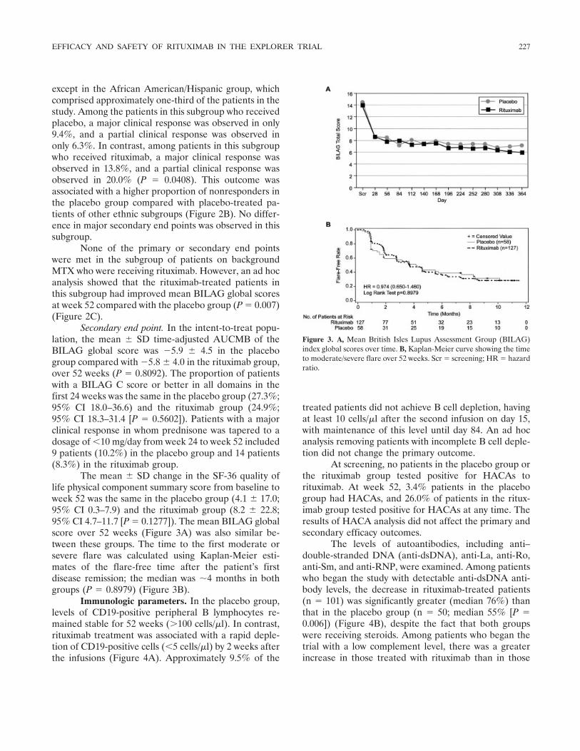
The mean � SD change in the SF-36 quality oflife physical component summary score from baseline toweek 52 was the same in the placebo group (4.1 � 17.0;95% CI 0.3–7.9) and the rituximab group (8.2 � 22.8;95% CI 4.7–11.7 [P � 0.1277]). The mean BILAG globalscore over 52 weeks (Figure 3A) was also similar be-tween these groups. The time to the first moderate orsevere flare was calculated using Kaplan-Meier esti-mates of the flare-free time after the patient’s firstdisease remission; the median was �4 months in bothgroups (P � 0.8979) (Figure 3B).
Immunologic parameters. In the placebo group,levels of CD19-positive peripheral B lymphocytes re-mained stable for 52 weeks (�100 cells/�l). In contrast,rituximab treatment was associated with a rapid deple-tion of CD19-positive cells (�5 cells/�l) by 2 weeks afterthe infusions (Figure 4A). Approximately 9.5% of the
treated patients did not achieve B cell depletion, havingat least 10 cells/�l after the second infusion on day 15,with maintenance of this level until day 84. An ad hocanalysis removing patients with incomplete B cell deple-tion did not change the primary outcome.
At screening, no patients in the placebo group orthe rituximab group tested positive for HACAs torituximab. At week 52, 3.4% patients in the placebogroup had HACAs, and 26.0% of patients in the ritux-imab group tested positive for HACAs at any time. Theresults of HACA analysis did not affect the primary andsecondary efficacy outcomes.
The levels of autoantibodies, including anti–double-stranded DNA (anti-dsDNA), anti-La, anti-Ro,anti-Sm, and anti-RNP, were examined. Among patientswho began the study with detectable anti-dsDNA anti-body levels, the decrease in rituximab-treated patients(n � 101) was significantly greater (median 76%) thanthat in the placebo group (n � 50; median 55% [P �0.006]) (Figure 4B), despite the fact that both groupswere receiving steroids. Among patients who began thetrial with a low complement level, there was a greaterincrease in those treated with rituximab than in those
Figure 3. A, Mean British Isles Lupus Assessment Group (BILAG)index global scores over time. B, Kaplan-Meier curve showing the timeto moderate/severe flare over 52 weeks. Scr � screening; HR � hazardratio.
EFFICACY AND SAFETY OF RITUXIMAB IN THE EXPLORER TRIAL 227

treated with placebo for C3 (median 129% versus 114%;P � 0.0029) (Figure 4C) and for C4 (173% versus 115%;P � 0.0045) (Figure 4D). The percentage of patientswith a low complement level at entry in whom the levelhad normalized at week 52 was also higher in therituximab group compared with the placebo group (forC3, 40.6% versus 31.3% [P � 0.3711; for C4, 51.0%versus 21.7% [P � 0.0188]).
Safety. AEs in the intent-to-treat population areshown in Table 2. The proportion of patients with anytreatment-emergent SAEs was similar in the placeboand rituximab groups (36.4% of placebo-treated subjectsand 37.9% rituximab-treated patients experienced seri-ous study-drug related AEs).
Infusion-related AEs occurred in similar percent-ages of each group during the first course of infusionsand decreased more in the placebo group than in therituximab group during the second course (8.0% versus13.6% for the first infusion, and 4.5% versus 14.8% forthe second infusion). These were primarily mild andtransient responses, although one SAE of angioedemawas reported.
Four serum sickness adverse events (1 of whichwas an SAE) occurred in the rituximab group (3 of the4 patients were HACA-positive, including the patientwho experienced the SAE) in comparison with none inthe placebo group. Two nonserious events were infusionrelated. Three of the 4 cases of serum sickness wereresolved within 1 week, while 1 case (nonserious) wasresolved within 3 weeks.
The most common AEs in the placebo groupwere nervous system (13.6%), general (10.2%), andgastrointestinal (10.2%) disorders, compared with ner-vous system (14.2%), gastrointestinal (14.2%), skin andsubcutaneous tissue (11.8%), and general disorders(10.7%) in the rituximab group.
There were more grade 3 and grade 4 neutrope-nia events in the rituximab group (7.7%) compared withthe placebo group (3.4%), including more cases of grade4 neutropenia (6 cases versus 0), but this did notcorrelate with significant infectious events. Of the 6patients in the rituximab group who had grade 4 neutro-penia, 3 patients were receiving MMF as backgroundtherapy, 2 were receiving AZA, and 1 was receivingMTX. Most cases of neutropenia (5 of 6 patients withgrade 4 neutropenia) were resolved at the subsequentmonthly visit.
The proportion of patients experiencing infec-tious AEs was equivalent between the 2 groups (forrituximab, 82.2%; for placebo, 83.0%), with upper respi-ratory tract infections being the most common (for
Figure 4. A, B cell depletion over time. Values are the means. B,Changes in the level of anti–double-stranded DNA (anti-dsDNA) overtime. C and D, Changes in complement C3 and C4 levels over time inpatients with low baseline levels of C3 and C4.
228 MERRILL ET AL
placebo, 46.6%; for rituximab, 49.1%). The proportionof patients with herpesvirus infections was lower in theplacebo group (7 patients [8.0%]) than in the rituximabgroup (26 patients [15.4%]), including rare oral andgenital outbreaks, as well as herpes zoster infection,which occurred in 3 placebo-treated patients (3.4%) andin 16 rituximab-treated patients (9.5%). In most patients(22 of 33), herpesvirus infections resolved within 1month. Sepsis occurred in 2.3% of placebo-treated pa-tients and in �1% of rituximab-treated patients.
The proportion of patients in whom serious in-fection developed (Table 2) was greater in the placebogroup (17.0%) than in the rituximab group (9.5%). Onepatient in the placebo group died (cardiopulmonaryarrest), and this death occurred 1 month after thepatient had withdrawn from the study. Three patients inthe rituximab group died, including 1 patient with aperforated bowel, 1 patient with multiple drug intoxica-tion, and 1 patient in whom the cause of death wasunknown. In addition, 1 neonatal death of a prematureinfant delivered at 33 weeks to a rituximab-treatedpatient was reported.
DISCUSSION
B cells are believed to play a central role in thepathogenesis of SLE. Preclinical studies (39) evaluatinganti-CD20 monoclonal antibody therapy and single-armopen-label studies of patients with SLE refractory totreatment (40) provided the rationale for a more rigor-ous study of rituximab in SLE. We report that anadequately powered, double-blind, placebo-controlledtrial of rituximab did not meet its prespecified primaryand secondary end points after 52 weeks of treatment.
Significant B cell depletion was obtained within 2weeks of rituximab administration; however, a subgroupof patients (9.5%) did not experience depletion. Incom-plete B cell depletion has been observed in murinemodels and humans and may involve cellular reconsti-tution of naive B cells or incomplete depletion in othertissues (39,41,42). The significance of this is unknown,but removing this group of patients did not impactefficacy as measured by the primary and secondary endpoints.
The EXPLORER trial was designed to enroll aheterogeneous population of patients with significantSLE disease activity that was inadequately controlled bybackground immunosuppressants. The purpose of thedesign was to avoid the pitfalls observed in previouslupus trials in which insufficient lupus disease activity atentry might have impeded identification of differencesbetween a test treatment and background therapy.
The EXPLORER population was maintained onthe background treatment throughout the trial, and botharms were given a steroid initiation and taper to controlimmediate disease activity. Therefore, sicker patientscould be enrolled, treated with an appropriate standardof care, and a novel definition of response could beinvestigated. The stringency of the standards for re-sponse in this study is illuminated by the fact that if anypatient had 1 new BILAG B score during the second halfof the study, that patient would be considered a treat-ment failure. Seventeen patients in the rituximab groupand 4 patients in the placebo group were categorized ashaving no clinical response solely on the basis of achiev-ing 1 new BILAG B score after 6 months. A BILAG Bscore signifies a range of moderate disease activity at anytime in a 4-week period, and the minimal cutoff pointscan be reached through relatively mild and transientdisease flares. This was confirmed by a study in whichonly 41% of patients with BILAG B disease flares weretreated (43). Thus, in all cases, 1 relatively minor diseaseflare would count as a treatment failure. It was notknown prior to this study whether its design would work
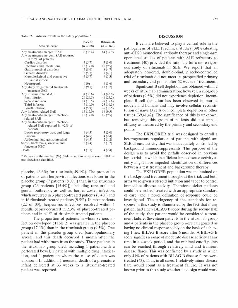
Table 2. Adverse events in the safety population*
Adverse eventPlacebo(n � 88)
Rituximab(n � 169)
Any treatment-emergent SAE 32 (36.4) 64 (37.9)Any treatment-emergent SAE reported
in �5% of patientsCardiac disorder 5 (5.7) 5 (3.0)Infections and infestations 15 (17.0) 16 (9.5)Gastrointestinal disorders 7 (8.0) 8 (4.7)General disorder 5 (5.7) 7 (4.1)Musculoskeletal and connective
tissue disorders5 (5.7) 9 (5.3)
Neutropenia 0 (0) 6 (3.6)Any study drug–related treatment-
emergent SAE8 (9.1) 13 (7.7)
Any infusion-related AE 34 (38.6) 74 (43.8)First infusion 26 (29.5) 46 (27.2)Second infusion 14 (16.5) 29 (17.6)Third infusion 7 (10.0) 23 (16.3)Fourth infusion 4 (5.9) 25 (18.5)
Any infusion-related SAE 15 (17.0) 16 (9.5)Any treatment-emergent infection-
related SAE15 (17.0) 16 (9.5)
Any treatment-emergent infection-related SAE reported in �2% ofpatients
Lower respiratory tract and lungs 4 (4.5) 5 (3.0)Bacterial 4 (4.5) 4 (2.4)Abdominal and gastrointestinal 4 (4.5) 2 (1.2)
Sepsis, bacteremia, viremia, andfungemia NEC
3 (3.4) 2 (1.2)
Death 1 (1.1) 4 (2.4)
* Values are the number (%). SAE � serious adverse event; NEC �not elsewhere classified.
EFFICACY AND SAFETY OF RITUXIMAB IN THE EXPLORER TRIAL 229

for or against the differentiation of treatment effects inSLE. Because negative results occurred, there are nodata to support or refute the robustness of this studydesign, although some hypotheses can be consideredbased on exploratory outcome data.
This study accomplished the important task ofenrolling demonstrably ill patients. Eighty-one percentof patients entered with �1 BILAG A score or �3BILAG B scores, and only 19.0% entered with theminimum of 2 BILAG B scores. Despite treatment withprednisone and immunosuppressants, most patients con-tinued to experience some clinical activity: only 27.3% ofplacebo-treated patients and 24.9% of rituximab-treatedpatients achieved a BILAG C score or less activity in allorgans at week 24, and the majority of withdrawals dueto clinical disease activity occurred prior to this timepoint. Furthermore, the primary end point definition ofresponse at week 52 was achieved in �30% of patients ineither group.
Despite some continued disease activity in bothgroups, the results suggested that placebo-treated andrituximab-treated patients improved during the firstmonth, when the largest steroid doses were given (Fig-ures 2C and 3A). An ad hoc ACR committee haspreviously defined a 7-point decrease in the BILAGscore as a clinically significant response (33). Accordingto this definition, both the placebo and rituximab groupsachieved a clinically significant response by day 28. Themean score did not subsequently increase again duringthe study in either group. This suggests that the benefitof initial steroid therapy was maintained by continuedbackground immunosuppression in the placebo group,preventing an increase in disease activity (back to base-line levels) after tapering of steroids, and that rituximabdid not add any incremental efficacy benefit comparedwith placebo.
One alternative interpretation of this observationis that both the placebo and rituximab groups receivedadequate treatment and achieved all that could beexpected using current measures. In this case, althoughthe efficacy of rituximab cannot be inferred, the possi-bility remains that potential benefits could have becomeapparent if patients in the placebo group had notmaintained their initial responses during the course ofthe trial. This is supported by the suggested efficacy ofrituximab observed in the African American/Hispanicsubgroup, although this finding may be spurious in thesetting of a trial with negative results and multipleanalyses. The decision of how aggressive backgroundtreatment should be in a population of sick patients is astudy design problem previously observed in trials of
SLE (44). Because African American and Hispanicpatients with SLE may have disease that is more refrac-tory to treatment (45), another hypothesis is that aunique biology underlies these different results. It ispossible that disease in these patients is more B celldriven, or that B cell depletion is less likely to stimulateother mechanisms that could confound the effects ofrituximab. In either case, biologic differences before andafter treatment could be tested in a future trial of B celldepletion therapy. A shared biology might be identifiedas more common in one ethnic group but is also likely tocross over ethnic differences and could be used to morerationally identify patients appropriate to treat.
At the end of the study, a difference in BILAGscores was observed between the placebo-treated andrituximab-treated patients whose background treatmentwas MTX. Caution should be used in interpreting thispost hoc analysis, given that rituximab did not showbenefit in this subgroup at the predefined primary andsecondary analyses. In addition, the sample size wassmall at the week 52 visit, when the withdrawal rate inthe 2 treatment groups was imbalanced (6 of 24 patientsin the placebo group [25.0%] and 19 of 46 patients in therituximab group [41.3%]). However, this does raise thepossibility that those who remained in the MTX-treatedplacebo subgroup were beginning to experience a dis-ease flare at the end of the study as compared withpatients in the placebo group who were receiving back-ground AZA or MMF. A separate ad hoc analysis of thepopulation of MTX-treated patients also revealed aseparation favoring rituximab when evaluating the phy-sician’s global score and the patient’s global score over52 weeks (data not shown).
Decreases in the level of anti-dsDNA autoanti-bodies and increases in complement C3 and C4 levelswere also greater in the rituximab group than in theplacebo group, confirming that rituximab had a biologiceffect that was previously observed in preliminary stud-ies. No significant change in the levels of anti–extractable nuclear antigen autoantibodies was ob-served, in contrast to what was observed in someprevious studies (40,46).
The proportion of HACA-positive rituximab-treated patients (26.0%) was fairly high for this popula-tion of patients who were being treated with intravenousand oral steroids while receiving a background immuno-suppressive drug. The incidences of AEs and SAEs werewell-balanced between groups, although the frequencyof treatment-emergent infectious SAEs was higher inthe placebo group than in the group of patients receivingrituximab. Immunosuppressive drugs and prednisone
230 MERRILL ET AL

could contribute to the AEs, but the balance of thesebackground treatments between the groups would makeit difficult to evaluate such a contribution. The incidenceof infusion-related events, including hypotension and/orfever, was similar during the first treatment course, andthe majority of events were mild-to-moderate in sever-ity. During the second course of treatment, the numberof infusion reactions was higher in the rituximab group.The incidence of neutropenia was also higher in therituximab group, and the occurrences were distributedacross all 3 background immunosuppressant therapies.Outside of the oncology setting, neutropenia has notbeen widely observed in patients receiving rituximab incombination with chemotherapeutic agents (47); how-ever, a case of neutropenia in a patient with severe RAwas recently reported (48). One case of neutropenia wasassociated with an infectious SAE. No cases of serumsickness were reported in the placebo group, although 1SAE and 3 AEs of serum sickness were reported in therituximab group. Three of the 4 patients who had serumsickness were HACA positive.
In summary, the EXPLORER trial demonstratedno difference in primary or secondary end points be-tween the placebo group and the rituximab group over52 weeks of treatment, in patients with moderate-to-severe SLE.
AUTHOR CONTRIBUTIONS
All authors were involved in drafting the article or revising itcritically for important intellectual content, and all authors approvedthe final version to be published. Dr. Merrill had full access to all of thedata in the study and takes responsibility for the integrity of the dataand the accuracy of the data analysis.Study conception and design. Merrill, Neuwelt, Wallace, Gordon,Isenberg, Hsieh, Zhang, Brunetta.Acquisition of data. Merrill, Neuwelt, Shanahan, Oates, Utset.Analysis and interpretation of data. Merrill, Neuwelt, Wallace, Lati-nis, Utset, Gordon, Isenberg, Hsieh, Zhang, Brunetta.
REFERENCES
1. Gill JM, Quisel AM, Rocca PV, Walters DT. Diagnosis of systemiclupus erythematosus. Am Fam Physician 2003;68:2179–86.
2. Rahman A, Isenberg DA. Systemic lupus erythematosus. N EnglJ Med 2008;358:929–39.
3. Stoll T, Seifert B, Isenberg DA. SLICC/ACR damage index isvalid, and renal and pulmonary organ scores are predictors ofsevere outcome in patients with systemic lupus erythematosus.Br J Rheumatol 1996;35:248–54.
4. Dooley MA, Hogan S, Jennette C, Falk R, for the GlomerularDisease Collaborative Network. Cyclophosphamide therapy forlupus nephritis: poor renal survival in black Americans. Kidney Int1997;51:1188–95.
5. Alarcon GS, Roseman JM, McGwin G Jr, Uribe A, Bastian HM,Fessler BJ, et al, for the LUMINA study group. Systemic lupus
erythematosus in three ethnic groups. XX. Damage as a predictorof further damage. Rheumatology (Oxford) 2004;43:202–5.
6. Lam GK, Petri M. Assessment of systemic lupus erythematosus.Clin Exp Rheumatol 2005;23 Suppl 39:S120–32.
7. Gladman DD, Urowitz MB, Rahman P, Ibanez D, Tam LS.Accrual of organ damage over time in patients with systemic lupuserythematosus. J Rheumatol 2003;30:1955–9.
8. Borchers AT, Keen CL, Shoenfeld Y, Gershwin ME. Surviving thebutterfly and the wolf: mortality trends in systemic lupus erythem-atosus. Autoimmun Rev 2004;3:423–53.
9. Lipsky PE. Systemic lupus erythematosus: an autoimmune diseaseof B cell hyperactivity. Nature Immunol 2001;2:764–6.
10. Mok CC, Lau CS. Pathogenesis of systemic lupus erythematosus.J Clin Pathol 2003;56:481–90.
11. Renaudineau Y, Pers JO, Bendaoud B, Jamin C, Youinou P.Dysfunctional B cells in systemic lupus erythematosus. Autoim-mun Rev 2004;3:516–23.
12. Swaak T, Smeenk R. Clinical significance of antibodies to doublestranded DNA (dsDNA) for systemic lupus erythematosus (SLE).Clin Rheumatol 1987;6:56–73.
13. Arbuckle MR, McClain MT, Rubertone MV, Scofield RH, DennisGJ, James JA, et al. Development of autoantibodies before theclinical onset of systemic lupus erythematosus. N Engl J Med2003;349:1526–33.
14. Anderson KC, Bates MP, Slaughenhoupt BL, Pinkus GS, Schloss-man SF, Nadler LM. Expression of human B cell-associatedantigens on leukemias and lymphomas: a model of human B celldifferentiation. Blood 1984;63:1424–33.
15. Maloney DG, Liles TM, Czerwinski DK, Waldichuk C, RosenbergJ, Grillo-Lopez A, et al. Phase I clinical trial using escalatingsingle-dose infusion of chimeric anti-CD20 monoclonal antibody(IDEC-C2B8) in patients with recurrent B-cell lymphoma. Blood1994;84:2457–66.
16. Reff ME, Carner K, Chambers KS, Chinn PC, Leonard JE, RaabR, et al. Depletion of B cells in vivo by a chimeric mouse humanmonoclonal antibody to CD20. Blood 1994;83:435–45.
17. Edwards JC, Szczepanski L, Szechinski J, Filipowicz-Sosnowska A,Emery P, Close DR, et al. Efficacy of B-cell-targeted therapy withrituximab in patients with rheumatoid arthritis. N Engl J Med2004;350:2572–81.
18. Cohen SB, Emery P, Greenwald MW, Dougados M, Furie RA,Genovese MC, et al. Rituximab for rheumatoid arthritis refractoryto anti–tumor necrosis factor therapy: results of a multicenter,randomized, double-blind, placebo-controlled, phase III trial eval-uating primary efficacy and safety at twenty-four weeks. ArthritisRheum 2006;54:2793–806.
19. Emery P, Fleischmann R, Filipowicz-Sosnowska A, Schechtman J,Szczepanski L, Kavanaugh A, et al. The efficacy and safety ofrituximab in patients with active rheumatoid arthritis despitemethotrexate treatment: results of a phase IIb randomized, dou-ble-blind, placebo-controlled, dose-ranging trial. Arthritis Rheum2006;54:1390–400.
20. Albert D, Khan S, Stansberry J, Kolasinski S, Tsai D, Kamoun M,et al. A phase I trial of rituximab (anti-CD20) for treatment ofsystemic lupus erythematosus [abstract]. Arthritis Rheum 2004;50Suppl:S446–7.
21. Ryan JP, Singer NG, Scalzi LV. Treatment of resistant SLE withrituximab administered without cyclophosphamide [abstract]. Ar-thritis Rheum 2004;50 Suppl:S413–4.
22. Leandro MJ, Edwards JC, Cambridge G, Ehrenstein MR, Isen-berg DA. An open study of B lymphocyte depletion in systemiclupus erythematosus. Arthritis Rheum 2002;46:2673–7.
23. Leandro MJ, Cambridge G, Edwards JC, Ehrenstein MR, Isen-berg DA. B-cell depletion in the treatment of patients withsystemic lupus erythematosus: a longitudinal analysis of 24 pa-tients. Rheumatology 2005;44:1542–5.
24. Van Vollenhoven RF, Gunnarsson I, Welin-Henriksson E, Sun-
EFFICACY AND SAFETY OF RITUXIMAB IN THE EXPLORER TRIAL 231

delin B, Osterborg A, Jacobson SH, et al. Biopsy-verified responseof severe lupus nephritis to treatment with rituximab (anti-CD20monoclonal antibody) plus cyclophosphamide after biopsy-docu-mented failure to respond to cyclophosphamide alone. ScandJ Rheumatol 2004;33:423–7.
25. Van Vollenhoven RF, Gunnarsson I, Welin-Henriksson E,Jonsdottir T, Sundelin B, Jacobsson S, et al. Rituximab pluscyclophosphamide in severe SLE: results in 15 patients who failedconventional immunosuppressive therapy [abstract]. ArthritisRheum 2005;52 Suppl:S741.
26. Cambridge G, Leandro MJ, Teodorescu M, Manson J, Rahman A,Isenberg DA, et al. B cell depletion therapy in systemic lupuserythematosus: effect on autoantibody and antimicrobial antibodyprofiles. Arthritis Rheum 2006;54:3612–22.
27. Smith KG, Jones RB, Burns SM, Jayne DR. Long-term compari-son of rituximab treatment for refractory systemic lupus erythem-atosus and vasculitis: remission, relapse, and re-treatment. Arthri-tis Rheum 2006;54:2970–82.
28. Tokunaga M, Saito K, Kawabata D, Imura Y, Fujii T, Nakay-amada S, et al. Efficacy of rituximab (anti-CD20) for refractorysystemic lupus erythematosus involving the central nervous system.Ann Rheum Dis 2007;66:470–5.
29. Sutter JA, Kwan-Morley J, Dunham J, Du YZ, Kamoun M, AlbertD, et al. A longitudinal analysis of SLE patients treated withrituximab (anti-CD20): factors associated with B lymphocyte re-covery. Clin Immunol 2008;126:282–90.
30. Lu TY, Jonsdottir T, van Vollenhoven RF, Isenberg DA. Pro-longed B-cell depletion following rituximab therapy in systemiclupus erythematosus: a report of two cases. Ann Rheum Dis2008;67:1493–4.
31. Kumar S, Benseler SM, Kirby-Allen M, Silverman ED. B-celldepletion for autoimmune thrombocytopenia and autoimmunehemolytic anemia in pediatric systemic lupus erythematosus. Pe-diatrics 2009;123:159–63.
32. Reynolds J, Toescu V, Yee C, Prabu A, Situnayake D, Gordon C.Effects of rituximab on resistant SLE disease including lunginvolvement. Lupus 2009;18:67–73.
33. American College of Rheumatology Ad Hoc Committee onSystemic Lupus Erythematosus Response Criteria. The AmericanCollege of Rheumatology response criteria for systemic lupuserythematosus clinical trials: measures of overall disease activity.Arthritis Rheum 2004;50:3418–26.
34. Hay EM, Bacon PA, Gordon C, Isenberg DA, Maddison P, SnaithML, et al. The BILAG index: a reliable and valid instrument formeasuring clinical disease activity in systemic lupus erythemato-sus. Q J Med 1993;86:447–58.
35. Isenberg DA, Gordon C, and the British Isles Lupus AssessmentGroup. From BILAG to BLIPS: disease activity assessment inlupus past, present and future. Lupus 2000;9:651–4.
36. Yu EB, Shikiar R, Howard K, Kalunian KC, Petrillo J, ThompsonC, et al. Validation of LUP-QOL: a lupus-specific measure ofhealth-related quality of life (HRQL) [abstract]. Ann Rheum Dis2006;65 Suppl II:601.
37. Ware JE Jr, Sherbourne CD. The MOS 36-item Short-Formhealth survey (SF-36). I. Conceptual framework and item selec-tion. Med Care 1992;30:473–83.
38. Ehrenstein MR, Conroy SE, Heath J, Latchman DS, Isenberg DA.The occurrence, nature and distribution of flares in a cohort ofpatients with systemic lupus erythematosus: a rheumatologicalview. Br J Rheumatol 1995;34:257–60.
39. Ahuja A, Shupe J, Dunn R, Kashgarian M, Kehry MR, ShlomchikMJ. Depletion of B cells in murine lupus: efficacy and resistance.J Immunol 2007;179:3351–61.
40. Tanaka Y, Yamamoto K, Takeuchi T, Nishimoto N, Miyasaka N,Sumida T, et al. A multicenter phase I/II trial of rituximab forrefractory systemic lupus erythematosus. Mod Rheumatol 2007;17:191–7.
41. Shlomchik MJ, Madaio MP, Ni D, Trounstein M, Huszar D. Therole of B cells in lpr/lpr-induced autoimmunity. J Exp Med1994;180:1295–306.
42. Gong Q, Ou Q, Ye S, Lee WP, Cornelius J, Diehl L, et al.Importance of cellular microenvironment and circulatory dynam-ics in B cell immunotherapy. J Immunol 2005;174:817–26.
43. Gordon C, Sutcliffe N, Skan J, Stoll T, Isenberg DA. Definitionand treatment of lupus flares measured by the BILAG index.Rheumatology 2003;42:1372–9.
44. Lewis EJ, Hunsicker LG, Lan SP, Rohde RD, Lachin JM, for theLupus Nephritis Collaborative Study Group. A controlled trial ofplasmapheresis therapy in severe lupus nephritis. N Engl J Med1992;326:1373–9.
45. Uribe AG, McGwin G Jr, Reveille JD, Alarcon GS. What have welearned from a 10-year experience with the LUMINA (Lupus inMinorities; Nature vs. nurture) cohort? Where are we heading?Autoimmun Rev 2004;3:321–9.
46. Anolik JH, Barnard J, Cappione A, Pugh-Bernard AE, Felgar RE,Looney RJ, et al. Rituximab improves peripheral B cell abnormal-ities in human systemic lupus erythematosus. Arthritis Rheum2004;11:3580–90.
47. Voog E, Morschhauser F, Solal-Celigny P. Neutropenia in patientstreated with rituximab. N Engl J Med 2003;348:2691–4.
48. Marotte H, Paintaud G, Watier H, Miossec P. Rituximab-relatedlate-onset neutropenia in a patient with severe rheumatoid arthri-tis. Ann Rheum Dis 2008;67:893–4.
APPENDIX A: EXPLORER TRIAL CLINICAL CENTERSAND INVESTIGATORS
Investigators for the EXPLORER trial, including the authors,are as follows: for the University of Alabama at Birmingham, GracielaS. Alarcon; for Arizona Arthritis and Rheumatology Associates,Paradise Valley, Arizona, Ralph Bennett; for the East Bay Rheuma-tology Research Institute, San Leandro, California, C. Michael Neu-welt; for Cedars-Sinai Medical Center, Los Angeles, California, DanielWallace; for Stanford Health Services/Blake Wilbur Clinic, Palo Alto,California, Eliza F. Chakravarty, Mark Genovese; for the University ofCalifornia, Los Angeles, Jennifer Grossman; for the University ofCalifornia, San Francisco, John Davis, Maria Dall’Era; for the Uni-versity of California, San Diego Thornton Hospital, La Jolla, KennethKalunian; for the Family Arthritis Center, Jupiter, Florida, HowardBusch; for Arthritis and Rheumatic Disease Specialties, Aventura,Florida, Norman Gaylis; for Emory University School of Medicine,Atlanta, Georgia, S. Sam Lim; for the Coeur d’Alene Arthritis Clinic,Coeur d’Alene, Idaho, Craig Wiesenhutter; for the IntermountainResearch Center, Boise, Idaho, James Loveless; for NorthwesternUniversity/Feinberg School of Medicine, Chicago, Illinois, RosalindRamsey-Goldman; for the University of Chicago, Chicago, Illinois,Tammy O. Utset; for the Tri-State Arthritis and RheumatologyCenter, Evansville, Indiana, Moges Sisay; for the University of KansasMedical Center, Kansas City, Kevin M. Latinis; for the Louisiana StateUniversity Health Science Center, Shreveport, Seth M. Berney; forJohns Hopkins University, Baltimore, Maryland, Michelle Petri; forThe Center for Rheumatology and Bone Research, Wheaton, Mary-land, Evan L. Siegel; for Tufts–New England Medical Center, Boston,Massachusetts, Elena Massarotti, Timothy E. McAlindon; for BethIsrael Deaconess Medical Center, Boston, Massachusetts, John Dono-hue, Lisa Fitzgerald; for the Michigan Arthritis Research Center,Brighton, James E. Dowd; for the University of Michigan HealthSystem, Ann Arbor, Joseph McCune; for Buffalo Rheumatology,Orchard Park, New York, Joseph Grisanti; for the Feinstein Institutefor Medical Research, Manhasset, New York, Meggan Mackay; for theHospital for Special Surgery, New York, New York, Michael Lockshin;for the Seligman Center for Advanced Therapeutics, New York, NewYork, Jill Buyon; for the North Shore Long Island Jewish Health
232 MERRILL ET AL

System, Lake Success, New York, Richard Furie; for Physician’s East,Greenville, North Carolina, Jeff Alloway; for Duke University MedicalCenter, Durham, North Carolina, Stacy Ardoin, Meggan Clowse,Joseph C. Shanahan; for the University of North Carolina Hospital,Chapel Hill, Mary Anne Dooley; for the Ohio State UniversityMedical Center, Columbus, Brad H. Rovin; for the Oklahoma MedicalResearch Foundation, Oklahoma City, Joan T. Merrill; for the Boneand Joint Hospital Research Department, Oklahoma City, Oklahoma,Larry Willis; for the Oklahoma Center for Arthritis Therapy andResearch, Tulsa, Ellen Zanetakis; for East Penn Rheumatology Asso-ciates, Bethlehem, Pennsylvania, Allen Samuels; for the Albert Ein-stein Medical Center, Philadelphia, Pennsylvania, Lawrence Brent,Ramesh Pappu; for the University of Pennsylvania Medical Center,Philadelphia, Robert A. Eisenberg; for the University of Pittsburgh
School of Medicine, Pittsburgh, Pennsylvania, Susan Manzi; for theMedical University of South Carolina, Charleston, Gary Gilkeson,James C. Oates; for Arthritis Associates, Hixson, Tennessee, JosephHuffstutter; for the Arthritis Centers of Texas, Dallas, Andrew Chu-bick, Linda Teague; for the Metroplex Clinical Research Center,Dallas, Texas, Stanley Cohen, Thomas D. Geppert; for the TexasResearch Center, Sugar Land, Texas, Angela McCain; for the HoustonInstitute for Clinical Research, Houston, Texas, Dale Halter; for theVirginia Commonwealth University Clinical Research Center, Rich-mond, W. Neal Roberts; for the University of Washington, Seattle,Carin Dugowson; for Seattle Rheumatology Associates, Seattle, Wash-ington, Philip Mease; for St. Joseph’s Health Care, London, Ontario,Canada, Janet E. Pope; for the University of Manitoba, Manitoba,Canada, Christine Peschken.
EFFICACY AND SAFETY OF RITUXIMAB IN THE EXPLORER TRIAL 233