Identification of miRNAs as potential modulators of tissue factor expression in patients with...
8
ORIGINAL ARTICLE Identification of miRNAs as potential modulators of tissue factor expression in patients with systemic lupus erythematosus and antiphospholipid syndrome R. TERUEL,* C. PE ´ REZ-SA ´ NCHEZ, J. CORRAL,* M. T. HERRANZ, à V. PE ´ REZ-ANDREU,* E. SAIZ,§ N. GARCI ´ A-BARBERA ´ ,* I. MARTI ´ NEZ-MARTI ´ NEZ,* V. ROLDA ´ N,* V. VICENTE,* C. LO ´ PEZ-PEDRERA and C. MARTI ´ NEZ* *Centro Regional de Hemodonacio ´ n, University of Murcia, Murcia; Unidad de Investigacio ´ n/Instituto de Investigacio ´ n Biome ´dica de Co ´ rdoba (IMIBIC), Hospital Universitario Reina Sofı ´a, Co ´ rdoba; àInternal Medicine Unit; and §Section of Rheumatology, Hospital Universitario Morales Meseguer, Murcia, Spain To cite this article: Teruel R, Pe ´ rez-Sa ´ nchez C, Corral J, Herranz MT, Pe ´rez-Andreu V, Saiz E, Garcı´a-Barbera ´ N, Martı ´nez-Martı ´nez I, Rolda ´ n V, Vicente V, Lo ´ pez-Pedrera C, Martı ´nez C. Identification of miRNAs as potential modulators of tissue factor expression in patients with systemic lupus erythematosus and antiphospholipid syndrome. J Thromb Haemost 2011; 9: 1985–92. Summary. Background: Tissue factor (TF) is the main initiator of the coagulation cascade and elements that may upregulate its expression might provoke thrombotic events. Systemic lupus erythematosus (SLE) and antiphospholipid syndrome (APS) are autoimmune diseases characterized by a high TF expression in monocytes. Objectives: To examine the role of microRNAs (miRNAs) in TF expression and to evaluate their levels in SLE and APS patients. Methods: An in silico search was performed to find potential putative binding sites of miRNAs in TF mRNA. In vitro validation was performed transfecting cells expressing TF (THP-1 and MDA-MB-231) with oligonucleo- tide miRNA precursors and inhibitors. Additionally, reporter assays were performed to test for the binding of miR-20a to TF mRNA. Levels of miRNAs and TF were measured by quantitative (qRT-PCR) in patients with APS and SLE. Results: Overexpression of miRNA precursors, but not inhib- itors, of two of the members of cluster miR-1792, for example miR-19b and miR-20a, in cells expressing TF decreased TF mRNA, protein levels, and procoagulant activity between 30% and 60%. Reporter assays showed that miR-20a binds to TF mRNA. Finally, we measured levels of miR-19b and miR-20a in monocytes from patients with APS and SLE and observed significantly lower miRNAs levels in comparison with healthy subjects inversely correlated with the levels of TF. Conclusions: Down-regulation of miR-19b and miR-20a observed in patients with SLE and APS could contribute to increased TF expression and thus provoke the hypercoagulable state characteristic of these patients. Keywords: antiphospholipid syndrome, miRNAs, systemic lu- pus erythematosus, thrombosis, tissue factor. Introduction Recently, a myriad of papers described a family of non-coding RNA as extremely relevant in many biological functions [1,2]. MicroRNAs (miRNAs) are approximately 21 to 24 nucleo- tide-long RNA derived from hairpin transcripts that have become a major focus of research in molecular biology where at least a thousand are predicted to operate in humans. Functional studies indicate that miRNAs participate in the regulation of almost every cellular process investigated and that changes in their expression are observed in human pathologies, including cancer and cardiovascular diseases [3– 6]. It is accepted that miRNAs post-transcriptionally control gene expression by regulating mRNA translation or stability in the cytoplasm by forming hetero-duplexes. What is certain is that the discovery of miRNAs has unraveled a new step towards decrypting the complexity of post-transcriptional regulation of a large number of eukaryotic genes. Interest- ingly, very recently a growing body of evidence shows that miRNAs may play a role in hemostasis [7–9]. The hemostatic system is a highly efficient and rapid system to avoid hemorrhage and thus to overcome the effects that are produced during vascular damage [10,11]. The hemostatic system responds through a series of proteolytic processes with a high efficacy. However, this system must be finely tuned and be spatially and temporally restricted to the site of the vessel injury. Thus, the hemostatic system is an equilibrium perfectly regulated between procoagulant and anticoagulant elements that regulate blood flowing under normal conditions but respond quickly and strongly to form the hemostatic plug to the vascular damage. Tissue factor (TF) is the primary physiological initiator of blood coagulation. TF is constitu- tively expressed in different cell types such as smooth muscle Correspondence: Constantino Martı´nez, Centro Regional de Hemodonacio´n, C/ Ronda de Garay S/N, 30003 Murcia, Spain. Tel: +34 968341990; fax: +34 968261914. E-mail: [email protected] Received 13 January 2011, accepted 21 June 2011 Journal of Thrombosis and Haemostasis, 9: 1985–1992 DOI: 10.1111/j.1538-7836.2011.04451.x ȑ 2011 International Society on Thrombosis and Haemostasis
Transcript of Identification of miRNAs as potential modulators of tissue factor expression in patients with...
ORIGINAL ARTICLE
Identification of miRNAs as potential modulators of tissuefactor expression in patients with systemic lupus erythematosusand antiphospholipid syndrome
R . T ERUEL ,* C . P EREZ -S ANCHEZ ,� J . C OR R A L , * M . T . H E R R A N Z ,� V . P EREZ -ANDREU ,* E . SA IZ ,§
N . GA RC I A -BA RBER A ,* I . MA RT I NEZ -MA RT I NEZ , * V . R OLD AN ,* V . V ICENTE ,* C . L O PEZ -PEDRERA�and C . MART I NEZ**Centro Regional de Hemodonacion, University of Murcia, Murcia; �Unidad de Investigacion/Instituto de Investigacion Biomedica de Cordoba
(IMIBIC), Hospital Universitario Reina Sofıa, Cordoba; �Internal Medicine Unit; and §Section of Rheumatology, Hospital Universitario Morales
Meseguer, Murcia, Spain
To cite this article: Teruel R, Perez-Sanchez C, Corral J, Herranz MT, Perez-Andreu V, Saiz E, Garcıa-Barbera N, Martınez-Martınez I, Roldan V,
Vicente V, Lopez-Pedrera C, Martınez C. Identification of miRNAs as potential modulators of tissue factor expression in patients with systemic lupus
erythematosus and antiphospholipid syndrome. J Thromb Haemost 2011; 9: 1985–92.
Summary. Background:Tissue factor (TF) is themain initiator
of the coagulation cascade and elements thatmay upregulate its
expression might provoke thrombotic events. Systemic lupus
erythematosus (SLE) and antiphospholipid syndrome (APS)
are autoimmune diseases characterized by a highTF expression
in monocytes. Objectives: To examine the role of microRNAs
(miRNAs) in TF expression and to evaluate their levels in SLE
and APS patients.Methods: An in silico search was performed
to find potential putative binding sites of miRNAs in TF
mRNA. In vitro validation was performed transfecting cells
expressing TF (THP-1 and MDA-MB-231) with oligonucleo-
tide miRNA precursors and inhibitors. Additionally, reporter
assays were performed to test for the binding of miR-20a to TF
mRNA. Levels of miRNAs and TF were measured by
quantitative (qRT-PCR) in patients with APS and SLE.
Results: Overexpression of miRNA precursors, but not inhib-
itors, of two of themembers of cluster miR-17�92, for example
miR-19b and miR-20a, in cells expressing TF decreased TF
mRNA, protein levels, and procoagulant activity between 30%
and 60%. Reporter assays showed that miR-20a binds to TF
mRNA. Finally, we measured levels of miR-19b and miR-20a
in monocytes from patients with APS and SLE and observed
significantly lower miRNAs levels in comparison with healthy
subjects inversely correlated with the levels of TF. Conclusions:
Down-regulation ofmiR-19b andmiR-20a observed in patients
with SLEandAPS could contribute to increasedTFexpression
and thus provoke the hypercoagulable state characteristic of
these patients.
Keywords: antiphospholipid syndrome, miRNAs, systemic lu-
pus erythematosus, thrombosis, tissue factor.
Introduction
Recently, a myriad of papers described a family of non-coding
RNA as extremely relevant in many biological functions [1,2].
MicroRNAs (miRNAs) are approximately 21 to 24 nucleo-
tide-long RNA derived from hairpin transcripts that have
become a major focus of research in molecular biology where
at least a thousand are predicted to operate in humans.
Functional studies indicate that miRNAs participate in the
regulation of almost every cellular process investigated and
that changes in their expression are observed in human
pathologies, including cancer and cardiovascular diseases [3–
6]. It is accepted that miRNAs post-transcriptionally control
gene expression by regulating mRNA translation or stability
in the cytoplasm by forming hetero-duplexes. What is certain
is that the discovery of miRNAs has unraveled a new step
towards decrypting the complexity of post-transcriptional
regulation of a large number of eukaryotic genes. Interest-
ingly, very recently a growing body of evidence shows that
miRNAs may play a role in hemostasis [7–9]. The hemostatic
system is a highly efficient and rapid system to avoid
hemorrhage and thus to overcome the effects that are
produced during vascular damage [10,11]. The hemostatic
system responds through a series of proteolytic processes with
a high efficacy. However, this system must be finely tuned and
be spatially and temporally restricted to the site of the vessel
injury. Thus, the hemostatic system is an equilibrium perfectly
regulated between procoagulant and anticoagulant elements
that regulate blood flowing under normal conditions but
respond quickly and strongly to form the hemostatic plug to
the vascular damage. Tissue factor (TF) is the primary
physiological initiator of blood coagulation. TF is constitu-
tively expressed in different cell types such as smooth muscle
Correspondence: Constantino Martınez, Centro Regional de
Hemodonacion, C/ Ronda de Garay S/N, 30003 Murcia, Spain.
Tel: +34 968341990; fax: +34 968261914.
E-mail: [email protected]
Received 13 January 2011, accepted 21 June 2011
Journal of Thrombosis and Haemostasis, 9: 1985–1992 DOI: 10.1111/j.1538-7836.2011.04451.x
� 2011 International Society on Thrombosis and Haemostasis
cells or fibroblasts, cells that come in contact with blood only
during vascular damage. In contrast, TF expression in blood
cells and endothelium is regulated through a number of
signaling pathways and occurs after cell activation with a
variety of agonists [12]. Indeed, a number of pathological
conditions show a dysregulation of the basal hemostatic
balance towards a hypercoagulable status related in certain
cases to a higher expression of TF and a higher level of
circulating TF-bearing microparticles [12,13]. Among these
pathologies, autoimmune diseases such as antiphospholipid
syndrome (APS) and systemic lupus erythematosus (SLE)
display increased TF expression in monocytes [14–16]. How-
ever, the exact pathogenic mechanisms responsible for the
increased thrombosis risk in patients with APS and SLE are
still unclear. Indeed, TF expression in endothelial cells and
monocytes, the main producers of TF-bearing microparticles,
is certainly fine-tuned and alterations in its modulation may
play a transcendental role in the development of thrombotic
events in APS and SLE patients. Very recently, miR-19a has
been shown to inhibit TF expression in cancer cell lines [17].
In the present study, we additionally showed that TF
expression may be regulated by miR-19b and miR-20a in
THP-1 cells and that down-regulation of these miRNAs in
monocytes from patients with APS and SLE may be a
relevant event in the observed hypercoagulable state of these
patients.
Methods
In silico studies
Several web databases and algorithms of miRNA target
prediction were used for the search of miRNA targeting TF.
We essentially used TargetScan [18] (release 5.1: http://
www.targetscan.org), which provides the prediction results
computed by the TargetScanS algorithm, PicTar (http://
pictar.mdc-berlin.de) [19] and miRanda (http://www.micror-
na.org/microrna/home.do) [20].
Cell culture and transfection
The following cell lines were used: MDA-MB-231 (breast
cancer) and THP-1 (monocytic leukemia). Cell lines were
obtained from the American Type Culture Collection (ATCC)
and cultured according to the ATCC protocols. Cell transfec-
tion was performed by standard procedures described in detail
in Data S1.
RNA isolation, qRT-PCR and Western blotting
Total RNA was isolated from cells using Trizol� Reagent
(Invitrogen, Madrid, Spain) and a reverse transcription (RT;
SuperScript First Strand, Invitrogen) reaction was per-
formed using 200 ng of total RNA from each sample
according to the manufacturer�s instructions. MiRNAs and
gene expression was quantified using Taqman� probes
purchased from Applied Biosystems (Madrid, Spain) See
Data S1 for qRT-PCR and Western blotting assays.
TF expression
Analysis of TF procoagulant activity was performed as
previously described with some modifications [21]. Briefly,
THP-1 cells (500 000 cells) were stimulated for 5 h (37 �C,5%CO2) with10 lg mL)1 lipolysaccharide (LPS) (#L-4391;
Sigma-Aldrich) after transfection with pre-miR or antagomiR.
Suspensions were centrifuged (350 · g, 5 min, 22 �C) and the
pellet was resuspended in 100 lL phosphate buffered saline
(PBS). Lysates were prepared by four times freeze/thawing in
liquid N2 and centrifuged (12 000 · g, 5 min, 22 �C). Super-natant were collected adjusted in protein content using BCA
assay, and incubated with 10 lg mL)1 FX (Stago, Asnieres,
France), 5 U mL)1 rFVIIa (NovoSeven; NovoNordisk SA,
Madrid, Spain) and 5 mmol L)1 CaCl2. FXa generation was
quantified in a Synergy 2 luminometer (Biotek, Bad Friedrich-
shall, Germany) at 405 nm after the addition of FXa substrate
(Instrumentation Laboratory, Milan, Italy).
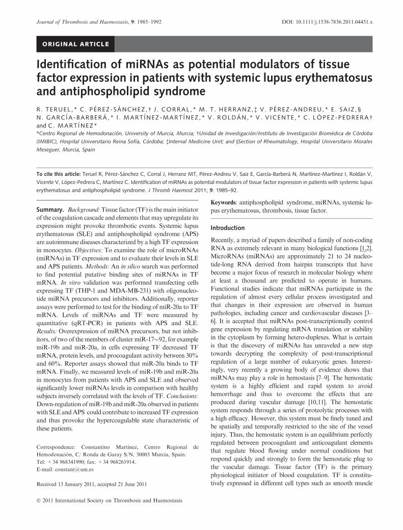
Fig. 1. Schematic representation of the binding sites of selected microRNAs (miRNAs) to tissue factor (TF) mRNA. (A) Putative binding sites localized
in TF mRNA (Ensembl transcript IDENST00000334047). Nucleotide A from ATG is considered as position +1. Mir-19a and mir-19b seeds are
located at +1137; miR-20a and miR-106b are located at position +1786. The ovals mark the position of the seed binding sites.
1986 R. Teruel et al
� 2011 International Society on Thrombosis and Haemostasis
Plasmid/luciferase construct
Constructs for luciferase assays were designed as described in
Data S1.
Luciferase assay
Twenty-four hours before transfection, HEK 293T (embryonic
kidney cells) were plated at 105/well in 24-well plates with
complete Dulbecco�s modified Eagle�s medium supplemented
with 10% fetal calf serum (FCS) without antibiotics. Cells were
transfected with miR-20a precursor or scrambled control
(Applied Biosystems) and 3¢UTR firefly luciferase reporter
plasmid (500 ng/well) along with 50 ng/well of renilla luciferase
control plasmid (pRLTK; Promega,Madison,WI,USA) using
Lipofectamine LTX (Invitrogen) according to the manufac-
turer�s instructions. Luciferase assays were performed as
described in Data S1.
Controls and patients
Ten patients [nine women, one man; 47 ± 13 years (range 35–
71)] fulfiling the Sapporo criteria for APS [22] were recruited.
Six patients suffered from arterial thrombosis (stroke; mean
interval from last thrombotic event: 10.2 ± 3.3 years) and four
patients had recurrent pregnancy loss (mean interval from last
pregnancy loss: 8.5 ± 3.1 years). Eight patients were antibody
positive. Twenty-seven patients [25 women, 2 men;
39 ± 14 years (range 18–68)] with SLE fulfiling the American
College of Rheumatology classification criteria [23] were
enrolled in this study. Three female patients had arterial
thrombosis (two secondary SAF and one patient had factor V
Leiden). Finally, 17 healthy controls were recruited from the
general population [five men, 12 women; 33 ± 10 years (range
19–73)]. None of the controls had a history of autoimmune
disease, bleeding disorders, thrombosis or pregnancy loss. All
subjects gave their informed consent according to the Decla-
ration of Helsinki to enter the study, which has been approved
by the Local Ethics Committee.
Membrane tissue factor expression
Monocytes were purified using standard procedures [15].
Monocyte TF expression was measured by flow cytometry.
See Data S1 for details.
Statistical analysis
Statistical analysis was performed using the Statistical Package
for Social Science (version 15.0; SPSS, Chicago, IL, USA). See
Data S1 for details.
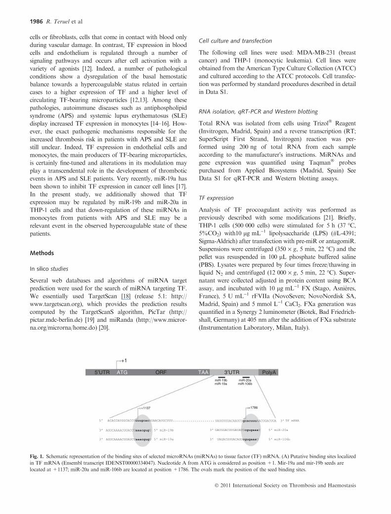
Fig. 2. Inhibition of tissue factor (TF) expression by miR-19b and miR-
20a in MDA-MB-231. MDA-MB-231 were transfected with
100 nmol L)1 precursors of miR-19b and miR-20a or a scrambled pre-
cursor (A) TF expression was measured by Western blotting with b-actinused as a loading control. Densitometry was performed and normalized
with respect to b-actin expression. (B) TF mRNA was measured by
quantitative (qRT-PCR) and normalized with respect to b-actin mRNA.
The normalized data were expressed as changes relative to the data of the
cells transfected with scrambled pre-miR and set as 100%. Differences
were analyzed by means of Student�s t-test. Statistical significance wastaken as P < 0.05. The data shown are mean ± SD representative of
four independent experiments with three replicates each.
Table 1 Binding in silico report
miRNA putative target
site in TargetScan & Pictar
TargetScan PicTar (Krek et al. 2005)
Seed
match
TargetScan
score percentile PCT
PicTar
score
Target site
number
Free energies
(Kcal mol)1)
hsa-miR-20a 8mer 97 0.95 3.20 1 )20.1hsa-miR-106b 8mer 96 0.95 2.99 1 )19.6hsa-miR-19a 8mer 92 0.65 2.84 1 )18.9hsa-miR-19b 8mer 92 0.65 2.84 1 )21.2
Tissue factor regulation by miRNAs 1987
� 2011 International Society on Thrombosis and Haemostasis
Results
In silico study of miRNA–TF mRNA interaction
The seed regions binding of miR-19a/miR-19b and miR-20a/
miR-106b were located at 1137 and 1786 bp from the
beginning of the ATG codon, respectively (Fig. 1). Scores,
free energies and characteristics of the seed region calculated by
Pictar and Targetscan algorithms are specified in Table 1. We
principally focused for the rest of the study on miR-20a.
Additionally, we also performed studies with miR-19b as miR-
19a has been recently shown to interact with TF mRNA [17].
Mir-19b and miR-20a inhibit TF expression
To validate the inhibition of endogenous TF transcripts by
miR-19b and miR-20a, we looked for a model of a tumor cell
line constitutively expressing TF. We chose the breast
carcinoma cell line MDA-MB-231 which has been shown to
express TF at high levels [24]. Overexpression of both
miRNAs by transfecting MDA-MB-231 cells with oligonu-
cleotide precursors significantly reduced the expression of
endogenous TF 48 h after transfection (Fig. 2A). We
observed an almost 60% decrease of TF protein expression
as assayed with densitometry after Western blot. The
inhibition of TF synthesis by miR-19b and miR-20a corre-
lated with a statistically significant reduction of TF mRNA
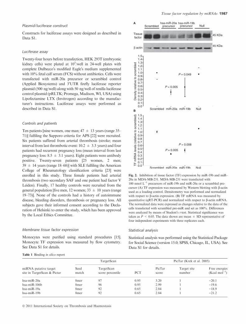
(Fig. 2B). To further validate the effect of miR-20a, we
transfected cells with miR-106b that shares the same binding
site. Time course experiments showed that only 6 h after
transfection, the three miRNAs decreased TF mRNA levels
by 20–60% (Fig. 3B). Concerning protein levels, the inhibi-
tion effect of the three miRNAs only appeared after 24-h
transfection and was maximal at that time (Fig. 3A).
Aiming to verify the effect of thesemiRNAs in a cell type that
inducibly expresses TF, we investigated their potential role in
inhibiting TF mRNA transcripted in THP-1, a human acute
monocytic leukemia cell line. In our experiment, we first
transfected THP-1 with pre-miR precursors and after 24-h cell
settlement, we challenged THP-1 with LPS for 2 h and checked
for the expression of TF mRNA. The present results showed
that overexpression ofmiR-19b andmiR-20a reduced the levels
of TF mRNA by 40–60% in comparison with cells transfected
with the scrambled precursor and later activated with LPS
(Fig. 4A). This reduction inmRNAwas also accompanied by a
statistically significant reduction of TF protein expression as
measuredbyTFprocoagulant activity (Fig. 4B).Concordantly,
the inhibition of miR-19b and miR-20a increased the levels of
TF mRNA by 15% and 12%, respectively (Fig. 4A). On the
other hand, the procoagulant activity in cells transfected with
anti-miR-20a significantly augmented by 50% in comparison
with cells transfected with the scrambled control (Fig. 4B).
Fig. 3. Time course inhibition of tissue factor (TF) expression in MDA-MB-231. MDA-MB-231 were transfected with 100 nmol L)1 precursors of miR-
19b, miR-20a, miR-106b or a scrambled precursor and collected after 6, 24 and 48 h. (A) TF expression was measured by Western blotting with s-actin
used as a loading control. Densitometry was performed and normalized with respect to b-actin expression. (B) TFmRNAwasmeasured by qRT-PCR and
normalized with respect to b-actin mRNA. The normalized data were expressed as changes relative to the data of the cells transfected with scrambled pre-
miR and set as 100%. Differences were analyzed by means of Student�s t-test. Statistical significance was taken as P < 0.05. The data shown are
mean ± SD representative of two independent experiments with duplicates (western blot) and three independent experiments with three replicates each
(qRT-PCR).
1988 R. Teruel et al
� 2011 International Society on Thrombosis and Haemostasis
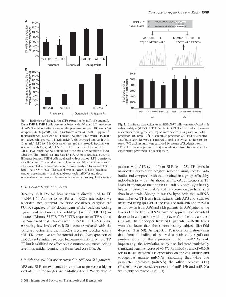
TF is a direct target of miR-20a
Recently, miR-19b has been shown to directly bind to TF
mRNA [17]. Aiming to test for a miR-20a interaction, we
generated two different luciferase constructs carrying the
3¢UTR sequence of TF downstream of the luciferase coding
region, and containing the wild-type (WT 3¢UTR TF) or
mutated (Mutate 3¢UTR TF) 3¢UTR sequence of TF without
the 7-mer seed that interacts with miR-20a. HEK-293T cells,
expressing low levels of miR-20a, were transfected with the
luciferase vectors and the miR-20a precursor together with a
pRL-TK control vector for normalization. Overexpression of
miR-20a substantially reduced luciferase activity inWT 3¢UTR
FT but it exhibited no effect on the mutated construct lacking
seven nucleotides forming the 8-mer seed core (Fig. 5).
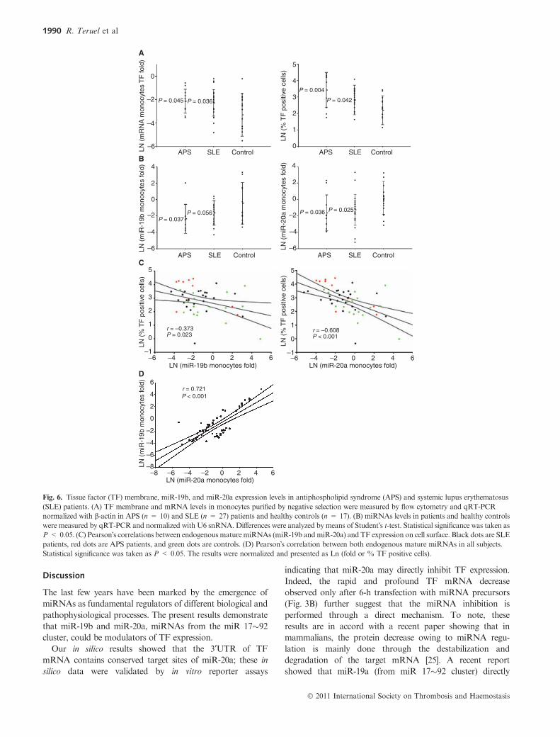
Mir-19b and mir-20a are decreased in APS and SLE patients
APS and SLE are two conditions known to provoke a higher
level of TF in monocytes and endothelial cells. We checked in
patients with APS (n = 10) or SLE (n = 23), TF levels in
monocytes purified by negative selection using specific anti-
bodies and compared with that obtained in a group of healthy
individuals (n = 17). As shown in Fig. 6A, differences in TF
levels in monocyte membrane and mRNA were significantly
higher in patients with APS and in a lesser degree from SLE
than in controls. Aiming to test the hypothesis that miRNA
may influence TF levels from patients with APS and SLE, we
measured using qRT-PCR the levels of miR-19b and mir-20a
inmonocytes fromAPS and SLE patients. In APS patients, the
levels of these two miRNAs have an approximate seven-fold
decrease in comparison with monocytes from healthy controls
(Fig. 6B). In monocytes from SLE patients, miR-20a levels
were also lower than those from healthy subjects (five-fold
decrease) (Fig. 6B). As expected, Pearson�s correlation using
data from all individuals showed a statistically significant
positive score for the expression of both miRNAs and,
importantly, the correlation study also indicated statistically
significant negative scores of)0.373 for miR-19b and of)0.608for miR-20a between TF expression on the cell surface and
endogenous mature miRNAs, indicating that while one
parameter decreases (miRNA) the other increases (TF)
(Fig. 6C). As expected, expression of miR-19b and miR-20a
was highly correlated (Fig. 6D).
Fig. 4. Inhibition of tissue factor (TF) expression by miR-19b and miR-
20a in THP-1. THP-1 cells were transfected with 100 nmol L)1 precursors
ofmiR-19b andmiR-20a or a scrambled precursor andwith 100 nmiRNA
antagonists (antagomiRs) and (A) activated after 24 h with 10 lg mL)1
lipolysaccharide (LPS) for 2 h.TFmRNAwasmeasuredbyqRT-PCRand
normalized with respect to b-actin mRNA, (B) activated after 24 h with
10 lg mL)1 LPS for 5 h. Cells were lysed and the cytosolic fraction was
incubated with 10 lg mL)1 FX, 5 U mL)1 rFVIIa and 5 mmol L)1
CaCl2. FXa generation was quantified at 405 nm after addition of FXa
substrate. The normal response was TF mRNA or procoagulant activity
difference between THP-1 cells incubated with or without LPS, transfected
with 100 nmol L)1 scrambled control and set as 100%. Differences with
cells transfected with scrambled controls were analyzed by means of Stu-
dent�s t-test, *P < 0.05. The data shown are mean ± SD of five inde-
pendent experiments with three replicates each (mRNA) and three
independent experiments with three replicates each (procoagulant activity).
Fig. 5. Luciferase expression assay. HEK293T cells were transfected with
either wild-type (WT) 3¢UTRTF orMutant 3¢UTRTF in which the seven
nucleotides forming the seed region were deleted, along with miR-20a
precursor (100 nmol L)1). A scrambled precursor was used as a control.
Luciferase activities were normalized to renilla activities. Differences be-
tween WT and mutants were analyzed by means of Student�s t-test,*P < 0.01. Results (mean ± SD) were obtained from four independent
experiments performed in quadruplicate.
Tissue factor regulation by miRNAs 1989
� 2011 International Society on Thrombosis and Haemostasis
Discussion
The last few years have been marked by the emergence of
miRNAs as fundamental regulators of different biological and
pathophysiological processes. The present results demonstrate
that miR-19b and miR-20a, miRNAs from the miR 17�92cluster, could be modulators of TF expression.
Our in silico results showed that the 3¢UTR of TF
mRNA contains conserved target sites of miR-20a; these in
silico data were validated by in vitro reporter assays
indicating that miR-20a may directly inhibit TF expression.
Indeed, the rapid and profound TF mRNA decrease
observed only after 6-h transfection with miRNA precursors
(Fig. 3B) further suggest that the miRNA inhibition is
performed through a direct mechanism. To note, these
results are in accord with a recent paper showing that in
mammalians, the protein decrease owing to miRNA regu-
lation is mainly done through the destabilization and
degradation of the target mRNA [25]. A recent report
showed that miR-19a (from miR 17�92 cluster) directly
Fig. 6. Tissue factor (TF) membrane, miR-19b, and miR-20a expression levels in antiphospholipid syndrome (APS) and systemic lupus erythematosus
(SLE) patients. (A) TF membrane and mRNA levels in monocytes purified by negative selection were measured by flow cytometry and qRT-PCR
normalized with b-actin in APS (n = 10) and SLE (n = 27) patients and healthy controls (n = 17). (B) miRNAs levels in patients and healthy controls
were measured by qRT-PCR and normalized with U6 snRNA. Differences were analyzed by means of Student�s t-test. Statistical significance was taken as
P < 0.05. (C) Pearson�s correlations between endogenousmaturemiRNAs (miR-19b andmiR-20a) and TF expression on cell surface. Black dots are SLE
patients, red dots are APS patients, and green dots are controls. (D) Pearson�s correlation between both endogenous mature miRNAs in all subjects.
Statistical significance was taken as P < 0.05. The results were normalized and presented as Ln (fold or % TF positive cells).
1990 R. Teruel et al
� 2011 International Society on Thrombosis and Haemostasis
inhibits TF in MDA-MB-231 cells in accordance with the
results obtained in the present study [17].
SLE and mainly APS are pathologies with a high incidence
of thrombotic episodes: deep vein thrombosis, stroke and
recurrent fetal loss among others. In particular, APS is
characterized by the presence of aPLs that may play a
significant role in the development of thrombosis. Several
studies have shown that aPLs induce the transcription of TF
in endothelial cells and monocytes [26–29]. However, the
mechanism that promotes this hypercoagulable state in
patients with APS is not yet fully elucidated. The levels of
TF observed in patients with APS and SLE are heteroge-
neous [16]. This variation can be explained by differences in
transcription, stability, or regulatory elements such as
miRNAs. In the present study, we confirm that monocytes
from patients with APS and SLE have a significantly higher
expression of membrane TF, directly related to the proco-
agulant activity of the molecule [30,31], in comparison with
controls. In addition, we showed that miRNA levels were
inversely correlated with the membrane TF levels. Obviously,
this type of correlation does not necessarily imply that the
decrease levels of miRNAs cause the increase levels of TF,
but our in vitro data are sufficiently suggestive to propose that
a low expression of miR-19b and miR-20a (�30% of the
mean level of healthy controls) may increase TF expression.
Indeed, the present study suggests that endogenous levels of
miR-19b and miR-20a may be important for the control of
TF expression. Indeed, inhibition of miR-20a in THP-1 cells
provoked a 50% increase of their procoagulant activity after
LPS induction in comparison with cells transfected with a
scrambled control. The mechanism by which miRNAs are
down-regulated in monocytes from APS and SLE patients is
a crucial point that remains to be clarified. A recent work
showed that LPS selectively increases the transcription of
certain miRNAs in a NF-jB-dependent manner in human
monocytes [32]. Thus, other processes not yet characterized
may be involved in the regulation of these miRNAs.
On the other hand, multiple causes may be responsible of
interindividual variations in miRNAs levels such as the
presence of single nucleotide polymorphisms (SNPs). In fact,
several studies suggest that a gain or a loss of miRNA function
is associated with disease progression and prognosis (see review
[33–35]) and that miRNAs are differentially expressed in
several diseases (see review [36]). A recent study [37] identified
16 miRNAs differentially expressed in peripheral blood
mononuclear cells from a SLE patient, showing that miR-17,
a member of the miR-17�92 cluster which may potentially
inhibit TF expression as our in silico results show (Table 1),
was downregulated. Thus, SNPs present in pri-, pre- and
mature-miRNA can potentially impair or enhance the pro-
cessing and expression of miRNAs and/or modulate the target
selection of these miRNAs [34]. These interindividual varia-
tions may subtly and precisely affect TF expression and thus be
a relevant element affecting the risk of thrombosis.
Finally, our studies could have potential therapeutic utility
in diseases such as SLE and APS where TF plays an
important role in the development of thrombotic episodes.
Although improvements in tissue specificity is required,
therapies based on miRNAs have already been tested
[36,38] and certainly in the coming years targeted therapeu-
tics will emerge as new and important instruments to control
certain diseases.
In conclusion, the present study suggests for the first time a
potential role of miRNAs in patients with SLE and APS, that
have a reduced expression of miR-19b and miR-20a that is
inversely correlated with TF membrane expression. These
results shed new light on new mechanisms that may regulate
the expression of TF and thus the occurrence of thrombotic
events. Inhibitors of TF expression are attractive therapeutic
targets for thrombosis occurrence. The results shown here
provide a target that may accomplish this objective and
potentially decrease the thrombotic risk.
Addendum
R. Teruel, C. Perez-Sanchez designed research, performed
research and analyzed data. J. Corral and C. Lopez-Pedrera
designed research, analyzed data and critically revised the
manuscript. M.T. Herranz, E. Saiz and V. Roldan collected
data and samples. V. Perez-Andreu and I. Martınez-Martınez
performed research and analyzed data. N. Garcıa-Barbera
performed research. V. Vicente critically revised the manu-
script. C. Martınez designed research, performed research,
analyzed data and wrote the manuscript.
Acknowledgements
The authors are grateful to J. Lozano, T. Iturbe and P. Conesa
for patient recruitment. R.T. holds FPI fellowships from
the Spanish Ministry of Science and Innovation (BES-2007-
16973). V.P.A. holds a Rıo Hortega Fellowship from ISCIII.
C.M. is an investigator from Fundacion para la Formacion e
Investigacion Sanitarias de la Region de Murcia (FFIS). This
work was supported by research grants from ISCIII and
FEDER (PI08/1506, EMER07/035), ISCIII (Red RECAVA
RD06/0014/0039), Fundacion Seneca (07703/GERM/07),
Research Grants from Junta de Andalucıa (PI0246 y P08-
CVI-04234), and Fondo de Investigacion Sanitaria (PS09/
01809).
Disclosure of Conflict of Interests
The authors state that they have no conflict of interest.
Supporting Information
Additional Supporting Informationmay be found in the online
version of this article:
Data S1. Methods.
Please note: Wiley-Blackwell are not responsible for the
content or functionality of any supporting materials supplied
Tissue factor regulation by miRNAs 1991
� 2011 International Society on Thrombosis and Haemostasis

by the authors. Any queries (other than missing material)
should be directed to the corresponding author for the article.
References
1 Boyd SD. Everything you wanted to know about small RNA but were
afraid to ask. Lab Invest 2008; 88: 569–78.
2 FabbriM, Croce CM, Calin GA.MicroRNAs.Cancer J 2008; 14: 1–6.
3 Garzon R, Fabbri M, Cimmino A, Calin GA, Croce CM.MicroRNA
expression and function in cancer. Trends Mol Med 2006; 12: 580–7.
4 Garzon R, Calin GA, Croce CM. MicroRNAs in Cancer. Annu Rev
Med 2009; 60: 167–79.
5 Small EM, Frost RJ, Olson EN. MicroRNAs add a new dimension to
cardiovascular disease. Circulation 2010; 121: 1022–32.
6 ZhangH, Li Y, LaiM. ThemicroRNAnetwork and tumormetastasis.
Oncogene 2010; 29: 937–48.
7 Fort A, Borel C, Migliavacca E, Antonarakis SE, Fish RJ, Neerman-
Arbez M. Regulation of fibrinogen production by microRNAs. Blood
2010; 116: 2608–15.
8 Muth M, Theophile K, Hussein K, Jacobi C, Kreipe H, Bock O.
Hypoxia-induced down-regulation of microRNA-449a/b impairs
control over targeted SERPINE1 (PAI-1) m. J Transl Med 2010; 8: 33.
9 Teruel R, Corral J, Perez-Andreu V, Martinez-Martinez I, Vicente V,
Martinez C. Potential role of miRNAs in developmental haemostasis.
PLoS ONE 2011; 6: e17648.
10 Arnout J, Hoylaerts MF, Lijnen HR. Haemostasis. Handb Exp
Pharmacol 2006; 176 Pt2: 1–41.
11 Furie B. Pathogenesis of thrombosis. Hematology Am Soc Hematol
Educ Program 2009; 255–8.
12 Shantsila E, Lip GY. The role of monocytes in thrombotic disorders.
Insights from tissue factor, monocyte-platelet aggregates and novel
mechanisms. Thromb Haemost 2009; 102: 916–24.
13 Zwicker JI, Liebman HA, Neuberg D, Lacroix R, Bauer KA, Furie
BC, Furie B. Tumor-derived tissue factor-bearing microparticles are
associated with venous thromboembolic events in malignancy. Clin
Cancer Res 2009; 15: 6830–40.
14 Dobado-Berrios PM, Lopez-Pedrera C, Velasco F, Aguirre MA,
Torres A, Cuadrado MJ. Increased levels of tissue factor mRNA in
mononuclear blood cells of patients with primary antiphospholipid
syndrome. Thromb Haemost 1999; 82: 1578–82.
15 Lopez-Pedrera C, Buendia P, Cuadrado MJ, Siendones E, Aguirre
MA, Barbarroja N, Montiel-Duarte C, Torres A, Khamashta M,
Velasco F. Antiphospholipid antibodies from patients with the anti-
phospholipid syndrome induce monocyte tissue factor expression
through the simultaneous activation of NF-kappaB/Rel proteins via
the p38 mitogen-activated protein kinase pathway, and of theMEK-1/
ERK pathway. Arthritis Rheum 2006; 54: 301–11.
16 Nojima J,MasudaY, Iwatani Y, Suehisa E, Futsukaichi Y, Kuratsune
H, Watanabe Y, Takano T, Hidaka Y, Kanakura Y. Tissue factor
expression on monocytes induced by anti-phospholipid antibodies as a
strong risk factor for thromboembolic complications in SLE patients.
Biochem Biophys Res Commun 2008; 365: 195–200.
17 Zhang X, Yu H, Lou JR, Zheng J, Zhu H, Popescu NI, Lupu F, Lind
SE, Ding WQ. MicroRNA-19 (miR-19) regulates tissue factor expres-
sion in breast cancer cells. J Biol Chem 2011; 286: 1429–35.
18 Lewis BP, Burge CB, Bartel DP. Conserved seed pairing, often flanked
by adenosines, indicates that thousands of human genes are microR-
NA targets. Cell 2005; 120: 15–20.
19 Krek A, Grun D, Poy MN, Wolf R, Rosenberg L, Epstein EJ,
Macmenamin P, da Piedade I, Gunsalus KC, Stoffel M, Rajewsky N.
Combinatorial microRNA target predictions.Nat Genet 2005; 37: 495–
500.
20 John B, Enright AJ, Aravin A, Tuschl T, Sander C, Marks DS.
Human MicroRNA targets. PLoS Biol 2004; 2: e363.
21 Gerrits AJ, Koekman CA, Yildirim C, Nieuwland R, Akkerman JW.
Insulin inhibits tissue factor expression in monocytes. J Thromb Hae-
most 2009; 7: 198–205.
22 Wilson WA, Gharavi AE, Koike T, Lockshin MD, Branch DW,
Piette JC, Brey R, Derksen R, Harris EN, Hughes GR, Triplett
DA, Khamashta MA. International consensus statement on pre-
liminary classification criteria for definite antiphospholipid syndrome:
report of an international workshop. Arthritis Rheum 1999; 42: 1309–
11.
23 Hochberg MC. Updating the American College of Rheumatology
revised criteria for the classification of systemic lupus erythematosus.
Arthritis Rheum 1997; 40: 1725.
24 Zhou JN, Ljungdahl S, Shoshan MC, Swedenborg J, Linder S. Acti-
vation of tissue-factor gene expression in breast carcinoma cells by
stimulation of the RAF-ERK signaling pathway. Mol Carcinog 1998;
21: 234–43.
25 Guo H, Ingolia NT, Weissman JS, Bartel DP. Mammalian microR-
NAs predominantly act to decrease target mRNA levels. Nature 2010;
466: 835–40.
26 Amengual O, Atsumi T, Khamashta MA, Hughes GR. The role
of the tissue factor pathway in the hypercoagulable state in patients
with the antiphospholipid syndrome. Thromb Haemost 1998; 79: 276–
81.
27 Kornberg A, Blank M, Kaufman S, Shoenfeld Y. Induction of tissue
factor-like activity in monocytes by anti-cardiolipin antibodies.
J Immunol 1994; 153: 1328–32.
28 Reverter JC, Tassies D, Font J,Monteagudo J, Escolar G, IngelmoM,
Ordinas A. Hypercoagulable state in patients with antiphospholipid
syndrome is related to high induced tissue factor expression on
monocytes and to low free protein s. Arterioscler Thromb Vasc Biol
1996; 16: 1319–26.
29 Zhou H, Wolberg AS, Roubey RA. Characterization of monocyte
tissue factor activity induced by IgG antiphospholipid antibodies and
inhibition by dilazep. Blood 2004; 104: 2353–8.
30 Cuadrado MJ, Lopez-Pedrera C, Khamashta MA, Camps MT,
Tinahones F, Torres A, Hughes GR, Velasco F. Thrombosis in pri-
mary antiphospholipid syndrome: a pivotal role for monocyte tissue
factor expression. Arthritis Rheum 1997; 40: 834–41.
31 Noguchi M, Sakai T, Kisiel W. Correlation between antigenic and
functional expression of tissue factor on the surface of cultured human
endothelial cells following stimulation by lipopolysaccharide endo-
toxin. Thromb Res 1989; 55: 87–97.
32 Bazzoni F, Rossato M, Fabbri M, Gaudiosi D, Mirolo M, Mori L,
Tamassia N, Mantovani A, Cassatella MA, Locati M. Induction and
regulatory function of miR-9 in human monocytes and neutrophils
exposed to proinflammatory signals. Proc Natl Acad Sci U S A 2009;
106: 5282–7.
33 ChenK, Song F, Calin GA,Wei Q, Hao X, ZhangW. Polymorphisms
in microRNA targets: a gold mine for molecular epidemiology.
Carcinogenesis 2008; 29: 1306–11.
34 Ryan BM, Robles AI, Harris CC. Genetic variation in microRNA
networks: the implications for cancer research. Nat Rev Cancer 2010;
10: 389–402.
35 Sun G, Yan J, Noltner K, Feng J, Li H, Sarkis DA, Sommer SS, Rossi
JJ. SNPs in humanmiRNA genes affect biogenesis and function.RNA
2009; 15: 1640–51.
36 Croce CM. Causes and consequences of microRNA dysregulation in
cancer. Nat Rev Genet 2009; 10: 704–14.
37 Dai Y, Huang YS, Tang M, Lv TY, Hu CX, Tan YH, Xu ZM, Yin
YB. Microarray analysis of microRNA expression in peripheral blood
cells of systemic lupus erythematosus patients. Lupus 2007; 16: 939–46.
38 Petri A, Lindow M, Kauppinen S. MicroRNA silencing in primates:
towards development of novel therapeutics. Cancer Res 2009; 69: 393–
5.
1992 R. Teruel et al
� 2011 International Society on Thrombosis and Haemostasis
![Impact of Persistent Antiphospholipid Antibodies on Symptomatic Thromboembolism In Children: A Systematic Review & Meta-Analysis [Observational Studies]](https://static.fdokumen.com/doc/165x107/6336bc8902a8c1a4ec02539e/impact-of-persistent-antiphospholipid-antibodies-on-symptomatic-thromboembolism.jpg)