Regulation of the germ stem cell niche as the foundation for adult spermatogenesis: A role for...
8
Please cite this article in press as: van den Driesche S, et al. Regulation of the germ stem cell niche as the foundation for adult spermatogenesis: A role for miRNAs? Semin Cell Dev Biol (2014), http://dx.doi.org/10.1016/j.semcdb.2014.04.006 ARTICLE IN PRESS G Model YSCDB-1559; No. of Pages 8 Seminars in Cell & Developmental Biology xxx (2014) xxx–xxx Contents lists available at ScienceDirect Seminars in Cell & Developmental Biology j ourna l h o me page: www.elsevier.com/locate/semcdb Review Regulation of the germ stem cell niche as the foundation for adult spermatogenesis: A role for miRNAs? Sander van den Driesche ∗ , Richard M. Sharpe, Philippa T.K. Saunders, Rod T. Mitchell MRC Centre for Reproductive Health, The Queen’s Medical Research Institute, The University of Edinburgh, 47 Little France Crescent, Edinburgh EH16 4TJ, UK a r t i c l e i n f o Article history: Available online xxx Keywords: Spermatogonial stem cell MicroRNA Testis Sertoli cell Self-renewal Niche a b s t r a c t Within the testis the spermatogonial stem cells reside in a unique microenvironment, or ‘niche’, which includes the surrounding somatic cells. The regulation of the balance between self-renewal and differ- entiation of spermatogonial stem cells determines the lifelong supply of spermatozoa by maintaining a population of undifferentiated spermatogonial stem cells and ensuring that adequate numbers of sper- matogonia undergo spermatogenesis. Mouse models have been instrumental in determining a large number of factors involved in regulating the spermatogonial stem cell self-renewal and/or differentiation. However, the precise mechanisms controlling regulation of the germ cell niche remain to be elucidated. Recently the discovery of microRNAs, which regulate gene expression at the post-transcriptional level, has provided new insight into testis biology, spermatogenesis and germ stem cell regulation. In this review we summarize the main factors involved in the regulation of the germ stem cell niche and describe the role of microRNA signaling in this regulation. © 2014 Elsevier Ltd. All rights reserved. Contents 1. Introduction . . . . . . . . . . . . . . . . . . . . . . . . . . . . . . . . . . . . . . . . . . . . . . . . . . . . . . . . . . . . . . . . . . . . . . . . . . . . . . . . . . . . . . . . . . . . . . . . . . . . . . . . . . . . . . . . . . . . . . . . . . . . . . . . . . . . . . . . . . 00 2. The germ stem cell niche . . . . . . . . . . . . . . . . . . . . . . . . . . . . . . . . . . . . . . . . . . . . . . . . . . . . . . . . . . . . . . . . . . . . . . . . . . . . . . . . . . . . . . . . . . . . . . . . . . . . . . . . . . . . . . . . . . . . . . . . . . . . . 00 3. Biogenesis of miRNAs and conditional Dicer knockout mouse models . . . . . . . . . . . . . . . . . . . . . . . . . . . . . . . . . . . . . . . . . . . . . . . . . . . . . . . . . . . . . . . . . . . . . . . . . . . . . . 00 4. MiRNAs and the germ stem cell niche . . . . . . . . . . . . . . . . . . . . . . . . . . . . . . . . . . . . . . . . . . . . . . . . . . . . . . . . . . . . . . . . . . . . . . . . . . . . . . . . . . . . . . . . . . . . . . . . . . . . . . . . . . . . . . . 00 5. Hormonal regulation of miRNA expression . . . . . . . . . . . . . . . . . . . . . . . . . . . . . . . . . . . . . . . . . . . . . . . . . . . . . . . . . . . . . . . . . . . . . . . . . . . . . . . . . . . . . . . . . . . . . . . . . . . . . . . . . . 00 6. MiRNA expression in human TGCC . . . . . . . . . . . . . . . . . . . . . . . . . . . . . . . . . . . . . . . . . . . . . . . . . . . . . . . . . . . . . . . . . . . . . . . . . . . . . . . . . . . . . . . . . . . . . . . . . . . . . . . . . . . . . . . . . . . 00 7. Future directions . . . . . . . . . . . . . . . . . . . . . . . . . . . . . . . . . . . . . . . . . . . . . . . . . . . . . . . . . . . . . . . . . . . . . . . . . . . . . . . . . . . . . . . . . . . . . . . . . . . . . . . . . . . . . . . . . . . . . . . . . . . . . . . . . . . . . . 00 Disclosure . . . . . . . . . . . . . . . . . . . . . . . . . . . . . . . . . . . . . . . . . . . . . . . . . . . . . . . . . . . . . . . . . . . . . . . . . . . . . . . . . . . . . . . . . . . . . . . . . . . . . . . . . . . . . . . . . . . . . . . . . . . . . . . . . . . . . . . . . . . . 00 Acknowledgements . . . . . . . . . . . . . . . . . . . . . . . . . . . . . . . . . . . . . . . . . . . . . . . . . . . . . . . . . . . . . . . . . . . . . . . . . . . . . . . . . . . . . . . . . . . . . . . . . . . . . . . . . . . . . . . . . . . . . . . . . . . . . . . . . . 00 References . . . . . . . . . . . . . . . . . . . . . . . . . . . . . . . . . . . . . . . . . . . . . . . . . . . . . . . . . . . . . . . . . . . . . . . . . . . . . . . . . . . . . . . . . . . . . . . . . . . . . . . . . . . . . . . . . . . . . . . . . . . . . . . . . . . . . . . . . . . . 00 1. Introduction Fertility in postpubertal males depends on the continued production of spermatozoa in the testis via the process of sper- matogenesis. Spermatogenesis involves a delicate balance between self-renewal and differentiation of spermatogonial stem cells (SSC) to ensure an endless production of mature spermatozoa [1]. In keeping with other stem cell systems, the SSCs reside in a unique microenvironment or ‘niche’, which is composed of the stem cell and the surrounding somatic cells. The germ stem cell niche is ∗ Corresponding author. Tel.: +44 0131 242 619; fax: +44 0131 242 6231. E-mail address: [email protected] (S. van den Driesche). composed of the growth factor environment that is provided by various somatic support cell populations in the testis [2,3]. In the adult testis, it has been shown that Sertoli cells, peritubular myoid cells, Leydig cells and the surrounding vasculature are all important components of the germ stem cell niche [3–5]. During fetal/early postnatal life the germ cells undergo a cru- cial period of development from gonocyte to spermatogonium, and it is thought that establishment of a suitable germ cell niche is a prerequisite for this to occur. This is important to ensure the estab- lishment of a supply of SSCs for future fertility, but also because failure of these cells to undergo differentiation in humans results in pre-neoplastic change into carcinoma in situ (CIS) cells which will ultimately lead to a testicular germ cell cancer (TGCC) in adult- hood [6,7]. In the postnatal period, active SSC self-renewal takes http://dx.doi.org/10.1016/j.semcdb.2014.04.006 1084-9521/© 2014 Elsevier Ltd. All rights reserved.
-
Upload
independent -
Category
Documents
-
view
0 -
download
0
Transcript of Regulation of the germ stem cell niche as the foundation for adult spermatogenesis: A role for...
Y
R
Rs
SM
a
AA
KSMTSSN
C
1
pmstkma
h1
ARTICLE IN PRESSG ModelSCDB-1559; No. of Pages 8
Seminars in Cell & Developmental Biology xxx (2014) xxx–xxx
Contents lists available at ScienceDirect
Seminars in Cell & Developmental Biology
j ourna l h o me page: www.elsev ier .com/ locate /semcdb
eview
egulation of the germ stem cell niche as the foundation for adultpermatogenesis: A role for miRNAs?
ander van den Driesche ∗, Richard M. Sharpe, Philippa T.K. Saunders, Rod T. MitchellRC Centre for Reproductive Health, The Queen’s Medical Research Institute, The University of Edinburgh, 47 Little France Crescent, Edinburgh EH16 4TJ, UK
r t i c l e i n f o
rticle history:vailable online xxx
eywords:permatogonial stem cellicroRNA
estis
a b s t r a c t
Within the testis the spermatogonial stem cells reside in a unique microenvironment, or ‘niche’, whichincludes the surrounding somatic cells. The regulation of the balance between self-renewal and differ-entiation of spermatogonial stem cells determines the lifelong supply of spermatozoa by maintaining apopulation of undifferentiated spermatogonial stem cells and ensuring that adequate numbers of sper-matogonia undergo spermatogenesis. Mouse models have been instrumental in determining a largenumber of factors involved in regulating the spermatogonial stem cell self-renewal and/or differentiation.
ertoli cellelf-renewaliche
However, the precise mechanisms controlling regulation of the germ cell niche remain to be elucidated.Recently the discovery of microRNAs, which regulate gene expression at the post-transcriptional level, hasprovided new insight into testis biology, spermatogenesis and germ stem cell regulation. In this reviewwe summarize the main factors involved in the regulation of the germ stem cell niche and describe therole of microRNA signaling in this regulation.
© 2014 Elsevier Ltd. All rights reserved.
ontents
1. Introduction . . . . . . . . . . . . . . . . . . . . . . . . . . . . . . . . . . . . . . . . . . . . . . . . . . . . . . . . . . . . . . . . . . . . . . . . . . . . . . . . . . . . . . . . . . . . . . . . . . . . . . . . . . . . . . . . . . . . . . . . . . . . . . . . . . . . . . . . . . 002. The germ stem cell niche . . . . . . . . . . . . . . . . . . . . . . . . . . . . . . . . . . . . . . . . . . . . . . . . . . . . . . . . . . . . . . . . . . . . . . . . . . . . . . . . . . . . . . . . . . . . . . . . . . . . . . . . . . . . . . . . . . . . . . . . . . . . . 003. Biogenesis of miRNAs and conditional Dicer knockout mouse models . . . . . . . . . . . . . . . . . . . . . . . . . . . . . . . . . . . . . . . . . . . . . . . . . . . . . . . . . . . . . . . . . . . . . . . . . . . . . . 004. MiRNAs and the germ stem cell niche . . . . . . . . . . . . . . . . . . . . . . . . . . . . . . . . . . . . . . . . . . . . . . . . . . . . . . . . . . . . . . . . . . . . . . . . . . . . . . . . . . . . . . . . . . . . . . . . . . . . . . . . . . . . . . . 005. Hormonal regulation of miRNA expression . . . . . . . . . . . . . . . . . . . . . . . . . . . . . . . . . . . . . . . . . . . . . . . . . . . . . . . . . . . . . . . . . . . . . . . . . . . . . . . . . . . . . . . . . . . . . . . . . . . . . . . . . . 006. MiRNA expression in human TGCC . . . . . . . . . . . . . . . . . . . . . . . . . . . . . . . . . . . . . . . . . . . . . . . . . . . . . . . . . . . . . . . . . . . . . . . . . . . . . . . . . . . . . . . . . . . . . . . . . . . . . . . . . . . . . . . . . . . 007. Future directions. . . . . . . . . . . . . . . . . . . . . . . . . . . . . . . . . . . . . . . . . . . . . . . . . . . . . . . . . . . . . . . . . . . . . . . . . . . . . . . . . . . . . . . . . . . . . . . . . . . . . . . . . . . . . . . . . . . . . . . . . . . . . . . . . . . . . . 00
Disclosure . . . . . . . . . . . . . . . . . . . . . . . . . . . . . . . . . . . . . . . . . . . . . . . . . . . . . . . . . . . . . . . . . . . . . . . . . . . . . . . . . . . . . . . . . . . . . . . . . . . . . . . . . . . . . . . . . . . . . . . . . . . . . . . . . . . . . . . . . . . . 00Acknowledgements . . . . . . . . . . . . . . . . . . . . . . . . . . . . . . . . . . . . . . . . . . . . . . . . . . . . . . . . . . . . . . . . . . . . . . . . . . . . . . . . . . . . . . . . . . . . . . . . . . . . . . . . . . . . . . . . . . . . . . . . . . . . . . . . . . 00References . . . . . . . . . . . . . . . . . . . . . . . . . . . . . . . . . . . . . . . . . . . . . . . . . . . . . . . . . . . . . . . . . . . . . . . . . . . . . . . . . . . . . . . . . . . . . . . . . . . . . . . . . . . . . . . . . . . . . . . . . . . . . . . . . . . . . . . . . . . . 00
. Introduction
Fertility in postpubertal males depends on the continuedroduction of spermatozoa in the testis via the process of sper-atogenesis. Spermatogenesis involves a delicate balance between
elf-renewal and differentiation of spermatogonial stem cells (SSC)o ensure an endless production of mature spermatozoa [1]. In
composed of the growth factor environment that is provided byvarious somatic support cell populations in the testis [2,3]. In theadult testis, it has been shown that Sertoli cells, peritubular myoidcells, Leydig cells and the surrounding vasculature are all importantcomponents of the germ stem cell niche [3–5].
During fetal/early postnatal life the germ cells undergo a cru-cial period of development from gonocyte to spermatogonium, and
Please cite this article in press as: van den Driesche S, et al. Reguspermatogenesis: A role for miRNAs? Semin Cell Dev Biol (2014), http
eeping with other stem cell systems, the SSCs reside in a uniqueicroenvironment or ‘niche’, which is composed of the stem cell
nd the surrounding somatic cells. The germ stem cell niche is
∗ Corresponding author. Tel.: +44 0131 242 619; fax: +44 0131 242 6231.E-mail address: [email protected] (S. van den Driesche).
ttp://dx.doi.org/10.1016/j.semcdb.2014.04.006084-9521/© 2014 Elsevier Ltd. All rights reserved.
it is thought that establishment of a suitable germ cell niche is aprerequisite for this to occur. This is important to ensure the estab-lishment of a supply of SSCs for future fertility, but also because
lation of the germ stem cell niche as the foundation for adult://dx.doi.org/10.1016/j.semcdb.2014.04.006
failure of these cells to undergo differentiation in humans resultsin pre-neoplastic change into carcinoma in situ (CIS) cells whichwill ultimately lead to a testicular germ cell cancer (TGCC) in adult-hood [6,7]. In the postnatal period, active SSC self-renewal takes
ING ModelY
2 ll & De
prsnHt
fordnfhaomametesshfTfiagtas
trp
Fteinw
ARTICLESCDB-1559; No. of Pages 8
S. van den Driesche et al. / Seminars in Ce
lace to establish the male germline, whereas in adulthood SSC self-enewal only occurs at certain times during specific stages of theeminiferous epithelial cycle when subpopulations of spermatogo-ia undergo the transition into differentiating spermatogonia [3].owever, increase in SSC self-renewal has also been observed after
esticular damage such as after chemotherapy [8].Most data on the mammalian germ stem cell niche derives
rom mouse studies and there is still little known about the biol-gy/regulation of the human germ stem cell niche, specificallyegarding the maintenance and regulation of SSC self-renewal andifferentiation. This ignorance is due partly to the absence of tech-iques for identifying and isolating pure populations of human SSCs
or in vitro study [9]. However, transplantation of testis cells fromumans into immunodeficient mice results in limited replicationnd maintenance of spermatogonia in the recipient seminifer-us tubules [10,11], suggesting a degree of conservation of theechanisms for self-renewal and SSC survival between human
nd mouse. Furthermore, prepubertal human and mouse sper-atogonia have remarkable conservation of their gene expression,
specially in the genes involved in SSC self-renewal [9]. Despitehese similarities there also exist a number of significant differ-nces. In the human testis there are no mitoses of differentiatingpermatogonia, which contrasts with the 6 rounds of mitotic divi-ions in the rodent testis [12]. In addition, transplantation studiesave demonstrated that human SSCs are unable to differentiate
ollowing incorporation into the SSC niche in the mouse testis [10].his suggests that the mechanisms regulating spermatogenic dif-erentiation in the rodent and human are different. Recently, themportance of microRNA (miRNA) signaling for spermatogenesisnd testicular function has been demonstrated using Sertoli- orerm cell-specific knockout of key enzymes in the miRNA biosyn-hesis pathway [13]. Hence, miRNA signaling in testicular somaticnd germ cells may comprise another level of regulation of the germtem cell niche.
Please cite this article in press as: van den Driesche S, et al. Reguspermatogenesis: A role for miRNAs? Semin Cell Dev Biol (2014), http
In this review we describe the main factors involved in regula-ion of the germ stem cell niche based on studies performed inodents. We also summarize how conditional deletions of com-onents of the miRNA biogenesis pathway in either Sertoli or
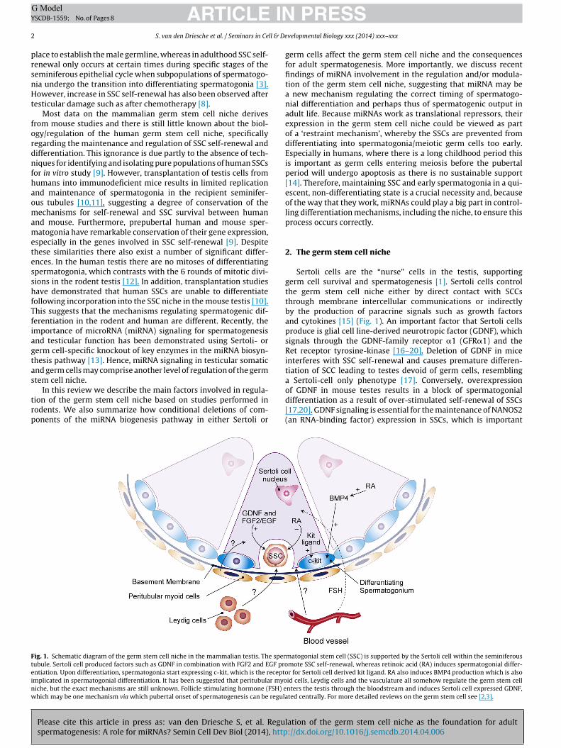
ig. 1. Schematic diagram of the germ stem cell niche in the mammalian testis. The sperubule. Sertoli cell produced factors such as GDNF in combination with FGF2 and EGF prontiation. Upon differentiation, spermatogonia start expressing c-kit, which is the receptomplicated in spermatogonial differentiation. It has been suggested that peritubular myoiche, but the exact mechanisms are still unknown. Follicle stimulating hormone (FSH) ehich may be one mechanism via which pubertal onset of spermatogenesis can be regula
PRESSvelopmental Biology xxx (2014) xxx–xxx
germ cells affect the germ stem cell niche and the consequencesfor adult spermatogenesis. More importantly, we discuss recentfindings of miRNA involvement in the regulation and/or modula-tion of the germ stem cell niche, suggesting that miRNA may bea new mechanism regulating the correct timing of spermatogo-nial differentiation and perhaps thus of spermatogenic output inadult life. Because miRNAs work as translational repressors, theirexpression in the germ stem cell niche could be viewed as partof a ‘restraint mechanism’, whereby the SSCs are prevented fromdifferentiating into spermatogonia/meiotic germ cells too early.Especially in humans, where there is a long childhood period thisis important as germ cells entering meiosis before the pubertalperiod will undergo apoptosis as there is no sustainable support[14]. Therefore, maintaining SSC and early spermatogonia in a qui-escent, non-differentiating state is a crucial necessity and, becauseof the way that they work, miRNAs could play a big part in control-ling differentiation mechanisms, including the niche, to ensure thisprocess occurs correctly.
2. The germ stem cell niche
Sertoli cells are the “nurse” cells in the testis, supportinggerm cell survival and spermatogenesis [1]. Sertoli cells controlthe germ stem cell niche either by direct contact with SCCsthrough membrane intercellular communications or indirectlyby the production of paracrine signals such as growth factorsand cytokines [15] (Fig. 1). An important factor that Sertoli cellsproduce is glial cell line-derived neurotropic factor (GDNF), whichsignals through the GDNF-family receptor �1 (GFR�1) and theRet receptor tyrosine-kinase [16–20]. Deletion of GDNF in miceinterferes with SSC self-renewal and causes premature differen-tiation of SCC leading to testes devoid of germ cells, resemblinga Sertoli-cell only phenotype [17]. Conversely, overexpression
lation of the germ stem cell niche as the foundation for adult://dx.doi.org/10.1016/j.semcdb.2014.04.006
of GDNF in mouse testes results in a block of spermatogonialdifferentiation as a result of over-stimulated self-renewal of SSCs[17,20]. GDNF signaling is essential for the maintenance of NANOS2(an RNA-binding factor) expression in SSCs, which is important
matogonial stem cell (SSC) is supported by the Sertoli cell within the seminiferousmote SSC self-renewal, whereas retinoic acid (RA) induces spermatogonial differ-r for Sertoli cell derived kit ligand. RA also induces BMP4 production which is alsoid cells, Leydig cells and the vasculature all somehow regulate the germ stem cellnters the testis through the bloodstream and induces Sertoli cell expressed GDNF,ted centrally. For more detailed reviews on the germ stem cell see [2,3].
ING ModelY
ll & De
f[Nottdciap
tieirffWrb[
sGiSo
redec[
cpdpnSaa[iihiSc[
hfdszabatrtt
ARTICLESCDB-1559; No. of Pages 8
S. van den Driesche et al. / Seminars in Ce
or preventing the differentiation of SSCs in the postnatal testis21]. Conversely, fibroblast growth factor 9 (FGF9) up-regulatesANOS2 and thereby acts as an inhibitor of meiotic differentiationf postnatal germ cells [22], perhaps indicating co-ordination ofhis with SSC differentiation control. Interestingly, the transcrip-ion factor Foxo1 is also required for SSC maintenance, as Foxo1eficiency results in severe impairment of SSC self-renewal and aomplete block of spermatogonial differentiation [23]. One of thedentified Foxo1 gene targets in SSC is Ret which, as mentionedbove, is part of the GDNF receptor complex, and Foxo1 signalingromotes high levels of Ret protein on the cell surface of SSCs [23].
FSH produced by the pituitary gland acts on the Sertoli cells inhe pubertal testis to initiate spermatogenesis [24], and FSH andts second messenger cyclic AMP (cAMP) are able to induce GDNFxpression in Sertoli cells [25,26], whereas retinoic acid (RA), whichs involved in spermatogonial differentiation (see below), down-egulates GDNF in Sertoli cells [25] (Fig. 1). Thus, GDNF is essentialor SSC proliferation and self-renewal and the control of SSC dif-erentiation, under the control of the hypothalamic-pituitary axis.
hilst GDNF is essential for SSC self-renewal, expression of itseceptor GFR�1 is not exclusive to undifferentiated spermatogonia,ut is also present in Apaired and Aaligned spermatogonial subtypes27–30].
Another factor that is required for SSC self-renewal is the tran-cription factor Ets-related molecule (ERM or ETV5) which, likeDNF, is expressed solely in Sertoli cells and its deletion results
n compromised Sertoli cell function, leading to premature loss ofSCs [31–34]. Interestingly, ERM/ETV5 has been shown to be onef the downstream GDNF targets in the kidney [35].
FGF2 and EGF in combination with GDNF can promote SSC self-enewal in vitro, although FGF2 and EGF on their own are notssential for SSC self-renewal as over time the number of SSCseclined in SSC enriched germ cell cultures cultured in the pres-nce of FGF2 or EGF without GDNF, indicating that self-renewal wasompromised and spermatogonial differentiation was promoted36,37].
Another important system for regulation of the germ stemell niche is the KIT signaling system (Fig. 1). C-kit is a receptorresent on the cell surface of differentiating spermatogonia thatistinguishes them from SSC. C-kit has been shown to mediateroliferation, survival and differentiation of type A spermatogo-ia in response to stimulation by kit ligand, which is produced byertoli cells [25,38–41]. This stimulates spermatogonia to undergo
number of mitotic divisions, forming the A2–A4, intermedi-te, and B spermatogonia in rodents before they enter meiosis42]. In Sertoli cells, cAMP and retinoic acid (RA) signaling [25,43]nduce the production of a paracrine factor called BMP4, which cannduce expression of c-kit in spermatogonia [43–45]. In prepubertaluman spermatogonia, higher levels of C-KIT mRNA are found than
n the prepubertal mouse, which may reflect the signal for someSCs to differentiate into type B spermatogonia as this is a pro-ess known to happen during the prepubertal period in humans9,46].
Sertoli cells produce RA, which is another paracrine factor thatas been demonstrated to be essential for spermatogonial dif-
erentiation [47] (Fig. 1). RA promotes differentiation of SSCs toifferentiated spermatogonia through several mechanisms. Oneuch mechanism is the down-regulation of promyelocytic leukemiainc finger (PLZF) [48], which is specifically expressed in SSCsnd upon deletion in mice impairs spermatogonial differentiationy affecting the expression of genes involved in SSC self-renewalnd differentiation [49–52]. One function of PLZF is to repress
Please cite this article in press as: van den Driesche S, et al. Reguspermatogenesis: A role for miRNAs? Semin Cell Dev Biol (2014), http
he expression of c-kit in spermatogonia [53] and as RA down-egulates PLZF in SSC this could be one mechanism that controlshe up-regulation of c-kit in spermatogonia to steer them downhe differentiation path.
PRESSvelopmental Biology xxx (2014) xxx–xxx 3
Treatment of postnatal spermatogonia with RA results inup-regulation of the b-Helix-Loop-Helix transcription factors, sper-matogenesis and oogenesis HLH1 (Sohlh1) and Sohlh2 [54]. BothSohlh1 and Sohlh2 are essential for spermatogonial differentia-tion as deletion of either leads to disappearance of c-kit-expressingspermatogonia in the prepubertal testis [55–59]. A strong correla-tion between expression of c-kit and both Sohlh1 and Sohlh2 inpostnatal spermatogonia has been shown [54], and a direct inter-action between c-kit and Sohlh1 was found in vivo in chromatinisolated from spermatogonia [54,58]. Therefore, up-regulation ofSohlh1 in spermatogonia by RA could be another mechanism bywhich RA increases expression of c-kit to promote spermatogonialdifferentiation. Involvement of doublesex-related transcriptionfactor (Dmrt1) is also likely in this process as c-kit expression issignificantly reduced in Dmrt1 conditional knockout mice [60]. Fur-thermore, when Dmrt1 is deleted from spermatogonia, loss of SSCswas observed as they entered meiosis precociously [60]. Dmrt1is important for repressing the RA-responsive, meiosis-inducinggenes Stra8 and Sohlh1, thereby preventing meiotic entry of differ-entiated spermatogonia [60]. Indeed, DMRT1 expression in germcells is considered as a likely master switch for controlling themitosis–meiosis transition [60], failure of which appears to be asso-ciated with the development of TGCC in humans [61].
3. Biogenesis of miRNAs and conditional Dicer knockoutmouse models
Conceptually, it is generally thought that in the male germ cellsare pre-programmed to go through their sequential developmentinto spermatozoa, provided that there is a supportive environment(the niche), determined by the surrounding somatic cells. How-ever, in situations when the niche is non-supportive, it might beexpected that other mechanisms may operate to literally hold thegerm cells under restraint. For example, during childhood in boys, itis essential for germ cells not to enter meiosis before puberty startsas there is no support system in place for sustaining germ cells thatenter the spermatogenic pathway. Small non-coding RNAs, such asmiRNAs have begun to attract attention as important regulators ofcell function, as they provide another level of translational controlof gene expression. Since miRNAs work as translational repressors,a logical role for them might be to act as part of a ‘restraint mech-anism’ that stops germ cells from entering meiosis too early andensuring that the right number of germ cells are ready to entermeiosis when the time is right.
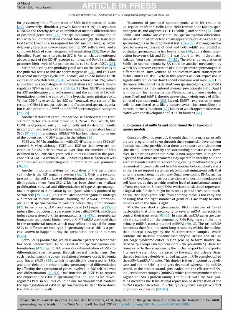
MiRNAs are short single-stranded RNA molecules of 19–23nucleotides that bind specifically to several mRNA molecules tocontrol their translation [62–65]. In animals, miRNA genes are usu-ally transcribed from the genome by RNA Polymerase II, formingprimary miRNA transcripts (pri-miRNA) (Fig. 2). The pri-miRNAmolecules then fold into stem-loop structures within the nucleusthat undergo cleavage by the Microprocessor complex, whichcontains the RNaseIII endonuclease enzyme Drosha and DGCR8(DiGeorge syndrome critical region gene 8), to form shorter iso-lated hairpin loops called precursor miRNA (pre-miRNA). These aretransported to the cytoplasm by the nuclear export factor exportin5 where the stem-loop is cleaved by the endoribonuclease Dicer,thereby forming a double-stranded mature miRNA complex calledthe miRNA:miRNA* duplex. This duplex is then unwound by a heli-case and the miRNA* strand gets degraded whereas the miRNAstrand, or the mature strand, gets loaded into the effector miRNA-induced silencer complex (miRISC), which contains members of the
lation of the germ stem cell niche as the foundation for adult://dx.doi.org/10.1016/j.semcdb.2014.04.006
Argonaute (AGO) protein family. The miRISC with the AGO pro-teins mediates the translational repression or degradation of themRNA targets. Therefore, miRNAs typically have a negative effecton protein expression [62–65].
ARTICLE IN PRESSG ModelYSCDB-1559; No. of Pages 8
4 S. van den Driesche et al. / Seminars in Cell & Developmental Biology xxx (2014) xxx–xxx
Fig. 2. MiRNA biogenesis pathway. The primary miRNA (Pri-miRNA) is transcribed from DNA by RNA polymerase II and gets processed into pre-miRNA structures of roughly70 nucleotides by the nuclear microprocessor complex, which is comprised of the endonucleases Drosha and DGRC8 (DiGeorge syndrome critical region 8). The pre-miRNAgets transported into the cytoplasm by exportin 5 where the RNase III endonuclease Dicer cleaves the stem loop structure to form 19–23 nucleotide small RNAs. Them plex (d
strtmpgSsBoc
ggdtede(tgitettieee
iRNA strand of the small RNAs gets loaded into the miRNA-induced silencing comegradation of target mRNAs.
Several studies have documented the preferential or exclu-ive expression of specific miRNAs in the immature and adultestis [66–79]. The overall importance of miRNA signaling foregulation of spermatogenesis has been demonstrated using cellype-specific conditional knockout studies of Dicer and other
iRNA biogenesis genes (Fig. 2), which show that miRNA-mediatedost-transcriptional control is an important regulator of spermato-enesis [69,80]. For example, conditional deletion of Dicer1 fromertoli cells in mice results in infertility due to complete absence ofpermatogenesis and progressive degeneration of the testis [81,82].oth studies demonstrated defects in early postnatal testis devel-pment and Sertoli cell proliferation, resulting in massive Sertoliell and germ cell apoptosis in the prepubertal testis [81,82].
Besides conditionally ablating Dicer from Sertoli cells, variousroups have reported studies in which they deleted Dicer fromerm cells [83–86]. This resulted in adult infertility or subfertilityue to spermatogenic arrest. The study by Hayashi et al. showedhat deletion of Dicer1 in germ cells caused a defect in the prolif-ration of male germ cells [83], whereas Maatouk et al. reportedefects in both sperm motility and the transition from round tolongated spermatids [85]. Both of these studies used the TNAP-Cretissue non-specific alkaline phosphatase) mouse line to generatehe ablation of Dicer in germ cells, but since the TNAP-Cre trans-enic mouse expresses Cre in only ∼50% of the germ cells, ands not totally specific to germ cells [86], the effects are difficulto interpret. Furthermore, TNAP-Cre expression begins as early asmbryonic day (E) 10 [87], making it very difficult to pinpointhe exact mechanism via which the spermatogenic defects arise inhese Dicer1 mutant mice, since Dicer ablation already takes place
Please cite this article in press as: van den Driesche S, et al. Reguspermatogenesis: A role for miRNAs? Semin Cell Dev Biol (2014), http
n (a proportion of) the early primordial germ cell population in thearly mouse embryo, thereby interfering with the development ofmbryonic germ cells. The studies by Romero et al. [86] and Liut al. [84] used a more specific germ cell expressing Cre mouse line,
miRISC), which includes the Argonaute (AGO) proteins and this complex mediates
namely Ddx4-Cre, which deleted Dicer just before birth at the stagewhen spermatogonia are just beginning to appear. Using this trans-genic line they concluded that Dicer1 expression in mouse germcells is not needed for SSC renewal and mitotic proliferation, but isnecessary for the differentiation of germ cells through meiotic andhaploid phases of spermatogenesis [84,86].
Several groups reported studies in which Dicer1 was deletedfrom spermatogonia after birth using transgenic mouse linesexpressing the Cre recombinase under the control of a Neurogenin3(Ngn3) [88] or Stra8 [89,90] promoter, which resulted in less severephenotypes when compared to the Romero et al. [86] and Liu et al.[84] studies. Ngn3 is expressed endogenously in male germ cellsstarting from postnatal day 5, and its expression has been shownin type Asingle, Apaired and Aaligned spermatogonia that gives rise toall differentiating germ cells [91]. This Ngn3Cre-Dicer1 transgenicmouse line did not show an effect on meiotic progression, but areduction in the number of haploid germ cells and an increase inthe number of apoptotic spermatocytes [88], whilst postnatal germcell-specific deletion of Dicer was demonstrated to be critical forthe normal organization of chromatin and nuclear shaping of elon-gated spermatids [88]. Interestingly, in the Ddx4Cre-Dicer1 mousemodel, upregulation of transposon expression was observed [86],whereas transposon expression was unaffected in the Ngn3Cre-Dicer1 knockout testis [88]. This indicates that, as well as miRNAbiogenesis, Dicer is also involved in transposon control in germcells in the perinatal phase of testis development, which takes placebefore Ngn3 expression. Deleting Dicer1 from early spermatogo-nia using the Stra8-Cre transgenic mouse lines resulted in similarphenotypes to the Nrg3Cre-Dicer1 model [89,90]. Another study
lation of the germ stem cell niche as the foundation for adult://dx.doi.org/10.1016/j.semcdb.2014.04.006
using the protamine 1 (Prm1)-Cre transgene to delete Dicer1 specif-ically in post-meiotic haploid male germ cells [92], demonstrateda less severe phenotype compared with those in which Dicer1 wasdeleted from pre-meiotic spermatogonia [88–90]. However, Chang
ING ModelY
ll & De
emiasflsd
4
mmcmussMaussRsMetpemii[gttnsn
5
iatg[tsscmswsEtrSit
ARTICLESCDB-1559; No. of Pages 8
S. van den Driesche et al. / Seminars in Ce
t al. did observe abnormal morphology in the elongated sper-atids indicating that post-meiotic differentiation was disrupted
n the Prm1Cre-Dicer1 mice [92]. Taken together, these studies show clear requirement for expression of Dicer1, and therefore themall non-coding RNA machinery, in both Sertoli and germ cellsor the onset of spermatogenesis and for male fertility. The ear-ier the deletion of Dicer takes place, the more severe effects onpermatogenesis are found, perhaps due to an accumulation ofefects.
. MiRNAs and the germ stem cell niche
Interestingly, a series of publications implicate miRNA involve-ent in regulation of the germ stem cell niche. Two particulariRNAs, miR-221 and miR-222 negatively regulate expression of
-kit at the post-transcriptional level [93]. Over-expression ofiR221/222 renders RA ineffective at inducing the transition ofndifferentiated spermatogonia into c-kit-positive differentiatedpermatogonia [93]. Furthermore, GDNF can up-regulate expres-ion of miR-221/222, whereas RA has the opposite effect [93].oreover, miR-146 interferes with the expression of c-kit, Stra8
nd Sohlh2 in RA-treated spermatogonia, suggesting that it mod-lates the effects of RA on spermatogonial differentiation [94]. RAignificantly induced expression of the Mirlet7 family miRNAs inpermatogonia, through suppression of Lin28, suggesting a role inA-induced spermatogonial differentiation [95]. The same grouphowed that RA down-regulates expression of members of their-17-92 (Mirc1) and Mir-106b-25 (Mirc3) clusters in undiffer-
ntiated spermatogonia, both in vitro and in vivo, suggesting thathe Mirlet7/Mir-17-92 (Mirc1)/Mir-106b-25 (Mirc3) clusters couldlay a role in SSC self-renewal and the proliferation of undiffer-ntiated spermatogonia [96]. Another study showed expression ofiRNA-20 and miRNA-106a in mouse SSCs and their involvement
n SSC self-renewal by targeting Stat3 and Cyclin D1, both involvedn spermatogonial differentiation, at the post-transcriptional level97]. Moreover, miR-21 is regulated by ERM/ETV5 in SSC-enrichederm cell cultures, and miR-21 was shown to be important in main-aining the SSC population in vitro, demonstrating for the first timehe involvement of a specific miRNA in SSC self-renewal [98]. Ineuronal cells miR-21 is induced by GDNF [99], raising the pos-ibility of a similar role in Sertoli cells within the germ stem celliche.
. Hormonal regulation of miRNA expression
Androgen regulation of spermatogenesis is fundamentallymportant, but how it does so is still unclear. Modulation of miRNAs,nd perhaps thus of the niche, is a potentially novel mechanismhat has only been superficially addressed. Interestingly, andro-ens are major regulators of miRNA expression in the prostate100–103], muscle [101,104] and liver [105]. Moreover, prenatalestosterone exposure alters fetal ovarian miRNA expression in theheep, and may lead to adult ovarian pathologies [106]; similartudies for the testis have not been reported. Using primary Sertoliell cultures and an in vivo rat model, upregulation of a subset ofiRNAs in Sertoli cells was reported after FSH or androgen suppres-
ion [107]. This included miR-23b, miR-30c, miR-30d and miR-690,hose predicted targets include genes important for focal adhe-
ion and regulation of the actin cytoskeleton, such as Pten andps15. Another study looked at adult Sertoli cells in mice and iden-ified a group of testosterone-dependent miRNAs playing a crucial
Please cite this article in press as: van den Driesche S, et al. Reguspermatogenesis: A role for miRNAs? Semin Cell Dev Biol (2014), http
ole in androgen-mediated events during spermatogenesis [108].ome of the miR-471 targets found in this study included Foxd1,mportant in Sertoli cell metabolism, and desmocollin-1 (Dsc1),hat plays a crucial role in cell-cell adhesion in epithelial cells
PRESSvelopmental Biology xxx (2014) xxx–xxx 5
[108]. Even though the miRNAs identified in this study differedfrom those identified in the Nicholls et al. study, both proposedthat testosterone-mediated inhibition of miRNAs in Sertoli cellscould facilitate essential gene expression necessary for progressionof spermatogenesis [107,108].
6. MiRNA expression in human TGCC
MiRNA expression has also been implicated in TGCC. In humansthese tumors present in young adulthood following transformationof pre-malignant carcinoma in situ (CIS) cells, which are believedto result from failure of differentiation of fetal germ cells. Cur-rently, CIS cells are considered to result from immaturity of thesomatic cell component of the germ stem cell niche [109]. In agenetic screen for miRNAs that work together with oncogenes incellular transformation, miR-372 and miR-373 were identified aspotential oncogenes involved in the development of human TGCC[110], whilst several miRNAs are specifically expressed in TGCC[111,112] and CIS cells [112]. Similarly, a subset of miRNAs wasshown to be differentially expressed in TGCC compared to nor-mal testicular tissue [113]. In human TGCC cell lines, repressionof the candidate tumor suppressor protein–tyrosine phosphatasenon-receptor type 23 (PTPN23) by miR-142-3p plays an impor-tant role in the pathogenesis of TGCCs by repressing expressionof PTPN23 [114]. TGCC is hypothesized to be part of a group ofmale reproductive health disorders, that include cryptorchidismand hypospadias, and comprises a testicular dysgenesis syndromewith a common origin in fetal life [115]. In the cryptorchid rat testisit was shown that the miRNA miR-135a was expressed at lowerlevels in the undescended testis and a decreased number of SSCswith Foxo1 activation was observed, suggesting a contribution ofmiR-135a to SSC maintenance by modulating Foxo1 activity [116].Since miRNAs are quick and easy to measure in blood or tissues, andas their levels can be altered by human diseases, they also haveunexplored potential as diagnostic tools and therapeutic targetsfor these diseases, as demonstrated in the case of (alcoholic) liverdisease [117–119]. It will be interesting to determine if there aremiRNAs measurable in the blood of testis cancer patients whichcould provide either a diagnosis and/or prognosis on their diseasestate.
7. Future directions
Specific miRNAs are expressed in the testis and appear toplay a role in SSC self-renewal and spermatogonial differentiation,although detailed understanding of the roles and importance ofmiRNAs in this context are lacking. For example, it is unclear ifSertoli cell expressed miRNAs act as secreted paracrine factors inthe germ stem cell niche, or whether they indirectly modulate thesecretion of other Sertoli cell factors that then affect germ cells.Similarly, miRNAs produced in the germ cells themselves could actdirectly upon target mRNAs within those germ cells or may acton mRNA targets in neighboring somatic cells. Another importantarea to explore is to determine the role of other somatic cell com-ponents (and their associated miRNAs) e.g. the peritubular myoidcells, the vasculature and Leydig cells in regulation of the germstem cell niche. Conceptually, it seems clear already that regula-tion of the miRNA networks in the germ stem cell niche and in thegerm cells themselves is likely to be critical for SSC/spermatogonialdevelopment and output.
lation of the germ stem cell niche as the foundation for adult://dx.doi.org/10.1016/j.semcdb.2014.04.006
Disclosure
The authors have nothing to disclose.
ING ModelY
6 ll & De
A
CtftGW
R
ARTICLESCDB-1559; No. of Pages 8
S. van den Driesche et al. / Seminars in Ce
cknowledgements
This work was supported in part by the UK Medical Researchouncil (Grants G33253 (SvdD, RMS) and G1100356/1 (PTKS)) andhe Wellcome Trust (Grant 098522, RTM). We thank Ronnie Grantor the figure illustrations. This work was supported in part byhe UK Medical Research Council (Grants G33253 (SvdD, RMS) and1100356/1 (PTKS)) and the Wellcome Trust (Grant 098522, RTM).e thank Ronnie Grant for the figure illustrations.
eferences
[1] Sharpe RM. Regulation of spermatogenesis. In: Knobil E, Neill JD, editors. Thephysiology of reproduction, vol. 1, 2nd ed. New York: Raven Press; 1994. p.1363–434.
[2] Li L, Xie T. Stem cell niche: structure and function. Annu Rev Cell Dev Biol2005;21:605–31.
[3] Oatley JM, Brinster RL. The germline stem cell niche unit in mammalian testes.Physiol Rev 2012;92:577–95.
[4] Xie T. Germline stem cell niches. In: Girard L, editor. StemBook (internet).Cambridge, MA: Harvard Stem Cell Institute; 2008.
[5] Yoshida S, Sukeno M, Nabeshima Y. A vasculature-associated niche forundifferentiated spermatogonia in the mouse testis. Science 2007;317:1722–6.
[6] Rajpert-De Meyts E. Developmental model for the pathogenesis of testicularcarcinoma in situ: genetic and environmental aspects. Hum Reprod Update2006;12:303–23.
[7] Skakkebaek NE. Possible carcinoma-in-situ of the testis. Lancet 1972;2:516–7.[8] Mitchell RT, Saunders PT, Sharpe RM, Kelnar CJ, Wallace WH. Male fertility
and strategies for fertility preservation following childhood cancer treatment.Endocr Dev 2009;15:101–34.
[9] Wu X, Schmidt JA, Avarbock MR, Tobias JW, Carlson CA, Kolon TF, et al. Pre-pubertal human spermatogonia and mouse gonocytes share conserved geneexpression of germline stem cell regulatory molecules. Proc Natl Acad Sci US A 2009;106:21672–7.
[10] Brinster RL. Germline stem cell transplantation and transgenesis. Science2002;296:2174–6.
[11] Nagano M, Patrizio P, Brinster RL. Long-term survival of human spermatogo-nial stem cells in mouse testes. Fertil Steril 2002;78:1225–33.
[12] Ehmcke J, Schlatt S. A revised model for spermatogonial expansion in man:lessons from non-human primates. Reproduction 2006;132:673–80.
[13] Yadav RP, Kotaja N. Small RNAs in spermatogenesis. Mol Cell Endocrinol2014;382:498–508.
[14] Chemes HE. Infancy is not a quiescent period of testicular development. Int JAndrol 2001;24:2–7.
[15] Rossi P, Dolci S. Paracrine mechanisms involved in the control of early stagesof mammalian spermatogenesis. Front Endocrinol 2013;4:181.
[16] Kubota H, Avarbock MR, Brinster RL. Growth factors essential for self-renewaland expansion of mouse spermatogonial stem cells. Proc Natl Acad Sci U S A2004;101:16489–94.
[17] Meng X, Lindahl M, Hyvonen ME, Parvinen M, de Rooij DG, Hess MW, et al.Regulation of cell fate decision of undifferentiated spermatogonia by GDNF.Science 2000;287:1489–93.
[18] Oatley JM, Avarbock MR, Brinster RL. Glial cell line-derived neurotrophicfactor regulation of genes essential for self-renewal of mouse spermato-gonial stem cells is dependent on Src family kinase signaling. J Biol Chem2007;282:25842–51.
[19] Viglietto G, Dolci S, Bruni P, Baldassarre G, Chiariotti L, Melillo RM, et al.Glial cell line-derived neutrotrophic factor and neurturin can act as paracrinegrowth factors stimulating DNA synthesis of Ret-expressing spermatogonia.Int J Oncol 2000;16:689–94.
[20] Yomogida K, Yagura Y, Tadokoro Y, Nishimune Y. Dramatic expansion ofgerminal stem cells by ectopically expressed human glial cell line-derivedneurotrophic factor in mouse Sertoli cells. Biol Reprod 2003;69:1303–7.
[21] Sada A, Hasegawa K, Pin PH, Saga Y. NANOS2 acts downstream of glial cellline-derived neurotrophic factor signaling to suppress differentiation of sper-matogonial stem cells. Stem Cells 2012;30:280–91.
[22] Barrios F, Filipponi D, Pellegrini M, Paronetto MP, Di Siena S, Geremia R, et al.Opposing effects of retinoic acid and FGF9 on Nanos2 expression and meioticentry of mouse germ cells. J Cell Sci 2010;123:871–80.
[23] Goertz MJ, Wu Z, Gallardo TD, Hamra FK, Castrillon DH. Foxo1 is required inmouse spermatogonial stem cells for their maintenance and the initiation ofspermatogenesis. J Clin Invest 2011;121:3456–66.
[24] Steinberger E. Hormonal control of mammalian spermatogenesis. Physiol Rev1971;51:1–22.
[25] Pellegrini M, Filipponi D, Gori M, Barrios F, Lolicato F, Grimaldi P, et al. ATRA
Please cite this article in press as: van den Driesche S, et al. Reguspermatogenesis: A role for miRNAs? Semin Cell Dev Biol (2014), http
and KL promote differentiation toward the meiotic program of male germcells. Cell Cycle 2008;7:3878–88.
[26] Tadokoro Y, Yomogida K, Ohta H, Tohda A, Nishimune Y. Homeostatic regula-tion of germinal stem cell proliferation by the GDNF/FSH pathway. Mech Dev2002;113:29–39.
PRESSvelopmental Biology xxx (2014) xxx–xxx
[27] Grasso M, Fuso A, Dovere L, de Rooij DG, Stefanini M, Boitani C, et al. Distribu-tion of GFRA1-expressing spermatogonia in adult mouse testis. Reproduction2012;143:325–32.
[28] Grisanti L, Falciatori I, Grasso M, Dovere L, Fera S, Muciaccia B, et al. Identi-fication of spermatogonial stem cell subsets by morphological analysis andprospective isolation. Stem Cells 2009;27:3043–52.
[29] Naughton CK, Jain S, Strickland AM, Gupta A, Milbrandt J. Glial cell-linederived neurotrophic factor-mediated RET signaling regulates spermatogo-nial stem cell fate. Biol Reprod 2006;74:314–21.
[30] Suzuki H, Sada A, Yoshida S, Saga Y. The heterogeneity of spermatogoniais revealed by their topology and expression of marker proteins includ-ing the germ cell-specific proteins Nanos2 and Nanos3. Dev Biol 2009;336:222–31.
[31] Chen C, Ouyang W, Grigura V, Zhou Q, Carnes K, Lim H, et al. ERM is requiredfor transcriptional control of the spermatogonial stem cell niche. Nature2005;436:1030–4.
[32] Morrow CM, Hostetler CE, Griswold MD, Hofmann MC, Murphy KM, CookePS, et al. ETV5 is required for continuous spermatogenesis in adult mice andmay mediate blood testes barrier function and testicular immune privilege.Ann N Y Acad Sci 2007;1120:144–51.
[33] Schlesser HN, Simon L, Hofmann MC, Murphy KM, Murphy T, Hess RA,et al. Effects of ETV5 (ets variant gene 5) on testis and body growth, timecourse of spermatogonial stem cell loss, and fertility in mice. Biol Reprod2008;78:483–9.
[34] Tyagi G, Carnes K, Morrow C, Kostereva NV, Ekman GC, Meling DD, et al. Lossof Etv5 decreases proliferation and RET levels in neonatal mouse testiculargerm cells and causes an abnormal first wave of spermatogenesis. Biol Reprod2009;81:258–66.
[35] Lu BC, Cebrian C, Chi X, Kuure S, Kuo R, Bates CM, et al. Etv4 and Etv5 arerequired downstream of GDNF and Ret for kidney branching morphogenesis.Nat Genet 2009;41:1295–302.
[36] Kanatsu-Shinohara M, Miki H, Inoue K, Ogonuki N, Toyokuni S, Ogura A, et al.Long-term culture of mouse male germline stem cells under serum-or feeder-free conditions. Biol Reprod 2005;72:985–91.
[37] Lee J, Kanatsu-Shinohara M, Inoue K, Ogonuki N, Miki H, Toyokuni S, et al. Aktmediates self-renewal division of mouse spermatogonial stem cells. Devel-opment 2007;134:1853–9.
[38] Dolci S, Pellegrini M, Di Agostino S, Geremia R, Rossi P. Signaling throughextracellular signal-regulated kinase is required for spermatogonial prolife-rative response to stem cell factor. J Biol Chem 2001;276:40225–33.
[39] Rossi P, Albanesi C, Grimaldi P, Geremia R. Expression of the mRNA forthe ligand of c-kit in mouse Sertoli cells. Biochem Biophys Res Commun1991;176:910–4.
[40] Rossi P, Dolci S, Albanesi C, Grimaldi P, Ricca R, Geremia R. Follicle-stimulatinghormone induction of steel factor (SLF) mRNA in mouse Sertoli cells andstimulation of DNA synthesis in spermatogonia by soluble SLF. Dev Biol1993;155:68–74.
[41] Rossi P, Lolicato F, Grimaldi P, Dolci S, Di Sauro A, Filipponi D, et al. Transcrip-tome analysis of differentiating spermatogonia stimulated with kit ligand.Gene Expr Patterns 2008;8:58–70.
[42] de Rooij DG. Proliferation and differentiation of spermatogonial stem cells.Reproduction 2001;121:347–54.
[43] Pellegrini M, Grimaldi P, Rossi P, Geremia R, Dolci S. Developmentalexpression of BMP4/ALK3/SMAD5 signaling pathway in the mouse testis: apotential role of BMP4 in spermatogonia differentiation. J Cell Sci 2003;116:3363–72.
[44] Carlomagno G, van Bragt MP, Korver CM, Repping S, de Rooij DG, van Pelt AM.BMP4-induced differentiation of a rat spermatogonial stem cell line causeschanges in its cell adhesion properties. Biol Reprod 2010;83:742–9.
[45] Nagano M, Ryu BY, Brinster CJ, Avarbock MR, Brinster RL. Maintenance ofmouse male germ line stem cells in vitro. Biol Reprod 2003;68:2207–14.
[46] Culty M. Gonocytes, the forgotten cells of the germ cell lineage. Birth DefectsRes C Embryo Today 2009;87:1–26.
[47] Hogarth CA, Griswold MD. The key role of vitamin A in spermatogenesis. JClin Invest 2010;120:956–62.
[48] Dann CT, Alvarado AL, Molyneux LA, Denard BS, Garbers DL, Porteus MH.Spermatogonial stem cell self-renewal requires OCT4, a factor downregulatedduring retinoic acid-induced differentiation. Stem Cells 2008;26:2928–37.
[49] Barna M, Merghoub T, Costoya JA, Ruggero D, Branford M, Bergia A, et al.Plzf mediates transcriptional repression of HoxD gene expression throughchromatin remodeling. Dev Cell 2002;3:499–510.
[50] Buaas FW, Kirsh AL, Sharma M, McLean DJ, Morris JL, Griswold MD, et al.Plzf is required in adult male germ cells for stem cell self-renewal. Nat Genet2004;36:647–52.
[51] Costoya JA, Hobbs RM, Barna M, Cattoretti G, Manova K, Sukhwani M, et al.Essential role of Plzf in maintenance of spermatogonial stem cells. Nat Genet2004;36:653–9.
[52] Payne C, Braun RE. Histone lysine trimethylation exhibits a dis-tinct perinuclear distribution in Plzf-expressing spermatogonia. Dev Biol2006;293:461–72.
[53] Filipponi D, Hobbs RM, Ottolenghi S, Rossi P, Jannini EA, Pandolfi PP,
lation of the germ stem cell niche as the foundation for adult://dx.doi.org/10.1016/j.semcdb.2014.04.006
et al. Repression of kit expression by Plzf in germ cells. Mol Cell Biol2007;27:6770–81.
[54] Barrios F, Filipponi D, Campolo F, Gori M, Bramucci F, Pellegrini M, et al.SOHLH1 and SOHLH2 control Kit expression during postnatal male germ celldevelopment. J Cell Sci 2012;125:1455–64.

ING ModelY
ll & De
ARTICLESCDB-1559; No. of Pages 8
S. van den Driesche et al. / Seminars in Ce
[55] Ballow D, Meistrich ML, Matzuk M, Rajkovic A. Sohlh1 is essential for sper-matogonial differentiation. Dev Biol 2006;294:161–7.
[56] Ballow DJ, Xin Y, Choi Y, Pangas SA, Rajkovic A. Sohlh2 is a germ cell-specificbHLH transcription factor. Gene Expr Patterns 2006;6:1014–8.
[57] Hao J, Yamamoto M, Richardson TE, Chapman KM, Denard BS, HammerRE, et al. Sohlh2 knockout mice are male-sterile because of degen-eration of differentiating type A spermatogonia. Stem Cells 2008;26:1587–97.
[58] Suzuki H, Ahn HW, Chu T, Bowden W, Gassei K, Orwig K, et al.SOHLH1 and SOHLH2 coordinate spermatogonial differentiation. Dev Biol2012;361:301–12.
[59] Toyoda S, Miyazaki T, Miyazaki S, Yoshimura T, Yamamoto M, Tashiro F, et al.Sohlh2 affects differentiation of KIT positive oocytes and spermatogonia. DevBiol 2009;325:238–48.
[60] Matson CK, Murphy MW, Griswold MD, Yoshida S, Bardwell VJ, Zarkower D.The mammalian doublesex homolog DMRT1 is a transcriptional gatekeeperthat controls the mitosis versus meiosis decision in male germ cells. Dev Cell2010;19:612–24.
[61] Jorgensen A, Nielsen JE, Almstrup K, Toft BG, Petersen BL, Rajpert-De Meyts E.Dysregulation of the mitosis-meiosis switch in testicular carcinoma in situ. JPathol 2013;229:588–98.
[62] Kim VN, Han J, Siomi MC. Biogenesis of small RNAs in animals. Nat Rev MolCell Biol 2009;10:126–39.
[63] Krol J, Loedige I, Filipowicz W. The widespread regulation of microRNA bio-genesis, function and decay. Nat Rev Genet 2010;11:597–610.
[64] Thomas M, Lieberman J, Lal A. Desperately seeking microRNA targets. NatStruct Mol Biol 2010;17:1169–74.
[65] Winter J, Jung S, Keller S, Gregory RI, Diederichs S. Many roads to matu-rity: microRNA biogenesis pathways and their regulation. Nat Cell Biol2009;11:228–34.
[66] Bao J, Li D, Wang L, Wu J, Hu Y, Wang Z, et al. MicroRNA-449 andmicroRNA-34b/c function redundantly in murine testes by targeting E2Ftranscription factor-retinoblastoma protein (E2F-pRb) pathway. J Biol Chem2012;287:21686–98.
[67] Buchold GM, Coarfa C, Kim J, Milosavljevic A, Gunaratne PH, MatzukMM. Analysis of microRNA expression in the prepubertal testis. PLoS ONE2010;5:e15317.
[68] Hossain MM, Sohel MM, Schellander K, Tesfaye D. Characterization andimportance of microRNAs in mammalian gonadal functions. Cell Tissue Res2012;349:679–90.
[69] McIver SC, Roman SD, Nixon B, McLaughlin EA. miRNA and mammalian malegerm cells. Hum Reprod Update 2012;18:44–59.
[70] McIver SC, Stanger SJ, Santarelli DM, Roman SD, Nixon B, McLaughlin EA. Aunique combination of male germ cell miRNAs coordinates gonocyte differ-entiation. PLoS ONE 2012;7:e35553.
[71] Mishima T, Takizawa T, Luo SS, Ishibashi O, Kawahigashi Y, Mizuguchi Y,et al. MicroRNA (miRNA) cloning analysis reveals sex differences in miRNAexpression profiles between adult mouse testis and ovary. Reproduction2008;136:811–22.
[72] Rakoczy J, Fernandez-Valverde SL, Glazov EA, Wainwright EN, Sato T, Takada S,et al. MicroRNAs-140-5p/140-3p modulate Leydig cell numbers in the devel-oping mouse testis. Biol Reprod 2013;88:143.
[73] Ro S, Park C, Sanders KM, McCarrey JR, Yan W. Cloning and expression profilingof testis-expressed microRNAs. Dev Biol 2007;311:592–602.
[74] Song R, Ro S, Michaels JD, Park C, McCarrey JR, Yan W. Many X-linked microRNAs escape meiotic sex chromosome inactivation. Nat Genet2009;41:488–93.
[75] Wainwright EN, Jorgensen JS, Kim Y, Truong V, Bagheri-Fam S, Davidson T,et al. SOX9 regulates microRNA miR-202-5p/3p expression during mousetestis differentiation. Biol Reprod 2013;89:34.
[76] Yan N, Lu Y, Sun H, Qiu W, Tao D, Liu Y, et al. Microarray profiling of microR-NAs expressed in testis tissues of developing primates. J Assist Reprod Genet2009;26:179–86.
[77] Yan N, Lu Y, Sun H, Tao D, Zhang S, Liu W, et al. A microarray for microRNAprofiling in mouse testis tissues. Reproduction 2007;134:73–9.
[78] Yang Q, Hua J, Wang L, Xu B, Zhang H, Ye N, et al. MicroRNA and piRNA profilesin normal human testis detected by next generation sequencing. PLoS ONE2013;8:e66809.
[79] Yu Z, Raabe T, Hecht NB. MicroRNA Mirn122a reduces expressionof the posttranscriptionally regulated germ cell transition protein 2(Tnp2) messenger RNA (mRNA) by mRNA cleavage. Biol Reprod 2005;73:427–33.
[80] Papaioannou MD, Nef S. microRNAs in the testis: building up male fertility. JAndrol 2010;31:26–33.
[81] Kim GJ, Georg I, Scherthan H, Merkenschlager M, Guillou F, Scherer G,et al. Dicer is required for Sertoli cell function and survival. Int J Dev Biol2010;54:867–75.
[82] Papaioannou MD, Pitetti JL, Ro S, Park C, Aubry F, Schaad O, et al. Ser-toli cell Dicer is essential for spermatogenesis in mice. Dev Biol 2009;326:250–9.
[83] Hayashi K, Chuva de Sousa Lopes SM, Kaneda M, Tang F, Hajkova P, Lao K, et al.
Please cite this article in press as: van den Driesche S, et al. Reguspermatogenesis: A role for miRNAs? Semin Cell Dev Biol (2014), http
MicroRNA biogenesis is required for mouse primordial germ cell developmentand spermatogenesis. PLoS ONE 2008;3:e1738.
[84] Liu D, Li L, Fu H, Li S, Li J. Inactivation of Dicer1 has a severe cumulative impacton the formation of mature germ cells in mouse testes. Biochem Biophys ResCommun 2012;422:114–20.
PRESSvelopmental Biology xxx (2014) xxx–xxx 7
[85] Maatouk DM, Loveland KL, McManus MT, Moore K, Harfe BD. Dicer1is required for differentiation of the mouse male germline. Biol Reprod2008;79:696–703.
[86] Romero Y, Meikar O, Papaioannou MD, Conne B, Grey C, Weier M, et al. Dicer1depletion in male germ cells leads to infertility due to cumulative meiotic andspermiogenic defects. PLoS ONE 2011;6:e25241.
[87] Lomeli H, Ramos-Mejia V, Gertsenstein M, Lobe CG, Nagy A. Targeted insertionof Cre recombinase into the TNAP gene: excision in primordial germ cells.Genesis 2000;26:116–7.
[88] Korhonen HM, Meikar O, Yadav RP, Papaioannou MD, Romero Y, Da Ros M,et al. Dicer is required for haploid male germ cell differentiation in mice. PLoSONE 2011;6:e24821.
[89] Greenlee AR, Shiao MS, Snyder E, Buaas FW, Gu T, Stearns TM, et al. Deregu-lated sex chromosome gene expression with male germ cell-specific loss ofDicer1. PLoS ONE 2012;7:e46359.
[90] Wu Q, Song R, Ortogero N, Zheng H, Evanoff R, Small CL, et al. The RNase IIIenzyme DROSHA is essential for microRNA production and spermatogenesis.J Biol Chem 2012;287:25173–90.
[91] Yoshida S, Takakura A, Ohbo K, Abe K, Wakabayashi J, Yamamoto M, et al.Neurogenin3 delineates the earliest stages of spermatogenesis in the mousetestis. Dev Biol 2004;269:447–58.
[92] Chang YF, Lee-Chang JS, Imam JS, Buddavarapu KC, Subaran SS, Sinha-HikimAP, et al. Interaction between microRNAs and actin-associated protein Arpc5regulates translational suppression during male germ cell differentiation.Proc Natl Acad Sci U S A 2012;109:5750–5.
[93] Yang QE, Racicot KE, Kaucher AV, Oatley MJ, Oatley JM. MicroRNAs 221 and222 regulate the undifferentiated state in mammalian male germ cells. Devel-opment 2013;140:280–90.
[94] Huszar JM, Payne CJ. MicroRNA 146 (Mir146) modulates spermatogonial dif-ferentiation by retinoic acid in mice. Biol Reprod 2013;88:15.
[95] Tong MH, Mitchell D, Evanoff R, Griswold MD. Expression of Mirlet7 familymicroRNAs in response to retinoic acid-induced spermatogonial differentia-tion in mice. Biol Reprod 2011;85:189–97.
[96] Tong MH, Mitchell DA, McGowan SD, Evanoff R, Griswold MD. Two miRNAclusters, Mir-17-92 (Mirc1) and Mir-106b-25 (Mirc3), are involved in theregulation of spermatogonial differentiation in mice. Biol Reprod 2012;86:72.
[97] He Z, Jiang J, Kokkinaki M, Tang L, Zeng W, Gallicano I, et al. MiRNA-20 and mirna-106a regulate spermatogonial stem cell renewal at thepost-transcriptional level via targeting STAT3 and Ccnd1. Stem Cells2013;31:2205–17.
[98] Niu Z, Goodyear SM, Rao S, Wu X, Tobias JW, Avarbock MR, et al. MicroRNA-21 regulates the self-renewal of mouse spermatogonial stem cells. Proc NatlAcad Sci U S A 2011;108:12740–5.
[99] Yoong LF, Wan G, Too HP. Glial cell-line derived neurotrophic factor andneurturin regulate the expressions of distinct miRNA precursors through theactivation of GFRalpha2. J Neurochem 2006;98:1149–58.
[100] Ambs S, Prueitt RL, Yi M, Hudson RS, Howe TM, Petrocca F, et al. Genomicprofiling of microRNA and messenger RNA reveals deregulated microRNAexpression in prostate cancer. Cancer Res 2008;68:6162–70.
[101] Narayanan R, Jiang J, Gusev Y, Jones A, Kearbey JD, Miller DD, et al. MicroR-NAs are mediators of androgen action in prostate and muscle. PLoS ONE2010;5:e13637.
[102] Waltering KK, Porkka KP, Jalava SE, Urbanucci A, Kohonen PJ, LatonenLM, et al. Androgen regulation of micro-RNAs in prostate cancer. Prostate2011;71:604–14.
[103] Wang G, Wang Y, Feng W, Wang X, Yang JY, Zhao Y, et al. Transcription factorand microRNA regulation in androgen-dependent and -independent prostatecancer cells. BMC Genomics 2008;2(9 Suppl.):S22.
[104] Wyce A, Bai Y, Nagpal S, Thompson CC. Research resource: The androgenreceptor modulates expression of genes with critical roles in muscle devel-opment and function. Mol Endorinol 2010;24:1665–74.
[105] Delic D, Grosser C, Dkhil M, Al-Quraishy S, Wunderlich F. Testosterone-induced upregulation of miRNAs in the female mouse liver. Steroids2010;75:998–1004.
[106] Luense LJ, Veiga-Lopez A, Padmanabhan V, Christenson LK. Developmentalprogramming: gestational testosterone treatment alters fetal ovarian geneexpression. Endocrinology 2011;152:4974–83.
[107] Nicholls PK, Harrison CA, Walton KL, McLachlan RI, O’Donnell L, Stanton PG.Hormonal regulation of sertoli cell micro-RNAs at spermiation. Endocrinology2011;152:1670–83.
[108] Panneerdoss S, Chang YF, Buddavarapu KC, Chen HI, Shetty G, Wang H,et al. Androgen-responsive microRNAs in mouse Sertoli cells. PLoS ONE2012;7:e41146.
[109] Hoei-Hansen CE, Holm M, Rajpert-De Meyts E, Skakkebaek NE. Histologicalevidence of testicular dysgenesis in contralateral biopsies from 218 patientswith testicular germ cell cancer. J Pathol 2003;200:370–4.
[110] Voorhoeve PM, le Sage C, Schrier M, Gillis AJ, Stoop H, Nagel R, et al. A geneticscreen implicates miRNA-372 and miRNA-373 as oncogenes in testiculargerm cell tumors. Cell 2006;124:1169–81.
[111] Gillis AJ, Stoop HJ, Hersmus R, Oosterhuis JW, Sun Y, Chen C, et al. High-
lation of the germ stem cell niche as the foundation for adult://dx.doi.org/10.1016/j.semcdb.2014.04.006
throughput microRNAome analysis in human germ cell tumours. J Pathol2007;213:319–28.
[112] Novotny GW, Belling KC, Bramsen JB, Nielsen JE, Bork-Jensen J, Almstrup K,et al. MicroRNA expression profiling of carcinoma in situ cells of the testis.Endocr Relat Cancer 2012;19:365–79.
ING ModelY
8 ll & De
A, et al. MicroRNAs: master regulators of ethanol abuse and toxicity. Alcohol
ARTICLESCDB-1559; No. of Pages 8
S. van den Driesche et al. / Seminars in Ce
[113] Bing Z, Master SR, Tobias JW, Baldwin DA, Xu XW, Tomaszewski JE. MicroRNAexpression profiles of seminoma from paraffin-embedded formalin-fixed tis-sue. Virchows Arch 2012;461:663–8.
[114] Tanaka K, Kondo K, Kitajima K, Muraoka M, Nozawa A, Hara T. Tumor-suppressive function of protein–tyrosine phosphatase non-receptor type 23in testicular germ cell tumors is lost upon overexpression of miR142-3p
Please cite this article in press as: van den Driesche S, et al. Reguspermatogenesis: A role for miRNAs? Semin Cell Dev Biol (2014), http
microRNA. J Biol Chem 2013;288:23990–9.[115] Sharpe RM, Skakkebaek NE. Testicular dysgenesis syndrome: mechanistic
insights and potential new downstream effects. Fertil Steril 2008;89:e33–8.[116] Moritoki Y, Hayashi Y, Mizuno K, Kamisawa H, Nishio H, Kurokawa S,
et al. Expression Profiling of microRNA in Cryptorchid Testes: miR-135a
PRESSvelopmental Biology xxx (2014) xxx–xxx
Contributes to the maintenance of spermatogonial stem cells by RegulatingFoxO1. J Urol 2014;119:1174–80.
[117] Bala S, Marcos M, Szabo G. Emerging role of microRNAs in liver diseases.World J Gastroenterol 2009;15:5633–40.
[118] Miranda RC, Pietrzykowski AZ, Tang Y, Sathyan P, Mayfield D, Keshavarzian
lation of the germ stem cell niche as the foundation for adult://dx.doi.org/10.1016/j.semcdb.2014.04.006
Clin Exp Res 2010;34:575–87.[119] Wang K, Zhang S, Marzolf B, Troisch P, Brightman A, Hu Z, et al. Circulating
microRNAs, potential biomarkers for drug-induced liver injury. Proc Natl AcadSci U S A 2009;106:4402–7.