Role of P-glycoprotein in transplacental transfer of methadone
Upload
independentCategory
view
0download
0
Immunopathology / OXLDL/β2-GPI COMPLEXES AND AUTOANTIBODIES
426 Am J Clin Pathol 2004;121:426-436426 DOI: 10.1309/2AUE6HD4W6TLEUU5
© American Society for Clinical Pathology
Oxidized Low-Density Lipoprotein/ββ2-Glycoprotein IComplexes and Autoantibodies to oxLig-1/ββ2-Glycoprotein Iin Patients With Systemic Lupus Erythematosus and Antiphospholipid Syndrome
Daniel Lopez,1 Ignacio Garcia-Valladares,2 Claudia A. Palafox-Sanchez, MD,2
Ignacio Garcia De La Torre, MD,2 Kazuko Kobayashi, PhD,3 Eiji Matsuura, PhD,3
and Luis R. Lopez, MD1
Key Words: Antiphospholipid antibodies; Autoimmunity; Oxidized-LDL antibodies
DOI: 10.1309/2AUE6HD4W6TLEUU5
A b s t r a c t
Oxidized low-density lipoprotein (oxLDL) interactswith β2-glycoprotein I (β2-GPI) via oxLDL-derivedspecific ligands (oxLig-1) forming complexes. Theprevalence and significance of oxLDL/β2-GPIcomplexes and antibodies to oxLig-1/β2-GPI wereevaluated in patients with systemic lupus erythematosus(SLE) and antiphospholipid syndrome (APS). TheoxLDL/β2-GPI complex was 69% positive (above mean+ 3 SD of control subjects) in 97 consecutive patientswith SLE, 62% in 40 patients with SLE with secondaryAPS, and 60% in 50 control patients with SLE withoutAPS. IgG anti–oxLig-1/β2-GPI antibody was positive in31 (32%) of 97 consecutive patients with SLE, in 26(65%) of 40 patients with SLE with secondary APS, andin 6 (19%) of 32 control patients with SLE. Anti–oxLig-1/β2-GPI antibodies were 93.7% specific with a positivepredictive value of 90.0% for APS, better thananticardiolipin antibodies (80.0% specific, 71.4%predictive value). These results confirm that oxLDL/β2-GPI complexes are common in SLE and suggest apossible immunogenic role in APS. In contrast, IgGanti–oxLig-1/β2-GPI antibodies not only are associatedwith but also are clinically useful risk factors for APS.
Vascular thromboembolic events, pregnancy morbidity(miscarriages and fetal loss), and thrombocytopenia in associ-ation with the presence of elevated serum levels of antiphos-pholipid antibodies are common clinical features of theantiphospholipid syndrome (APS). APS is classified asprimary if there is no coexisting autoimmune disease orsecondary when present in the context of an autoimmunedisorder. There is considerable evidence to suggest a patho-genic role of antiphospholipid antibodies in the developmentof these clinical features.1-3 Antiphospholipid antibodies are aheterogeneous group of autoantibodies characterized by theirreactivity to anionic phospholipids, phospholipid/proteincomplexes, and certain proteins presented on suitablesurfaces in the absence of phospholipids, ie, activated cellmembranes and oxygenated polystyrene.4-6
Several plasma proteins that participate in coagulationand interact with anionic phospholipids have been reported tofunction as antiphospholipid cofactors, eg, β2-glycoprotein I(β2-GPI), prothrombin, protein C, protein S, and annexin V.β2-GPI is the most extensively studied of the cofactors andhas been shown to be a relevant antigenic target for antiphos-pholipid antibodies.7,8 β2-GPI is a 50-kd, single-chainpolypeptide composed of 326 amino acid residues, arrangedin 5 homologous repeats known as complement controlprotein domains. β2-GPI’s fifth domain contains a patch ofpositively charged amino acids that likely represents thebinding region for phospholipids.9-11
Oxidized low-density lipoprotein (oxLDL) has an impor-tant pathogenic role in early events leading toatherosclerosis,12,13 and oxLDL has been shown to be aproinflammatory chemotactic agent for macrophages and Tlymphocytes, cells with a central role in atherogenesis.14

Immunopathology / ORIGINAL ARTICLE
Am J Clin Pathol 2004;121:426-436 427427 DOI: 10.1309/2AUE6HD4W6TLEUU5 427
© American Society for Clinical Pathology
During the 1980s, oxLDL was localized in atheroscleroticlesions of rabbit and man,15 and it can be immunogenic, asantibodies to oxLDL have been demonstrated in patientswith autoimmune disorders such as systemic lupus erythe-matosus (SLE) and APS.16,17 More recently, the premature(or accelerated) development of atherosclerosis has beenrecognized in patients with autoimmune diseases.18-20 Thetraditional risk factors for atherosclerosis failed to accountfor these changes,21 and alternative mechanisms have beenproposed such as increased levels of autoantibodies to Lp(a),oxLDL, and phospholipids, as well as certain biochemicaland genetic abnormalities.22 An immune component in thepathogenesis of atherosclerosis is becoming apparent, andantiphospholipid antibodies might be possible participants.23
β2-GPI also has been localized in human atheroscleroticlesions by immunohistochemical staining,24 which suggestsa role for β2-GPI (and antiphospholipid antibodies) in athero-sclerosis. Hasunuma et al25 reported that Cu2+-oxidizedLDL, unlike native LDL, binds to β2-GPI. In vitromacrophage uptake of oxLDL was partly but significantlydecreased when β2-GPI was added compared with oxLDLalone. This uptake was inhibited by polyinosinic acid, ascavenger receptor blocker. The addition of an antiphospho-lipid antibody, either β2-GPI-dependent anticardiolipin (aCL)or anti-β2-GPI, resulted in a significant increase of complexuptake by macrophages. Because the increased uptake wasnot affected by polyinosinic acid, it was suggested thatmacrophage Fcγ receptors were involved. This mechanismmight be relevant to the development of atherosclerosis inpatients with APS. Kobayashi et al26 recently demonstrated acovalent interaction between oxLDL and β2-GPI thatresulted in a “stable” (or nondissociable) oxLDL/β2-GPIcomplex. The ligand on the oxLDL molecule (oxLig-1, 7-ketocholesteryl-9-carboxynonanoate) responsible for theinteraction with β2-GPI has been identified and isolated.Increased in vitro macrophage uptake also has been reportedwhen oxLig-1/β2-GPI antibody complexes were used.27,28 Inaddition, high serum levels of oxLDL/β2-GPI complexes andautoantibodies to these complexes have been associated withvenous and arterial thrombosis in patients with APS.26,29
The objectives of the present study were to determinethe presence of oxLDL/β2-GPI complexes and IgG autoanti-bodies to oxLig-1/β2-GPI and to further investigate theirassociation with APS in patients with autoimmune disease.Two SLE populations, one consisting of consecutive patientswith SLE to assess the prevalence of the complexes and anti-bodies and another of selected patients with SLE (with andwithout APS) were studied to evaluate their association withAPS. Our results show that oxidation of LDL and its interac-tion with β2-GPI to form stable circulating complexes arecommon in SLE and APS but not in rheumatoid arthritis(RA) and that serum levels of these complexes fluctuate
widely with time. Autoantibodies to oxLig-1/β2-GPI werepresent in some consecutive patients with SLE and frequentin selected patients with SLE with secondary APS. Theseautoantibodies seem to correlate with measures of diseaseactivity (using the SLE disease activity index [DAI]),suggesting a pathogenic role in the development of APS andatherosclerosis in patients with SLE.
Materials and Methods
Subjects
Two separate populations of patients with autoimmunediseases were studied. To study the first population, we usedserum samples from 97 consecutive patients with SLE and120 with RA. The patients with SLE attended the Rheuma-tology Clinic, Western General Hospital, Guadalajara,Mexico, from January to October 2001, and the patients withRA from April to August 2002. The samples were stored at–20°C until tested. The diagnoses of SLE and RA wereestablished according to American College of RheumatologyClassification Criteria.30 Control samples consisted of 20serum samples from patients with syphilis and 34 fromhealthy blood bank donors. Six (30%) of the syphilissamples were positive for IgG aCL antibodies and none foranti-β2-GPI antibodies.
Of the patients with SLE, 93 (96%) were women, and ofthe patients with RA, 102 (85.0%) were women. The meanage for patients with SLE was 31 years (range, 18-82 years)and for patients with RA was 49 years (range, 19-80 years).Informed consent was obtained from all patients and institu-tional review board approval from Western General Hospital.
For 6 patients with SLE, at least 4 serum samples wereobtained at different intervals during a follow-up period of 12months. The hospital records were reviewed, and clinical andserologic measures of disease activity corresponding to thetime of the samples were recorded. Scoring for the SLE-DAIwas as follows: 0, inactive; 1 to 5, minimal; 6 to 10,moderate; more than 10, high.31 SLE-DAI scores were gener-ated by the attending rheumatologist from 24 weighted clin-ical and serologic descriptors of disease activity.Anti–double-stranded DNA antibodies, C3 and C4 serumlevels, and erythrocyte sedimentation rate were also recorded.
To study the second population, we used serum samplesfrom 90 selected patients with SLE classified into 2subgroups: 50 without APS and no clinical history ofantiphospholipid antibodies and 40 with secondary APS.The clinical diagnosis was established according to theSapporo criteria for the classification of APS.32 Of thepatients with SLE, 82 were females and 8 were males. Themean age was 38.7 years (range, 17-74 years). A separate

Lopez et al / OXLDL/β2-GPI COMPLEXES AND AUTOANTIBODIES
428 Am J Clin Pathol 2004;121:426-436428 DOI: 10.1309/2AUE6HD4W6TLEUU5
© American Society for Clinical Pathology
group of 60 serum samples from healthy blood donors wereused as control samples.
Monoclonal Antibodies
The following monoclonal antibodies were used todevelop the enzyme-linked immunosorbent assay (ELISA)tests for measuring oxLDL/β2-GPI complexes andanti–oxLig-1/β2-GPI antibodies: WB-CAL-1 monoclonalantibody reactive to β2-GPI (IgG2a, κ) derived from a NZWx BXSB F1 mouse, a model of spontaneous APS,33 andEY2C9 monoclonal anti-β2-GPI antibody (IgM) establishedfrom peripheral blood lymphocytes of patients with APS.34
Both monoclonal antibodies bind only to β2-GPI/negativelycharged phospholipid (or oxLDL) complexes and not withmonomeric (free) β2-GPI in solution. 1D2 is an IgG murinemonoclonal antibody specific to human apolipoprotein B-100. 1D2 equally reacts with native and oxLDL.
Purification of Human ββ2-GPI
Human β2-GPI was purified from fresh normal plasmaas previously described35 with slight modifications. Briefly,human plasma was precipitated with 70% perchloric acid,extensively dialyzed against tris(hydroxymethyl)amino-methane–sodium chloride buffer (pH 8.0), and concentratedbefore loading into a heparin column (Amersham Biosciences,Piscataway, NJ). Pooled β2-GPI fractions again were dialyzedagainst sodium acetate–sodium chloride buffer (pH 4.8) andconcentrated. This preparation was then loaded into acarboxymethylcellulose column (Sigma-Aldrich, St Louis,MO), and β2-GPI fractions were pooled, dialyzed againstsodium acetate–sodium chloride buffer, concentrated atapproximately 1 mg/mL, and stored at –70°C until used. Theβ2-GPI preparation contained more than 95% of protein, and a50-kd, single, diffuse band was demonstrated by sodiumdodecyl sulfate polyacrylamide gel electrophoresis. In addi-tion, the immunoreactivity of the purified β2-GPI was checkedby using an anti-β2-GPI ELISA procedure before use.
LDL Purification and Oxidation
LDL was isolated by ultracentrifugation of fresh normalhuman plasma in EDTA–potassium bromide solutions asdescribed.36 LDL (d = 1.019-1.063 g/mL) was adjusted to aconcentration of 100 µg/mL based on the protein concentra-tion. The LDL fraction was oxidized with a 5-µmol/Lconcentration of copper sulfate in a 10-mmol/L concentra-tion of Hepes buffer and a 150-mmol/L concentration ofsodium chloride, pH 7.4 (Hepes buffer), at 37°C for 12hours. Oxidation was terminated by the addition of EDTA(at a final concentration of 1 mmol/L), and oxLDL wasdialyzed extensively against Hepes buffer containing EDTA.The degree of oxidation was measured using the thiobarbi-turic acid reactive substance procedure.37
ELISA Procedure for oxLDL/ββ2-GPI Complexes
Monoclonal antibody against β2-GPI (WB-CAL-1) wascoated onto Immulon 2HB microplates (DynexTechnologies, Chantilly, VA) by incubating 50 µL per wellof 5 µg/mL of WB-CAL-1 in phosphate-buffered saline(PBS), pH 7.4, overnight at 2°C to 4°C. WB-CAL-1 is anIgG murine monoclonal antibody against human β2-GPIused in this assay to capture oxLDL/β2-GPI complexes viaits reactivity with β2-GPI.
The plate was blocked with PBS–1% nonfat dry milkfor 1 hour. Then, 100 µL of serum samples diluted 1:25 inPBS–nonfat dry milk containing a 10-mmol/L concentrationof magnesium chloride were added to the appropriatemicrowells and incubated for 2 hours at room temperature.Magnesium chloride dissociates intermediate oxLDL/β2-GPIcomplexes (ie, electrostatically bound), permitting thespecific detection of nondissociable and covalently boundcomplexes present in serum samples.26 The microwells werewashed 4 times with PBS–0.05% polysorbate 20 betweeneach step. Biotinylated 1D2 (antihuman apolipoprotein B-100) antibody diluted in PBS–nonfat dry milk was added tothe microwells and incubated for 1 hour at room tempera-ture, followed by horseradish peroxidase–streptavidin. Colorwas developed with tetramethylbenzidine–hydrogenperoxide, and the reaction was stopped with 0.36N sulfuricacid. Optical density was read at a wavelength of 450 nm(650 nm reference).
The intra-assay precision (coefficient of variationpercentage) ranged from 7.2% to 12.3% for weakly and 4.5%to 8.9% for moderately and strongly reactive samples. Theserum oxLDL/β2-GPI complex concentration (expressed inunits per milliliter) was calculated against a reference curvebuilt with 2-fold serial dilutions of oxLDL/β2-GPI complexsolution. The complexes were prepared in advance by incu-bating equal amounts of Cu2+-oxLDL and purified human β2-GPI, pH 7.4, for 12 hours at 37°C. The unit value was derivedarbitrarily from the protein concentration of the oxLDL/β2-GPI complex used in the reference curve. A normal cutoffvalue for the assay was established by testing serum samplesfrom healthy blood donors (mean + 3 SD). ❚Figure 1❚ showsthat only oxidized (not native) LDL reacted with exogenoushuman β2-GPI forming oxLDL/β2-GPI complexes.
ELISA for IgG Anti–oxLig-1/ββ2-GPI Antibodies
The ELISA procedure described by Kobayashi et al27
was used with slight modifications. We coated 50 µL of 100µg/mL of oxLig-1 (7-ketocholesteryl-9-carboxynonanoate) inethanol onto Immulon 2HB microplates by evaporation. Thesynthesis and characterization of oxLig-1 has been reported.28
The plate was blocked with 1% bovine serumalbumin (BSA) for 1 hour at room temperature andwashed. Then 50 µL of 30 µg/mL of β2-GPI in PBS–3%

Immunopathology / ORIGINAL ARTICLE
Am J Clin Pathol 2004;121:426-436 429429 DOI: 10.1309/2AUE6HD4W6TLEUU5 429
© American Society for Clinical Pathology
BSA was added to oxLig-1–coated microwells to permitcomplex formation. These oxLig-1/β2-GPI complexesserved as antigenic substrates to capture patients’ anti-bodies. Subsequently, 50 µL of serum or plasma samplesdiluted 1:100 in PBS–3% BSA were added to the micro-wells and incubated for 1 hour at room temperature. Themicrowells were washed 4 times with PBS–0.05%polysorbate 20 between steps. Diluted horseradish peroxi-dase–conjugated antihuman IgG antibody was added tothe microwells and incubated for 1 hour. Color was devel-oped with tetramethylbenzidine–hydrogen peroxide, andthe reaction was stopped with 0.36N sulfuric acid. Opticaldensity was read at a wavelength of 450 nm (650 nmreference).
The intra-assay precision (coefficient of variationpercentage) ranged from 7.4% to 12.6% for weakly and5.5% to 9.9% for moderately and strongly reactivesamples. EY2C9 is an IgM monoclonal antibody againstβ2-GPI (derived from a patient with APS), and it was usedin this assay only as a reference human antibody reactingto the β2-GPI of the antigenic substrate, to select stronglyreactive samples to be used as control samples. However,the IgG oxLig-1/β2-GPI antibody concentration ofpatients’ samples (expressed in units per milliliter as statedearlier) was calculated against a standard curve preparedwith a selected positive sample. A normal cutoff value for
the assay was established by testing serum samples fromhealthy blood donors (mean + 3 SD).
ELISA for aCL and Anti-ββ2-GPI Antibodies
All APS samples were tested for IgG aCL and anti-β2-GPI antibodies by using commercially available ELISA testkits (Corgenix, Westminster, CO) according to the manufac-turer’s instructions.38 The IgG aCL ELISA test requiresexogenous bovine β2-GPI, thus measuring β2-GPI-dependentantiphospholipid antibodies. The anti-β2-GPI ELISA usespurified human β2-GPI as the antigen and detects anti-β2-GPI antibodies in the absence of exogenous phospholipids.
Statistical Analysis
Statistical analysis was performed with a SigmaStatprogram (SPSS Science, Chicago, IL). The Student t test wasperformed to compare the results between different groupsand the Fisher exact test to assess the relationship betweenantibodies and clinical manifestations. Sensitivity, speci-ficity, positive predictive value (PPV), and odds ratio ofanti–oxLig-1/β2-GPI antibodies were calculated by 2 × 2contingency table analysis. We also calculated 95% confi-dence intervals for the odds ratios. The Pearson productmoment correlation was performed to assess the associationof individual values between variables. A P value of .05 orless was considered significant.
Results
Serum oxLDL/ββ2-GPI Complexes in ConsecutivePatients With SLE
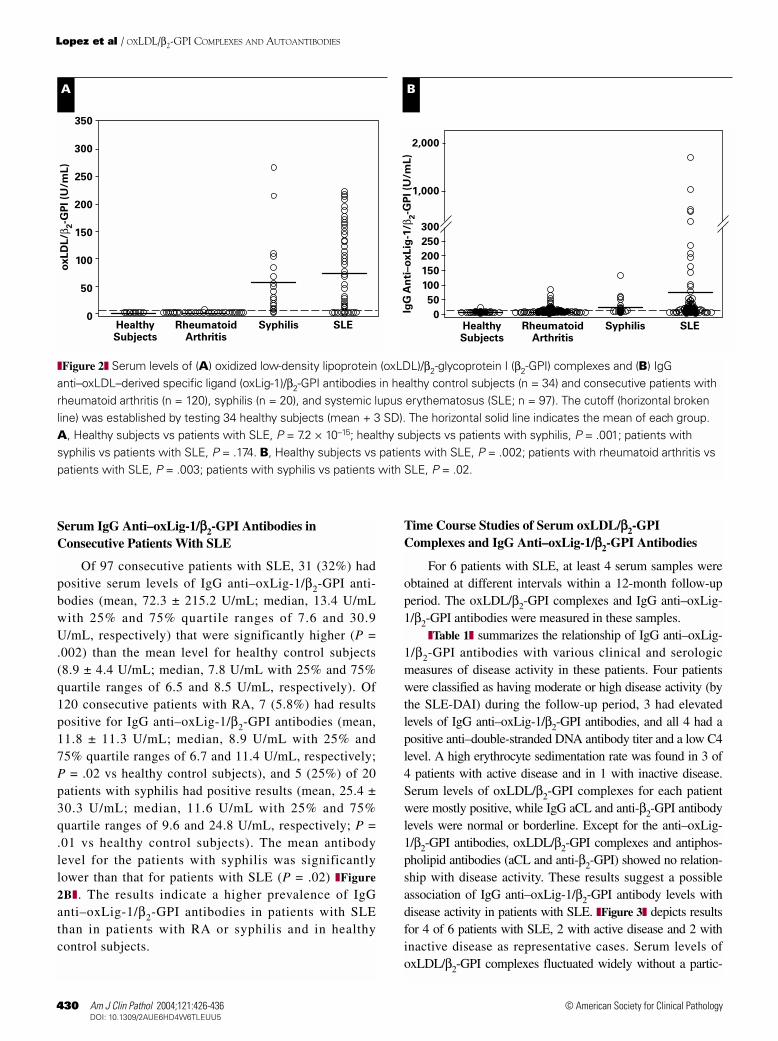
Of 97 consecutive patients with SLE, 67 (69%) hadpositive serum levels of oxLDL/β2-GPI complex with amean level of 73.2 ± 78.1 U/mL, which was significantlyhigher than the mean level for healthy control subjects (1.3 ±0.4 U/mL; P = 7.2 × 10⁻¹⁵). Only 3 (2.5%) of 120 consecu-tive patients with RA had positive results for oxLDL/β2-GPIwith a mean of 1.4 ± 0.7 U/mL, which was statisticallydifferent from that for healthy control subjects (P = .09). Incontrast with the RA group, 13 (65%) of 20 patients withsyphilis had positive results for oxLDL/β2-GPI with a meanof 56.2 ± 71.5 U/mL, which was significantly higher than thelevel for the healthy control subjects (P = .001). The meanlevel for the patients with syphilis was lower than forpatients with SLE, but this difference was not statisticallysignificant (P = .174) ❚Figure 2A❚. These results indicate thatoxidation of LDL and its complex formation with β2-GPI arecommon in SLE and syphilis. Although RA is a systemicautoimmune disease, oxLDL/β2-GPI complexes were notdetected in our RA patient population.
OD
(450 n
m)
2.0
1.5
1.0
0.5
0.02 4 6 8 10
LDL (µg/mL of ApoB equivalent)
❚Figure 1❚ Complex formation between oxidized low-densitylipoprotein (oxLDL) and β2-glycoprotein I (β2-GPI). Increasingconcentrations of oxLDL (open circles) or native LDL (closedcircles) were incubated together with purified human β2-GPI inImmulon 2HB microwells coated with anti-β2-GPI monoclonalantibody (WB-CAL-1). Bound LDL was detected withbiotinylated human apolipoprotein B-100 monoclonal antibody(1D2) and avidin–horseradish peroxidase. Results areexpressed as the mean optical density (OD) of triplicate testing± SD (error bars). For proprietary information, see the text.
Lopez et al / OXLDL/β2-GPI COMPLEXES AND AUTOANTIBODIES
430 Am J Clin Pathol 2004;121:426-436430 DOI: 10.1309/2AUE6HD4W6TLEUU5
© American Society for Clinical Pathology
Serum IgG Anti–oxLig-1/ββ2-GPI Antibodies inConsecutive Patients With SLE
Of 97 consecutive patients with SLE, 31 (32%) hadpositive serum levels of IgG anti–oxLig-1/β2-GPI anti-bodies (mean, 72.3 ± 215.2 U/mL; median, 13.4 U/mLwith 25% and 75% quartile ranges of 7.6 and 30.9U/mL, respectively) that were significantly higher (P =.002) than the mean level for healthy control subjects(8.9 ± 4.4 U/mL; median, 7.8 U/mL with 25% and 75%quartile ranges of 6.5 and 8.5 U/mL, respectively). Of120 consecutive patients with RA, 7 (5.8%) had resultspositive for IgG anti–oxLig-1/β2-GPI antibodies (mean,11.8 ± 11.3 U/mL; median, 8.9 U/mL with 25% and75% quartile ranges of 6.7 and 11.4 U/mL, respectively;P = .02 vs healthy control subjects), and 5 (25%) of 20patients with syphilis had positive results (mean, 25.4 ±30.3 U/mL; median, 11.6 U/mL with 25% and 75%quartile ranges of 9.6 and 24.8 U/mL, respectively; P =.01 vs healthy control subjects). The mean antibodylevel for the patients with syphilis was significantlylower than that for patients with SLE (P = .02) ❚Figure
2B❚. The results indicate a higher prevalence of IgGanti–oxLig-1/β2-GPI antibodies in patients with SLEthan in patients with RA or syphilis and in healthycontrol subjects.
Time Course Studies of Serum oxLDL/ββ2-GPIComplexes and IgG Anti–oxLig-1/ββ2-GPI Antibodies
For 6 patients with SLE, at least 4 serum samples wereobtained at different intervals within a 12-month follow-upperiod. The oxLDL/β2-GPI complexes and IgG anti–oxLig-1/β2-GPI antibodies were measured in these samples.
❚Table 1❚ summarizes the relationship of IgG anti–oxLig-1/β2-GPI antibodies with various clinical and serologicmeasures of disease activity in these patients. Four patientswere classified as having moderate or high disease activity (bythe SLE-DAI) during the follow-up period, 3 had elevatedlevels of IgG anti–oxLig-1/β2-GPI antibodies, and all 4 had apositive anti–double-stranded DNA antibody titer and a low C4level. A high erythrocyte sedimentation rate was found in 3 of4 patients with active disease and in 1 with inactive disease.Serum levels of oxLDL/β2-GPI complexes for each patientwere mostly positive, while IgG aCL and anti-β2-GPI antibodylevels were normal or borderline. Except for the anti–oxLig-1/β2-GPI antibodies, oxLDL/β2-GPI complexes and antiphos-pholipid antibodies (aCL and anti-β2-GPI) showed no relation-ship with disease activity. These results suggest a possibleassociation of IgG anti–oxLig-1/β2-GPI antibody levels withdisease activity in patients with SLE. ❚Figure 3❚ depicts resultsfor 4 of 6 patients with SLE, 2 with active disease and 2 withinactive disease as representative cases. Serum levels ofoxLDL/β2-GPI complexes fluctuated widely without a partic-
oxLD
L/β
2-G
PI (U
/mL)
350
300
250
200
150
100
50
0HealthySubjects
RheumatoidArthritis
Syphilis SLE
IgG
An
ti–o
xLig
-1/β
2-G
PI (U
/mL)
2,000
1,000
300
250
200
150
100
50
0HealthySubjects
RheumatoidArthritis
Syphilis SLE
A B
❚Figure 2❚ Serum levels of (A) oxidized low-density lipoprotein (oxLDL)/β2-glycoprotein I (β2-GPI) complexes and (B) IgGanti–oxLDL–derived specific ligand (oxLig-1)/β2-GPI antibodies in healthy control subjects (n = 34) and consecutive patients withrheumatoid arthritis (n = 120), syphilis (n = 20), and systemic lupus erythematosus (SLE; n = 97). The cutoff (horizontal brokenline) was established by testing 34 healthy subjects (mean + 3 SD). The horizontal solid line indicates the mean of each group.A, Healthy subjects vs patients with SLE, P = 7.2 × 10⁻¹⁵; healthy subjects vs patients with syphilis, P = .001; patients withsyphilis vs patients with SLE, P = .174. B, Healthy subjects vs patients with SLE, P = .002; patients with rheumatoid arthritis vspatients with SLE, P = .003; patients with syphilis vs patients with SLE, P = .02.

Immunopathology / ORIGINAL ARTICLE
Am J Clin Pathol 2004;121:426-436 431431 DOI: 10.1309/2AUE6HD4W6TLEUU5 431
© American Society for Clinical Pathology
❚Table 1❚Relationship Between IgG Anti–oxLig-1/ββ2-GPI Antibodies and Disease Activity in Patients With SLE*
Mean IgG Patient Anti–oxLig-1/ Mean IgG aCL Mean IgG Anti- No. ββ2-GPI (U/mL) SLE-DAI† Anti-dsDNA‡ C3 Level C4 Level ESR Level (GPL U) ββ2-GPI Level (U)
1 9.8 Inactive Negative NA NA High 6.5 0.92 234.5 Moderate Positive N Low N 23.2 2.63 38.3 High Positive N Low High 11.5 2.84 9.9 Moderate Positive Low Low High 4.4 0.85 24.5 Moderate Positive N Low High 7.4 0.76 9.1 Inactive Negative N N N 5.7 0.6
aCL, anticardiolipin; β2-GPI, β2-glycoprotein I; DAI, disease activity index; dsDNA, double-stranded DNA; ESR, erythrocyte sedimentation rate; GPL, IgG phospholipid units;N, normal; NA, not available; oxLig-1, oxidized low-density lipoprotein–derived specific ligand; SLE, systemic lupus erythematosus.
* Bold type indicates an abnormal value. Reference ranges are as follows: C3, 60-220 mg%; C4, 20-40 mg%; ESR (Westergren method), <20 mm/h; IgG aCL, <23 GPL U; IgGanti-β2-GPI, <20 U.
† Scoring for the SLE-DAI is as follows: 0, inactive; 1-5, minimal; 6-10, moderate; >10, high.‡ By immunofluorescence crithidia assay; a positive titer was defined as >1:10.
oxLD
L/β
2-G
PI C
om
ple
x (
U/m
L)
250
200
150
100
50
0
3 4 5 6 7 8 9 10 11Time (Month of the Year)
IgG
An
tib
od
y L
evel
700
600
500
400
300
200
100
0
Time (Month of the Year)
IgG Anti–oxLig-1/β2-GPI (U/mL)
IgG aCL (GPL U)
IgG aβ2-GPI (U)
3 4 5 6 7 8 9 10 11
oxLD
L/β
2-G
PI C
om
ple
x (
U/m
L)
250
200
150
100
50
0
0 1 2 3 4 5 6 7 8 9 10 Time (Month of the Year)
IgG
An
tib
od
y L
evel
100
80
60
40
20
0
0 1 2 3 4 5 6 7 8 9 10
Time (Month of the Year)
IgG Anti–oxLig-1/β2-GPI (U/mL)
IgG aCL (GPL U)
IgG aβ2-GPI (U)
A B
C D
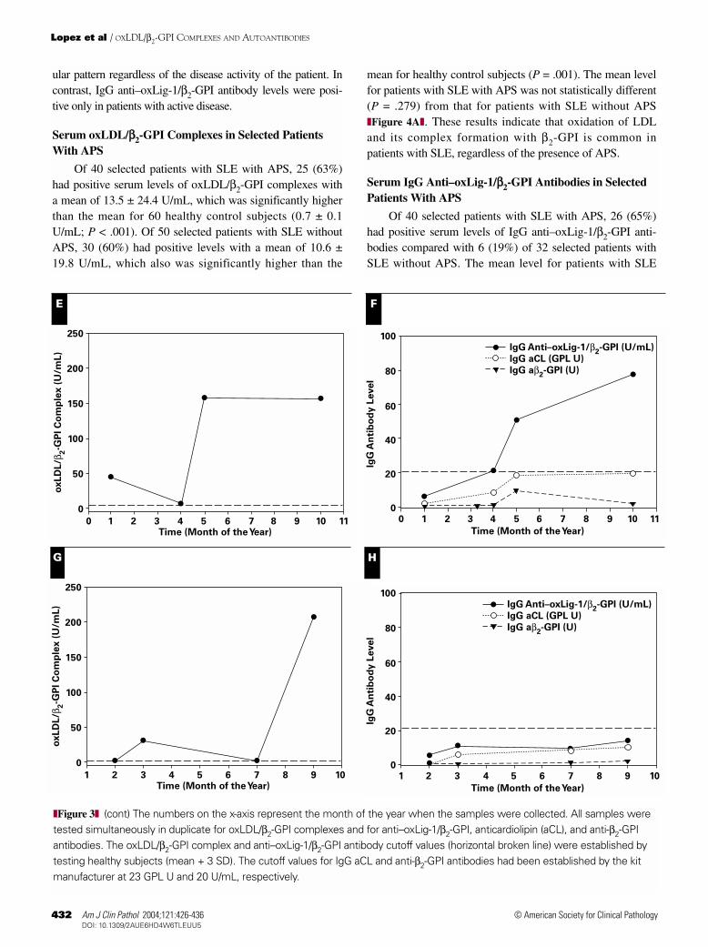
❚Figure 3❚ Serum levels of oxidized low-density lipoprotein (oxLDL)/β2-glycoprotein I (β2-GPI) complexes (A, C, E, G) withcorresponding IgG anti–oxLDL–derived specific ligand (anti–oxLig-1)/β2-GPI antibodies (B, D, F, H) in 4 of 6 patients with systemiclupus erythematosus (SLE) tested over time. According to the SLE disease activity index (see text), patients 1 (A and B) and 6 (Gand H) had inactive disease and patients 2 (C and D) and 3 (E and F), active disease. (cont next page)
Lopez et al / OXLDL/β2-GPI COMPLEXES AND AUTOANTIBODIES
432 Am J Clin Pathol 2004;121:426-436432 DOI: 10.1309/2AUE6HD4W6TLEUU5
© American Society for Clinical Pathology
ular pattern regardless of the disease activity of the patient. Incontrast, IgG anti–oxLig-1/β2-GPI antibody levels were posi-tive only in patients with active disease.
Serum oxLDL/ββ2-GPI Complexes in Selected PatientsWith APS
Of 40 selected patients with SLE with APS, 25 (63%)had positive serum levels of oxLDL/β2-GPI complexes witha mean of 13.5 ± 24.4 U/mL, which was significantly higherthan the mean for 60 healthy control subjects (0.7 ± 0.1U/mL; P < .001). Of 50 selected patients with SLE withoutAPS, 30 (60%) had positive levels with a mean of 10.6 ±19.8 U/mL, which also was significantly higher than the
mean for healthy control subjects (P = .001). The mean levelfor patients with SLE with APS was not statistically different(P = .279) from that for patients with SLE without APS❚Figure 4A❚. These results indicate that oxidation of LDLand its complex formation with β2-GPI is common inpatients with SLE, regardless of the presence of APS.
Serum IgG Anti–oxLig-1/ββ2-GPI Antibodies in SelectedPatients With APS
Of 40 selected patients with SLE with APS, 26 (65%)had positive serum levels of IgG anti–oxLig-1/β2-GPI anti-bodies compared with 6 (19%) of 32 selected patients withSLE without APS. The mean level for patients with SLE
❚Figure 3❚ (cont) The numbers on the x-axis represent the month of the year when the samples were collected. All samples weretested simultaneously in duplicate for oxLDL/β2-GPI complexes and for anti–oxLig-1/β2-GPI, anticardiolipin (aCL), and anti-β2-GPIantibodies. The oxLDL/β2-GPI complex and anti–oxLig-1/β2-GPI antibody cutoff values (horizontal broken line) were established bytesting healthy subjects (mean + 3 SD). The cutoff values for IgG aCL and anti-β2-GPI antibodies had been established by the kitmanufacturer at 23 GPL U and 20 U/mL, respectively.
oxLD
L/β
2-G
PI C
om
ple
x (
U/m
L)
250
200
150
100
50
0
0 1 2 3 4 5 6 7 8 9 10 11Time (Month of the Year)
IgG
An
tib
od
y L
evel
100
80
60
40
20
00 1 2 3 4 5 6 7 8 9 10 11
Time (Month of the Year)
IgG Anti–oxLig-1/β2-GPI (U/mL)
IgG aCL (GPL U)
IgG aβ2-GPI (U)
oxLD
L/β
2-G
PI C
om
ple
x (
U/m
L)
250
200
150
100
50
0
1 2 3 4 5 6 7 8 9 10 Time (Month of the Year)
IgG
An
tib
od
y L
evel
100
80
60
40
20
01 2 3 4 5 6 7 8 9 10
Time (Month of the Year)
IgG Anti–oxLig-1/β2-GPI (U/mL)
IgG aCL (GPL U)
IgG aβ2-GPI (U)
E F
G H

Immunopathology / ORIGINAL ARTICLE
Am J Clin Pathol 2004;121:426-436 433433 DOI: 10.1309/2AUE6HD4W6TLEUU5 433
© American Society for Clinical Pathology
with APS (24.4 ± 28.4 U/mL) was significantly higher thanthe mean for SLE control patients without APS (9.1 ± 5.1U/mL; P = .0008) and the mean for 43 healthy controlsubjects (5.7 ± 1.4 U/mL; P = 8.6 × 10⁻⁵) ❚Figure 4B❚. Theresults indicate a higher prevalence and serum levels of IgGanti–oxLig-1/β2-GPI antibodies in patients with SLE withAPS patients compared with patients with SLE without ahistory of APS, which suggests a possible pathogenic rolefor these antibodies in APS.
Relationship of IgG Anti–oxLig-1/ββ2-GPI With aCL andWith Anti-ββ2-GPI Antibodies
Owing to the prominent presence of β2-GPI in the anti-genic mixture used to detect IgG anti–oxLig-1/β2-GPI anti-bodies, the relationship of these antibodies with β2-GPI-dependent aCL and anti-β2-GPI antibodies was evaluated in40 selected patients with SLE with secondary APS. ❚Figure
5❚ shows the relationship of IgG anti–oxLig-1/β2-GPI anti-bodies with IgG aCL and with anti-β2-GPI antibodies. Thedistribution of IgG anti–oxLig-1/β2-GPI vs anti-β2-GPI anti-bodies followed a pattern different from that of aCL anti-bodies. This pattern suggests the presence of antibodies withat least 2 different reactivities: to β2-GPI and to oxLig-1/β2-GPI. Although preliminary, these results might indicate thatIgG anti–oxLig-1/β2-GPI antibodies represent a distinctsubset of antiphospholipid antibodies.
Comparative Clinical Performance
The clinical performance (relative sensitivity, specificity,and PPV) of IgG anti–oxLig-1/β2-GPI antibodies for clinicalmanifestations of APS in 90 selected patients with SLE wasevaluated by 2 × 2 contingency table analysis. IgG anti–oxLig-1/β2-GPI antibodies were 45% sensitive and 93.7% specific,with a PPV of 90% for APS (P < .001). ❚Table 2❚ shows thecomparison of the results of IgG anti–oxLig-1/β2-GPI anti-bodies with those of IgG aCL and anti-β2-GPI antibodies. IgGaCL antibodies were 62.5% sensitive, compared with 35% foranti-β2-GPI and 45% for anti–oxLig-1/β2-GPI antibodies. Thisis due to more false-positive results observed in the SLEcontrol group as evidenced by the lower specificity and PPVfor IgG aCL antibodies. Thus, IgG anti–oxLig-1/β2-GPI anti-bodies are better predictors of APS.
Discussion
Oxidation of LDL occurs in vivo and has an importantpathogenic role in atherogenesis.12,13 More than 60% of thepatients with SLE in the present study had elevated serumlevels of oxLDL/β2-GPI complexes as compared with 2.5%in RA and 0% in healthy control subjects (Figure 2A).However, the prevalence and the mean level of oxLDL/β2-GPI complexes in patients with SLE without a history of APSwere similar to those for patients with APS (Figure 4A). Thechronic inflammation of the vasculature frequently seen in
oxLD
L/β
2-G
PI (U
/mL)
140
120
100
80
60
40
20
0HealthySubjects
SLE Secondary APS
IgG
An
ti–o
xLig
-1/β
2-G
PI (U
/mL)
160
140
120
100
80
60
40
20
0HealthySubjects
SLE Secondary APS
A B
❚Figure 4❚ Serum levels of (A) oxidized low-density lipoprotein (oxLDL)/β2-glycoprotein I (β2-GPI) complexes and (B) IgGanti–oxLDL–derived specific ligand (anti–oxLig-1)/β2-GPI antibodies in healthy control subjects (A, n = 60; B, n = 43), selectedpatients with systemic lupus erythematosus (SLE) (without antiphospholipid syndrome [APS]; A, n = 50; B, n = 32) and withsecondary APS (A and B, n = 40). The cutoff (horizontal broken line) was established by testing 60 healthy subjects (mean + 3SD). The horizontal solid lines indicate the mean for each group. A, Healthy subjects vs patients with SLE and secondary APS, P= .001; patients with SLE and no APS vs patients with SLE and secondary APS, P = .279. B, Healthy subjects vs patients withSLE and secondary APS, P = 8.6 × 10⁻⁵; patients with SLE and no APS vs patients with SLE and secondary APS, P = .0008.

Lopez et al / OXLDL/β2-GPI COMPLEXES AND AUTOANTIBODIES
434 Am J Clin Pathol 2004;121:426-436434 DOI: 10.1309/2AUE6HD4W6TLEUU5
© American Society for Clinical Pathology
patients with certain autoimmune diseases might account forthe increased oxidative modification of LDL and its interac-tion with β2-GPI. These results are in agreement with recentreports that oxidation of LDL and interaction with β2-GPI arecommon in patients with these diseases.26,29 CirculatingoxLDL/β2-GPI complexes have been implicated as athero-genic autoantigens, and their presence might represent a riskfactor or an indirect but significant contributor to thrombosisand atherosclerosis.26 The presence of autoantibodies againstoxLig-1/β2-GPI in patients with SLE with secondary APS(Figure 4B), together with the common occurrence ofoxLDL/β2-GPI complexes, might help explain the accelerated(or premature) development of vascular complications,including atherosclerosis, seen in some of these patients.
In contrast with SLE, oxLDL/β2-GPI complexes were notfound in RA (Figure 2A). To our knowledge, autoantibodiesagainst oxLDL have not been reported in this disease.However, a high incidence of atherosclerosis has been
described in patients with RA.18 The lack of circulatingoxLDL/β2-GPI complexes might help to explain the low preva-lence of autoantibodies to the complex (Figure 2B) and the lowincidence of thromboembolic complications seen in RA. It ispossible that other mechanism(s), different from oxidation ofLDL, might contribute to the development of atherosclerosis inRA. A recent review proposed several potential proatherogenicmechanisms, including dyslipidemias such as Lp(a), medica-tions (steroids, methotrexate), proinflammatory cytokines(interleukins 1 and 6, tumor necrosis factor α), acute phasereactants (C-reactive protein, serum amyloid A), and newer riskfactors such as hyperhomocysteinemia.18
The oxLig-1/β2-GPI complexes (Figure 2A) weredetected in 13 (65%) of a small group (n = 20) of patients withsyphilis. The presence of these complexes suggests that theinfectious process also might result in vascular inflammationleading to oxidation of LDL and β2-GPI complex formation,perhaps similar to SLE. About 25% of the patients with
IgG
An
ti–o
xLig
-1/β
2-G
PI (U
/mL)
160
140
120
100
80
60
40
20
0
0 20 40 60 80 100
IgG aCL (GPL U)
IgG
An
ti–o
xLig
-1/β
2-G
PI (U
/mL)
160
140
120
100
80
60
40
20
0
0 40 80 120 160 200
IgG Anti–β2-GPI (U)
A B
❚Figure 5❚ Correlation between IgG anti–oxLDL–derived specific ligand (anti–oxLig-1)/β2-glycoprotein I (β2-GPI) antibodies andantiphospholipid antibodies determined by enzyme-linked immunosorbent assay in 40 selected patients with systemic lupuserythematosus with secondary antiphospholipid syndrome. A, IgG anti–oxLig-1/β2-GPI vs IgG anticardiolipin (aCL) (r = 0.737; P= 5.96 × 10⁻⁸; n = 40). B, IgG anti–oxLig-1/β2-GPI vs IgG anti-β2-GPI (r = 0.325; P = .04; n = 40). The straight line represents thebest-fit linear regression.
❚Table 2❚Association Between IgG Anti–oxLig-1/ββ2-GPI Antibodies and Clinical Manifestations of APS in Patients With SLE
Antibody Sensitivity (%) Specificity (%) PPV (%) P* OR (95% CI)
IgG anti–oxLig-1/β2-GPI 45.0 93.7 90.0 <.001 12.3 (2.6-58.6)IgG aCL 62.5 80.0 71.4 <.001 6.7 (2.6-17.1)IgG anti-β2-GPI 35.0 98.0 93.3 <.001 26.4 (3.3-212.1)
aCL, anticardiolipin; APS, antiphospholipid syndrome; β2-GPI, β2-glycoprotein I; CI, confidence interval; OR, odds ratio; oxLig-1, oxidized low-density lipoprotein–derivedspecific ligand; PPV, positive predictive value; SLE, systemic lupus erythematosus.
* Fisher exact test.
Immunopathology / ORIGINAL ARTICLE
Am J Clin Pathol 2004;121:426-436 435435 DOI: 10.1309/2AUE6HD4W6TLEUU5 435
© American Society for Clinical Pathology
syphilis showed low positive levels of IgG anti–oxLig-1/β2-GPI antibodies (Figure 2B). These antibody titers had astronger correlation with IgG aCL (r = 0.889) compared withanti-β2-GPI (r = 0.229) antibodies (data not shown). Theseresults not only show that anti–oxLig-1/β2-GPI antibodiesmight be different from anti-β2-GPI antibodies, but alsosuggest that oxLDL/β2-GPI complexes can induce the produc-tion of low levels of “infectious” anti–oxLig-1/β2-GPI anti-bodies. Unlike patients with APS, no “autoimmune” anti-β2-GPI antibodies were detected in this group of patients withsyphilis. The exact mechanism(s) and possible pathogeniceffect of these antibodies in thrombosis and atherosclerosisremain to be elucidated.
Kobayashi et al26 recently demonstrated that magnesiumchloride dissociates intermediate forms of oxLDL/β2-GPIcomplexes bound by electrostatic interactions. Our ELISAsystem included magnesium chloride in the sample diluent topermit the detection of “stable” nondissociable oxLDL/β2-GPIcomplexes bound by covalent interactions. It can be hypothe-sized that these stable complexes might be clinically relevant.The wide fluctuation in serum levels of oxLDL/β2-GPIcomplexes in some patients with SLE (Figure 3) suggests thatoxidation of LDL and/or the formation of complexes with β2-GPI in circulation is an active process. It is possible that adysfunction of some regulatory mechanism(s) might accountfor these findings. For example, the HDL-associated enzymeparaoxonase (PON) has antioxidant activity that protects LDLfrom oxidation.39 Decreased PON activity has been reported inpatients with aCL antibodies.40 Furthermore, IgG anti-β2-GPIantibodies were associated with reduced PON activity inpatients with SLE and patients with primary APS.41 Vascularinjury (biochemical or immunologic) as seen in patients withautoimmune disease might affect PON activity or any otherantioxidant mechanism, triggering the oxidative aforemen-tioned changes. In addition, it is possible that patients withAPS have circulating immune complexes (antibody/oxLig-1/β2-GPI), as suggested by the coexistence of oxLDL/β2-GPIand autoantibodies. These immune complexes might beremoved from circulation by macrophages via Fcγ receptors, amechanism also relevant in atherogenesis.26
High serum levels of IgG anti–oxLig-1/β2-GPI anti-bodies were demonstrated in 31 (32%) of 97 consecutiveSLE patients (Figure 2B). The APS status of the consecutivepatients with SLE with IgG anti–oxLig-1/β2-GPI antibodieswas not known; however, the 32% prevalence correspondswith the expected general prevalence or risk of APS inSLE.3-5 High serum levels of these antibodies were found in4 patients with SLE with persistent clinical and serologicmeasures of disease activity (Figure 3 and Table 1).Although this is a small group of patients, these resultssuggest that IgG anti–oxLig-1/β2-GPI antibodies might serveas indicators of disease activity in patients with SLE and
might contribute to the pathogenesis of APS. Taken together,these findings provide additional support to the hypothesisthat oxidative stress has an important role in antiphospho-lipid antibody production and the development of APS.
We found no correlation between serum levels ofoxLDL/β2-GPI complexes and IgG anti–oxLig-1/β2-GPI anti-bodies in patients with SLE with APS (data not shown). It ispossible that high complex levels might result in increasedantibody binding and immune complex formation, depleting“free” autoantibodies. However, the possible relationshipbetween oxLDL/β2-GPI complexes and the autoantibodies isillustrated by the presence of IgG anti–oxLig-1/β2-GPI anti-bodies only in patients with SLE with a history of APS andthe report of a stronger association of anti–oxLig-1/β2-GPIantibodies with thrombosis in patients with high levels vs lowlevels of oxLDL/β2-GPI complexes.26
Serum levels of IgG anti–oxLig-1/β2-GPI antibodiesshowed a stronger correlation with IgG aCL (r = 0.737) thanwith anti-β2-GPI (r = 0.325) antibodies in patients with SLEwith APS (Figure 5). On the other hand, the PPVs for APSof IgG anti–oxLig-1/β2-GPI (90%) and anti-β2-GPI (93.3%)antibodies were better than IgG aCL (71.4%) antibodies inour study population (Table 2). These results suggest thatIgG anti–oxLig-1/β2-GPI antibodies might represent a subsetof antiphospholipid antibodies that coexist with otherautoantibodies. Additional studies using purified antibodiesand/or inhibition experiments are needed to further charac-terize these antibodies. The results of this study suggest thatmeasuring oxLDL/β2-GPI complexes and IgG anti–oxLig-1/β2-GPI antibodies by ELISA might be useful in the sero-logic evaluation of APS and that they are possible contribu-tors to the development of atherosclerosis in some patientswith autoimmune diseases such as SLE.
From 1Corgenix, Westminster, CO; 2 Department of Immunologyand Rheumatology, Ministry of Health Western General Hospital,University of Guadalajara, Guadalajara, Mexico; and3Department of Cell Chemistry, Okayama University GraduateSchool of Medicine and Dentistry, Okayama, Japan.
Address reprint requests to Dr Luis R. Lopez: Corgenix,12061 Tejon St, Westminster, CO 80234.
References1. Harris EN, Chan JKH, Asherson RA, et al. Thrombosis,
recurrent fetal loss and thrombocytopenia: predictive value ofthe anticardiolipin antibody test. Arch Intern Med.1986;146:2153-2156.
2. Ginsburg KS, Liang MH, Newcomer L, et al. Anticardiolipinantibodies and the risk for ischemic stroke and venousthrombosis. Ann Intern Med. 1992;117:997-1002.
3. Bick RL, Baker WF. Antiphospholipid syndrome andthrombosis. Semin Thromb Hemost. 1999;25:333-350.
4. Hughes GRV, Harris EN, Gharavi AE. The anticardiolipinsyndrome. J Rheumatol. 1986;13:486-489.

Lopez et al / OXLDL/β2-GPI COMPLEXES AND AUTOANTIBODIES
436 Am J Clin Pathol 2004;121:426-436436 DOI: 10.1309/2AUE6HD4W6TLEUU5
5. Gharavi AE, Harris EN, Asherson RA, et al. Anticardiolipinantibodies: isotype distribution and phospholipid specificity.Ann Rheum Dis. 1987;46:1-6.
6. Roubey RAS. Autoantibodies to phospholipid-bindingplasma proteins: a new view of lupus anticoagulants and other“antiphospholipid” antibodies. Blood. 1994;84:2858-2867.
7. Matsuura E, Igarashi Y, Fujimoto M, et al. Anticardiolipincofactor(s) and differential diagnosis of autoimmune diseases.Lancet. 1990;336:177-178.
8. McNeil HP, Simpson RJ, Chesterman CN, et al.Antiphospholipid antibodies are directed against a complexantigen that includes a lipid-binding inhibitor of coagulation:beta2-glycoprotein I (apolipoprotein H). Proc Natl Acad Sci U S A. 1990;87:4120-4124.
9. Bouma B, de Groot PG, van den Elsen JMH, et al. Adhesionmechanism of human β2-glycoprotein I to phospholipidsbased on its crystal structure. EMBO J. 1999;18:5166-5174.
10. Hoshino M, Hagihara Y, Nishii I, et al. Identification of thephospholipid-binding site of human β2-glycoprotein I domain V byheteronuclear magnetic resonance. J Mol Biol. 2000;304:927-939.
11. Sheng Y, Kandiah DA, Krilis SA. β2-glycoprotein I: targetantigen for “antiphospholipid” antibodies: immunological andmolecular aspects. Lupus. 1998;7(suppl 2):S5-S9.
12. Steinberg D. Low density lipoprotein oxidation and itspathobiological significance. J Biol Chem. 1997;272:20963-20966.
13. Berliner JA, Heinecke JW. The role of oxidized lipoproteinsin atherogenesis. Free Radic Biol Med. 1996;20:707-727.
14. McMurray HF, Parthasarathy S, Steinberg D. Oxidativelymodified low density lipoprotein is a chemoattractant forhuman T lymphocytes. J Clin Invest. 1993;92:1004-1008.
15. Yla-Herttuala S, Palinski W, Rosenfeld ME, et al. Evidencefor the presence of oxidatively modified low densitylipoprotein in atherosclerotic lesions of rabbit and man. J ClinInvest. 1989;85:1086-1095.
16. Salonen JT, Yla-Herttuala S, Yamamoto R, et al.Autoantibodies against oxidized LDL and progression ofcarotid atherosclerosis. Lancet. 1992;339:883-887.
17. Vaarala O, Alfthan G, Jauhiainen M, et al. Crossreaction betweenantibodies to oxidized low density lipoprotein and to cardiolipinin systemic lupus erythematosus. Lancet. 1993;341:923-925.
18. Van Doornum S, McColl G, Wicks IP. Acceleratedatherosclerosis: an extraarticular feature of rheumatoidarthritis? Arthritis Rheum. 2002;46:862-873.
19. Ward MM. Premature morbidity from cardiovascular andcerebrovascular diseases in women with systemic lupuserythematosus. Arthritis Rheum. 1999;42:338-346.
20. Aranow C, Ginzler EM. Epidemiology of cardiovascular diseasein systemic lupus erythematosus. Lupus. 2000;9:166-169.
21. Esdaile JM, Abrahamowicz M, Grodzicky T, et al. TraditionalFramingham risk factors fail to fully account for acceleratedatherosclerosis in systemic lupus erythematosus. ArthritisRheum. 2001;44:2331-2337.
22. Lockshin MD, Salmon JE, Roman MJ. Atherosclerosis andlupus: a work in progress [editorial]. Arthritis Rheum.2001;44:2215-2217.
23. Vaarala O. Antiphospholipid antibodies and atherosclerosis.Lupus. 1996;5:442-447.
24. George J, Harats D, Gilburd B, et al. Immunolocalization ofβ2-glycoprotein I (apolipoprotein H) to humanatherosclerotic plaques: potential implications for lesionprogression. Circulation. 1999;99:2227-2230.
25. Hasunuma Y, Matsuura E, Makita Z, et al. Involvement of β2-glycoprotein I and anticardiolipin antibodies in oxidativelymodified low density lipoprotein uptake by macrophages. ClinExp Immunol. 1997;107:569-573.
26. Kobayashi K, Kishi M, Atsumi T, et al. Circulating oxidizedlow density lipoprotein forms complexes with β2-glycoproteinI: implication as an atherogenic autoantigen. J Lipid Res.2003;44:716-726.
27. Kobayashi K, Matsuura E, Liu Q, et al. A specific ligand forβ2-glycoprotein I mediates autoantibody-dependent uptake ofoxidized low density lipoprotein by macrophages. J Lipid Res.2001;42:697-709.
28. Liu Q, Kobayashi K, Furukawa J, et al. ω-Carboxyl variants of7-ketocholesteryl esters are ligands for β2-glycoprotein I andmediate antibody-dependent uptake of oxidized LDL bymacrophages. J Lipid Res. 2002;43:1486-1495.
29. Zhao D, Ogawa H, Wang X, et al. Oxidized low-densitylipoprotein and autoimmune antibodies in patients withantiphospholipid syndrome with a history of thrombosis. Am JClin Pathol. 2001;116:760-767.
30. Tan EM, Cohen AS, Fries JF, et al. The 1982 revised criteriafor the classification of systemic lupus erythematosus. ArthritisRheum. 1982;25:1271-1277.
31. Bombardier C, Gladman DD, Urowitz MB, et al. Derivationof the SLEDAI: a disease activity index for lupus patients.Arthritis Rheum. 1992;35:630-640.
32. Wilson WA, Gharavi AE, Koike T, et al. Internationalconsensus statement on preliminary classification criteria fordefinite antiphospholipid syndrome: report of an internationalworkshop. Arthritis Rheum. 1999;42:1309-1311.
33. Hashimoto Y, Kawamura M, Ichikawa K, et al. Anticardiolipinantibodies in NZW x BXSB F1 mice: a model ofantiphospholipid syndrome. J Immunol. 1992;149:1063-1068.
34. Ichikawa K, Khamashta MA, Koike T, et al. β2-glycoprotein Ireactivity of monoclonal anticardiolipin antibodies frompatients with the antiphospholipid syndrome. Arthritis Rheum.1994;37:1453-1461.
35. Finlayson JS, Mushinski JF. Separation of subfractions of humanβ2-glycoprotein I. Biochim Biophys Acta. 1967;147:413-420.
36. Havel RJ, Eder HA, Bragdon JH. The distribution andchemical composition of ultracentrifugally separatedlipoproteins in human serum. J Clin Invest. 1955;43:1345-1353.
37. Ohkawa H, Ohishi N, Yagi K. Assay for lipid peroxides inanimal tissues by thiobarbituric acid reaction. Anal Biochem.1979;95:351-358.
38. Lopez LR, Santos ME, Espinoza LR, et al. Clinicalsignificance of immunoglobulin A versus immunoglobulins Gand M anti-cardiolipin antibodies in patients with systemiclupus erythematosus: correlation with thrombosis,thrombocytopenia, and recurrent abortion. Am J Clin Pathol.1992;98:449-454.
39. Durrington PN, Mackness B, Mackness MI. Paraoxonase andatherosclerosis. Arterioscler Thromb Vasc Biol. 2001;21:473-480.
40. Lambert M, Boullier A, Hachulla E, et al. Paraoxonaseactivity is dramatically decreased in patients positive foranticardiolipin antibodies. Lupus. 2000;9:299-300.
41. Delgado-Alves J, Ames PRJ, Donohue S, et al. Antibodies tohigh-density lipoprotein and β2-glycoprotein I are inverselycorrelated with paraoxonase activity in systemic lupuserythematosus and primary antiphospholipid syndrome.Arthritis Rheum. 2002;46:2686-2694.
Copyright © 2022 FDOKUMEN


![Impact of Persistent Antiphospholipid Antibodies on Symptomatic Thromboembolism In Children: A Systematic Review & Meta-Analysis [Observational Studies]](https://static.fdokumen.com/doc/165x107/6336bc8902a8c1a4ec02539e/impact-of-persistent-antiphospholipid-antibodies-on-symptomatic-thromboembolism.jpg)