Activatory and Inhibitory Fcγ Receptors Augment Rituximab-mediated Internalization of CD20...
30
FcγR augment internalisation of RTX-ligated CD20 1 Activatory and inhibitory Fcγ receptors augment rituximab-mediated internalisation of CD20 independent of signalling via the cytoplasmic domain Andrew T. Vaughan 1 , Claude H. T. Chan 1 , Christian Klein 2 , Martin J. Glennie 1 , Stephen A. Beers 1* and Mark S. Cragg 1* 1 From: Antibody and Vaccine Group, Cancer Sciences Unit, University of Southampton, Faculty of Medicine, General Hospital, Southampton, United Kingdom. SO16 6YD 2 Roche Pharmaceutical Research and Early Development, Roche Innovation Center Zurich, Wagistrasse 18, CH-8952 Schlieren, Switzerland *SAB and MSC are the senior authors and contributed equally to the project. Running title: FcγR augment internalisation of RTX-ligated CD20 To whom correspondence should be addressed: Mark S. Cragg, Antibody and Vaccine Group, Cancer Sciences Unit, University of Southampton Faculty of Medicine, General Hospital, Southampton, United Kingdom. SO16 6YD, Fax: +44 (0) 2380 704061; Email: [email protected] Keywords: Antibody; Fc-gamma receptor; Receptor endocytosis; CD20; B cell; Lymphoma; Lipid raft; Signaling____________________________________________________________________________ Background: Fcγ receptor (FcγR) IIb augments internalisation of CD20 from the surface of B cells in response to rituximab treatment. Results: Activatory and inhibitory FcγR augment internalisation, independent of the FcγR cytoplasmic domain. Conclusion: Active signalling is not required for FcγR-augmented internalisation of CD20 in response to rituximab treatment. Significance: FcγR may play a structural role in augmenting CD20 internalisation. ABSTRACT Type I anti-CD20 mAb such as rituximab and ofatumumab engage with the inhibitory FcγR, FcγRIIb on the surface of B cells, resulting in immunoreceptor tyrosine-based inhibitory motif (ITIM) phosphorylation. Internalisation of the CD20:mAb:FcγRIIb complex follows, the rate of which correlates with FcγRIIb expression. In contrast, although type II anti- CD20 mAb such as tositumomab and obinutuzumab also interact with and activate FcγRIIb, this interaction fails to augment the rate of CD20:mAb internalisation, raising the question of whether ITIM phosphorylation plays any role in this process. We have assessed the molecular requirements for the internalisation process and demonstrate that in contrast to internalisation of IgG immune complexes, FcγRIIb-augmented internalisation of rituximab-ligated CD20 occurs independently of the FcγRIIb ITIM, indicating that signalling downstream of FcγRIIb is not required. In transfected cells, activatory FcγRI, FcγRIIa and FcγRIIIa augmented internalisation of rituximab-ligated CD20 in a similar manner. However, FcγRIIa mediated a slower rate of internalisation than cells expressing equivalent levels of the highly homologous FcγRIIb. The difference was maintained in cells expressing FcγRIIa and FcγRIIb lacking cytoplasmic domains and in which the transmembrane domains had been exchanged. This difference may be due to increased degradation of FcγRIIa, which traffics to lysosomes independently of rituximab. We conclude that the cytoplasmic domain of FcγR is not required for promoting internalisation of rituximab-ligated CD20. Instead, we propose that FcγR provide a structural role in augmenting endocytosis that differs from that employed during the endocytosis of immune complexes.___________ Anti-CD20 mAb are classified as type I (rituximab (RTX)-like) or type II (tositumomab- http://www.jbc.org/cgi/doi/10.1074/jbc.M114.593806 The latest version is at JBC Papers in Press. Published on January 7, 2015 as Manuscript M114.593806 Copyright 2015 by The American Society for Biochemistry and Molecular Biology, Inc. by guest on January 18, 2015 http://www.jbc.org/ Downloaded from
Transcript of Activatory and Inhibitory Fcγ Receptors Augment Rituximab-mediated Internalization of CD20...

FcγR augment internalisation of RTX-ligated CD20
1
Activatory and inhibitory Fcγ receptors augment rituximab-mediated internalisation of CD20 independent
of signalling via the cytoplasmic domain
Andrew T. Vaughan1, Claude H. T. Chan1, Christian Klein2, Martin J. Glennie1, Stephen A. Beers1*
and Mark S. Cragg1*
1From: Antibody and Vaccine Group, Cancer Sciences Unit, University of Southampton, Faculty of
Medicine, General Hospital, Southampton, United Kingdom. SO16 6YD
2Roche Pharmaceutical Research and Early Development, Roche Innovation Center Zurich, Wagistrasse
18, CH-8952 Schlieren, Switzerland
*SAB and MSC are the senior authors and contributed equally to the project.
Running title: FcγR augment internalisation of RTX-ligated CD20
To whom correspondence should be addressed: Mark S. Cragg, Antibody and Vaccine Group, Cancer
Sciences Unit, University of Southampton Faculty of Medicine, General Hospital, Southampton, United
Kingdom. SO16 6YD, Fax: +44 (0) 2380 704061; Email: [email protected]
Keywords: Antibody; Fc-gamma receptor; Receptor endocytosis; CD20; B cell; Lymphoma; Lipid raft;
Signaling____________________________________________________________________________
Background: Fcγ receptor (FcγR) IIb augments
internalisation of CD20 from the surface of B cells
in response to rituximab treatment.
Results: Activatory and inhibitory FcγR augment
internalisation, independent of the FcγR
cytoplasmic domain.
Conclusion: Active signalling is not required for
FcγR-augmented internalisation of CD20 in
response to rituximab treatment.
Significance: FcγR may play a structural role in
augmenting CD20 internalisation.
ABSTRACT
Type I anti-CD20 mAb such as rituximab
and ofatumumab engage with the inhibitory
FcγR, FcγRIIb on the surface of B cells, resulting
in immunoreceptor tyrosine-based inhibitory
motif (ITIM) phosphorylation. Internalisation
of the CD20:mAb:FcγRIIb complex follows, the
rate of which correlates with FcγRIIb
expression. In contrast, although type II anti-
CD20 mAb such as tositumomab and
obinutuzumab also interact with and activate
FcγRIIb, this interaction fails to augment the
rate of CD20:mAb internalisation, raising the
question of whether ITIM phosphorylation
plays any role in this process. We have assessed
the molecular requirements for the
internalisation process and demonstrate that in
contrast to internalisation of IgG immune
complexes, FcγRIIb-augmented internalisation
of rituximab-ligated CD20 occurs independently
of the FcγRIIb ITIM, indicating that signalling
downstream of FcγRIIb is not required. In
transfected cells, activatory FcγRI, FcγRIIa and
FcγRIIIa augmented internalisation of
rituximab-ligated CD20 in a similar manner.
However, FcγRIIa mediated a slower rate of
internalisation than cells expressing equivalent
levels of the highly homologous FcγRIIb. The
difference was maintained in cells expressing
FcγRIIa and FcγRIIb lacking cytoplasmic
domains and in which the transmembrane
domains had been exchanged. This difference
may be due to increased degradation of FcγRIIa,
which traffics to lysosomes independently of
rituximab. We conclude that the cytoplasmic
domain of FcγR is not required for promoting
internalisation of rituximab-ligated CD20.
Instead, we propose that FcγR provide a
structural role in augmenting endocytosis that
differs from that employed during the
endocytosis of immune complexes.___________
Anti-CD20 mAb are classified as type I
(rituximab (RTX)-like) or type II (tositumomab-
http://www.jbc.org/cgi/doi/10.1074/jbc.M114.593806The latest version is at JBC Papers in Press. Published on January 7, 2015 as Manuscript M114.593806
Copyright 2015 by The American Society for Biochemistry and Molecular Biology, Inc.
by guest on January 18, 2015http://w
ww
.jbc.org/D
ownloaded from

FcγR augment internalisation of RTX-ligated CD20
2
like) based on functional differences that they
mediate in various in vitro assays (1). Type I mAb
cause redistribution of CD20 into lipid rafts,
favouring potent complement dependent
cytotoxicity; whereas Type II mAb are ineffective
in these assays but more potently elicit homotypic
adhesion and a non-apoptotic lysosomal form of
cell death (2-6). We recently observed that in
addition, type I anti-CD20 mAb mediate rapid
internalisation of CD20 from the cell surface,
thereby reducing antibody efficacy, whereas type II
mAb do not (7,8). We subsequently showed that
internalisation of type I anti-CD20 mAb was
greatly augmented by their engagement with
FcγRIIb on the cell surface via antibody bipolar
bridging and that the rate of internalisation
positively correlated with cell surface expression of
FcγRIIb (8). Higher expression of target cell
FcγRIIb was associated with reduced survival or
response in cancer patients treated with RTX
therapy in two retrospective trials (8,9).
Previously we have proposed that in contrast
to the treatment of cancer, CD20 internalisation
may be advantageous in the treatment of
autoimmune disease (10), where rituximab therapy
has proven beneficial (11). Its mechanism of action
is still poorly understood, but it has been suggested
that type I anti-CD20 mAb promote a regulatory B
cell response that can suppress autoimmune
responses (12). FcγRIIb is down regulated on B
cells of patients with systemic lupus erythematosis
(SLE) (13), but is up regulated on a subset of
regulatory B cells (14). Therefore, FcγRIIb‐mediated internalisation of CD20 in response to
type I mAb‐ligation may result in preferential
clearance of pathogenic FcγRIIb‐low cells in SLE,
whilst sparing FcγRIIb‐high regulatory B cells.
Thus, it is of great interest to elucidate the
mechanism by which interaction between type I
anti-CD20 mAb and FcγRIIb promotes
internalisation of the CD20:mAb:FcγRIIb complex
in order to design strategies to inhibit the process
and improve therapy in the treatment of
malignancy, or augment it in situations such as SLE
where internalisation may prove beneficial.
Given our initial observations that type I anti-
CD20 mAb appeared to be unique in their ability to
interact with and activate FcγRIIb in cis (8), we
theorised that activation of the ITIM and signalling
via the FcγR initiated the endocytic process,
analogous to the interaction between FcγRIIb2 and
immune complexes (15,16). Endocytosis of
immune complex in the form of heat aggregated
human IgG (ahIgG) is dependent on the expression
of a complete ITIM within the cytoplasmic domain
of FcγRIIb (15,16) and is completely abrogated in
cells expressing mutated forms of the receptor in
which the ITIM has been truncated (15).
Furthermore, ahIgG remains on the surface of cells
expressing the b1 isoform of FcγRIIb due to an
extra 19 amino acids in the cytoplasmic domain that
excludes the receptor from clathrin-coated pits (16).
We have previously observed that both b1 and
b2 isoforms of FcγRIIb are equally effective at
augmenting internalisation of RTX-ligated CD20
(10), raising the possibility that the mechanism of
endocytosis is different from the internalisation of
immune complex. We have also found that the
majority of mAb directed to a range of B cell
receptors interact with and activate FcγRIIb via
antibody bipolar bridging, with the extent of
activation related to the level of mAb bound to the
cell surface (10). The type II anti-CD20 mAb
tositumomab also activated FcγRIIb, although to a
much lesser extent than RTX (10). The presence of
FcγRIIb failed to alter the rate of internalisation of
most mAb-ligated receptors, raising the question as
to whether activation of FcγRIIb and signalling via
the ITIM is indeed the mechanism by which type I
anti-CD20 mAb augment internalisation of CD20.
Here we have investigated this question, as
well as whether expression of other activatory FcγR
can promote internalisation of CD20 in response to
type I anti-CD20 mAb ligation; and the underlying
molecular mechanism.
EXPERIMENTAL PROCEDURES
Cell lines-The Burkitt’s lymphoma cell lines
Ramos and Raji were obtained from the European
Collection of Cell Cultures and maintained in
complete cell culture media (RPMI 1640, 4mM L-
glutamine, 1mM sodium pyruvate and 10% (v/v)
FCS (all from Life Technologies)).
Generation of wild type (WT) and mutant
human FcγR constructs-Human FcγRIIb1,
FcγRIIb2 and truncated FcγRIIbΔcyt expression
vectors were constructed previously (8,10). Human
FcγRI and human FcγRIIIa V158 were amplified
from complementary DNA obtained from primary
human leukocytes using specific primers and
by guest on January 18, 2015http://w
ww
.jbc.org/D
ownloaded from

FcγR augment internalisation of RTX-ligated CD20
3
cloned into the pIRES vector (Clontech) co-
expressing the FcR common γ-chain amplified
from the same cells. Human FcγRIIa was amplified
from human leukocytes using specific primers and
cloned into the pCIPURO expression vector. The
pCIPURO vector was constructed by sub cloning the
puromycin resistance gene from Ppuro (Clontech)
into pCI-neo (Promega) via PuvII/BamHI sites. To
generate a truncated mutant version of FcγRIIa
lacking the intracellular domain (FcγRIIaΔcyt), a
stop mutation at residue 244 was introduced using
the Quickchange Multi Site-Directed Mutagenesis
Kit (Stratagene) according to the manufacturer’s
instructions. FcγRIIbΔcyt and FcγRIIaΔcyt were
used to construct TGI230-232IAT and IAT224-
226TGI transmembrane mutants, respectively by
site-directed mutagenesis.
Cell transfection-FcγRIIb1, FcγRIIb2 and
FcγRIIbΔcyt transfected Ramos cells were
generated previously (8,10). To produce other
transfectants, cells were transfected by
nucleofection using Kit V on the Nuclefector I
device according to the manufacturer’s instructions
(Lonza). Ramos and Raji cells were transfected
using programmes O-06 and M-13, respectively.
Stable transfectants of Ramos cells expressing
FcγRIIb and FcγRIIa variants were selected with
1mg/ml G418 (Geneticin, Life Technologies) and
1µg/ml puromycin (Life Technologies),
respectively and screened for cell surface FcγRII
expression using AT10-PE (AbD, Serotec).
Transient transfectants of Raji cells were used 24
hours after electroporation with 4µg FcγRIIIa
V158/γ-chain or FcγRI/γ-chain and screened for
cell surface FcγR expression using 3G8-APC
(Biolegend) or 10.1-APC (Biolegend),
respectively.
Monoclonal antibody production and
labelling-Rituximab was gifted by Southampton
General Hospital oncology pharmacy. Non-
radiolabelled tositumomab was gifted by Professor
T. Illidge (University of Manchester, United
Kingdom). F(ab’)2 fragments of RTX were
produced as described previously (17). AT10 was
produced in-house (18) and used to generate F(ab’)2
fragments prior to labelling with Alexa Fluor 647
(A647) using the A647 labelling kit according to
the manufacturer’s instructions (Life
Technologies). RTX, RTX F(ab’)2 and
tositumomab were labelled with A488-TFP ester
according to the manufacturer’s instructions (Life
Technologies).
Preparation of heat-aggregated human IgG-
Human IgG was treated at 62oC for 30 minutes to
induce aggregation. Heat aggregated human IgG
was then separated from the monomeric fraction by
size exclusion HPLC.
Antibody internalisation assays-
Internalisation of A488-labelled mAb was
quantified as reported previously (7,8,10) using the
following formula: % cell-surface mAb remaining
on B cells = (mean fluorescence intensity (MFI) of
unquenched cells-MFI of quenched cells)/MFI of
unquenched cells x 100. The MFI of unstained cells
was subtracted as background. Internalisation of
ahIgG was quantified by treating cells with
20µg/ml ahIgG for 30 minutes at 4oC. After
washing, cells were divided in to two fractions. One
half was maintained at 4oC (time zero fraction)
whilst the other half was incubated at 37oC
(internalised fraction) for the times indicated. Both
fractions were then stained with A488-labelled
polyclonal goat anti-human IgG (Jackson
ImmunoResearch Laboratories) and the MFI was
quantified by flow cytometry. Internalisation was
expressed as the proportion of ahIgG remaining at
the cell surface compared to time zero using the
following formula: % cell-surface ahIgG
remaining = (MFI of internalised fraction/MFI of
time zero fraction) x 100.
Western blotting-For the detection of
phosphorylated FcγRIIb (pFcγRIIb), cells were
incubated with 5µg/ml RTX for 30 minutes, or left
untreated as described previously (8,10).
Membranes were then probed with EP926Y (rabbit
anti-human FcγRIIb (phospho-specific),
Cambridge Bioscience). For the detection of total
FcγRIIa/b, cells plated at 4 x 10⁶/ml were pre-
incubated at 37oC for 30 minutes before the
addition of A488-labelled RTX (5µg/ml) or
20µg/ml ahIgG for the indicated times, or left
untreated. Cells were then washed in ice cold PBS
and resuspended in lysis buffer as described
previously (10,19). Membranes were probed with
MAB1330 (mouse anti-human FcγRII, R&D
Systems) in TBS-Tween 0.05%/5% BSA/0.05%
sodium azide at 4oC overnight, then with donkey
anti-mouse IgG HRP-linked F(ab’)2 for 1 hour at
room temperature, washed and the signal visualised
using ECL reagents and light sensitive film (all GE
by guest on January 18, 2015http://w
ww
.jbc.org/D
ownloaded from

FcγR augment internalisation of RTX-ligated CD20
4
Healthcare Lifesciences). Where quantification was
required, densitometry was employed using ImageJ
software (National Institutes of Health). All
quantities were normalised to α tubulin to control
for variations in protein loading.
FcγRIIa/b internalisation assay by cell surface
biotinylation-Biotinylation of cell surface proteins
was performed as described previously (20).
Briefly, 4 x 10⁷ cells were resuspended in 10ml
0.25mg/ml sulfo-NHS-SS-biotin (Thermo
Scientific) for 1 hour at 4oC. Cells were then
washed twice with 25mM L-lysine (Sigma-
Aldrich)/5% FCS/PBS followed by 5% FCS/PBS
and resuspended in full tissue culture media. Cells
were returned to 37oC and either left untreated or
treated with A488-labelled RTX (5µg/ml). At each
time point, cells were treated 3 times with 100mM
sodium 2-mercaptoethanesulfonate (MESNa)/50mM Tris/100mM NaCl pH 8.5 (Sigma-
Aldrich) or buffer lacking MESNa, followed by 2
washes with 5mg/ml iodoacetamide (Sigma-
Aldrich) and a final wash with PBS. Cells were then
lysed in lysis buffer and added to 85µl NeutrAvidin
agarose beads (Thermo Scientific) and left at 4oC
overnight on a rotator. Beads were washed 5 times
with wash buffer (Thermo Scientific) and treated as
for western blotting described above. Blots were
then probed for FcγRII. Densitometry was
employed and internalisation at each time point was
quantified and expressed as the proportion of
FcγRIIa/b present at 0 hours using the following
formula: % internalised FcγR = (intensity of
internalised band after MESNa treatment/intensity
of non-MESNa treated band at 0 hours) x 100.
FcγRIIa/b recycling assay by cell surface
biotinylation-Cells were biotinylated as described
above and then treated with A488-labelled RTX
(5µg/ml) for 30 minutes or 2 hours as indicated.
After treatment with 100mM MESNa/50mM
Tris/100mM NaCl pH 8.5 as above, cells were
resuspended in 1ml full tissue culture media and
divided into two fractions. One fraction was treated
immediately with MESNa for a second time or
buffer lacking MESNa (0 hour fraction). The
second fraction was returned to 37oC for 2 hours to
allow recycling of proteins to the cell surface (2
hour fraction). This fraction was then also treated
with MESNa or buffer alone. Finally, the cells were
lysed in lysis buffer and added to NeutrAvidin
agarose beads as above. Blots were then probed for
FcγRII. Densitometry was employed and the data
was expressed as a proportion of the band intensity
at 0 hours using the following formula: relative
density of FcγR (%) = (intensity of band/intensity
of band at 0 hours (Non-MESNa treated)) x 100.
Blots were also probed for CD22 as a positive
control (H-221, rabbit polyclonal IgG (Santa Cruz
Biotechnology)).
Confocal microscopy-To determine the
intracellular trafficking of RTX and FcγRIIa/b,
cells were incubated with A488–labelled RTX for
the times indicated and then washed and fixed with
2% paraformaldehyde as described previously (8).
For detection of FcγRIIa/b and LAMP-1, cells were
permeabilised with 0.3% saponin and incubated
with A647–labelled AT10 F(ab’)2 and biotin
conjugated anti-human LAMP-1 (eBioscience),
respectively. Cells were then washed, stained with
streptavidin–A547 (Life Technologies) and
transferred onto slides.
Statistical analysis-Analyses were performed
using the Mann-Whitney U test for unpaired
samples and the Wilcoxon signed ranks test for
paired samples with a two-tailed hypothesis using
Graphpad Prism version 6.00 for Windows
(Graphpad software).
RESULTS
The intracellular domain of FcγRIIb is not
required for FcγRIIb-augmented internalisation of
RTX-We previously observed a lack of correlation
between the ability of cell-surface bound mAb to
interact with and activate FcγRIIb via bipolar
antibody bridging and increased internalisation of
the mAb-ligated receptor (10). This led us to
question whether phosphorylation of the ITIM of
FcγRIIb was necessary for augmenting CD20
internalisation in response to RTX ligation.
To investigate this, we transfected FcγRIIb-ve
Ramos cells with a truncated version of FcγRIIb
(FcγRIIbΔcyt) lacking the ITIM-containing
cytoplasmic domain as used previously (10).
Colonies expressing low, medium or high levels of
the receptor were selected (Figure 1A), reflecting
the expression of FcγRIIb on normal human B cells,
primary lymphoma cells overexpressing the
receptor, and an even higher (likely non-
physiological) level of FcγRIIb, respectively.
Western blots were conducted to confirm the
lower molecular weight of FcγRIIbΔcyt in
by guest on January 18, 2015http://w
ww
.jbc.org/D
ownloaded from

FcγR augment internalisation of RTX-ligated CD20
5
transfected cells (Figure 1B). Cells transfected with
a low level of WT FcγRIIb2 (8,10) or empty vector
were used as positive and negative controls,
respectively. As expected, WT FcγRIIb2 had a
molecular weight of approximately 32kDa. Lysates
prepared from FcγRIIbΔcyt transfectants displayed
reduced molecular weight bands at 27kDa,
consistent with the absence of the intracellular
domain. The intensity of the FcγRIIbΔcyt bands
also reflected their differing surface expression.
Transfectants were then treated with A488-labelled
RTX and probed for pFcγRIIb (Figure 1C). As
expected (8,10), treatment with RTX reliably
activated FcγRIIb in lysates prepared from cells
transfected with WT but not truncated transfectants,
confirming the absence of the ITIM in FcγRIIbΔcyt
transfected cells.
To determine if FcγRIIbΔcyt could augment
internalisation of CD20 in response to RTX
ligation, transfectants were cultured with A488-
labelled RTX for 2 hours, and the proportion of
RTX remaining on the cell surface quantified
(Figure 1D). Expression of FcγRIIbΔcyt at a level
normally seen on B cells (FcγRIIbΔcyt low)
resulted in a significant increase in internalised
RTX compared with FcγRIIb-ve controls (p<0.01),
indicating that phosphorylation of the FcγRIIb
ITIM is not required for this activity. Furthermore,
there was no significant difference in the rate of
RTX internalisation mediated by FcγRIIbΔcyt
transfectants and cells expressing WT FcγRIIb2,
suggesting that absence of the ITIM had no effect
on the rate of internalisation mediated by the
truncated receptor. As observed previously with
FcγRII b1 (10) and b2 (8) transfected cells, there
was a dose-dependent increase in the internalisation
of RTX in cells expressing higher levels of
FcγRIIbΔcyt (Figure 1D).
Budde et al. previously observed an absolute
requirement for the FcγRIIb2 ITIM in the
internalisation of ahIgG, suggesting that FcγRIIb-
augmented internalisation of RTX occurs by an
alternative mechanism to the endocytosis of
immune complexes. However, these results were
generated using transfectants of the A20 IIA1.6
mouse B cell line (15). Therefore, in order to
confirm the importance of the ITIM in
internalisation of ahIgG in human B cells, we
measured the rate of internalisation of ahIgG in our
Ramos FcγRIIb transfectants. Initially we
attempted to quantify the rate of internalisation of
A488-labelled ahIgG using the same method as for
figure 1D, but saw incomplete quenching of the
fluorescent signal at time zero, possibly due to the
inability of the secondary anti-A488 Ab to fully
penetrate the immune complex. Thus, we adopted
an alternative method to quantify internalisation in
which we treated cells with unlabelled ahIgG and
measured the level remaining on the cell surface
over 60 minutes using a secondary A488-labelled
anti-human IgG Ab (see Experimental Procedures,
Figure 1E). As expected (15), almost all ahIgG had
been internalised from the surface of cells
expressing WT FcγRIIb2 by 30 minutes, with only
a low level remaining after 60 minutes (median;
11.04% of time zero fraction). In contrast, a
substantial proportion of ahIgG (median; 53.42% of
time zero fraction) remained on the cell surface of
cells expressing FcγRIIb1. The level of
internalisation was further reduced in FcγRIIbΔcyt
transfectants, which retained the majority of ahIgG
on the cell surface after 1 hour (median; 89.18% of
time zero fraction), confirming a requirement for
the cytoplasmic domain in the endocytosis of
immune complexes in human B cells.
Activatory FcγR augment the internalisation
of CD20 in response to ligation by RTX-Having
established that the intracellular ITIM-containing
domain of FcγRIIb was dispensable for promoting
internalisation of CD20, we asked if expression of
other IgG-binding receptors, in particular activatory
FcγR could also augment CD20 internalisation in
response to RTX ligation. Although normal human
B cells only express the inhibitory FcγR,
Gamberale et al. observed heterogeneous
expression of FcγRIIa in malignant B cells from
patients with CLL (21). FcγRIIa shares ~93%
homology with FcγRIIb in the extracellular and
transmembrane domains, but differs substantially
in the intracellular domain (22) as it contains an
immunoreceptor tyrosine-based activatory motif
(ITAM) rather than an ITIM (23). From the high
degree of homology between the extracellular and
transmembrane domains of FcγRIIa and FcγRIIb,
we anticipated that FcγRIIa would also augment
internalisation of RTX-ligated CD20.
To investigate this we transfected FcγRIIa-ve
Ramos cells with WT FcγRIIa and selected
colonies expressing a low level of FcγRIIa (WT
FcγRIIa low), comparable to the level of FcγRIIb
by guest on January 18, 2015http://w
ww
.jbc.org/D
ownloaded from

FcγR augment internalisation of RTX-ligated CD20
6
on WT FcγRIIb2 low transfectants, and a colony
expressing very high levels of expression (FcγRIIa
high) (Figure 2A). Cells were cultured with A488-
labelled RTX and the proportion of mAb remaining
on the surface quantified at 1 and 6 hours. WT
FcγRIIb2 low and empty vector transfected cells
were included as positive and negative controls,
respectively (Figure 2B).
Expression of WT FcγRIIa at a low level
resulted in a significant increase in the rate of CD20
internalisation in response to RTX ligation at 1 and
6 hours, compared to controls, but to a much lesser
extent than cells expressing WT FcγRIIb2. There
was a faster rate of internalisation in cells
expressing a high level of WT FcγRIIa, confirming
that FcγRIIa was able to augment internalisation of
RTX-ligated CD20. However, the rate of
internalisation mediated by the high level of WT
FcγRIIa was also slower than observed in Ramos
cells expressing WT FcγRIIb1 at a comparably high
level as demonstrated previously (10). As with cells
expressing FcγRIIb, a slower rate of CD20
internalisation was observed in response to ligation
with the type II anti-CD20 mAb tositumomab,
compared to RTX in cells expressing WT FcγRIIa
(Figure 2C).
Having established that FcγRIIa could
augment the internalisation of RTX from the cell
surface, albeit less efficiently than FcγRIIb, in order
to understand the underlying mechanism, we were
interested to see if other Fc-binding receptors had
similar activity, or whether it was restricted to
FcγRIIa/b. Unlike FcγRIIa/b, FcγRI and FcγRIIIa
do not express an intrinsic signalling domain (24).
Instead, they associate with the ITAM-containing γ
chain via homologous sequences in the
transmembrane domains (25). Association between
FcγRI and FcγRIIIa and the γ chain in the
endoplasmic reticulum protects the FcγR from
degradation and is necessary for expression on the
cell surface (26). We therefore transfected Ramos
cells with FcγRI and FcγRIIIa alongside the γ
chain, but were unable to generate stable
transfectants. We were also unable to detect
expression of these receptors after transient
transfection (data not shown), perhaps because co-
association of the FcγR and the γ chain failed to
occur, preventing cell surface expression.
We were able to successfully transiently
transfect Raji cells with FcγRI or FcγRIIIa as
determined by flow cytometry (Figure 3A). 24
hours after transfection, cells were cultured with
A488-labelled anti-CD20 mAb for 2 hours, and the
proportion of mAb remaining on the cell surface
was quantified and compared between FcγR-ve and
FcγR+ve cells (Figure 3B).
We observed a significant increase in the rate
of CD20 internalisation in response to RTX ligation
in Raji cells transiently expressing FcγRI and
FcγRIIIa, demonstrating that the ability to mediate
this activity is not restricted to cells expressing
FcγRIIa/b. A lower rate of internalisation was
observed in response to RTX F(ab’)2, indicating
that the effect was dependent on the Fc:FcγR
interaction as has been observed previously (8,10).
The difference in rate of CD20 internalisation
mediated by FcγRIIa and FcγRIIb in response to
RTX ligation is due to greater degradation of
FcγRIIa-The slower rate of CD20 internalisation
promoted by expression of FcγRIIa on Ramos cells
compared to FcγRIIb2 was unexpected given the
high degree of homology between the extracellular
and transmembrane domains of FcγRIIa and
FcγRIIb, and the higher affinity of FcγRIIa for
IgG1 (27). However, Zhang et al. recently
demonstrated that subsequent to ahIgG binding and
internalisation, FcγRIIa and FcγRIIb were
divergently sorted (22). FcγRIIa was degraded in
the lysosome along with the bound ahIgG, whilst
FcγRIIb was recycled back to the plasma
membrane, leaving behind the ahIgG to be
degraded in the lysosome. We have previously
demonstrated co-internalisation of FcγRIIb with
CD20 upon ligation with type I anti-CD20 mAb
(8,10) and anticipated that the same occurs with
FcγRIIa. Given the greater efficacy of FcγRIIb, we
hypothesised that FcγRIIa was degraded within the
lysosome alongside CD20 following interaction
with RTX, whilst a proportion of FcγRIIb was
recycled back to the membrane as occurs in
response to ahIgG, allowing further rounds of
interaction with mAb-ligated CD20 on the cell
surface.
Zhang and colleagues investigated the sorting
of FcγRIIa and FcγRIIb in a transfected hamster
fibroblast cell line and in human monocyte-derived
macrophages, but not B cells (22). Therefore, to
confirm whether FcγRIIa and FcγRIIb were also
differentially degraded in human B cells, Ramos
by guest on January 18, 2015http://w
ww
.jbc.org/D
ownloaded from

FcγR augment internalisation of RTX-ligated CD20
7
cells expressing equivalent levels of WT FcγRIIa or
FcγRIIb2 were incubated with ahIgG and the
proportion of total FcγR remaining was quantified
at 1, 2 and 6 hours by western blotting (Figure 4).
Although the reduction of FcγRIIa was less
pronounced than that observed by Zhang et al. (22),
in agreement with their findings, by 6 hours there
was a lower proportion of FcγRIIa remaining
(median; 67.53% of untreated cells), compared to
FcγRIIb (median; 100.5% of untreated cells), which
remained stable over the duration of the assay.
After establishing that FcγRIIa was also
preferentially degraded after engagement of
immune complex in human B cells, we were
interested if the same was true after interaction with
RTX-ligated CD20. Ramos cells expressing
equivalent levels of WT FcγRIIb1, FcγRIIb2 or
FcγRIIa were cultured with A488-labelled RTX
and the proportion of total FcγR remaining was
quantified at 1, 2 and 6 hours by western blotting
(Figure 5). In contrast to treatment with ahIgG, the
proportion of FcγRII b1, b2 and FcγRIIa was
reduced over time in cells cultured with RTX,
compared to untreated cells. Although the loss of
FcγRIIa was slightly faster than that of FcγRIIb, it
was not clear if this difference was sufficient to
explain the slower rate of CD20 internalisation
mediated by FcγRIIa compared to FcγRIIb.
As the extracellular and transmembrane
domains of FcγRIIa and FcγRIIb are ~93%
identical, we decided to focus on whether the
divergent intracellular domains were responsible
for the differences in the rate of RTX internalisation
observed between cells expressing the two
receptors. We have already observed that the
intracellular domain of FcγRIIb is dispensable for
promoting increased internalisation of mAb-ligated
CD20 (Figure 1), so we next investigated whether
the same was true for FcγRIIa. Furthermore, if the
divergent intracellular domain was responsible for
the slower rate of internalisation mediated by
FcγRIIa, we anticipated that removal of it would
augment the rate of CD20 internalisation in
response to RTX-ligation.
We transfected FcγR-ve Ramos cells with a
truncated version of FcγRIIa (FcγRIIaΔcyt) lacking
the ITAM-containing cytoplasmic domain. A clone
expressing a low level of receptor, comparable to
that expressed on WT FcγRIIa low transfectants,
was selected (Figure 6B). WT FcγRIIa low,
FcγRIIaΔcyt low, WT FcγRIIb2 low and
FcγRIIbΔcyt low cells were cultured with A488-
labelled RTX for 6 hours and the proportion of cell
surface mAb remaining was quantified. Ramos
cells transfected with empty vector were included
as negative controls (Figure 6C).
As already observed, expression of WT
FcγRIIa and WT FcγRIIb augmented
internalisation of RTX-ligated CD20, with WT
FcγRIIa less effective than WT FcγRIIb. Mutated
receptors lacking the intracellular domain also
augmented internalisation, but loss of the
intracellular domain from FcγRIIa failed to increase
the rate to that observed in cells expressing
FcγRIIbΔcyt, with the difference between
FcγRIIaΔcyt and FcγRIIbΔcyt remaining (Figure
6C). These results confirmed that the intracellular
domain of FcγRIIa was also dispensable for
augmenting internalisation of RTX-ligated CD20
and was not responsible for the slower rate
observed, compared to FcγRIIb.
In addition to measuring the rate of
internalisation of RTX, we also measured the total
FcγR remaining in cells expressing FcγRIIaΔcyt
and FcγRIIbΔcyt by western blotting after
treatment with A488-labelled RTX for 1, 2 and 6
hours (Figure 7A and B). In contrast to cells
expressing WT FcγRIIa, the level of FcγRIIaΔcyt
was rapidly reduced by 1 hour (median; 48.7% of
untreated cells) in cells cultured with RTX, before
increasing over the remainder of the experiment.
Conversely, levels of FcγRIIb were maintained at 1
hour (median; 101.5% of untreated cells) and were
only slightly reduced by 6 hours (median; 82.79%
of untreated cells). These results suggested that
similarly to FcγRIIb, FcγRIIa efficiently interacts
with RTX on the surface of cells, but is then rapidly
internalised and degraded, in contrast to FcγRIIb.
The difference in the rate of internalisation of RTX-
ligated CD20 mediated by FcγRIIa and FcγRIIb is
consistent with divergent sorting of the two
receptors within lysosomes after internalisation as
previously described (22).
Zhang and colleagues demonstrated that
divergent intracellular sorting of FcγRIIa and
FcγRIIb occurred independently of the Fc-binding
extracellular domains of the receptors, suggesting
that differences between the transmembrane or
intracellular domains were responsible (22). We
have already demonstrated that the intracellular
by guest on January 18, 2015http://w
ww
.jbc.org/D
ownloaded from

FcγR augment internalisation of RTX-ligated CD20
8
domains of FcγRIIa and FcγRIIb are dispensable
for their ability to augment internalisation of RTX-
ligated CD20, so we focussed on whether
differences in the transmembrane domains were
responsible for the slower rate of internalisation
mediated by FcγRIIa.
To investigate this possibility we generated
mutated versions of FcγRIIaΔcyt and FcγRIIbΔcyt,
in which the transmembrane domains were
exchanged between the two receptors. We
transfected Ramos cells with these constructs to
generate transfectants expressing either an
FcγRIIaΔcyt extracellular domain with an FcγRIIb
transmembrane domain (IIaΔcytTmIIb), or an
FcγRIIbΔcyt extracellular domain with an FcγRIIa
transmembrane domain (IIbΔcytTmIIa). Colonies
were selected in which the expression level was
comparable to those used previously (Figure 6A
and B).
Cells were cultured with A488-labelled RTX
for 6 hours and the proportion of total mAb
remaining on the cell surface quantified (Figure
6C). Exchange of the transmembrane domains
failed to reverse the difference observed between
cells expressing FcγRIIa and FcγRIIb. Expression
of the FcγRIIa transmembrane domain actually
further augmented the rate of internalisation
mediated by IIbΔcytTmIIA, whereas expression of
the FcγRIIb transmembrane domain made no
difference to the rate of internalisation mediated by
IIaΔcytTmIIb. These results demonstrate that
neither the intracellular nor transmembrane
domains were responsible for the slower rate of
CD20 internalisation mediated by FcγRIIa in
response to RTX-ligation. Instead, the difference
may be determined by subtle differences between
the extracellular domains.
We measured the total FcγR remaining in cells
expressing IIaΔcytTmIIb and IIbΔcytTmIIa by
western blotting after treatment with A488-labelled
RTX for 1, 2 and 6 hours (Figure 7C and D). Levels
of IIbΔcytTmIIa were maintained over the duration
of the assay, reaching their lowest levels at 1 hour
(median; 92.24% of untreated cells). In contrast, the
majority of IIaΔcytTmIIb was lost from cells by 6
hours (median; 46.41% of untreated cells), but with
delayed kinetics compared to cells expressing
FcγRIIaΔcyt (Figure 7A and B).
The lack of any role for the cytoplasmic and
transmembrane domains in determining the
difference in the rate of internalisation of RTX-
ligated CD20 mediated by FcγRIIa and FcγRIIb,
suggested that the difference may not be due to
divergent sorting of the two receptors after
internalisation as observed in cells treated with
ahIgG (22), which was demonstrated to be
independent of the extracellular domains. However,
in an attempt to rule out this possibility, we adopted
a reversible biotinylation strategy to specifically
look at the internalisation and recycling of
FcγRIIa/b in response to RTX treatment. We
treated Ramos cells transfected with WT FcγRIIb1,
WT FcγRIIa, FcγRIIbΔcyt or FcγRIIaΔcyt with
membrane impermeable sulfo-NHS-SS-biotin to
biotinylate cell-surface proteins including
FcγRIIa/b and then cultured them in the presence or
absence of A488-labelled RTX (Figures 8 and 9).
Immediately following biotinylation of cell
surface proteins (0 hour), total cell surface
FcγRIIa/b was immunoprecipitated with
streptavidin coated beads and quantified by western
blot (Figure 8A and C; Figure 9A and C). Treatment
with MESNa at this time point to reduce the
disulphide bond present in the cell surface sulfo-
NHS-SS-biotin, liberating the biotin component
demonstrated the reversibility of this process,
resulting in almost total loss of FcγRIIa/b. Increases
in FcγRIIa/b over time indicate a decrease in cell-
surface accessible protein due to internalisation.
In untreated cells expressing WT FcγRIIb1
and WT FcγRIIa, the amount of FcγRII observed in
the assay increased over the time course, indicating
that the receptor was constitutively being
internalised in resting cells (Figure 8B and D). The
same was true in cells expressing FcγRIIbΔcyt and
FcγRIIaΔcyt (Figure 9B and D). A lower molecular
weight band appeared in cells expressing WT
FcγRIIb1 and WT FcγRIIa, which became more
prominent over time. This band was absent from
cells expressing the truncated receptors, suggesting
that FcγRIIa/b was cleaved within, or proximal to
the cytoplasmic domain following internalisation.
After treatment with RTX there was a small
increase in the level of internalised WT FcγRIIa/b1
compared to untreated cells at 1 hour (Figure 8),
with a smaller increase at later time points
compared to untreated cells, possibly due to
increased protein degradation observed in response
to RTX (Figure 5). As in untreated cells, a lower
molecular weight band appeared in response to
by guest on January 18, 2015http://w
ww
.jbc.org/D
ownloaded from

FcγR augment internalisation of RTX-ligated CD20
9
RTX treatment. In cells expressing FcγRIIbΔcyt or
FcγRIIaΔcyt there was also an increased level of
protein at early timepoints compared to untreated
cells, suggesting that RTX treatment increases the
rate of receptor internalisation (Figure 9).
Interestingly, the rate of internalisation was largely
similar between cells regardless of whether they
were expressing FcγRIIa or FcγRIIb, despite the
different rates of internalisation of RTX-ligated
CD20 mediated by the two FcγR.
Zhang and colleagues demonstrated that
FcγRIIb2 is recycled back to the cell surface after
internalisation in response to ahIgG, in contrast to
FcγRIIa, which is degraded in the lysosome (20).
Thus, we used the reversible biotinylation strategy
to specifically look at recycling of FcγRIIa/b.
Ramos cells transfected with WT FcγRIIb1, WT
FcγRIIa, FcγRIIbΔcyt and FcγRIIaΔcyt were
treated with sulfo-NHS-SS-biotin and then cultured
in the presence of A488-labelled RTX to stimulate
internalisation of the FcγR. Following treatment
with MESNa to remove cell-surface biotin, cells
were returned to 37oC to allow recycling of proteins
to the cell surface, followed by a second treatment
with MESNa (Figure 10). Any decrease in the level
of FcγRIIa/b detected by western blotting after the
second MESNa treatment represents an increase in
cell-surface accessible protein due to recycling.
In cells expressing WT FcγRIIb1 and FcγRIIa
there was a reduction in the level of FcγR detected
after 2 hours prior to MESNa treatment in response
to RTX (Figure 10A and B), consistent with
degradation of the receptors after internalisation
(Figure 5). This coincided with an increase in the
level of the lower molecular weight form of the
receptor, suggesting continued cleavage following
internalisation. There was a further decrease in both
WT FcγRIIb1 and FcγRIIa after MESNa treatment,
suggesting that a proportion of both receptors were
recycled back to the cell surface. Interestingly, the
lower molecular weight form of the receptors
appeared to be preferentially recycled compared to
the higher molecular weight band (Figure 10A),
possibly accounting for the low levels present in
untreated cells (Figure 8A and C). Similar to the
WT receptors, there was a decrease in the level of
FcγRIIbΔcyt after 2 hours prior to MESNa
treatment in response to RTX (Figure 10C and D),
consistent with degradation of the receptor after
internalisation (Figure 7). In constrast, the level of
FcγRIIaΔcyt was maintained over time. After
treatment with MESNa there was a decrease in the
level of both FcγRIIbΔcyt and FcγRIIaΔcyt,
suggesting recycling of both receptors to the cell
surface independently of the cytoplasmic domain
(Figure 10C and D). Once again, there was little
difference in the proportion of recycled FcγR
between cells expressing FcγRIIa and FcγRIIb.
Recycling of CD22 was also measured in cells as a
positive control (Figure 10E), which is
constitutively endocytosed and recycled (20),
illustrating the validity of our assay.
The lack of a large difference in the rate of
internalisation and recycling between FcγRIIa and
FcγRIIb in response to RTX as measured using
reversible biotinylation led us to theorise that in
contrast to divergent sorting of the
CD20:RTX:FcγRIIa and CD20:RTX:FcγRIIb
trimeric complexes after internalisation, FcγRIIa
may be internalised and traffic to lysosomes
independently of RTX-ligated CD20 after
interaction within the plasma membrane.
To investigate this theory, we treated Ramos
cells transfected with WT FcγRIIb1, WT FcγRIIa,
FcγRIIbΔcyt and FcγRIIaΔcyt with A488-labelled
RTX. After 1 hour and 5 hours, cells were fixed,
permeabilised and stained with A647-labelled
AT10 F(ab’)2 and biotinylated LAMP-1 followed
by streptavidin-labelled A547 to follow the
trafficking of RTX-ligated CD20, FcγRII and
lysosomes, respectively by confocal microscopy
(Figure 11). At both the 1 hour and 5 hour time
points, RTX staining was highly punctate in cells
transfected with WT and FcγRIIbΔcyt as has been
demonstrated previously in primary CLL cells
expressing FcγRIIb (7,8). AT10 F(ab’)2 staining
was similarly punctate, with all FcγRIIb completely
co-localising with RTX, suggesting close
interaction between the two, as demonstrated
previously (8). After 5 hours, there was also some
co-localisation between RTX:AT10 F(ab’)2 and
LAMP-1 staining, consistent with CD20, RTX and
FcγRIIb being internalised as a trimeric complex
and trafficking to lysosomes.
In contrast to cells transfected with FcγRIIb,
RTX staining was less punctate and more diffuse in
cells transfected with WT and FcγRIIaΔcyt (Figure
11). There was some co-localisation between RTX
and AT10 F(ab’)2 in cells transfected with WT
FcγRIIa after 1 hour and 5 hours, suggesting the
by guest on January 18, 2015http://w
ww
.jbc.org/D
ownloaded from
FcγR augment internalisation of RTX-ligated CD20
10
occurrence of antibody bipolar bridging. However,
a large degree of the AT10 F(ab’)2 staining was co-
localised with LAMP-1 after 1 hour, with almost
complete co-localisation by 5 hours. RTX staining
was absent from many of the areas in which AT10
F(ab’)2 and LAMP-1 were co-localised, particularly
after 5 hours, suggesting that WT FcγRIIa may
traffic to the lysosomes independently of RTX,
which remains in the plasma membrane. In cells
expressing FcγRIIaΔcyt, the majority of the AT10
F(ab’)2 staining co-localised with RTX in the
plasma membrane. Intensity of the AT10 F(ab’)2
staining was reduced, consistent with the rapid
reduction in total FcγRIIaΔcyt as measured by
western blotting (Figure 7A). The intensity of AT10
F(ab’)2 staining was increased in FcγRIIaΔcyt
expressing cells after 5 hours (Figure 11),
consistent with increasing levels of total FcγRIIa
(Figure 7A). However, AT10 F(ab’)2 remained co-
localised with RTX in these cells, with little co-
localisation with LAMP-1.
DISCUSSION
We previously observed that most mAb
directed at receptors expressed on the surface of B
cells interact with and activate FcγRIIb expressed
in cis via antibody bipolar bridging, with the level
of activation related to the amount of mAb bound
to the cell surface. However, we saw a lack of
correlation between the ability of mAb to activate
FcγRIIb and the rate of internalisation of the
receptor:mAb complex (10), with the majority of
mAb unaffected by the presence of FcγRIIb. This
suggested that activation of FcγR was insufficient
to augment internalisation, leading us to question
whether phosphorylation of the ITIM was required
for FcγRIIb-augmented internalisation of CD20 in
response to type I anti-CD20 mAb ligation.
Using truncated variants of FcγRIIb, we have
demonstrated here that the cytoplasmic domain is
not required for mediating this activity, which
explains our previous data showing no difference in
the ability of the b1 and b2 isoforms of FcγRII to
augment internalisation of mAb-ligated CD20 (10).
These data clearly indicate that FcγRIIb functions
differently in augmenting the internalisation of
CD20 compared with endocytosis of ahIgG.
The finding that active signalling via the ITIM
was not required for FcγRIIb-mediated
internalisation of mAb-ligated CD20 led us to
consider whether ITAM-containing activatory
FcγR could also promote internalisation. Transient
expression of FcγRI and FcγRIIIa on Raji cells
demonstrated that these activatory FcγR augmented
internalisation of CD20 in response to RTX. We
also found that expression of the activatory receptor
FcγRIIa augmented internalisation of RTX-ligated
CD20, but at a much slower rate than FcγRIIb. This
may help to explain the heterogeneity observed in
the rate of RTX-mediated internalisation observed
between patients with CLL (8,10). There is a
correlation between rate of internalisation of RTX-
ligated CD20 and cell surface FcγRII expression as
measured by staining with the pan FcγRII mAb
AT10, but there is still substantial variation
between patients that express the receptor at
equivalent levels (8). AT10 binds to both FcγRIIa
and FcγRIIb, and as both FcγRIIa and -b may be
expressed on CLL cells (21), expression of FcγRIIa
would be predicted to mediate a slower rate of
internalisation than the equivalent expression of
FcγRIIb.
We initially assumed that divergence in the
cytoplasmic domains would be responsible for the
different rates of CD20 internalisation observed
between cells expressing FcγRIIa and FcγRIIb in
response to RTX ligation. This assumption was
based on studies demonstrating the importance of
the cytoplasmic domain in intracellular sorting of
FcγRIIa and FcγRIIb (22), and in determining the
ability of the b1 and b2 isoforms of FcγRIIb to
mediate endocytosis of immune complexes (16).
However, all these studies utilised immune
complexes in the form of ahIgG to activate the
FcγR. Previously we surmised that the interaction
between type I anti-CD20 mAb and FcγR via
antibody bipolar bridging was analogous to that
mediated by immune complexes. However, the
results of this study suggest that this is not the case.
Consistent with this conclusion, we found that the
difference in rate of CD20 internalisation mediated
by cells expressing FcγRIIa and FcγRIIb was
independent of differences between the FcγR
cytoplasmic domains. We then focussed on whether
differences in the transmembrane domains were
responsible for the slower rate of internalisation
mediated by FcγRIIa. Although there are only 3
adjacent amino acids that differ between the
transmembrane regions of the two receptors,
mutations within the transmembrane region of both
by guest on January 18, 2015http://w
ww
.jbc.org/D
ownloaded from

FcγR augment internalisation of RTX-ligated CD20
11
FcγRIIa and FcγRIIb have been associated with the
ability of the receptors to translocate to lipid rafts
(28,29). However, although differential ability to
enter lipid rafts may have been involved in the
reduced ability of FcγRIIa to augment
internalisation of RTX-ligated CD20, the exchange
of transmembrane domains between FcγRIIa and
FcγRIIb suggested that differences between the two
receptors in the extracellular domains were
responsible.
FcγRIIaΔcyt was rapidly degraded upon RTX
treatment, suggesting that it was internalised
independently of RTX-ligated CD20, which was
internalized relatively slowly. This conclusion is
supported by the high degree of FcγRIIa:lysosomal
co-localisation in cells transfected with WT
FcγRIIa, suggesting that despite the slower rate of
degradation, WT FcγRIIa trafficks rapidly to the
lysosomes upon engagement with RTX, whilst
RTX-ligated CD20 remains in the plasma
membrane. In contrast, WT and FcγRIIbΔcyt
remained co-localised with RTX, consistent with it
remaining as a trimeric CD20:RTX:FcγRIIb
complex as suggested previously (8). Detachment
and internalisation of FcγRIIa from the CD20:RTX
complex may explain the slower rate of CD20
internalisation mediated by this receptor.
It is unclear why the rate of degradation was so
varied between the WT, truncated and
transmembrane mutant forms of FcγRIIa in
response to RTX stimulation. One possibility is that
they were all internalised at approximately the same
rate upon interaction with RTX-ligated CD20, but
were degraded at different rates within the
lysosome. This would explain why WT FcγRIIa
was detectable within lysosomes at 1 hour, due to
the continued presence of intact protein, whilst
FcγRIIaΔcyt, which was degraded much quicker,
was only detectable in the plasma membrane. This
is supported by the reversible biotinylation
experiments demonstrating only minor differences
in the rate of internalisation between the WT and
truncated forms of FcγRIIa in response to RTX.
It could be argued that the presence of
significant amounts of only WT FcγRIIa within the
lysosomes was due to divergent sorting of FcγRIIa
and FcγRIIb by the mechanism described by Zhang
et al. subsequent to internalisation of trimeric
CD20:RTX:FcγRII complexes and not due to
independent internalisation of the FcγR. However,
several lines of evidence argue against this
possibility. Firstly, Zhang et al. described co-
localisation of both ahIgG and FcγRIIa within
lysosomes, suggesting that they were internalised
as a dimeric complex (22). In contrast, we observed
WT FcγRIIa in lysosomes without co-localised
RTX, suggesting that it was internalised
independently of RTX-ligated CD20. Secondly,
the rapid rate of degradation of FcγRIIaΔcyt
contrasts with the slow rate of RTX internalisation
in these cells. These data suggest that rapid
internalisation of FcγRIIaΔcyt occurred, despite
being unable to observe it within lysosomes by
confocal microscopy. Thirdly, the differences
between FcγRIIa and FcγRIIb observed by Zhang
and colleagues were due to differences between the
cytoplasmic or transmembrane domains of the two
FcγR (20), in contrast to our data, in which the
differences were due to variation between the
extracellular domains. Finally, using a reversible
biotinylation approach we observed little difference
in internalisation and recycling between FcγRIIa
and FcγRIIb in response to RTX treatment.
Given the high degree of homology with
FcγRIIb, and the higher affinity of FcγRIIa for
IgG1, we initially predicted that FcγRIIa would
promote a faster rate of CD20 internalisation than
cells expressing FcγRIIb in response to RTX-
ligation. FcγR affinities were determined by surface
plasmon resonance using FcγR immobilised on a
solid surface with monomeric or ahIgG added in
solution (27). However, this experimental set up
may be more representative of trans interactions
between FcγR on the surface of cells and IgG in
solution and does not necessarily represent cis
interactions between mAb and FcγR interacting
within a phospholipid membrane on the same cell
surface. Although pure speculation, it is possible
that FcγRIIa may have a lower affinity for IgG
present in cis. Lower affinity might explain why
FcγRIIa detaches from RTX-ligated CD20 prior to
internalisation and degradation, resulting in the
slower rate of CD20 internalisation than cells
expressing FcγRIIb.
The observation that active signalling via FcγR
is not required to augment endocytosis of the
CD20:mAb:FcγR complex suggests that FcγR may
play a more physical/structural role in the process
that is absent in response to ligation of cell surface
receptors by most other mAb. We are now
by guest on January 18, 2015http://w
ww
.jbc.org/D
ownloaded from

FcγR augment internalisation of RTX-ligated CD20
12
investigating alternative mechanisms by which
antibody bipolar bridging may augment
internalisation of CD20 that are independent of
FcγR-mediated signalling. We have observed here
and in previous studies (8,10) that RTX-ligated
CD20 is internalised faster than tositumomab-
ligated CD20 even in B cells that do not express
FcγR, suggesting that the type I mAb inherently
initiate endocytosis independently of FcγR
interaction. The function of FcγR expression may
be to augment this process by binding to and
recruiting RTX-ligated CD20 to sites within the
membrane where endocytosis has already been
initiated. Thus, FcγR may increase the amount of
CD20 that is internalised per endocytic event, as
opposed to increasing the rate of endocytosis per se.
If FcγRIIa has a lower affinity for RTX than
FcγRIIb within the plasma membrane, it may be
less able to recruit RTX-ligated CD20 to sites of
endocytosis, resulting in less endocytosis of the
receptor overall. This may also explain why RTX
staining remains more diffuse in cells expressing
FcγRIIa, compared to FcγRIIb.
Another potential mechanism involves lipid
rafts. The raft redistributing properties of CD20
were first described by Deans et al. (30-32) and we
subsequently demonstrated that type I, but not type
II anti-CD20 mAb cause CD20 to translocate to
these domains (3). This activity also corresponds
with their ability to mediate internalisation of the
CD20:mAb:FcγRIIb complex. FcγRIIb can be
found localised to both raft and non-raft regions of
the plasma membrane in untreated B cells, but the
proportion of raft-associated receptor is increased
upon co-engagement with the BCR (33).
Redistribution of CD20 and FcγRIIb to lipid rafts
may initiate endocytosis, as has been observed for
other receptor-ligand complexes and viruses (34).
Although most mAb interact with FcγRIIb (10), it
is possible that only interaction between mAb and
FcγRIIb within lipid rafts is sufficient to initiate
endocytosis of the mAb:receptor complex and that
interaction in non-raft regions may be insufficient
to augment internalisation. This may explain why
type II anti-CD20 mAb do not mediate
internalisation of CD20 and why tositumomab
stimulates phosphorylation of FcγRIIb to a lesser
extent than RTX (10). By failing to mediate
redistribution of CD20 to lipid rafts, interaction
with FcγRIIb may be restricted to non-raft regions,
whereas RTX may interact with both raft and non-
raft fractions. FcγRIIa also translocates to lipid rafts
upon crosslinking (35), so if this mechanism is
important, it is unclear why FcγRIIa mediates a
slower rate of CD20 internalisation than FcγRIIb.
The rate and extent of FcγRIIa and FcγRIIb to
redistribute to lipid rafts have not been directly
compared, so it is possible that there is a difference
between these two FcγR. Alternatively, if FcγRIIa
binds to RTX with lower affinity in the plasma
membrane, it may detach from RTX after
recruitment to raft fractions, or partition to different
raft microdomains than CD20, explaining why the
receptor is internalised independently of the RTX-
ligated receptor. We are currently investigating the
importance of raft redistribution of both CD20 and
FcγRIIb in promoting the internalisation of CD20
in response to type I anti-CD20 mAb ligation.
In conclusion, we have demonstrated that both
inhibitory and activatory FcγR can augment
internalisation of CD20 in response to ligation by
type I anti-CD20 mAb, independent of signalling
via the cytoplasmic domain. This verifies our
previous conclusions that screening of potential
therapeutic mAb for their ability to activate FcγR
expressed in cis is insufficient to predict whether a
mAb will remain cell surface localized.
Alternatively, FcγR may play a structural role in
augmenting internalisation of type I anti-CD20
mAb-ligated CD20, possibly involving recruitment
of CD20 to sites of endocytosis or via redistribution
of CD20 to lipid raft domains.
by guest on January 18, 2015http://w
ww
.jbc.org/D
ownloaded from

FcγR augment internalisation of RTX-ligated CD20
13
REFERENCES
1. Glennie, M. J., French, R. R., Cragg, M. S., and Taylor, R. P. (2007) Mechanisms of killing by
anti-CD20 monoclonal antibodies. Mol. Immunol. 44, 3823-3837
2. Chan, H. T., Hughes, D., French, R. R., Tutt, A. L., Walshe, C. A., Teeling, J. L., Glennie, M. J.,
and Cragg, M. S. (2003) CD20-induced lymphoma cell death is independent of both caspases and
its redistribution into triton X-100 insoluble membrane rafts. Cancer Res. 63, 5480-5489
3. Cragg, M. S., Morgan, S. M., Chan, H. T., Morgan, B. P., Filatov, A. V., Johnson, P. W., French,
R. R., and Glennie, M. J. (2003) Complement-mediated lysis by anti-CD20 mAb correlates with
segregation into lipid rafts. Blood 101, 1045-1052
4. Cragg, M. S., and Glennie, M. J. (2004) Antibody specificity controls in vivo effector mechanisms
of anti-CD20 reagents. Blood 103, 2738-2743
5. Beers, S. A., Chan, C. H., James, S., French, R. R., Attfield, K. E., Brennan, C. M., Ahuja, A.,
Shlomchik, M. J., Cragg, M. S., and Glennie, M. J. (2008) Type II (tositumomab) anti-CD20
monoclonal antibody out performs type I (rituximab-like) reagents in B-cell depletion regardless
of complement activation. Blood 112, 4170-4177
6. Ivanov, A., Beers, S. A., Walshe, C. A., Honeychurch, J., Alduaij, W., Cox, K. L., Potter, K. N.,
Murray, S., Chan, C. H., Klymenko, T., Erenpreisa, J., Glennie, M. J., Illidge, T. M., and Cragg,
M. S. (2009) Monoclonal antibodies directed to CD20 and HLA-DR can elicit homotypic adhesion
followed by lysosome-mediated cell death in human lymphoma and leukemia cells. J. Clin. Invest.
119, 2143-2159
7. Beers, S. A., French, R. R., Chan, H. T., Lim, S. H., Jarrett, T. C., Vidal, R. M., Wijayaweera, S.
S., Dixon, S. V., Kim, H., Cox, K. L., Kerr, J. P., Johnston, D. A., Johnson, P. W., Verbeek, J. S.,
Glennie, M. J., and Cragg, M. S. (2010) Antigenic modulation limits the efficacy of anti-CD20
antibodies: implications for antibody selection. Blood 115, 5191-5201
8. Lim, S. H., Vaughan, A. T., Ashton-Key, M., Williams, E. L., Dixon, S. V., Chan, H. T., Beers, S.
A., French, R. R., Cox, K. L., Davies, A. J., Potter, K. N., Mockridge, C. I., Oscier, D. G., Johnson,
P. W., Cragg, M. S., and Glennie, M. J. (2011) Fc gamma receptor IIb on target B cells promotes
rituximab internalization and reduces clinical efficacy. Blood 118, 2530-2540
9. Lee, C. S., Ashton-Key, M., Cogliatti, S., Rondeau, S., Schmitz, S. F., Ghielmini, M., Cragg, M.
S., and Johnson, P. (2015) Expression of the inhibitory Fc gamma receptor IIB (FCGR2B, CD32B)
on follicular lymphoma cells lowers the response rate to rituximab monotherapy (SAKK 35/98).
Brit. J. Haematol. 168, 145-148
10. Vaughan, A. T., Iriyama, C., Beers, S. A., Chan, C. H., Lim, S. H., Williams, E. L., Shah, V.,
Roghanian, A., Frendeus, B., Glennie, M. J., and Cragg, M. S. (2014) Inhibitory FcgammaRIIb
(CD32b) becomes activated by therapeutic mAb in both cis and trans and drives internalization
according to antibody specificity. Blood 123, 669-677
11. Rosman, Z., Shoenfeld, Y., and Zandman-Goddard, G. (2013) Biologic therapy for autoimmune
diseases: an update. BMC. med. 11, 88
12. Hu, C. Y., Rodriguez-Pinto, D., Du, W., Ahuja, A., Henegariu, O., Wong, F. S., Shlomchik, M. J.,
and Wen, L. (2007) Treatment with CD20-specific antibody prevents and reverses autoimmune
diabetes in mice. J. Clin. Invest. 117, 3857-3867
13. Su, K., Yang, H., Li, X., Gibson, A. W., Cafardi, J. M., Zhou, T., Edberg, J. C., and Kimberly, R.
P. (2007) Expression profile of FcgammaRIIb on leukocytes and its dysregulation in systemic lupus
erythematosus. J. Immunol. 178, 3272-3280
by guest on January 18, 2015http://w
ww
.jbc.org/D
ownloaded from

FcγR augment internalisation of RTX-ligated CD20
14
14. Qian, L., Qian, C., Chen, Y., Bai, Y., Bao, Y., Lu, L., and Cao, X. (2012) Regulatory dendritic cells
program B cells to differentiate into CD19hiFcgammaIIbhi regulatory B cells through IFN-beta
and CD40L. Blood 120, 581-591
15. Budde, P., Bewarder, N., Weinrich, V., Schulzeck, O., and Frey, J. (1994) Tyrosine-containing
sequence motifs of the human immunoglobulin G receptors FcRIIb1 and FcRIIb2 essential for
endocytosis and regulation of calcium flux in B cells. J. Biol. Chem. 269, 30636-30644
16. Miettinen, H. M., Matter, K., Hunziker, W., Rose, J. K., and Mellman, I. (1992) Fc receptor
endocytosis is controlled by a cytoplasmic domain determinant that actively prevents coated pit
localization. J. Cell Biol. 116, 875-888
17. Glennie, M. J., McBride, H. M., Worth, A. T., and Stevenson, G. T. (1987) Preparation and
performance of bispecific F(ab' gamma)2 antibody containing thioether-linked Fab' gamma
fragments. J Immunol. 139, 2367-2375
18. Greenman, J., Tutt, A. L., George, A. J., Pulford, K. A., Stevenson, G. T., and Glennie, M. J. (1991)
Characterization of a new monoclonal anti-Fc gamma RII antibody, AT10, and its incorporation
into a bispecific F(ab')2 derivative for recruitment of cytotoxic effectors. Mol. Immunol. 28, 1243-
1254
19. Walshe, C. A., Beers, S. A., French, R. R., Chan, C. H., Johnson, P. W., Packham, G. K., Glennie,
M. J., and Cragg, M. S. (2008) Induction of cytosolic calcium flux by CD20 is dependent upon B
Cell antigen receptor signaling. J. Biol. Chem. 283, 16971-16984
20. O'Reilly, M. K., Tian, H., and Paulson, J. C. (2011) CD22 is a recycling receptor that can shuttle
cargo between the cell surface and endosomal compartments of B cells. Journal of immunology
186, 1554-1563
21. Gamberale, R., Fernandez-Calotti, P., Sanjurjo, J., Arrossagaray, G., Avalos, J. S., Geffner, J., and
Giordano, M. (2005) Signaling capacity of FcgammaRII isoforms in B-CLL cells. Leuk. Res. 29,
1277-1284
22. Zhang, C. Y., and Booth, J. W. (2010) Divergent intracellular sorting of Fc{gamma}RIIA and
Fc{gamma}RIIB2. J. Biol. Chem. 285, 34250-34258
23. Van den Herik-Oudijk, I. E., Capel, P. J., van der Bruggen, T., and Van de Winkel, J. G. (1995)
Identification of signaling motifs within human Fc gamma RIIa and Fc gamma RIIb isoforms.
Blood 85, 2202-2211
24. Ravetch, J. V., and Bolland, S. (2001) IgG Fc receptors. Annu. Rev. Immunol. 19, 275-290
25. Kim, M. K., Huang, Z. Y., Hwang, P. H., Jones, B. A., Sato, N., Hunter, S., Kim-Han, T. H., Worth,
R. G., Indik, Z. K., and Schreiber, A. D. (2003) Fcgamma receptor transmembrane domains: role
in cell surface expression, gamma chain interaction, and phagocytosis. Blood 101, 4479-4484
26. van Vugt, M. J., Heijnen, A. F., Capel, P. J., Park, S. Y., Ra, C., Saito, T., Verbeek, J. S., and van
de Winkel, J. G. (1996) FcR gamma-chain is essential for both surface expression and function of
human Fc gamma RI (CD64) in vivo. Blood 87, 3593-3599
27. Bruhns, P., Iannascoli, B., England, P., Mancardi, D. A., Fernandez, N., Jorieux, S., and Daeron,
M. (2009) Specificity and affinity of human Fcgamma receptors and their polymorphic variants for
human IgG subclasses. Blood 113, 3716-3725
28. Bournazos, S., Hart, S. P., Chamberlain, L. H., Glennie, M. J., and Dransfield, I. (2009) Association
of FcgammaRIIa (CD32a) with lipid rafts regulates ligand binding activity. J. Immunol. 182, 8026-
8036
29. Kono, H., Kyogoku, C., Suzuki, T., Tsuchiya, N., Honda, H., Yamamoto, K., Tokunaga, K., and
Honda, Z. (2005) FcgammaRIIB Ile232Thr transmembrane polymorphism associated with human
systemic lupus erythematosus decreases affinity to lipid rafts and attenuates inhibitory effects on B
cell receptor signaling. Hum. Mol. Genet. 14, 2881-2892
30. Polyak, M. J., Tailor, S. H., and Deans, J. P. (1998) Identification of a cytoplasmic region of CD20
required for its redistribution to a detergent-insoluble membrane compartment. J. Immunol. 161,
3242-3248
by guest on January 18, 2015http://w
ww
.jbc.org/D
ownloaded from

FcγR augment internalisation of RTX-ligated CD20
15
31. Li, H., Ayer, L. M., Lytton, J., and Deans, J. P. (2003) Store-operated cation entry mediated by
CD20 in membrane rafts. J. Biol. Chem. 278, 42427-42434
32. Li, H., Ayer, L. M., Polyak, M. J., Mutch, C. M., Petrie, R. J., Gauthier, L., Shariat, N., Hendzel,
M. J., Shaw, A. R., Patel, K. D., and Deans, J. P. (2004) The CD20 calcium channel is localized to
microvilli and constitutively associated with membrane rafts: antibody binding increases the
affinity of the association through an epitope-dependent cross-linking-independent mechanism. J.
Biol. Chem. 279, 19893-19901
33. Aman, M. J., Tosello-Trampont, A. C., and Ravichandran, K. (2001) Fc gamma RIIB1/SHIP-
mediated inhibitory signaling in B cells involves lipid rafts. J. Biol. Chem. 276, 46371-46378
34. Lajoie, P., and Nabi, I. R. (2007) Regulation of raft-dependent endocytosis. J. Cell. Mol. Med. 11,
644-653
35. Garcia-Garcia, E., Brown, E. J., and Rosales, C. (2007) Transmembrane mutations to
FcgammaRIIA alter its association with lipid rafts: implications for receptor signaling. J. Immunol
.178, 3048-3058
by guest on January 18, 2015http://w
ww
.jbc.org/D
ownloaded from

FcγR augment internalisation of RTX-ligated CD20
16
ACKNOWLEDGEMENTS
We would like to thank all members of the Antibody and Vaccine group and particularly the production
team for their help and assistance with this project. We would also like to acknowledge Dr. Sonya James
for assistance with confocal microscopy.
FOOTNOTES
The abbreviations used are: FcγR, Fcγ receptor; ITIM, immunoreceptor tyrosine-based inhibitory motif;
RTX, rituximab; SLE, systemic lupus erythematosus; ahIgG, heat-aggregated human IgG; A, Alexa Fluor;
MFI, mean fluorescence intensity; pFcγRIIB, phosphorylated FcγRIIb; MESNa, Sodium 2-mercaptoethanesulfonate; ITAM, immunoreceptor tyrosine-based activatory motif; IIaΔcytTmIIb,
FcγRIIaΔcyt IAT224-226TGI; IIbΔcytTmIIa, FcγRIIbΔcyt TGI230-232IAT.
Conflict of interest disclosure: C.K is a paid employee of Roche. M.J.G. acts as a consultant to a number
of biotech companies to write general antibody expert reports and receives institutional payments and
royalties from antibody patents and licenses. M.S.C. serves as a consultant for and received grant funding
from BioInvent International, and has previously served as an ad hoc consultant for Roche. The remaining
authors declare no competing financial interests.
FIGURE LEGENDS
FIGURE 1. The intracellular domain of FcγRIIb is not required for FcγRIIb-augmented internalisation of
CD20 in response to ligation with RTX. (A) Ramos cells were transfected with empty vector, WT
FcγRIIb2 or FcγRIIbΔcyt and stable transfectants selected expressing different levels of the receptor.
Control cells (filled histogram), WT FcγRIIb2 low (solid black line), FcγRIIbΔcyt low (solid grey line),
FcγRIIbΔcyt medium (dotted line) and FcγRIIbΔcyt high cells (dashed line) were labelled with AT10-PE
and assessed by flow cytometry. (B) Lysates of Ramos transfectants were blotted for FcγRII and α tubulin
as a loading control. Sample lanes: 1. Empty vector, 2. WT FcγRIIb2 low, 3. WT FcγRIIb2 low, 4.
FcγRIIbΔcyt low, 5. FcγRIIbΔcyt medium, 6. FcγRIIbΔcyt high. (C) Ramos transfectants were treated
with 5µg/ml A488-labelled RTX for 30 minutes, or left untreated. Lysates were then blotted for pFcγRIIb
and α tubulin as a loading control. Sample lanes: 1, 2. Empty vector, 3, 4. WT FcγRIIb2 low, 5, 6.
FcγRIIbΔcyt low, 7, 8. FcγRIIbΔcyt medium, 9,10. FcγRIIbΔcyt high. - = NT, + = RTX treated. (D)
Ramos transfectants were cultured with 5µg/ml A488-labelled RTX for 2 hours. The proportion of total
mAb remaining on the cell surface was assessed by flow cytometry after treatment of cells with anti-A488
to quench cell surface fluorescence. Transfectants were compared using the Mann-Whitney U test,
NS=not significant, n=6-7. (E) Ramos transfectants were cultured with 20µg/ml ahIgG for 1 hour. The
proportion of total Ab remaining on the cell surface after 30 and 60 minutes was assessed by flow
cytometry after treatment of cells with A488-labelled anti-human IgG, n=6. Horizontal bars represent the
median.
FIGURE 2. Expression and activity of FcγRIIa in regulating the rate of internalisation of RTX. (A)
Ramos cells were transfected with pCDNA3 or pCI Puro empty vectors, WT FcγRIIb2 or WT FcγRIIa
and stable transfectants selected expressing different levels of the receptors. pCDNA3 empty vector
(filled black histogram), pCI Puro empty vector (filled grey histogram), FcγRIIb2 low (solid black line),
FcγRIIa low (solid grey line) and FcγRIIa high (dotted line) transfectants were labelled with AT10-PE
and assessed by flow cytometry. (B and C) Ramos transfectants were cultured with 5µg/ml A488-labelled
mAb for 6 hours. The proportion of total mAb remaining on the cell surface was assessed by flow
cytometry at 1 and 6 hours after treatment of cells with anti-A488 to quench cell surface fluorescence. (B)
RTX internalisation. WT FcγRIIb2 low transfectants were compared with pCDNA3 empty vector
by guest on January 18, 2015http://w
ww
.jbc.org/D
ownloaded from

FcγR augment internalisation of RTX-ligated CD20
17
controls and WT FcγRIIa transfectants were compared with pCI Puro empty vector controls using the
Mann-Whitney U test, ***=p<0.001, n=7-8. (C) RTX and Tositumomab internalisation at 6 hours.
Internalisation of RTX was compared to Tositumomabumomab using the Wilcoxon Signed ranks test,
*=p<0.05, n=6-8. Horizontal bars represent the median.
FIGURE 3. Activity of FcγRIIIa and FcγRI in regulating the rate of internalisation of anti-CD20 mAb.
(A) Raji cells were transiently transfected with WT FcγRIIIa V158/γ-chain or WT FcγRI/γ-chain and 24
hours later, cells were labelled with 3G8-APC or 10.1-APC respectively. Expression of each receptor on
transfected cells (lower panels) was compared to untransfected cells (upper panels) as assessed by flow
cytometry. Gating illustrates the strategy used to define FcγR+ve and FcγR-ve cells in the transfected
population. Numbers represent the percentage of events within the FcγR+ve gate. (B) Raji transfectants
were cultured with 5µg/ml A488-labelled anti-CD20 mAb for 2 hours. The proportion of total mAb
remaining on the cell surface of FcγR+ve and FcγR-ve cells was assessed by flow cytometry after
treatment with anti-A488 to quench cell surface fluorescence. Paired samples were compared using the
Wilcoxon Signed ranks test and unpaired samples were compared using the Mann-Whitney U test,
NS=not significant, *=p<0.05, ***=p<0.001, n=6-7. Horizontal bars represent the median.
FIGURE 4. WT FcγRIIa is degraded in transfected Ramos cells treated with ahIgG. Ramos cells
transfected with WT FcγRIIb2 or WT FcγRIIa were cultured with 20µg/ml ahIgG for 6 hours, or left
untreated. The total expression of FcγRIIa/b was assessed at 0, 1, 2 and 6 hours by blotting for FcγRII and
α tubulin as a loading control, - = NT, + = RTX treated. (A) One representative example of three. (B)
Densitometry data demonstrating the proportion of FcγRIIa/b remaining in RTX treated cells, expressed
as a percentage of untreated cells, n=3. Horizontal bars represent the median.
FIGURE 5. WT FcγRIIb1, WT FcγRIIb2 and WT FcγRIIa are degraded in transfected Ramos cells
treated with RTX. Ramos cells transfected with WT FcγRIIb1 (black squares), WT FcγRIIb2 (white
squares) or WT FcγRIIa (grey circles) were cultured with 5µg/ml A488-labelled RTX for 6 hours or left
untreated. The total expression of FcγRIIa/b was assessed at 0, 1, 2 and 6 hours by blotting for FcγRII
and α tubulin as a loading control (A) One representative example. - = NT, + = RTX treated. (B)
Densitometry demonstrating the proportion of FcγRIIa/b remaining in RTX treated cells, expressed as a
percentage of untreated cells, n=3-5. Horizontal bars represent the median.
FIGURE 6. The intracellular and transmembrane region of FcγRIIa is not responsible for the slower rate
of internalisation of CD20 mediated by FcγRIIa in response to ligation by RTX. Ramos cells were
transfected with (A) pCDNA3 empty vector (filled black histogram), WT FcγRIIb2 (solid black line),
FcγRIIbΔcyt (solid grey line) and IIbΔcytTmIIa (dotted line) or (B) pCI Puro empty vector (filled black
histogram), WT FcγRIIa (solid black line), FcγRIIaΔcyt (solid grey line) and IIaΔcytTmIIb (dotted line).
Stable transfectants were selected expressing comparable cell surface levels of FcγRIIb (A) and FcγRIIa
(B) as assessed by flow cytometry after staining with AT10-PE. (C) Ramos transfectants were cultured
with 5µg/ml A488-labelled RTX for 6 hours. The proportion of total mAb remaining on the cell surface
was assessed by flow cytometry after treatment of cells with anti-A488 to quench cell surface
fluorescence. Transfectants were compared using the Mann-Whitney U test, **=p<0.01, n=6. Δcyt Tm
mutant receptor = IIbΔcytTmIIa and IIaΔcytTmIIb transfectants. Horizontal bars represent the median.
FIGURE 7. Truncated variants of FcγRIIa are preferentially degraded in response to RTX treatment,
compared to truncated variants of FcγRIIb. Ramos cells transfected with FcγRIIbΔcyt, FcγRIIaΔcyt (A and
B), IIbΔcytTmIIa and IIaΔcytTmIIb (C and D) were cultured with 5µg/ml A488-labelled RTX for 6 hours
or left untreated. The total expression of FcγRIIa/b was assessed at 0, 1, 2 and 6 hours by blotting for FcγRII
and α tubulin as a loading control. (A and C) One representative example - = NT, + = RTX treated. (B and
by guest on January 18, 2015http://w
ww
.jbc.org/D
ownloaded from

FcγR augment internalisation of RTX-ligated CD20
18
D) Densitometry demonstrating the proportion of FcγRIIa/b remaining in RTX treated cells, expressed as
a percentage of untreated cells, n=3-4. Horizontal bars represent the median.
FIGURE 8. Internalisation of truncated FcγIIb1 and FcγRIIa is augmented by RTX treatment. Ramos cells
transfected with WT FcγRIIb1 or WT FcγRIIa were treated with sulfo-NHS-SS-biotin at 4oC to biotinylate
cell surface proteins and cultured with 5µg/ml A488-labelled RTX for 6 hours at 37oC or left untreated.
Internalisation of FcγRIIb1/a was assessed at 0, 1, 2 and 6 hours by treating cells with MESNa to remove
cell surface biotin, followed by immunoprecipitation of internalised biotinylated proteins with streptavidin
coated beads. Precipitated proteins were blotted for FcγRII. (A and C) Representative examples of WT
FcγRIIb1 and WT FcγRIIa transfectants left untreated (left panel) or treated with RTX (right panel),
respectively - = not treated with MESNa, + = MESNa treated. (B and D) Densitometry demonstrating the
proportion of internalised WT FcγRIIb1 (B) and WT FcγRIIa (D) in untreated cells (left panel) and cells
treated with RTX (right panel), expressed as a percentage of non-MESNa treated cells, n=3. Horizontal
bars represent the median.
FIGURE 9. Internalisation of truncated FcγRIIa/b is augmented by RTX treatment. Ramos cells transfected
with FcγRIIbΔcyt or FcγRIIaΔcyt were treated with sulfo-NHS-SS-biotin at 4oC to biotinylate cell surface
proteins and cultured with 5µg/ml A488-labelled RTX for 2 hours at 37oC or left untreated. Internalisation
of FcγRIIb/a was assessed at 0, 30, 60 and 120 minutes by treating cells with MESNa to remove cell surface
biotin, followed by immunoprecipitation of internalised biotinylated proteins with streptavidin coated
beads. Precipitated proteins were blotted for FcγRII. (A and C) Representative examples of FcγRIIbΔcyt
and FcγRIIaΔcyt transfectants left untreated (left panel) or treated with RTX (right panel), respectively - =
not treated with MESNa, + = MESNa treated. (B and D) Densitometry demonstrating the proportion of
internalised FcγRIIbΔcyt (B) and FcγRIIaΔcyt (D) in untreated cells (left panel) and cells treated with RTX
(right panel), expressed as a percentage of non-MESNa treated cells, n=3. Horizontal bars represent the
median.
FIGURE 10. Differential recycling of FcγRIIb1 and FcγRIIa is not responsible for the slower rate of
internalisation of CD20 mediated by FcγRIIa in response to ligation by RTX. Ramos cells transfected with
WT FcγRIIb1, WT FcγRIIa, FcγRIIbΔcyt or FcγRIIaΔcyt were treated with sulfo-NHS-SS-biotin at 4oC
to biotinylate cell surface proteins and cultured with 5µg/ml A488-labelled RTX for 2 hours (A and B) or
30 minutes (C and D) to allow endocytosis. Cells were then treated with MESNa to remove cell surface
biotin. Cells were either treated with MESNa for a second time or buffer lacking MESNa (0 hour as
indicated) or returned to 37oC for a further 2 hours to allow recycling of internalised proteins (2 hours).
After further MESNa treatment or treatment with buffer lacking MESNa (2 hours as indicated), internalised
biotinylated proteins were immunoprecipitated with streptavidin coated beads. Precipitated proteins were
blotted for FcγRII. (A and C) Representative examples demonstrating recycling of WT FcγRIIb1, WT
FcγRIIa, FcγRIIbΔcyt and FcγRIIaΔcyt, respectively - = not treated with MESNa, + = MESNa treated. (B
and D) Densitometry demonstrating the relative density of internalised WT FcγRIIb1 (B, left panel), WT
FcγRIIa (B, right panel), FcγRIIbΔcyt (D, left panel) or FcγRIIaΔcyt (D, right panel), expressed as a
percentage of 0 hour, non-MESNa treated cells, n=4. Horizontal bars represent the median. (E)
Representative examples demonstrating recycling of CD22 in Ramos cells transfected with WT FcγRIIb1,
WT FcγRIIa, FcγRIIbΔcyt or FcγRIIaΔcyt.
FIGURE 11. FcγRIIa traffics to the lysosomes independently of RTX-ligated CD20. Ramos cells
transfected with WT FcγRIIb1, WT FcγRIIa, FcγRIIbΔcyt and FcγRIIaΔcyt were cultured with 5µg/ml
A488-labelled RTX. After 1 (A) or 5 (B) hours cells were fixed, permeabilised and stained with A647-
labelled AT10 F(ab’)2 to label FcγRII and biotin-conjugated LAMP-1 followed by A547-labelled
streptavidin to label lysosomes.
by guest on January 18, 2015http://w
ww
.jbc.org/D
ownloaded from

FcγR augment internalisation of RTX-ligated CD20
19
FIGURE 1
Cell
su
rfac
e R
TX
(%
)
20
40
60
80
100
FcγRII
B
C
A
1 2 3 4 5 6 7 8 9 10
pFcγRIIb
α tubulin
Empty vector
WT FcγRIIb2 low
FcγRIIbΔcyt low
FcγRIIbΔcyt medium
FcγRIIbΔcyt high
D
NS
1 2 3 4 5 6
FcγRII
α tubulin
30 kDa
20 kDa
- + - + - + - + - +
30 kDa
20 kDa
125
100
75
50
25
Ce
ll s
urf
ac
e a
hIg
G(%
of
tim
e z
ero
)
WT FcγRIIb2 low
WT FcγRIIb1 low
FcγRIIbΔcyt low
E
30 60 30 60 30 60Cell line
Time (minutes)
by guest on January 18, 2015http://w
ww
.jbc.org/D
ownloaded from

FcγR augment internalisation of RTX-ligated CD20
20
FIGURE 2
Cell
su
rfa
ce
RT
X (
%)
20
40
60
80
100
FcγRII
B
A
1 hr 6 hr
******
***
***
***
***
Empty vector (pCDNA3) WT FcγRIIa low
WT FcγRIIa high
WT FcγRIIb2 low
Empty vector (pCI Puro)
Cell
su
rfa
ce
mA
b(%
)20
40
60
80
100 *
Empty
vector
WT
FcγRIIa
low
RTX Tositumomab
C
*
WT
FcγRIIa
high
6 hr
Cell line Cell line
by guest on January 18, 2015http://w
ww
.jbc.org/D
ownloaded from

FcγR augment internalisation of RTX-ligated CD20
21
FIGURE 3
by guest on January 18, 2015http://w
ww
.jbc.org/D
ownloaded from

FcγR augment internalisation of RTX-ligated CD20
22
FIGURE 4
by guest on January 18, 2015http://w
ww
.jbc.org/D
ownloaded from

FcγR augment internalisation of RTX-ligated CD20
23
FIGURE 5
by guest on January 18, 2015http://w
ww
.jbc.org/D
ownloaded from

FcγR augment internalisation of RTX-ligated CD20
24
FIGURE 6
by guest on January 18, 2015http://w
ww
.jbc.org/D
ownloaded from

FcγR augment internalisation of RTX-ligated CD20
25
FIGURE 7
by guest on January 18, 2015http://w
ww
.jbc.org/D
ownloaded from
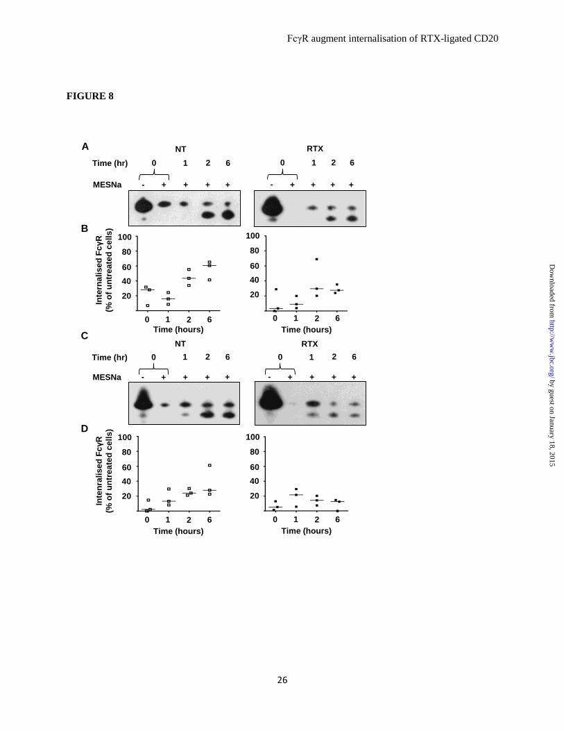
FcγR augment internalisation of RTX-ligated CD20
26
FIGURE 8
A
1 2
- + +
0
+ +
NT
21 60
20
40
60
80
100
Inte
rna
lis
ed
FcγR
(% o
f u
ntr
ea
ted
ce
lls
)
C
B
D
Time (hours)
6Time (hr)
MESNa
1 2
- + +
0
+ +
RTX
6
21 60
20
40
60
80
100
Time (hours)
1 2
- + +
0
+ +
NT
6Time (hr)
MESNa
21 60
20
40
60
80
100
Time (hours)
1 2
- + +
0
+ +
RTX
6
Inte
nra
lis
ed
FcγR
(% o
f u
ntr
ea
ted
ce
lls
)
21 60
20
40
60
80
100
Time (hours)
by guest on January 18, 2015http://w
ww
.jbc.org/D
ownloaded from

FcγR augment internalisation of RTX-ligated CD20
27
FIGURE 9
30 120030 120
A
30 60
- + +
0
+ +
NT
600
20
40
60
80
100
Inte
rna
lis
ed
FcγR
(% o
f u
ntr
ea
ted
ce
lls
)B
Time (hours)
120Time (min)
MESNa
30 60
- + +
0
+ +
RTX
120
60
20
40
60
80
100
Time (hours)
30 120030 120
C
30 60
- + +
0
+ +
NT
600
20
40
60
80
100
Inte
rna
lis
ed
FcγR
(% o
f u
ntr
ea
ted
ce
lls
)D
Time (hours)
120Time (min)
MESNa
30 60
- + +
0
+ +
RTX
120
60
20
40
60
80
100
Time (hours)
by guest on January 18, 2015http://w
ww
.jbc.org/D
ownloaded from

FcγR augment internalisation of RTX-ligated CD20
28
FIGURE 10
+ +
-- + +
A
- + -
0hr
+
50
150
100
Rela
tive
de
ns
ity o
f F
cγR
(% o
f u
ntr
ea
ted
ce
lls
)
B
Time (min)
MESNa
2hr
200
0hr
2hr
- + - +
2hr0hr
-- + +
0hr
50
150
100
200
2hr
- + - +
2hr0hr
FcγRIIb1 low FcγRIIa low
FcγRIIb1 low
FcγRIIa low
--
C
- + -
0hr
+
200
Rela
tive
de
ns
ity o
f F
cγR
(% o
f u
ntr
ea
ted
ce
lls
)
D
Time (min)
MESNa
E
2hr
300
0hr
2hr
- + - +
2hr0hr
100
-- + +
0hr 2hr
FcγRIIbΔcyt
FcγRIIaΔcyt
200
300
100
FcγRIIbΔcyt FcγRIIaΔcyt
by guest on January 18, 2015http://w
ww
.jbc.org/D
ownloaded from

FcγR augment internalisation of RTX-ligated CD20
29
FIGURE 11
by guest on January 18, 2015http://w
ww
.jbc.org/D
ownloaded from

Beers and Mark S. CraggA.Christian Klein, Martin J. Glennie, Stephen
Andrew T. Vaughan, Claude H. T. Chan, signalling via the cytoplasmic domaininternalisation of CD20 independent ofaugment rituximab-mediated
receptorsγActivatory and inhibitory FcImmunology:
published online January 7, 2015J. Biol. Chem.
10.1074/jbc.M114.593806Access the most updated version of this article at doi:
.JBC Affinity SitesFind articles, minireviews, Reflections and Classics on similar topics on the
Alerts:
When a correction for this article is posted•
When this article is cited•
to choose from all of JBC's e-mail alertsClick here
http://www.jbc.org/content/early/2015/01/07/jbc.M114.593806.full.html#ref-list-1
This article cites 0 references, 0 of which can be accessed free at
by guest on January 18, 2015http://w
ww
.jbc.org/D
ownloaded from