B-cell activation influences T-cell polarization and outcome of anti-CD20 B-cell depletion in...
15
ORIGINAL ARTICLE B-Cell Activation Influences T-Cell Polarization and Outcome of Anti-CD20 B-Cell Depletion in Central Nervous System Autoimmunity Martin S. Weber, MD, 1 Thomas Prod’homme, PhD, 1 Juan C. Patarroyo, BS, 1 Nicolas Molnarfi, PhD, 1 Tara Karnezis, PhD, 2 Klaus Lehmann-Horn, MD, 3 Dimitry M. Danilenko, DVM, PhD, 4 Jeffrey Eastham-Anderson, MS, 4 Anthony J. Slavin, PhD, 5 Christopher Linington, PhD, 6 Claude C. A. Bernard, PhD, 2 Flavius Martin, MD, 4 and Scott S. Zamvil, MD, PhD 1 Objective: Clinical studies indicate that anti-CD20 B-cell depletion may be an effective multiple sclerosis (MS) therapy. We investigated mechanisms of anti-CD20-mediated immune modulation using 2 paradigms of experimental autoimmune encephalomyelitis (EAE). Methods: Murine EAE was induced by recombinant myelin oligodendrocyte glycoprotein (rMOG), a model in which B cells are considered to contribute pathogenically, or MOG peptide (p)35-55, which does not require B cells. Results: In EAE induced by rMOG, B cells became activated and, when serving as antigen-presenting cells (APCs), promoted differentiation of proinflammatory MOG-specific Th1 and Th17 cells. B-cell depletion prevented or reversed established rMOG-induced EAE, which was associated with less central nervous system (CNS) inflammation, elimination of meningeal B cells, and reduction of MOG-specific Th1 and Th17 cells. In contrast, in MOG p35-55- induced EAE, B cells did not become activated or efficiently polarize proinflammatory MOG-specific T cells, similar to naive B cells. In this setting, anti-CD20 treatment exacerbated EAE, and did not impede development of Th1 or Th17 cells. Irrespective of the EAE model used, B-cell depletion reduced the frequency of CD4 þ CD25 þ Foxp3 þ regulatory T cells (Treg), and increased the proinflammatory polarizing capacity of remaining myeloid APCs. Interpretation: Our study highlights distinct roles for B cells in CNS autoimmunity. Clinical benefit from anti-CD20 treatment may relate to inhibition of proinflammatory B cell APC function. In certain clinical settings, however, elimination of unactivated B cells, which participate in regulation of T cells and other APC, may be undesirable. Differences in immune responses to MOG protein and peptide may be important considerations when choosing an EAE model for testing novel B cell-targeting agents for MS. ANN NEUROL 2010;68:369–383 T he central nervous system (CNS) has traditionally been viewed as an immune-privileged compartment with limited and well-controlled access for immune cells. B cells and plasma cells, however, are commonly found in active multiple sclerosis (MS) lesions, 1 and the presence of oligo- clonal antibodies within the cerebrospinal fluid remains a hallmark finding in the diagnosis of MS. Myelin-specific antibodies have been identified in areas of vesicular View this article online at wileyonlinelibrary.com. DOI: 10.1002/ana.22081 Received Nov 24, 2009, and in revised form Apr 13, 2010. Accepted for publication Apr 30, 2010. Address correspondence to Dr Zamvil, Department of Neurology, University of California, San Francisco, 513 Parnassus Avenue, S-268, San Francisco, CA 94143-0114. E-mail: [email protected] Current affiliation for M.S.W.: Department of Neurology, Technische Universita ¨ t Mu ¨ nchen, Munich, Germany. From the 1 Department of Neurology and Program in Immunology, University of California, San Francisco, CA; 2 Immunology and Stem Cell Laboratories, Monash University, Melbourne, Australia; 3 Department of Neurology, Technische Universita ¨t Mu ¨ nchen, Munich, Germany; 4 Department of Immunology, Genentech, South San Francisco, CA; 5 Department of Immunology and Inflammation, Boehringer-Ingelheim, Ridgefield, CT; and 6 Division of Clinical Neurosciences, University of Glasgow, Glasgow, United Kingdom. Additional Supporting Information can be found in the online version of this article. V C 2010 American Neurological Association 369
Transcript of B-cell activation influences T-cell polarization and outcome of anti-CD20 B-cell depletion in...
ORIGINAL ARTICLE
B-Cell Activation Influences T-CellPolarization and Outcome of Anti-CD20B-Cell Depletion in Central Nervous
System Autoimmunity
Martin S. Weber, MD,1 Thomas Prod’homme, PhD,1 Juan C. Patarroyo, BS,1
Nicolas Molnarfi, PhD,1 Tara Karnezis, PhD,2 Klaus Lehmann-Horn, MD,3
Dimitry M. Danilenko, DVM, PhD,4 Jeffrey Eastham-Anderson, MS,4
Anthony J. Slavin, PhD,5 Christopher Linington, PhD,6 Claude C. A. Bernard, PhD,2
Flavius Martin, MD,4 and Scott S. Zamvil, MD, PhD1
Objective: Clinical studies indicate that anti-CD20 B-cell depletion may be an effective multiple sclerosis (MS)therapy. We investigated mechanisms of anti-CD20-mediated immune modulation using 2 paradigms ofexperimental autoimmune encephalomyelitis (EAE).Methods: Murine EAE was induced by recombinant myelin oligodendrocyte glycoprotein (rMOG), a model in which B cellsare considered to contribute pathogenically, or MOG peptide (p)35-55, which does not require B cells.Results: In EAE induced by rMOG, B cells became activated and, when serving as antigen-presenting cells (APCs),promoted differentiation of proinflammatory MOG-specific Th1 and Th17 cells. B-cell depletion prevented orreversed established rMOG-induced EAE, which was associated with less central nervous system (CNS) inflammation,elimination of meningeal B cells, and reduction of MOG-specific Th1 and Th17 cells. In contrast, in MOG p35-55-induced EAE, B cells did not become activated or efficiently polarize proinflammatory MOG-specific T cells, similarto naive B cells. In this setting, anti-CD20 treatment exacerbated EAE, and did not impede development of Th1 orTh17 cells. Irrespective of the EAE model used, B-cell depletion reduced the frequency of CD4þCD25þFoxp3þ
regulatory T cells (Treg), and increased the proinflammatory polarizing capacity of remaining myeloid APCs.Interpretation: Our study highlights distinct roles for B cells in CNS autoimmunity. Clinical benefit from anti-CD20treatment may relate to inhibition of proinflammatory B cell APC function. In certain clinical settings, however,elimination of unactivated B cells, which participate in regulation of T cells and other APC, may be undesirable.Differences in immune responses to MOG protein and peptide may be important considerations when choosing anEAE model for testing novel B cell-targeting agents for MS.
ANN NEUROL 2010;68:369–383
The central nervous system (CNS) has traditionally been
viewed as an immune-privileged compartment with
limited and well-controlled access for immune cells. B cells
and plasma cells, however, are commonly found in active
multiple sclerosis (MS) lesions,1 and the presence of oligo-
clonal antibodies within the cerebrospinal fluid remains a
hallmark finding in the diagnosis of MS. Myelin-specific
antibodies have been identified in areas of vesicular
View this article online at wileyonlinelibrary.com. DOI: 10.1002/ana.22081
Received Nov 24, 2009, and in revised form Apr 13, 2010. Accepted for publication Apr 30, 2010.
Address correspondence to Dr Zamvil, Department of Neurology, University of California, San Francisco, 513 Parnassus Avenue, S-268, San Francisco,
CA 94143-0114. E-mail: [email protected]
Current affiliation for M.S.W.: Department of Neurology, Technische Universitat Munchen, Munich, Germany.
From the 1Department of Neurology and Program in Immunology, University of California, San Francisco, CA; 2Immunology and Stem Cell Laboratories,
Monash University, Melbourne, Australia; 3Department of Neurology, Technische Universitat Munchen, Munich, Germany; 4Department of Immunology,
Genentech, South San Francisco, CA; 5Department of Immunology and Inflammation, Boehringer-Ingelheim, Ridgefield, CT; and 6Division of Clinical
Neurosciences, University of Glasgow, Glasgow, United Kingdom.
Additional Supporting Information can be found in the online version of this article.
VC 2010 American Neurological Association 369
demyelination,2 suggesting that they directly promote CNS
damage. The observation that plasma exchange was benefi-
cial in MS patients with histologic evidence of CNS anti-
body deposition3 provided further support for a pathogenic
role of antibodies. Besides serving as the source for anti-
body-secreting plasma cells, B cells express major histocom-
patibility complex (MHC) class II molecules constitutively
and may participate as antigen-presenting cells (APCs). B
cells are capable of processing native antigen and are very
efficient APCs when they recognize the same antigen as
the responding T cells.4,5 As processing of native myelin
antigen by CNS resident or infiltrating APCs is required
for initiation of CNS autoimmune inflammation and clini-
cal disease,6,7 myelin-specific B cells may have an impor-
tant role in the activation of encephalitogenic T cells in
the pathogenesis of CNS autoimmune disease.
With greater appreciation that B cells may have dual
humoral and cellular roles in MS pathogenesis, interest in
use of selective B cell-depleting agents for therapy has
intensified.8,9 Promising results were obtained in clinical
trials testing a monoclonal antibody targeting CD20 (Rit-
uxan), a cell surface protein that is expressed on immature
and mature B cells, but not on differentiated plasma cells.
Treatment with Rituxan was beneficial in patients with
relapsing-remitting MS8 and in a subgroup of primary pro-
gressive MS patients with evidence of active CNS inflam-
mation.9 Anti-CD20–mediated B-cell depletion was also
clinically beneficial in a small open-label study in patients
with neuromyelitis optica (NMO),10 a CNS demyelinating
disease associated with aquaporin-4–specific antibodies.
The purpose of our investigation was to elucidate the
immunologic consequences of anti-CD20 therapy in 2
related models of experimental autoimmune encephalomy-
elitis (EAE).11 In 1 model, EAE was induced by immuni-
zation with recombinant myelin oligodendrocyte glycopro-
tein (rMOG), which generates a population of antigen-
activated B cells and promotes development of antibodies
against MOG protein. B-cell depletion prevented rMOG-
induced EAE and reversed paralysis when treatment was
initiated after EAE onset. In established EAE, anti-CD20–
depleted B cells within the CNS. B-cell depletion decreased
the frequency of peripheral and CNS encephalitogenic
Th1 and Th17 cells and was associated with reduced se-
rum titers of myelin-specific antibodies. These findings
highlight the pathogenic role of activated B cells in CNS
autoimmune disease, and provide mechanisms of action in
support of B-cell depletion for treatment of MS.
In the second model, EAE was induced by immuniza-
tion with MOG peptide (p) 35-55, which binds MHC II
directly on lymphoid APC without processing,6 and leads to
peripheral activation of encephalitogenic T cells.6,12 Using
this protocol, considered B-cell independent, MOG protein-
specific B cells were not activated. In contrast to the benefit
observed in EAE elicited by MOG protein, B-cell depletion
exacerbated clinical and histologic EAE in this model, and de-
velopment of Th1 and Th17 cells was not dampened. In both
rMOG and peptide-induced EAE, CD20-mediated B-cell
depletion reduced the frequency of CD4þCD25þFoxp3þ
regulatory T cells (Treg) and augmented the proinflammatory
function of remaining myeloid APCs. These observations
indicate that in the absence of proinflammatory B cell func-
tion, depletion of unactivated (naive) B cells may not be ad-
vantageous. The results of this study highlight key differences
between MOG protein and MOG peptide EAE models, and
underscore the importance of B-T crosstalk in pathogenesis
and regulation of CNS autoimmunity.
Materials and Methods
MiceC57BL/6 female mice, 5 to 8 weeks of age, as well as lMT
mice were purchased from Jackson Laboratories (Bar Harbor,
MN). hCD20 transgenic (Tg) C57BL/6 mice13,14 were used
for anti-CD20–mediated B-cell depletion. In these mice,
hCD20 recapitulates the expression of endogenous murine
CD20,15 and treatment with the murine antihuman CD20
antibody (clone 2h7) results in rapid depletion of B cells (Gong
et al,13 Supplementary Fig 1A). Untreated or isotype control-
treated hCD20 Tg mice developed EAE indistinguishable from
wild-type mice (see Supplementary Fig 1B). C57BL/6 MOG
35-55–specific T-cell receptor (TCR) Tg mice16 were kindly
provided by V. K. Kuchroo (Harvard). JHT mice17 were
obtained from K. Rajewsky (Harvard).
PeptidesMouse MOG p35-55 (MEVGWYRSPFSRVVHLYRNGK) was
synthesized by Auspep (Parkville, Australia). Recombinant mouse
MOG (1-117) was synthesized, purified and refolded as previously
described.18 Ovalbumin (OVA) p323-339 (ISQAVHAAHAEI-
NEAGR) was synthesized by Abgent, Inc. (San Diego, CA). Intact
OVA was purchased from Sigma-Aldrich (St. Louis, MO).
EAE InductionEight to 12-week-old female C57BL/6 or hCD20 Tg C57BL/6
mice were injected subcutaneously with 25lg MOG p35-55 or
100lg rMOG 1-117 in complete Freund adjuvant (DIFCO Labo-
ratories, Detroit, MI). After immunization and 48 hours later,
mice received an intravenous injection of 200ng pertussis toxin.
Individual animals were observed daily and clinical scores were
assessed as follows: 0 ¼ no clinical disease, 1 ¼ loss of tail tone
only, 2 ¼ mild monoparesis or paraparesis, 3 ¼ severe paraparesis,
4 ¼ paraplegia and/or quadraparesis, and 5 ¼ moribund or death.
Anti-CD20 TreatmentTo ensure maximal B-cell depletion when examining anti-CD20
in prevention of EAE, anti-CD20 mice received weekly intraperi-
toneal (i.p.) injections of 200lg of a-hCD20 monoclonal antibody
ANNALS of Neurology
370 Volume 68, No. 3
(m2h7) or antiragweed immunoglobulin (Ig)G2a-isotype control
monoclonal antibody starting 21 days prior to immunization. For
evaluation of anti-hCD20 treatment in established EAE, mice
were randomized to weekly treatment once they developed an
EAE disease score �2.
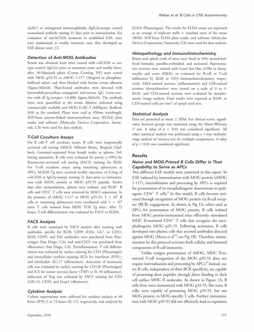
Detection of Anti-MOG AntibodiesSerum was obtained from mice treated with a-hCD20 or iso-
type control (IgG2a) prior to treatment onset and weekly there-
after. 96-Maxisorb plates (Costar, Corning, NY) were coated
with MOG p35-55 or rMOG 1-117 (10lg/ml in phosphate-
buffered saline) and then blocked with bovine serum albumin
(Sigma-Aldrich). Plate-bound antibodies were detected with
horseradish-peroxidase–conjugated anti-mouse IgG (cross-reac-
tive with all Ig isotypes; 1:6,000; Sigma-Aldrich). The antibody
titers were quantified at the serum dilution indicated using
commercially available anti-MOG 8.18C-5 (Millipore, Bedford,
MA) as the standard. Plates were read at 450nm wavelength.
SOFTmax enzyme-linked immunosorbent assay (ELISA) plate
reader and software (Molecular Devices Corporation, Sunny-
vale, CA) were used for data analysis.
T-Cell Coculture AssaysFor B cell–T cell coculture assays, B cells were magnetically
activated cell sorting (MACS; Miltenyi Biotec, Bergisch Glad-
bach, Germany)-separated from lymph nodes or spleens. Fol-
lowing separation, B cells were evaluated for purity (>99%) by
fluorescence-activated cell sorting (FACS) staining for B220.
For T-cell coculture assays using remaining splenocytes as
APCs, hCD20 Tg mice received weekly injections of 0.2mg of
a-hCD20 or IgG2a-isotype starting 21 days prior to immuniza-
tion with rMOG protein or MOG p35-55 peptide. Twelve
days after immunization, spleens were isolated, and B220þ B
cells and CD3þ T cells were removed by MACS separation. In
the presence of rMOG 1-117 or MOG p35-55, 5 � 105 B
cells or remaining splenocytes were cocultured with 1 � 104
naive T cells isolated from MOG TCR Tg mice. After 72
hours, T-cell differentiation was evaluated by FACS or ELISA.
FACS AnalysisB cells were examined by FACS analysis after staining with
antibodies specific for B220, CD95 (FAS), GL7, or CD21.
B220, CD95, and FAS antibodies were purchased from Phar-
mingen (San Diego, CA), and anti-CD21 was purchased from
eBioscience (San Diego, CA). Proinflammatory T cell differen-
tiation was evaluated by surface staining for CD3 (Pharmingen)
and intracellular cytokine staining (ICS) for interferon (IFN)-cand interleukin (IL)-17 (eBioscience). Activation of monocytic
cells was evaluated by surface staining for CD11b (Pharmingen)
and ICS for tumor necrosis factor (TNF) or IL-10 (eBiosience).
Induction of Treg was evaluated by FACS staining for CD4
(GK1.5), CD25, and Foxp3 (eBiosience).
Cytokine AnalysisCulture supernatants were collected for cytokine analysis at 48
hours (IFN-c) or 72 hours (IL-17), respectively, and analyzed by
ELISA (Pharmingen). The results for ELISA assays are expressed
as an average of triplicate wells 6 standard error of the mean
(SEM). SOFTmax ELISA plate reader and software (Molecular
Devices Corporation, Sunnyvale, CA) were used for data analysis.
Histopathology and ImmunohistochemistryBrains and spinal cords of mice were fixed in 10% neutral-buf-
fered formalin, paraffin-embedded, and sectioned. Representa-
tive sections were stained with Luxol fast blue (LFB) or hema-
toxylin and eosin (H&E), or evaluated for B-cell or T-cell
infiltration by B220 or CD3 immunohistochemistry, respec-
tively. H&E-stained sections (inflammation) and LFB-stained
sections (demyelination) were scored on a scale of 0 to 4.
B220- and CD3-stained sections were evaluated by morpho-
metric image analysis. Final results were reported as B220- or
CD3-stained cells per mm2 of spinal cord area.
Statistical AnalysisData are presented as mean 6 SEM. For clinical scores, signifi-
cance between groups was examined using the Mann-Whitney
U test. A value of p < 0.01 was considered significant. All
other statistical analysis was performed using a 1-way multiple-
range analysis of variance test for multiple comparisons. A value
of p < 0.01 was considered significant.
Results
Naive and MOG-Primed B Cells Differ in TheirCapability to Serve as APCsTwo different EAE models were examined in this report. In
EAE induced by immunization with MOG protein (rMOG
1-117), internalization and processing by APCs is required
for presentation of its encephalitogenic determinant to path-
ogenic CD4þ T cells.6 In this model, B cells become acti-
vated through recognition of MOG protein via B-cell recep-
tor (BCR) engagement. As shown in Fig 1A, when used as
APCs for presentation of MOG protein, B cells isolated
from MOG protein-immunized mice efficiently stimulated
MHC II-restricted CD4þ T cells that recognize the ence-
phalitogenic MOG p35-55. Following activation, B cells
developed into plasma cells that secreted antibodies directed
against MOG (Marta et al19; see Fig 1B). Therefore, immu-
nization by this protocol activates both cellular and humoral
components of B-cell immunity.
Unlike antigen presentation of rMOG, MHC II-re-
stricted T-cell recognition of the MOG p35-55 does not
require internalization and processing by APCs.6 Instead, na-
ive B cells, independent of their BCR specificity, are capable
of presenting short peptides through direct binding to their
cell surface MHC II molecules. As shown in Figure 1A, B
cells from mice immunized with MOG p35-55, like naive B
cells, were capable of presenting MOG p35-55, but not
MOG protein, to MOG-specific T cells. Further, immuniza-
tion with MOG p35-55 did not efficiently lead to expansion
Weber et al: B Cells in CNS Autoimmunity
September, 2010 371
of MOG-specific B cells, and was not associated with a sig-
nificant antibody response (Lyons et al20; see Fig 1C).
Kinetics of Anti-CD20–Mediated B-CellDepletion Differs in Distinct TissueMicroenvironmentsAnti-CD20 treatment was investigated in human (h) CD20
Tg C57BL/6 mice.13,14 These mice develop EAE in a man-
ner that is indistinguishable from wild-type C57BL/6 mice
(see Supplementary Fig 1). Data indicate that kinetics of B-
cell depletion in different tissue microenvironments may
depend on vascular access of anti-CD20 antibodies.13 Deple-
tion of mature (B220þCD21þ) B cells was examined in
blood, bone marrow, lymph nodes, spleen, and the perito-
neal cavity at various time points following a single anti-
CD20 treatment of unimmunized hCD20 Tg mice. A hier-
archy in tissue susceptibility to CD20-mediated B-cell deple-
tion was evident13; reduction of B cells was detected in blood
and bone marrow at 3 hours, and in lymph nodes and spleen
at 2 days (Fig 2). B-cell depletion in the peritoneum was
slower; at 2 days, peritoneal B cells were reduced by approxi-
mately 30%, and at 7 days by 95%. There was >99%
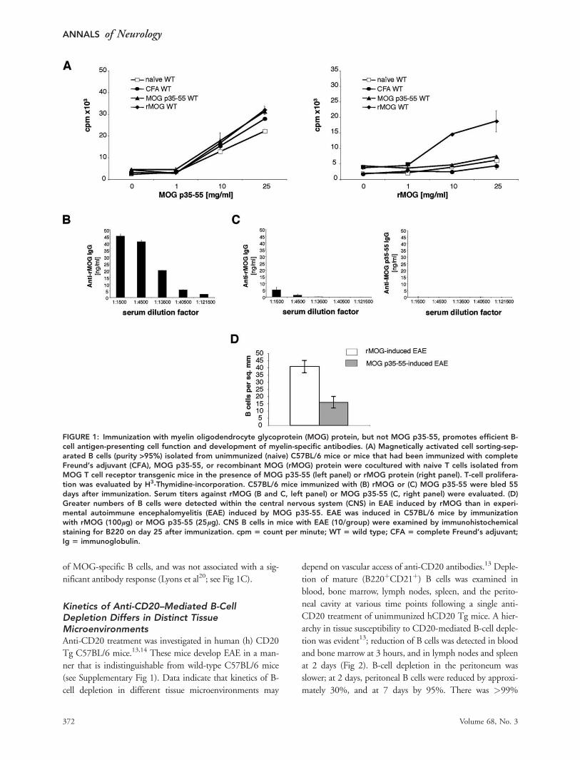
FIGURE 1: Immunization with myelin oligodendrocyte glycoprotein (MOG) protein, but not MOG p35-55, promotes efficient B-cell antigen-presenting cell function and development of myelin-specific antibodies. (A) Magnetically activated cell sorting-sep-arated B cells (purity >95%) isolated from unimmunized (naive) C57BL/6 mice or mice that had been immunized with completeFreund’s adjuvant (CFA), MOG p35-55, or recombinant MOG (rMOG) protein were cocultured with naive T cells isolated fromMOG T cell receptor transgenic mice in the presence of MOG p35-55 (left panel) or rMOG protein (right panel). T-cell prolifera-tion was evaluated by H3-Thymidine-incorporation. C57BL/6 mice immunized with (B) rMOG or (C) MOG p35-55 were bled 55days after immunization. Serum titers against rMOG (B and C, left panel) or MOG p35-55 (C, right panel) were evaluated. (D)Greater numbers of B cells were detected within the central nervous system (CNS) in EAE induced by rMOG than in experi-mental autoimmune encephalomyelitis (EAE) induced by MOG p35-55. EAE was induced in C57BL/6 mice by immunizationwith rMOG (100lg) or MOG p35-55 (25lg). CNS B cells in mice with EAE (10/group) were examined by immunohistochemicalstaining for B220 on day 25 after immunization. cpm 5 count per minute; WT 5 wild type; CFA 5 complete Freund’s adjuvant;Ig 5 immunoglobulin.
ANNALS of Neurology
372 Volume 68, No. 3
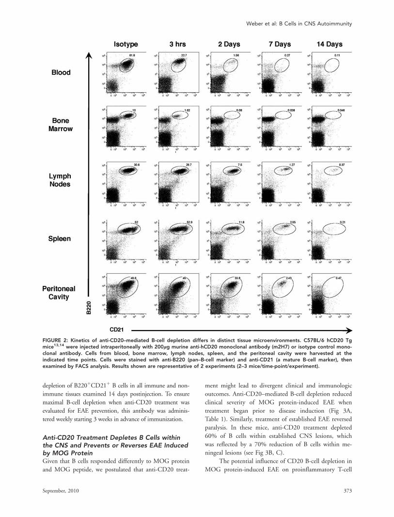
depletion of B220þCD21þ B cells in all immune and non-
immune tissues examined 14 days postinjection. To ensure
maximal B-cell depletion when anti-CD20 treatment was
evaluated for EAE prevention, this antibody was adminis-
tered weekly starting 3 weeks in advance of immunization.
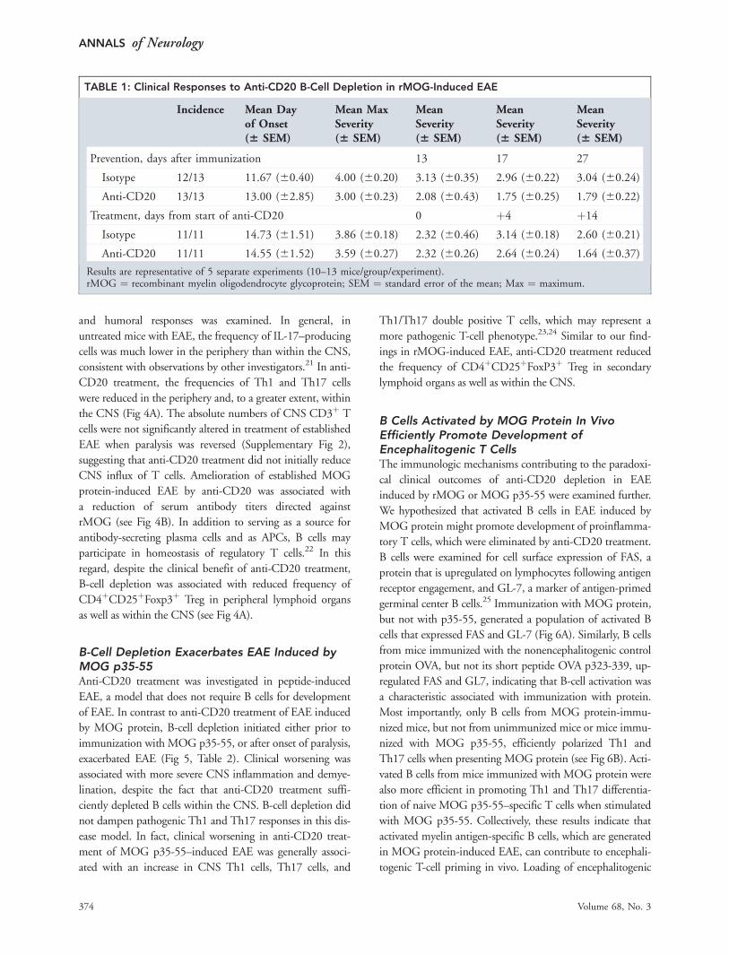
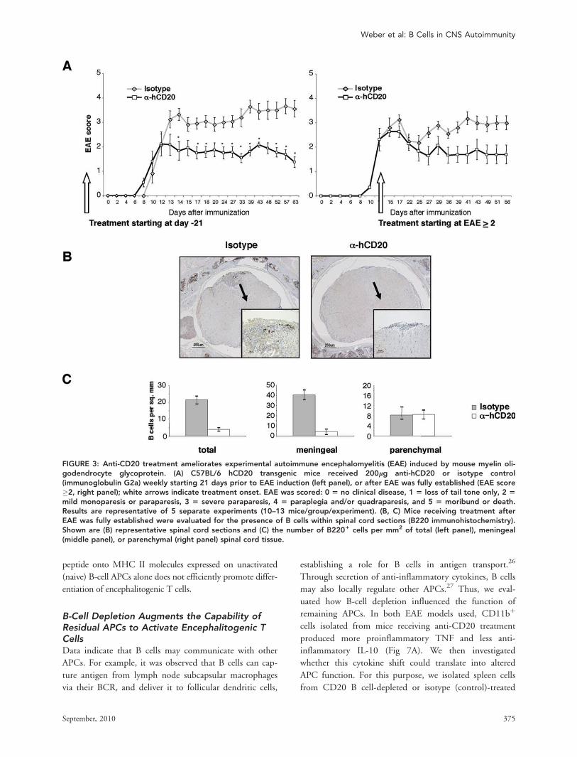
Anti-CD20 Treatment Depletes B Cells withinthe CNS and Prevents or Reverses EAE Inducedby MOG ProteinGiven that B cells responded differently to MOG protein
and MOG peptide, we postulated that anti-CD20 treat-
ment might lead to divergent clinical and immunologic
outcomes. Anti-CD20–mediated B-cell depletion reduced
clinical severity of MOG protein-induced EAE when
treatment began prior to disease induction (Fig 3A,
Table 1). Similarly, treatment of established EAE reversed
paralysis. In these mice, anti-CD20 treatment depleted
60% of B cells within established CNS lesions, which
was reflected by a 70% reduction of B cells within me-
ningeal lesions (see Fig 3B, C).
The potential influence of CD20 B-cell depletion in
MOG protein-induced EAE on proinflammatory T-cell
FIGURE 2: Kinetics of anti-CD20–mediated B-cell depletion differs in distinct tissue microenvironments. C57BL/6 hCD20 Tgmice13,14 were injected intraperitoneally with 200lg murine anti-hCD20 monoclonal antibody (m2H7) or isotype control mono-clonal antibody. Cells from blood, bone marrow, lymph nodes, spleen, and the peritoneal cavity were harvested at theindicated time points. Cells were stained with anti-B220 (pan–B-cell marker) and anti-CD21 (a mature B-cell marker), thenexamined by FACS analysis. Results shown are representative of 2 experiments (2–3 mice/time-point/experiment).
Weber et al: B Cells in CNS Autoimmunity
September, 2010 373
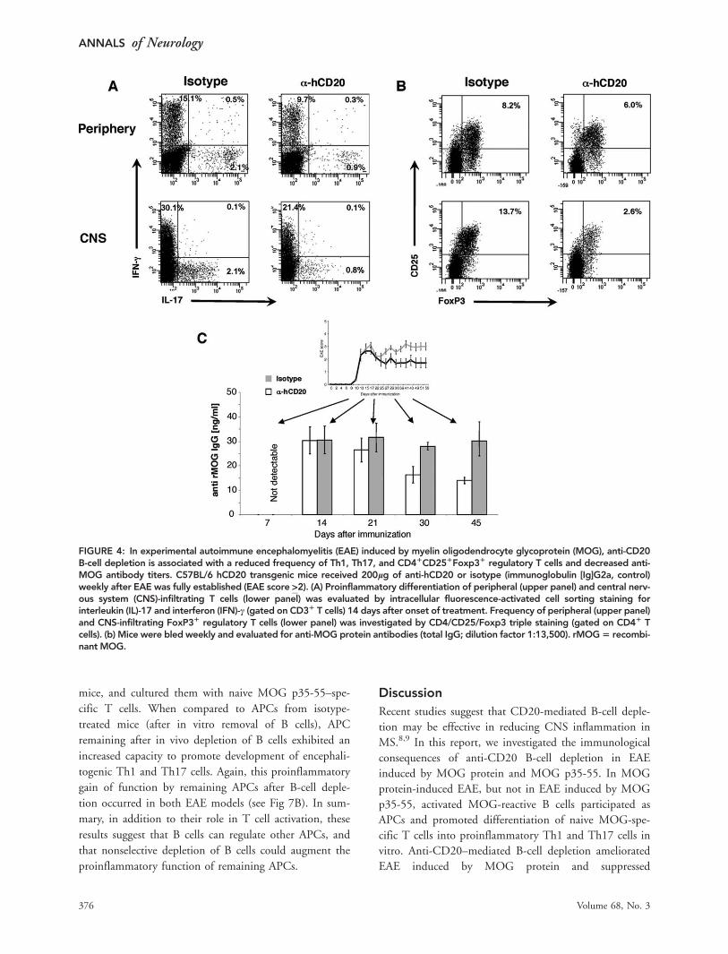
and humoral responses was examined. In general, in
untreated mice with EAE, the frequency of IL-17–producing
cells was much lower in the periphery than within the CNS,
consistent with observations by other investigators.21 In anti-
CD20 treatment, the frequencies of Th1 and Th17 cells
were reduced in the periphery and, to a greater extent, within
the CNS (Fig 4A). The absolute numbers of CNS CD3þ T
cells were not significantly altered in treatment of established
EAE when paralysis was reversed (Supplementary Fig 2),
suggesting that anti-CD20 treatment did not initially reduce
CNS influx of T cells. Amelioration of established MOG
protein-induced EAE by anti-CD20 was associated with
a reduction of serum antibody titers directed against
rMOG (see Fig 4B). In addition to serving as a source for
antibody-secreting plasma cells and as APCs, B cells may
participate in homeostasis of regulatory T cells.22 In this
regard, despite the clinical benefit of anti-CD20 treatment,
B-cell depletion was associated with reduced frequency of
CD4þCD25þFoxp3þ Treg in peripheral lymphoid organs
as well as within the CNS (see Fig 4A).
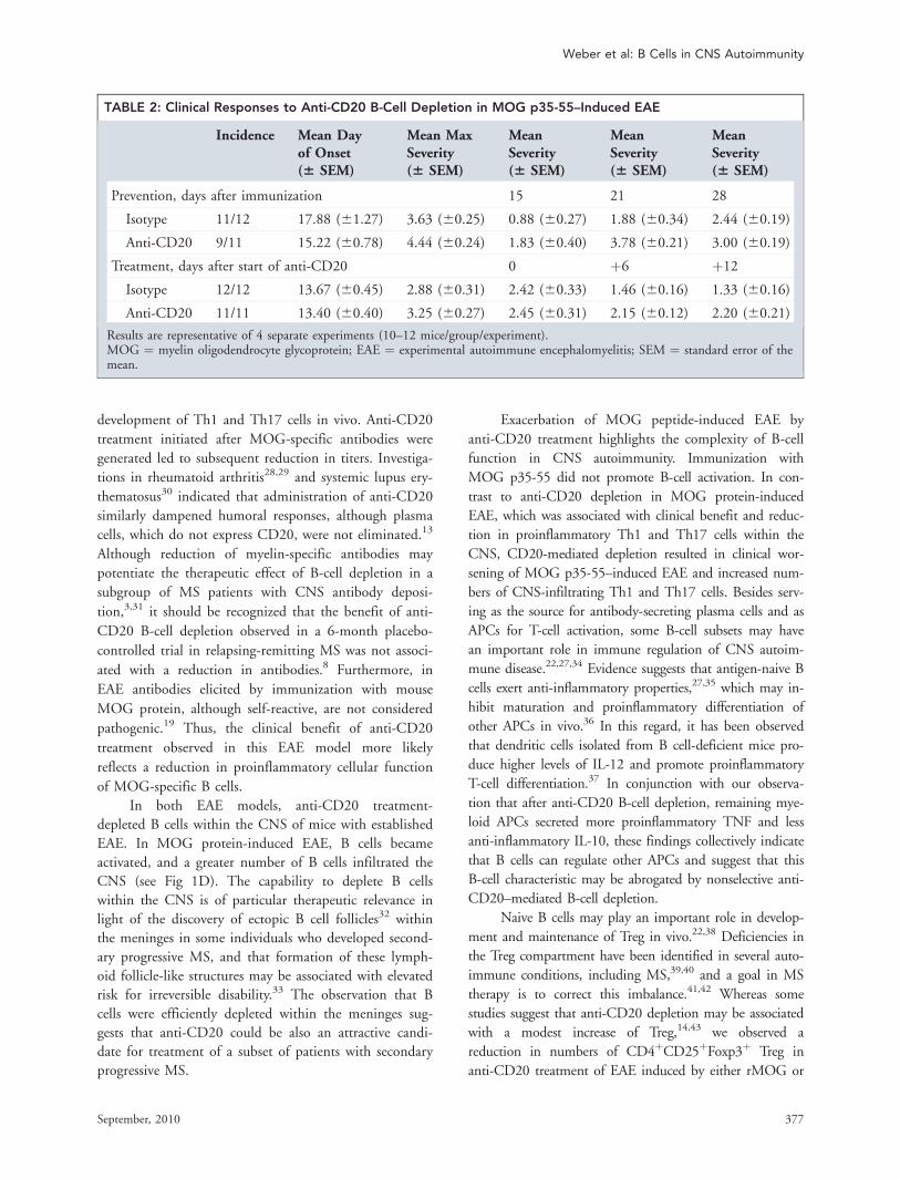
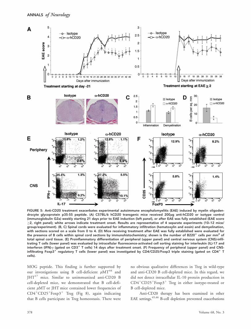
B-Cell Depletion Exacerbates EAE Induced byMOG p35-55Anti-CD20 treatment was investigated in peptide-induced
EAE, a model that does not require B cells for development
of EAE. In contrast to anti-CD20 treatment of EAE induced
by MOG protein, B-cell depletion initiated either prior to
immunization with MOG p35-55, or after onset of paralysis,
exacerbated EAE (Fig 5, Table 2). Clinical worsening was
associated with more severe CNS inflammation and demye-
lination, despite the fact that anti-CD20 treatment suffi-
ciently depleted B cells within the CNS. B-cell depletion did
not dampen pathogenic Th1 and Th17 responses in this dis-
ease model. In fact, clinical worsening in anti-CD20 treat-
ment of MOG p35-55–induced EAE was generally associ-
ated with an increase in CNS Th1 cells, Th17 cells, and
Th1/Th17 double positive T cells, which may represent a
more pathogenic T-cell phenotype.23,24 Similar to our find-
ings in rMOG-induced EAE, anti-CD20 treatment reduced
the frequency of CD4þCD25þFoxP3þ Treg in secondary
lymphoid organs as well as within the CNS.
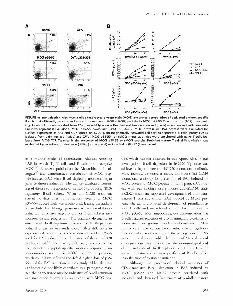
B Cells Activated by MOG Protein In VivoEfficiently Promote Development ofEncephalitogenic T CellsThe immunologic mechanisms contributing to the paradoxi-
cal clinical outcomes of anti-CD20 depletion in EAE
induced by rMOG or MOG p35-55 were examined further.
We hypothesized that activated B cells in EAE induced by
MOG protein might promote development of proinflamma-
tory T cells, which were eliminated by anti-CD20 treatment.
B cells were examined for cell surface expression of FAS, a
protein that is upregulated on lymphocytes following antigen
receptor engagement, and GL-7, a marker of antigen-primed
germinal center B cells.25 Immunization with MOG protein,
but not with p35-55, generated a population of activated B
cells that expressed FAS and GL-7 (Fig 6A). Similarly, B cells
from mice immunized with the nonencephalitogenic control
protein OVA, but not its short peptide OVA p323-339, up-
regulated FAS and GL7, indicating that B-cell activation was
a characteristic associated with immunization with protein.
Most importantly, only B cells from MOG protein-immu-
nized mice, but not from unimmunized mice or mice immu-
nized with MOG p35-55, efficiently polarized Th1 and
Th17 cells when presenting MOG protein (see Fig 6B). Acti-
vated B cells from mice immunized with MOG protein were
also more efficient in promoting Th1 and Th17 differentia-
tion of naive MOG p35-55–specific T cells when stimulated
with MOG p35-55. Collectively, these results indicate that
activated myelin antigen-specific B cells, which are generated
in MOG protein-induced EAE, can contribute to encephali-
togenic T-cell priming in vivo. Loading of encephalitogenic
TABLE 1: Clinical Responses to Anti-CD20 B-Cell Depletion in rMOG-Induced EAE
Incidence Mean Dayof Onset(6 SEM)
Mean MaxSeverity(6 SEM)
MeanSeverity(6 SEM)
MeanSeverity(6 SEM)
MeanSeverity(6 SEM)
Prevention, days after immunization 13 17 27
Isotype 12/13 11.67 (60.40) 4.00 (60.20) 3.13 (60.35) 2.96 (60.22) 3.04 (60.24)
Anti-CD20 13/13 13.00 (62.85) 3.00 (60.23) 2.08 (60.43) 1.75 (60.25) 1.79 (60.22)
Treatment, days from start of anti-CD20 0 þ4 þ14
Isotype 11/11 14.73 (61.51) 3.86 (60.18) 2.32 (60.46) 3.14 (60.18) 2.60 (60.21)
Anti-CD20 11/11 14.55 (61.52) 3.59 (60.27) 2.32 (60.26) 2.64 (60.24) 1.64 (60.37)
Results are representative of 5 separate experiments (10–13 mice/group/experiment).rMOG ¼ recombinant myelin oligodendrocyte glycoprotein; SEM ¼ standard error of the mean; Max ¼ maximum.
ANNALS of Neurology
374 Volume 68, No. 3
peptide onto MHC II molecules expressed on unactivated
(naive) B-cell APCs alone does not efficiently promote differ-
entiation of encephalitogenic T cells.
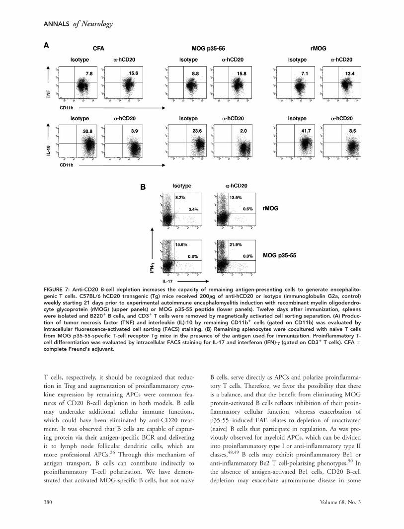
B-Cell Depletion Augments the Capability ofResidual APCs to Activate Encephalitogenic TCellsData indicate that B cells may communicate with other
APCs. For example, it was observed that B cells can cap-
ture antigen from lymph node subcapsular macrophages
via their BCR, and deliver it to follicular dendritic cells,
establishing a role for B cells in antigen transport.26
Through secretion of anti-inflammatory cytokines, B cells
may also locally regulate other APCs.27 Thus, we eval-
uated how B-cell depletion influenced the function of
remaining APCs. In both EAE models used, CD11bþ
cells isolated from mice receiving anti-CD20 treatment
produced more proinflammatory TNF and less anti-
inflammatory IL-10 (Fig 7A). We then investigated
whether this cytokine shift could translate into altered
APC function. For this purpose, we isolated spleen cells
from CD20 B cell-depleted or isotype (control)-treated
FIGURE 3: Anti-CD20 treatment ameliorates experimental autoimmune encephalomyelitis (EAE) induced by mouse myelin oli-godendrocyte glycoprotein. (A) C57BL/6 hCD20 transgenic mice received 200lg anti-hCD20 or isotype control(immunoglobulin G2a) weekly starting 21 days prior to EAE induction (left panel), or after EAE was fully established (EAE score�2, right panel); white arrows indicate treatment onset. EAE was scored: 0 5 no clinical disease, 1 5 loss of tail tone only, 2 5
mild monoparesis or paraparesis, 3 5 severe paraparesis, 4 5 paraplegia and/or quadraparesis, and 5 5 moribund or death.Results are representative of 5 separate experiments (10–13 mice/group/experiment). (B, C) Mice receiving treatment afterEAE was fully established were evaluated for the presence of B cells within spinal cord sections (B220 immunohistochemistry).Shown are (B) representative spinal cord sections and (C) the number of B2201 cells per mm2 of total (left panel), meningeal(middle panel), or parenchymal (right panel) spinal cord tissue.
Weber et al: B Cells in CNS Autoimmunity
September, 2010 375
mice, and cultured them with naive MOG p35-55–spe-
cific T cells. When compared to APCs from isotype-
treated mice (after in vitro removal of B cells), APC
remaining after in vivo depletion of B cells exhibited an
increased capacity to promote development of encephali-
togenic Th1 and Th17 cells. Again, this proinflammatory
gain of function by remaining APCs after B-cell deple-
tion occurred in both EAE models (see Fig 7B). In sum-
mary, in addition to their role in T cell activation, these
results suggest that B cells can regulate other APCs, and
that nonselective depletion of B cells could augment the
proinflammatory function of remaining APCs.
Discussion
Recent studies suggest that CD20-mediated B-cell deple-
tion may be effective in reducing CNS inflammation in
MS.8,9 In this report, we investigated the immunological
consequences of anti-CD20 B-cell depletion in EAE
induced by MOG protein and MOG p35-55. In MOG
protein-induced EAE, but not in EAE induced by MOG
p35-55, activated MOG-reactive B cells participated as
APCs and promoted differentiation of naive MOG-spe-
cific T cells into proinflammatory Th1 and Th17 cells in
vitro. Anti-CD20–mediated B-cell depletion ameliorated
EAE induced by MOG protein and suppressed
FIGURE 4: In experimental autoimmune encephalomyelitis (EAE) induced by myelin oligodendrocyte glycoprotein (MOG), anti-CD20B-cell depletion is associated with a reduced frequency of Th1, Th17, and CD41CD251Foxp31 regulatory T cells and decreased anti-MOG antibody titers. C57BL/6 hCD20 transgenic mice received 200lg of anti-hCD20 or isotype (immunoglobulin [Ig]G2a, control)weekly after EAE was fully established (EAE score >2). (A) Proinflammatory differentiation of peripheral (upper panel) and central nerv-ous system (CNS)-infiltrating T cells (lower panel) was evaluated by intracellular fluorescence-activated cell sorting staining forinterleukin (IL)-17 and interferon (IFN)-c (gated on CD31 T cells) 14 days after onset of treatment. Frequency of peripheral (upper panel)and CNS-infiltrating FoxP31 regulatory T cells (lower panel) was investigated by CD4/CD25/Foxp3 triple staining (gated on CD41 Tcells). (b) Mice were bled weekly and evaluated for anti-MOG protein antibodies (total IgG; dilution factor 1:13,500). rMOG5 recombi-nant MOG.
ANNALS of Neurology
376 Volume 68, No. 3
development of Th1 and Th17 cells in vivo. Anti-CD20
treatment initiated after MOG-specific antibodies were
generated led to subsequent reduction in titers. Investiga-
tions in rheumatoid arthritis28,29 and systemic lupus ery-
thematosus30 indicated that administration of anti-CD20
similarly dampened humoral responses, although plasma
cells, which do not express CD20, were not eliminated.13
Although reduction of myelin-specific antibodies may
potentiate the therapeutic effect of B-cell depletion in a
subgroup of MS patients with CNS antibody deposi-
tion,3,31 it should be recognized that the benefit of anti-
CD20 B-cell depletion observed in a 6-month placebo-
controlled trial in relapsing-remitting MS was not associ-
ated with a reduction in antibodies.8 Furthermore, in
EAE antibodies elicited by immunization with mouse
MOG protein, although self-reactive, are not considered
pathogenic.19 Thus, the clinical benefit of anti-CD20
treatment observed in this EAE model more likely
reflects a reduction in proinflammatory cellular function
of MOG-specific B cells.
In both EAE models, anti-CD20 treatment-
depleted B cells within the CNS of mice with established
EAE. In MOG protein-induced EAE, B cells became
activated, and a greater number of B cells infiltrated the
CNS (see Fig 1D). The capability to deplete B cells
within the CNS is of particular therapeutic relevance in
light of the discovery of ectopic B cell follicles32 within
the meninges in some individuals who developed second-
ary progressive MS, and that formation of these lymph-
oid follicle-like structures may be associated with elevated
risk for irreversible disability.33 The observation that B
cells were efficiently depleted within the meninges sug-
gests that anti-CD20 could be also an attractive candi-
date for treatment of a subset of patients with secondary
progressive MS.
Exacerbation of MOG peptide-induced EAE by
anti-CD20 treatment highlights the complexity of B-cell
function in CNS autoimmunity. Immunization with
MOG p35-55 did not promote B-cell activation. In con-
trast to anti-CD20 depletion in MOG protein-induced
EAE, which was associated with clinical benefit and reduc-
tion in proinflammatory Th1 and Th17 cells within the
CNS, CD20-mediated depletion resulted in clinical wor-
sening of MOG p35-55–induced EAE and increased num-
bers of CNS-infiltrating Th1 and Th17 cells. Besides serv-
ing as the source for antibody-secreting plasma cells and as
APCs for T-cell activation, some B-cell subsets may have
an important role in immune regulation of CNS autoim-
mune disease.22,27,34 Evidence suggests that antigen-naive B
cells exert anti-inflammatory properties,27,35 which may in-
hibit maturation and proinflammatory differentiation of
other APCs in vivo.36 In this regard, it has been observed
that dendritic cells isolated from B cell-deficient mice pro-
duce higher levels of IL-12 and promote proinflammatory
T-cell differentiation.37 In conjunction with our observa-
tion that after anti-CD20 B-cell depletion, remaining mye-
loid APCs secreted more proinflammatory TNF and less
anti-inflammatory IL-10, these findings collectively indicate
that B cells can regulate other APCs and suggest that this
B-cell characteristic may be abrogated by nonselective anti-
CD20–mediated B-cell depletion.
Naive B cells may play an important role in develop-
ment and maintenance of Treg in vivo.22,38 Deficiencies in
the Treg compartment have been identified in several auto-
immune conditions, including MS,39,40 and a goal in MS
therapy is to correct this imbalance.41,42 Whereas some
studies suggest that anti-CD20 depletion may be associated
with a modest increase of Treg,14,43 we observed a
reduction in numbers of CD4þCD25þFoxp3þ Treg in
anti-CD20 treatment of EAE induced by either rMOG or
TABLE 2: Clinical Responses to Anti-CD20 B-Cell Depletion in MOG p35-55–Induced EAE
Incidence Mean Dayof Onset(6 SEM)
Mean MaxSeverity(6 SEM)
MeanSeverity(6 SEM)
MeanSeverity(6 SEM)
MeanSeverity(6 SEM)
Prevention, days after immunization 15 21 28
Isotype 11/12 17.88 (61.27) 3.63 (60.25) 0.88 (60.27) 1.88 (60.34) 2.44 (60.19)
Anti-CD20 9/11 15.22 (60.78) 4.44 (60.24) 1.83 (60.40) 3.78 (60.21) 3.00 (60.19)
Treatment, days after start of anti-CD20 0 þ6 þ12
Isotype 12/12 13.67 (60.45) 2.88 (60.31) 2.42 (60.33) 1.46 (60.16) 1.33 (60.16)
Anti-CD20 11/11 13.40 (60.40) 3.25 (60.27) 2.45 (60.31) 2.15 (60.12) 2.20 (60.21)
Results are representative of 4 separate experiments (10–12 mice/group/experiment).MOG ¼ myelin oligodendrocyte glycoprotein; EAE ¼ experimental autoimmune encephalomyelitis; SEM ¼ standard error of themean.
Weber et al: B Cells in CNS Autoimmunity
September, 2010 377
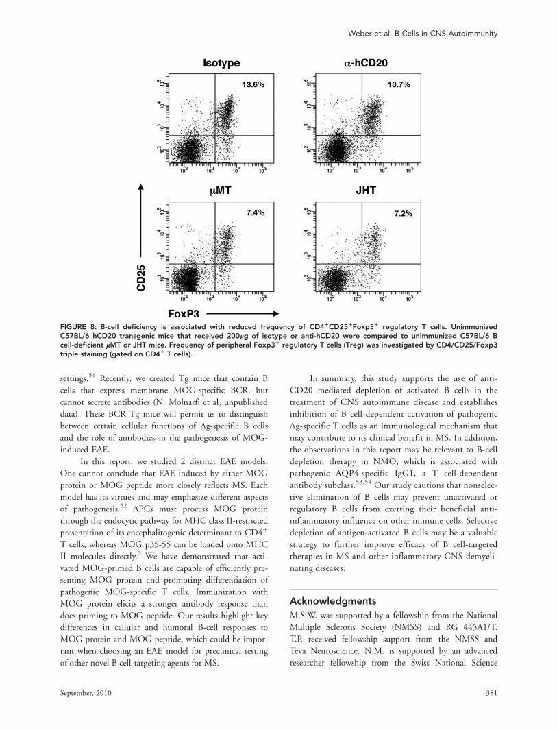
MOG peptide. This finding is further supported by
our investigations using B cell-deficient lMT44 and
JHT17 mice. Similar to unimmunized anti-CD20 B
cell-depleted mice, we demonstrated that B cell-defi-
cient lMT or JHT mice contained lower frequencies of
CD4þCD25þFoxp3þ Treg (Fig 8), again indicating
that B cells participate in Treg homeostasis. There were
no obvious qualitative differences in Treg in wild-type
and anti-CD20 B cell-depleted mice. In this regard, we
did not detect intracellular IL-10 protein production in
CD4þCD25þFoxp3þ Treg in either isotype-treated or
B cell-depleted mice.
Anti-CD20 therapy has been examined in other
EAE settings.45,46 B-cell depletion prevented exacerbations
FIGURE 5: Anti-CD20 treatment exacerbates experimental autoimmune encephalomyelitis (EAE) induced by myelin oligoden-drocyte glycoprotein p35-55 peptide. (A) C57BL/6 hCD20 transgenic mice received 200lg anti-hCD20 or isotype control(immunoglobulin G2a) weekly starting 21 days prior to EAE induction (left panel), or after EAE was fully established (EAE score�2, right panel); white arrows indicate treatment onset. Results are representative of 4 separate experiments (10–12 mice/group/experiment). (B, C) Spinal cords were evaluated for inflammatory infiltration (hematoxylin and eosin) and demyelination,with sections scored on a scale from 0 to 4. (D) Mice receiving treatment after EAE was fully established were evaluated forthe presence of B cells within spinal cord sections by immunohistochemistry; shown is the number of B2201 cells per mm2 oftotal spinal cord tissue. (E) Proinflammatory differentiation of peripheral (upper panel) and central nervous system (CNS)-infil-trating T cells (lower panel) was evaluated by intracellular fluorescence-activated cell sorting staining for interleukin (IL)-17 andinterferon (IFN)-c (gated on CD31 T cells) 14 days after treatment onset. (F) Frequency of peripheral (upper panel) and CNS-infiltrating Foxp31 regulatory T cells (lower panel) was investigated by CD4/CD25/Foxp3 triple staining (gated on CD41 Tcells).
ANNALS of Neurology
378 Volume 68, No. 3
in a murine model of spontaneous relapsing-remitting
EAE in which Tg T cells and B cells both recognize
MOG.46 A recent publication by Matsushita and col-
leagues45 also demonstrated exacerbation of MOG pep-
tide-induced EAE when B cell-depleting treatment began
prior to disease induction. The authors attributed worsen-
ing of disease to the absence of an IL-10–producing (B10)
regulatory B-cell subset. When anti-CD20 treatment
started 14 days after immunization, severity of MOG
p35-55–induced EAE was ameliorated, leading the authors
to conclude that although protective at the time of disease
induction, at a later stage, B cells or B-cell subsets may
promote disease progression. The apparent divergence in
outcome of B-cell depletion in reversal of MOG peptide-
induced disease in our study could reflect differences in
experimental procedures, such as dose of MOG p35-55
used for EAE induction, or the nature of the anti-CD20
antibody used.47 One striking difference, however, is that
they detected a peptide-specific antibody response upon
immunization with their MOG p35-55 preparation,
which could have reflected the 4-fold higher dose of p35-
55 used for EAE induction in their study. Although those
antibodies did not likely contribute in a pathogenic man-
ner, their appearance may be indicative of B-cell activation
and maturation following immunization with MOG pep-
tide, which was not observed in this report. Also, in our
investigation, B-cell depletion in hCD20 Tg mice was
achieved using a mouse anti-hCD20 monoclonal antibody.
More recently, we tested a mouse antimouse (m) CD20
monoclonal antibody for prevention of EAE induced by
MOG protein or MOG peptide in non-Tg mice. Consist-
ent with our findings using mouse anti-hCD20, anti-
mCD20 treatment suppressed development of proinflam-
matory T cells and clinical EAE induced by MOG pro-
tein, whereas it promoted development of proinflamma-
tory T cells and exacerbated clinical EAE induced by
MOG p35-55. Most importantly, our demonstration that
B cells regulate secretion of proinflammatory cytokines by
monocytes is in agreement with the observation by Mat-
sushita et al that certain B-cell subsets have regulatory
function, whereas others support the pathogenesis of CNS
autoimmune disease. Unlike the results of Matsushita and
colleagues, our data indicate that the immunological and
clinical outcome of B-cell depletion is determined by the
activation status and antigen-specificity of B cells, rather
than the time of treatment initiation.
Although the paradoxical clinical outcomes of
CD20-mediated B-cell depletion in EAE induced by
MOG p35-55 and MOG protein correlated with
increased and decreased frequencies of proinflammatory
FIGURE 6: Immunization with myelin oligodendrocyte glycoprotein (MOG) generates a population of activated antigen-specificB cells that efficiently process and present recombinant MOG (rMOG) protein to MOG p35-55 T-cell receptor (TCR) transgenic(Tg) T cells. (A) B cells isolated from C57BL/6 wild type mice that had not been immunized (naive) or immunized with completeFreund’s adjuvant (CFA) alone, MOG p35-55, ovalbumin (OVA) p323-339, MOG protein, or OVA protein were evaluated forsurface expression of FAS and GL7 (gated on B2201). (B) magnetically activated cell sorting-separated B cells (purity >95%)isolated from unimmunized (naive) and CFA-, MOG p35-55-, or rMOG-immunized mice were cocultured with naive T cells iso-lated from MOG TCR Tg mice in the presence of MOG p35-55 or rMOG protein. Proinflammatory T-cell differentiation wasevaluated by secretion of interferon (IFN)-c (upper panel) or interleukin (IL)-17 (lower panel).
Weber et al: B Cells in CNS Autoimmunity
September, 2010 379
T cells, respectively, it should be recognized that reduc-
tion in Treg and augmentation of proinflammatory cyto-
kine expression by remaining APCs were common fea-
tures of CD20 B-cell depletion in both models. B cells
may undertake additional cellular immune functions,
which could have been eliminated by anti-CD20 treat-
ment. It was observed that B cells are capable of captur-
ing protein via their antigen-specific BCR and delivering
it to lymph node follicular dendritic cells, which are
more professional APCs.26 Through this mechanism of
antigen transport, B cells can contribute indirectly to
proinflammatory T-cell polarization. We have demon-
strated that activated MOG-specific B cells, but not naive
B cells, serve directly as APCs and polarize proinflamma-
tory T cells. Therefore, we favor the possibility that there
is a balance, and that the benefit from eliminating MOG
protein-activated B cells reflects inhibition of their proin-
flammatory cellular function, whereas exacerbation of
p35-55–induced EAE relates to depletion of unactivated
(naive) B cells that participate in regulation. As was pre-
viously observed for myeloid APCs, which can be divided
into proinflammatory type I or anti-inflammatory type II
classes,48,49 B cells may exhibit proinflammatory Be1 or
anti-inflammatory Be2 T cell-polarizing phenotypes.50 In
the absence of antigen-activated Be1 cells, CD20 B-cell
depletion may exacerbate autoimmune disease in some
FIGURE 7: Anti-CD20 B-cell depletion increases the capacity of remaining antigen-presenting cells to generate encephalito-genic T cells. C57BL/6 hCD20 transgenic (Tg) mice received 200lg of anti-hCD20 or isotype (immunoglobulin G2a, control)weekly starting 21 days prior to experimental autoimmune encephalomyelitis induction with recombinant myelin oligodendro-cyte glycoprotein (rMOG) (upper panels) or MOG p35-55 peptide (lower panels). Twelve days after immunization, spleenswere isolated and B2201 B cells, and CD31 T cells were removed by magnetically activated cell sorting separation. (A) Produc-tion of tumor necrosis factor (TNF) and interleukin (IL)-10 by remaining CD11b1 cells (gated on CD11b) was evaluated byintracellular fluorescence-activated cell sorting (FACS) staining. (B) Remaining splenocytes were cocultured with naive T cellsfrom MOG p35-55-specific T-cell receptor Tg mice in the presence of the antigen used for immunization. Proinflammatory T-cell differentiation was evaluated by intracellular FACS staining for IL-17 and interferon (IFN)-c (gated on CD31 T cells). CFA 5
complete Freund’s adjuvant.
ANNALS of Neurology
380 Volume 68, No. 3
settings.51 Recently, we created Tg mice that contain B
cells that express membrane MOG-specific BCR, but
cannot secrete antibodies (N. Molnarfi et al, unpublished
data). These BCR Tg mice will permit us to distinguish
between certain cellular functions of Ag-specific B cells
and the role of antibodies in the pathogenesis of MOG-
induced EAE.
In this report, we studied 2 distinct EAE models.
One cannot conclude that EAE induced by either MOG
protein or MOG peptide more closely reflects MS. Each
model has its virtues and may emphasize different aspects
of pathogenesis.52 APCs must process MOG protein
through the endocytic pathway for MHC class II-restricted
presentation of its encephalitogenic determinant to CD4þ
T cells, whereas MOG p35-55 can be loaded onto MHC
II molecules directly.6 We have demonstrated that acti-
vated MOG-primed B cells are capable of efficiently pre-
senting MOG protein and promoting differentiation of
pathogenic MOG-specific T cells. Immunization with
MOG protein elicits a stronger antibody response than
does priming to MOG peptide. Our results highlight key
differences in cellular and humoral B-cell responses to
MOG protein and MOG peptide, which could be impor-
tant when choosing an EAE model for preclinical testing
of other novel B cell-targeting agents for MS.
In summary, this study supports the use of anti-
CD20–mediated depletion of activated B cells in the
treatment of CNS autoimmune disease and establishes
inhibition of B cell-dependent activation of pathogenic
Ag-specific T cells as an immunological mechanism that
may contribute to its clinical benefit in MS. In addition,
the observations in this report may be relevant to B-cell
depletion therapy in NMO, which is associated with
pathogenic AQP4-specific IgG1, a T cell-dependent
antibody subclass.53,54 Our study cautions that nonselec-
tive elimination of B cells may prevent unactivated or
regulatory B cells from exerting their beneficial anti-
inflammatory influence on other immune cells. Selective
depletion of antigen-activated B cells may be a valuable
strategy to further improve efficacy of B cell-targeted
therapies in MS and other inflammatory CNS demyeli-
nating diseases.
Acknowledgments
M.S.W. was supported by a fellowship from the National
Multiple Sclerosis Society (NMSS) and RG 445A1/T.
T.P. received fellowship support from the NMSS and
Teva Neuroscience. N.M. is supported by an advanced
researcher fellowship from the Swiss National Science
FIGURE 8: B-cell deficiency is associated with reduced frequency of CD41CD251Foxp31 regulatory T cells. UnimmunizedC57BL/6 hCD20 transgenic mice that received 200lg of isotype or anti-hCD20 were compared to unimmunized C57BL/6 Bcell-deficient lMT or JHT mice. Frequency of peripheral Foxp31 regulatory T cells (Treg) was investigated by CD4/CD25/Foxp3triple staining (gated on CD41 T cells).
Weber et al: B Cells in CNS Autoimmunity
September, 2010 381
Foundation (PA00A-119532). Support for this work was
provided to S.S.Z. by the National Institute of Health
(RO1 AI073737, RO1 AI059709, and RO1 NS063008),
the NMSS (RG 3622 and 3913), Dana Foundation,
Guthy Jackson Charitable Foundation, and Maisin Foun-
dation. C.C.A.B. was supported by grants from the
Baker Foundation, the National Health and Medical
Research Council of Australia and the NMSS (CCAB:
RG3844A2/1).
We thank Dr. P. A. Nelson for helpful discussion,
and C. Refino of Genentech and the Genentech Histol-
ogy and Immunohistochemistry Laboratories for process-
ing and staining brain and spinal cord specimens.
Authorship
M.S.W. and T.P. contributed equally to this study.
Potential Conflicts of Interest
Dmitry Danilenko is an employee of Genentech. Christo-
pher Linington received a grant from the Multiple Sclerosis
Society. FlaviusMartin is an employee of Genentech, as well
as an owner of stock. Anthony Slavin was an employee of
Amgen, Inc. and Novartis and is currently an employee of
Boehringer-Ingelheim. Dr. Slavin was paid money for two
patents - WO 99/41247 Treatment of multiple sclerosis
using COP-1 and Th2-enhancing cytokines EP
105488020010007758 Treatment of multiple sclerosis
using COP-1 and Th2-enhancing cytokines (Brigham and
Women’sHospital, Boston,MA) andUS 2005152896Anti-
galanin antibodies and uses thereof WO 2005/058961
Antibodies specific for human Galanin, and uses thereof
(Amgen, Inc.). Dr. Slavin also owns stock in Amgen,
Novartis, Gilead, Elan, Merck, Prana Biotechnology,
MetLife Inc. and Natus Medical Inc. as part of a mutual
fund/IRA account. Scott Zamvil was paid an honoraria, as
well as had travel and accommodations paid for by
Genentech to attend a Genentech PPMS Advisory Board
Meeting in November 2006, as well as a Genentech meeting
on B cells and B cell depletion in MS in September, 2007.
References
1. Prineas JW, Connell F. The fine structure of chronically active mul-tiple sclerosis plaques. Neurology 1978;28:68–75.
2. Genain CP, Cannella B, Hauser SL, Raine CS. Identification of au-toantibodies associated with myelin damage in multiple sclerosis.Nat Med 1999;5:170–175.
3. Keegan M, Konig F, McClelland R, et al. Relation between hu-moral pathological changes in multiple sclerosis and response totherapeutic plasma exchange. Lancet 2005;366:579–582.
4. Constant S, Schweitzer N, West J, et al. B lymphocytes can becompetent antigen-presenting cells for priming CD4þ T cells toprotein antigens in vivo. J Immunol 1995;155:3734–3741.
5. Constant S, Sant’Angelo D, Pasqualini T, et al. Peptide and pro-tein antigens require distinct antigen-presenting cell subsets forthe priming of CD4þ T cells. J Immunol 1995;154:4915–4923.
6. Slavin AJ, Soos JM, Stuve O, et al. Requirement for endocyticantigen processing and influence of invariant chain and H-2M defi-ciencies in CNS autoimmunity. J Clin Invest 2001;108:1133–1139.
7. Tompkins SM, Padilla J, Dal Canto MC, et al. De novo centralnervous system processing of myelin antigen is required for theinitiation of experimental autoimmune encephalomyelitis. J Immu-nol 2002;168:4173–4183.
8. Hauser SL, Waubant E, Arnold DL, et al. B-cell depletion with rit-uximab in relapsing-remitting multiple sclerosis. N Engl J Med2008;358:676–688.
9. Hawker K, O’Connor P, Freedman MS, et al. Rituximab in patientswith primary progressive multiple sclerosis: results of a random-ized double-blind placebo-controlled multicenter trial. Ann Neurol2009;66:460–471.
10. Cree BA, Lamb S, Morgan K, et al. An open label study of the effectsof rituximab in neuromyelitis optica. Neurology 2005;64:1270–1272.
11. Steinman L, Zamvil SS. How to successfully apply animal studies inexperimental allergic encephalomyelitis to research on multiplesclerosis. Ann Neurol 2006;60:12–21.
12. Mendel I, Kerlero de Rosbo N, Ben-Nun A. A myelin oligodendro-cyte glycoprotein peptide induces typical chronic experimentalautoimmune encephalomyelitis in H-2b mice: fine specificity and Tcell receptor V beta expression of encephalitogenic T cells. Eur JImmunol 1995;25:1951–1959.
13. Gong Q, Ou Q, Ye S, et al. Importance of cellular microenviron-ment and circulatory dynamics in B cell immunotherapy. J Immu-nol 2005;174:817–826.
14. Hu CY, Rodriguez-Pinto D, Du W, et al. Treatment with CD20-spe-cific antibody prevents and reverses autoimmune diabetes inmice. J Clin Invest 2007;117:3857–3867.
15. Uchida J, Lee Y, Hasegawa M, et al. Mouse CD20 expression andfunction. Int Immunol 2004;16:119–129.
16. Bettelli E, Pagany M, Weiner HL, et al. Myelin oligodendrocyte glyco-protein-specific T cell receptor transgenic mice develop spontaneousautoimmune optic neuritis. J Exp Med 2003;197:1073–1081.
17. Chen J, Trounstine M, Alt FW, et al. Immunoglobulin gene rear-rangement in B cell deficient mice generated by targeted deletionof the JH locus. Int Immunol 1993;5:647–656.
18. Clements CS, Reid HH, Beddoe T, et al. The crystal structure ofmyelin oligodendrocyte glycoprotein, a key autoantigen inmultiple sclerosis. Proc Natl Acad Sci U S A 2003;100:11059–11064.
19. Marta CB, Oliver AR, Sweet RA, et al. Pathogenic myelin oligo-dendrocyte glycoprotein antibodies recognize glycosylated epi-topes and perturb oligodendrocyte physiology. Proc Natl AcadSci U S A 2005;102:13992–13997.
20. Lyons JA, San M, Happ MP, Cross AH. B cells are critical to inductionof experimental allergic encephalomyelitis by protein but not by ashort encephalitogenic peptide. Eur J Immunol 1999;29:3432–3439.
21. Korn T, Anderson AC, Bettelli E, Oukka M. The dynamics of effec-tor T cells and Foxp3þ regulatory T cells in the promotion andregulation of autoimmune encephalomyelitis. J Neuroimmunol2007;191:51–60.
22. Mann MK, Maresz K, Shriver LP, et al. B cell regulation ofCD4þCD25þ T regulatory cells and IL-10 via B7 is essential for re-covery from experimental autoimmune encephalomyelitis. J Immu-nol 2007;178:3447–3456.
23. Kroenke MA, Carlson TJ, Andjelkovic AV, Segal BM. IL-12- and IL-23-modulated T cells induce distinct types of EAE based on histol-ogy, CNS chemokine profile, and response to cytokine inhibition.J Exp Med 2008;205:1535–1541.
ANNALS of Neurology
382 Volume 68, No. 3
24. Kebir H, Ifergan I, Alvarez JI, et al. Preferential recruitment ofinterferon-gamma-expressing T H 17 cells in multiple sclerosis.Ann Neurol 2009;66:390–402.
25. Cervenak L, Magyar A, Boja R, Laszlo G. Differential expression ofGL7 activation antigen on bone marrow B cell subpopulations andperipheral B cells. Immunol Lett 2001;78:89–96.
26. Phan TG, Grigorova I, Okada T, Cyster JG. Subcapsular encounterand complement-dependent transport of immune complexes bylymph node B cells. Nat Immunol 2007;8:992–1000.
27. Fillatreau S, Sweenie CH, McGeachy MJ, et al. B cells regulateautoimmunity by provision of IL-10. Nat Immunol 2002;3:944–950.
28. Edwards JC, Szczepanski L, Szechinski J, et al. Efficacy of B-cell-targeted therapy with rituximab in patients with rheumatoid arthri-tis. N Engl J Med 2004;350:2572–2581.
29. Cambridge G, Stohl W, Leandro MJ, et al. Circulating levels of Blymphocyte stimulator in patients with rheumatoid arthritis follow-ing rituximab treatment: relationships with B cell depletion, circu-lating antibodies, and clinical relapse. Arthritis Rheum 2006;54:723–732.
30. Anolik JH, Barnard J, Owen T, et al. Delayed memory B cell re-covery in peripheral blood and lymphoid tissue in systemic lupuserythematosus after B cell depletion therapy. Arthritis Rheum2007;56:3044–3056.
31. Lucchinetti C, Bruck W, Parisi J, et al. Heterogeneity of multiplesclerosis lesions: implications for the pathogenesis of demyelin-ation. Ann Neurol 2000;47:707–717.
32. Serafini B, Rosicarelli B, Magliozzi R, et al. Detection of ectopic B-cell follicles with germinal centers in the meninges of patients withsecondary progressive multiple sclerosis. Brain Pathol 2004;14:164–174.
33. Magliozzi R, Howell O, Vora A, et al. Meningeal B-cell follicles insecondary progressive multiple sclerosis associate with early onsetof disease and severe cortical pathology. Brain 2007;130:1089–1104.
34. Bouaziz JD, Yanaba K, Tedder TF. Regulatory B cells as inhibitorsof immune responses and inflammation. Immunol Rev 2008;224:201–214.
35. Evans JG, Chavez-Rueda KA, Eddaoudi A, et al. Novel suppres-sive function of transitional 2 B cells in experimental arthritis. JImmunol 2007;178:7868–7878.
36. De Smedt T, Van Mechelen M, De Becker G, et al. Effect of inter-leukin-10 on dendritic cell maturation and function. Eur J Immunol1997;27:1229–1235.
37. Moulin V, Andris F, Thielemans K, et al. B lymphocytes regulatedendritic cell (DC) function in vivo: increased interleukin 12 pro-duction by DCs from B cell-deficient mice results in T helper celltype 1 deviation. J Exp Med 2000;192:475–482.
38. Reichardt P, Dornbach B, Rong S, et al. Naive B cells generateregulatory T cells in the presence of a mature immunologic syn-apse. Blood 2007;110:1519–1529.
39. Viglietta V, Baecher-Allan C, Weiner HL, Hafler DA. Loss of func-tional suppression by CD4þCD25þ regulatory T cells in patientswith multiple sclerosis. J Exp Med 2004;199:971–979.
40. Kukreja A, Cost G, Marker J, et al. Multiple immuno-regulatorydefects in type-1 diabetes. J Clin Invest 2002;109:131–140.
41. Hong J, Li N, Zhang X, et al. Induction of CD4þCD25þ regulatoryT cells by copolymer-I through activation of transcription factorFoxp3. Proc Natl Acad Sci U S A 2005;102:6449–6454.
42. Putheti P, Soderstrom M, Link H, Huang YM. Effect of glatirameracetate (Copaxone) on CD4þCD25 high T regulatory cells andtheir IL-10 production in multiple sclerosis. J Neuroimmunol 2003;144:125–131.
43. Sfikakis PP, Souliotis VL, Fragiadaki KG, et al. Increased expres-sion of the FoxP3 functional marker of regulatory T cells followingB cell depletion with rituximab in patients with lupus nephritis.Clin Immunol 2007;123:66–73.
44. Kitamura D, Roes J, Kuhn R, Rajewsky K. A B cell-deficient mouseby targeted disruption of the membrane exon of the immuno-globulin mu chain gene. Nature 1991;350:423–426.
45. Matsushita T, Yanaba K, Bouaziz JD, et al. Regulatory B cells in-hibit EAE initiation in mice while other B cells promote diseaseprogression. J Clin Invest 2008;118:3420–3430.
46. Pollinger B, Krishnamoorthy G, Berer K, et al. Spontaneous relaps-ing-remitting EAE in the SJL/J mouse: MOG-reactive transgenic Tcells recruit endogenous MOG-specific B cells. J Exp Med 2009;206:1303–1316.
47. Hamaguchi Y, Xiu Y, Komura K, et al. Antibody isotype-specificengagement of Fcgamma receptors regulates B lymphocytedepletion during CD20 immunotherapy. J Exp Med 2006;203:743–753.
48. Kim HJ, Ifergan I, Antel JP, et al. Type 2 monocyte and microgliadifferentiation mediated by glatiramer acetate therapy in patientswith multiple sclerosis. J Immunol 2004;172:7144–7153.
49. Weber MS, Prod’homme T, Youssef S, et al. Type II monocytesmodulate T cell-mediated central nervous system autoimmune dis-ease. Nat Med 2007;13:935–943.
50. Harris DP, Haynes L, Sayles PC, et al. Reciprocal regulation ofpolarized cytokine production by effector B and T cells. NatImmunol 2000;1:475–482.
51. Goetz M, Atreya R, Ghalibafian M, et al. Exacerbation of ulcerativecolitis after rituximab salvage therapy. Inflamm Bowel Dis 2007;13:1365–1368.
52. Steinman L, Zamvil SS. Virtues and pitfalls of EAE for the develop-ment of therapies for multiple sclerosis. Trends Immunol 2005;26:565–571.
53. Bradl M, Misu T, Takahashi T, et al. Neuromyelitis optica: patho-genicity of patient immunoglobulin in vivo. Ann Neurol 2009;66:630–643.
54. Bennett JL, Lam C, Kalluri SR, et al. Intrathecal pathogenic anti-aquaporin-4 antibodies in early neuromyelitis optica. Ann Neurol2009;66:617–629.
Weber et al: B Cells in CNS Autoimmunity
September, 2010 383