Absence of Mature Peripheral B Cell Populations in Mice with Concomitant Defects in B Cell Receptor...
15
of June 13, 2013. This information is current as Signaling Defects in B Cell Receptor and BAFF-R Populations in Mice with Concomitant Absence of Mature Peripheral B Cell Wasif N. Khan Kristen L. Hoek, Gianluca Carlesso, Emily S. Clark and http://www.jimmunol.org/content/183/9/5630 doi: 10.4049/jimmunol.0901100 2009; 183:5630-5643; ; J Immunol References http://www.jimmunol.org/content/183/9/5630.full#ref-list-1 , 47 of which you can access for free at: cites 76 articles This article Subscriptions http://jimmunol.org/subscriptions is online at: The Journal of Immunology Information about subscribing to Permissions http://www.aai.org/ji/copyright.html Submit copyright permission requests at: Email Alerts http://jimmunol.org/cgi/alerts/etoc Receive free email-alerts when new articles cite this article. Sign up at: Print ISSN: 0022-1767 Online ISSN: 1550-6606. Immunologists, Inc. All rights reserved. Copyright © 2009 by The American Association of 9650 Rockville Pike, Bethesda, MD 20814-3994. The American Association of Immunologists, Inc., is published twice each month by The Journal of Immunology by guest on June 13, 2013 http://www.jimmunol.org/ Downloaded from
-
Upload
independent -
Category
Documents
-
view
0 -
download
0
Transcript of Absence of Mature Peripheral B Cell Populations in Mice with Concomitant Defects in B Cell Receptor...

of June 13, 2013.This information is current as
SignalingDefects in B Cell Receptor and BAFF-RPopulations in Mice with Concomitant Absence of Mature Peripheral B Cell
Wasif N. KhanKristen L. Hoek, Gianluca Carlesso, Emily S. Clark and
http://www.jimmunol.org/content/183/9/5630doi: 10.4049/jimmunol.0901100
2009; 183:5630-5643; ;J Immunol
Referenceshttp://www.jimmunol.org/content/183/9/5630.full#ref-list-1
, 47 of which you can access for free at: cites 76 articlesThis article
Subscriptionshttp://jimmunol.org/subscriptions
is online at: The Journal of ImmunologyInformation about subscribing to
Permissionshttp://www.aai.org/ji/copyright.htmlSubmit copyright permission requests at:
Email Alertshttp://jimmunol.org/cgi/alerts/etocReceive free email-alerts when new articles cite this article. Sign up at:
Print ISSN: 0022-1767 Online ISSN: 1550-6606. Immunologists, Inc. All rights reserved.Copyright © 2009 by The American Association of9650 Rockville Pike, Bethesda, MD 20814-3994.The American Association of Immunologists, Inc.,
is published twice each month byThe Journal of Immunology
by guest on June 13, 2013http://w
ww
.jimm
unol.org/D
ownloaded from

Absence of Mature Peripheral B Cell Populations in Micewith Concomitant Defects in B Cell Receptor and BAFF-RSignaling1
Kristen L. Hoek,2* Gianluca Carlesso,2,3* Emily S. Clark,† and Wasif N. Khan4*†
Generation of mature B lymphocytes from early (T1) and late transitional (T2) precursors requires cooperative signaling throughBCR and B cell-activating factor receptor 3 (BR3). Recent studies have shown that BCR signaling positively regulates NF-�B2,suggesting BCR regulation of BR3 signaling. To investigate the significance of signal integration from BCR and BR3 in B celldevelopment and function, we crossed Btk-deficient mice (btk�/�), which are developmentally blocked between the T2 and themature follicular B cell stage as a result of a partial defect in BCR signaling, and A/WySnJ mice, which possess a mutant BR3defective in propagating intracellular signals that results in a severely reduced peripheral B cell compartment, although all B cellsubsets are present in relatively normal ratios. A/WySnJ � btk�/� mice display a B cell-autonomous defect, resulting in a devel-opmental block at an earlier stage (T1) than either mutation alone, leading to the loss of mature splenic follicular and marginalzone B cells, as well as the loss of peritoneal B1 and B2 cell populations. The competence of the double mutant T1 B cells to respondto TLR4 and CD40 survival and activation signals is further attenuated compared with single mutations as evidenced by severelyreduced humoral immune responses in vivo and proliferation in response to anti-IgM, LPS, and anti-CD40 stimulation in vitro.Thus, BCR and BR3 independently and in concert regulate the survival, differentiation, and function of all B cell populations atand beyond T1, earliest transitional stage. The Journal of Immunology, 2009, 183: 5630–5643.
S ignaling through BCR and B cell-activating factor receptor3 (BR3)5 controls the development of fully functional ma-ture B lymphocytes in the periphery (reviewed in Refs. 1
and 2). Early B cell development occurs in the bone marrow (BM),and upon surface expression of IgM (BCR) the immature B cellsemigrate to the spleen. In the spleen, immature B cells proceedthrough several “transitional” periods of development, termed theearly transitional (T1) and late transitional (T2) stages, before de-velopment concludes with the generation of mature follicular (Fo)or marginal zone (MZ) B cells (3–5). Selection processes, which
occur in both the BM and the secondary lymphoid tissues, ensurethe elimination of self-reactive immature B cell clones and theexpansion of clones that can efficiently respond to foreign Agswhile maintaining tolerance to self-Ags (6, 7). In the periphery,both negative and positive B cell selection processes have beensuggested to occur at the T1 and T2 transitional stages (3, 7–10).All of these selection processes are likely regulated by signalsemanating from the BCR. In this regard, cell surface Ig and theassociated signaling modules Ig�/� or the receptor proximal ty-rosine kinase Syk have been shown to play key roles in signaltransmission from both the pre-BCR in early B cell development inthe BM and the BCR following the cell surface display of IgMin immature and mature B cell populations in the periphery(11–14). As such, targeted or inducible gene inactivation ofeither Ig�/� or Syk leads to a B cell developmental block at thepro-B cell to pre-B cell transition in the BM as well as at the T1to T2 transition in the spleen, demonstrating a role for BCRsignaling during this transition (12, 14 –17). Additionally, lossof function of the BCR signal transducer Btk results in a B celldevelopmental block at the T2 to mature Fo B cell transition(18 –21). The B cell deficiency caused by loss of Btk functionis referred to as Xid (X-linked deficiency) in mice. Xid miceserve as a prototype for defective function of the BCR signa-losome, as mice deficient for other components of this signa-losome also display an Xid-like phenotype (22).
Recent studies suggest that in addition to the BCR signals,interaction with and response to growth factors is also requiredfor the progression of transitional B cells into mature B cellpopulations. In this regard, B cell-activating factor belonging tothe TNF family (BAFF)-mediated signaling is vital for periph-eral B cell development and survival. BAFF serves as a majorsurvival and growth factor for peripheral B cells (23–28). BAFFbinds three receptors of the TNF-R family: BCMA (B cell mat-uration Ag), which is only detected on germinal center (GC) Bcells and terminally differentiated plasma cells (29 –31); TACI
*Department of Microbiology and Immunology, Vanderbilt University School ofMedicine, Nashville, TN 37232; and †Department of Microbiology and Immunology,Miller School of Medicine, University of Miami, Miami, FL 33136
Received for publication April 6, 2009. Accepted for publication August 24, 2009.
The costs of publication of this article were defrayed in part by the payment of pagecharges. This article must therefore be hereby marked advertisement in accordancewith 18 U.S.C. Section 1734 solely to indicate this fact.1 This study is supported in part by National Institutes of Health (NIH) GrantAI060729 (to W.N.K.). K.L.H. was supported in part by NIH Grants T32 HL69715-0and F32-AI069770-01 and E.C. was supported in part by NIH Grant AI060729 andNational Institute of Mental Health Grant 2R32 MH018917-21 for BiopsychosocialResearch Training in Immunology and AIDS (to Neil Schneiderman, Department ofPsychology, University of Miami, Miami, FL).2 K.L.H. and G.C. contributed equally to this work.3 Current address: MedImmune, LLC. One MedImmune Way, Department of Respi-ratory Inflammation and Autoimmunity, Gaithersburg, MD 20878.4 Address correspondence and reprint requests to Dr. Wasif N. Khan, Miller Schoolof Medicine, University of Miami, 1600 Northwest 10th Avenue, Room 3347A,Rosenstiel Medical Sciences Building, Miami, FL 33136. E-mail address: [email protected] Abbreviations used in this paper: BR3, B cell-activating factor 3; BAFF, Bcell-activating factor belonging to the TNF family; BM, bone marrow; DNP,dinitrophenyl; Fo, follicular; GC, germinal center; HSA, human serum albumin;IKK, I�B kinase; MZ, marginal zone; T1, early transitional stage; T2, late tran-sitional stage; TD, T cell dependent; TI-I, T cell independent type 1; TNP, trini-trophenyl; wt, wild type.
Copyright © 2009 by The American Association of Immunologists, Inc. 0022-1767/09/$2.00
The Journal of Immunology
www.jimmunol.org/cgi/doi/10.4049/jimmunol.0901100
by guest on June 13, 2013http://w
ww
.jimm
unol.org/D
ownloaded from

(transmembrane activator and cyclophilin ligand interactor),which is highly expressed on splenic T2 and MZ B cells (31);and BR3 (also known as BAFF-R), which is detectable on allsplenic B cell subsets, with the highest expression on T2 andMZ B cells and the least expression on T1 B cells. BAFF re-ceptors are not detectable at significant levels in BM B cells.Consistent with an important role for BR3 and BAFF in B celldevelopment, gene-targeted deletion studies reveal that the lossof BAFF or BR3 results in a partial block at the T1 to T2transition, as well as a severe deficiency of mature B cells,whereas TACI and/or BCMA appear to be dispensable for Bcell development (32–35). Likewise, mice containing a mutantBR3 that is impaired in transducing BAFF-mediated signals(A/WySnJ) also display B cell deficiencies and decreased B cellsurvival, albeit less severe than with complete deletion of BR3(28, 32, 34, 36, 37).
Several lines of evidence suggest that BCR signaling and BR3signaling cooperatively regulate B cell maturation and survival.For example, a previous report identified BAFF as a B cell-acti-vating factor because human B cell proliferation and Ab produc-tion were increased in vitro following BCR ligation in the presenceof BAFF (38). Consistent with these human B cell studies, anadditional early study found that stimulation with BAFF increasedsurvival, as well as BCR-induced differentiation, of murine pri-mary B cells at and beyond the T2 stage of peripheral B cell de-velopment (39). Emerging evidence also suggests that the depen-dence of B cell survival on both BCR and BR3 signals involves acrosstalk between the receptors. For example, BCR stimulationup-regulates BR3 and NF-�B2 expression in T2 and mature B cells(40–42). Additionally, BCR signaling increases the expression ofp100, a substrate downstream of BR3, in the activation of an al-ternative NF-�B pathway (40, 42). Our recent studies also dem-onstrate that BCR-induced BR3 expression and p100 expressionrequire Btk, suggesting an important role for this tyrosine kinase infacilitating crosstalk between the two receptors (40). Thus, theBCR-signalosome likely plays a critical role in coupling BCR tothe BR3 pathway in the survival of T2 and mature B cells. Takentogether, currently available evidence suggests that BR3 liesdownstream of BCR in B cell survival and that signaling throughboth receptors coordinately controls peripheral B cell differentia-
tion and survival; although activity of the BCR signalosome drivesB cell selection and the generation of the mature peripheral B cellcompartment, BAFF-mediated signaling has been shown to pro-vide vitally important signals that regulate survival and metabolicfitness during B cell developmental processes in the periphery(25). However, it is unknown whether all BR3 functions are reg-ulated by BCR signaling during peripheral B cell development andsurvival.
Given that interference with BCR or BR3 signaling profoundlybut distinctly effects B cell development, we investigated the con-sequences of concomitant defective signaling from both BCR andBR3 in the development and function of peripheral B cell popu-lations by crossing Btk-deficient mice with A/WySnJ mice. TheA/WySnJ � btk�/� double mutant mice display a B cell develop-mental block at the T1 stage, resulting in accumulation of T1 Bcells and an almost complete loss of mature Fo and MZ B cells inthe spleen as well as the virtual disappearance of all peritoneal (B1and B2) B cells. Consistent with reduced mature B cell subpopu-lations, natural Ab production and primary immune responses to Tcell-independent type 1 (TI-I) and T cell-dependent (TD) Ags werereduced in single btk�/� or A/WySnJ mutant mice; however, sec-ondary immune responses to the TD Ag were relatively normal,suggesting that T cell help can rescue the single mutant B cells intoAb-producing plasma and memory B cells. In contrast to wild-type(wt) and single mutant mice, the A/WySnJ � btk�/� double mutantmice did not respond well to TI-I (trinitrophenyl (TNP)-LPS,which stimulates B cells via TLR4) or TD (dinitrophenyl (DNP)-human serum albumin (HSA), which activates T cell help to Bcells via CD40L-CD40 interaction) immunization and were notrescued following secondary TD immunization. Thus, independentsignaling from BCR and BR3 contributes to the development ofperipheral B cells from early transitional (T1) precursors. Further-more, T1 B cells that develop in A/WySnJ � btk�/� double mutantmice display attenuated competence to respond to other survivaland activation factors, including TLR4 or CD40 agonists. Thus, inaddition to the recently reported crosstalk between these two path-ways, in this study we show that independent signaling functionsof BCR and BR3 are critical for the development of B cells, asevidenced by the more severe B cell developmental defect we
FIGURE 1. Early B cell develop-ment in the BM is not adversely af-fected in A/WySnJ � btk�/� mice.Flow cytometric analysis of freshlyisolated BM cells were labeled withAbs directed against B220, IgM, IgD,and CD43 to identify B cell popula-tions from wt, A/WySnJ, btk�/�, andA/WySnJ � btk�/� mice. A, B220 andIgM staining of total BM cells revealimmature (Imm), transitional (Trans),and mature recirculating B cell pop-ulations in the BM. B, The B220�
IgM� population from A was stainedwith CD43 to define pro- and pre-Bcell populations. C, IgMlowIgD� ma-ture B cells in total BM. Data are rep-resentative of multiple experiments.
5631The Journal of Immunology
by guest on June 13, 2013http://w
ww
.jimm
unol.org/D
ownloaded from

observe in A/WySnJ � btk�/� double mutant mice compared witheither mutant alone.
Materials and MethodsMice
A/WySnJ and B6.SJL mice were obtained from The Jackson Laboratory.The generation of Btk-deficient (btk�/�) mice has been described previ-ously (20); however, during our studies they were backcrossed to C57BL/6for six generations. A/WySnJ � btk�/� double mutant mice were generatedby crossing A/WySnJ and btk�/� mice. The wt and the double mutant micefrom this cross were bred for six generations. All mice used for experi-mentation were at least 6 wk of age. Mice were treated humanely in ac-cordance with federal and state government guidelines, and their use wasapproved by the Institutional Animal Committee of Vanderbilt UniversitySchool of Medicine (Nashville, TN).
Cell isolation and in vitro culture conditions
For primary B cell purification, single-cell suspensions were prepared frompooled spleens of wt, A/WySnJ, btk�/�, and A/WySnJ � btk�/� mice. Cellsuspensions were depleted of RBCs by hypotonic lysis with water. B cellswere enriched either by autoMACS depletion (Miltenyi Biotec) using anti-CD43 conjugated microbeads (Miltenyi Biotec) or by using the B cellenrichment kit (BD Biosciences). The purity of B cells was between 92 and98% as confirmed by flow cytometric analysis using anti-CD19 and anti-IgM Abs (BD Biosciences). For some experiments, AA4high immature Bcells (primarily T1) were sorted using suboptimal concentration of biotin-ylated AA4.1 (1 �l per 18 � 106 splenocytes; eBioscience), followed bystreptavidin particles (BD Biosciences). The purity of B cells was �95%as confirmed by flow cytometric analysis (as above), and the purity of T1B cells ranged from 60 to 80% as confirmed by flow cytometric analysisusing anti-IgM, anti-IgD, and anti-CD21 Abs (BD Biosciences). Enrichedprimary mouse B cells were cultured in RPMI 1640 (HyClone) containing10% FCS, 50 nM 2-ME, 4 mM L-glutamine, and penicillin/streptomycin.Cells were nonstimulated or stimulated with goat anti-mouse IgM F(ab�)2
(Jackson ImmunoResearch Laboratories), LPS (Alexis Laboratories), anti-mouse CD40 (BD Biosciences), or PMA and ionomycin (Calbiochem), forthe indicated times in the indicated concentrations.
BM reconstitution
B6.SJL (congenic wt) mice expressing the CD45.1 allele or A/WySnJ �btk�/� mice expressing the CD45.2 allele were lethally irradiated (twodoses of 500 rad each 4 h apart) in a Mark-1 137Cs irradiator (J. L. Shep-herd and Associates). Sixteen hours later, animals were injected with 10 �106 total BM cells from wt or A/WySnJ � btk�/� donors via retro-orbital
injection. Eight weeks following BM cell injection, animals were sacrificedand B cell populations in the spleen were analyzed.
Flow cytometry
For phenotypic analysis, single-cell suspensions were prepared fromspleens, BM, and thymus of wt, A/WySnJ, btk�/� and A/WySnJ �btk�/� mice. Spleen cell suspensions were depleted of RBCs by hypo-tonic lysis with water. To identify B cell subpopulations, splenocyteswere stained with anti-IgM-7-amino-4-methylcoumarin-3-acetic acid-NHS ester (Jackson ImmunoResearch Laboratories), anti-IgD-FITC, anti-HSA-PE, anti-CD21-allophycocyanin, anti-CD19-allophycocyanin-Cy7, andanti-CD23-biotin, which was visualized by streptavidin-PerCP-Cy5.5 (all fromBD Biosciences), and AA4.1-PE-Cy7 (eBioscience). To identify B cell pop-ulations in the BM, cells were stained with anti-IgD-FITC, anti-CD25-PE,anti-B220-PerCP-Cy5.5, and anti-CD43-biotin, visualized by streptavidin-PE-Cy7(allfromBDBiosciences),andanti-IgM-allophycocyanin(JacksonImmuno-Research Laboratories). To identify T cell populations in the spleen, cells werestained with anti-CD4-FITC, anti-CD8-PE, and anti-CD3-PerCP-Cy5.5 (BDBiosciences). To identify T cell population in the thymus, cells were stainedwith anti-CD44-FITC, anti-CD25-PE, anti-CD8-PerCP-Cy5.5, and anti-CD4-allophycocyanin. For phenotypic analysis of peripheral lymphoid compart-ments in BM reconstitution experiments, CD45.1-PE and CD45.2-FITC(eBioscience) were used to distinguish donor and recipient cells. Followingculture, B cells were analyzed for up-regulation of activation markers usinganti-CD69-PerCP-Cy5.5 Ab (BD Biosciences). In some experiments, cellswere labeled with CFSE (Invitrogen) before culture to measure proliferation.Data was acquired on a BD LSR II flow cytometer and analyzed using theFlowJo software package (Tree Star).
Immunizations and ELISA for detection of Ab isotypes
For the determination of Igs (total Ig, IgM, IgG1, IgG2a, IgG2c, and IgG3)produced in vivo by wt, A/WySnJ, btk�/�, and A/WySnJ � btk�/� mice, theSBA clonotyping system (Southern Biotechnology Associates) was usedaccording to the manufacturer’s instructions. Briefly, plates were coatedwith 5 �g/ml capture Ab, and serum (diluted 1/1,000) was incubated andrevealed by HRP-labeled secondary Abs. Results are plotted as the con-centration of each Ig isotype. To measure TI-1 immune responses,A/WySnJ, btk�/�, and A/WySnJ � btk�/� mice were immunized with 30�g of TNP-LPS (1 mg/kg) in PBS i.p. Mice were bled before immunizationand 7 days postimmunization to measure the primary immune response.TNP-specific ELISA was performed for each Ig isotype (SBA clonotypingsystem, Southern Biotechnology Associates) by using TNP-BSA (50 �g/ml) to coat plates. The serum (diluted 1/250) was analyzed and revealed byHRP-labeled secondary Abs. To measure TD immune responses, A/WySnJ,btk�/�, and A/WySnJ � btk�/� mice were immunized with 200 �g ofDNP-HSA in alum i.p. Mice were bled before immunization and 7 days
Table I. B cell percentages in the BMa
B220� (%) Pro-Pre-B Cells (%)
Immature andTransitional B
Cells (%)
MatureRecirculatingB Cells (%)
wt (n � 5) 37.6 � 10.9 26.1 � 7.5 7.6 � 3.8 2.2 � 1.1A/WySnJ (n � 5) 31.0 � 9.7 21.6 � 6.9 5.2 � 1.1 1.4 � 0.7Btk�/� (n � 5) 24.1 � 5.7* 17.3 � 5.1 4.7 � 2.0 0.7 � 0.6*A/WySnJ � btk�/� (n � 7) 24.0 � 2.9* 18.7 � 2.2* 3.4 � 1.9*,# 0.69 � 0.5*
a B cell subsets were identified from total live bone marrow cells by antibodies directed against B220, IgM, IgD, and CD25.�, Statistically significant ( p � 0.03) compared with wt control animals.#, Statistically significant ( p � 0.04) compared with A/WySnJ animals.
Table II. B cell numbers in the BMa
B220� (n) Pro-Pre-B Cells (n)
Immature andTransitionalB Cells (n)
MatureRecirculatingB Cells (n)
wt (n � 5) 21.2 � 13.0 14.5 � 1.7 4.5 � 3.3 1.1 � 0.4A/WySnJ (n � 5) 16.3 � 6.1 11.3 � 4.0 2.8 � 0.9 0.71 � 0.3Btk�/� (n � 5) 16.4 � 6.7 11.9 � 5.5 3.4 � 2.1 0.4 � 0.3*A/WySnJ � btk�/� (n � 7) 12.8 � 5.0 9.8 � 3.5 1.9 � 1.3 0.32 � 0.2*,#
a Numbers (n) are given in millions.�, Statistically significant ( p � 0.01) compared with wt control animals.#, Statistically significant ( p � 0.02) compared with A/WySnJ animals.
5632 LACK OF MATURE B CELLS IN BCR AND BR3 SIGNALING-DEFECTIVE MICE
by guest on June 13, 2013http://w
ww
.jimm
unol.org/D
ownloaded from
postimmunization to measure the primary immune response. Mice werebled 14 days postimmunization and then reimmunized to measure the sec-ondary immune response 28 days postimmunization. DNP-specific ELISAwas performed for each Ig isotype (SBA clonotyping system, SouthernBiotechnology Associates) by using DNP-keyhole limpet hemocyanin (50�g/ml) to coat the plates. The serum (diluted 1/250) was analyzed andrevealed by HRP -labeled secondary Abs. Results are plotted as the meanOD of each Ig isotype.
Immunohistochemistry
Spleens from nonimmunized and immunized mice were formalin fixedimmediately upon harvest, paraffin embedded, and sectioned. Sectionswere stained with Abs directed against B220 (Alexa Fluor 488) and CD3(Alexa Fluor 564) to visualize B and T cells. Slides were analyzed using aZeiss Axiovert 200M fluorescence microscope and the Axiovision 4.6 dataanalysis software program.
Statistical analysis
Data were compared with the Student’s t test. Values of p � 0.05 wereconsidered statistically significant.
ResultsLoss of Fo, MZ, and peritoneal B cells in A/WySnJ � btk�/�
mice
Btk-deficient mice have been used as a prototype for deficienciesin BCR-mediated signaling, as Btk is required for proper functionof the BCR signalosome downstream of the BCR signaling mod-ules Ig�/Ig�. The requirement for Btk in B cell signaling and
development is evidenced by the block in B cell development thatoccurs between the T2 and Fo B cell stages in btk�/� mice.A/WySnJ mice harbor a natural mutation in the cytoplasmic tail ofBR3 that renders BR3-BAFF signaling axis defective. These micedisplay a severe reduction in the number of splenic B cells, al-though the proportions of splenic B cell subpopulations remainrelatively similar to those of wt controls. A/WySnJ (and baff-r�/�)B cells display decreased survival in response to BAFF stimula-tion, suggesting that BR3-BAFF interaction provides an importantsurvival signal during peripheral B cell development. Thus, signalsemanating from both the BCR (via Btk) and BR3 have been shownto be vitally important for the generation of mature B cells. Ad-ditionally, cross-talk between these two receptor-mediated signalpathways has recently been described; BCR-mediated signalinginduces BR3 expression on the B cell surface and induces p100, asubstrate downstream of BR3, in activation of the alternativeNF-�B pathway (42). Both of these processes require BCR-depen-dent Btk function (40). These observations suggest that interfer-ence with either BCR or BR3 profoundly but differentially effectsB cell development. Therefore, to determine the affects of impairedsignaling from both receptors on B cell development, we crossedA/WySnJ mice with btk�/� mice and examined B cell developmentin double mutant A/WySnJ � btk�/� mice.
Flow cytometric analysis of the BM revealed that there is no sig-nificant impairment in early B cell development in A/WySnJ � btk�/�
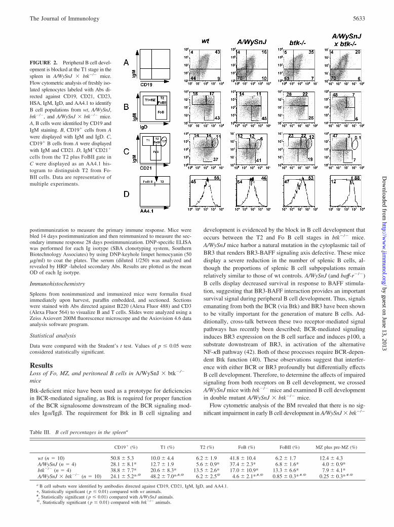
FIGURE 2. Peripheral B cell devel-opment is blocked at the T1 stage in thespleen in A/WySnJ � btk�/� mice.Flow cytometric analysis of freshly iso-lated splenocytes labeled with Abs di-rected against CD19, CD21, CD23,HSA, IgM, IgD, and AA4.1 to identifyB cell populations from wt, A/WySnJ,btk�/�, and A/WySnJ � btk�/� mice.A, B cells were identified by CD19 andIgM staining. B, CD19� cells from Awere displayed with IgM and IgD. C,CD19� B cells from A were displayedwith IgM and CD21. D, IgM�CD21�
cells from the T2 plus FoBII gate inC were displayed as an AA4.1 his-togram to distinguish T2 from Fo-BII cells. Data are representative ofmultiple experiments.
Table III. B cell percentages in the spleena
CD19� (%) T1 (%) T2 (%) FoB (%) FoBII (%) MZ plus pre-MZ (%)
wt (n � 10) 50.8 � 5.3 10.0 � 4.4 6.2 � 1.9 41.8 � 10.4 6.2 � 1.7 12.4 � 4.3A/WySnJ (n � 4) 28.1 � 8.1* 12.7 � 1.9 5.6 � 0.9* 37.4 � 2.3* 6.8 � 1.6* 4.0 � 0.9*btk�/� (n � 4) 38.8 � 7.7* 20.6 � 8.3* 13.5 � 2.6* 17.0 � 10.9* 13.3 � 6.6* 7.9 � 4.1*A/WySnJ � btk�/� (n � 10) 24.1 � 5.2*,@ 48.2 � 7.0*,#,@ 6.2 � 2.5@ 4.6 � 2.1*,#,@ 0.85 � 0.3*.#.@ 0.25 � 0.3*.#.@
a B cell subsets were identified by antibodies directed against CD19, CD21, IgM, IgD, and AA4.1.�, Statistically significant ( p � 0.01) compared with wt animals.#, Statistically significant ( p � 0.01) compared with A/WySnJ animals.@, Statistically significant ( p � 0.01) compared with btk�/� animals.
5633The Journal of Immunology
by guest on June 13, 2013http://w
ww
.jimm
unol.org/D
ownloaded from

mice compared with single mutant mice (Fig. 1, A and B, andTable I); however, the double mutant mice have slightly reducednumbers of pro-pre-B cells and immature B cells (Table II). Fur-thermore, the proportions and numbers of mature recirculating Bcells, which express lower levels of IgM and higher levels of IgDwere further reduced in A/WySnJ � btk�/� mice compared withsingle mutant animals (Fig. 1, A and C, and Table II).
To investigate the effects of loss of mature recirculating B cellson the peripheral B cell compartment, we analyzed splenic B cellsfrom A/WySnJ � btk�/� mice compared with those from singlemutant mice. Although the proportion and number of CD19� Bcells was reduced in A/WySnJ � btk�/� mice compared withbtk�/� mice, it was not significant when compared with A/WySnJmice (Fig. 2A and Tables III and IV). Upon further analysis, wefound that B cells in the spleens of A/WySnJ � btk�/� mice werecomprised almost entirely of T1 (CD19�IgM�IgD�CD21�
AA4�) B cells with a relatively small proportion of T2 (CD19�
IgM�IgD�CD21�AA4�) B cells and the virtual absence of ma-ture Fo (CD19�IgMlowIgD�CD21�AA4�) and MZ (CD19�
IgMhighIgD�CD21highAA4�) B cells, as well as the newlydescribed Fo II B cell population (CD19�IgMhighIgD�CD21�
AA4�) (Fig. 2, B–D, and Table III) (18). Consistent with increasedproportions, splenic T1 B cell numbers were increased in
A/WySnJ � btk�/� mice compared with single mutant animals,whereas FoBI, FoBII, and MZ B cell numbers were significantlyreduced in A/WySnJ � btk�/� mice compared with A/WySnJ orbtk�/� single mutant animals (Table IV). Accumulation of T1 Bcells suggests that a block in B cell development occurs at the T1stage in the spleens in A/WySnJ � btk�/� mice.
Given the severe B cell deficiency, the splenic architecture andlymphoid follicles were examined by histology. In contrast toA/WySnJ and btk�/� mice and consistent with a more severe B celldeficiency, spleens from A/WySnJ � btk�/� double mutant miceshowed smaller lymphoid follicles (Fig. 3A). Additionally, the Bcell compartment in the follicles of the spleen, which is comprisedprimarily of T2 and mature Fo B cells, was analyzed by immuno-histochemical analysis. As shown in Fig. 3B, B cells in spleensfrom wt and btk�/� mice form large and dense B cell zones, al-though follicular architecture is not well organized. Consistentwith a previous report, the B cell zones in the spleens of A/WySnJmice are smaller and poorly organized compared with the controls,whereas the T cell zones are expanded (43). The B cell zones in thespleens of A/WySnJ � btk�/� mice are severely reduced in sizeand number compared with single mutant animals, and the splenicarchitecture is disordered with very few dispersed B cells. Thisresult is consistent with an accumulation of predominantly T1 B
FIGURE 3. Defective splenic Bcell organization in A/WySnJ �btk�/� mice. H&E staining (A) andimmunofluorescent staining (B) ofspleens from wt, A/WySnJ, btk�/�,and A/WySnJ � btk�/� (A�B) miceare shown; green, B220; red, CD3.The B cell areas from each genotypeare calculated in the graph; ��, p �0.005; and ����, p � 0.0001.
Table IV. B cell numbers in the spleena
CD19� (n) T1 (n) T2 (n) FoB (n) FoBII (n) MZ plus pre-MZ (n)
wt (n � 10) 40.4 � 13.4 4.4 � 2.8 2.6 � 1.4 16.2 � 4.8 2.5 � 1.1 4.9 � 1.9A/WySnJ (n � 4) 10.8 � 1.7* 1.4 � 0.3 0.60 � 0.07* 4.1 � 0.9* 0.75 � 0.24* 0.44 � 0.16*btk�/� (n � 4) 17.2 � 7.4* 3.2 � 1.2 2.2 � 0.6 3.5 � 3.3* 1.9 � 0.4* 1.6 � 1.3*A/WySnJ � btk�/� (n � 10) 11.7 � 5.2* 5.9 � 3.0# 0.70 � 0.31*,@ 0.61 � 0.44*,#,@ 0.11 � 0.05*,#,@ 0.033 � 0.02*,#,@
a Numbers (n) are given in millions.�, Statistically significant ( p � 0.01) compared with wt animals.#, Statistically significant ( p � 0.01) compared with A/WySnJ animals.@, Statistically significant ( p � 0.01) compared with btk�/� animals.
5634 LACK OF MATURE B CELLS IN BCR AND BR3 SIGNALING-DEFECTIVE MICE
by guest on June 13, 2013http://w
ww
.jimm
unol.org/D
ownloaded from

cells, which reside outside the follicles, in the double mutant mice.Together, these data demonstrate that the compound loss of Btk(and, thus, impaired BCR-mediated signaling) and functional BR3disrupts splenic architecture, leads to a drastic reduction in B cells,and results in a reduction of both the frequency and size of thesplenic follicular structures.
Btk and BR3 function appear to be dispensable for T cell de-velopment; therefore, as expected, the development of conven-tional T cells in the thymus and spleen remained unaltered inA/WySnJ � btk�/� mice; T cell numbers as well as the ratio ofCD4:CD8 cells remains similar to that observed in control animals(data not shown). Additionally, T cell zones in follicles of thespleen are present in all animals (Fig. 3B).
In addition to the splenic B cell deficiencies, there is also asevere deficiency of peritoneal cavity B cells in A/WySnJ � btk�/�
mice. In contrast to wt and A/WySnJ mice, which possess all peri-toneal cavity B cell populations, and btk�/� mice, which possesperitoneal B2 B cells but lack B1 B cells, there is an almost com-plete absence of all peritoneal cavity B cells in A/WySnJ � btk�/�
mice (Fig. 4 and Tables V and VI). Taken together, these datasuggest that development of all peripheral B cells, no matter theirlocation, requires signaling through both the BCR and BR3.
B cell developmental defects are cell intrinsic in A/WySnJ �btk�/� mice
After observing that peripheral B cell development is impaired inA/WySnJ � btk�/� mice, we wanted to determine whether thesedefects were intrinsic to the mutant B cells or whether additionalfactors (such as aberrant splenic architecture) contribute to im-paired B cell development. We therefore generated BM chimerasusing B6.SJL mice, a congenic wt strain of mice that expresses theCD45.1 allele, whereas all of the strains we have used in this study
express the CD45.2 allele. Thus, through flow cytometric staining,cells originating from either a donor or a recipient animal can bedistinguished. Following lethal irradiation, B6.SJL animals wereinjected with BM cells derived from wt or A/WySnJ � btk�/�
mice, A/WySnJ � btk�/� mice were injected with BM cells de-rived from B6.SJL animals, and B cell populations were analyzed8 wk later. Reconstitution ranged from 60 to 96% complete asdetermined by flow cytometry, and in no case did the remainingrecipient cells significantly contribute to the B cell compartment(Tables VII and VIII). Fig. 5 reveals that B cell developmentaldefects are intrinsic to B cells from A/WySnJ � btk�/� mice, asevidenced by the absence of mature B cells in B6.SJL mice re-constituted with BM from A/WySnJ � btk�/� mice. In contrast,A/WySnJ � btk�/� mice reconstituted with BM derived fromB6.SJL mice possess all splenic B cell populations, revealing thatthe splenic architecture (beyond the lack of mature B cells) is notimpaired in A/WySnJ � btk�/� animals.
Reduced serum Ig levels in A/WySnJ � btk�/� mice
Because B cell development was impaired in A/WySnJ � btk�/�
mice, we wanted to determine whether B cell functional responseswere also blunted in double mutant mice. Serum levels of Ig iso-types from nonimmunized wt, A/WySnJ, btk�/�, and A/WySnJ �btk�/� mice were determined by ELISA (Fig. 6). Serum levels ofall Ig isotypes were further reduced in A/WySnJ � btk�/� doublemutant mice relative to either single mutation alone.
A/WySnJ � btk�/� mice display defective humoral immuneresponses to the TI-I Ag TNP-LPS
Our results show that A/WySnJ � btk�/� mice possess primarilyT1 B cells and have reduced serum Abs; however, these experi-ments do not reveal the functional capacity of these T1 B cells in
FIGURE 4. Absence of peritonealcavity B cells in A/WySnJ � btk�/�
mice. Shown is flow cytometric anal-ysis of cells isolated from the perito-neal cavity of wt, A/WySnJ, btk�/�,and A/WySnJ � btk�/� mice stainedwith Abs directed against B220, IgM,IgD, and CD5 for the identification ofperitoneal B cell populations. A, Peri-toneal cells are displayed with IgMand IgD to identify B1 and B2 B cellpopulations. B, IgM�IgD� B1 B cellsfrom A were displayed with B220 andCD5 to identify B1-a and B1-b B cellpopulations. Data are a representativeof multiple experiments.
Table V. B cell percentages in the peritoneal cavitya
B2 (CD5�) (%) B1 (%) B1-a (%) B1-b (%)
wt (n � 4) 10.8 � 1.9 51.8 � 5.2 21.1 � 5.0 11.4 � 7.9Btk�/� (n � 4) 10.7 � 0.49 2.0 � 0.36 0.1 � 0.11 0.5 � 0.1A/WySnJ (n � 4) 4.6 � 0.17 22 � 2.1 11 � 2.3 4.2 � 1.0A/WySnJ � btk�/� (n � 6) 0.15 � 0.16*.#.@ 0.86 � 1.0 0.046 � 0.06 0.39 � 0.6
a B cell subsets were identified by antibodies directed against B220, CD5, IgM and IgD.�, Statistically significant ( p � 0.005) compared with wt animals.#, Statistically significant ( p � 0.05) compared with A/WySnJ animals.@, Statistically significant ( p � 0.00001) compared with btk�/� animals.
5635The Journal of Immunology
by guest on June 13, 2013http://w
ww
.jimm
unol.org/D
ownloaded from

the context of an immune response involving complex interactionswith accessory cells and functional expression of costimulatorymolecules. In this regard, recent studies have shown that wt T1 Bcells respond to bacterial TLR ligands both in vitro and in vivo byproliferation, differentiation into Ab-producing plasma cells, andthe production of IgM and IgG Abs. Furthermore, BAFF has beenshown to enhance proliferation and Ig isotype switching in vitro(44). Therefore it was of particular interest to investigate the ca-pacity of mutant T1 B cells in A/WySnJ � btk�/� mice to produceAbs in response to the TI-I Ag, TNP-LPS. The wt, A/WySnJ,btk�/�, and A/WySnJ � btk�/� mice were immunized with TNP-LPS, and immunogen-specific Ab titers were measured 7 days af-ter immunization (Fig. 7A). In response to TNP-LPS immuniza-tion, wt mice produced a significant amount of TNP-specific IgMand IgGs (IgG1, IgG2a, IgG2b, and IgG3). A/WySnJ and btk�/�
mice also produced detectable amounts of total TNP-specific Igscompared with base-line Ab titers; however, the amount of Absproduced was significantly decreased compared with the wt controlanimals. Notably, the A/WySnJ mutation did not alter the TNP-specific IgM response, possibly due to an intact B1 B cell com-partment. In contrast, a decrease in the production of TNP-specificIgGs in A/WySnJ mice suggests that BR3 signaling plays a role inisotype switching in response to TI-I Ags. Except for IgG2b andIgM, the levels of all Ab isotypes were somewhat reduced inbtk�/� mice. These findings are consistent with previous reports(45, 46). Compared with single mutant animals, A/WySnJ �btk�/� mice were severely impaired in the production of signifi-cant levels of all Ag-specific Ig isotypes in response to TNP-LPS,although a small increase in TNP-specific IgM and IgG2b Abs wasdetected.
To investigate whether alterations in the splenic architecturecontributed to the reduced immune responses in A/WySnJ �btk�/� mice, we performed immunohistochemical analysis ofspleens from TNP-LP- immunized mutant and control mice. Asexpected, we observed an increase in the size and frequency of Bcell zones in the spleens of wt mice (Fig. 7B). Consistent with ourprevious findings (46), B cell zones in the spleens of btk�/� micealso appear to increase in response to TNP-LPS immunization,although to a lesser extent than in wt animals (Fig. 7, B and C). Inaddition, we found that A/WySnJ mice are also impaired in theincrease of B cell zones in response to TNP-LPS immunization, as
evidenced by their reduced frequency and size. In contrast to wt aswell as either single mutant, the B cell zones do not increase infrequency or size in the spleens of A/WySnJ � btk�/� mutant miceafter TNP-LPS immunization (Fig. 7, B and C). Thus, the lack ofboth BCR and BR3 signaling prevents BCR plus TLR-4-inducedexpansion of the remaining B cells (T1) in response to TI-I Agencounter. Taken together with decreased Ab production, this re-sult suggests that the T1 cells from double mutant A/WySnJ �btk�/� mice are more severely defective in TLR-4 signaling,which promotes plasmacytic differentiation, isotypic switching,and Ab secretion.
A/WySnJ � btk�/� mice display defective humoral immuneresponses to the TD Ag DNP-HSA
Although a lack of mature B cells in A/WySnJ � btk�/� micepredicts reduced Ab responses to antigenic challenge, it also pro-vides a unique opportunity to test whether T cell help can promotethe differentiation of T1 B cells into Ab-producing plasma cells. Inthis regard, prior studies have shown that T cell help can rescue wtT1 cells from BCR-induced apoptosis (47). Therefore, we immu-nized A/WySnJ � btk�/� and control mice with the T cell-depen-dent Ag DNP-HSA. Ab titers in the serum of wt, A/WySnJ, btk�/�,and A/WySnJ � btk�/� mice were analyzed preimmunization and7 days postimmunization (Fig. 8A). In response to DNP-HSA im-munization, wt mice produced significant levels of DNP-specificIgs, including increased IgM, IgG1, IgG2a, and IgG2b. A/WySnJmice responded similarly to wt mice in their DNP-specific IgMprofile; however, all other isotypes were decreased compared withwt, as previously reported (43). Btk�/� mice displayed a reductionin levels of DNP-specific Igs, including IgM, compared withA/WySnJ mice, which is consistent with the finding that primaryimmune responses to TD Ags are defective in Xid mice (48–51).In contrast to either single mutant animal, A/WySnJ � btk�/� dou-ble mutant mice were unable to mount any Ab response followingDNP-HSA immunization; no increase in Ig levels was detected(Fig. 8A). Immunohistochemical staining of the spleens fromDNP-HAS-immunized mice reveals presence of GCs in wt,A/WySnJ, and btk�/� mice, albeit the GCs were smaller and lessfrequent in single mutant mice relative to wt mice (Fig. 8, B andC). In contrast, fewer and smaller B cell zones, which did not organizeinto GCs, were observed in immunized A/WySnJ � btk�/� mice
Table VI. B cell numbers in the peritoneal cavitya
B2 (CD5�) (n) B1 (n) B1-a (n) B1-b (n)
wt (n � 10) 0.062 � 0.028 0.308 � 0.16 0.068 � 0.04 0.028 � 0.01Btk�/� (n � 4) 0.028 � 0.003 0.003 � 0.001 0.0051 � 0.005 0.0029 � 0.0018A/WySnJ (n � 4) 0.022 � 0.007 0.135 � 0.056 0.015 � 0.007 0.0057 � 0.0027A/WySnJ � btk�/� (n � 6) 0.00042 � 0.0003*,#,@ 0.0009 � 0.0007 0.00008 � 0.00001 0.00006 � 0.0001
a Numbers (n) are given in millions.�, Statistically significant ( p � 0.01) compared with wt animals.#, Statistically significant ( p � 0.005) compared with A/WySnJ animals.@, Statistically significant ( p � 0.00005) compared with btk�/� animals.
Table VII. Donor B cell percentages in the spleena
Donor Cells (%) CD19� (%) T1 (%) T2 (%) FoB (%) MZ (%)
wt into B6.SJL (n � 3) 89.6 � 0.6 31.8 � 2.5 16.3 � 1.2 14.9 � 1.3 36.2 � 2.3 8.6 � 1.9A � b into B6.SJL (n � 4) 69.3 � 13.9 19.3 � 9.0 43.5 � 13.7* 5.4 � 0.8* 9.0 � 3.0* 3.0 � 0.9*B6.SJL into A � b (n � 3) 84.1 � 1.5 32.5 � 10.7 13.5 � 2.6 22.3 � 1.2 37.5 � 4.7 5.4 � 0.9
a Donor B cell subsets were identified by antibodies directed against CD45.1, CD45.2, CD19, IgM, IgD, and AA4.1. A� B refers to A/WySnJ � btk�/�
mice.�, Statistically significant ( p � 0.02) compared with B6.SJL reconstituted with wt BM.
5636 LACK OF MATURE B CELLS IN BCR AND BR3 SIGNALING-DEFECTIVE MICE
by guest on June 13, 2013http://w
ww
.jimm
unol.org/D
ownloaded from

(Fig. 8, B and C). These results demonstrate that A/WySnJ andbtk�/� single mutant mice respond to TD Ag at a reduced level,consistent with the impaired production of Ag-specific Igs. Con-sistent with a lack of Ag-specific Abs, no significant expansion ofB cell zones or formation of GCs was observed in A/WySnJ �btk�/� mice (Fig. 8, B and C).
We also tested the possibility that the TD immune responses aredelayed in the double mutant mice by measuring secondary immuneresponses to DNP-HSA immunization. Following primary immuni-zation, animals were reimmunized on day 14 after primary immuni-zation, and spleens were collected 28 days after primary immuniza-tion. The analysis of B cell zones and GCs by immunohistochemistryreveals the presence of GCs in A/WySnJ, and btk�/�; however, theGCs in the single mutant mice were smaller relative to those in the wtmice (Fig. 8, D and E). In contrast to single mutant animals, B cellzones and GCs were barely detectable in A/WySnJ � btk�/� mice(Fig. 8, D and E). Consistent with severely reduced B cell zones andGCs, only a small anti-DNP response (mainly IgG1) was detectable inA/WySnJ � btk�/� mice (data not shown). These results demonstratethat A/WySnJ � btk�/� mice are severely defective in TD immuneresponses and suggest that the transitional B cells produced inA/WySnJ � btk�/� double mutant mice are not capable of respondingto TD antigenic challenge significantly.
Impaired in vitro B cell proliferation in A/WySnJ � btk�/�
mice
Following the observations that B cell development and Ab re-sponses were impaired in double mutant mice, we wanted to de-termine whether these defects were B cell autonomous. Becausethe splenic B cell compartment of A/WySnJ � btk�/� mice iscomprised almost entirely of T1 B cells, we sorted AA4.1 highlyexpressing cells (which were 60–80% T1 B cells for all geno-types, as determined by flow cytometry; Fig. 9A) from each ge-notype to ensure that similar cell populations were compared in
our in vitro studies. AA4.1high B cells from wt, A/WySnJ, btk�/�,and A/WySnJ � btk�/� mice were labeled with CFSE and culturedfor 3 days in the presence of LPS, anti-IgM, and anti-CD40 plusIL-4 as surrogates for TI-I, TI-II, and TD immune responses invitro. Proliferation of B cells, as measured by dilution of CFSE,was analyzed by flow cytometry. As shown in Fig. 9B, wtAA4.1high cells were mildly responsive to anti-IgM stimulationand are quite responsive to LPS and anti-CD40 plus IL-4 stimu-lation, as evidenced by the dilution of CFSE. AA4.1high cells fromA/WySnJ and btk�/� mice were refractory to anti-IgM stimulationbut did proliferate to varying degrees in response to LPS and anti-CD40 plus IL-4. In contrast, AA4.1high cells from A/WySnJ �btk�/� mice do not respond to anti-IgM or LPS stimulation andproliferate only weakly to anti-CD40 plus IL-4 stimulation. How-ever, T1 cells from A/WySnJ � btk�/� mice are not entirely de-ficient for signaling potential from each of these ligands comparedwith controls, as AA4.1high cells from all genotypes were able toup-regulate the early activation marker CD69 in response to 12 hof stimulation with anti-IgM, LPS, and anti-CD40 (Fig. 9C). Thus,the impairment of mutant B cells to respond to multiple stimuli invitro may explain the developmental and functional defects weobserved in vivo in A/WySnJ, btk�/�, and A/WySnJ � btk�/�
mutant mice.
DiscussionSignals emanating from the BCR and BR3 play pivotal roles in thedevelopment of transitional B cells into fully functional mature Bcells in the secondary lymphoid organs; whereas BCR signalingdrives the generation of a functional mature B lymphocyte reper-toire by promoting the differentiation of transitional precursors inthe spleen and is necessary for maintaining self-tolerance, BR3-mediated signals promote the metabolic fitness and survival ofcells undergoing this differentiation process (25, 27, 52). Recentstudies have shown that BCR signaling regulates the expression
FIGURE 5. B cell developmental defects are intrinsic in A/WySnJ � btk�/� mice. Flow cytometric analysis of splenocytes isolated from wt andA/WySnJ � btk�/� (A�B) mice, as well as lethally irradiated B6.SJL mice reconstituted with BM from wt and A/WySnJ � btk�/� (mutant) mice orA/WySnJ � btk�/� (mutant) mice lethally irradiated and reconstituted with BM from B6.SJL mice. Freshly isolated splenocytes were labeled with Absdirected against CD45.1 and CD45.2 to identify donor and recipient cells, as well as Abs directed against CD19, IgM, IgD, CD23, and AA4.1 for B cellsubset identification. A, Donor B cells were identified by CD19 and IgM staining. B, CD19� cells from A were displayed with IgM and IgD to identifyB cell populations (n � 3).
Table VIII. Donor B cell numbers in the spleena
Donor cells (n) CD19� (n) T1 (n) T2 (n) FoB (n) MZ (n)
wt into B6.SJL (n � 3) 66.4 � 13.8 21.0 � 4.5 3.4 � 0.8 3.1 � 0.6 7.7 � 2.0 1.8 � 0.6A � B into B6.SJL (n � 4) 5.2 � 2.5* 1.1 � 1.0 0.58 � 0.63* 0.07 � 0.07* 0.08 � 0.05* 0.03 � 0.03*B6.SJL into A � b (n � 3) 39.8 � 4.3* 12.8 � 3.5 1.9 � 0.4 2.9 � 0.9 4.9 � 1.7 0.68 � 0.28
a Numbers (n) are given in millions. A� B refers to A/WySnJ � btk�/� mice.�, Statistically significant ( p � 0.01) compared with B6.SJL reconstituted with wt BM.
5637The Journal of Immunology
by guest on June 13, 2013http://w
ww
.jimm
unol.org/D
ownloaded from

and function of BR3 beginning at the T2 stage, suggesting thatintegrated signaling by these two receptors regulates late stages ofB cell development (40–42). Furthermore, Btk regulates BCR-induced expression of BR3 and NF-�B2, which is essential for thesurvival function of BR3 (40). To investigate the biologic signif-icance of concomitant BCR and BR3 signaling in peripheral B celldevelopment, we have analyzed mutant mice that are partially de-fective in both BCR (btk�/�) and BR3 (A/WySnJ) signaling. Wefound that the compound deficiency of signaling from both BR3and BCR (A/WySnJ � btk�/� mice) leads to a complete block inperipheral B cell development at the most immature (T1) stage inthe spleen; this developmental block results in a block of the emer-gence of late transitional (T2 and T3) and mature splenic B cell(FoBI, FoBII, and MZB) populations as well as both peritoneal B1and B2 B cell populations in the mouse. Although the B cell com-partment is variably affected in either of the single mutants—btk�/� mice display a specific severe reduction of mature splenicFo B cells and peritoneal B1 B cells whereas A/WySnJ mice dis-
play a generalized reduction in all splenic B cell populations—thenonemergence of all mature B cells in our A/WySnJ � btk�/� miceis a far more severe B cell developmental defect than that observedin either single mutant alone. This defect reveals that in addition tothe crosstalk between BCR and BR3, the independent functions ofthese receptors facilitate development of all peripheral B cells be-yond the T1 stage, which serves as a BCR-sensitive checkpoint fornegative selection.
Mutant models of BCR and BR3 signaling have established arole for these receptors in late transitional and mature B cell sur-vival. For example, A/WySnJ mice have previously been shown todisplay a severe, generalized decrease in all splenic B cells, al-though all subsets can still be detected (53–56). In several studies,a minor increase in T1 B cells as well as a decrease in mature Foand MZ B cells in A/WySnJ mice has been reported (53, 54),whereas other studies did not find these differences (56). Althoughthere was a significant decrease in numbers, we observed approxi-mately similar proportions of all splenic B cell subpopulations except
FIGURE 6. Serum Igs are decreased inA/WySnJ � btk�/� mice. Serum obtainedfrom wt (circles), A/WySnJ (triangles),btk�/� (diamonds) and A/WySnJ � btk�/�
(A � B; squares) mice was screened byELISA for identification of Igs. Data are dis-played as the concentration of each Ig iso-type (n � 4). IgG2c is expressed in mice ona C57BL/6 background (btk�/� mice),whereas mice with most other backgroundsdo not express IgG2c but do express IgG2a.
5638 LACK OF MATURE B CELLS IN BCR AND BR3 SIGNALING-DEFECTIVE MICE
by guest on June 13, 2013http://w
ww
.jimm
unol.org/D
ownloaded from

MZ B cells in A/WySnJ mice compared with wt controls (Fig. 2and Table I). However, we found that the peritoneal B1 as well asB2 cell populations in A/WySnJ mice were comparable to wt con-trols (Fig. 3). The BCR signaling defect that arises due to the lossof Btk leads to a block in B cell development between the T2 andFo I B cell stage and results in reduced mature follicular B cells inthe spleen; however, development of MZ B cells appears to beonly marginally affected (20, 77). Furthermore, an absence of B1cells and a reduction in B2 cells has also been reported in theperitoneal cavity of btk�/� mice (20). In addition to these knownphenotypes, we found an increased proportion of T1 B cells inbtk�/� mice compared with controls, suggesting that Btk signalingmay play a role in T1 B cell function as well (Fig. 2A and TableI). In this regard, we observed an increase in both T1 proportionsand numbers in A/WySnJ � btk�/� mice compared with eithersingle mutant. This block in B cell development at the T1 stage inA/WySnJ � btk�/� mice, which results in the accumulation of T1B cells, is radical and reveals that parallel BCR-dependent andBR3-dependent signaling pathways are more critical for T1 celldifferentiation into later stages of transitional B cell populationsthan previously appreciated. Additionally, the virtual lack of B cellfollicles in the spleens of double mutant mice is consistent with asevere reduction in all Fo B cell populations. Furthermore, thecomplete absence of B2 cells in the peritoneal cavity in theA/WySnJ � btk�/� mice, which is significantly more severe thanin btk�/� mice, suggests that this B cell population may also re-quire normal splenic T1 B cell development for its generation.
Following immunization with TNP-LPS, a TI- type 1 Ag thatactivates B cells via TLR4, A/WySnJ or btk�/� single mutant mice
produced only modest TNP-specific Ab responses relative to wtmice, and A/WySnJ � btk�/� mice were unable to generate animmune response. This may be explained by a known function forBtk in TLR4 signaling and the regulation of Ig isotype switchingby BR3 (46, 57, 58). In addition to defective TI-I immune re-sponses, the primary immune responses to DNP-HSA (TD Ag) inA/WySnJ or btk�/� single mutant animals were also significantlyimpaired compared with wt animals, although DNP-specific re-sponses were more severely impaired in btk�/� mice comparedwith A/WySnJ mice. Following secondary immunization, Ab re-sponses in A/WySnJ mice were comparable to those of wt controls,whereas btk�/� mice mounted only minor Ab responses.A/WySnJ � btk�/� mice lacked both primary and secondary Abresponses to DNP-HSA. Thus, T1 cells generated in A/WySnJ �btk�/� mice are functionally incompetent to respond to TLR4 orCD40 signaling, perhaps because they lack the signaling compo-nent(s) necessary for a productive response to TLR4 and CD40engagement. In this regard, we observed a lack of proliferativeresponse of A/WySnJ � btk�/� T1 B cells to anti-CD40 and LPSin vitro (Fig. 9), which is consistent with the idea that T1 B cellsproduced in mice with impaired BCR and BR3 signaling lack thesignaling capacity necessary for both TLR4- and CD40-mediatedsignaling. Because signaling from both receptors converges onNF-�B to promote activation and proliferation of B cells, it ispossible that reduced immune responses to TD and TI-I Ags andlack of B cell proliferation in response to TLR4 and CD40 stim-ulation in A/WySnJ � btk�/� T1 B cells compared with eitherbtk�/� or A/WySnJ T1 B cells is attributable to defects in NF-�Bactivation.
FIGURE 7. Humoral immune re-sponses to the TI-I Ag TNP-LPS aredefective in A/WySnJ � btk�/� mice.A, Serum obtained from wt (E),A/WySnJ (A/W; triangles), btk�/�(di-amonds), and A/WySnJ � btk�/�
(A�B; squares) mice before immuni-zation (closed symbols) and 7 dayspostimmunization with TNP-LPS (opensymbols) was screened by ELISA forthe production of TNP-specific Igs.Data are displayed as the OD values ofeach TNP-specific Ig isotype (n � 4).The p values for Ig isotypes in TNP-LPS immunized mice are as follows: �,p � 0.05; ��, p � 0.005; ���, p �0.0005; and ����, p � 0.0001. B, Im-munofluorescent staining of spleensfrom wt, A/WySnJ, btk�/�, andA/WySnJ � btk�/� mice on day 7postimmunization with TNP-LPS;green, B220; red, CD3. C, The num-ber of GCs (top) and the number of Bcell areas within the GC areas (bot-tom) from each genotype were calcu-lated and are shown in the graphs.
5639The Journal of Immunology
by guest on June 13, 2013http://w
ww
.jimm
unol.org/D
ownloaded from

Stimulation through the BCR leads to the recruitment of multi-ple signaling molecules into a membrane microdomain. This sig-naling complex, termed the BCR signalosome, is necessary for thetransduction of BCR-induced signals. Thus, the loss of any com-ponent of the BCR signalosome leads to a block in B cell devel-opment predominantly between the T2 and FoBI stages, termedXid. B cells from all of these mutant animal models, including Btk,share one common feature: the failure to activate the classicalNF-�B pathway following anti-IgM stimulation (59–62). Like-wise, mice in which BR3 function is impaired (A/WySnJ) displaydefects in peripheral B cell development beyond the T1 stage;however, all B cell subpopulations are still present in A/WySnJmice. The significant reduction in B cell numbers is likely due todefective BR3-mediated survival signaling, which at least in partinvolves the activation of NF-�B. In this regard, early studies ofBAFF function in primary B cells revealed that BR3-mediatedsignaling activates the alternative NF-�B pathway, as evidenced
by IkB kinase (IKK) �-dependent processing of p100 to p52 (23,63–65). Additionally, processing of p100 is impaired in B cellsfrom A/WySnJ mice, confirming a role for BR3 signaling in theregulation of alternative NF-�B activity in response to BAFF (64).The classical NF-�B pathway has also been shown to be activatedfollowing stimulation via BR3, although less robustly than the al-ternative pathway (66–69). Moreover, primary B cells deficientfor p50 and/or p52 also display impaired survival following stim-ulation with soluble BAFF in vitro, further supporting the role forboth classical and alternative NF-�B pathways in BAFF-mediatedB cell survival (23, 66, 67).
The requirement for NF-�B activity in the generation of a func-tional nonself reactive B cell repertoire is further evidenced byimpaired B cell development and function in animals lacking var-ious signaling components that regulate the nuclear translocationof NF-�B DNA binding subunits, as well as the NF-�B subunitsthemselves (reviewed in Ref. 70). For example, conditional loss of
FIGURE 8. Humoral immune re-sponses to the TD Ag DNP-HSA aredefective in A/WySnJ � btk�/� mice.A, Serum obtained from wt (circles),A/WySnJ (A/W; triangles), btk�/� (di-amonds), and A/WySnJ � btk�/�
(A�B; squares) mice before immuni-zation (closed symbols) and 7 dayspostimmunization with DNP-HSA(open symbols) was screened byELISA for production of DNP-spe-cific Igs. Data are displayed as the ODvalues of each DNP-specific Ig iso-type (n � 4). The p values for the Igisotypes in DNP-HSA immunizedmice are as follows: �, p � 0.05; ��,p � 0.005; ���, p � 0.0005; and����, p � 0.0001. B and D, Immuno-fluorescent staining of spleens fromwt, A/WySnJ, btk�/�, and A/WySnJ �btk�/� mice on day 7 (B) and day 28(D) postimmunization with DNP-HSA; green, B220; red, CD3. C andE, The number of GCs and the num-ber B cell areas within the GCs fromeach genotype are calculated in thegraphs.
5640 LACK OF MATURE B CELLS IN BCR AND BR3 SIGNALING-DEFECTIVE MICE
by guest on June 13, 2013http://w
ww
.jimm
unol.org/D
ownloaded from

IKK�, IKK�, or IKK� results in a generalized decrease in B cellnumbers and impaired survival of the remaining cells (71–73),whereas loss of the inhibitor of NF-�B (IkB�) results in an in-crease in B cells that are hyper-responsive to stimulation (74). Lossof the individual NK-�B subunits RelA, RelB, c-Rel, p50, and p52variably affects B cell generation and function, typically includingdecreased survival and hypo-responsiveness to BCR-mediated sig-nals, whereas constitutively active p50 leads to an inflammatoryphenotype (75). Additionally, compound loss of both p50 (classi-cal NF-�B) and p52 (alternative NF-�B) leads to a block in B celldevelopment at the T1 stage in the spleen (23, 76). The B celldevelopmental block observed in our double mutant A/WySnJ �btk�/� mice closely resembles that observed in p50�/� � p52�/�
double mutant animals. Because signaling from both BCR andBR3 has been shown to activate NF-�B, and both classical andalternative NF-�B have been shown to be important for B celldevelopment and function, it is tempting to speculate that the Bcell developmental block we observe in A/WySnJ � btk�/� micearises due to the inability of these cells to activate both arms ofNF-�B activity. Effective lack of NF-�B activation might also ex-plain the peritoneal B cell phenotype that we observed inA/WySnJ � btk�/� mice, which is far more severe than that ob-served in either single mutant alone. In addition to defectiveNF-�B activation, B cell defects in the A/WySnJ � btk�/� micemay also arise from impaired activation of the PI3K/Akt pathway,which is critical for promoting survival and metabolic fitness of Bcells via mTOR (mammalian target of rapamycin) and Pim-2 de-pendent pathways and for preventing apoptosis by inhibiting theinduction of Bim (25, 27).
In this study, by using mutant mice (A/WySnJ � btk�/�) that arepartially defective in both BCR and BR3 signaling, we have dem-onstrated that BCR- and BR3-mediated signals regulate the gen-eration of fully functional peripheral B cells. Although we ob-served no overt defects in early B cell development in the BM,development in the spleen was blocked at the T1 stage with thenonemergence of mature splenic B cells. This coincided with analmost complete loss of peritoneal cavity B cells. Investigation of
humoral immunity in A/WySnJ � btk�/� mice revealed a decreasein serum Igs at the basal level as well as following primary im-munization with TD Ags. This result was not completely unex-pected due to the lack of mature B cells of any sort in A/WySnJ �btk�/� mice. In this regard, the defective Ab response to immu-nization with TI-I Ag (TNP-LPS) may result from defective TLR-and BR3-induced B cell proliferation and Ig class switching. How-ever, even secondary immunization with the TD Ag (DNP-HSA)was unable to induce significant Ab production or B cell expansionin A/WySnJ � btk�/� mice, revealing that the T1 B cells presentin the double mutant animals are unable to be efficiently recruitedinto an immune response. Finally, B cells from A/WySnJ � btk�/�
mice were defective in B cell proliferation to multiple agonists invitro, possibly explaining the in vivo defects. Together, our pre-viously published work and the data presented in this study revealthat both integrated and independent BR3 and BCR signaling con-tribute to the development and function of mature peripheral Bcells.
AcknowledgmentsWe thank the Vanderbilt Immunohistochemistry Core for technical assis-tance with sample preparation and immunofluorescence analysis and Dr.George McNamara for technical assistance with immunofluorescence mi-croscopy. Microscopic analyses was performed at Diabetes Research In-stitute, University of Miami, Miami, FL. Flow cytometry experiments wereperformed in the Vanderbilt Medical Center Flow Cytometry and Immu-nology Shared Resource.
DisclosuresThe authors have no financial conflict of interest.
References1. Cancro, M. P., and J. F. Kearney. 2004. B cell positive selection: road map to the
primary repertoire? J. Immunol. 173: 15–19.2. Patke, A., I. Mecklenbrauker, and A. Tarakhovsky. 2004. Survival signaling in
resting B cells. Curr. Opin. Immunol. 16: 251–255.3. Allman, D., R. C. Lindsley, W. DeMuth, K. Rudd, S. A. Shinton, and
R. R. Hardy. 2001. Resolution of three nonproliferative immature splenic B cellsubsets reveals multiple selection points during peripheral B cell maturation.J. Immunol. 167: 6834–6840.
FIGURE 9. In vitro proliferationof AA4.1high B cells from A/WySnJ �btk�/� mice is impaired in responseto multiple stimuli. A, Postsort flowcytometric analysis of MACS-en-riched AA4.1high B cells from wt,A/WySnJ, btk�/�, and A/WySnJ �btk�/� mice. B, AA4.1high B cellswere labeled with CFSE and culturedfor 3 days in the presence of anti-IgM(�-IgM; 5 �g/ml), LPS (1 �g/ml), oranti-CD40 (5 �g/ml) plus IL-4 (10ng/ml). Proliferation was measured asthe dilution of CFSE in live (7-ami-noactinomycin D-negative) cells. C,AA4.1high B cells were cultured for12 h in the presence of anti-IgM (�-IgM; 10 �g/ml), LPS (1 �g/ml), oranti-CD40 (5 �g/ml). Activation wasmeasured as the up-regulation ofCD69 in live (7-aminoactinomycinD-negative) cells. Data are represen-tative of multiple experiments.
5641The Journal of Immunology
by guest on June 13, 2013http://w
ww
.jimm
unol.org/D
ownloaded from
4. Loder, F., B. Mutschler, R. J. Ray, C. J. Paige, P. Sideras, R. Torres,M. C. Lamers, and R. Carsetti. 1999. B cell development in the spleen takes placein discrete steps and is determined by the quality of B cell receptor-derivedsignals. J. Exp. Med. 190: 75–89.
5. Srivastava, B., W. J. Quinn III, K. Hazard, J. Erikson, and D. Allman. 2005.Characterization of marginal zone B cell precursors. J. Exp. Med. 202:1225–1234.
6. Pillai, S. 1999. The chosen few? Positive selection and the generation of naive Blymphocytes. Immunity 10: 493–502.
7. Rolink, A. G., C. Schaniel, J. Andersson, and F. Melchers. 2001. Selection eventsoperating at various stages in B cell development. Curr. Opin. Immunol. 13:202–207.
8. King, L. B., A. Norvell, and J. G. Monroe. 1999. Antigen receptor-induced signaltransduction imbalances associated with the negative selection of immature Bcells. J. Immunol. 162: 2655–2662.
9. Petro, J. B., R. M. Gerstein, J. Lowe, R. S. Carter, N. Shinners, and W. N. Khan.2002. Transitional type 1 and 2 B lymphocyte subsets are differentially respon-sive to antigen receptor signaling. J. Biol. Chem. 277: 48009–48019.
10. Su, T. T., and D. J. Rawlings. 2002. Transitional B lymphocyte subsets operateas distinct checkpoints in murine splenic B cell development. J. Immunol. 168:2101–2110.
11. Fuentes-Panana, E. M., G. Bannish, N. Shah, and J. G. Monroe. 2004. BasalIg�/Ig� signals trigger the coordinated initiation of pre-B cell antigen receptor-dependent processes. J. Immunol. 173: 1000–1011.
12. Reichlin, A., Y. Hu, E. Meffre, H. Nagaoka, S. Gong, M. Kraus, K. Rajewsky,and M. C. Nussenzweig. 2001. B cell development is arrested at the immature Bcell stage in mice carrying a mutation in the cytoplasmic domain of immuno-globulin �. J. Exp. Med. 193: 13–23.
13. Takata, M., H. Sabe, A. Hata, T. Inazu, Y. Homma, T. Nukada, H. Yamamura,and T. Kurosaki. 1994. Tyrosine kinases Lyn and Syk regulate B cell receptor-coupled Ca2� mobilization through distinct pathways. EMBO J. 13: 1341–1349.
14. Turner, M., P. J. Mee, P. S. Costello, O. Williams, A. A. Price, L. P. Duddy,M. T. Furlong, R. L. Geahlen, and V. L. Tybulewicz. 1995. Perinatal lethality andblocked B-cell development in mice lacking the tyrosine kinase Syk. Nature 378:298–302.
15. Chung, J. B., M. Silverman, and J. G. Monroe. 2003. Transitional B cells: step bystep towards immune competence. Trends Immunol. 24: 343–349.
16. Kraus, M., M. B. Alimzhanov, N. Rajewsky, and K. Rajewsky. 2004. Survival ofresting mature B lymphocytes depends on BCR signaling via the Ig�/� het-erodimer. Cell 117: 787–800.
17. Niiro, H., and E. A. Clark. 2002. Regulation of B-cell fate by antigen-receptorsignals. Nat. Rev. Immunol. 2: 945–956.
18. Cariappa, A., C. Boboila, S. T. Moran, H. Liu, H. N. Shi, and S. Pillai. 2007. Therecirculating B cell pool contains two functionally distinct, long-lived, posttran-sitional, follicular B cell populations. J. Immunol. 179: 2270–2281.
19. Cariappa, A., M. Tang, C. Parng, E. Nebelitskiy, M. Carroll, K. Georgopoulos,and S. Pillai. 2001. The follicular versus marginal zone B lymphocyte cell fatedecision is regulated by Aiolos, Btk, and CD21. Immunity 14: 603–615.
20. Khan, W. N., F. W. Alt, R. M. Gerstein, B. A. Malynn, I. Larsson, G. Rathbun,L. Davidson, S. Muller, A. B. Kantor, L. A. Herzenberg, et al. 1995. DefectiveB cell development and function in Btk-deficient mice. Immunity 3: 283–299.
21. Satterthwaite, A. B., Z. Li, and O. N. Witte. 1998. Btk function in B cell devel-opment and response. Semin. Immunol. 10: 309–316.
22. Fruman, D. A., A. B. Satterthwaite, and O. N. Witte. 2000. Xid-like phenotypes:a B cell signalosome takes shape. Immunity 13: 1–3.
23. Claudio, E., K. Brown, S. Park, H. Wang, and U. Siebenlist. 2002. BAFF-inducedNEMO-independent processing of NF-�B2 in maturing B cells. Nat. Immunol. 3:958–965.
24. Morrison, M. D., W. Reiley, M. Zhang, and S. C. Sun. 2005. An atypical tumornecrosis factor (TNF) receptor-associated factor-binding motif of B cell-activat-ing factor belonging to the TNF family (BAFF) receptor mediates induction ofthe noncanonical NF-�B signaling pathway. J. Biol. Chem. 280: 10018–10024.
25. Patke, A., I. Mecklenbrauker, H. Erdjument-Bromage, P. Tempst, andA. Tarakhovsky. 2006. BAFF controls B cell metabolic fitness through a PKC�-and Akt-dependent mechanism. J. Exp. Med. 203: 2551–2562.
26. Rolink, A. G., J. Tschopp, P. Schneider, and F. Melchers. 2002. BAFF is asurvival and maturation factor for mouse B cells. Eur. J. Immunol. 32:2004–2010.
27. Woodland, R. T., C. J. Fox, M. R. Schmidt, P. S. Hammerman, J. T. Opferman,S. J. Korsmeyer, D. M. Hilbert, and C. B. Thompson. 2008. Multiple signalingpathways promote B lymphocyte stimulator dependent B-cell growth and sur-vival. Blood 111: 750–760.
28. Yan, M., J. R. Brady, B. Chan, W. P. Lee, B. Hsu, S. Harless, M. Cancro,I. S. Grewal, and V. M. Dixit. 2001. Identification of a novel receptor for Blymphocyte stimulator that is mutated in a mouse strain with severe B cell de-ficiency. Curr. Biol. 11: 1547–1552.
29. Avery, D. T., S. L. Kalled, J. I. Ellyard, C. Ambrose, S. A. Bixler, M. Thien,R. Brink, F. Mackay, P. D. Hodgkin, and S. G. Tangye. 2003. BAFF selectivelyenhances the survival of plasmablasts generated from human memory B cells.J. Clin. Invest. 112: 286–297.
30. Klein, B., K. Tarte, M. Jourdan, K. Mathouk, J. Moreaux, E. Jourdan,E. Legouffe, J. De Vos, and J. F. Rossi. 2003. Survival and proliferation factorsof normal and malignant plasma cells. Int. J. Hematol. 78: 106–113.
31. Ng, L. G., A. P. Sutherland, R. Newton, F. Qian, T. G. Cachero, M. L. Scott,J. S. Thompson, J. Wheway, T. Chtanova, J. Groom, et al. 2004. B cell-activatingfactor belonging to the TNF family (BAFF)-R is the principal BAFF receptor
facilitating BAFF costimulation of circulating T and B cells. J. Immunol. 173:807–817.
32. Harless, S. M., V. M. Lentz, A. P. Sah, B. L. Hsu, K. Clise-Dwyer, D. M. Hilbert,C. E. Hayes, and M. P. Cancro. 2001. Competition for BLyS-mediated signalingthrough Bcmd/BR3 regulates peripheral B lymphocyte numbers. Curr Biol. 11:1986–1989.
33. Lentz, V. M., M. P. Cancro, F. E. Nashold, and C. E. Hayes. 1996. Bcmd governsrecruitment of new B cells into the stable peripheral B cell pool in the A/WySnJmouse. J. Immunol. 157: 598–606.
34. Sasaki, Y., S. Casola, J. L. Kutok, K. Rajewsky, and M. Schmidt-Supprian. 2004.TNF family member B cell-activating factor (BAFF) receptor-dependent and-independent roles for BAFF in B cell physiology. J. Immunol. 173: 2245–2252.
35. Schiemann, B., J. L. Gommerman, K. Vora, T. G. Cachero, S. Shulga-Morskaya,M. Dobles, E. Frew, and M. L. Scott. 2001. An essential role for BAFF in thenormal development of B cells through a BCMA-independent pathway. Science293: 2111–2114.
36. Miller, D. J., and C. E. Hayes. 1991. Phenotypic and genetic characterization ofa unique B lymphocyte deficiency in strain A/WySnJ mice. Eur. J. Immunol. 21:1123–1130.
37. Thompson, J. S., S. A. Bixler, F. Qian, K. Vora, M. L. Scott, T. G. Cachero,C. Hession, P. Schneider, I. D. Sizing, C. Mullen, et al. 2001. BAFF-R, a newlyidentified TNF receptor that specifically interacts with BAFF. Science 293:2108–2111.
38. Schneider, P., F. MacKay, V. Steiner, K. Hofmann, J. L. Bodmer, N. Holler,C. Ambrose, P. Lawton, S. Bixler, H. Acha-Orbea, et al. 1999. BAFF, a novelligand of the tumor necrosis factor family, stimulates B cell growth. J. Exp. Med.189: 1747–1756.
39. Batten, M., J. Groom, T. G. Cachero, F. Qian, P. Schneider, J. Tschopp,J. L. Browning, and F. Mackay. 2000. BAFF mediates survival of peripheralimmature B lymphocytes. J. Exp. Med. 192: 1453–1466.
40. Castro, I., J. A. Wright, B. Damdinsuren, K. L. Hoek, G. Carlesso, N. P. Shinners,R. M. Gerstein, R. T. Woodland, R. Sen, and W. N. Khan. 2009. B cell receptor-mediated sustained c-Rel activation facilitates late transitional B cell survivalthrough control of B cell activating factor receptor and NF-�B2. J. Immunol. 182:7729–7737.
41. Smith, S. H., and M. P. Cancro. 2003. Cutting edge: B cell receptor signalsregulate BLyS receptor levels in mature B cells and their immediate progenitors.J. Immunol. 170: 5820–5823.
42. Stadanlick, J. E., M. Kaileh, F. G. Karnell, J. L. Scholz, J. P. Miller,W. J. Quinn III, R. J. Brezski, L. S. Treml, K. A. Jordan, J. G. Monroe, et al.2008. Tonic B cell antigen receptor signals supply an NF-�B substrate for pro-survival BLyS signaling. Nat. Immunol. 9: 1379–1387.
43. Miller, D. J., K. D. Hanson, J. A. Carman, and C. E. Hayes. 1992. A singleautosomal gene defect severely limits IgG but not IgM responses in B lympho-cyte-deficient A/WySnJ mice. Eur. J. Immunol. 22: 373–379.
44. Ueda, Y., D. Liao, K. Yang, A. Patel, and G. Kelsoe. 2007. T-independentactivation-induced cytidine deaminase expression, class-switch recombina-tion, and antibody production by immature/transitional 1 B cells. J. Immunol.178: 3593–3601.
45. Brorson, K. A., M. V. Krasnokutsky, and K. E. Stein. 1995. Immunoglobulinisotype switching in xid mice. Mol. Immunol. 32: 487–494.
46. Khan, W. N., A. Nilsson, E. Mizoguchi, E. Castigli, J. Forsell, A. K. Bhan,R. Geha, P. Sideras, and F. W. Alt. 1997. Impaired B cell maturation in micelacking Bruton’s tyrosine kinase (Btk) and CD40. Int. Immunol. 9: 395–405.
47. Sater, R. A., P. C. Sandel, and J. G. Monroe. 1998. B cell receptor-inducedapoptosis in primary transitional murine B cells: signaling requirements and mod-ulation by T cell help. Int. Immunol. 10: 1673–1682.
48. Boswell, H. S., M. I. Nerenberg, I. Scher, and A. Singer. 1980. Role of accessorycells in B cell activation. III. Cellular analysis of primary immune response def-icits in CBA/N mice: presence of an accessory cell-B cell interaction defect.J. Exp. Med. 152: 1194–1309.
49. Press, J. L., and C. A. Giorgetti. 1986. Clonal analysis of the primary and sec-ondary B cell responses of neonatal, adult, and xid mice to (T,G)-A–L. J. Im-munol. 137: 784–790.
50. Ridderstad, A., G. J. Nossal, and D. M. Tarlinton. 1996. The xid mutation di-minishes memory B cell generation but does not affect somatic hypermutationand selection. J. Immunol. 157: 3357–3365.
51. Scher, I., A. K. Berning, and R. Asofsky. 1979. X-linked B lymphocyte defect inCBA/N mice. IV. Cellular and environmental influences on the thymus-depen-dent IgG anti-sheep red blood cell response. J. Immunol. 123: 477–486.
52. Otipoby, K. L., Y. Sasaki, M. Schmidt-Supprian, A. Patke, R. Gareus,M. Pasparakis, A. Tarakhovsky, and K. Rajewsky. 2008. BAFF activates Akt andErk through BAFF-R in an IKK1-dependent manner in primary mouse B cells.Proc. Natl. Acad. Sci. USA 105: 12435–12438.
53. Amanna, I. J., J. P. Dingwall, and C. E. Hayes. 2003. Enforced bcl-xL geneexpression restored splenic B lymphocyte development in BAFF-R mutant mice.J. Immunol. 170: 4593–4600.
54. Mayne, C. G., I. J. Amanna, F. E. Nashold, and C. E. Hayes. 2008. Systemicautoimmunity in BAFF-R-mutant A/WySnJ strain mice. Eur. J. Immunol. 38:587–598.
55. Rahman, Z. S., and T. Manser. 2004. B cells expressing Bcl-2 and a signaling-impaired BAFF-specific receptor fail to mature and are deficient in the formationof lymphoid follicles and germinal centers. J. Immunol. 173: 6179–6188.
56. Shulga-Morskaya, S., M. Dobles, M. E. Walsh, L. G. Ng, F. MacKay, S. P. Rao,S. L. Kalled, and M. L. Scott. 2004. B cell-activating factor belonging to the TNFfamily acts through separate receptors to support B cell survival and T cell-independent antibody formation. J. Immunol. 173: 2331–2341.
5642 LACK OF MATURE B CELLS IN BCR AND BR3 SIGNALING-DEFECTIVE MICE
by guest on June 13, 2013http://w
ww
.jimm
unol.org/D
ownloaded from

57. Alugupalli, K. R., S. Akira, E. Lien, and J. M. Leong. 2007. MyD88- and Bru-ton’s tyrosine kinase-mediated signals are essential for T cell-independent patho-gen-specific IgM responses. J. Immunol. 178: 3740–3749.
58. Gray, P., A. Dunne, C. Brikos, C. A. Jefferies, S. L. Doyle, and L. A. O’Neill.2006. MyD88 adapter-like (Mal) is phosphorylated by Bruton’s tyrosine kinaseduring TLR2 and TLR4 signal transduction. J. Biol. Chem. 281: 10489–10495.
59. Matsuda, S., Y. Mikami, M. Ohtani, M. Fujiwara, Y. Hirata, A. Minowa,Y. Terauchi, T. Kadowaki, and S. Koyasu. 2009. Critical role of class IA PI3Kfor c-Rel expression in B lymphocytes. Blood 115: 1037–1044.
60. Petro, J. B., and W. N. Khan. 2001. Phospholipase C-�2 couples Bruton’s ty-rosine kinase to the NF-�B signaling pathway in B lymphocytes. J. Biol. Chem.276: 1715–1719.
61. Petro, J. B., S. M. Rahman, D. W. Ballard, and W. N. Khan. 2000. Bruton’styrosine kinase is required for activation of I�B kinase and nuclear factor �B inresponse to B cell receptor engagement. J. Exp. Med. 191: 1745–1754.
62. Tan, J. E., S. C. Wong, S. K. Gan, S. Xu, and K. P. Lam. 2001. The adaptorprotein BLNK is required for B cell antigen receptor-induced activation of nu-clear factor-� B and cell cycle entry and survival of B lymphocytes. J. Biol.Chem. 276: 20055–20063.
63. Kanakaraj, P., T. S. Migone, B. Nardelli, S. Ullrich, Y. Li, H. S. Olsen,T. W. Salcedo, T. Kaufman, E. Cochrane, Y. Gan, et al. 2001. BLyS binds to Bcells with high affinity and induces activation of the transcription factors NF-�Band ELF-1 Cytokine 13: 25–31.
64. Kayagaki, N., M. Yan, D. Seshasayee, H. Wang, W. Lee, D. M. French,I. S. Grewal, A. G. Cochran, N. C. Gordon, J. Yin, et al. 2002. BAFF/BLySreceptor 3 binds the B cell survival factor BAFF ligand through a discrete surfaceloop and promotes processing of NF-kappaB2. Immunity 17: 515–524.
65. Mukhopadhyay, A., J. Ni, Y. Zhai, G. L. Yu, and B. B. Aggarwal. 1999. Iden-tification and characterization of a novel cytokine, THANK, a TNF homologuethat activates apoptosis, nuclear factor-�B, and c-Jun NH2-terminal kinase.J. Biol. Chem. 274: 15978–15981.
66. Enzler, T., G. Bonizzi, G. J. Silverman, D. C. Otero, G. F. Widhopf,A. Anzelon-Mills, R. C. Rickert, and M. Karin. 2006. Alternative and classicalNF-�B signaling retain autoreactive B cells in the splenic marginal zone andresult in lupus-like disease. Immunity 25: 403–415.
67. Hatada, E. N., R. K. Do, A. Orlofsky, H. C. Liou, M. Prystowsky,I. C. MacLennan, J. Caamano, and S. Chen-Kiang. 2003. NF-�B1 p50 is requiredfor BLyS attenuation of apoptosis but dispensable for processing of NF-�B2 p100to p52 in quiescent mature B cells. J. Immunol. 171: 761–768.
68. Shinners, N. P., G. Carlesso, I. Castro, K. L. Hoek, R. A. Corn, R. T. Woodland,M. L. Scott, D. Wang, and W. N. Khan. 2007. Bruton’s tyrosine kinase mediatesNF-�B activation and B cell survival by B cell-activating factor receptor of theTNF-R family. J. Immunol. 179: 3872–3880.
69. Zarnegar, B., J. Q. He, G. Oganesyan, A. Hoffmann, D. Baltimore, and G. Cheng.2004. Unique CD40-mediated biological program in B cell activation requiresboth type 1 and type 2 NF-�B activation pathways. Proc. Natl. Acad. Sci. USA101: 8108–8113.
70. Pasparakis, M., T. Luedde, and M. Schmidt-Supprian. 2006. Dissection of theNF-�B signalling cascade in transgenic and knockout mice. Cell Death Differ. 13:861–872.
71. Kaisho, T., K. Takeda, T. Tsujimura, T. Kawai, F. Nomura, N. Terada, andS. Akira. 2001. I�B kinase � is essential for mature B cell development andfunction. J. Exp. Med. 193: 417–426.
72. Li, Z. W., S. A. Omori, T. Labuda, M. Karin, and R. C. Rickert. 2003. IKK � isrequired for peripheral B cell survival and proliferation. J. Immunol. 170:4630–4637.
73. Pasparakis, M., M. Schmidt-Supprian, and K. Rajewsky. 2002. I�B kinase sig-naling is essential for maintenance of mature B cells. J. Exp. Med. 196: 743–752.
74. Chen, C. L., N. Singh, F. E. Yull, D. Strayhorn, L. Van Kaer, and L. D. Kerr.2000. Lymphocytes lacking I� B-� develop normally, but have selective defectsin proliferation and function. J. Immunol. 165: 5418–5427.
75. Ishikawa, H., E. Claudio, D. Dambach, C. Raventos-Suarez, C. Ryan, andR. Bravo. 1998. Chronic inflammation and susceptibility to bacterial infections inmice lacking the polypeptide (p) 105 precursor (NF-�B1) but expressing p50.J. Exp. Med. 187: 985–996.
76. Franzoso, G., L. Carlson, L. Xing, L. Poljak, E. W. Shores, K. D. Brown,A. Leonardi, T. Tran, B. F. Boyce, and U. Siebenlist. 1997. Requirement forNF-�B in osteoclast and B-cell development. Genes Dev. 11: 3482–3496.
77. Khan, W. N., N. P. Shinners, I. Castro, and K. L. Hoek. 2009. FBAFF receptorregulation of peripheral B lymphocyte survival and development. In BLyS Li-gands and Receptors. M. P. Cancro, eds. In press.
5643The Journal of Immunology
by guest on June 13, 2013http://w
ww
.jimm
unol.org/D
ownloaded from