THE PURIFIED PROTEIN DERIVATIVE OF TURBERCULIN, A B-CELL MITOGEN THAT DISTINGUISHES IN ITS ACTION...
12
THE PURIFIED PROTEIN DERIVATIVE OF TURBERCULIN, A B-CELL MITOGEN THAT DISTINGUISHES IN ITS ACTION RESTING, SMALL B CELLS FROM ACTIVATED B-CELL BLASTS By JAN ANDERSSON,* WALDEMAR LERNHARDT,:[: AND FRITZ MELCHERS:]: From the Biomedicum, University of Uppsala, Uppsala, Sweden; and the Basel Institute for Immunology, Basel 5, Switzerland A variety of substances, many of them of bacterial origin, stimulate murine B lymphocytes polyclonally to growth, and to maturation into a state of Ig secretion (1). We have previously found, under improved tissue-culture conditions for murine lymphocytes (2, 3), that one of three splenic B lymphocytes can be stimulated as a single precursor to the clonal growth and maturation of >100 IgM-secreting cells per clone in vitro (4, 5) by the mitogens lipopolysaccharide (LPS) 1 (6), lipoprotein (LP) (7), and Nocardia (NOC) (8). A major part of all these splenic B cells has, as mitogen- activated blasts, reactivities, and therefore probably receptors, for more than one of these mitogens. Thus, a large part of all blasts generated by a first mitogen that needs the continuous presence of mitogen for further clonal growth can continue to grow with another mitogen (9). We show in this paper that PPD, the purified protein derivative of tuberculin (PPD tuberculin) is a B-cell mitogen (10) for only activated B-cell blasts, but not for resting, small B lymphocytes. Thus, small, resting B cells from mouse spleen purified by velocity sedimentation (11) are not activated to growth, whereas the medium- and large-size cells from spleen containing background plaque-forming cells (PFC) (5), as well as B-cell blasts generategl by the mitogen LPS, can continue to grow and mature into IgM-secreting PFC (IgM-PFC) in large numbers in the presence of PPD tuberculin. We, therefore, now distinguish between some mitogens, such as LPS, LP, or NOC which can stimulate resting cells and activated blasts to growth and others, such as PPD tuberculin, which can only stimulate activated blasts. Small B cells, on the other hand, do not remain in their resting state after treatment with PPD tuberculin, but are activated to the blast state in which they secrete Ig molecules, in the absence of cellular DNA-synthesis. PPD tuberculin, therefore, can distinguish in its stimulatory action reactions in B cells leading to maturation into Ig secretion, from those leading to growth. * Biomedicum, Universityof Uppsala, Uppsala, Sweden. Supported by the Swedish Cancer Society. :~Basel Institute for Immunology, Basel, Switzerland. 1Abbreviations used in this paper: BSS, balanced salt solution; Go, resting state of the cell cycle; Gx, cell- cycle phase of an activated cell after mitosisand before DNA synthesis; G2, cell-cyclephase of an activated cell after DNA synthesis and before mitosis; HRC, horse erythrocytes; IgM-PFC, IgM-secretingplaque- formingcells; LP, lipoprotein; LPS, lipopolysaccharide;M, mitosis; NO(], Nocardia; PFC, plaque-forming cell(s); PPD tuberculin, purified protein derivative of tuberculin; S-phase, DNA-synthesis phase; SRC, sheep erythrocyte(s);TNP~0-SRC,trinitrophenyl-substituted SRC. J. Exp. M~D.© The RockefellerUniversity Press • 0022-1007/79/12/1339/12 $1.00 1339 Volume 150 December1979 1339-1350 on January 12, 2015 jem.rupress.org Downloaded from Published December 1, 1979
-
Upload
independent -
Category
Documents
-
view
1 -
download
0
Transcript of THE PURIFIED PROTEIN DERIVATIVE OF TURBERCULIN, A B-CELL MITOGEN THAT DISTINGUISHES IN ITS ACTION...

THE PURIFIED PROTEIN DERIVATIVE OF TURBERCULIN, A
B-CELL MITOGEN THAT DISTINGUISHES IN ITS ACTION
RESTING, SMALL B CELLS FROM ACTIVATED B-CELL BLASTS
By JAN ANDERSSON,* WALDEMAR LERNHARDT,:[: AND FRITZ MELCHERS:]:
From the Biomedicum, University of Uppsala, Uppsala, Sweden; and the Basel Institute for Immunology, Basel 5, Switzerland
A variety of substances, many of them of bacterial origin, stimulate murine B lymphocytes polyclonally to growth, and to maturat ion into a state of Ig secretion (1). We have previously found, under improved tissue-culture conditions for murine lymphocytes (2, 3), that one of three splenic B lymphocytes can be stimulated as a single precursor to the clonal growth and maturat ion of >100 IgM-secreting cells per clone in vitro (4, 5) by the mitogens lipopolysaccharide (LPS) 1 (6), lipoprotein (LP) (7), and Nocardia (NOC) (8). A major part of all these splenic B cells has, as mitogen- activated blasts, reactivities, and therefore probably receptors, for more than one of these mitogens. Thus, a large part of all blasts generated by a first mitogen that needs the continuous presence of mitogen for further clonal growth can continue to grow with another mitogen (9).
We show in this paper that PPD, the purified protein derivative of tuberculin (PPD tuberculin) is a B-cell mitogen (10) for only activated B-cell blasts, but not for resting, small B lymphocytes. Thus, small, resting B cells from mouse spleen purified by velocity sedimentation (11) are not activated to growth, whereas the medium- and large-size cells from spleen containing background plaque-forming cells (PFC) (5), as well as B-cell blasts generategl by the mitogen LPS, can continue to grow and mature into IgM-secreting PFC (IgM-PFC) in large numbers in the presence of PPD tuberculin. We, therefore, now distinguish between some mitogens, such as LPS, LP, or NOC which can stimulate resting cells and activated blasts to growth and others, such as PPD tuberculin, which can only stimulate activated blasts. Small B cells, on the other hand, do not remain in their resting state after treatment with PPD tuberculin, but are activated to the blast state in which they secrete Ig molecules, in the absence of cellular DNA-synthesis. PPD tuberculin, therefore, can distinguish in its stimulatory action reactions in B cells leading to maturat ion into Ig secretion, from those leading to growth.
* Biomedicum, University of Uppsala, Uppsala, Sweden. Supported by the Swedish Cancer Society. :~ Basel Institute for Immunology, Basel, Switzerland. 1 Abbreviations used in this paper: BSS, balanced salt solution; Go, resting state of the cell cycle; Gx, cell-
cycle phase of an activated cell after mitosis and before DNA synthesis; G2, cell-cycle phase of an activated cell after DNA synthesis and before mitosis; HRC, horse erythrocytes; IgM-PFC, IgM-secreting plaque- forming cells; LP, lipoprotein; LPS, lipopolysaccharide; M, mitosis; NO(], Nocardia; PFC, plaque-forming cell(s); PPD tuberculin, purified protein derivative of tuberculin; S-phase, DNA-synthesis phase; SRC, sheep erythrocyte(s); TNP~0-SRC, trinitrophenyl-substituted SRC.
J. Exp. M~D. © The Rockefeller University Press • 0022-1007/79/12/1339/12 $1.00 1339 Volume 150 December 1979 1339-1350
on January 12, 2015jem
.rupress.orgD
ownloaded from
Published December 1, 1979
1340 PURIFIED PROTEIN DERIVATIVE OF TUBERCULIN
M a t e r i a l s a n d M e t h o d s Animals. C57BL/6J F1 nu/nu mice, 6-8 wk of age, and Lewis strain rats, 4 wk of age, were
obtained from the Institut f/fir Biologisch-Medizinische Forschung A.G., F/illinsdorf, Switzer- land.
Cell Cultures. Mouse spleen cells were prepared as described previously (2, 3) and cultured in a modified Dulbecco's-modified Eagle's medium containing albumin, transferrin, and which contained soybean lipid (12) (instead of fetal calf serum), 2-mercaptoethanol (5 X 10 -5 M), and kanamycin (Gibco Bio-Cult, Irvine, Scotland). Mass cultures were initiated at 3 × 10⁵ cells/ml. Cultures at limiting dilutions of reactive B cells contained 3 X 10⁶ rat thymus cells/ ml as growth-supporting filler cells (2). Separation of cells of different size was done by velocity sedimentation using a gradient of 2-4% bovine serum albumin (Sigma Chemical Co., St. Louis, Mo.) in balanced salt solution (BSS) (11). After separation cell fractions were washed with BSS and then either recultured in mass cultures at 1-3 × 10 ~ cells/ml or in limiting dilutions in 3 × 10⁶ rat filler cells/ml (Results). To minimize contaminations of medium- and large-size cells in the small cells fractions, cells in the ascending part of the peak of small cells, near the peak o f • • • 6 the even smaller erythrocytes (11), were taken. In a typxcal separatxon experiment, 10 cells of a selected fraction of small cells incorporated < 15 cpm [3H]thymidine (2 Ci/mMol, 10 #Ci/ ml) in 1 h into acid-precipitable material, and contained 100-600 background IgM-PFC. Selected fractions of medium- and large-size cells incorporated 750-3,000 cpm [3H]thymidine and contained 1-5 × 10⁴ background IgM-PFC, whereas 106 original, unseparated spleen cells incorporated 150-300 cpm [3H]thymidine and contained 2-8 × 10³ background IgM-PFC.
Mitogens. The smooth form of LPS from Salmonella abortus-equi was kindly prepared for us by Dr. C. Galanos and Dr. O. Liideritz, Max Planck Institut ffir Immunbiologie, Freiburg, West Germany. It was routinely used at 50 #g/ml in cultures. Both small, resting B cells as well as B- cell blasts showed their optimum activation at this concentration. Several batches of PPD tuberculin (Results) were obtained from Statens Serum Institute, Copenhagen, Denmark. Unless specified in the Results, they were routinely used at 100/~g/ml in cultures.
Assays. Cell concentrations in culture were determined with a Biirker hemocytometer. Cultures were assayed for the number of cells secreting either IgM or IgG1 and IgG2 by the protein A-sheep erytbrocyte (SRC) plaque assay (13). Protein A was prepared for us by Dr. H. Wigzell, Biomedicum, University of Uppsala, Uppsala, Sweden, or commercially obtained from Pharmacia AB, Uppsala, Sweden. Antibodies against purified MOPC 104E 19S IgM 0~, /L) against MOPC21 IgG (','1, J¢), and against PC-5 IgGz (Y2, to) were raised in rabbits by repeated injections of 0.5-1 mg of the myeloma protein in incomplete Freund's adjuvants and titrated for their optimal developing capacity in the plaque assay with myeloma protein-secreting myeloma cell lines and with LPS-stimulated spleen cells. Complement was from Gibco Bio- Cult. Plaque assays specific for trinitrophenyl-substituted SRC (TNP30-SRC), horse ecythrocytes (HRC), or SRC were done as described previously (5) and detected IgM-secreting, direct, specific PFC.
R e s u l t s
Growth Induction of Small, Resting B Cells by PPD Tuberculin and by LPS. Spleen cells of C 5 7 B L / 6 J n u / n u mice were separa ted by velocity sed imenta t ion (11, 14) into small , rest ing cells and into med ium- and large-size cells (Mater ia ls and Methods) . The separa ted small cells were thereaf ter cu l tu red in the absence or presence of PPD tubercul in or LPS in a modif ied Dulbecco 's m e d i u m con ta in ing the serum subst i tutes t ransferr in, serum a lbumin , and l ipids (12) to increase survival of lymphocytes in cu l ture and to exclude possible mitogens con ta ined in serum. For 5 d thereafter , cells were counted in a hemocytometer . Small- and large-size cells were dis t inguished.
Fig. 1 c shows tha t in the absence of a d d e d mitogen, the cells r emained small , and the cell concent ra t ion r ema ined prac t ica l ly constant over the 5-d period. In the presence of LPS (Fig. 1 a), the n u m b e r of small cells d r o p p e d wi th in a day by one- third , then remained prac t ica l ly constant for the next 4 d. T h e drop in the n u m b e r of
on January 12, 2015jem
.rupress.orgD
ownloaded from
Published December 1, 1979

JAN ANDERSSON, WALDEMAR LERNHARDT, AND FRITZ MELCHERS 1341
SMALL CELLS
m E
0
10 6
10 5
a
LPS
totaIz~ blasts
/o 5 g
PPD
_ small
0 ~0-0 0 ~ / 0 blasts o
C
no additions
" - ° ~ e • • ~mal l
(= to ta l )
Time of mitogenic stimulation (days) F[c. I. Blast transformation and growth of small, resting B cells by PPD tuberculin and by LPS. 4.2 × 10⁶ small splenic lymphocytes/ml from C57BL/6J nu/nu mice were cultured with LPS (50 ~g/ml) (a), PPD (100 /~g/ml) (b), or with no additions (c). Cells were counted each day and distinguished under the microscope for large (O) and small (0) cells.
small cells in the LPS-containing cultures could nearly be accounted for by the number of cells (~ 1 × 10⁵/ml) initiating growth, and thereby, becoming blasts. The number of blast cells increased exponentially up to day 5, a result expected from previous results of LPS-induced B-cell growth (3). Consequently the total number of cells in culture increased up to day 5.
In the presence of PPD tuberculin, a drop in the number of small cells within the first 24 h of stimulation similar to that observed with LPS could be seen (Fig. 1 b). However, the total number of cells remained ,nstant and did not increase over the 5-d period. Approximately the same number o5 blast cells appeared within 24 h of stimulation as it had been observed in the LPS-stimulated cell cultures. These PPD tuberculin-induced blasts, however, apparently did not grow because their number remained practically constant for the next 4 d. The size of these blasts increased with time of stimulation.
PPD tuberculin-induced B-cell blasts also did not incorporate significant amounts of [3H]thymidine into DNA, very much like the unstimulated cell population, and in contrast to LPS-induced [3H]thymidine uptake in such stimulated cells (Table I).
We conclude that PPD tuberculin induces blast transformation in a part, approx- imately one-third to one-fourth, of all small, resting splenic lymphocytes of C57BL/ 6J nu /nu mice, but does not initiate growth of these cells.
Induction of Small, Resting B Cells to Ig Secretion by PPD Tuberculin and by LPS. Separated, small, resting spleen cells of C57BL/6J nu /nu mice were induced, as expected, by LPS to the development of IgM-PFC in numbers comparable to those observed in previous experiments (12) (Fig. 2). Approximately 10% of all blast cells
on January 12, 2015jem
.rupress.orgD
ownloaded from
Published December 1, 1979
1342 PURIFIED PROTEIN DERIVATIVE OF TUBERCULIN
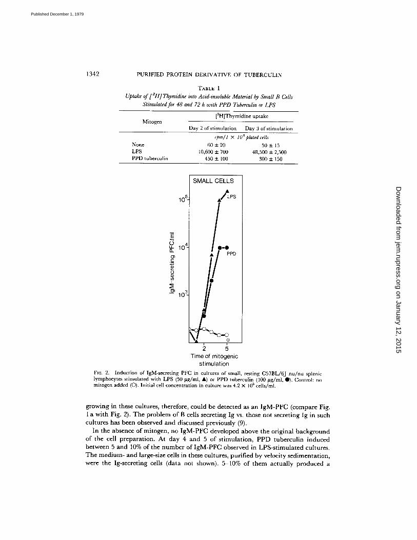
TABLE I Uptake of [aH] Thymidine into Acid-insoluble Material by Small B Cells
Stimulated for 48 and 72 h with PPD Tuberculin or LPS
Mitogen [3H]Thymidine uptake
Day 2 of stimulation Day 3 of stimulation
cpm/l X 105 plated cells None 60 + 20 50 _ 15 LPS 10,600 + 700 48,500 + 2,500 PPD tuberculin 450 + 100 300 + 150
10 5
10 4 t - "
t
~ 10 a_
SMALL CELLS
/ L P S
I 0 - 0 / PPD
r
2 5 Time of mitogenic
stimulation FIG. 2. Induction of IgM-secreting PFC in cultures of small, resting C57BL/6J nu/nu splenic lymphocytes stimulated with LPS (50 #g/m[, A) or PPD tuberculin (100 #g/ml, 0). Control: no mitogen added (C)). Initial cell concentration in culture was 4.2 x 10⁵ cells/ml.
growing in these cultures, therefore, could be detected as an I g M - P F C (compare Fig. 1 a wi th Fig. 2). The p rob lem of B cells secret ing Ig vs. those not secret ing Ig in such cul tures has been observed and discussed previously (9).
In the absence of mitogen, no I g M - P F C developed above the or iginal background of the cell p repara t ion . At day 4 and 5 of s t imula t ion , P P D tubercul in induced between 5 and 10% of the n u m b e r of I g M - P F C observed in LPS-s t imula t ed cultures. The med ium- and large-size cells in these cultures, pur i f ied by velocity sed imenta t ion , were the Ig-secreting cells (da ta not shown). 5-10% of them ac tua l ly p roduced a
on January 12, 2015jem
.rupress.orgD
ownloaded from
Published December 1, 1979

JAN ANDERSSON, WALDEMAR LERNHARDT, AND FRITZ MELCHERS 1343
plaque (for this problem, see above), and the repertoire of these secreting cells was found to be polyclonal. Thus, 1 in 2 × 10 a PPD tuberculin-induced IgM-PFC were SRC specific, 1 in 450 HRC specific, and 1 in 100 TNPs0-SRC specific; very much like the repertoire of LPS-induced IgM-PFC obtained after growth and maturation and as published previously (5). Of all blast cells in these cultures (Fig. 1 b) ~5-10% could be detected as IgM-PFC.
PPD tuberculin also induced small, resting cells into cells secreting IgG1 and/or IgG2, detected in the protein A-SRC plaque assay (Materials and Methods) at day 5 of stimulation. Compared with the LPS-induced maturation to IgG1- and IgG2- secreting cells (7.5 × 10⁴ PFC/4 × 105 plated cells) PPD tuberculin stimulated much fewer such cells (1 × 10³ PFC/4 × 105 plated cells). Compared to the number of PPD tuberculin-induced IgM-PFC ( - 104/4 × 105 cultured cells [Fig. 2]) the number of PPD tuberculin-induced IgG1- and IgG2-secreting cells (1 × 10a/4 × 105) was
10%. We conclude that PPD tuberculin, although not stimulating growth of small,
resting B cells, induces polyclonal maturation to blast-like, large IgM- and IgG- secreting cells.
Growth Stimulation of Activated B-Cell Blasts by PPD Tuberculin and by LPS. Activated B-cell blasts can be generated from resting B cells of spleen in a 48-h incubation period with B-cell mitogens such as LPS (9). They can be separated from nonactivated small cells by velocity sedimentation. Such LPS-activated B-cell blast populations contain >98% Ig+ cells and are, therefore, highly enriched. Another source of activated B-cell blasts are the medium- and large-size cells of spleen, which are, however, contaminated by large cells of other origins, such as macrophages etc. Both types of B-cell blasts need the presence of the growth-stimulating mitogens early after division in the cell cycle to go through the next round of division. This requirement in each cell cycle of a growing clone of B cells has been used previously to assay given B-cell blast populations for their reactivities toward several different B-cell mitogens (9). The same experimental principle of restimulation of growth and maturation is used here to study the growth- and maturation-stimulating properties of PPD tuberculin with LPS-activated and with background-activated B-cell blasts.
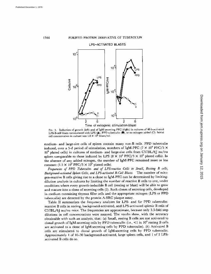
LPS-activated B-cell blasts were cultured in the absence or presence of LPS (the homologous mitogen) as controls, or in the presence of PPD tuberculin. Growth and induction to IgM-PFC was measured (Fig. 3).
The numbers of growing cells and the numbers of IgM-PFC induced by PPD tuberculin compared to the homologous mitogen LPS show that approximately one- half of the LPS-induced B-cell blasts can continue to grow and to mature to IgM secretion in the presence of the heterologous B-cell mitogen PPD tuberculin. Two conclusions can be reached from these results: (a) B-cell blasts generated by LPS- activation of resting C57BL/6J nu /nu spleen cells (6-8 wk old) contain more than one mitogen reactivity for, and therefore probably more than one receptor for LPS and for PPD tuberculin and (b) PPD tuberculin, although unable to activate and stimulate resting B cells to growth, is able to stimulate activated B-cell blasts to growth and secretion.
Medium- and large-size cells, enriched from spleen cells by velocity sedimentation, were also tested for their susceptibility to stimulation by PPD tuberculin. There was a minor increase in the total number of cells in culture, as a result of the fact that the
on January 12, 2015jem
.rupress.orgD
ownloaded from
Published December 1, 1979
1344 PURIFIED P R O T E I N DERIVATIVE OF T U B E R C U L I N
LPS-ACTIVATED BLASTS
10 7
10 6. 0
10 5
LPS
10"
o4 f ,°-o\
• O~ 0 0
Time of mitogenic st imulation (days) FXC~. 3. Induction of growth (left) and of IgM-secreting PFC (right) in cultures of 48-h-activated LPS-B-cell blasts restimulated with LPS (A), PPD tuberculin (O), or no mitogen added (O). Initial cell concentration in culture was 1.8 × 10⁵ blasts/ml.
medium- and large-size cells of spleen contain many non-B cells. PPD tuberculin induced, over a 5-d period of stimulation, numbers of IgM-PFC (7 × 10⁴ PFC/3 X 105 plated cells) in cultures of medium- and large-size cells from C57BL/6J nu /nu spleen comparable to those induced by LPS (8 × 10⁴ PFC/3 X 105 plated cells). In the absence of any added mitogen, the number of IgM-PFC remained more or less constant (1.5 × 10³ PFC/3 × 10 ~ plated cells).
Frequencies of PPD Tuberculin- and of LPS-reactive Cells in Small, Resting B cells, Background-activated Spleen Cells, and LPS-activated B-Cell Blasts. The number of mito- gen-reactive B cells giving rise to a clone to IgM-PFC can be determined by limiting- dilution analysis in cultures by limiting the number of reactive B cells to one, under conditions where every growth-inducible B cell (resting or blast) will be able to grow and mature into a clone of secreting cells (2). Such clones of secreting cells, developed in medium containing thymus filler cells and the appropriate mitogen (LPS or PPD tuberculin) are detected by the protein A-SRC plaque assay.
Table II summarizes the frequency analyses for LPS- and for PPD tuberculin- reactive B cells in resting, background-activated, and LPS-activated splenic B cells of C57BL/6J nu /nu mice. The frequencies are approximate, because only 3.3-fold step dilutions in cell concentrations were assayed. The results show, with the accuracy obtainable with such an analysis, that: (a) Small, resting B cells are not activated to clonal growth of IgM-secreting cells by PPD tuberculin (i.e., <1 in 103 resting B cells are activated to a clone of IgM-secreting cells by PPD tuberculin). (b) Activated B cells are stimulated to clonal growth of IgM-secreting cells by PPD tuberculin. Approximately 1 of 10-30 background-activated, large spleen cells, and 1 of 2 LPS- activated B cells do so.
on January 12, 2015jem
.rupress.orgD
ownloaded from
Published December 1, 1979
JAN ANDERSSON, WALDEMAR LERNHARDT, AND FRITZ MELCHERS
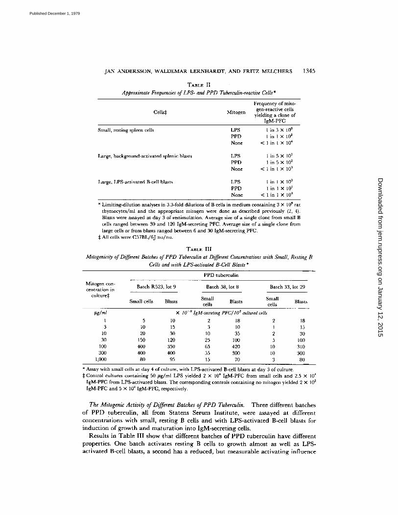
TABLE II
Approximate Frequencies of LPS- and PPD Tuberculin-reactive Cells *
1345
Cells~ Mitogen
Frequency of mito- gen-reactive cells
yielding a clone of IgM-PFC
Small, resting spleen cells
Large, background-activated splenic blasts
Large, LPS-activated B-cell blasts
LPS 1 in 5 × l0 ° PPD 1 in 1 × 10 s None < 1 in 1 × 10⁴
LPS 1 in 5 × 101 PPD 1 in 5 × 10 2 None < 1 in 1 × 10 a
LPS 1 in 1 × 10 ° PPD 1 in 1 × 10 I None < l i n 1 × 10 s
* Limiting-dilution analyses in 3.3-fold dilutions of B cells in medium containing 3 × l O 6 r a t
thymocytes/ml and the appropriate mitogen were done as described previously (2, 4). Blasts were assayed at day 3 of restimulation. Average size of a single clone from small B cells ranged between 30 and 120 IgM-seereting PFC. Average size of a single clone from large cells or from blasts ranged between 6 and 30 IgM-secreting. PFC.
:]: All cells were C57BL/6J nu/nu.
TABLE III
Mitogenicity of Different Batches of PPD Tuberculin at Different Concentrations with Small, Resting B Cells and with LPS-activated B-CeU Blasts *
PPD tuberculin
Mitogen con- centration in
culture~:
Batch R523, lot 9 Batch 38, lot 8 Batch 33, lot 29
Small Small Small cells Blasts Blasts Blasts cells cells
~g/ml × lO-~IgM-secreting PFC/lOncultured cel~ 1 5 10 2 18 2 18 3 10 15 3 10 1 15
10 20 30 10 35 2 30 30 150 120 25 100 5 100
100 400 350 65 420 l0 310 300 400 400 55 300 10 300
1,000 80 95 15 70 3 80
* Assay with small cells at day 4 of culture, with LPS-activated B-cell blasts at day 3 of culture. ~: Control cultures containing 50 #g/ml LPS yielded 2 X 10⁴ IgM-PFC from small cells and 2.5 X 10⁴
IgM-PFC from LPS-activated blasts. The corresponding controls containing no mitogen yielded 2 X 10² IgM-PFC and 5 × 10² IgM-PFC, respectively.
The Mitogenic Activity of Different Batches of PPD Tuberculin. T h r e e d i f f e r e n t b a t c h e s
o f P P D t u b e r c u l i n , a l l f r o m S t a t e n s S e r u m I n s t i t u t e , w e r e a s s a y e d a t d i f f e r e n t
c o n c e n t r a t i o n s w i t h s m a l l , r e s t i n g B ce l l s a n d w i t h L P S - a c t i v a t e d B-ce l l b l a s t s fo r
i n d u c t i o n o f g r o w t h a n d m a t u r a t i o n i n t o I g M - s e c r e t i n g cel ls .
R e s u l t s in T a b l e I I I s h o w t h a t d i f f e r e n t b a t c h e s o f P P D t u b e r c u l i n h a v e d i f f e r e n t
p r o p e r t i e s . O n e b a t c h a c t i v a t e s r e s t i n g B ce l l s t o g r o w t h a l m o s t as w e l l as L P S -
a c t i v a t e d B-ce l l b l a s t s , a s e c o n d h a s a r e d u c e d , b u t m e a s u r a b l e a c t i v a t i n g i n f l u e n c e
on January 12, 2015jem
.rupress.orgD
ownloaded from
Published December 1, 1979
1346 PURIFIED PROTEIN DERIVATIVE OF TUBERCULIN
on resting cells, whereas a third, which was used throughout the studies reported in this paper (above), has no measurable activating capacity for resting cells. As seen from the data, this is so at concentrations of PPD tuberculin between 1 mg/ml and 1 /~g/ml. All batches activate LPS-induced B-cell blasts equally well. The optimal stimulatory capacity for B-blasts is between 100 and 300/~g/ml PPD tuberculin.
It is evident that some PPD tuberculin batches will have to be purified to separate mitogens that stimulate resting, as well as activated cells from mitogens that stimulate only activated cells.
Discussion
B-cell mitogens have been defined as molecules which have the property of inducing the growth of B-cell clones in a polyclonal fashion, irrespective of the specificities of VH/VL-regions expressed in the mitogen-reactive B cells and their clonal descendants. Most mitogens so far studied will also induce B-cell maturat ion to Ig secretion. Conditions can be found in which only growth, but no maturat ion into Ig secretion (15), or no growth, and only maturat ion into Ig secretion (16), can be observed after mitogenic stimulation. The latter, maturat ion without growth, was so far only observed with normal lymphocytes when DNA-synthesis was inhibited through inhibition of the deoxynucleotide metabolism of the B cell.
The results in this paper, for the first time, define a B-cell mitogen as a polyclonal activator for maturat ion to Ig secretion only. The improved tissue-culture conditions allow even the detection of IgG-secreting cells in the absence of growth, which indicates that the switch from IgM to IgG synthesis and secretion observed in single clones of mitogen-activated B cells (17) may occur without cell division and in a single cell stimulated by PPD tuberculin to maturat ion only.
Maturat ion into Ig secretion has previously been found to be antagonistic to the growth of B cells. Induction to maturat ion effected by such mitogens as PPD tuberculin may, therefore, delete the activated B cell from the repertoire of growth- inducible B cells of the system and may, thereby, constitute suppression or deletion of such B cells. It seems premature to speculate on the physiological role of such mitogens as long as the connections of mitogenicity, antigen specificity, and possible further requirements in immune reactions leading to stimulation or suppression of B-cells have not been further clarified.
Earlier experiments, performed under conditions where there were very limited possibilities for murine B lymphocytes to grow and to mature to Ig secretion (18), identified PPD tuberculin as a B-cell mitogen which appeared to activate B-cell subpopulations characterized as late in their development to Ig-secreting cells (19, 20). Somewhat in contrast to these results, it was found in other experiments, that LPS and PPD tuberculin activated largely the same B-cell populations, indicating that B cells could be reactive to more than one mitogen (20, 21). Our results presented in this paper, i.e., that LPS can stimulate resting B cells as well as activated B-cell blasts, whereas PPD tuberculin can only stimulate activated B-cell blasts, but not resting B cells, force us to reinterpret these earlier results. It appears now most likely that already activated, background B-cell blasts of spleen have been further stimulated under these earlier, more limited conditions of in vitro cultures. This action of PPD tuberculin on B cells would certainly support the previous interpretation that PPD stimulated late B cells (20, 22), if one accepts an activated B-cell blast on its way to
on January 12, 2015jem
.rupress.orgD
ownloaded from
Published December 1, 1979
JAN ANDERSSON, WALDEMAR LERNHARDT, AND FRITZ MELCHERS 1347
becoming a mature plasma cells as a distinct stage of B-cell development. Alternately, it is possible that part of the earlier discrepancies in experimental
results were a result of the different mitogenic action of different batches of PPD tuberculin (see Table III). It is also not excluded that at the cell concentrations (i.e., 5 × 10⁶ cell/ml) used in the earlier experiments, interactions of other cells with the resting B cells may render the resting B cells activated, and consequently susceptible, to stimulation by PPD tuberculin. This latter possibility will have to be probed experimentally with more-carefully purified, resting B-cell populations.
Our studies on the action of PPD tuberculin on B cells have certainly been inspired by studies on T-cell activation (23, 24). In these studies, it was shown that resting, small T-cells, carefully purified from B cells, activated large cells and macrophages could not be stimulated by mitogens (factors) contained in supernates ofconcanavalin A-treated spleen cells (23) unless these resting cells had previously been treated with antigen or with concanavalin A. Antigen, in this case allogeneic cells, or concanavalin A alone, on the other hand, did not stimulate the resting T cells to growth. Our results with B cells and their activation by PPD tuberculin point to a striking similarity in the distinction of a resting, from an activated, state of a lymphocyte and may suggest a similar mode of activation of T and B cells by antigens plus mitogens.
We have, on occasions, observed similar stimulatory effects with selected batches of fetal calfsera and transferrin preparations on activated B-cell blasts, whereas the same batches were nonstimulatory for resting cells. This appears of importance wherever B- cell activation by cooperative factors and by anti-Ig antibodies is studied in the presence of fetal calf serum (25, 26). In particular, exPerimental conditions should be critically assessed for the presence of PPD tuberculin-like mitogens, whenever the action of anti-Ig antibodies on B cells leads to stimulation.
T-cell dependent help in the antigen-specific activation and stimulation of resting B cells is another likely candidate for such a PPD tuberculin-like mitogen for B cells, whether the mitogen itself is directly supplied by T cells, or indirectly via the activation of mitogen-producing third cells such as macrophages. Such action of T- cell help may be in effect where in vitro differentiation of human leukemic B lymphocytes to Ig-secreting plasma ceils has been observed (27).
The cell cycle of growing eukaryotic cells is characterized by two events which are separated in time: ceil division (mitosis [M]) and DNA-synthesis (S-phase). The gap after M and preceding S-phase is called Ga, the gap after S-phase and preceding M is called G2. Activated, growing B cells need the action o fa mitogen (such as LPS etc., 9) after M early in G1 to proceed through the next cell cycle and to complete M (W. Lernhardt and F. Melchers. Manuscript in preparation.). PPD tuberculin, as much as the commonly known B-cell mitogens LPS or LP (6, 7), is able to convey this type of mitogenesis to B cells. PPD tuberculin is, therefore, not deficient in its ability to stimulate growth. Because PPD tuberculin is able to do so with a large number (approximately one-half) of all LPS-activated B-cell blasts, we have to conclude in close similarity to conclusions previously reached for LPS-, LP-, and NOC-reactive B cells (9), that a majority (~50%) of all splenic B cells (of a C57BL/6J nu /nu mouse, 6-8 wk of age) are reactive to more than one mitogen, now including PPD tuberculin, and, therefore, probably possess more than one mitogen receptor. It should be recalled here that a direct action of these B-cell mitogens on B cells, under circumvention of help from T cells or adherent cells, is concluded from the finding that growth of
on January 12, 2015jem
.rupress.orgD
ownloaded from
Published December 1, 1979

1348 PURIFIED PROTEIN DERIVATIVE OF TUBERCULIN
single clones of B cells in semi-solid media is dependent on the presence of such B-cell mitogens (28, 29).
Resting cells do not synthesize DNA and do not divide. Their resting state has been called the G0-phase of the cell cycle. Activation of resting cells, i.e., of resting B cells by LPS, results in an activation of DNA-synthesis followed by M. From the G0-phase cells, therefore, B cells first enter the S-phase and then G2-phase and M of the cell cycle. Thereafter, they proceed through Ga, S-phase, Gz, and M and so on, as long as the stimulatory mitogen is present (above). The results of this paper establish for B cells that G0-phase B cells require, in part, different, and probably more, signals to become activated and stimulated to growth than do activated B cells to enter the next cell cycle. Mitogens, such as PPD tuberculin, which do not activate resting G0-phase B lymphocytes, but which will stimulate activated Gl-phase B-cell blasts, pose the intriguing problem as to what renders a resting G0-phase B cell susceptible to stimulation by such Gl-phase-only mitogens.
The most likely reaction which could render a resting G0-phase B cell susceptible to stimulation by Gl-phase-only mitogens is the binding of antigen to surface Ig molecules. This may induce changes in the surface membrane which may expose a functionally hidden receptor for the mitogen. G0-Gl-phase mitogens, such as LPS, LP, NOC, and mitogens in fetal calf serum, as a result of their lipophilic nature, may induce such changes in G0-phase B cells, by their very ability to insert themselves into the lipid bilayer of the B-cell surface membrane, which, of course, also contains the mitogen-specific receptors. Such a difference in action of G0-Gl-phase and Ga-phase- only mitogens may, in fact, offer an explanation for the observed difference in activating potential between T-independent antigens of type 1 (hapten on LPS, LP, NOC, and Brucella abortus) and such of type 2 (pneumococcal polysaccharide, dextran, and levan).
Finally, it appears still feasible to expect (30, 31) that recognitions by the B cells additional to the binding of antigen to surface Ig molecules, as i.e., of H-2-encoded molecules, may be required to change a G0-phase B cell to its activated form susceptible for Gl-phase-only mitogens.
S u m m a r y
The purified protein derivative of tuberculin (PPD tuberculin) stimulates approx- imately one of two lipopolysaccharide (LPS)-activated B-cell blasts of C57BL/6J nu / nu spleen cells to continued clonal growth and maturation to IgM and IgG secretion. It also stimulates background, in vivo-activated large cells of normal C57BL/6J nu / nu spleen to growth and Ig secretion, at a frequency of ~ 1 of 100 large spleen cells. PPD tuberculin, therefore, is a polyclonal B-cell activator for B-cell blasts. Many single murine splenic B cells (~50%) appear to have reactivities, and therefore probably receptors, for LPS and PPD tuberculin.
PPD tuberculin does not stimulate small, resting B cells to growth as measured by the number of cells in culture and by thymidine uptake. However, it stimulates approximately one-fourth of all spleen cells to blast transformation. The large-size blast cells secrete IgM and, therefore, form plaques in the protein A plaque assay. IgG-secreting, plaque-forming cells develop at later stages of stimulation, indicating that the switch from IgM to IgG may occur without division in single, stimulated B cells. Stimulation of resting B cells to maturation by PPD tuberculin is polyclonal.
on January 12, 2015jem
.rupress.orgD
ownloaded from
Published December 1, 1979
JAN ANDERSSON, WALDEMAR LERNHARDT, AND FRITZ MELCHERS 1349
Thus, - 1 in 102 IgM-secre t ing p laque- fo rming cells form plaques wi th t r in i t rophenyl - subs t i tu ted sheep erythrocytes , 1 in 450 do so wi th horse erythrocytes , and 1 in 103 with sheep erythrocytes . Fur the rmore , the n u m b e r of Ig-secret ing cells deve loping from small , rest ing cells wi thout growth in cul tures wi th or wi thout filler t hymus cells suggests polyc lonal ac t iva t ion by P P D tubercu l in to m a t u r a t i o n only of at least one out of four small , splenic B cells.
The able technical assistance of Mrs. Deborah Norman is gratefully acknowledged.
Received for publication l l June 1979.
R e f e r e n c e s
1. M611er, G., editor. 1972. Lymphocyte activation by mitogens. Transplant. Rev. 11:1. 2. Andersson, J., A. Coutinho, W. Lernhardt, and F. Melchers. 1977. Clonal growth and
maturation to immunoglobulin secretion m vitro of every growth-inducible B-lymphocyte. Cell. 10:27.
3. Melchers, F., A. Coutinho, G. Heinrich, and J. Andersson. 1975. Continuous growth of mitogen-reactive B-lymphocytes. Scand. J. Immunol. 4:853.
4. Andersson, J., A. Coutinho, and F. Melchers. 1977. Frequencies of mitogen-reactive B-cells in the mouse. I. Distribution in different lymphoid organs from different inbred strains of mice at different ages.J. Exp. Med. 145:1511.
5. Andersson, J., A. Coutinho, and F. Melchers. 1977. Frequencies of mitogen-reactive B cells in the mouse. II. Frequencies of B cells producing antibodies which lyse sheep or horse erythrocytes, and trinitrophenylated or nitroiodophenylated sheep erythrocytes. J. Exp. Med. 145:1520.
6. Andersson, J., O. Sj6berg, and G. M611er. 1972. Induction ofimmunoglobulin and antibody synthesis "in vitro" by lipopolysaccharide. Eur. J. Immunol. 2:349.
7. Melchers, F., V. Braun, and C. Galanos. 1975. The lipoprotein of the outer membrane of Escherichia coli: a B lymphocyte mitogen. J. Exp. Med. 142:473.
8. Bona, C., C. Damais, and L. Chedid. 1974. Blastic transformation of mouse spleen lymphocytes by a water-soluble mitogen extracted from Nocardia. Proc. Natl. Acad. Sci. U. S. A. 71:1602.
9. Andersson, J., A. Coutinho, and F. Melchers. 1979. Mitogen-activated B-cell blasts reactive to more than one mitogen.J. Exp. Med. 149:553.
10. Sultzer, B. M., and B. S. Nilsson. 1972. PPD-tuberculin--a B cell mitogen. Nat. New Biol. 240:198.
11. Miller, R. G., and R. A. Phillips. 1969. Separation of cells by velocity sedimentation. J. Cell, Physiol. 73:191.
12. Iscove, N. N., and F. Melchers. 1978. Complete replacement of serum by albumin, transferrin, and soybean lipid in cultures of lipopolysaccharide-reactive B lymphocytes. J. Exp. Med. 147:923.
13. Gronowicz, E., A. Coutinho, and F. Melchers. 1976. A plaque assay for all cells secreting Ig of a given type or class. Eur. J. Immunol. 6:588.
14. Andersson,J., L. Lafleur, and F. Melchers. 1974. IgM in bone marrow-derived lymphocytes. Synthesis, surface deposition, turnover and carbohydrate composition in unstimulated mouse B cells. Eur. J. Immunol. 4:170.
15. Andersson, J., W. W. Bullock, and F. Melchers. 1974. Inhibition of mitogenic stimulation of mouse lymphocytes by anti-mouse immunoglobulin antibodies. I. Mode of action. Eur. J. hnmunol. 4:715.
16. Andersson, J., and F. Melchers. 1974. Maturation of mitogen-activated bone marrow- derived lymphocytes in the absence of proliferation. Eur. J. Immunol. 4:533.
on January 12, 2015jem
.rupress.orgD
ownloaded from
Published December 1, 1979

1350 PURIFIED PROTEIN DERIVATIVE OF TUBERCULIN
17. Andersson, J., A. Coutinho, and F. Melchers. 1978. The switch from IgM to IgG secretion in single mitogen-stimulated B-cell clones..]. Exp. Med. 147:1744.
18. Coutinho, A., G. Mfller, J. Andersson, and W. W. Bullock. 1973. In vitro activation of mouse lymphocytes in serum-free medium: effects o fT and B cell mitogens on proliferation and antibody synthesis. Eur. J. Irnmunol. 3:299.
19. Gronowicz, E., A. Coutinho, and G. M611er. 1974. Differentiation of B cells: sequential appearance of responsiveness to polyclonal activators. Stand. J. Immunol. 3:413.
20. Gronowicz, E., and A. Coutinho. 1976. Heterogeneity of B cells: direct evidence of selective triggering of distinct subpopulations by polyclonal activators. Stand. J. lmmunol. 5:55.
21. Melchers, F., and J. Andersson. 1974. The kinetics of proliferation and maturation of mitogen-activated bone marrow-derived lymphocytes. Eur. J. Immunol. 4:687.
22. Gronowicz, E., and A. Coutinho. 1974. Selective triggering of B cell subpopulations by mitogens. Eur. J. Immunol. 4:771.
23. Andersson, J., K.-O. Gr6nvik, E.-L. Larsson, and A. Coutinho. Studies on T-lymphocyte activation. I. Requirements for the mitogen-dependent production of T-cell growth factors. Eur. J. Irnmunol. In press.
24. Coutinho, A., E.-L. Larsson, K.-O. Gr6nvik, and J. Andersson. Studies on T-lymphocyte activation. II. The target cells for Con A-induced growth factors. Eur. J. Immunol. In press.
25. Kishimoto, T., T. Miyahe, Y. Nishizawa, T. Watanabe, and Y. Yamamura. 1975. Trigger- ing mechanism of B lymphocytes. I. Effect of anti-immunoglobulin and enhancing soluble factor on differentiation and proliferation of B cells. J. Immunol. 115:1179.
26. Sieckman, D. G., R. Asofsky, D. E. Mosier, I. M. Zitron, and W. E. Paul. 1978. Activation of mouse lymphocytes by anti-immunoglobulin. I. Parameters of the proliferative response. J. Exp. Med. 147:814.
27. Fu, S. M., N. Chiorazzi, H. G. Kunkel, J. P. Halper, and S. R. Harris. 1978. Induction of in vitro differentiation and immunogtobulin synthesis of human leukemic B lymphocytes. J. Exp. Med. 148:1570.
28. Metcalf, D. 1976. Role of mercaptoethanol and endotoxin in stimulating B lymphocyte colony formation in vitro. J. Immunol. 166:635.
29. Kincade, P. W., P. Ralph, and M. A. S. Moore. 1976. Growth of B lymphocytes clones in semi-solid culture is mitogen dependent.J. Exp. Med. 143:1265.
30. Sprent, J. 1978. Role of H-2 gene products in the function of T-helper cells from normal and chimeric mice in vivo. Immunol. Rev. 42:108.
31. Katz, D. H., B.J. Skidmore, L. R. Katz, and C. A. Bogowitz. 1978. Adaptive differentiation of murine lymphocytes. I. Both T- and B-lymphocytes differentiating in Fx --~ parental chimeras manifest preferential cooperative activity for partner lymphocytes derived from the same parental type corresponding to the chimeric host.J. Exp. Med. 148:727.
on January 12, 2015jem
.rupress.orgD
ownloaded from
Published December 1, 1979