Flavokawain B induced cytotoxicity in two breast cancer cell ...
14
RESEARCH ARTICLE Open Access Flavokawain B induced cytotoxicity in two breast cancer cell lines, MCF-7 and MDA- MB231 and inhibited the metastatic potential of MDA-MB231 via the regulation of several tyrosine kinases In vitro Nadiah Abu 1,2 , M. Nadeem Akhtar 3 , Swee Keong Yeap 4 , Kian Lam Lim 5 , Wan Yong Ho 6 , Mohd Puad Abdullah 2 , Chai Ling Ho 2 , Abdul Rahman Omar 4 , Jamil Ismail 3 and Noorjahan Banu Alitheen 2* Abstract Background: The kava-kava plant (Piper methysticum) is traditionally consumed by the pacific islanders and has been linked to be involved in several biological activities. Flavokawain B is a unique chalcone, which can be found in the roots of the kava-kava plant. In this study, the operational mechanism of the anti-cancer activity of a synthetic Flavokawain B (FKB) on two breast cancer cell lines, MCF-7 and MDA-MB231 was investigated. Method: Several in vitro assays were attempted such as MTT, flow cytometry of cell cycle analysis, annexin V analysis, and JC-1 analysis to detect apoptosis. Moreover, in vitro metastasis assays were also performed such as transwell migration assay, invasion assay, rat aorta ring and HUVEC tube formation. Molecular analysis of related genes and proteins were conducted using real-time PCR and proteome profiler analysis. Results: Based on our results, apoptosis was induced when both MCF-7 and MDA-MB231 were treated with FKB. A significant G2/M arrest was seen in MDA-MB231 cells. Additionally, FKB also inhibited the in vitro migration and invasion in MDA-MB231 cells in a dose dependent manner. Moreover, FKB can be a potential inhibitor in angiogenesis as it suppressed the formation of vessels in HUVEC cells as well as in the ex-vivo rat aortic ring assay. Conclusion: Our findings suggested that FKB also regulated several receptor tyrosine kinases. Overall, FKB is not only a potential candidate to be an anti-cancer agent, but as an anti-metastatic agent as well. Keywords: Breast cancer, Flavokawain, Proliferation, Metastasis, Tyrosine kinase Background Cancer is still a worrying disease despite the progressive scientific research conducted. In fact, breast cancer still remains one of the most deadliest cancer among women [1]. Approximately around 1 out of 8 women will de- velop breast cancer at some point in their lives [1]. According to the cancer statistics reported, around 14 % of breast-cancer related cases ended up being fatal [1]. Additionally, breast cancer has a tendency to become malignant [1]. More than half of the patients diagnosed with breast cancer have already developed metastatic breast cancer [2]. Metastasis is usually the main cause of cancer-related deaths [2, 3]. Therefore, it is extremely crucial to not only treat, but also prevent this disease from becoming malignant. Natural-derived drugs have become a more favourable option in the pharmaceutical industry [4, 5]. In fact, in the development of new anti- cancer drugs, natural products still remain as one of the major players in providing the database [6]. The kava-kava plant extract has been used as traditional remedies to treat various illnesses [7]. Interestingly, there has been a correlation between the consumption of the * Correspondence: [email protected] 2 Faculty of Biotechnology and Bimolecular Sciences, Universiti Putra Malaysia, UPM Serdang 43400Selangor, Darul Ehsan, Malaysia Full list of author information is available at the end of the article © 2016 Abu et al. Open Access This article is distributed under the terms of the Creative Commons Attribution 4.0 International License (http://creativecommons.org/licenses/by/4.0/), which permits unrestricted use, distribution, and reproduction in any medium, provided you give appropriate credit to the original author(s) and the source, provide a link to the Creative Commons license, and indicate if changes were made. The Creative Commons Public Domain Dedication waiver (http://creativecommons.org/publicdomain/zero/1.0/) applies to the data made available in this article, unless otherwise stated. Abu et al. BMC Complementary and Alternative Medicine (2016) 16:86 DOI 10.1186/s12906-016-1046-8
-
Upload
khangminh22 -
Category
Documents
-
view
3 -
download
0
Transcript of Flavokawain B induced cytotoxicity in two breast cancer cell ...

RESEARCH ARTICLE Open Access
Flavokawain B induced cytotoxicity in twobreast cancer cell lines, MCF-7 and MDA-MB231 and inhibited the metastaticpotential of MDA-MB231 via the regulationof several tyrosine kinases In vitroNadiah Abu1,2, M. Nadeem Akhtar3, Swee Keong Yeap4, Kian Lam Lim5, Wan Yong Ho6, Mohd Puad Abdullah2,Chai Ling Ho2, Abdul Rahman Omar4, Jamil Ismail3 and Noorjahan Banu Alitheen2*
Abstract
Background: The kava-kava plant (Piper methysticum) is traditionally consumed by the pacific islanders and hasbeen linked to be involved in several biological activities. Flavokawain B is a unique chalcone, which can be foundin the roots of the kava-kava plant. In this study, the operational mechanism of the anti-cancer activity of a syntheticFlavokawain B (FKB) on two breast cancer cell lines, MCF-7 and MDA-MB231 was investigated.
Method: Several in vitro assays were attempted such as MTT, flow cytometry of cell cycle analysis, annexin V analysis,and JC-1 analysis to detect apoptosis. Moreover, in vitro metastasis assays were also performed such as transwellmigration assay, invasion assay, rat aorta ring and HUVEC tube formation. Molecular analysis of related genes andproteins were conducted using real-time PCR and proteome profiler analysis.
Results: Based on our results, apoptosis was induced when both MCF-7 and MDA-MB231 were treated with FKB.A significant G2/M arrest was seen in MDA-MB231 cells. Additionally, FKB also inhibited the in vitro migration andinvasion in MDA-MB231 cells in a dose dependent manner. Moreover, FKB can be a potential inhibitor in angiogenesisas it suppressed the formation of vessels in HUVEC cells as well as in the ex-vivo rat aortic ring assay.
Conclusion: Our findings suggested that FKB also regulated several receptor tyrosine kinases. Overall, FKB is not only apotential candidate to be an anti-cancer agent, but as an anti-metastatic agent as well.
Keywords: Breast cancer, Flavokawain, Proliferation, Metastasis, Tyrosine kinase
BackgroundCancer is still a worrying disease despite the progressivescientific research conducted. In fact, breast cancer stillremains one of the most deadliest cancer among women[1]. Approximately around 1 out of 8 women will de-velop breast cancer at some point in their lives [1].According to the cancer statistics reported, around 14 %of breast-cancer related cases ended up being fatal [1].Additionally, breast cancer has a tendency to become
malignant [1]. More than half of the patients diagnosedwith breast cancer have already developed metastaticbreast cancer [2]. Metastasis is usually the main cause ofcancer-related deaths [2, 3]. Therefore, it is extremelycrucial to not only treat, but also prevent this diseasefrom becoming malignant. Natural-derived drugs havebecome a more favourable option in the pharmaceuticalindustry [4, 5]. In fact, in the development of new anti-cancer drugs, natural products still remain as one of themajor players in providing the database [6].The kava-kava plant extract has been used as traditional
remedies to treat various illnesses [7]. Interestingly, therehas been a correlation between the consumption of the
* Correspondence: [email protected] of Biotechnology and Bimolecular Sciences, Universiti Putra Malaysia,UPM Serdang 43400Selangor, Darul Ehsan, MalaysiaFull list of author information is available at the end of the article
© 2016 Abu et al. Open Access This article is distributed under the terms of the Creative Commons Attribution 4.0International License (http://creativecommons.org/licenses/by/4.0/), which permits unrestricted use, distribution, andreproduction in any medium, provided you give appropriate credit to the original author(s) and the source, provide a link tothe Creative Commons license, and indicate if changes were made. The Creative Commons Public Domain Dedication waiver(http://creativecommons.org/publicdomain/zero/1.0/) applies to the data made available in this article, unless otherwise stated.
Abu et al. BMC Complementary and Alternative Medicine (2016) 16:86 DOI 10.1186/s12906-016-1046-8

kava extracts with the incidence of cancer [8]. Some ofthe most interesting components that can be foundfrom the kava root extracts include the flavokawains[9]. There are three types of flavokawains that can befound and the differences lies in the side chain [10].Flavokawain B has gained worldwide interest due to itspromising anti-cancer activity as reported by severalstudies [11, 12]. This chalcone was reported to bepresent in two other types of plant species; the alpiniaand piper family [9, 13]. Flavokawain B was found topossess several fascinating biological activities such as,anti-inflammatory, antinociceptive and anti-cancer ac-tivities [13, 14].The fact that receptor tyrosine kinases (RTK) play an
important role in cancer progression was an impactful dis-covery [15]. There are various RTKs and downstream pro-teins involved in breast cancer and metastasis such asVEGF, MMP9, GLUT1 and FOXM1. Flavonoids havebeen reported to regulate some of these RTKs and thushave the potential to become promising anti-canceragents. Nevertheless, to the best of our knowledge, studieson the effects of flavokawain B on breast cancer cells invitro and on the RTKs have not yet been reported. There-fore, this study aims to elucidate the anti-metastatic effectsof flavokawain B on breast cancer cells in vitro.
ResultsFlavokawain B inhibited the proliferation of MCF-7 andMDA-MB231 in vitroMTT analysis was performed to analyze the cytotoxiceffects of flavokawain B on two breast cancer cell lines,MCF-7 and MDA-MB231, as well as on the non-transformed mammary epithelial cell line, MCF-10A.The cells were treated with 7 different concentrations offlavokawain B for 72 h. As in Fig. 1a, the half-inhibitoryconcentration (IC50) of flavokawain B on MDA-MB231(12.3 μM) was much lower compared to MCF-7(33.8 μM). Nonetheless, the IC50 of flavokawain B on thepositive control cell line, MCF-10A was considerablyhigher than the two cancer cell lines. The BrdU incorp-oration assay was also conducted to analyze the anti-proliferative effects of flavokawain B. Fig. 1b depicts thatthe percentage of BrdU incorporation is negatively cor-related with the dose of flavokawain B. Flavokawain Bimpeded the proliferation of both MCF-7 and MDA-MB231 in a dose-dependent manner.
Flavokawain B induced G2/M arrest and apoptosis inMDA-MB231 and MCF-7To investigate the effects of flavokawain B on the cellcycle progression in MDA-MB231 and MCF-7, theFACS cell cycle analysis was performed. Three differentconcentrations of flavokawain B were used as treatmentand the cells were analysed at 12 h and 24 h post-
treatment. Based on Fig. 2 the changes in the G2/Mphase in MDA-MB231 cells were significant as the doseincreases. After 24 h, the percentage of cells in the subG0/G1 phase also increased as the dose escalates. InMCF-7 cells however, there was no significant G2/Marrest seen at both time points, as depicted in Fig. 2a(Additional file 1: Figure S1).One paramater of apoptosis was examined by detect-
ing the externalization of phosphatidylserine. The cellswere stained with Annexin V-FITC and Propidium Iod-ide before being subjected to flow cytometry. Accordingto Fig. 2b (Additional file 2: Figure S2), in both MCF-7and MDA-MB231 a pattern can be seen where there is ashift in the percentage of cells from viable to early apop-tosis and to late apoptosis/necrosis as the dose escalates.
Flavokawain B inhibited migration and invasion in vitroand suppressed the formation of tube-like vessels in vitroand ex vivoFor the in vitro motility assessment, the scratch (woundhealing) assay was used in MDA-MB231. The percent-age of wound closure was compared between all treat-ments of group with the control as showed in Fig. 3a. Inthe control group of the MDA-MB231 cells, the area ofthe wound was completely covered with migrated cellsafter 24 h (100 ± 0.1 %). In the treated groups, the per-centage of wound closure is negatively correlated to thedose of flavokawain B given. For the IC12.5, IC50 andIC75treated groups, the wound closure is approximately65 ± 4.9, 28.6 ± 5.6 and 5.4 ± 2.7 % respectively.The transwell assay was also performed to investigate
the effects of flavokawain B on the motility and invasive-ness of MDA-MB231. The percentage of migrated cellswas inversely proportional to the dose of flavokawain Baccording to Fig. 3b. In MDA-MB231 treated cells, thehigher the dose of flavokawain B; the lower the percent-age of migrated cells through the transwell. For the IC50
treated group, the percentage of migrated cells in MDA-MB231 was 15.4 %. The matrigel-transwell assay wasconducted using MDA-MB231 cells only since it ishighly invasive as compared to MCF-7. Similar to the invitro migration assay, as well as the wound-healingassay, the percentage of invaded cells is negatively corre-lated to the dose of flavokawain B. In the flavokawain B-treated groups, IC12.5, IC25 and IC75 the percentage ofinvaded cells were 54.1, 24.7 and 15.5 % correspondinglyas in Fig. 3c.For the in vitro angiogenesis assessment, the HUVEC
tube formation assay was conducted, followed by treat-ment with three different concentrations of flavokawainB. In the IC12.5 treated group, there was no significantinhibition in the formation of tube-like vessels as repre-sented in Fig. 4a. The percentage of tube formation inIC25-treated group was significantly suppressed with
Abu et al. BMC Complementary and Alternative Medicine (2016) 16:86 Page 2 of 14

23.6 % tube formation. There was a total inhibition inthe formation of tube in the IC50-treated group. The ex-vivo rat aortic ring assay was also conducted to furtherunderstand the anti-angiogenic potential of flavokawainB as shown in Fig. 4b. This also suggests that flavoka-wain B regulates angiogenesis in a dose-dependentmanner.
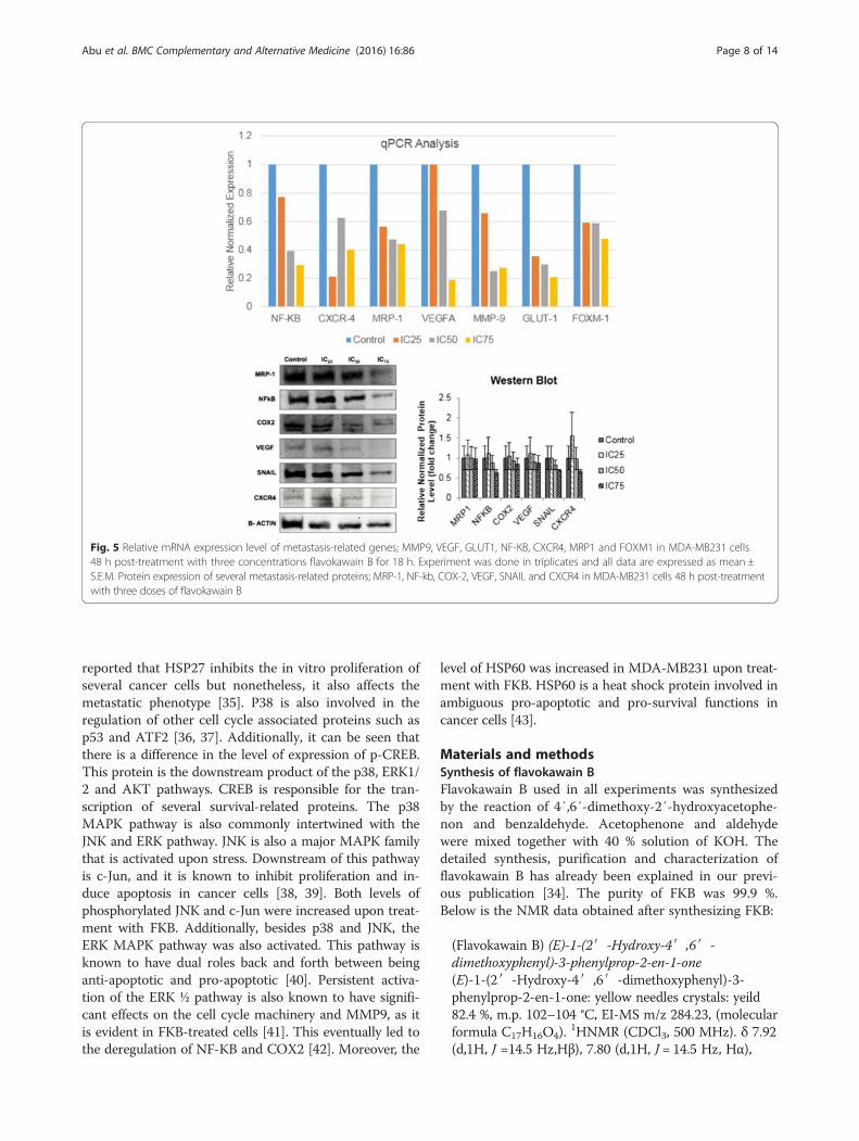
Flavokawain B regulated several metastasis-related pro-teins and genes, and tyrosine kinases in MDA-MB231To measure the relative mRNA level of metastasis-relatedgenes, quantitative real-time analysis was performed asdisplayed in Fig. 5. Upon induction with three doses of fla-vokawain B, the mRNA expression of all genes in MDA-MB231 treated cells; MMP9, VEGF, GLUT1 and FOXM1
were down regulated. The significance of these findingswere dose-dependent; the lower the dose of flavokawainB given, the less significant the expression of mRNA.The expressions of the mRNA level were consideredsignificant if it were more than 2-fold. In the westernblot analysis on the other hand, the protein expressionof inflammation-related proteins such as NF-KB andCOX-2 were decreased in a dose-dependent manner.Likewise, the same pattern can be seen in othermetastasis-related proteins such as VEGF, SNAIL andCXCR4 according to Fig. 5.Based on the proteome profiler analysis, flavokawain
B regulated several important tyrosine kinases in MDA-MB231. In Fig. 6, in MDA-MB231, flavokawain B up-regulated the phosphorylation of p-p38 alpha, p-CREB,
Fig. 1 a Percentage of viability of three cell lines (MCF-7, MDA-MB231 and MCF-10a) when treated with seven doses of flavokawain B measuredthrough the MTT assay after 72 h. b Percentage of BrdU incorporation in both MCF-7 and MDA-MB231 upon treatment with three different concentrationsof flavokawain B for 48 h. All data are expressed as mean ± S.E.M with three biological replicates
Abu et al. BMC Complementary and Alternative Medicine (2016) 16:86 Page 3 of 14

24 h
48 h
72 h
Per
cent
age
of C
ells
(%
)
MCF-7 MDA-MB231
0
10
20
30
40
50
60
70
80
90
100
Viable Early Apoptosis LateApoptosis/Necrosis
0
10
20
30
40
50
60
70
80
90
100
Viable Early Apoptosis LateApoptosis/Necrosis
0
10
20
30
40
50
60
70
80
90
100
Viable Early Apoptosis LateApoptosis/Necrosis
0
10
20
30
40
50
60
70
80
90
100
Viable Early Apoptosis LateApoptosis/Necrosis
0
10
20
30
40
50
60
70
80
90
100
Viable Early Apoptosis LateApoptosis/Necrosis
0
10
20
30
40
50
60
70
80
90
100
Viable Early Apoptosis LateApoptosis/Necrosis
Flavokawain B
ControlFKB IC25FKB IC50FKB IC75
* **
**
* * *
* *
**
*
*
* * *
**
*
**
*
*
**
**
*
*
** *
*
* * * *
**
* *
**
*
A
MC
F-7
MD
A-M
B23
1
CONTROL IC25 IC50 IC75
ANNEXIN V-FITC
PRO
PID
IUM
IO
DID
E
B
Fig. 2 (See legend on next page.)
Abu et al. BMC Complementary and Alternative Medicine (2016) 16:86 Page 4 of 14

p-HSP27, p-JNK, p-AKT, p-ERK, p-HSP60, p-WNK1,p-c-Jun and p-p53.
DiscussionFrom our findings, it can be suggested that FKB inducesapoptosis and inhibits proliferation in both MCF-7 andMDA-MB231 cells in vitro as well as halting some of thesteps in the metastatic cascade in MDA-MB231 cells invitro. FKB possess anti-proliferative effects and is select-ive in its actions as evidenced by the MTT and BrdU as-says. It has been documented in some studies that FKBinduced G2/M arrest in several cancer cell lines [12–14].It is observed that FKB induced G2/M arrest in MDA-MB231 cells in the early stages of treatment. Metastasisaccounts for the major cause of cancer-related mortal-ities especially in breast cancer patients [16]. Thereforeit is essential to not only inhibit the growth of cancercells, but hamper the metastatic process as well. Theability of cancer cells to migrate and invade extracellulartissues contributes greatly to the likelihood of formingsecondary tumors. Both migration and invasion are cru-cial steps in metastasis and FKB was shown to impedeboth of these processes in vitro dose-dependently. Besidesthat, angiogenesis, the formation of blood vessels, is alsoan essential step in metastasis because the formation ofsecondary tumors is highly dependent on the adequacy ofblood and nutrient supply [17]. FKB was not only able toimpede the motility and invasiveness of MDA-MB231 invitro, but also thwart the formation of vessel-like struc-tures in vitro and ex vivo.To further understand the roleof FKB in suppressing metastasis, the expression of 4metastasis-related genes were measured in FKB-treatedcells. MMP9 is a matrix metalloproteinase protein that isactively involved in metastasis, especially relating to angio-genesis [18, 19]. In breast cancer particularly, MMP9 hasbeen found to be expressed in tumors expanding to sec-ondary sites [20]. The down regulation of MMP9 coulddecrease the metastatic ability of cancer cells. Apart fromthat, cancer cells have a high demand of energy and in-creased metabolism; therefore the need of substantial en-ergy is required [21]. The Warburg effect is a populardoctrine regarding the active level of aerobic glycolysis incancer cells [22]. GLUT1 is a glucose transporter thatplays an important role in the uptake of glucose by gly-colysis in tumor and is often associated in malignant phe-notypes of cancer [22]. In FKB-treated cells, theexpression of GLUT1 at the mRNA level is decreased in
both cell lines. One of the most infamous biomarkers usedto detect metastasis is VEGF. Most often, VEGF is overex-pressed in tumors as it promotes angiogenesis as well asaccelerating metastasis [23]. Moreover, FOXM1 is a tran-scription factor involving in cell cycle progression [24].Nevertheless, it has been noted that FOXM1 also plays arole in the development of cancer on a wider scale [25].FOXM1 has been found to promote metastasis in associ-ation with MMP9 as well as VEGF [24]. In FKB-treatedcells, it can be seen that FOXM1 was down regulated inall treated groups in a dose-dependent manner.Cancer progression as well as the metastatic process
itself is often associated with inflammation [26, 27]. Infact, it has been put forward that inflammation actuallyaccelerates the metastasis process altogether [27]. NF-KB is a well-known protein to be involved in inflamma-tion and cancer. In fact, through the inhibition of NF-KB in a breast cancer model, the EMT process, a subsetin the whole metastatic cascade, is decreased [28]. Fromour results, it can be suggested that FKB decreases theprotein expression of NF-KB depending on the dosegiven. This overall implies that FKB may inhibit theinflammatory process in cancer cells, and eventuallymetastasis. Additionally, FKB was also found to impedethe expression of COX-2, another inflammation-relatedprotein. In breast cancer specifically, the suppression ofCOX-2 has been a targeted therapeutic approach intreating this disease [29, 30]. As seen in FKB-treatedcells, similar to the expression of NF-KB, there was adecrement in the level of expression in COX-2 as well.There are several key regulators in the EMT process in-cluding E-cadherin, Snail, Slug and Twist [31]. In fact,the pro-inflammatory protein, NF-KB is also commonlyassociated with the activation of EMT; NF-KB isinvolved in EMT by regulating and stabilizing theprotein Snail. It has been studied by the depletion ofSnail; the manifestation of metastasis is reduced bothin vitro and in vivo [31]. Moreover, breast cancer isregularly known to metastasize to the bones, lymphnodes and lungs. One of the major important playersin this process is the CXCR4 [32]. CXC-chemokinereceptor 4 (CXCR4) belongs to the G-protein coupledreceptor group and is regularly involved in breastcancer metastasis [32].Tyrosine kinases play major roles in the apoptosis and
metastasis pathways. Based on the proteome profiler re-sults in MDA-MB231, the phosphorylation of p38alpha
(See figure on previous page.)Fig. 2 a Histogram cell cycle analysis of MCF-7 and MDA-MB231 after 12 h and 24 h of treatment with three different doses of flavokawain B. bRepresentative histogram analysis of the Annexin V-FITC/PI apoptosis assay via FACS Calibur. Both MCF-7 and MDA-MB231 were treated with three differentconcentrations of flavokawain B for 48 h. (Quantitative analyses of cell cycle and annexin V assays are presented in Additional files 1 and 2: Figures S1 andS2 respectively. All data are expressed as mean ± S.E.M with three biological replicates)
Abu et al. BMC Complementary and Alternative Medicine (2016) 16:86 Page 5 of 14

Fig. 3 (See legend on next page.)
Abu et al. BMC Complementary and Alternative Medicine (2016) 16:86 Page 6 of 14

increased in the FKB-treated group. P38 mitogen-activated protein kinase is regularly involved in cell pro-liferation, survival and differentiation [33]. Additionallythe phosphorylation of p38 MAPK is known to lead tothe activation of several other important molecules
including heat shock protein 27 [34]. HSP27 was signifi-cantly up regulated in the FKB-treated cells. This heatshock protein is activated by several other factors includ-ing TNF-alpha, interleukins and growth factors [34].HSP27 has dual roles in cancer progression, it has been
Fig. 4 a Tube formation analysis of HUVEC cells when treated with three different doses of flavokawain B for 18 h. Experiment was done intriplicates and all data are expressed as mean ± S.E.M. Magnification: 20X. b Sprouting of vessels from the ex-vivo rat aortic ring assay followingthe treatment of three different doses of flavokawain B. The aortas were excised from Sprague-drawley rats and the aorta was incubated for7 days. Experiment was done in triplicates and all data are expressed as mean ± S.E.M. Magnification: 10X
(See figure on previous page.)Fig. 3 a Percentage of wound closure in MDA-MB231 cells after treatments with three different concentrations of flavokawain B. Representativeimages of the wound in MDA-MB231 at 0 hourand 24 h upon treatment with flavokawain B. b In vitro matrigel-transwell invasion assay was performedon MDA-MB231 treated cells after being serum-starved for 24 h. Cells were allowed to invade an extracellular matrix (matrigel) concurrently beingtreated with three different doses of flavokawain B. c In vitro transwell migration assay was conducted in MDA-MB231 cells when treated with threedifferent concentrations of flavokawain B for 24 h. Cells were allowed to migrate through an 8 μM pore with FBS as a chemo-attractant. Magnification:20X. Data are expressed as mean ± S.E.M. (p < 0.05)
Abu et al. BMC Complementary and Alternative Medicine (2016) 16:86 Page 7 of 14
reported that HSP27 inhibits the in vitro proliferation ofseveral cancer cells but nonetheless, it also affects themetastatic phenotype [35]. P38 is also involved in theregulation of other cell cycle associated proteins such asp53 and ATF2 [36, 37]. Additionally, it can be seen thatthere is a difference in the level of expression of p-CREB.This protein is the downstream product of the p38, ERK1/2 and AKT pathways. CREB is responsible for the tran-scription of several survival-related proteins. The p38MAPK pathway is also commonly intertwined with theJNK and ERK pathway. JNK is also a major MAPK familythat is activated upon stress. Downstream of this pathwayis c-Jun, and it is known to inhibit proliferation and in-duce apoptosis in cancer cells [38, 39]. Both levels ofphosphorylated JNK and c-Jun were increased upon treat-ment with FKB. Additionally, besides p38 and JNK, theERK MAPK pathway was also activated. This pathway isknown to have dual roles back and forth between beinganti-apoptotic and pro-apoptotic [40]. Persistent activa-tion of the ERK ½ pathway is also known to have signifi-cant effects on the cell cycle machinery and MMP9, as itis evident in FKB-treated cells [41]. This eventually led tothe deregulation of NF-KB and COX2 [42]. Moreover, the
level of HSP60 was increased in MDA-MB231 upon treat-ment with FKB. HSP60 is a heat shock protein involved inambiguous pro-apoptotic and pro-survival functions incancer cells [43].
Materials and methodsSynthesis of flavokawain BFlavokawain B used in all experiments was synthesizedby the reaction of 4′,6′-dimethoxy-2′-hydroxyacetophe-non and benzaldehyde. Acetophenone and aldehydewere mixed together with 40 % solution of KOH. Thedetailed synthesis, purification and characterization offlavokawain B has already been explained in our previ-ous publication [34]. The purity of FKB was 99.9 %.Below is the NMR data obtained after synthesizing FKB:
(Flavokawain B) (E)-1-(2′-Hydroxy-4′,6′-dimethoxyphenyl)-3-phenylprop-2-en-1-one(E)-1-(2′-Hydroxy-4′,6′-dimethoxyphenyl)-3-phenylprop-2-en-1-one: yellow needles crystals: yeild82.4 %, m.p. 102–104 °C, EI-MS m/z 284.23, (molecularformula C17H16O4).
1HNMR (CDCl3, 500 MHz). δ 7.92(d,1H, J =14.5 Hz,Hβ), 7.80 (d,1H, J = 14.5 Hz, Hα),
Fig. 5 Relative mRNA expression level of metastasis-related genes; MMP9, VEGF, GLUT1, NF-KB, CXCR4, MRP1 and FOXM1 in MDA-MB231 cells48 h post-treatment with three concentrations flavokawain B for 18 h. Experiment was done in triplicates and all data are expressed as mean ±S.E.M. Protein expression of several metastasis-related proteins; MRP-1, NF-kb, COX-2, VEGF, SNAIL and CXCR4 in MDA-MB231 cells 48 h post-treatmentwith three doses of flavokawain B
Abu et al. BMC Complementary and Alternative Medicine (2016) 16:86 Page 8 of 14

Fig. 6 Proteome Profiler TM using Phospo-RTK using MDA-MB231 cell lines treated with flavokawain B for 48 h. Experiment was done in triplicatesand all data are expressed as mean ± S.E.M
Abu et al. BMC Complementary and Alternative Medicine (2016) 16:86 Page 9 of 14

7.62 (br,d, 2H, H-2,6), 7.46 (m, 3H,H-3, 4, 5), 6.12 (br,s,1H, H-3′), 5.98 (br,s, 1H, H-5′), 3.93 (s, 3H, OMe,C-6′), 3.85 (s, 3H, OMe, C-4′).
Ethical approvalAccording to the Universiti Putra Malaysia’s InstitutionalAnimal Care and Use Committee (IACUC), the usage ofcell lines obtained from a commercial source does notrequire an ethical approval from the committee. For theex vivo assays, the experiments were done in accordanceto the Institutional Animal Care and Use Committeeguidelines. The ethical approval for the ex vivo studywas sought and obtained from the Universiti PutraMalaysia’ Institutional Animal Care and Use Committee(IACUC).
Cell cultureThe cell lines MCF-7, MDA-MB231 and MCF-10A wereobtained from the ATCC collection (ATCC, Rockville,MD, USA). MCF-7 was maintained in RPMI supple-mented with 10 % fetal bovine serum (FBS), whileMDA-MB231 was maintained in DMEM, supplementedwith 10 % FBS. MCF-10A on the other hand, was main-tained in DMEM-F12 media supplemented with hydro-cortisone (0.5 μg/ml), insulin (10 μg/ml), hEGF (20 ng/ml) and 10 % (v/v) FBS. All the cells were kept in a 37 °C incubator with 5 % CO2.
MTT assayThis assay was performed as outlined by Mosmann(1983) [45] with minimal modifications. The cells wereseeded in a 96-well plate at a concentration of 0.8x10⁵
cells/ml. Flavokawain B was added to the wells at the de-sired concentrations one day after the seeding. After thedesignated incubation time, MTT solution (5 mg/ml)was added at a volume of 20 μl in each well and the cellswere incubated for an additional three hours. Then, thesolution was removed, and 100 μl of DMSO was addedto solubilize the crystals. The plates were then readusing the μquant plate reader at the wavelength of570 nm (Bio-tek instruments, USA). The followingmethod of calculation was used to determine the per-centage of viable cells:
Percentage of Cell Viability¼ A570Control =A570Sample½ � x 100 %
Cell treatmentBased on the MTT assay, subsequent assays were per-formed according to the results obtained. FKB was dis-solved in DMSO and the percentage of administrationto the cells was less than 0.1 %. Three different concen-trations of flavokawain B were used in the treatment of
MCF-7 and MDA-MB231 as outlined in Table 1. Thecontrol sample was treated with the vehicle (DMSO)at 0.1 %.
BrdU incorporation assayThe BrdU incorporation was measured using the BrdUCell Proliferation Kit (Merck, Germany). The cells wereseeded in a 96 well plate at a concentration of 0.8x10⁵
cells/ml and left overnight. The next day, the cells weretreated with three different concentrations of flavoka-wain B. 24 h before fixing the cells, 20 μl of BrdU wasadded to each well. After the respective incubationhours, the cells were fixed and denatured using a fixingsolution. The plates were then stored at 4 °C. Then, theplates were washed twice before adding 100 μl of the de-tector antibody into each well for 1 h. Next, 100 μl ofGoat anti-mouse Ig G-HRP conjugated was added for30 min. Afterwards, the plates were incubated with100 μl of the TMB substrate for roughly 30 min. Finally,100 μl of stop solution was added and the absorbancewas measured at 450 nm, using the μquant plate reader(Bio-tek Instruments, USA).
Cell cycle analysisThe cells were seeded in 6 well plates at a density of2.4x105 cells/ml. The following day, the cells weretreated with three different concentrations of flavoka-wain B. After 12 and 24 h of treatment, the cells weredetached using trypsin, washed with PBS and collected.The resulting pellet was fixed in 80 % ethanol and storedat −20 °C. After a week, the fixed cells were washed withPBS and treated with RNAse and Triton-X 100, andwere then stained with propidium iodide (PI) for 15 min(Sigma, St Louis, MO, USA). Afterwards, the cells weresubjected to flow cytometric analysis using the FACSCalibur flow cytometer (Becton Dickinson, USA).
Annexin V/FITC assayThe Annexin V assay was carried out using the AnnexinV Kit (BD Pharmingen, USA). Cells were seeded in 6well plates (2.4x10⁵ cells/ml) and were treated with thedesired concentrations of flavokawain B. After 24, 48and 72 h of treatment, the treated cells were collectedand harvested according to the desired time points. The
Table 1 The doses of flavokawain B used in the treatment ofMCF-and MDA-MB231 in all of the assays
Cell line
Flavokawain B (μM) MCF-7 MDA-MB231
IC25 10.5 6
IC50 31.1 12.3
IC75 40.5 24.6
Abu et al. BMC Complementary and Alternative Medicine (2016) 16:86 Page 10 of 14

resulting pellets were immediately resuspended in theprovided binding buffer and subsequently stained with5 μ lof Annexin V-FITC and 5 μl of PI. The subsequentmixture was left to incubate at room temperature for15 min. Afterwards, the cells were analyzed using theFACS system (Becton Dickinson, Franklin Lakes, NJ,USA).
Wound healing assayThe cells were seeded in 6 well plates to total confluencefor 24 h. On the day of the treatment, a wound was in-troduced in the middle of the wells using a sterile yellowtip pipette. Afterwards, the media was replaced withfresh media along with the addition of the designatedcompounds. The migration of cells was photographed at10x magnification for every 3 h up until 24 h. The rateof migration was calculated based on the followingformula.
% Percentage of Wound closure¼ ðLength of wound at 0 h− nð Þ hð Þ=Length of wound at 0 hÞ � 100
Transwell migration/Invasion assayThe transwell migration/invasion assay was performedusing an 8 μM pore cell culture insert (BD, USA).Matrigel (BD, USA) was diluted at a ratio of 1:3 withserum-free media and was applied to the top chamberof the cell culture inserts for the invasion assay,whereas in the migration assay, the inserts were notcoated. The matrigel-coated inserts were left for 2 h tosolidify before being used. Next, 2x10⁵ cells/ml wereseeded in the upper chamber of the inserts after beingserum-starved for 24 h. The lower chamber was filled withmedia containing 10 % of FBS. The cells were incubatedin a humidified chamber at 37 °C for 24 h. Afterwards, thenon-migrating/invading cells were removed using a cottonswab. The migrated/invaded cells were fixed in methanolfor 30 min and were stained with crystal violet for 30 minbefore being photographed.
HUVEC tube formation assayThis assay was performed as described by Arnaoutova etal. [46] with slight modifications. Prior to the assay,50 μl of Matrigel (BD, USA) was seeded in a 96 wellplate for 2 h. HUVEC cells at a density of 1 x10⁵ cells/ml were seeded in the pre-coated 96 well plates along-side the treatments. The cells were incubated in a 37 °C-humidified chamber for 18 h before being photographedunder an inverted light microscope (Nikon, Japan) at40x magnification.
Rat aortic ring assayFor this experiment, 6-weeks old, male Sprague Drawleyrats were used. Freshly removed aortas were rinsed inice-cold PBS supplemented with penicillin-streptomycinbefore being cut to 1 mm-1.5 mm segments. The seg-ments were placed on top of a matrigel-coated 48 wellplate. Another aliquot of 150 μl of ice-cold matrigel wasplaced on top of the segments to sandwich it in betweentwo layers of matrigel, and was allowed to solidify for2 h at 37 °C. The segments were incubated in ECMmedia (Sciencell, USA) for 24 h before being replacedwith fresh ECM media containing three different concen-trations of Flavokawain B. The aortic rings were photo-graphed on day 7 using an inverted light microscope(Nikon, Japan). The Animal Care and Use Committee,Universiti Putra Malaysia (UPM/FPV/PS/3.2.1.551/AUP-R168) approved this study according to the guidelinesfrom the committee.
Quantitative real-time PCRAfter the treatment with flavokawain B, total RNA wasisolated using the RNaeasy Kit according to the manu-facturer’s protocol (Qiagen, Germany). The purity andconcentration of the isolated RNA were measured usinga Nano-spectrophotometer (Beckman Coulter, USA).Next, 1 μg of the RNA was converted to cDNA usingthe QuantiTect Reverse Transcription Kit according tothe manufacturer’s protocol (Qiagen, Hilden, Germany).Afterwards, real-time PCR was carried out using theSYBRSelect Master Mix (Invitrogen, USA) on the iCy-cler IQ5 (Bio-Rad, Hercules, USA). The program for thePCR reaction was initiated at 95 °C for 10 min. This wasfollowed by 40 cycles of denaturation at 95 °C for 10 sand annealing/extension step at 60 °C. Table 2 illustratesthe gene, accession number and sequence of the primersused. The analysis of the qPCR results were performedbased on the efficiency of the primers using the GeNormanalysis and two housekeeping genes (GAPDH andBeta-actin) to normalize. The difference in the foldchange was analysed between the normalized untreated(control) and FKB-treated samples.
Western blotTotal protein lysates were obtained by lysing the cellswith ice –cold RIPA buffer supplemented with phos-phatase inhibitor cocktail (Roche, Canada). The quantifi-cation of protein was measured using the Bradford assay(Sigma, USA). Next, 100 μg of each sample proteinswere subjected to a 10 % SDS-Page before the proteinswere transferred onto a PVDF membrane (Roche,Canada) using the Pierce Fast Semi-Dry Blotter (Pierce,USA). Afterwards, the membranes were blocked in 0.5 %skimmed milk overnight. The following day, the mem-branes were washed in TBST for 10 min to a total of 3
Abu et al. BMC Complementary and Alternative Medicine (2016) 16:86 Page 11 of 14

washes and were incubated with the antibodies, anti-SNAIL (ab82846, Abcam, USA), anti-CXCR4 (ab2074,Abcam, USA), anti-VEGF (ab1316, Abcam, USA), anti-COX2 (ab15191, Abcam, USA), anti-NFKB (AHP 1342,Abdserotec, USA), anti-MRP1(ab32574, Abcam, USA).Afterwards, the membranes were incubated in the ap-propriate secondary antibodies conjugated with HRP.The western blots were then developed under chemilu-minescence condition (SuperSignal West Pico, Pierce,USA) using the ChemiDocXRS machine (Bio-rad, USA).The bands were then analyzed using the Quantity One1D Analysis software (Bio-rad, USA).
Proteome profiler array TM
The proteome profiler antibody array was employed todetermine the effects of flavokawain B on the phos-phorylation of several tyrosine kinases. This assay wasdone according to the manufacturer’s protocol. Thecell lysates were incubated with the designated mem-branes overnight at 4 °C. The following day, the mem-branes were washed three times and were thenincubated with the freshly prepared antibody cocktailfor 2 h. Afterwards, the membranes were washed forthree times again, before being incubated with thestreptavidin-horseradish-peroxidase for 30 min at roomtemperature. Then, the membranes were developed underchemiluminescence conditions using the ChemiDOC XRS(Bio-rad, USA).
Statistical analysisAll experiments were done in triplicates and the averagevalues were obtained. The statistical analysis was per-formed using the GraphPad Prism (version 4.0). For theexperimental analysis the one-way ANOVA was per-formed followed by Tukey’s post hoc test. The signifi-cance was set at p < 0.05. The comparison of statisticalsignificance was done between the control (treated) andflavokawain B treated groups.
ConclusionIn conclusion, FKB remains a promising anti-cancer agentin treating breast cancer especially metastatic breastcancer. It is in our future interest to do further in-depthstudy to investigate the effects of FKB on p38 pathway,NRF-2 pathwa, thioredoxin pathway and other cell stress-related pathways. Further studies are required to trulyunderstand the functional machinery of FKB.
Additional files
Additional file 1: Figure S1. Bar chart analysis of the cell cycle assay inthree independent replicates of both MCF-7 and MDA-MB231. All data areexpressed as mean ± S.E.M with three biological replicates. (TIF 103 kb)
Additional file 2: Figure S2. Bar chart analysis of the Annexin V assayin three independent replicates of both MCF-7 and MDA-MB231 after 24, 48and 72 h of tretmanet with FKB. All data are expressed as mean ± S.E.M withthree biological replicates. (TIF 94 kb)
Table 2 Sequence, accession number, length of amplicon and the name of the gene of the primers used in the qPCR analysis
Accession number Gene Amplicon length Sequence
NM_001101.3 ACTB 184 F: 5′-AGAGCTACGAGCTGCCTGAC-3′
R: 5′-AGCACTGTGTTGGCGTACAG-3′
NM_002046.4 GAPDH 206 F: 5- GGATTTGGTCGTATTGGGC-3
R: 5- TGGAAGATGGTGATGGGATT-3
NM_001287044.1 VEGF 195 F: 5-GCTGTGGACTTGAGTTGGG-3
R: 5-GCTGGGTTTGTCGGTGTT-3
NM_001243089.1 FOXM1 175 F: 5-ATACGTGGATTGAGGACCACT-3
R: 5-TCCAATGTCAAGTAGCGGTTG −3
NM_006516.2 GLUT1 168 F: 5- ACAACACTGGAGTCATCAATGC −3
R: 5-CCACAGAGAAGGAGCCAATCA-3
NM_004994.2 MMP9 266 F: 5-GAGTGGCAGGGGGAAGATGC-3
R: 5-CCTCAGGGCACTGCAGGATG-3
NM_019898 MRP1 117 F: 5-GTCGGGGCATATTCCTGGC-3
R: 5-CTGAAGACTGAACTCCCTTCCT-3
NM_003998 NF-KB 104 F: 5-AACAGAGAGGATTTCGTTTCCG-3
R: 5-TTTGACCTGAGGGTAAGACTTCT-3
NM_003467 CXCR4 96 F: 5-ACGCCACCAACAGTCAGAG-3
R: 5-AGTCGGGAATAGTCAGCAGGA-3
Abu et al. BMC Complementary and Alternative Medicine (2016) 16:86 Page 12 of 14

AbbreviationsAKT: RAC-alpha serine/threonine-protein kinase; ATF-2: activatingtranscription factor 2; COX-2: prostaglandin-endoperoxide synthase 2;CREB: cAMP response element-binding protein; CXCR4: C-X-C chemokinereceptor type 4; ERK1/2: extracellular-signal-regulated kinases;FKB: flavokawain B; GLUT-1: glucose transporter 1; HSP-27: heat shock protein27; HSP60: heat shock protein 60.; JNK: c-Jun N-terminal kinases;MAPK: mitogen-activated protein kinases; MMP-9: matrix metallopeptidase 9;MRP-1: multidrug resistance-associated protein 1; NFKB: nuclear factor kappa-light-chain-enhancer of activated B cells; SNAIL: zinc finger protein SNAI1;VEGF: vascular endothelial growth factor.
Competing interestsThe authors declare that they have no competing interests.
Authors’ contributionsDesigned the experiments: SKY, NA. Synthesized the compound: MNA, NA, JI.Performed the experiments: NA, KLL, SKY, MNA. Analyzed the data: SKY, NA,KLL, WYH, and NBA. Provided the Reagents/Materials/analysis tools: SKY,MNA, ARO, MPA, CLH and NBA. Manuscript preparation: NA, SKY, MNA, andNBA. All authors read and approved the final manuscript.
AcknowledgmentsThis study was supported by Fundamental Research Grant Scheme (FRGS/1/2012/SG05/UPM/02/5) from the Ministry of Education, Government ofMalaysia. This work was partially supported by the Universiti Malaysia Pahang(internal grant No. RDU 120373).
Author details1Bright Sparks Unit, Universiti Malaya, 50603 Kuala Lumpur, Malaysia. 2Facultyof Biotechnology and Bimolecular Sciences, Universiti Putra Malaysia, UPMSerdang 43400Selangor, Darul Ehsan, Malaysia. 3Faculty of Industrial Sciences& Technology, Universiti Malaysia Pahang, Lebuhraya Tun Razak 26300,Kuantan, Pahang, Malaysia. 4Institute of Bioscience, Universiti Putra Malaysia,UPM Serdang 43400Selangor, Darul Ehsan, Malaysia. 5Faculty of Medicineand Health Sciences, Universiti Tunku Abdul Rahman, Lot PT 21144, JalanSungai Long, Bandar Sungai Long, Cheras, 43000 Selangor, Malaysia. 6Schoolof Biomedical Sciences, University of Nottingham Malaysia Campus, JalanBroga, Semenyih 43500, Selangor, Malaysia.
Received: 8 October 2015 Accepted: 12 February 2016
References1. Siegel R, Naishadham D, Jemal A. Cancer statistics, 2013. CA Cancer J Clin.
2013;63:11–30.2. Lu J et al. Breast cancer metastasis: challenges and opportunities. Cancer
Res. 2009;69:4951–3.3. Zijl FV, Krupitza G, Mikulits W. Initial steps of metastasis: cell invasion and
endothelial transmigration. Mutation Research/Reviews in MutationResearch. 2011;728:23–34.
4. Govind P. Some important anticancer herbs : a review. InternationalResearch Journal of Pharamacy. 2011;2:45–53.
5. Rocha AB, Lopes RM, Schwartsmann G. Natural products in anticancertherapy. Curr Opin Pharmacol. 2001;1:364–9.
6. Newman DJ, Cragg GM. Natural products as sources of New drugs over the30 years from 1981 to 2010†. J Nat Prod. 2012;75:311–35.
7. Lebot V, Merlin M, Lindstrom L. Kava: The Pacific Elixir: The Definitive Guide to ItsEthnobotany, History, and Chemistry (ed. Bear&Co) (Yale University Press, 1997).
8. Steiner G. The correlation between cancer incidence and kavaconsumption. Hawaii Med. 2000;J59:420–2.
9. Dharmaratne HRW, Nanayakkara NPD, Khan IA. Kavalactones from Pipermethysticum, and their 13 C NMR spectroscopic analyses. Phytochemistry.2002;59:429–33.
10. Abu N et al. The flavokawains: uprising medicinal chalcones. Cancer Cell Int.2013;12:1–7.
11. Tang Y, Simoneau AR, Xie J. Effects of the kava chalcone flavokawain a differin bladder cancer cells with wild-type versus mutant p53. Cancer PreventionResearch (Phila). 2008;1:1–24.
12. Tang Y et al. Flavokawain B, a kava chalcone, induces apoptosis via up-regulationof death-receptor 5 and Bim expression in androgen receptor negative,
hormonal refractory prostate cancer cell lines and reduces tumor growth.Int J Cancer. 2010;127:1758–68.
13. Sakai T. et al. Flavokawain B, a Kava Chalcone, Induces Apoptosis in SynovialSarcoma Cell Lines. J Orthopaedic Res 1–6 (2011);30:(7).
14. Kuo Y-F et al. Flavokawain B, a novel chalcone from Alpinia pricei Hayatawith potent apoptotic activity: Involvement of ROS and GADD153 upstreamof mitochondria-dependent apoptosis in HCT116 cells. Free Radic Biol Med.2010;49:214–27.
15. Arora A, Scholar EM. Role of tyrosine kinase inhibitors in cancer therapy.J Pharmacol Exp Ther. 2005;315:971–9.
16. Weigelt B, Peterse JL, van ’t Veer LJ. Breast cancer metastasis. Markers AndModels Nature Reviews. 2005;5:591–603.
17. Fidler I. Angiogenesis and cancer metastasis. Cancer. 2000;J6:134–41.18. Rundhaug JE. Matrix metalloproteinases, angiogenesis, and cancer. Clin
Cancer Res. 2003;9:551–4.19. Yoon S-O, Park S-J, Yun C-H, Chung A-S. Roles of matrix metalloproteinases
in tumor metastasis and angiogenesis. J Biochem Mol Biol. 2003;36:128–37.20. Mendes O, Kim H-T, Stoica G. Expression of MMP2, MMP9 and MMP3 in breast
cancer brain metastasis in a Rat model. Clin Exp Metastasis. 2005;22:237–46.21. Airley R et al. Glucose transporter glut-1 expression correlates with tumor
hypoxia and predicts metastasis-free survival in advanced carcinoma of thecervix. Clin Cancer Res. 2001;7:928–34.
22. Robey I et al. Regulation of the Warburg effect in early-passage breastcancer cells. Neoplasia. 2008;10:745–56.
23. Kim L, Huang S, Lu W, Lev D, Price J. Vascular endothelial growth factorexpression promotes the growth of breast cancer brain metastases in nudemice. Clin Exp Metastasis. 2004;21:107–18.
24. Raychaudhuri P, Park HJ. FoxM1: a master regulator of tumor metastasis.Cancer Res. 2012;71:4329–33.
25. Koo C-Y, Muir KW, Lam EW-F. FOXM1: from cancer initiation to progressionand treatment. Biochimica et Biophysica Acta (BBA) - Gene RegulatoryMechanisms. 2012;1819:28–37.
26. Finger EC, Giaccia AJ. Hypoxia, inflammation, and the tumor microenvironmentin metastatic disease. Cancer Metastasis Rev. 2010;29:285–93.
27. Wu Y, Zhou B. Inflammation: a driving force speeds cancer metastasis. CellCycle. 2009;8:3267–73.
28. Huber M et al. NF-kappaB is essential for epithelial-mesenchymal transition andmetastasis in a model of breast cancer progression. J Clin Invest. 2004;114:569–81.
29. Karavitis J, Zhang M. COX2 regulation of breast cancer bone metastasis.OncoImmunology. 2013;2:1–4.
30. Singh B et al. COX-2 involvement in breast cancer metastasis to bone.Oncogene. 2007;26:3789–96.
31. Wang Y, Zhou BP. Epithelial-mesenchymal transition in breast cancerprogression and metastasis. Chin J Cancer. 2011;30:603–11.
32. Mukherjee D, Zhao J. The role of Chemokine receptor CXCR4 in breastcancer metastasis. American Journal of Cancer Research. 2013;3:46–57.
33. Dhillon A, Hagan S, Rath O, Kolch W. MAP kinase signalling pathways incancer. Oncogene. 2007;26:11.
34. Stokoe D, En K, Campbell DG, Cohen P, Oaeste M. Idet%tification ofMAPKAP kinase 2 as a maj.or enzyme responsible for the phosphorylationof the small mammahan heat shock proteins. FEBS. 1992;313:6.
35. Aldrian S et al. Overexpression of Hsp27 affects the metastatic phenotype ofhuman melanoma cells in vitro. Cell Stress Chaperones. 2002;7:8.
36. Thornton TM, Rincon M. Non-classical P38 Map kinase functions: cell cyclecheckpoints and survival. Int J Biol Sci. 2009;5:8.
37. Wagner EF, Nebreda Á. Signal integration by JNK and p38 MAPK pathwaysin cancer development. Nat Rev Cancer. 2009;9:13.
38. Bossy-Wetzel E, Bakiri L, Yaniv M. Induction of apoptosis by the transcriptionfactor c-Jun. The EMBO Journal. 1997;16:14.
39. Podar K. et al. Up-Regulation of c-Jun Inhibits Proliferation and InducesApoptosis via Caspase-Triggered c-Abl Cleavage in Human MultipleMyeloma. Cancer Research 67 (2007):1680-1688.
40. Lu Z, Xu S. ERK1/2 MAP kinases in cell survival and apoptosis. Life. 2006;58:10.41. Schramek H. MAP kinases: from intracellular signals to physiology and
disease. Physiology. 2002;17:5.42. Deng J et al. Crossregulation of NF-κB by the APC/GSK-3β/β-catenin
pathway. Mol Carcinog. 2004;39:7.43. Chandra D, Choy G, Tang D. Cytosolic accumulation of HSP60 during
apoptosis with or without apparent mitochondrial release: evidence that itspro-apoptotic or pro-survival functions involve differential interactions withcaspase-3. J Biol Chem. 2007;282:12.
Abu et al. BMC Complementary and Alternative Medicine (2016) 16:86 Page 13 of 14

44. Azam SM et al. Possible participation of nitric oxide⁄cyclic guanosinemonophosphate⁄protein kinase C⁄ATP-sensitive K+ channels pathway inthe systemic Antinociception of flavokawin B. Basic & Clinical Pharmacology& Toxicology. 2011;108:400–5.
45. Mosmann T. Rapid colorimetric assay for cellular growth and survival:application to proliferation and cytotoxicity assays. J Immunol Methods.1983;65:55–63.
46. Arnaoutova I, Kleinman HK. In vitro angiogenesis: endothelial cell tubeformation on gelled basement membrane extract. Nat Protoc. 2010;5:628–35.
• We accept pre-submission inquiries
• Our selector tool helps you to find the most relevant journal
• We provide round the clock customer support
• Convenient online submission
• Thorough peer review
• Inclusion in PubMed and all major indexing services
• Maximum visibility for your research
Submit your manuscript atwww.biomedcentral.com/submit
Submit your next manuscript to BioMed Central and we will help you at every step:
Abu et al. BMC Complementary and Alternative Medicine (2016) 16:86 Page 14 of 14