Regulation of Myocardial Contractility and Cell Size by Distinct PI3K-PTEN Signaling Pathways
Upload
independentCategory
view
0download
0
I D E N T I F I C A T I O N O F A T C E L L - D E R I V E D B C E L L G R O W T H
F A C T O R D I S T I N C T F R O M I N T E R L E U K I N 2
By MAUREEN HOWARD,* JOHN FARRAR, MARY HILFIKER, BARBARA JOHNSON, KIYOSHI TAKATSU, TOSHIYUKI HAMAOKA, AND
WILLIAM E. PAUL
From the Laboratoo~ of Immunology, National Institute of Allergy and Infectious Diseases, and Laboratory of Microbiology and Immunology, National Institute of Dental Research, National Institutes of Health,
Bethesda, Maryland 20205; and the Institute for Cancer Research, Osaka University Medical School, Osaka 553Japan
The importance of murine monokines and lymphokines in T cell immune responses has been emphasized by the development o f T cell cloning technology (1) and the identification of cloned tumor lines that respond to or secrete these factors (2, 3). Such studies have shown that the monokine interleukin 1 (IL-1) 1 induces certain T cells to secrete interleukin 2 (IL-2), and that this lymphokine can maintain continuous in vitro proliferation of some T cell subsets. In contrast, the interaction of monokines and lymphokines with B cells is poorly understood, part ly because of the lack of assays for B cell function in which contaminat ing accessory cells capable of mediat ing secondary effects have been excluded. We have recently described a procedure for the long-term culture of normal mouse B lymphocytes in which induced supernatants from a T cell hybr idoma sustain slow proliferation of l ipopolysaccharide-activated B cells for periods of up to 12 mo (4). This procedure implied the existence of a T cell- derived growth factor that interacts with activated B lymphocytes to maintain proliferation. Here we identify such a factor in induced supernatants from the mouse t hym om a EL4. This material is distinct from previously recognized factors, including IL-2.
Ma te r i a l s a n d M e t h o d s Mice. BALB/c mice were obtained from The Jackson Laboratory, Bar Harbor, ME and
used at 8-12 wk of age. Anti-lgM Antibody. Affinity column-purified goat anti-mouse immunoglobulin (Ig) specific
for # heavy chains (anti-#) was prepared as previously described (5). Factor Preparations. EL4 supernatant was prepared by stimulating a cloned subline of EL4
thymoma with 10 ng/ml phorbol myristate acetate (PMA) as described elsewhere (2). Cell-free supernatants were collected after 48 h and depleted of PMA by absorption on activated charcoal (6). B151KI2 supernatant was collected from the cloned T cell hybridoma B151K12 produced by Takatsu and colleagues (7).
B Cell Purification. Small splenic B cells were enriched as described by Leibson et al. (8). BALB/c mice were treated with 0.04 mi anti-thymocyte serum (Microbiological Associates,
* Supported by a C. J. Martin Fellowship from the National Health and Medical Research Council of Australia.
1 Abbreviations used in this paper: AFC, antibody-forming cells; BCGF, B cell growth factor; IL- 1, interleukin 1; IL-2, interleukin 2; PFC, plaque-forming cells; PMA, phorbol myristate acetate; SDS-PAGE, sodium dodecyl sulfate-polyacrylamide gel eleetrophoresis; SRC, sheep erythrocytes; TRF, T cell-replacing factor.
914 J. ExP. MED. © The Rockefeller University Press • 0022-1007/82/03/0914/10 $1.00 Volume 155 March 1982 914-923
on Decem
ber 20, 2015jem
.rupress.orgD
ownloaded from
Published March 1, 1982

B CELL GROWTH FACTOR DISTINCT FROM INTERLEUKIN 2 915
Walkersville, MD), and spleen cells harvested 2 d later were depleted of maerophages, activated lymphocytes, and other adherent cells by passage over Sephadex G10 as described by Ly and Mishell (9). T cells and null cells were subsequently removed by two treatments with a battery of cytotoxic antibodies (4°C, 45 min), followed by guinea pig complement at a 1:4 dilution (37°C, 45 rain). The antibodies comprised a monoelonal mouse anti-Thy-l.2 antibody (New England Nuclear, Boston, MA), a noncytotoxic monoelonal rat anti Lyt-1 antibody (generously provided by J. Ledbetter, Stanford University, Paolo Alto, CA) combined with a cytotoxie monoclonal mouse anti-rat x antibody (MAR 18.5, generously provided by L. Lanier, University of New Mexico, Albuquerque, NM), and a monoclonal mouse cell antibody specific for certain splenic null cells (MK 2.2, generously provided by P. Marrack, National Jewish Hospital, Denver, CO). The cells obtained by this method comprised 20-30% of the initial B cell population, were all small lymphocytes by density gradient analysis, were 98-99% Ig-positive by immunofluorescence, and showed the normal range of B ci~ll immunocompetence in a variety of filler-supplemented assay systems (unpublished data).
B Cell Co-Stimulator Assay. Purified B cells were cultured at densities ranging from 5 × 10⁴ to 5 × 10⁵ cells/well in 200 #1 of medium in flat-bottomed 96-well microtiter plates (3596; Costar, Data Packaging, Cambridge, MA). The culture medium was RPMI 1640 (Gibeo Laboratories, Grand Island, NY) supplemented with 10% fetal calf serum (Reheis, Kankakee, IL), penicillin (50 #g/ml), streptomycin (50/~g/ml), gentamicin (100/~g/ml), L-glutamine (200 mM), and 2-mereaptoethanol (5 × 10 -5 M). Some cultures contained goat anti-# antibody (1- 50/~g/ml) and/or dilutions of the various factor preparations. Cultures were incubated at 37°C in a humidified atmosphere of air containifi~ 7.5% CO2 for 3 d. The proliferative response of these cultures was determined by adding [ H]thymidine for the last 16 h of culture and measuring [3H]thymidine incorporation as previously described (5).
Absorption oflL-2. IL-2-containing supernatants were depleted of IL-2 by absorbing them twice with the IL-2-dependent cytotoxic T cell clone CT6, each time incubating 107 CT6 cells with 1 ml ofsupernatant for 2 h at 37°C (10).
Gel Filtration Analysis. PMA-induced EL4 supernatants were fractionated on a calibrated AcA54 column exactly as described previously (2).
IL-2 Assay. IL-2 activity was measured by culturing CT6 cells at 104 cells/well for 24 h in medium containing various dilutions of the test supernatant. Proliferation was quantitated by the addition of [3H]thymidine during the final 5 h.
B Cell Growth Factor Assay. The standard B cell growth factor assay developed in this study involved the co-culture of 5 × 10⁴ purified B cells/well, anti-IgM antibodies at a final concentration of 5 #g/ml, and varying dilutions of PMA-induced EL4 supernatant either before or after fractionation. Cultures were incubated for 3 d, with a final 16-h pulse of [SH]thymidine.
For both IL-2 and B cell growth factor assays, a relative unit of activity was defined as the amount of material required to produce 50% of the proliferation caused by a saturating amount of EL4 supernatant. Thus, the inverse of the dilution that produced 50% of the proliferation obtained with a saturating level of EL4 supernatant defined the number of relative units per milliliter.
Enumeration of Antibody-forming Cells (AFC). 5 × 10⁵ purified B cells were cultured in microtiter wells, together with 10/~1 of 0.1% erythrocytes (SRC) per well and various factor preparations. SRC-specific direct hemolytic plaque-forming cells (PFC) were measured 4 d later.
Resu l t s
T Cell-mediated Enhancement ofAnti-lg-induced B Cell Proliferation. A short-term B cell co-st imulator assay was developed to identify stimuli required for B cell proliferation. This assay, based on Parker 's (11) demonstrat ion of a T cell supernatant enhancing the proliferation o f an t i - IgM stimulated B cells, involved the short-term co-culture of highly purified resting B lymphocytes with ant i-IgM, together with supernatant obtained from st imulated EL4 cells. When 5 × 10⁵ purified B cells were cultured in 200 ~1 of culture medium containing affinity-purified goat ant i - IgM antibodies at a
on Decem
ber 20, 2015jem
.rupress.orgD
ownloaded from
Published March 1, 1982

916 HOWARD ET AL.
final concentration of 50/~g/ml, a good proliferative response was obtained (Fig. 1), as has been reported from this (5) and other (12) laboratories. However, this response rapidly deteriorated to near background levels when the anti-IgM concentration or the B cell density was decreased to 10/~g/ml or 5 × 10⁴ cells/well, respectively (Fig. 1). To ascertain whether T cell-derived helper factors were limiting in these experi- ments, they were repeated, this time with the addition of a supernatant obtained from a subline of EL4 thymoma after stimulation with PMA. Such supernatants have been shown to be potent sources of T cell-derived lymphokines (2, 10). As little as 5% EL4 supernatant was found to produce two striking effects: (a) a shift in antibody dose/response, such that 5 #g /ml anti-IgM plus EL4 supernatant induced levels of
= 60t70 5x 10⁵ B CELLS/WELL t105 B CELLS/WELL // /~ _-Sx 104 B CELLS/WELL
~4~ + EL4Supt.
[anti- lgM I ~g / ml
F]o. 1. Proliferative response of purified B cells to anti-IgM, cultured in the presence (0) or absence (O) of EL4 supernatant added at a final concentration of 5%. All groups were cultured in triplicate and incubated for 3 d with the addition of [3H]thymidine for the last 16 h.
FIG. 2. cells.
BCG ASSAY4 2O 16 ~1~//~.~ EL4 supt.
CT6 abs EL supt
12~ ~ l *I × ~ 4
0 1 I I I I I _1 I I 1-~ 15 ~ IL.2 ASSAY
10 EL4 supt.
5 E L , , ° o ,
0 I l I I I L [ I I L J
1/2 1/8 1/32 11128 1/512 112048 SUPT. DILUTION
BCGF and IL-2 activity in EL4 supernatant before and after absorption on cloned CT6 T
on Decem
ber 20, 2015jem
.rupress.orgD
ownloaded from
Published March 1, 1982
B CELL GROWTH FACTOR DISTINCT FROM INTERLEUKIN 2 917
proliferation greater than those induced by 50/~g/ml anti-IgM alone (Fig. 1); and (b) a shift in cell density/response such that a significant response to 5-50 #g/ml anti- IgM could be obtained with as few as 5 X 10⁴ B cells/well (Fig. 1). We show elsewhere that 5% EL4 supernatant is in fact saturating in these experiments (M. Howard, B. Johnson, and W. E. Paul, manuscript submitted). In all subsequent experiments, the combination of 5 × 10⁴ B cells/well and 5/~g/ml anti-IgM was chosen as the routine condition for assaying the T cell-derived B cell co-stimulating factor present in EL4 supernatants.
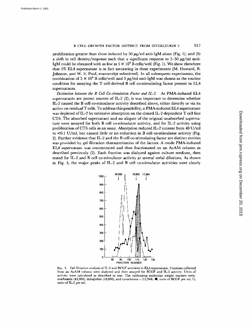
Distinction between the B Cell Co-stimulation Factor and IL-2. As PMA-induced EL4 supernatants are potent sources of IL-2 (2), it was important to determine whether IL-2 caused the B cell co-stimulator activity described above, either directly or via its action on residual T cells. To address this possibility, a PMA-induced EL4 supernatant was depleted of IL-2 by extensive absorption on the cloned IL-2-dependent T cell line CT6. The absorbed supernatant and an aliquot of the original unabsorbed superna- tant were assayed for both B cell co-stimulator activity, and for IL-2 activity using proliferation of CT6 cells as an assay. Absorption reduced IL-2 content from 40 U / m l to <0.1 U/ml , but caused little or no reduction in B cell co-stimulator activity (Fig. 2). Further evidence that IL-2 and the B cell co-stimulating factor are distinct entities was provided by gel filtration charaGterization of the factors. A crude PMA-induced EL4 supernatant was concentrated and then fractionated on an AcA54 column as described previously (2). Each fraction was dialyzed against culture medium, then tested for IL-2 and B cell co-stimulator activity at several serial dilutions. As shown in Fig. 3, the m a j o r peaks of IL-2 and B cell co-stimulator activities were clearly
1000
9OO
800
700
6OO
500
4OO
300
200
100
0
43,500 18,800 12,384
80 90 100 110 120 130 FRACTION NUMBER
Fie. 3. Gel filtration analysis of IL-2 and BCGF activities in EL4 supernatant. Fractions collected from an AcA54 column were dialyzed and then assayed for BCGF and IL-2 activity. Units of activity were calculated as described in text. The calibrating molecular weight markers were: ovalbumin (43,500), myoglobin (18,800), and cytochrome c (12,384). O, units of BCGF per ml; O, units of IL-2 per ml.
on Decem
ber 20, 2015jem
.rupress.orgD
ownloaded from
Published March 1, 1982
918 H O W A R D ET AL.
separable by this procedure, with their approximate molecular weights being 30,000- 35,000 and 16,500-18,500, respectively. The 16,500-18,500 Mr material produced little or no proliferation in control B cell cultures from which anti-IgM antibodies had been omitted. This result was repeated in two further experiments using AcA54 fractions prepared from separate PMA-induced EL4 supernatants. The molecular weight estimate for IL-2 obtained in these experiments agrees with that reported by us (13) and by three other laboratories on the basis of gel filtration analysis (14-16). We have recently confirmed the molecular weight estimate for B cell co-stimulator activity by sodium dodecyl sulfate (SDS)-polyacrylamide gel electrophoresis (PAGE) analysis (J. Farrar, M. Howard, J. Fuller-Farrar, and W. E. Paul, manuscript in preparation). For convenience, the 18,000 Mr B cell co-stimulator from the EL4 thymoma was termed B cell growth factor (BCGF).
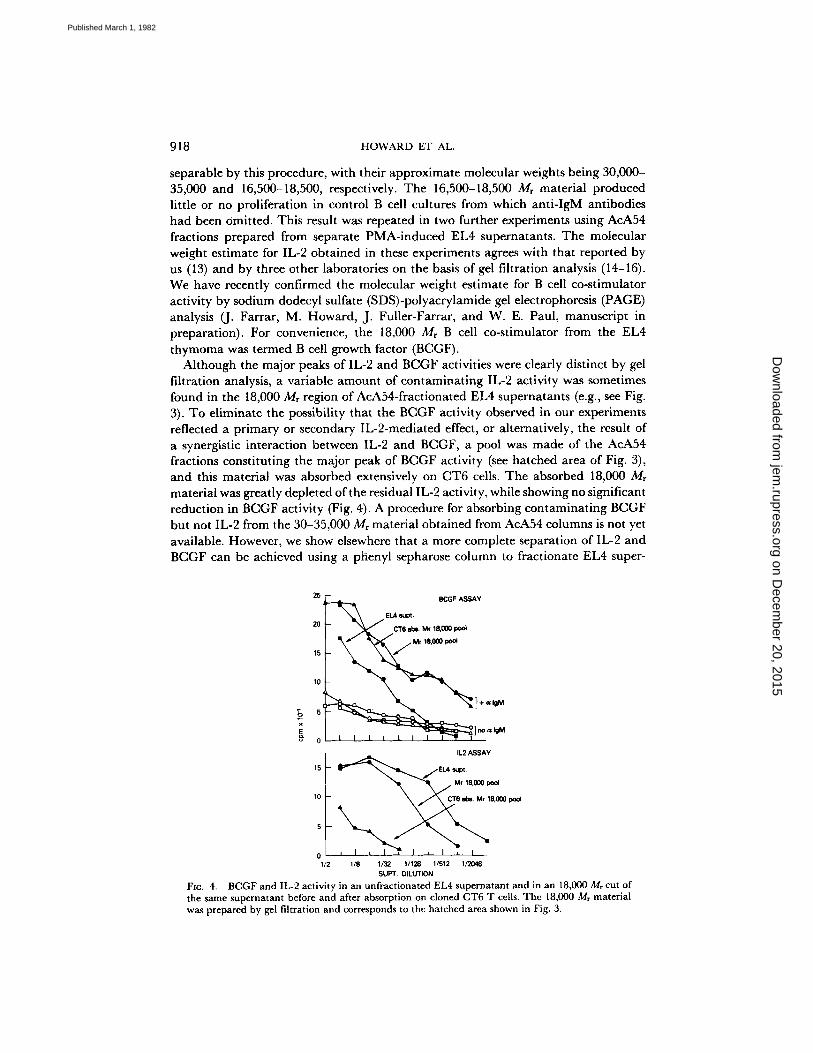
Although the major peaks of IL-2 and BCGF activities were clearly distinct by gel filtration analysis, a variable amount of contaminating IL-2 activity was sometimes found in the 18,000 Mr region of AcA54-fractionated EL4 supernatants (e.g., see Fig. 3). To eliminate the possibility that the BCGF activity observed in our experiments reflected a primary or secondary IL-2-mediated effect, or alternatively, the result of a synergistic interaction between IL-2 and BCGF, a pool was made of the AcA54 fractions constituting the major peak of BCGF activity (see hatched area of Fig. 3), and this material was absorbed extensively on CT6 cells. The absorbed 18,000 Mr material was greatly depleted of the residual IL-2 activity, while showing no significant reduction in BCGF activity (Fig. 4). A procedure for absorbing contaminating BCGF but not IL-2 from the 30-35,000 Mr material obtained from AcA54 columns is not yet available. However, we show elsewhere that a more complete separation of IL-2 and BCGF can be achieved using a phenyl sepharose column to fractionate EL4 super-
25 ~ BCGF ASSAY
I \ \ EL4 8LIpL
20 i - ~ / C T 6 abe. Mr 18,000 pool
I \ " -~ /M, ,8=o~
11.2 ASSAY
15 ~ p 1 / E L 4 supt. " ~ ~ , , , , / Mr 18,000 pool
10 CT6 ~oi. Mr 15,000 pool
0 I I I I ~ I I I ~ I 1/2 1/8 1/32 1/128 1/512 112048
SUPT, DILUTION FIG. 4. BCGF and IL-2 activity in an unfractionated EL4 supernatant and in an 18,000 Mr cut of the same supernatant before and after absorption on cloned CT6 T cells. The 18,000 M, material was prepared by gel filtration and corresponds to the hatched area shown in Fig. 3.
on Decem
ber 20, 2015jem
.rupress.orgD
ownloaded from
Published March 1, 1982

B CELL GROWTH FACTOR DISTINCT FROM INTERLEUKIN 2 919
T A B L E I
Nonspecific Factor Requirements for an SRC-specific AFC Response
Group
Factor additions to the B cell cultures*
Anti-SRC AFC per 10 n B cells¶
IL-2:]: BCGF§ TRFII Experi- Experi- ment 1 ment 2
A - - - - - - 0 0 B 1:200 - - - - 2 0 C - - 1:200 - - 0 0 D - - 1:4 0 0 E 1:200 1:200 - - 2 0 F 1:200 - - 1:4 8 2 G 1:200 1:4 8 10 H 1:200 1:200 1:4 76 93
I 1 :20 - - - - 0 N D * *
J - - 1:20 - - 0 ND K - - - - 1:4 0 ND L 1:20 1:20 - - 0 ND M 1:20 - - 1:4 7 ND N - - 1:20 1:4 0 ND O 1:20 1:20 1:4 120 ND
* Factors were added at the dilutions indicated simultaneously with SRC on day 0 to cultures containing 5 × 10⁵ B cells.
:~ A pool of active fractions from a preparative isoelectric focusing experiment. Preparation free of BCGF.
§ A pool of active fractions from a preparative isoeleetric focusing experiment. Preparation free of IL-2.
[I Unfractionated B 151K 12 hybridoma supernatant. Preparation free of IL-2 and BCGF.
¶ Antibody-forming cells measured on day 4. All groups comprised triplicate cultures.
** Not determined.
n a t a n t s (10). T h e B C G F - f r e e I L - 2 - c o n t a i n i n g m a t e r i a l o b t a i n e d by this p r o c e d u r e
t o t a l l y lacks t he ab i l i ty to synerg ize w i t h a n t i - I g M an t i bod i e s for i n d u c t i o n o f
po lye lona l B cell p ro l i f e r a t i on (10).
Requirement for BCGF in an AFC Response. T o fu r the r exp lo re t he role o f B C G F in
B cell i m m u n e responses, t he s t imul i r e q u i r e d for A F C p r o d u c t i o n by h i g h l y pu r i f i ed
B cells were inves t iga ted . As expec ted , B cells d e p l e t e d o f accessory cells, as desc r ibed
above , fa i led to g e n e r a t e SRC -spec i f i c A F C w h e n c u l t u r e d w i t h S R C for 4 d in a
s t a n d a r d in v i t ro A F C assay ( T a b l e I, g roup A). T o test w h e t h e r B C G F a n d / o r IL-2
cou ld c o m p e n s a t e for the d e p l e t e d accessory cell func t ion , it was des i rab le to use
p r e p a r a t i o n s o f these factors p u r e r t h a n those o b t a i n e d f rom A c A 5 4 c o l u m n f rac t ion-
a t ion . A full b i o c h e m i c a l c h a r a c t e r i z a t i o n o f B C G F is p r e sen t ed e l sewhere (J. Fa ra r ,
M. H o w a r d , J . F u l l e r - F a r r a r , a n d W. E. Pau l , m a n u s c r i p t in p r e p a r a t i o n ) , a n d shows
t h a t m o r e c o m p l e t e s epa ra t i on o f B C G F a n d IL-2 is possible e i the r b y p h e n y l
sepharose c h r o m a t o g r a p h y , by S D S - P A G E , or by isoelectr ic-focusing. T h u s , r e la t ive ly
p u r e p r e p a r a t i o n s o f B C G F a n d IL-2 were p r o d u c e d by isoelectr ic focus ing o f a P M A -
i n d u c e d E L 4 s u p e r n a t a n t , a n d an a m o u n t o f e a c h found to be s a t u r a t i n g in the
a p p r o p r i a t e p ro l i f e r a t i on assay was a d d e d to cu l tu res c o n t a i n i n g pur i f i ed B cells a n d
S R C an t igen . B o t h l y m p h o k i n e s fa i led to i n d u c e a n t i g e n - a c t i v a t e d B cells to deve lop
on Decem
ber 20, 2015jem
.rupress.orgD
ownloaded from
Published March 1, 1982

920 HOWARD ET AL.
into AFC, either individually or in combination (Table I, groups B, C, E, I, J, and L). It has recently been proposed (11, I7) that antigen-induced AFC responses require two functionally distinct nonspecific T cell factors: one to enhance B cell proliferation and the other to provide a differentiation signal. To provide an independent source of the latter, we used supernatant from the T cell-replacing factor (TRF)-secreting hybridoma BI51K12 produced by Takatsu and colleagues (7). This supernatant was found to lack both BCGF and IL-2 activity (K. Nakanishi and M. Howard, unpub- lished data) and was insufficient by itself or in combination with either BCGF or IL- 2 to support the production of specific AFC in cultures of purified B cells and SRC antigen (Table I, groups D, F, G, K, M, and N). However, an excellent SRC-specific AFC response was obtained in B cell cultures containing a combination of antigen, BCGF, IL-2, and B151K12 supernatant as a source of TRF (Table I, groups H and O). These responses were antigen specific both in terms of requiring the presence of antigen and of not including generation of AFC with unrelated specificities (unpub- lished data). From these data, we conclude that the B cell proliferation-enhancing factor BCGF is required for the production of SRC-specific AFC. In view of the high number of purified B cells required for the experiments summarized in Table I, the chance for residual T cell and macrophage contamination is markedly increased. Thus, it should not be assumed that the other factors required for AFC production (i.e., IL-2 and B151K12 supernatant) are acting directly on B cells.
Discussion
We describe in this report a factor, BCGF, that synergizes with anti-IgM antibodies to cause po!yclonal B cell proliferation. The small number of highly purified target B cells and the absolute requirement for anti-IgM in the proliferation experiments make it highly unlikely that BCGF is acting on a contaminating non-B cell present in the cultures. The action of BCGF is seemingly restricted to DNA synthesis and does not include AFC production unless additional factors are provided. When these additional factors are provided, BCGF is found to be an essential component of an antigen- specific AFC response. The evidence presented qualifies BCGF as a new T cell lymphokine. It can be distinguished both functionally and by molecular weight from other previously described T cell factors, namely IL-2 (1, 13-16), TRF (18), colony- stimulating factor (19), interleukin 3 (20), and B cell replication and maturation factor (21). BCGF does not induce proliferation of phytohemagglutin-stimulated thymocytes (unpublished data) and so is distinct from the monokine IL-1 (22). The biochemical properties of this molecule are presented in greater detail elsewhere (J. Farrar, M. Howard, J. Fuller-Farrar, and W. E. Paul, manuscript in preparation) and further support its novelty. The proportion of B cells responsive to BCGF is unclear. The levels of proliferation obtained in these studies are consistent with involvement of the entire anti-IgM-activated B cell pool, a pool recently quantitated to be -50% of normal B cells (23). BCGF-Iike activity can be found in antigen- induced supernatants from normal T cell helper lines propagated in long-term culture as described by Kimoto and Fathman (24) (K. Nakanishi and M. Howard, unpub- lished data). We are currently investigating the phenotype of normal cells that produce BCGF.
An additional point of interest to emerge from this study is the role for the Ig receptor in B cell activation. Although the sequence in which anti-IgM and BCGF
on Decem
ber 20, 2015jem
.rupress.orgD
ownloaded from
Published March 1, 1982

B CELL GROWTH FACTOR DISTINCT FROM INTERLEUKIN 2 921
signal the B cell cannot be implied from the above experiments, we have found that proliferation can be extended beyond the 3-d co-stimulator assay by the continued addition of BCGF but not anti-IgM (unpublished observations). In light of this, we propose the following model of B cell activation: that BCGF has no action on resting B cells; that anti-Ig delivers a nonproliferative signal to resting B cells, possibly causing the expression of receptors for BCGF; and that BCGF delivers a proliferation signal to these anti-Ig-activated, growth factor-sensitive B cells. Thus, the overall concept is analogous to currently proposed models for T cell activation (25-27). We wish to emphasize this model pertains only to the 50% of B cells that can be activated- by anti-IgM (23) and thus does not exclude the existence of a separate subset of B cells that interact with T cells in an H-2-restricted manner. Such a division of B lymphocytes is in fact consistent with the recent work of Singer et al. (28). Similarly, we cannot exclude the possibility that other lymphokines or monokines may be involved in enhancing, modifying, or regulating this entire process.
S u m m a r y
We report here a factor (B cell growth factor) found in induced supernatants of the mouse thymoma EL4 that co-stimulates with anti-IgM antibodies in short-term cultures of purified B lymphocytes to induce polyclonal B cell proliferation but not antibody-forming cell production. The factor is not mitogenic for resting B cells and interacts with anti-IgM-activated B cells in a non-H-2-restricted manner. Absorption studies and molecular weight analysis reveal the factor is distinct from interleukin 2. This factor synergises with antigen, interleukin 2, and an interleukin 2-free, B cell growth factor-free T cell supernatant that contains T cell-replacing factor to produce erythrocyte-specific plaque-forming cells in cultures of highly purified B cells.
We thank A. DeFranco and R. Asofsky for providing affinity-purified goat anti-mouse IgM antibodies, and J. Fuller-Farrar for assistance with gel filtration analyses.
Received for publication 18 November 1981.
References
1. Moiler, G., editor. 1980. T cell clones. Immunol. Rev. Vol. 54. 2. Farrar, J., J. Fuller-Farrar, P. Simon, M. Hilfiker, B. Stadler, and W. Farrar. 1980.
Thymoma production of T cell growth factor (interleukin 2). J. Immunol. 125:2555. 3. Gillis, S., and S. Mizel. 1981. T cell lymphoma model for the analysis of interleukin 1-
mediated T cell activation. Proc. NatL Acad. Sci. U. S. A. 78:1133. 4. Howard, M., S. Kessler, T. Chused, and W. Paul. 1981. Long-term culture of normal
mouse B lymphocytes. Proc. NatL Acad. Sci. U. S. A. 78:5788. 5. Sieckmann, D., R. Asofsky, D. Mosier, I. Zitron, and W. E. Paul. 1978. Activation of mouse
lymphocytes by anti-immunoglobulin. I. Parameters of the proliferative response. J. Exp. Med. 147:814.
6. Fuller-Farrar, J., M. Hilfiker, W. Farrar, andJ. Farrar. 1981. PMA enhances the production of interleukin 2. Cell, Immunol. 58"156.
7. Takatsu, K., K. Tanaka, A. Tominaga, Y. Kumahara, and T. Hamaoka. 1980. Antigen- induced T cell-replacing factor (TRF). III. Establishment of T cell hybrid clone continu- ously producing TRF and functional analysis of released TRF.J. Immunol. 195:2646.
on Decem
ber 20, 2015jem
.rupress.orgD
ownloaded from
Published March 1, 1982
922 HOWARD ET AL.
8. Leibson, H., P. Marrack, and J. Kappler. 1981. B cell helper factors. I. Requirement for both IL2 and another 40,000 Mr factor.J. Exp. Med. 154:1681.
9. Ly, L., and R. Mishell. 1974. Separation of mouse spleen cells by passage through Sephadex G-10.J. Immunol. Methods. 5:239.
10. Farrar, J., W. Benjamin, M. Hilfiker, M. Howard, W. Farrar, and J. Fuller-Farter. 1982. The biochemistry, biology, and role of IL2 in the induction of cytotoxic T cells and antibody-forming B cell responses. Immunol. Rev. In press.
11. Parker, D. 1980. Induction and suppression of polyclonal antibody responses by anti-Ig reagents and antigen-nonspecific helper factors. Immunol. Rev. 52:115.
12. Parker, D. 1975. Stimulation of mouse lymphocytes by insoluble anti-mouse immunoglob- ulin. Nature ( Lond.). 258:361.
13. Farrar, J., P. Simon, W. Koopman, and J. Fuiler-Bonar. 1978. Biochemical relationship of thymocyte mitogenic factor and factors enhancing humoral and cell-mediated immune responses.J. Immunol. 121:1353.
14. Chen, D., and G. DiSabato. 1976. Further studies on the thymocyte stimulating factor. Cell. Immunol. 22:211.
15. Shaw, J., v . Monticone, G. Mills, and V. Paetkau. 1978. Effects of co-stimulator on immune responses in vitro. J. Immunol. 120:1974.
16. Watson, J., s. Gillis, j . Marbrook, D. Mochizuki, and K. Smith. 1979. Biochemical and biological characteristics of lymphocyte regulatory molecules. I. Purification of a class of murine lymphokines. J. Exp. Med. 150:849.
17. Swain, S., G. Dennert, J. Warner, and R. Dutton. 1981. Culture supernatant of a stimulated T cell line have helper activity that synergizes with IL2 in the response of B cells to antigen. Proc. Natl. Acad. Sci. U. S. A. 78:2517.
18. Schimpl, A., L. Hubner, C. Wong, and E. Wecker. 1980. Distinction between T helper cell replacing factor (TCRF) and T cell growth factor (TCGF). Behring Inst. Mitt. 67:221.
19. Nicola, N., A. Burgess, and D. Metcalf. 1979. Similar molecular properties of granulocyte- macrophage colony-stimulating factors produced by different mouse organs in vitro and in vivo. J. Biol. Chem. 254:5290.
20. Hapel, A., J. Lee, W. Farrar, and J. Ihle. 1981. Establishment of continous cultures of Thy 1.2 +, Lyt 1 +2- T cells with purified interleukin 3. Cell. 25:179.
21. Andersson, J., and F. Melchers. 1981. T cell-dependent activation of resting B cells: requirement for both nonspecific unrestricted and antigen-specific Ia-restricted soluble factors. Proc. Natl. A~d. Sci. U. S. A. 78:2497.
22. Lachman, L., M. Hacker, G. Blyden, and R. Handschumacher. 1977. Preparation of lymphocyte-activating factor from continuous murine macrophage cell lines. Cell. Immunol. 34:416.
23. DeFranco, A., E. Raveche, R. Asofsky, and W. Paul. 1982. Frequency of B lymphocytes responsive to anti-immunoglobulin. J. Exp. Med. In press.
24. Kimoto, M., and C. Fathman. 1980. Antigen-reactive T cell clones. I. Transcomplementing hybrid I-A region gene products functions effectively in antigen presentation.J. Exp. Med. 152:759.
25. Lafferty, K., H. Warren, J. Woolnough, and D. Talmage. 1978. Immunological induction of T lymphocytes: role of antigen and the lymphocyte co-stimulator. Blood Cells. 4:395.
26. Larsson, E., and A. Coutinho. 1979. On the role of mitogenic lectins in T-cell triggering. Nature ( Lond.). 280:239.
27. Andersson, J., K. Gronvik, E. Larsson, and A. Coutinho. 1979. Studies on T-lymphocyte activation. I. Requirement for the mitogenic-dependent production ofT-cell growth factors. Fur. J. Immunol. 9:581.
on Decem
ber 20, 2015jem
.rupress.orgD
ownloaded from
Published March 1, 1982
B CELL GROWTH FACTOR DISTINCT FROM INTERLEUKIN 2 923
28. Singer, A., P. Morrissey, K. Hathcock, A. Ahmed, I. Scher, and R. Hodes. 1981. Role of the major histocompatibility complex in T cell activation of B cell subpopulations. Lyb-5 + and Lyb-5- B cell subpopulations differ in their requirement for major histocompatibility complex-restricted T cell recognition.,]. Exp. Med. 154:501.
on Decem
ber 20, 2015jem
.rupress.orgD
ownloaded from
Published March 1, 1982
Copyright © 2022 FDOKUMEN