An Fcγ Receptor-Dependent Mechanism Drives Antibody-Mediated Target-Receptor Signaling in Cancer...
13
Cancer Cell Article An Fcg Receptor-Dependent Mechanism Drives Antibody-Mediated Target-Receptor Signaling in Cancer Cells Nicholas S. Wilson, 1 Becky Yang, 1 Annie Yang, 1 Stefanie Loeser, 2 Scot Marsters, 1 David Lawrence, 1 Yun Li, 1 Robert Pitti, 1 Klara Totpal, 3 Sharon Yee, 3 Sarajane Ross, 3 Jean-Michel Vernes, 4 Yanmei Lu, 4 Cam Adams, 5 Rienk Offringa, 2 Bob Kelley, 5 Sarah Hymowitz, 6 Dylan Daniel, 1,7 Gloria Meng, 4 and Avi Ashkenazi 1, * 1 Department of Molecular Oncology 2 Department of Discovery Immunology 3 Department of Cancer Signaling and Translational Oncology 4 Department of Assay & Automation Technology 5 Department of Antibody Engineering 6 Department of Structural Biology Genentech, Inc., 1 DNA Way, South San Francisco, CA 94080, USA 7 Present address: Oncology Department, Novartis Institutes for Biomedical Research, 4560 Horton Street, Emeryville CA 94608-2916, USA *Correspondence: [email protected] DOI 10.1016/j.ccr.2010.11.012 SUMMARY Antibodies to cell-surface antigens trigger activatory Fcg receptor (FcgR)-mediated retrograde signals in leukocytes to control immune effector functions. Here, we uncover an FcgR mechanism that drives anti- body-dependent forward signaling in target cells. Agonistic antibodies to death receptor 5 (DR5) induce cancer-cell apoptosis and are in clinical trials; however, their mechanism of action in vivo is not fully defined. Interaction of the DR5-agonistic antibody drozitumab with leukocyte FcgRs promoted DR5-mediated tumor- cell apoptosis. Whereas the anti-CD20 antibody rituximab required activatory FcgRs for tumoricidal function, drozitumab was effective in the context of either activatory or inhibitory FcgRs. A CD40-agonistic antibody required similar FcgR interactions to stimulate nuclear factor-kB activity in B cells. Thus, FcgRs can drive antibody-mediated receptor signaling in target cells. INTRODUCTION The interaction of antigen-bound antibodies with FcgRs on the surface of leukocytes triggers retrograde signals into these FcgR- bearing cells. FcgRs fall into two signaling subclasses: activatory or inhibitory (Nimmerjahn and Ravetch, 2008). With the exception of human glycophosphatidylinositol-anchored FcgRIIIB, all mouse and human activatory FcgRs signal either through a cyto- plasmic immunoreceptor tyrosine-based activating motif (ITAM), or via the ITAM-containing common g chain. The engagement of leukocyte activatory FcgRs by antibody-antigen complexes may lead to various biologic effects, including cytokine secretion, oxidative burst, increased phagocytosis, and enhanced anti- body-dependent, cell-mediated cytotoxicity (ADCC). ADCC is mediated by release of perforin and cytotoxic granules contain- ing granzyme B, which, upon entry into target cells, activates apoptotic executioner caspases such as caspase-3 (Bolitho et al., 2007; Martin et al., 1996; Wilson et al., 2009). In addition to expressing activatory FcgRs, innate immune cells such as macrophages, monocytes, dendritic cells (DCs), mast cells, and granulocytes, express an inhibitory FcgR (FcgRIIB) (Amigor- ena et al., 1992; Nimmerjahn and Ravetch, 2008). FcgRIIB Significance Therapeutic antibodies targeting antigens on cancer cells often rely on antibody-dependent, cell-mediated cytotoxicity (ADCC) for efficacy: ADCC is controlled by antibody interaction with activatory versus inhibitory Fcg receptors (FcgRs) on immune cells. A class of antibodies aims to trigger tumor-cell death via the proapoptotic receptors DR4 and DR5; how these agents work in vivo is not completely defined. We discovered that FcgRs on tumor-associated leukocytes provide a crosslinking scaffold that promotes antibody-dependent, DR5-mediated apoptosis in cancer cells. Both activa- tory and inhibitory FcgRs can support DR5 activation, unlike ADCC. A similar FcgR-based mechanism mediates antibody- dependent apoptosis activation through DR4, and NF-kB stimulation via a more distant receptor, CD40. These results have implications for clinical testing and optimization of agonistic antibodies. Cancer Cell 19, 101–113, January 18, 2011 ª2011 Elsevier Inc. 101
-
Upload
independent -
Category
Documents
-
view
0 -
download
0
Transcript of An Fcγ Receptor-Dependent Mechanism Drives Antibody-Mediated Target-Receptor Signaling in Cancer...
Cancer Cell
Article
An Fcg Receptor-Dependent Mechanism DrivesAntibody-Mediated Target-Receptor Signalingin Cancer CellsNicholas S.Wilson,1 Becky Yang,1 Annie Yang,1 Stefanie Loeser,2 ScotMarsters,1 David Lawrence,1 Yun Li,1 Robert Pitti,1
Klara Totpal,3 Sharon Yee,3 Sarajane Ross,3 Jean-Michel Vernes,4 Yanmei Lu,4 Cam Adams,5 Rienk Offringa,2
Bob Kelley,5 Sarah Hymowitz,6 Dylan Daniel,1,7 Gloria Meng,4 and Avi Ashkenazi1,*1Department of Molecular Oncology2Department of Discovery Immunology3Department of Cancer Signaling and Translational Oncology4Department of Assay & Automation Technology5Department of Antibody Engineering6Department of Structural Biology
Genentech, Inc., 1 DNA Way, South San Francisco, CA 94080, USA7Present address: Oncology Department, Novartis Institutes for Biomedical Research, 4560 Horton Street, Emeryville CA 94608-2916, USA
*Correspondence: [email protected] 10.1016/j.ccr.2010.11.012
SUMMARY
Antibodies to cell-surface antigens trigger activatory Fcg receptor (FcgR)-mediated retrograde signals inleukocytes to control immune effector functions. Here, we uncover an FcgR mechanism that drives anti-body-dependent forward signaling in target cells. Agonistic antibodies to death receptor 5 (DR5) inducecancer-cell apoptosis and are in clinical trials; however, their mechanism of action in vivo is not fully defined.Interaction of the DR5-agonistic antibody drozitumabwith leukocyte FcgRs promoted DR5-mediated tumor-cell apoptosis. Whereas the anti-CD20 antibody rituximab required activatory FcgRs for tumoricidal function,drozitumab was effective in the context of either activatory or inhibitory FcgRs. A CD40-agonistic antibodyrequired similar FcgR interactions to stimulate nuclear factor-kB activity in B cells. Thus, FcgRs can driveantibody-mediated receptor signaling in target cells.
INTRODUCTION
The interaction of antigen-bound antibodies with FcgRs on the
surfaceof leukocytes triggers retrograde signals into theseFcgR-
bearing cells. FcgRs fall into two signaling subclasses: activatory
or inhibitory (Nimmerjahn andRavetch, 2008).With the exception
of human glycophosphatidylinositol-anchored FcgRIIIB, all
mouse and human activatory FcgRs signal either through a cyto-
plasmic immunoreceptor tyrosine-based activatingmotif (ITAM),
or via the ITAM-containing common g chain. The engagement of
leukocyte activatory FcgRs by antibody-antigen complexes may
Significance
Therapeutic antibodies targeting antigens on cancer cells of(ADCC) for efficacy: ADCC is controlled by antibody interacton immune cells. A class of antibodies aims to trigger tumohow these agents work in vivo is not completely defined. Wprovide a crosslinking scaffold that promotes antibody-depentory and inhibitory FcgRs can support DR5 activation, unlike Adependent apoptosis activation through DR4, and NF-kB stimuimplications for clinical testing and optimization of agonistic a
C
lead to various biologic effects, including cytokine secretion,
oxidative burst, increased phagocytosis, and enhanced anti-
body-dependent, cell-mediated cytotoxicity (ADCC). ADCC is
mediated by release of perforin and cytotoxic granules contain-
ing granzyme B, which, upon entry into target cells, activates
apoptotic executioner caspases such as caspase-3 (Bolitho
et al., 2007; Martin et al., 1996; Wilson et al., 2009). In addition
to expressing activatory FcgRs, innate immune cells such as
macrophages, monocytes, dendritic cells (DCs), mast cells,
and granulocytes, express an inhibitory FcgR (FcgRIIB) (Amigor-
ena et al., 1992; Nimmerjahn and Ravetch, 2008). FcgRIIB
ten rely on antibody-dependent, cell-mediated cytotoxicityion with activatory versus inhibitory Fcg receptors (FcgRs)r-cell death via the proapoptotic receptors DR4 and DR5;e discovered that FcgRs on tumor-associated leukocytesdent, DR5-mediated apoptosis in cancer cells. Both activa-DCC. A similar FcgR-based mechanism mediates antibody-lation via a more distant receptor, CD40. These results haventibodies.
ancer Cell 19, 101–113, January 18, 2011 ª2011 Elsevier Inc. 101
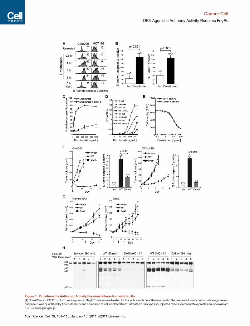
Figure 1. Drozitumab’s Antitumor Activity Requires Interaction with FcgRs
(A) Colo205 and HCT116 colon tumors grown in Rag2�/�mice were treated for the indicated time with drozitumab. The percent of tumor cells containing cleaved
caspase-3 was quantified by flow cytometry and compared to cells isolated from untreated or isotype (Iso) injected mice. Representative profiles are shown from
n = 3–4 mice per group.
Cancer Cell
DR5-Agonistic Antibody Activity Requires FcgRs
102 Cancer Cell 19, 101–113, January 18, 2011 ª2011 Elsevier Inc.
Cancer Cell
DR5-Agonistic Antibody Activity Requires FcgRs
contains a cytoplasmic immunoreceptor tyrosine-based inhibi-
tory motif (ITIM), which negatively regulates the magnitude of
signaling by ITAM-containing activatory receptors (Takai et al.,
1996). Different IgG subclasses have varying affinity and speci-
ficity for distinct FcgRs (Nimmerjahn et al., 2005; Nimmerjahn
and Ravetch, 2006). In humans, IgG1 and IgG3 function as
proinflammatory IgG subclasses, based on their higher affinity
for activatory versus inhibitory FcgRs. In mice, this latter feature
characterizes the IgG2a and IgG2b subclasses (Dijstelbloem
et al., 2001; Nimmerjahn and Ravetch, 2006).
Many cancer-therapeutic antibodies that bind cancer cell-
surface antigens require ADCC to mediate tumor-cell elimination
in vivo. Indeed, evidence frompreclinical animalmodels suggests
that ADCC is an important component of the therapeutic activity
of human IgG1 antibodies that target the CD20 B cell-differentia-
tion antigen (e.g., rituximab) (Maloney et al., 1997; de Haij
et al., 2010), human epidermal growth factor receptor 2 (HER2)
(trastuzumab) (Barok et al., 2007; Clynes et al., 2000; Musolino
et al., 2008), epidermal growth factor receptor (cetuximab)
(Hara et al., 2008), fibroblast growth factor receptor 3 (R3Mab)
(Qing et al., 2009), CD52 (alemtuzumab) (Golay et al., 2006), and
EphA2 receptor (Bruckheimer et al., 2009).
Proapoptotic receptor agonists (PARAs) activate the extrinsic
apoptotic pathway by engaging death-domain-containing tumor
necrosis factor receptor superfamily (TNFRSF) members on
tumor cells (Ashkenazi, 2008a; Wilson et al., 2009). PARAs
include recombinant human Apo2L/TRAIL (dulanermin), and
agonistic antibodies that target the human receptors DR4 or
DR5 (Ashkenazi, 2008a, 2008b; Ashkenazi and Herbst, 2008;
Johnstone et al., 2008). Several DR4 and DR5 agonistic
antibodies have demonstrated potent antitumor efficacy in
preclinical cancer models and are now in clinical trials. Drozitu-
mab is a human DR5-specific IgG1 antibody (Adams et al.,
2008; Camidge et al., 2010). Drozitumab mediates DR5
clustering and binding of the apical signaling adaptor FADD,
which in turn recruits the apoptosis-initiating protease, cas-
pase-8, to assemble a death-inducing signaling complex
(DISC) (Wilson et al., 2009). Once stimulated, caspase-8 acti-
vates executioner caspases such as caspase-3 and -7, either
directly, or via the cell-intrinsic apoptotic pathway. Caspase-3
and -7 cleave multiple cellular substrates, inducing apoptotic
cell death.
In the present study, we investigated the in vivo mechanism of
action of agonistic antibodies targeting DR5 and DR4.
(B) Caspase-3 activity was correlated with an increase in TUNEL-positive Colo20
(C) Caspase-3 activation in Colo205 tumor cells cultured with drozitumab alone
(D) Binding of the wild-type (WT) or DANA drozitumab to recombinant murine Fc
(E) Colo205 cells treated with a dose titration ofWT or DANA drozitumab in combin
with the AlamarBlue cell viability assay after 48 hr.
(F) Rag2�/� mice bearing Colo205 or OCI-LY19 tumors were treated with WT o
Histograms: Caspase-3 activation was quantified ex vivo in established Colo205
(G) Rag2�/� mice bearing Ramos-RA1 or OCI-LY19 tumors received the following
an isotype IgG control antibody. Treatment regimens were administered on days
(H) Rag2�/� mice with pre-established (approximately 300 mm3) Colo205 tumo
180 min after treatment and homogenized in lysis buffer. DR5 was immunoprec
in the DISC was evaluated by immunoprecipitation of DR5 and immunoblotting
test was used to calculate statistical significance.
Error bars in (B)–(D) indicate the standard deviation (SD). Error bars in (F) and (G) in
sentative of two or more independent experiments. See also Figure S1.
C
RESULTS
Drozitumab Requires Interaction with FcgRs to ExertAntitumor ActivityWe quantified the extent and kinetics of drozitumab-induced
apoptosis in xenografted tumor cells by measuring caspase-3
cleavage and DNA fragmentation ex vivo after in vivo antibody
administration. Drozitumab induced marked activation of cas-
pase-3 in the CD45-negative (CD45neg) tumor-cell fraction of
Colo205 or HCT116 colon carcinoma xenografts, peaking at
�3 hr (Figure 1A). A significant increase in caspase-3 activity
correlated with DNA fragmentation after drozitumab treatment
(Figure 1B), confirming apoptotic tumor-cell death.
Various agonistic human DR4 and DR5 antibodies display
maximal proapoptotic signaling upon artificial Fc crosslinking
(Ichikawa et al., 2001; Li et al., 2008; Natoni et al., 2007; Pukac
et al., 2005; Yada et al., 2008; Zhang et al., 2007a). Indeed,
drozitumab-induced caspase-3 activation and cell death in
diverse cancer cell lines were augmented by crosslinking with
an antihuman Fc-specific F(ab’)2 reagent (Figure 1C; see Fig-
ure S1A available online) (Adams et al., 2008). In vivo, however,
drozitumab displayed potent antitumor activity in the absence
of any exogenous crosslinking reagent (Figure 1A). We hypothe-
sized that FcgRs, expressed on immune cells in the tumormicro-
environment, may facilitate drozitumab’s in vivo activity bymedi-
ating ADCC or through some other mechanism. To explore this,
we generated a drozitumab variant with two mutations in the
IgG1 Fc region (D265A and N297A, dubbed DANA), which elim-
inate FcgR binding (Shields et al., 2001). Wild-type (WT) drozitu-
mab bound to murine FcgRs with the following rank order of
affinity: I > IV > (IIB, III), whereas drozitumab-DANA did not
bind to any of these receptors (Figure 1D). WT drozitumab bound
also to human FcgRs, with the following rank order of affinity:
I > IIIA-V158 > (IIIA-F158,IIA) > IIB, whereas drozitumab-DANA
did not exhibit detectable binding (data not shown). The DANA
mutations did not affect drozitumab binding to the salvaging
neonatal FcgR (FcgRn), which is important for maintaining
proper antibody half-life in plasma (Roopenian et al., 2003);
accordingly, the WT and DANA variants displayed similar phar-
macokinetic properties in mice (data not shown). Furthermore,
the two variants were equally potent at inducing death of
Colo205 cells in vitro in presence of a F(ab’)2 crosslinking
reagent (Figure 1E), confirming that the DANA mutations do
not compromise drozitumab’s ability to interact with DR5.
5 cells 4 hr following drozitumab or isotype (Iso) antibody treatment.
or combined with 1 mg/ml of anti-human Fc-specific (Fab’)2 (anti-Fc) reagent.
gR (I, IIB, III, or IV).
ation with the anti-Fc crosslinking reagent) (1 mg/ml). Cell survival was analyzed
r DANA drozitumab or an isotype control antibody on day 0 (n = 10/group).
or OCI-LY19 tumors 4 hr after WT or DANA drozitumab treatment.
treatment; wild-type anti-CD20 (WT), the anti-CD20-DANA mutant (DANA), or
0, 7, and 14 following after group-out (n = 8–10/group).
rs were treated with WT or DANA drozitumab. Tumors were dissected 60 or
ipitated from equal tumor weights and caspase-8 recruitment and processing
for caspase-8 (n = 5 individual tumors/group). Where indicated, a Student’s t
dicate the standard error of the mean (SEM). Data shown in Figure 1 are repre-
ancer Cell 19, 101–113, January 18, 2011 ª2011 Elsevier Inc. 103

Figure 2. Cell Lines Expressing Either Activatory or Inhibitory FcgRs Similarly Support Drozitumab-Induced Caspase-3 Activation in Tumor
Cells
(A) CFSE-labeled Colo205 target cells were cocultured with HEK293 cell lines stably expressing murine FcgRIIB, III, or IV or Jurkat cells stably expressing FcgRI
(at a 1:5 ratio). Caspase-3 activation was assessed by flow cytometry 4 hr after drozitumab (250 ng/ml) addition. Drozitumab binding to murine FcgRs was
inhibited by preblocking FcgR expressing cells for 30 min at 4�C with 1 mg/ml of murine IgG2a isotype antibody (FcgRI), anti-FcgRIIB/III (anti-FcgRIIB/III), or
anti-FcgRIV (anti-FcgRIV).
(B) Caspase-3 activation was monitored by flow cytometry in CFSE-labeled Colo205 cells cocultured for 4 hr with HEK293 cells expressing human FcgRIIB and
drozitumab (250 ng/ml). HEK293-FcgRIIB cells were preblocked for 30 min at 4�C with anti-FcgRIIB (anti-FcgRIIB).
Error bars indicate the SD. Data shown are representative of three independent experiments.
Cancer Cell
DR5-Agonistic Antibody Activity Requires FcgRs
To examine whether FcgRs are important for the antitumor
efficacy of drozitumab, we treated Rag2–/– mice (Shinkai et al.,
1992) bearing various human tumor xenografts with the two
antibody variants. WT drozitumab caused tumor regression or
significantly restricted tumor progression in a number of models
(Figure 1F; Figure S1B); in contrast, the DANA variant displayed
little to no antitumor activity. As compared with WT, the DANA
variant induced �65%–80% less caspase-3 activity in Colo205
and OCI-LY19 B cell lymphoma tumors (Figure 1F, histograms),
with similar reductions in apoptosis as measured by the TUNEL
or hypodiploid DNA assays (data not shown). A DANA derivative
of anti-CD20 antibody also displayed markedly less antitumor
activity as compared to WT anti-CD20 in two models of B cell
lymphoma (Figure 1G). Hence, both the anti-DR5 and anti-
CD20 antibodies require FcgR interactions for optimal efficacy.
To assess the activation of DR5 at the biochemical level, we
performed ex vivo analysis of the formation of a DR5-associated
DISC in tumors. We treated Rag2–/– mice harboring Colo205
tumors with the WT or DANA drozitumab variants, immunopre-
cipitated DR5 from tumor-cell lysates, and used immunoblot
analysis to detect caspase-8 recruitment into the DISC; as well
as caspase-8 activation, as indicated by its proteolytic process-
ing (Figure 1H). Caspase-8 recruitment was evident within 60min
of WT drozitumab treatment, with substantial processing and
appearance of the p18 catalytic subunit by 180 min. These
events were markedly less pronounced upon treatment with
the DANA variant. Similar caspase-8 processing occurred in
HCT116 tumor xenografts after 180 min of WT drozitumab treat-
ment (Figure S1C).
Various Mouse and Human FcgRs Can SupportDrozitumab-Mediated Apoptosis In VitroTwo key FcgR-dependent mechanisms may contribute to drozi-
tumab’s efficacy: immune cell-mediated ADCC and/or DR5
signaling. Importantly, each could account for an apoptotic
104 Cancer Cell 19, 101–113, January 18, 2011 ª2011 Elsevier Inc.
phenotype, as both involve caspase-3 activation (Bolitho et al.,
2007;Martin et al., 1996;Wilson et al., 2009).We therefore exam-
ined the capacity of individual activatory and inhibitory FcgRs to
support apoptosis stimulation by drozitumab. We cocultured
transfected cell lines stably expressing individual FcgRs with
CFSE-labeled Colo205 target cells, added drozitumab, and
measured apoptosis after 4 hr by quantifying caspase-3
cleavage in the CFSEhigh tumor-cell population. All cell lines
expressing murine or human FcgRs, including those bearing
the inhibitory receptor FcgRIIB, were capable of supporting dro-
zitumab-mediated caspase-3 activation (Figures 2A and 2B;
data not shown); preincubation of the cell lines with FcgR-block-
ing antibodies inhibited caspase-3 activation. Hence, both acti-
vatory and inhibitory FcgRs can support drozitumab-mediated
DR5 activation in vitro.
FcgR-Expressing Cells in the Tumor MicroenvironmentSupport Drozitumab-Mediated Apoptosis in Tumor CellsTo better define the interaction between drozitumab and FcgRs
within the tumor microenvironment, we profiled the FcgRs
expressed by enriched CD45high leukocytes extracted from
Colo205 tumors (Figure S2A). The largest tumor-associated
leukocyte population consisted of myelomonocytic cells, identi-
fied as F4/80highCD11cint-highCD11bhigh (CD11bhighF4/80high)
(Figure S2B). Other, less abundant leukocytic populations also
were detected in the tumors using specific cell-surface lineage
markers (Figures S2C and S2D).
Next, we analyzed expression of FcgRIIB, III and IV on tumor-
associated leukocytes, as compared with phenotypically similar
immune-cell populations in the spleen of Rag2–/– mice (Fig-
ure 3A). Myeloid immune cells reportedly express both activatory
and inhibitory FcgRs, whereas NK cells only express activatory
FcgRIII (Nimmerjahn and Ravetch, 2006). One caveat with distin-
guishing between mouse FcgRIIB and FcgRIII is that the
available 2.4G2 antibody recognizes a common polymorphic
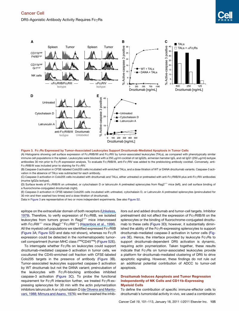
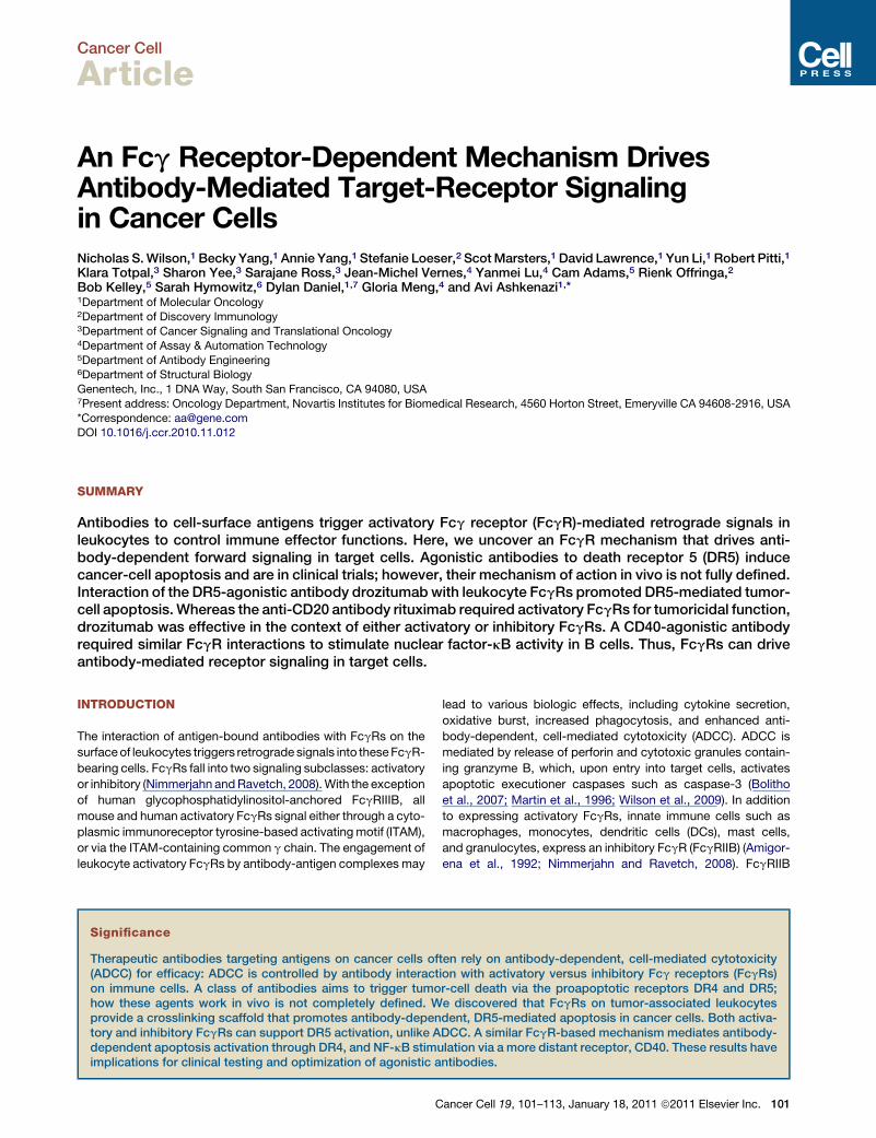
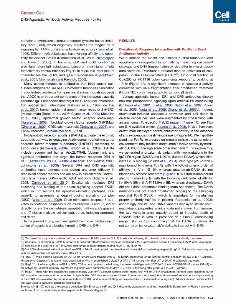
Figure 3. FcgRs Expressed by Tumor-Associated Leukocytes Support Drozitumab-Mediated Apoptosis in Tumor Cells
(A) Histograms showing cell surface expression of FcgRIIB/IIII and FcgRIV by tumor-associated leukocytes (TALs), as compared with phenotypically similar
immune cell populations in the spleen. Leukocytes were blocked with a 250 mg/ml cocktail of rat IgG2b, armenian hamster IgG, and rat IgG1 (250 mg/ml) isotype
antibodies 30 min prior to FcgR expression analysis. To evaluate FcgRIIB/III, anti-FcgRIV was added to the preblocking antibody cocktail. Conversely, anti-
FcgRIIB/III was included prior to staining for FcgRIV.
(B) Caspase-3 activation in CFSE-labeled Colo205 cells incubated with enriched TALs, and a dose titration of WT or DANA drozitumab variants. Caspase-3 acti-
vation in the absence of TALs was subtracted for each antibody.
(C) Caspase-3 activation in Colo205 cells incubated with drozitumab and TALs, either untreated or pretreated with anti-FcgRIIB/III plus anti-FcgRIV antibodies
(murine IgG2a isotype).
(D) Surface levels of FcgRIIB/III on untreated, or cytochalasin D or latrunculin A pretreated splenocytes from Rag2–/– mice (left), and cell surface binding of
a fluorochrome-conjugated drozitumab (right).
(E) Caspase-3 activation in CFSE-labeled Colo205 cells incubated with untreated, cytochalasin D, or Latrunculin A pretreated splenocytes (preincubated for
30 min and then washed two times) and a dose titration of drozitumab.
Data in Figure 3 are representative of two or more independent experiments. See also Figure S2.
Cancer Cell
DR5-Agonistic Antibody Activity Requires FcgRs
epitope on the extracellular domain of both receptors (Unkeless,
1979). Therefore, to verify expression of FcgRIIB, we isolated
leukocytes from tumors grown in Rag2–/– mice intercrossed
with FcgRIII–/– mice (Rag2–/–FcgRIII–/–) (Hazenbos et al., 1996).
All the myeloid cell populations we identified expressed FcgRIIB
(Figure 3A; Figure S2D and data not shown), whereas no FcgR
expression could be detected in the nonhematopoietic tumor-
cell compartment (human MHC class InegCD45neg) (Figure S2E).
To interrogate whether FcgRs on leukocytes could support
drozitumab-mediated caspase-3 activation in tumor cells, we
cocultured the CD45-enriched cell fraction with CFSE-labeled
Colo205 targets in the presence of antibody (Figure 3B).
Tumor-associated leukocytes supported caspase-3 activation
by WT drozitumab but not the DANA variant; preincubation of
the leukocytes with FcgR-blocking antibodies inhibited
caspase-3 activation (Figure 3C). To probe the functional
requirement for FcgR interaction further, we treated FcgR-ex-
pressing splenocytes for 30 min with the actin polymerization
inhibitors latrunculin A or cytochalasin D (de Oliveira and Manto-
vani, 1988; Mimura and Asano, 1976); we then washed the inhib-
C
itors out and added drozitumab and tumor-cell targets. Inhibitor
pretreatment did not affect the expression of FcgRIIB/III on the
splenocytes or the binding of fluorochrome-conjugated drozitu-
mab to these cells (Figure 3D); however, it substantially dimin-
ished the ability of the FcgR-expressing splenocytes to support
drozitumab-mediated caspase-3 activation in tumor cells (Fig-
ure 3E). Hence, the interface provided by leukocyte FcgRs to
support drozitumab-dependent DR5 activation is dynamic,
requiring actin poymerization. Taken together, these results
indicate that FcgRs on tumor-associated leukocytes provide
a platform for drozitumab-mediated clustering of DR5 to drive
apoptotic signaling. However, these findings do not rule out
an additional potential contribution of ADCC to tumor-cell
apoptosis.
Drozitumab Induces Apoptosis and Tumor RegressionIndependently of NK Cells and CD11b-ExpressingMyeloid CellsTo define the contribution of specific immune-effector cells to
drozitumab’s tumoricidal activity in vivo, we used a combination
ancer Cell 19, 101–113, January 18, 2011 ª2011 Elsevier Inc. 105
Figure 4. The Antitumor Activity of Drozitumab Is Independent of Tumor-Associated NK or Myeloid Effector Cell Activity
(A) Colo205 tumors were grown in NK cell containing Rag2�/� or NK cell deficient (Rag2�/�IL2Rg–/–, DKO)mice.Mice were assigned into cohorts and treatedwith
drozitumab or an isotype IgG control antibody (n = 10/group).
(B) Caspase-3 activation was assessed in established (�500 mm3) Colo205 tumors grown in Rag2�/� or DKO mice 4 hr after drozitumab or isotype (Iso) control
antibody treatment.
(C) Beginning 1 day prior to Colo205 tumor cell inoculation, DKOmicewere dosed daily with PBS,mouse aCD11b or isotype control antibody. Fourteen days after
tumor cell inoculation, tumor-bearing mice (�300 mm3) were treated withWT or DANA drozitumab, or an isotype control antibody. Caspase-3 activation in tumor
cells was evaluated ex vivo 4 hr after antibody treatment by flow cytometry.
Error bars shown in panel (A)–(C) indicate the SEM Student’s t test was used to calculate statistical significance. See also Figure S3.
Cancer Cell
DR5-Agonistic Antibody Activity Requires FcgRs
of genetically deficient mice and antibody-based cell depletion
regimens. We examined dependence on NK-cell-mediated
ADCC by inoculating Colo205 tumor cells into Rag2–/– mice, or
into mice deficient in both Rag2 and the interleukin 2 receptor
common g-chain (Rag2–/–IL2Rg–/–, dubbed DKO), which lack
NK cells (Colucci et al., 1999). Drozitumab induced equally
potent Colo205 tumor regression and caspase-3 activation in
both mouse strains (Figures 4A and 4B). We obtained similar
results with H2122 nonsmall cell lung cancer xenografts, or using
antibody-based depletion of NK cells in Colo205 tumor-bearing
Rag2�/� mice (data not shown). To further assess the contribu-
tion of myeloid effector cells, we blocked their recruitment into
tumors by treating NK cell-deficient mice daily with anti-CD11b
antibody, starting one day before tumor-cell inoculation (Smyth
et al., 2006; Zhang et al., 1997). Anti-CD11b treatment itself
did not affect tumor growth (Figure S3A), while gating on the
remaining CD45high cell fraction confirmed myeloid-cell
exclusion from the tumor (Figure S3B). Tumor-associated
CD45highCD11bneg cells isolated from NK cell-deficient mice
coexpressed the DC lineage marker CD11c and MHC class II,
but not for the macrophage marker F4/80 (Figure S3C), suggest-
ing a DC-like phenotype. These cells expressed both activatory
FcgRIII and inhibitory FcgRIIB (Figure S3D). WT drozitumab
induced indistinguishable tumor-cell caspase-3 activation in
mice treated with either an isotype control or an anti-CD11b anti-
body, while the DANA variant was markedly less effective (Fig-
ure 4C). These results indicate that drozitumab can induce
apoptosis in cancer cells independently of tumor-associated
NK or CD11b-expressing myeloid cells. It remains possible
nonetheless that in some tumor types ADCCmay also contribute
to drozitumab’s antitumor activity. Although not formally demon-
strated, these results suggest that FcgR-expressing DC-like
cells are sufficient to support drozitumab’s activity in vivo.
106 Cancer Cell 19, 101–113, January 18, 2011 ª2011 Elsevier Inc.
Drozitumab Exerts Intact Antitumor Activity in Absenceof Activatory FcgRsTo further dissect the contribution FcgRs to drozitumab’s anti-
tumor activity, we devised an in vivo strategy that eliminates
interaction with activatory FcgRs. First, we intercrossed
Rag2–/– mice with FcgRI–/–FcgRIII–/– mice. To ablate interaction
with the remaining activatory FcgRIV, we took advantage of
the failure of this FcgR to bind to muIgG1 (Nimmerjahn et al.,
2005), by generating a muIgG1 variant of drozitumab (Figure 5A).
To confirm that FcgRIV cannot support activity of murine (mu)
IgG1-drozitumab, we used cell lines expressing either FcgRIIB
or FcgRIV. FcgRIIB-expressing cells facilitated caspase-3 acti-
vation in Colo205 cells by either human (hu) IgG1- or muIgG1-
drozitumab (Figure 5B). In contrast, FcgRIV-expressing cells
supported proapoptotic activity of the huIgG1 but not the
muIgG1 drozitumab variant (Figure 5C). Specific FcgR-blocking
antibodies attenuated caspase-3 activation by both drozitumab
isotypes (data not shown). In Rag2–/–FcgRI/III–/– mice, themIgG1
isotype can interact only with the inhibitory FcgRIIB. Nonethe-
less, the tumoricidal activity of muIgG1-drozitumab was compa-
rable in Rag2–/– and Rag2–/–FcgRI/III–/– mice, and consistent with
that of huIgG1-drozitumab in Rag2–/– mice (Figure 5D). We ob-
tained similar results with a muIgG1 variant of an agonistic
anti-DR4 antibody (Chuntharapai et al., 2001) (Figure 5E). These
findings indicate that both activatory and inhibitory FcgRs on
leukocytes are capable of driving antibody-mediated proapop-
totic DR5 or DR4 signaling in cancer cells.
The tumoricidal activity of the anti-CD20 antibody rituximab
is significantly impaired in mice lacking activatory FcgRs
(Clynes et al., 2000). To confirm a similar phenotype in Rag2–/–
FcgRI/III–/– mice, we used isotypic huIgG1 andmuIgG1 rituximab
versions. As compared with the huIgG1 isotype, muIgG1-
rituximab displayed significantly less activity against BJAB
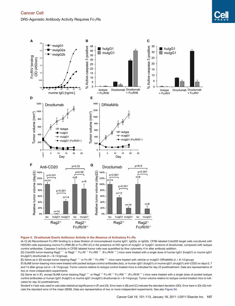
Figure 5. Drozitumab Exerts Antitumor Activity in the Absence of Activatory FcgRs
(A–C) (A) Recombinant FcgRIV binding to a dose titration of noncomplexed murine IgG1, IgG2a, or IgG2b. CFSE-labeled Colo205 target cells cocultured with
HEK293 cells expressing murine FcgRIIB (B) or FcgRIV (C) in the presence of 250 ng/ml of muIgG1 or huIgG1 versions of drozitumab, compared with isotype
control antibodies. Caspase-3 activity in CFSE-labeled tumor cells was quantified by flow cytometry 4 hr after antibody addition.
(D) Colo205 tumor-bearing Rag2�/� or Rag2�/�FcgRI�/�FcgRIII�/� (FcgRI/III�/�) mice were treated with a single dose of human IgG1 (huIgG1) or murine IgG1
(muIgG1) drozitumab (n = 8–12/group).
(E) Same as in (D) except tumor-bearing Rag2�/� or FcgRI�/�FcgRIII�/� mice were treated with vehicle or muIgG1-DR4aMAb (n = 8–12/group).
(F) BJAB tumor-bearing mice were treated with pooled isotype control antibodies (Iso), or human IgG1 (huIgG1) or murine IgG1 (muIgG1) anti-CD20 on days 0, 7
and 14 after group out (n = 8–14/group). Tumor volume relative to isotype control treated mice is indicated for day 22 posttreatment. Data are representative of
two or more independent experiments.
(G) Same as in (F), except BJAB tumor-bearing Rag2�/� or Rag2�/�FcgRI�/�FcgRIII�/� (FcgRI/III�/�) mice were treated with a single dose of pooled isotype
control antibodies or human IgG1 (huIgG1) or murine IgG1 (muIgG1) drozitumab (n = 8–14/group). Tumor volume relative to isotype control treated mice is indi-
cated for day 22 posttreatment.
Student’s t test was used to calculate statistical significance in (F) and (G). Error bars in (B) and (C) indicate the standard deviation (SD). Error bars in (D)–(G) indi-
cate the standard error of the mean (SEM). Data are representative of two or more independent experiments. See also Figure S4.
Cancer Cell
DR5-Agonistic Antibody Activity Requires FcgRs
Cancer Cell 19, 101–113, January 18, 2011 ª2011 Elsevier Inc. 107

Cancer Cell
DR5-Agonistic Antibody Activity Requires FcgRs
Burkitt’s B cell lymphoma xeongrafts in Rag2–/– mice (Figure 5F).
Antitumor efficacy of muIgG1-rituximab was further impaired in
Rag2–/–FcgRI/III–/– mice, consistent with previous evidence
that FcgRIIB interactions inhibit effector-cell activity (Clynes
et al., 2000; Nimmerjahn and Ravetch, 2008). In contrast,
FcgRIIB sufficiently supported activity of either the murine or
human IgG1 variants of drozitumab against BJAB tumors (Fig-
ure 5G). A suboptimal dose of huIgG1 drozitumab (0.1 mg/kg)
was less active in Rag2–/–FcgRIIB–/– mice as compared with
Rag2–/– mice (Figures S4A and S4B), suggesting that FcgRIIB
is not only sufficient but indeed contributes significantly to drozi-
tumab-mediated DR5 activation.
Inkeepingwith the invivo results, FcgR-expressingsplenocytes
isolated from Rag2–/–, or Rag2–/–FcgRI/III–/–, or Rag2–/–
FcgRIIB–/– mice supported tumor-cell caspase-3 activation
ex vivo by either human or murine IgG1-drozitumab (Figure S4C).
Moreover, muIgG1-drozitumab induced tumor-cell caspase-8
activation in xenografted Rag2–/–FcgRI/III–/– mice, while infusion
with an FcgRIIB-blocking antibody before mIgG1-drozitumab
treatment attenuated this effect (Figure S4D). Thus, FcgRIIB is
sufficient to support drozitumab-induced DISC activation in vivo.
Polymorphisms in Human FcgRIIA and FcgRIIIA AffectDrozitumab Binding and Proapoptotic ActivityTwo human FcgR polymorphisms have been associated with
improved progression-free survival in lymphoma patients
treated with rituximab (Cartron et al., 2002; Weng et al., 2004;
Weng and Levy, 2003). A similar correlation was reported for
breast cancer therapy with trastuzumab and colorectal cancer
treatment with cetuximab (Bibeau et al., 2009; Musolino et al.,
2008; Zhang et al., 2007b). The most characterized human
FcgR polymorphism with improved binding to human IgG1 is in
activatory FcgRIIIA, wherein valine versus phenylalanine at posi-
tion 158 (FcgRIIIA158V or FcgRIIIA158F) confers a 3- to 5-fold
higher affinity for human IgG1 (Koene et al., 1997; Presta,
2008; Shields et al., 2001). A polymorphism in human FcgRIIA,
with histidine versus arginine at position 131 (FcgRIIA131H or
FcgRIIA131R), also improves binding to human IgG1 (Bruhns
et al., 2009).
Consistent with other antibodies (Bruhns et al., 2009), huIgG1-
drozitumab exhibited approximately 2- to 3-fold higher affinity
for recombinant FcgRIIA131H versus FcgRIIA131R (Figure 6A).
This increase was seen with noncomplexed and complexed
drozitumab, using anti-k antibody to form complexes and
enhance FcgR-binding avidity. Avidity may increase upon simul-
taneous binding of multiple drozitumab molecules to DR5-ex-
pressing tumor cells and FcgR-bearing leukocytes. To examine
this possibility, we generated transfected HEK293 cell lines
stably expressing FcgRIIA131H or FcgRIIA131R and measured
binding to huIgG subclasses (Figure S5A). Cells expressing
FcgRIIA131H as compared with FcgRIIA131R showed markedly
improved binding to huIgG1 and huIgG2, with similar binding
to huIgG3 and inferior binding to huIgG4 (Figure S5B). Further-
more, cells expressing FcgRIIA131H as compared to FcgRIIA131R
were superior at facilitating drozitumab-induced caspase-3 acti-
vation (Figure 6B). Human IgG1 binds with higher affinity to
FcgRIIIA158V versus FcgRIIIA158F (Figure 6C) (Bruhns et al.,
2009; Koene et al., 1997; Presta, 2008; Shields et al., 2001).
Indeed, cells expressing FcgRIIIA158V also were superior to
108 Cancer Cell 19, 101–113, January 18, 2011 ª2011 Elsevier Inc.
those expressing FcgRIIIA158F at supporting drozitumab-medi-
ated caspase-3 activation in Colo205 cells (Figure 6D). Hence,
stronger FcgR interactions can further augment drozitumab’s
proapoptotic activity.
NF-kB Activation by Anti-CD40 Agonistic AntibodyRequires FcgRsWe next evaluated whether a similar FcgR-dependent mecha-
nism extended to antibody-based activation of another TNFRSF
member, CD40. To this end, we took advantage of the selective
coexpression of FcgRIIB with CD40 (TNFRSF5) on B cells. We
isolated splenic B cells from WT or FcgRIIB–/– mice and exam-
ined the ability of the agonistic anti-CD40 antibody FGK-45
(Elgueta et al., 2009; Saijo et al., 2002) to stimulate NF-kB
signaling and cytokine production. Despite equivalent levels of
CD40 expression, FGK-45 induced substantially less phosphor-
ylation of IkBa, indicating weaker NF-kB activation, in FcgRIIB–/–
versus WT B cells (Figure 6E; Figure S5C). Crosslinking of FGK-
45 with an anti-rat Fc-specific F(ab’)2 reagent reconstituted the
antibody’s activity on FcgRIIB–/– B cells (Figure 6F). Further anal-
ysis based on phosphorylation of the p65 subunit of NF-kB
confirmed that activation by FGK-45 was severely impaired
in FcgRIIB–/– versus WT B cells yet rescued by crosslinking
(Figure S5D). Consistent with these results, FGK-45 induced
markedly less interleukin (IL)-6 and IL-12/23 p40 cytokine
production in FcgRIIB–/– as compared toWTB cells (Figure S5E).
In contrast, IL-6 production in response to lipopolysaccharide
(LPS) was indistinguishable (Figure S5F). These results suggest
that a similar FcgR-dependent mechanism applies to antibody-
mediated activation of diverse signaling functions by structurally
distinct TNFRSF members.
DISCUSSION
FcgRs play important roles in modulating antibody function and
host immunity, by transmitting differential retrogade signals into
leukocytes through activatory or inhibitory sequence motifs
(Nimmerjahn and Ravetch, 2008). ADCC is a key mechanism of
innate immunity, allowing effector cells to attack and eliminate
antibody-decorated target cells upon activatory FcgR stimula-
tion; in contrast, signaling through inhibitory FcgRs negatively
regulates ADCC (Nimmerjahn and Ravetch, 2008). ADCC is
implicated also as a critical component of the anticancer action
of several antibodies that target tumor-associated antigens
(Barok et al., 2007; Bruckheimer et al., 2009; Clynes et al.,
2000; Kimura et al., 2007; Kurai et al., 2007; Musolino et al.,
2008; Peipp et al., 2008; Qing et al., 2009; Schneider-Merck
et al., 2010). Elimination of activatory-FcgR interactions signifi-
cantly attenuates the efficacy of the therapeutic agents rituximab
and trastuzumab in preclinical models (Clynes et al., 2000),
consistent with the importance of ADCC for antitumor activity.
Various anti-DR5 and anti-DR4 antibodies have been under
investigation, with more than 20 human cancer clinical trials
(Ashkenazi, 2002, 2008b; Ashkenazi and Herbst, 2008; John-
stone et al., 2008; Yang et al., 2010). Drozitumab was well toler-
ated in a Phase Ia study in patients with advanced malignancy
(Camidge et al., 2010). While drozitumab displays much stronger
proapoptotic activity in vitro upon artificial Fc crosslinking, it
exhibits potent antitumor activity in vivo in various cancermodels
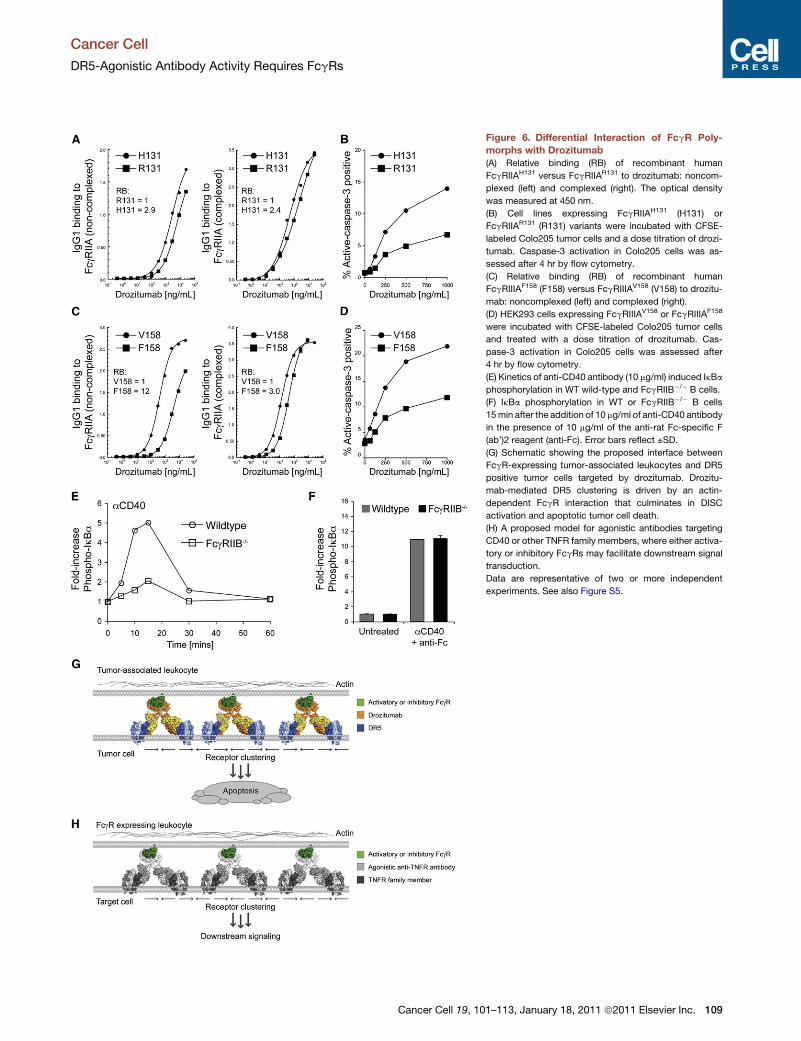
Figure 6. Differential Interaction of FcgR Poly-
morphs with Drozitumab
(A) Relative binding (RB) of recombinant human
FcgRIIAH131 versus FcgRIIAR131 to drozitumab: noncom-
plexed (left) and complexed (right). The optical density
was measured at 450 nm.
(B) Cell lines expressing FcgRIIAH131 (H131) or
FcgRIIAR131 (R131) variants were incubated with CFSE-
labeled Colo205 tumor cells and a dose titration of drozi-
tumab. Caspase-3 activation in Colo205 cells was as-
sessed after 4 hr by flow cytometry.
(C) Relative binding (RB) of recombinant human
FcgRIIIAF158 (F158) versus FcgRIIIAV158 (V158) to drozitu-
mab: noncomplexed (left) and complexed (right).
(D) HEK293 cells expressing FcgRIIIAV158 or FcgRIIIAF158
were incubated with CFSE-labeled Colo205 tumor cells
and treated with a dose titration of drozitumab. Cas-
pase-3 activation in Colo205 cells was assessed after
4 hr by flow cytometry.
(E) Kinetics of anti-CD40 antibody (10 mg/ml) induced IkBa
phosphorylation in WT wild-type and FcgRIIB�/� B cells.
(F) IkBa phosphorylation in WT or FcgRIIB�/� B cells
15min after the addition of 10 mg/ml of anti-CD40 antibody
in the presence of 10 mg/ml of the anti-rat Fc-specific F
(ab’)2 reagent (anti-Fc). Error bars reflect ±SD.
(G) Schematic showing the proposed interface between
FcgR-expressing tumor-associated leukocytes and DR5
positive tumor cells targeted by drozitumab. Drozitu-
mab-mediated DR5 clustering is driven by an actin-
dependent FcgR interaction that culminates in DISC
activation and apoptotic tumor cell death.
(H) A proposed model for agonistic antibodies targeting
CD40 or other TNFR family members, where either activa-
tory or inhibitory FcgRs may facilitate downstream signal
transduction.
Data are representative of two or more independent
experiments. See also Figure S5.
Cancer Cell
DR5-Agonistic Antibody Activity Requires FcgRs
Cancer Cell 19, 101–113, January 18, 2011 ª2011 Elsevier Inc. 109
Cancer Cell
DR5-Agonistic Antibody Activity Requires FcgRs
in the absence of exogenous crosslinkers (Adams et al., 2008;
Edgington et al., 2009; Zinonos et al., 2009). This intriguing obser-
vation prompted us to interrogate how drozitumab operates
in vivo. We found that interaction with FcgRs expressed by
tumor-associated leukocytes is necessary, while either activa-
tory or inhibitory FcgRs are sufficient for optimal proapoptotic
signaling by drozitumab. FcgRs on tumor-associated leukocytes
appear to provide a dynamic platform that facilitates drozitumab-
mediatedDR5 engagement, DISC assembly, and apoptosis acti-
vation in tumor cells (see model in Figure 6G). Thus, leukocyte
FcgRs can promote antibody-mediated signaling through
a cognate receptor on target cells. This may have implications
not only for DR5 antibodies but also more broadly for antibodies
directed to other cell-surface receptors within and perhaps even
outside the TNFR superfamily (Figure 6H).
To test whether FcgRs are important for drozitumab’s proa-
poptotic activity, we generated a mutant variant of drozitumab,
which lacked FcgR interaction while retaining DR5 binding.
The DANA mutant displayed significantly impaired antitumor
activity, demonstrating that FcgR interactions are critical for
drozitumab’s efficacy. We obtained similar results with a DANA
variant of anti-CD20 antibody, which exertsmuch of its antitumor
activity via an activatory FcgR-dependent ADCC. Since ADCC
and DR5 signaling can trigger effector-caspase activation (Boli-
tho et al., 2007; Chowdhury and Lieberman, 2008; Cullen and
Martin, 2008; Martin et al., 1996; Wilson et al., 2009), both mech-
anisms might account for the apoptotic phenotype in drozitu-
mab-treated tumors. To address this, we tested the dependence
of drozitumab’s efficacy on activatory versus inhibitory FcgRs.
In vitro, both types of FcgR were capable of supporting drozi-
tumab-induced caspase activation in tumor-cell targets. Activity
was also facilitated ex vivo by isolated tumor-associated leuko-
cytes, which expressed a spectrum of activatory and inhibitory
FcgRs. Using genetic models in conjunction with a murine
IgG1 version of drozitumab, we found that the inhibitory
FcgRIIB was sufficient, if not more important, to support drozitu-
mab’s antitumor activity. We obtained a similar result with
a DR4-agonistic antibody, suggesting a common mechanism
for antibody-driven proapoptotic receptor activation. This
contrasted with rituximab, which showed significantly impaired
tumoricidal activity in context of inhibitory FcgRs. Hence, anti-
DR5 or -DR4 agonistic antibodies can rely on interaction with
either activatory and/or inhibitory FcgRs in the tumor microenvi-
ronment to drive DR5 or DR4 activation (Figure 6E). Moreover, an
anti-CD40 agonistic antibody relied on FcgRIIB on B cells for
stimulation of NF-kB and cytokine production. Given that
CD40 lacks a death domain, this finding illustrates a common
FcgR-dependent mechanism for antibody-mediated activation
of diverse TNFRSF members to induce distinct signaling
outcomes.
An antibody specific to murine DR5, clone MD5.1, has been
used as a tumoricidal agent in several mouse models of cancer
(Frew et al., 2008; Shanker et al., 2008; Smyth et al., 2006; Stagg
et al., 2008; Takeda et al., 2004; Teng et al., 2007; Uno et al.,
2006; van der Most et al., 2009). In contrast to drozitumab,
MD5.1 appears to require aspects of innate and adaptive
immunity to elicit optimal antitumor effects (Takeda et al.,
2004; Uno et al., 2006). Further, the inhibitory FcgRIIB was not
sufficient to support MD5.1-mediated antitumor activity in
110 Cancer Cell 19, 101–113, January 18, 2011 ª2011 Elsevier Inc.
mice, and MD5.1’s efficacy was dependent on immune effector
function (Takeda et al., 2004). The differences between MD5.1
and drozitumab may be related to unique features of the DR5
signaling pathway in mice versus humans. A potential advantage
of drozitumab’s independence of adaptive immunity stems from
the fact that the tumor microenvironment is often immunosup-
pressive (Rabinovich et al., 2007), a feature that can be exacer-
bated by chemotherapy (Zitvogel et al., 2008). Other agonistic
antibodies to human DR5 may operate more similarly to drozitu-
mab, given that their activity is augmented by Fc crosslinking
and that they exhibit antitumor efficacy in immunodeficient
mice without exogenous crosslinkers (Natoni et al., 2007; Yada
et al., 2008).
Cells expressing the FcgRIIA131H and FcgRIIIA158V variants,
which displayed higher affinity for drozitumab than the respec-
tive FcgRIIA131R and FcgRIIIA158F polymorphs, supported
substantially greater proapoptotic activity. The FcgRIIA131H
and FcgRIIIA158V variants have been correlated with improved
effectiveness in cancer patients treated with rituximab, trastuza-
mab, or cetuximab (Bibeau et al., 2009; Cartron et al., 2002;
Musolino et al., 2008; Weng et al., 2004; Weng and Levy, 2003;
Zhang et al., 2007b). It will be important to search for similar
correlations in clinical studies with DR5- and DR4-agonisic anti-
bodies to assess whether FcgR polymorphisms affect efficacy.
Furthermore, it may be possible to improve the potency of
agonistic antibodies by modifying the amino acid or carbohy-
drate components of the Fc region to enhance affinity for FcgRs
(Carter, 2006; Jefferis, 2009; Lazar et al., 2006; Presta et al.,
2002; Satoh et al., 2006).
In conclusion, our studies identify an FcgR-based mechanism
that drives antibody-mediated forward signaling in target cells.
These findings may have implications for agonistic antibodies
directed to various TNFRSF members, and perhaps more
broadly, for antibody action in vivo (Andreu et al., 2010; Chan
and Carter, 2010; Smith and Clatworthy, 2010; Weiner et al.,
2010). Our findings may aid in the optimization of agonistic
antibodies and help guide their clinical investigation.
EXPERIMENTAL PROCEDURES
Cell Lines
All cell lines were maintained in RPMI medium supplemented with L-glutamine
and 10% fetal bovine serum (FBS) under conditions of 5%CO2 at 37�C. Jurkat
cells expressing full-length murine FcgRI, and HEK293 and expressing full-
length mouse FcgRIIB, III, and IV, and human FcgRIIB, FcgRIIA-H131, or
FcgRIIA-R131 and FcgRIIIA-V158 or FcgRIIIA-F158 variants have been previ-
ously described. In brief, cells were transfected with full-length FcgRs in
pCMV.PD vector and puromycin selected. Flow cytometric sorting was used
to generate single cell clones, and FcgR expression was confirmed by cell
surface staining. The common g chain was coexpressed, with the exception
of cells expressing FcgRIIB.
In Vitro Cell Viability and Caspase-3 Assays
Cell viability following drozitumab treatment alone or in combination with an
antihuman (Fab’)2 reagent was determined using the AlamarBlue cell viability
assay. Caspase-3 processing in tumor cells was monitored by flow cytometry
using the cleaved caspase-3-specific antibody (clone C92-605). Colo205
tumor cells were labeled with carboxyfluorescein succinimidyl ester (CFSE),
as per the manufacturer’s instructions (Invitrogen). Caspase-3 cleavage in all
coculture experiments was assessed after 4 hr by gating on CFSEhigh tumor
cells. Anti-CD45 was included as an additional parameter where tumor cells
were cocultured with CD45-enriched leukocytes. Where indicated, the

Cancer Cell
DR5-Agonistic Antibody Activity Requires FcgRs
background level of caspase-3 activation induced byWT or DANA drozitumab
alone was subtracted for clarity.
Mouse Models
Female mice (6- to 12-weeks-old) were used as indicated: C57Bl6.Rag2–/–
and Balb/c.Rag2–/– mice were obtained from Taconic, Inc. Balb/c.Rag2–/–
FcgRIII–/–, Balb/B6 (mix).Rag2–/–FcgRI–/–FcgRIII–/–, C57Bl6.Rag2–/–FcgRIIB–/–,
Balb/c.Rag2–/–IL2g–/–, and C57Bl6.Rag2–/–IL2g–/– were bred and maintained
at Genentech, Inc. under specific pathogen-free conditions. Procedures
involving animals were reviewed and approved by the Institutional Animal
Care and Use Committee, Genentech, Inc., and conform to the relevant regu-
latory standards.
Tumor Xenograft Models
Mice were injected subcutaneously with 5 3 106 cancer cells. Tumors were
measured in two dimensions using a caliper. Tumor volume was calculated
using the formula: V = 0.5axb2, where a and b are the long and the short diam-
eters of the tumor, respectively. For antitumor efficacy studies, mice bearing
�200 mm3 tumors were randomly assigned into groups and injected intraper-
itoneally with drozitumab (human IgG1 (WT) or DANA IgG1 (DANA), or mouse
IgG1 variant), DR4aMAb (clone 4H6, mouse IgG1), rituximab (human or mouse
IgG1), or an isotype- control antibody. The DANA mutations in human IgG1
have been previously described (Shields et al., 2001).
Death-Inducing Signaling Complex (DISC), Cell Lysate, and
Immunoblot Analyses
Colo205 tumors (<500 mm3) were treated with 10 mg/kg of WT or DANA
drozitumab, or an isotype control antibody. Caspase-8 recruitment and pro-
cessing from total lysates or DR5 immunoprecipitated fractions were moni-
tored by western blotting, as previously described (Adams et al., 2008).
SUPPLEMENTAL INFORMATION
Supplemental Information includes Supplemental Experimental Procedures
and five figures and can be found with this article online at doi:10.1016/j.ccr.
2010.11.012.
ACKNOWLEDGMENTS
We thank Mark Sliwkowski and Ira Mellman for helpful discussion and insight,
Qian Gong and Qinglin Ou for providing cell lines stably expressing individual
human and mouse FcgRs, and the Genentech core research flow cytometry
group for assistance.
Received: May 18, 2010
Revised: September 8, 2010
Accepted: November 3, 2010
Published: January 18, 2011
REFERENCES
Adams, C., Totpal, K., Lawrence, D., Marsters, S., Pitti, R., Yee, S., Ross, S.,
Deforge, L., Koeppen, H., Sagolla, M., et al. (2008). Structural and functional
analysis of the interaction between the agonistic monoclonal antibody
Apomab and the proapoptotic receptor DR5. Cell Death Differ. 15, 751–761.
Amigorena, S., Bonnerot, C., Drake, J.R., Choquet, D., Hunziker, W., Guillet,
J.G., Webster, P., Sautes, C., Mellman, I., and Fridman, W.H. (1992).
Cytoplasmic domain heterogeneity and functions of IgG Fc receptors in B
lymphocytes. Science 256, 1808–1812.
Andreu, P., Johansson, M., Affara, N.I., Pucci, F., Tan, T., Junankar, S., Korets,
L., Lam, J., Tawfik, D., DeNardo, D.G., et al. (2010). FcRgamma activation
regulates inflammation-associated squamous carcinogenesis. Cancer Cell
17, 121–134.
Ashkenazi, A. (2002). Targeting death and decoy receptors of the tumour-
necrosis factor superfamily. Nat. Rev. Cancer 2, 420–430.
Ashkenazi, A. (2008a). Directing cancer cells to self-destruct with pro-
apoptotic receptor agonists. Nat. Rev. Drug Discov. 7, 1001–1012.
C
Ashkenazi, A. (2008b). Targeting the extrinsic apoptosis pathway in cancer.
Cytokine Growth Factor Rev. 19, 325–331.
Ashkenazi, A., and Herbst, R.S. (2008). To kill a tumor cell: the potential of
proapoptotic receptor agonists. J. Clin. Invest. 118, 1979–1990.
Barok, M., Isola, J., Palyi-Krekk, Z., Nagy, P., Juhasz, I., Vereb, G., Kauraniemi,
P., Kapanen, A., Tanner, M., Vereb, G., and Szollosi, J. (2007). Trastuzumab
causes antibody-dependent cellular cytotoxicity-mediated growth inhibition
of submacroscopic JIMT-1 breast cancer xenografts despite intrinsic drug
resistance. Mol. Cancer Ther. 6, 2065–2072.
Bibeau, F., Lopez-Crapez, E., Di Fiore, F., Thezenas, S., Ychou, M., Blanchard,
F., Lamy, A., Penault-Llorca, F., Frebourg, T., Michel, P., et al. (2009). Impact of
Fc{gamma}RIIa-Fc{gamma}RIIIa polymorphisms and KRAS mutations on the
clinical outcome of patients with metastatic colorectal cancer treated with
cetuximab plus irinotecan. J. Clin. Oncol. 27, 1122–1129.
Bolitho, P., Voskoboinik, I., Trapani, J.A., and Smyth, M.J. (2007). Apoptosis
induced by the lymphocyte effector molecule perforin. Curr. Opin. Immunol.
19, 339–347.
Bruckheimer, E.M., Fazenbaker, C.A., Gallagher, S., Mulgrew, K., Fuhrmann,
S., Coffman, K.T., Walsh, W., Ready, S., Cook, K., Damschroder, M., et al.
(2009). Antibody-dependent cell-mediated cytotoxicity effector-enhanced
EphA2 agonist monoclonal antibody demonstrates potent activity against
human tumors. Neoplasia 11, 509–517.
Bruhns, P., Iannascoli, B., England, P., Mancardi, D.A., Fernandez, N., Jorieux,
S., and Daeron, M. (2009). Specificity and affinity of human Fcgamma recep-
tors and their polymorphic variants for human IgG subclasses. Blood 113,
3716–3725.
Camidge, D.R., Herbst, R.S., Gordon, M.S., Eckhardt, S.G., Kurzrock, R.,
Durbin, B., Ing, J., Tohnya, T.M., Sager, J., Ashkenazi, A., et al. (2010). A phase
I safety and pharmacokinetic study of the death receptor 5 agonistic antibody
PRO95780 in patients with advanced malignancies. Clin. Cancer Res. 16,
1256–1263.
Carter, P.J. (2006). Potent antibody therapeutics by design. Nat. Rev.
Immunol. 6, 343–357.
Cartron, G., Dacheux, L., Salles, G., Solal-Celigny, P., Bardos, P., Colombat,
P., and Watier, H. (2002). Therapeutic activity of humanized anti-CD20 mono-
clonal antibody and polymorphism in IgG Fc receptor FcgammaRIIIa gene.
Blood 99, 754–758.
Chan, A.C., and Carter, P.J. (2010). Therapeutic antibodies for autoimmunity
and inflammation. Nat. Rev. Immunol. 10, 301–316.
Chowdhury, D., and Lieberman, J. (2008). Death by a thousand cuts: granzyme
pathways of programmed cell death. Annu. Rev. Immunol. 26, 389–420.
Chuntharapai, A., Dodge, K., Grimmer, K., Schroeder, K., Marsters, S.A.,
Koeppen, H., Ashkenazi, A., and Kim, K.J. (2001). Isotype-dependent inhibi-
tion of tumor growth in vivo by monoclonal antibodies to death receptor 4.
J. Immunol. 166, 4891–4898.
Clynes, R.A., Towers, T.L., Presta, L.G., and Ravetch, J.V. (2000). Inhibitory Fc
receptors modulate in vivo cytoxicity against tumor targets. Nat. Med. 6,
443–446.
Colucci, F., Soudais, C., Rosmaraki, E., Vanes, L., Tybulewicz, V.L., and Di
Santo, J.P. (1999). Dissecting NK cell development using a novel alymphoid
mouse model: investigating the role of the c-abl proto-oncogene in murine
NK cell differentiation. J. Immunol. 162, 2761–2765.
Cullen, S.P., and Martin, S.J. (2008). Mechanisms of granule-dependent
killing. Cell Death Differ. 15, 251–262.
de Haij, S., Jansen, J.H., Boross, P., Beurskens, F.J., Bakema, J.E., Bos, D.L.,
Martens, A., Verbeek, J.S., Parren, P.W., van deWinkel, J.G., and Leusen, J.H.
(2010). In vivo cytotoxicity of type I CD20 antibodies critically depends on Fc
receptor ITAM signaling. Cancer Res. 70, 3209–3217.
de Oliveira, C.A., andMantovani, B. (1988). Latrunculin A is a potent inhibitor of
phagocytosis by macrophages. Life Sci. 43, 1825–1830.
Dijstelbloem, H.M., van de Winkel, J.G., and Kallenberg, C.G. (2001).
Inflammation in autoimmunity: receptors for IgG revisited. Trends Immunol.
22, 510–516.
ancer Cell 19, 101–113, January 18, 2011 ª2011 Elsevier Inc. 111
Cancer Cell
DR5-Agonistic Antibody Activity Requires FcgRs
Edgington, L.E., Berger, A.B., Blum, G., Albrow, V.E., Paulick, M.G., Lineberry,
N., and Bogyo, M. (2009). Noninvasive optical imaging of apoptosis by
caspase-targeted activity-based probes. Nat. Med. 15, 967–973.
Elgueta, R., Benson, M.J., de Vries, V.C., Wasiuk, A., Guo, Y., and Noelle, R.J.
(2009). Molecular mechanism and function of CD40/CD40L engagement in the
immune system. Immunol. Rev. 229, 152–172.
Frew, A.J., Lindemann, R.K., Martin, B.P., Clarke, C.J., Sharkey, J., Anthony,
D.A., Banks, K.M., Haynes, N.M., Gangatirkar, P., Stanley, K., et al. (2008).
Combination therapy of established cancer using a histone deacetylase inhib-
itor and a TRAIL receptor agonist. Proc. Natl. Acad. Sci. USA 105, 11317–
11322.
Golay, J., Cortiana, C., Manganini, M., Cazzaniga, G., Salvi, A., Spinelli, O.,
Bassan, R., Barbui, T., Biondi, A., Rambaldi, A., and Introna, M. (2006). The
sensitivity of acute lymphoblastic leukemia cells carrying the t(12;21) translo-
cation to campath-1H-mediated cell lysis. Haematologica 91, 322–330.
Hara, M., Nakanishi, H., Tsujimura, K., Matsui, M., Yatabe, Y., Manabe, T., and
Tatematsu, M. (2008). Interleukin-2 potentiation of cetuximab antitumor
activity for epidermal growth factor receptor-overexpressing gastric cancer
xenografts through antibody-dependent cellular cytotoxicity. Cancer Sci. 99,
1471–1478.
Hazenbos, W.L., Gessner, J.E., Hofhuis, F.M., Kuipers, H., Meyer, D., Heijnen,
I.A., Schmidt, R.E., Sandor, M., Capel, P.J., Daeron, M., et al. (1996). Impaired
IgG-dependent anaphylaxis and Arthus reaction in Fc gamma RIII (CD16) defi-
cient mice. Immunity 5, 181–188.
Ichikawa, K., Liu, W., Zhao, L., Wang, Z., Liu, D., Ohtsuka, T., Zhang, H.,
Mountz, J.D., Koopman, W.J., Kimberly, R.P., and Zhou, T. (2001).
Tumoricidal activity of a novel anti-human DR5 monoclonal antibody without
hepatocyte cytotoxicity. Nat. Med. 7, 954–960.
Jefferis, R. (2009). Glycosylation as a strategy to improve antibody-based ther-
apeutics. Nat. Rev. Drug Discov. 8, 226–234.
Johnstone, R.W., Frew, A.J., and Smyth, M.J. (2008). The TRAIL apoptotic
pathway in cancer onset, progression and therapy. Natl. Rev. 8, 782–798.
Kimura, H., Sakai, K., Arao, T., Shimoyama, T., Tamura, T., and Nishio, K.
(2007). Antibody-dependent cellular cytotoxicity of cetuximab against tumor
cells with wild-type or mutant epidermal growth factor receptor. Cancer Sci.
98, 1275–1280.
Koene, H.R., Kleijer, M., Algra, J., Roos, D., von demBorne, A.E., and de Haas,
M. (1997). Fc gammaRIIIa-158V/F polymorphism influences the binding of IgG
by natural killer cell Fc gammaRIIIa, independently of the Fc gammaRIIIa-48L/
R/H phenotype. Blood 90, 1109–1114.
Kurai, J., Chikumi, H., Hashimoto, K., Yamaguchi, K., Yamasaki, A., Sako, T.,
Touge, H., Makino, H., Takata, M., Miyata, M., et al. (2007). Antibody-depen-
dent cellular cytotoxicity mediated by cetuximab against lung cancer cell lines.
Clin. Cancer Res. 13, 1552–1561.
Lazar, G.A., Dang, W., Karki, S., Vafa, O., Peng, J.S., Hyun, L., Chan, C.,
Chung, H.S., Eivazi, A., Yoder, S.C., et al. (2006). Engineered antibody Fc vari-
ants with enhanced effector function. Proc. Natl. Acad. Sci. USA 103, 4005–
4010.
Li, J., Knee, D.A., Wang, Y., Zhang, Q., Johnson, J.A., Cheng, J., He, H., Miller,
C., Li, Z., Kowal, C., et al. (2008). LBY135, a novel anti-DR5 agonistic antibody
induces tumor cell-specific cytotoxic activity in human colon tumor cell lines
and xenografts. Drug Dev. Res. 69, 69–82.
Maloney, D.G., Grillo-Lopez, A.J., White, C.A., Bodkin, D., Schilder, R.J.,
Neidhart, J.A., Janakiraman, N., Foon, K.A., Liles, T.M., Dallaire, B.K., et al.
(1997). IDEC-C2B8 (Rituximab) anti-CD20 monoclonal antibody therapy in
patients with relapsed low-grade non-Hodgkin’s lymphoma. Blood 90,
2188–2195.
Martin, S.J., Amarante-Mendes, G.P., Shi, L., Chuang, T.H., Casiano, C.A.,
O’Brien, G.A., Fitzgerald, P., Tan, E.M., Bokoch, G.M., Greenberg, A.H., and
Green, D.R. (1996). The cytotoxic cell protease granzyme B initiates apoptosis
in a cell-free system by proteolytic processing and activation of the ICE/CED-3
family protease, CPP32, via a novel two-step mechanism. EMBO J. 15, 2407–
2416.
112 Cancer Cell 19, 101–113, January 18, 2011 ª2011 Elsevier Inc.
Mimura, N., and Asano, A. (1976). Synergistic effect of colchicine and cytocha-
lasin D on phagocytosis by peritoneal macrophages. Nature 261, 319–321.
Musolino, A., Naldi, N., Bortesi, B., Pezzuolo, D., Capelletti, M., Missale, G.,
Laccabue, D., Zerbini, A., Camisa, R., Bisagni, G., et al. (2008).
Immunoglobulin G fragment C receptor polymorphisms and clinical efficacy
of trastuzumab-based therapy in patients with HER-2/neu-positive metastatic
breast cancer. J. Clin. Oncol. 26, 1789–1796.
Natoni, A., MacFarlane, M., Inoue, S., Walewska, R., Majid, A., Knee, D.,
Stover, D.R., Dyer, M.J., and Cohen, G.M. (2007). TRAIL signals to apoptosis
in chronic lymphocytic leukaemia cells primarily through TRAIL-R1 whereas
cross-linked agonistic TRAIL-R2 antibodies facilitate signalling via TRAIL-
R2. Br. J. Haematol. 139, 568–577.
Nimmerjahn, F., Bruhns, P., Horiuchi, K., and Ravetch, J.V. (2005).
FcgammaRIV: a novel FcR with distinct IgG subclass specificity. Immunity
23, 41–51.
Nimmerjahn, F., and Ravetch, J.V. (2006). Fcgamma receptors: old friends and
new family members. Immunity 24, 19–28.
Nimmerjahn, F., and Ravetch, J.V. (2008). Fcgamma receptors as regulators of
immune responses. Nat. Rev. Immunol. 8, 34–47.
Peipp, M., Schneider-Merck, T., Dechant, M., Beyer, T., van Bueren, J.J.,
Bleeker, W.K., Parren, P.W., van de Winkel, J.G., and Valerius, T. (2008).
Tumor cell killing mechanisms of epidermal growth factor receptor (EGFR)
antibodies are not affected by lung cancer-associated EGFR kinase muta-
tions. J. Immunol. 180, 4338–4345.
Presta, L.G. (2008). Molecular engineering and design of therapeutic anti-
bodies. Curr. Opin. Immunol. 20, 460–470.
Presta, L.G., Shields, R.L., Namenuk, A.K., Hong, K., and Meng, Y.G. (2002).
Engineering therapeutic antibodies for improved function. Biochem. Soc.
Trans. 30, 487–490.
Pukac, L., Kanakaraj, P., Humphreys, R., Alderson, R., Bloom, M., Sung, C.,
Riccobene, T., Johnson, R., Fiscella, M., Mahoney, A., et al. (2005). HGS-
ETR1, a fully human TRAIL-receptor 1 monoclonal antibody, induces cell
death inmultiple tumour types in vitro and in vivo. Br. J. Cancer 92, 1430–1441.
Qing, J., Du, X., Chen, Y., Chan, P., Li, H., Wu, P., Marsters, S., Stawicki, S.,
Tien, J., Totpal, K., et al. (2009). Antibody-based targeting of FGFR3 in bladder
carcinoma and t(4;14)-positive multiple myeloma in mice. J. Clin. Invest. 119,
1216–1229.
Rabinovich, G.A., Gabrilovich, D., and Sotomayor, E.M. (2007).
Immunosuppressive strategies that are mediated by tumor cells. Annu. Rev.
Immunol. 25, 267–296.
Roopenian, D.C., Christianson, G.J., Sproule, T.J., Brown, A.C., Akilesh, S.,
Jung, N., Petkova, S., Avanessian, L., Choi, E.Y., Shaffer, D.J., et al. (2003).
TheMHCclass I-like IgG receptor controls perinatal IgG transport, IgG homeo-
stasis, and fate of IgG-Fc-coupled drugs. J. Immunol. 170, 3528–3533.
Saijo, K., Mecklenbrauker, I., Santana, A., Leitger, M., Schmedt, C., and
Tarakhovsky, A. (2002). Protein kinase C beta controls nuclear factor
kappaB activation in B cells through selective regulation of the IkappaB kinase
alpha. J. Exp. Med. 195, 1647–1652.
Satoh, M., Iida, S., and Shitara, K. (2006). Non-fucosylated therapeutic anti-
bodies as next-generation therapeutic antibodies. Expert Opin. Biol. Ther. 6,
1161–1173.
Schneider-Merck, T., Lammerts van Bueren, J.J., Berger, S., Rossen, K., van
Berkel, P.H., Derer, S., Beyer, T., Lohse, S., Bleeker, W.K., Peipp, M., et al.
(2010). Human IgG2 antibodies against epidermal growth factor receptor
effectively trigger antibody-dependent cellular cytotoxicity but, in contrast to
IgG1, only by cells of myeloid lineage. J. Immunol. 184, 512–520.
Shanker, A., Brooks, A.D., Tristan, C.A., Wine, J.W., Elliott, P.J., Yagita, H.,
Takeda, K., Smyth, M.J., Murphy,W.J., and Sayers, T.J. (2008). Treatingmeta-
static solid tumors with bortezomib and a tumor necrosis factor-related
apoptosis-inducing ligand receptor agonist antibody. J. Natl. Cancer Inst.
100, 649–662.
Shields, R.L., Namenuk, A.K., Hong, K., Meng, Y.G., Rae, J., Briggs, J., Xie, D.,
Lai, J., Stadlen, A., Li, B., et al. (2001). High resolution mapping of the binding
site on human IgG1 for Fc gamma RI, Fc gamma RII, Fc gamma RIII, and FcRn
Cancer Cell
DR5-Agonistic Antibody Activity Requires FcgRs
and design of IgG1 variants with improved binding to the Fc gamma R. J. Biol.
Chem. 276, 6591–6604.
Shinkai, Y., Rathbun, G., Lam, K.P., Oltz, E.M., Stewart, V., Mendelsohn, M.,
Charron, J., Datta, M., Young, F., Stall, A.M., et al. (1992). RAG-2-deficient
mice lack mature lymphocytes owing to inability to initiate V(D)J rearrange-
ment. Cell 68, 855–867.
Smith, K.G., and Clatworthy, M.R. (2010). FcgammaRIIB in autoimmunity and
infection: evolutionary and therapeutic implications. Nat. Rev. Immunol. 10,
328–343.
Smyth, M.J., Hayakawa, Y., Cretney, E., Zerafa, N., Sivakumar, P., Yagita, H.,
and Takeda, K. (2006). IL-21 enhances tumor-specific CTL induction by anti-
DR5 antibody therapy. J. Immunol. 176, 6347–6355.
Stagg, J., Sharkey, J., Pommey, S., Young, R., Takeda, K., Yagita, H.,
Johnstone, R.W., and Smyth, M.J. (2008). Antibodies targeted to TRAIL
receptor-2 and ErbB-2 synergize in vivo and induce an antitumor immune
response. Proc. Natl. Acad. Sci. USA 105, 16254–16259.
Takai, T., Ono, M., Hikida, M., Ohmori, H., and Ravetch, J.V. (1996).
Augmented humoral and anaphylactic responses in Fc gamma RII-deficient
mice. Nature 379, 346–349.
Takeda, K., Yamaguchi, N., Akiba, H., Kojima, Y., Hayakawa, Y., Tanner, J.E.,
Sayers, T.J., Seki, N., Okumura, K., Yagita, H., and Smyth, M.J. (2004).
Induction of tumor-specific T cell immunity by anti-DR5 antibody therapy.
J. Exp. Med. 199, 437–448.
Teng, M.W., Westwood, J.A., Darcy, P.K., Sharkey, J., Tsuji, M., Franck, R.W.,
Porcelli, S.A., Besra, G.S., Takeda, K., Yagita, H., et al. (2007). Combined
natural killer T-cell based immunotherapy eradicates established tumors in
mice. Cancer Res. 67, 7495–7504.
Unkeless, J.C. (1979). Characterization of a monoclonal antibody directed
against mouse macrophage and lymphocyte Fc receptors. J. Exp. Med.
150, 580–596.
Uno, T., Takeda, K., Kojima, Y., Yoshizawa, H., Akiba, H., Mittler, R.S., Gejyo,
F., Okumura, K., Yagita, H., and Smyth, M.J. (2006). Eradication of established
tumors in mice by a combination antibody-based therapy. Nat. Med. 12,
693–698.
van der Most, R.G., Currie, A.J., Cleaver, A.L., Salmons, J., Nowak, A.K.,
Mahendran, S., Larma, I., Prosser, A., Robinson, B.W., Smyth, M.J., et al.
(2009). Cyclophosphamide chemotherapy sensitizes tumor cells to TRAIL-
dependent CD8 T cell-mediated immune attack resulting in suppression of
tumor growth. PLoS ONE 4, e6982.
C
Weiner, L.M., Surana, R., and Wang, S. (2010). Monoclonal antibodies: versa-
tile platforms for cancer immunotherapy. Nat. Rev. Immunol. 10, 317–327.
Weng, W.K., Czerwinski, D., Timmerman, J., Hsu, F.J., and Levy, R. (2004).
Clinical outcome of lymphoma patients after idiotype vaccination is correlated
with humoral immune response and immunoglobulin G Fc receptor genotype.
J. Clin. Oncol. 22, 4717–4724.
Weng, W.K., and Levy, R. (2003). Two immunoglobulin G fragment C receptor
polymorphisms independently predict response to rituximab in patients with
follicular lymphoma. J. Clin. Oncol. 21, 3940–3947.
Wilson, N.S., Dixit, V., and Ashkenazi, A. (2009). Death receptor signal trans-
ducers: nodes of coordination in immune signaling networks. Nat. Immunol.
10, 348–355.
Yada, A., Yazawa, M., Ishida, S., Yoshida, H., Ichikawa, K., Kurakata, S., and
Fujiwara, K. (2008). A novel humanized anti-human death receptor 5 antibody
CS-1008 induces apoptosis in tumor cells without toxicity in hepatocytes. Ann.
Oncol. 19, 1060–1067.
Yang, A., Wilson, N.S., and Ashkenazi, A. (2010). Proapoptotic DR4 and
DR5 signaling in cancer cells: toward clinical translation. Curr. Opin. Cell
Biol. 10.1016/j.ceb.2010.08.001.
Zhang, L., Yoshimura, T., andGraves, D.T. (1997). Antibody toMac-1 ormono-
cyte chemoattractant protein-1 inhibits monocyte recruitment and promotes
tumor growth. J. Immunol. 158, 4855–4861.
Zhang, L., Zhang, X., Barrisford, G.W., and Olumi, A.F. (2007a). Lexatumumab
(TRAIL-receptor 2mAb) induces expression of DR5 and promotes apoptosis in
primary and metastatic renal cell carcinoma in a mouse orthotopic model.
Cancer Lett. 251, 146–157.
Zhang, W., Gordon, M., Schultheis, A.M., Yang, D.Y., Nagashima, F., Azuma,
M., Chang, H.M., Borucka, E., Lurje, G., Sherrod, A.E., et al. (2007b). FCGR2A
and FCGR3A polymorphisms associated with clinical outcome of epidermal
growth factor receptor expressing metastatic colorectal cancer patients
treated with single-agent cetuximab. J. Clin. Oncol. 25, 3712–3718.
Zinonos, I., Labrinidis, A., Lee, M., Liapis, V., Hay, S., Ponomarev, V.,
Diamond, P., Zannettino, A.C., Findlay, D.M., and Evdokiou, A. (2009).
Apomab, a fully human agonistic antibody to DR5, exhibits potent antitumor
activity against primary and metastatic breast cancer. Mol. Cancer Ther. 8,
2969–2980.
Zitvogel, L., Apetoh, L., Ghiringhelli, F., and Kroemer, G. (2008). Immunological
aspects of cancer chemotherapy. Nat. Rev. Immunol. 8, 59–73.
ancer Cell 19, 101–113, January 18, 2011 ª2011 Elsevier Inc. 113