Cholesterol Regulates Syntaxin 6 Trafficking at trans-Golgi Network Endosomal Boundaries
Upload
trinitytheologyCategory
view
0download
0
Molecular Biology of the CellVol. 11, 3155–3168, September 2000
RGS4 and RGS2 Bind Coatomer and Inhibit COPIAssociation with Golgi Membranes and IntracellularTransportBrandon M. Sullivan,* Kimberly J. Harrison-Lavoie,† VladimirMarshansky,* Herbert Y. Lin,* John H. Kehrl,‡ Dennis A. Ausiello,* DennisBrown,* and Kirk M. Druey§i
*Renal Unit, Program in Membrane Biology, Massachusetts General Hospital, Charlestown,Massachusetts 02129; †Institute of Cancer Research, Chester Beatty Laboratories, London SW3 6JB,United Kingdom; ‡B-Cell Molecular Immunology Section, Laboratory of Immunoregulation, and§Molecular Signal Transduction Section, Laboratory of Allergic Diseases, National Institute of Allergyand Infectious Diseases, National Institutes of Health, Bethesda, Maryland 20892
Submitted February 2, 2000; Revised June 5, 2000; Accepted July 7, 2000Monitoring Editor: Suzanne R. Pfeffer
COPI, a protein complex consisting of coatomer and the small GTPase ARF1, is an integralcomponent of some intracellular transport carriers. The association of COPI with secretorymembranes has been implicated in the maintenance of Golgi integrity and the normal functioningof intracellular transport in eukaryotes. The regulator of G protein signaling, RGS4, interactedwith the COPI subunit b9-COP in a yeast two-hybrid screen. Both recombinant RGS4 and RGS2bound purified recombinant b9-COP in vitro. Endogenous cytosolic RGS4 from NG108 cells andRGS2 from HEK293T cells cofractionated with the COPI complex by gel filtration. Binding ofb9-COP to RGS4 occurred through two dilysine motifs in RGS4, similar to those contained in someaminoglycoside antibiotics that are known to bind coatomer. RGS4 inhibited COPI binding toGolgi membranes independently of its GTPase-accelerating activity on Gia. In RGS4-transfectedLLC-PK1 cells, the amount of COPI in the Golgi region was considerably reduced compared withthat in wild-type cells, but there was no detectable difference in the amount of either Golgi-associated ARF1 or the integral Golgi membrane protein giantin, indicating that Golgi integritywas preserved. In addition, RGS4 expression inhibited trafficking of aquaporin 1 to the plasmamembrane in LLC-PK1 cells and impaired secretion of placental alkaline phosphatase fromHEK293T cells. The inhibitory effect of RGS4 in these assays was independent of GTPase-accelerating activity but correlated with its ability to bind COPI. Thus, these data support thehypothesis that these RGS proteins sequester coatomer in the cytoplasm and inhibit its recruit-ment onto Golgi membranes, which may in turn modulate Golgi–plasma membrane or intra-Golgitransport.
INTRODUCTION
RGS (regulator of G protein signaling) proteins inhibit sig-naling pathways induced by heterotrimeric G proteins by
acting as GTPase-accelerators for Ga subunits of the Gi andGq classes, resulting in faster deactivation and a decreasedlifetime for activated Ga to interact with effectors (Berman etal., 1996b; Watson et al., 1996; Berman and Gilman, 1998).Signaling pathways such as MAPK activation induced bystimulation of G protein–coupled receptors are inhibited byRGS protein expression (Druey et al., 1996; Huang et al.,1997; Yan et al., 1997). In earlier studies, we found thatalthough RGS4 is membrane-associated, the protein ismainly localized to the cytoplasm in the neuronal cell lineNG108 (Druey et al., 1998). In addition, a closely related RGSprotein, GAIP (Ga-interacting protein), was shown to local-ize in the cytosol and with Golgi-derived vesicles (both
i Corresponding author. E-mail address: [email protected] used: AMF, magnesium aluminum fluoride; ER,endoplasmic reticulum; FPLC, fast performance liquid chroma-tography; GAP, GTPase-activating protein; GFP, Green Fluores-cent Protein; HA, hemagglutinin; HEK293T, human embryonickidney 293; IgG, immunoglobulin G; PM, plasma membrane;RGS, regulator of G protein signaling; SEAP, secretion of humanplacental alkaline phosphatase; TCs, transport carriers.
© 2000 by The American Society for Cell Biology 3155
clathrin-coated and other) in rat liver and Madin-Darbycanine kidney cells (De Vries et al., 1998; Wylie et al., 1999).The intracellular localization of these proteins suggests abroader physiological role for RGS4 and RGS-GAIP in theregulation of processes in addition to plasma membrane–associated signaling events.
To identify proteins that might interact with RGS4 tomodulate novel pathways, we performed yeast two-hybridinteraction cloning (Hollenberg et al., 1995). We screened amouse embryonic stem cell library with a full-length RGS4–LexA fusion protein and repeatedly isolated a cDNA encod-ing a portion of b9-COP, a subunit of coatomer. The majorityof coatomer exists as a cytosolic protein complex of sevensubunits (a-, b-, b9-, g-, d-, e-, and z-COP) that, along withthe monomeric GTP-binding protein ARF1 (ADP-ribosyla-tion factor), is recruited onto secretory membranes to formCOPI coats (Serafini et al., 1991; Waters et al., 1991; Lowe andKreis, 1995; Cosson and Letourneur, 1997). Defects incoatomer subunits lead to severely disrupted intracellulartransport in both yeast and mammalian cells (Duden et al.,1994; Guo et al., 1994, 1996; Schekman and Orci, 1996), andmicroinjection of antibodies to b-COP disrupts Golgi integ-rity and blocks forward membrane trafficking of viral sto-matitis virus glycoprotein (Pepperkok et al., 1993; Peter et al.,1993).
The precise physiological function of the COPI coat iscontroversial. Some evidence suggests that COPI mediatesforward trafficking of membrane-bound components uponexit from the endoplasmic reticulum (ER) by facilitatingtheir concentration and sorting from resident proteins intransport carriers (TCs) (Klausner et al., 1992; Peters et al.,1995; Gaynor and Emr, 1997; Presley et al., 1997; Scales et al.,1997). An equal body of evidence supports a role for ARF1/COPI in the recycling of resident Golgi enzymes back to theER (Letourneur et al., 1994; Pelham, 1994; Schekman andMellman, 1997). Recent imaging studies of Green Fluores-cent Protein (GFP)-tagged coatomer subunits in living cellssuggest that COPI predominates in TCs, shuttling antero-grade cargo from the ER to the Golgi. Furthermore, thisforward transport appears to be accompanied by progres-sive segregation of COPI and anterograde cargo-rich do-mains in TCs, suggesting that COPI may sequester cargobound for ER retrieval (Shima et al., 1999).
The recruitment of coatomer onto Golgi membranes isstimulated by the activation (GTP binding) and membraneassociation of ARF1, which may interact directly with the b-and g-COP subunits (Donaldson et al., 1992; Palmer et al.,1993; Zhao et al., 1997, 1999). Inactivation of ARF1 byARFGAP-initiated GTPase activity promotes COPI mem-brane dissociation. Coatomer has also been shown to bind toa C-terminal dilysine motif, KKXX (where X is any aminoacid), contained in the cytoplasmic tail of a family of ER-resident type I transmembrane proteins (p23 and p24 fam-ily) (Cosson and Letourneur, 1994; Fiedler et al., 1996; Sohnet al., 1996). The KKXX–coatomer interaction is mediated bythe g-COP subunit (Fiedler et al., 1996; Sohn et al., 1996;Harter and Wieland, 1998). Two recent studies have estab-lished that coatomer can be precipitated by some aminogly-coside antibiotics such as neomycin that contain two closelyspaced amino groups (Hudson and Draper, 1997) and thatsuch compounds interfere with COPI binding to Golgi mem-branes and coatomer function (Hu et al., 1999). These studies
suggest that the compounds may interact with a dilysine-binding site on coatomer, because the effects can be repro-duced by dilysine itself.
In this report, we describe and characterize the novelinteraction between coatomer and at least two RGS proteins,RGS4 and RGS2. We found that RGS4 and RGS2 inhibitedCOPI binding to Golgi membranes and that these effectsdepended on two internal dilysine motifs in RGS4 similar tothe COPI-binding motifs in some aminoglycoside antibiot-ics. Although the b9-COP binding site on RGS4 mappedpartially inside the conserved RGS (G protein–binding) box,these effects were independent of the enzymatic GTPase-accelerating (GAP) activity of RGS4 on Gia, implying a noveland perhaps unrelated role for this G protein regulatorymolecule in intracellular membrane trafficking.
MATERIALS AND METHODS
Yeast Two-Hybrid ScreeningRGS4 was used as the bait to screen a mouse embryonic stem celllibrary constructed as described (Hollenberg et al., 1995). A PCRfragment containing the coding region of rat RGS4 was subclonedin-frame with the LexA coding region into the vector pLexA. Thisplasmid was transformed into L40 yeast, which contain HIS3 drivenby four LexA operators. One liter of RGS4/L40 was transformedwith 0.5 mg of a mixture of mouse embryonic libraries (d 9.5 and10.5) by the lithium acetate method. After overnight recovery inyeast complete medium (Trp2Leu2Ura2), transformants wereplated on medium to select for histidine prototrophy(Trp2Leu2Ura2Lys2His2). After 5 d, histidine-positive colonieswere lysed in liquid nitrogen and assayed for b-galactosidase activ-ity on filters. Positive colonies were further analyzed after loss ofbait plasmid (by plating on low-adenine–containing medium).These yeast were mated with an opposite mating phenotype strain(AMR70) containing either pLexA-RGS4 or pLexA-lamin as a neg-ative control. Plasmid DNA from colonies that were repeatedlyb-galactosidase positive in the mating assay specifically with RGS4was isolated and transformed into HB101 bacteria by electropora-tion. These plasmids were sequenced with a pLEXA-specific primerby automated sequencing.
Plasmids and ProteinsGFP–wild-type RGS4 and truncation mutant inserts were gener-ated by PCR and subcloned in-frame with the C terminus of GFPinto the vector pEGFP-C1 (Clontech, Palo Alto, CA). The prepa-ration of His-tagged RGS4 and mutagenesis have been describedpreviously (Watson et al., 1996; Druey and Kehrl, 1997). Recom-binant His-tagged MEK1 was purchased from Santa Cruz Bio-technology (Santa Cruz, CA). Recombinant truncated thioredox-in–RGS4 fusion proteins were generated by subcloning PCRproducts in-frame with thioredoxin into the BamHI–XbaI sites ofpHis-Thio A (Invitrogen, Carlsbad, CA). Plasmids were trans-formed into BL21(DE3)pLysS Escherichia coli and induced with 1mM isopropylthio-b-galactoside. Thio-fusion proteins were pu-rified on Thio-bond resin according to the manufacturer’s in-structions. GST–b9-COP was constructed by excision of full-length b9-COP from pBluescript as a blunt (filled in EcoRI site)–XhoI fragment, which was ligated into blunt (filled in BamHIsite)–XhoI-digested pGEX5X-1 (Amersham Pharmacia Biotech,Arlington Heights, IL) to create an in-frame GST fusion. Thisplasmid was transformed into BL21(DE3)pLysS bacterial strainand induced with 1 mM isopropylthio-b-galactoside at 30°C for1.5 h., and recombinant protein was purified on glutathione–Sepharose as described elsewhere (Rohman and Harrison-Lavoie,2000).
B.M. Sullivan et al.
Molecular Biology of the Cell3156
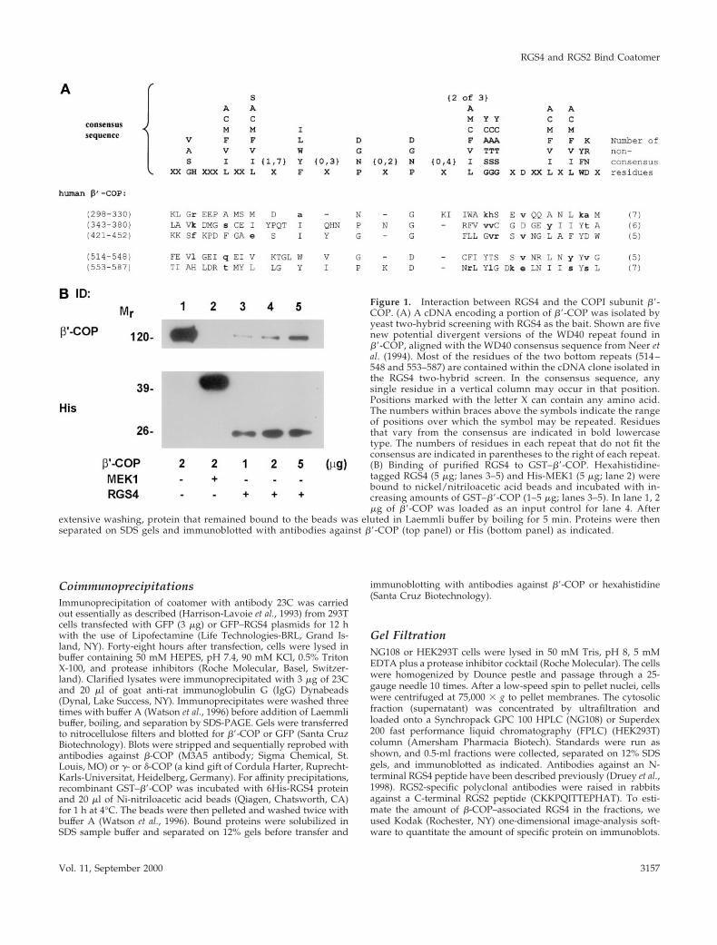
CoimmunoprecipitationsImmunoprecipitation of coatomer with antibody 23C was carriedout essentially as described (Harrison-Lavoie et al., 1993) from 293Tcells transfected with GFP (3 mg) or GFP–RGS4 plasmids for 12 hwith the use of Lipofectamine (Life Technologies-BRL, Grand Is-land, NY). Forty-eight hours after transfection, cells were lysed inbuffer containing 50 mM HEPES, pH 7.4, 90 mM KCl, 0.5% TritonX-100, and protease inhibitors (Roche Molecular, Basel, Switzer-land). Clarified lysates were immunoprecipitated with 3 mg of 23Cand 20 ml of goat anti-rat immunoglobulin G (IgG) Dynabeads(Dynal, Lake Success, NY). Immunoprecipitates were washed threetimes with buffer A (Watson et al., 1996) before addition of Laemmlibuffer, boiling, and separation by SDS-PAGE. Gels were transferredto nitrocellulose filters and blotted for b9-COP or GFP (Santa CruzBiotechnology). Blots were stripped and sequentially reprobed withantibodies against b-COP (M3A5 antibody; Sigma Chemical, St.Louis, MO) or g- or d-COP (a kind gift of Cordula Harter, Ruprecht-Karls-Universitat, Heidelberg, Germany). For affinity precipitations,recombinant GST–b9-COP was incubated with 6His-RGS4 proteinand 20 ml of Ni-nitriloacetic acid beads (Qiagen, Chatsworth, CA)for 1 h at 4°C. The beads were then pelleted and washed twice withbuffer A (Watson et al., 1996). Bound proteins were solubilized inSDS sample buffer and separated on 12% gels before transfer and
immunoblotting with antibodies against b9-COP or hexahistidine(Santa Cruz Biotechnology).
Gel FiltrationNG108 or HEK293T cells were lysed in 50 mM Tris, pH 8, 5 mMEDTA plus a protease inhibitor cocktail (Roche Molecular). The cellswere homogenized by Dounce pestle and passage through a 25-gauge needle 10 times. After a low-speed spin to pellet nuclei, cellswere centrifuged at 75,000 3 g to pellet membranes. The cytosolicfraction (supernatant) was concentrated by ultrafiltration andloaded onto a Synchropack GPC 100 HPLC (NG108) or Superdex200 fast performance liquid chromatography (FPLC) (HEK293T)column (Amersham Pharmacia Biotech). Standards were run asshown, and 0.5-ml fractions were collected, separated on 12% SDSgels, and immunoblotted as indicated. Antibodies against an N-terminal RGS4 peptide have been described previously (Druey et al.,1998). RGS2-specific polyclonal antibodies were raised in rabbitsagainst a C-terminal RGS2 peptide (CKKPQITTEPHAT). To esti-mate the amount of b-COP–associated RGS4 in the fractions, weused Kodak (Rochester, NY) one-dimensional image-analysis soft-ware to quantitate the amount of specific protein on immunoblots.
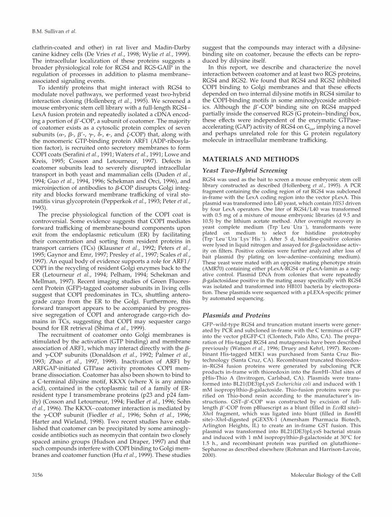
Figure 1. Interaction between RGS4 and the COPI subunit b9-COP. (A) A cDNA encoding a portion of b9-COP was isolated byyeast two-hybrid screening with RGS4 as the bait. Shown are fivenew potential divergent versions of the WD40 repeat found inb9-COP, aligned with the WD40 consensus sequence from Neer etal. (1994). Most of the residues of the two bottom repeats (514 –548 and 553–587) are contained within the cDNA clone isolated inthe RGS4 two-hybrid screen. In the consensus sequence, anysingle residue in a vertical column may occur in that position.Positions marked with the letter X can contain any amino acid.The numbers within braces above the symbols indicate the rangeof positions over which the symbol may be repeated. Residuesthat vary from the consensus are indicated in bold lowercasetype. The numbers of residues in each repeat that do not fit theconsensus are indicated in parentheses to the right of each repeat.(B) Binding of purified RGS4 to GST–b9-COP. Hexahistidine-tagged RGS4 (5 mg; lanes 3–5) and His-MEK1 (5 mg; lane 2) werebound to nickel/nitriloacetic acid beads and incubated with in-creasing amounts of GST–b9-COP (1–5 mg; lanes 3–5). In lane 1, 2mg of b9-COP was loaded as an input control for lane 4. After
extensive washing, protein that remained bound to the beads was eluted in Laemmli buffer by boiling for 5 min. Proteins were thenseparated on SDS gels and immunoblotted with antibodies against b9-COP (top panel) or His (bottom panel) as indicated.
RGS4 and RGS2 Bind Coatomer
Vol. 11, September 2000 3157

Figure 2. (A) Coimmunoprecipitation of RGS4 and coatomer from HEK293TT cells. GFP–RGS4 full-length and deletion (87–205 and 131–205)fusion proteins were expressed in HEK293TT cells and immunoprecipitated with antibody to b9-COP as described in MATERIALS ANDMETHODS. The entire amount of each immunoprecipitation was separated on SDS gels, transferred to nitrocellulose membranes, and blotted withantibodies against GFP or b-, b9-, g-, or d-COP as shown in the left panel (IP: b9-COP). Equal amounts of protein from postimmuno-
B.M. Sullivan et al.
Molecular Biology of the Cell3158
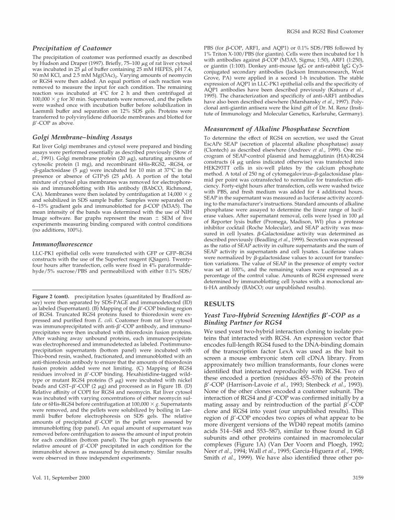
Precipitation of CoatomerThe precipitation of coatomer was performed exactly as describedby Hudson and Draper (1997). Briefly, 75–100 mg of rat liver cytosolwas incubated in 25 ml of buffer containing 25 mM HEPES, pH 7.4,50 mM KCl, and 2.5 mM Mg(OAc)2. Varying amounts of neomycinor RGS4 were then added. An equal portion of each reaction wasremoved to measure the input for each condition. The remainingreaction was incubated at 4°C for 2 h and then centrifuged at100,000 3 g for 30 min. Supernatants were removed, and the pelletswere washed once with incubation buffer before solubilization inLaemmli buffer and separation on 12% SDS gels. Proteins weretransferred to polyvinylidene difluoride membranes and blotted forb9-COP as above.
Golgi Membrane–binding AssaysRat liver Golgi membranes and cytosol were prepared and bindingassays were performed essentially as described previously (Stow etal., 1991). Golgi membrane protein (20 mg), saturating amounts ofcytosolic protein (1 mg), and recombinant 6His-RGS2, -RGS4, or-b-galactosidase (5 mg) were incubated for 10 min at 37°C in thepresence or absence of GTPgS (25 mM). A portion of the totalmixture of cytosol plus membranes was removed for electrophore-sis and immunoblotting with His antibody (BAbCO, Richmond,CA). Membranes were then isolated by centrifugation at 14,000 3 gand solubilized in SDS sample buffer. Samples were separated on6–15% gradient gels and immunoblotted for b-COP (M3A5). Themean intensity of the bands was determined with the use of NIHImage software. Bar graphs represent the mean 6 SEM of fiveexperiments measuring binding compared with control conditions(no additions, 100%).
ImmunofluorescenceLLC-PK1 epithelial cells were transfected with GFP or GFP–RGS4constructs with the use of the Superfect reagent (Qiagen). Twenty-four hours after transfection, cells were fixed in 4% paraformalde-hyde/5% sucrose/PBS and permeabilized with either 0.1% SDS/
PBS (for b-COP, ARF1, and AQP1) or 0.1% SDS/PBS followed by1% Triton X-100/PBS (for giantin). Cells were then incubated for 1 hwith antibodies against b-COP (M3A5, Sigma; 1:50), ARF1 (1:250),or giantin (1:100). Donkey anti-mouse IgG or anti-rabbit IgG Cy3-conjugated secondary antibodies (Jackson Immunoresearch, WestGrove, PA) were applied in a second 1-h incubation. The stableexpression of AQP1 in LLC-PK1 epithelial cells and the specificity ofAQP1 antibodies have been described previously (Katsura et al.,1995). The characterization and specificity of anti-ARF1 antibodieshave also been described elsewhere (Marshansky et al., 1997). Poly-clonal anti-giantin antisera were the kind gift of Dr. M. Renz (Insti-tute of Immunology and Molecular Genetics, Karlsruhe, Germany).
Measurement of Alkaline Phosphatase SecretionTo determine the effect of RGS4 on secretion, we used the GreatEscAPe SEAP (secretion of placental alkaline phosphatase) assay(Clontech) as described elsewhere (Andreev et al., 1999). One mi-crogram of SEAP-control plasmid and hemagglutinin (HA)-RGS4constructs (4 mg unless indicated otherwise) was transfected intoHEK293TT cells in six-well plates by the calcium phosphatemethod. A total of 250 ng of cytomegalovirus–b-galactosidase plas-mid per point was cotransfected to normalize for transfection effi-ciency. Forty-eight hours after transfection, cells were washed twicewith PBS, and fresh medium was added for 4 additional hours.SEAP in the supernatant was measured as luciferase activity accord-ing to the manufacturer’s instructions. Standard amounts of alkalinephosphatase were assayed to determine the linear range of lucif-erase values. After supernatant removal, cells were lysed in 100 mlof Reporter lysis buffer (Promega, Madison, WI) plus a proteaseinhibitor cocktail (Roche Molecular), and SEAP activity was mea-sured in cell lysates. b-Galactosidase activity was determined asdescribed previously (Beadling et al., 1999). Secretion was expressedas the ratio of SEAP activity in culture supernatants and the sum ofSEAP activity in supernatants and cell lysates. Luciferase valueswere normalized by b-galactosidase values to account for transfec-tion variations. The value of SEAP in the presence of empty vectorwas set at 100%, and the remaining values were expressed as apercentage of the control value. Amounts of RGS4 expressed weredetermined by immunoblotting cell lysates with a monoclonal an-ti-HA antibody (BAbCO; our unpublished results).
RESULTS
Yeast Two-Hybrid Screening Identifies b*-COP as aBinding Partner for RGS4We used yeast two-hybrid interaction cloning to isolate pro-teins that interacted with RGS4. An expression vector thatencodes full-length RGS4 fused to the DNA-binding domainof the transcription factor LexA was used as the bait toscreen a mouse embryonic stem cell cDNA library. Fromapproximately two million transformants, four clones wereidentified that interacted reproducibly with RGS4. Two ofthese encoded a portion (residues 455–576) of the proteinb9-COP (Harrison-Lavoie et al., 1993; Stenbeck et al., 1993).None of the other clones encoded a coatomer subunit. Theinteraction of RGS4 and b9-COP was confirmed initially by amating assay and by reintroduction of the partial b9-COPclone and RGS4 into yeast (our unpublished results). Thisregion of b9-COP encodes two copies of what appear to bemore divergent versions of the WD40 repeat motifs (aminoacids 514–548 and 553–587), similar to those found in Gbsubunits and other proteins contained in macromolecularcomplexes (Figure 1A) (Van Der Voorn and Ploegh, 1992;Neer et al., 1994; Wall et al., 1995; Garcia-Higuera et al., 1998;Smith et al., 1999). We have also identified three other po-
Figure 2 (cont). precipitation lysates (quantitated by Bradford as-say) were then separated by SDS-PAGE and immunodetected (ID)as labeled (Supernatant). (B) Mapping of the b9-COP binding regionof RGS4. Truncated RGS4 proteins fused to thioredoxin were ex-pressed and purified from E. coli. Coatomer from rat liver cytosolwas immunoprecipitated with anti-b9-COP antibody, and immuno-precipitates were then incubated with thioredoxin fusion proteins.After washing away unbound proteins, each immunoprecipitatewas electrophoresed and immunodetected as labeled. Postimmuno-precipitation supernatants (bottom panel) were incubated withThio-bond resin, washed, fractionated, and immunoblotted with ananti-thioredoxin antibody to ensure that the amounts of thioredoxinfusion protein added were not limiting. (C) Mapping of RGS4residues involved in b9-COP binding. Hexahistidine-tagged wild-type or mutant RGS4 proteins (5 mg) were incubated with nickelbeads and GST–b9-COP (2 mg) and processed as in Figure 1B. (D)Relative affinity of COPI for RGS4 and neomycin. Rat liver cytosolwas incubated with varying concentrations of either neomycin sul-fate or 6His-RGS4 before centrifugation at 100,000 3 g. Supernatantswere removed, and the pellets were solubilized by boiling in Lae-mmli buffer before electrophoresis on SDS gels. The relativeamounts of precipitated b9-COP in the pellet were assessed byimmunoblotting (top panel). An equal amount of supernatant wasremoved before centrifugation to assess the amount of input proteinfor each condition (bottom panel). The bar graph represents therelative amount of b9-COP precipitated in each condition for theimmunoblot shown as measured by densitometry. Similar resultswere observed in three independent experiments.
RGS4 and RGS2 Bind Coatomer
Vol. 11, September 2000 3159

tential divergent “WD40 repeat–like” motifs between resi-dues 298 and 452. All five of these are shown in Figure 1A.Six other WD40 repeats and one other WD40 repeat–likemotif have already been identified in b9-COP (Harrison-Lavoie et al., 1993; Stenbeck et al., 1993; Csukai et al., 1997). InGb, the only WD40 repeat protein for which the crystalstructure has been resolved to date, the WD40 repeats formthe blades of a toroidal propeller structure (Wall et al., 1995).It has been suggested that all WD40 repeat–containing pro-teins are likely to form propeller structures, which mayfacilitate interaction with multiple protein partners (Garcia-Higuera et al., 1998). Therefore, it is possible that b9-COPmay contain one or more such propeller structures.
To confirm that the RGS4–b9-COP interaction was direct,we incubated various amounts of purified, recombinantGST–b9-COP with hexahistidine-tagged RGS4 (or a controlcytoplasmic protein, 6His-MEK1) bound to nickel beads,washed away unbound proteins, and subjected affinity-pu-rified proteins to electrophoresis and immunoblotting withantibodies directed against either b9-COP or hexahistidine(Figure 1B). In this assay, b9-COP bound the immobilizedRGS4 in a concentration-dependent manner, with a maxi-mum retrieval of ;14% of the input. Similar binding affin-ities were observed for two RGS4 mutant proteins that donot bind Gia and have no measurable GAP activity,RGS4(L159F) and RGS4(1–158) (Druey and Kehrl, 1997;Srinivasa et al., 1998), and for 6His-RGS2 (our unpublishedresults). These studies demonstrate that RGS4 and RGS2bind b9-COP directly and that the N-terminal half of RGS4 issufficient for the interaction.
RGS4 Binds the Coatomer Complex throughConserved Dilysine MotifsTo determine whether RGS4 associates with intact coatomercomplex as opposed to free cytosolic b9-COP and to confirm
Figure 3. Cofractionation of endogenous RGS proteins and COPI.(A) Endogenous RGS4 cofractionates with b-COP in NG108 cells bygel filtration. Protein molecular weight standards were loaded ontoa Synchropack GPC 100 HPLC column; the elution profile is shownin the chromatograph for thyroglobulin (669,000), aldolase(150,000), and chymotrypsinogen (25,000) molecular weight stan-dards (solid line). NG108 cytosol was loaded onto the column, and0.5-ml fractions were collected. The entire amount of protein in peakfractions was separated on 12% gels, transferred to nitrocellulose,and probed with specific antibodies against b-COP or RGS4 asindicated. (B) Characterization of RGS2 antibody. 6His-RGS2 or-RGS4 (50 ng) was electrophoresed and immunoblotted with RGS2antiserum. The antiserum was preincubated with either a controlpeptide (FLAG; left panel) or immunizing peptide (middle panel)(both at 10 mg/ml) for 30 min at room temperature before immu-nodetection. Right panel shows lysate from HEK293T cells (100–150mg of protein) treated with PMA (100 ng/ml for 2 h) (lane 1) or6His-RGS2 as a positive control (lane 2) electrophoresed and blottedwith RGS2 antiserum. (C) Endogenous RGS2 cofractionates withb9-COP in HEK293T cells. The cytosolic fraction from PMA-treatedHEK293T cells was loaded onto a Superdex 200 FPLC column andfractionated as in A. Molecular weight marker migrations are indi-cated by arrows (thyroglobulin, 669,000; g-globulin, 158,000; andovalbumin, 44,000). Peak protein-containing fractions were sepa-rated by SDS-PAGE and immunodetected with antibodies for b9-COP (top panel) or RGS2 (bottom panel).
B.M. Sullivan et al.
Molecular Biology of the Cell3160
that this interaction occurs in intact cells, we used an anti-body directed against b9-COP (Harrison-Lavoie et al., 1993)to immunoprecipitate endogenous coatomer from lysates ofHEK293T cells that were transfected with either control(GFP) or GFP–RGS4 constructs encoding the full-length pro-tein or various N-terminal deletions. The immunoprecipita-tions were then analyzed for the presence of variouscoatomer subunits by immunoblotting with available COPIantibodies and were also analyzed for GFP–RGS proteins byimmunoblotting with anti-GFP antibodies. As shown in Fig-ure 2A (IP: b9-COP), the b9-COP antibody immunoprecipi-tated b-, b9-, g-, and d-COP as well as full-length GFP–RGS4and GFP–RGS4(87–205), but it failed to immunoprecipitateGFP–RGS4(131–205) or the GFP-only control. The four COPIsubunits could also be detected in postimmunoprecipitationlysates, as could the GFP-only control and all of the GFP–RGS4 fusion proteins, including GFP–RGS4(131–205) (Fig-ure 2A, Supernatant). The identities of the lower-molecular-weight proteins detected with anti-GFP antibodies are
uncertain, but because they are detectable with the GFPantibody, they are likely to be proteins prematurely trun-cated at their C termini or proteolytic cleavage products.These experiments suggest that RGS4 associates with theentire COPI complex or at least a subcomplex containing b-,b9-, g-, and d-COP rather than free b9-COP. They also indi-cate that the b9-COP binding site(s) on RGS4 lies betweenresidues 87 and 130, which encompasses the N-terminal halfof the RGS domain.
Amino acids 87–130 in RGS4 contain several importantregions for contact with its known substrate, Ga. Based oncrystallographic data for RGS4 complexed with AlF4
2-Gia1,residues 84–88 and 128–130 form direct contacts with the Gprotein (Tesmer et al., 1997). However, b9-COP did not in-hibit the GAP activity of RGS4 toward Gia1 (our unpub-lished results). Thus, we hypothesized that because RGS4binding to b9-COP did not block G protein binding alloster-ically, the binding site(s) probably involved residues that donot contact the G protein directly. We noted in this region
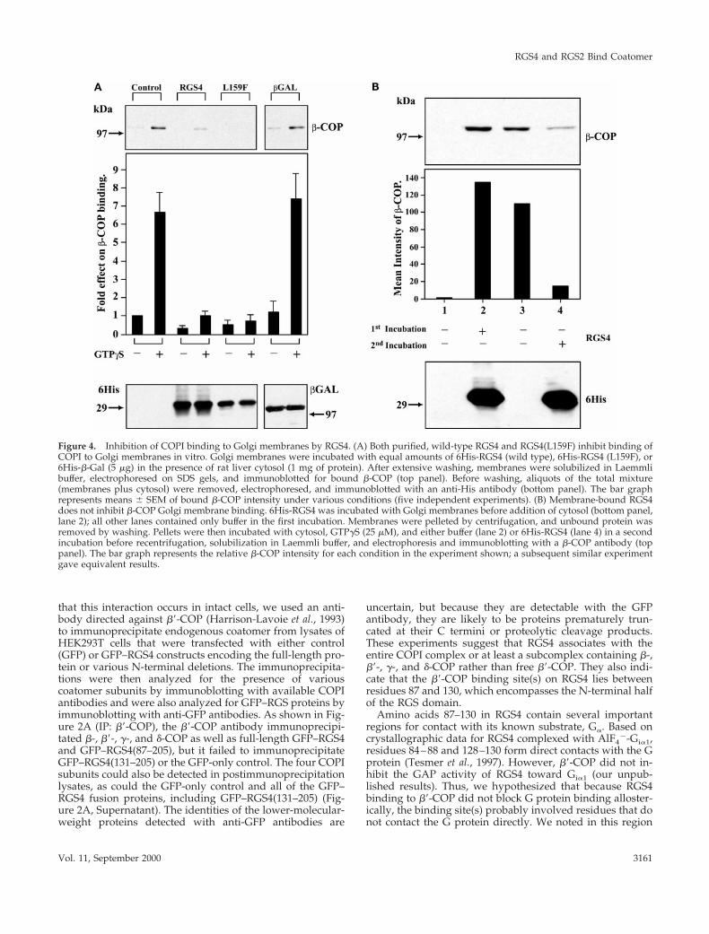
Figure 4. Inhibition of COPI binding to Golgi membranes by RGS4. (A) Both purified, wild-type RGS4 and RGS4(L159F) inhibit binding ofCOPI to Golgi membranes in vitro. Golgi membranes were incubated with equal amounts of 6His-RGS4 (wild type), 6His-RGS4 (L159F), or6His-b-Gal (5 mg) in the presence of rat liver cytosol (1 mg of protein). After extensive washing, membranes were solubilized in Laemmlibuffer, electrophoresed on SDS gels, and immunoblotted for bound b-COP (top panel). Before washing, aliquots of the total mixture(membranes plus cytosol) were removed, electrophoresed, and immunoblotted with an anti-His antibody (bottom panel). The bar graphrepresents means 6 SEM of bound b-COP intensity under various conditions (five independent experiments). (B) Membrane-bound RGS4does not inhibit b-COP Golgi membrane binding. 6His-RGS4 was incubated with Golgi membranes before addition of cytosol (bottom panel,lane 2); all other lanes contained only buffer in the first incubation. Membranes were pelleted by centrifugation, and unbound protein wasremoved by washing. Pellets were then incubated with cytosol, GTPgS (25 mM), and either buffer (lane 2) or 6His-RGS4 (lane 4) in a secondincubation before recentrifugation, solubilization in Laemmli buffer, and electrophoresis and immunoblotting with a b-COP antibody (toppanel). The bar graph represents the relative b-COP intensity for each condition in the experiment shown; a subsequent similar experimentgave equivalent results.
RGS4 and RGS2 Bind Coatomer
Vol. 11, September 2000 3161

(99/100 and 112/113) the presence of two dilysine-contain-ing motifs reminiscent of those found in some aminoglyco-side antibiotics known to precipitate COPI. Because thesepositively charged regions represent candidate contact siteswith the negatively charged WD40 repeat–like motifs ofb9-COP, we sought to determine the precise residues in-volved in binding by two methods. First, we generatedtruncated RGS4 proteins fused to thioredoxin, incubatedthem with rat liver cytosol as a source of coatomer, andimmunoprecipitated b9-COP as before. Immunoprecipitateswere separated on SDS gels and blotted with antibodiesagainst b9-COP or thioredoxin. Although full-length Thio-RGS4 and Thio-RGS4(1–105) coimmunoprecipitated with b9-COP, neither Thio-RGS4(1–98) nor Thio-RGS4(131–205)bound (Figure 2B). These results show that RGS4 residues99–105 are necessary to bind b9-COP. Second, we generated6His-RGS4 proteins with mutations in the dilysine motif of
the b9-COP binding region (K100E) or a proximate dilysinemotif as a control (KK112/113AA), bound them to nickelbeads, and incubated them with GST–b9-COP. After wash-ing away unbound proteins, we separated bound proteinsby SDS-PAGE and blotted for b9-COP or hexahistidine. Sur-prisingly, although the wild-type protein bound as before,either mutation in RGS4 abolished b9-COP binding (Figure2C). The inability of either mutant protein to bind b9-COPwas probably not due to misfolding, because both mutantsdemonstrated enzymatic GAP activity similar to the wild-type protein and proteins containing mutations of residuesthat surround the downstream dilysine motif (KA110/111AS or IY114/115AA) bound b9-COP as well as wild-typeRGS4 (our unpublished results). Based on these results, al-though RGS4 contains two internal KK motifs in other re-gions of the protein, either of the two dilysine-containingsites involving residues 99/100 or 112/113 are required for
Figure 5. Cytosolic dispersion of b-COP and reduction in COPI association with Golgi in RGS4-expressing cells. Kidney LLC-PK1 epithelialcells were transfected with plasmids encoding GFP alone (A), GFP–RGS4 (wild type) (B), or GFP–RGS4(131–205) (C) and stained withantibodies against b-COP. Left column represents GFP-, GFP–RGS4 (wild type)-, and GFP–RGS4(131–205)-expressing cells; middle columnshows endogenous b-COP distribution; and right column is the merge of the two images in each row. Arrowheads indicate the Golgi complexof nontransfected cells (control), and arrows indicate the Golgi complex of transfected LLC-PK1 cells in the same cell culture. Asterisksrepresent mistargeted and dispersed b-COP in RGS4-transfected cells. Bars, 5 mm.
B.M. Sullivan et al.
Molecular Biology of the Cell3162

b9-COP binding, and perhaps both synergize to bind b9-COP.
To estimate the affinity of b9-COP for the internal KK-containing sequences, we compared the ability of neomycin,an aminoglycoside antibiotic, and RGS4 to precipitate COPIfrom rat liver cytosol (Figure 2D). Cytosol was incubatedwith various concentrations of either neomycin or 6His-RGS4 and then centrifuged at 100,000 3 g. The pellets werewashed, and proteins were solubilized and separated bySDS-PAGE. Gels were transferred and immunoblotted forb9-COP. As has been shown previously, high concentrationsof neomycin (1 mM) precipitated COPI (Figure 2D, lane 3).However, micromolar concentrations of RGS4 precipitated asimilar amount of coatomer as 1 mM neomycin, whereasneomycin in micromolar concentrations had little effect (Fig-ure 2D, lanes 2 and 4, respectively). These studies suggestthat the affinity of COPI for RGS4 is substantially greaterthan its affinity for neomycin; thus, high concentrations ofneomycin or other dilysine compounds could have an RGS4-like effect in vitro.
To determine the physiological relevance of the RGS–COPI interaction, we sought to demonstrate an associationof endogenous RGS proteins with native coatomer in vivo.Although NG108 cells contain detectable amounts of cyto-solic RGS4 protein, we have not been able to localize theprotein ultrastructurally in these cells because of technicaldifficulties related to cell preservation. Instead, we gel fil-tered NG108 cytosol and blotted the fractions for RGS4 andb-COP (Figure 3A). Although purified, bacterially expressedRGS4 eluted from an HPLC column with a profile consistentwith its monomeric molecular weight of ;24,000 (Berman et
al., 1996a), whereas virtually the entire cellular complementof RGS4 in NG108 cells migrated with an apparent molecu-lar weight of .150,000. Approximately 74% of the cytosolicb-COP in these cells overlapped with fractions containingRGS4. Similarly, we investigated whether RGS2, which alsocontains the conserved dilysine motifs, associated withCOPI. Although RGS2 is relatively ubiquitously expressed,we did not detect this protein in NG108 cells, so we frac-tionated HEK293T cells, which contain endogenous RGS2mRNA (our unpublished results). RGS2 protein was de-tected with a polyclonal antiserum raised against a C-termi-nal RGS2 peptide. This antiserum was specific for RGS2 inthat it recognized only 6His-RGS2 and not 6His-RGS4 (Fig-ure 3B, left panel). In addition, blocking studies showed thatthe RGS2 signal was abrogated by preincubating the anti-body with immunizing peptide (but not a control peptide)before immunoblotting (Figure 3B, middle panel). The anti-body also detected a protein band of similar mobility toRGS2 in HEK293T cells, and this protein was markedlyup-regulated by treatment of the cells with phorbol 12-myristate 13-acetate (PMA) (Figure 3B, right panel; our un-published results). Therefore, we fractionated PMA-treatedHEK293T cytosol by FPLC and blotted for b9-COP (Figure3C, top panel) and RGS2 (bottom panel). Virtually all of thecytosolic RGS2 was contained in the high-molecular-weightfractions containing b9-COP. Thus, the comigration of cyto-solic RGS4 and RGS2 with COPI on a size fractionationcolumn is compatible with the notion that at least somenative RGS4 and RGS2 associates with COPI complexes inthese two cell types.
Figure 6. Intact Golgi morphology in RGS4-expressing cells. LLC-PK1 cells were transiently transfected with plasmids encoding either GFPor GFP–RGS4 before staining with giantin (A) or ARF1 (B) antibodies and visualization by immunofluorescence microscopy. Left columnrepresents GFP–RGS4-expressing cells; middle column shows endogenous giantin (A) or ARF1 (B) distribution; and right column is themerge of the two images in each row. Arrowheads indicate the Golgi region of nontransfected (control) cells, and arrows delineate the Golgiregion of cells in the same culture transfected with GFP–RGS4 (wild type). Asterisks indicate the nucleus of GFP–RGS4 (wild type)-transfected cells (middle column). Bars, 5 mm.
RGS4 and RGS2 Bind Coatomer
Vol. 11, September 2000 3163
RGS4 Inhibits COPI Binding to Golgi MembranesThe overlap in distribution of RGS4 with COPI coupled withthe interaction of RGS4 with coatomer via the b9-COP sub-unit led us to hypothesize that RGS4 may affect COPI re-cruitment onto Golgi membranes. To test this possibility, weincubated isolated Golgi stacks with cytosolic coatomer andrecombinant 6His-RGS4 and measured the amount of mem-brane binding of b-COP by immunoblotting. Under controlconditions, COPI bound to Golgi membranes, and GTPgS, asshown previously, enhanced binding (Stow et al., 1991).When RGS4 (but not a control protein, 6His-b-galactosidase,which also contains a dilysine motif) was added to cytosolbefore addition to Golgi membranes, COPI binding wasdramatically inhibited (Figure 4A). In a similar experiment,the addition of recombinant 6His-RGS2 also inhibited COPIGolgi binding (our unpublished results). Like the directbinding of RGS4 to b9-COP, the inhibitory effect of RGS4 on
COPI binding to Golgi membranes did not depend on bind-ing of RGS4 to Gia or its GAP activity, because RGS4(L159F)inhibited COPI binding. However, when RGS4 was incu-bated with Golgi membranes before the addition of cytosolicCOPI, RGS4 bound to Golgi membranes but failed to inhibitsubsequent COPI binding (Figure 4B). These results suggestthat the consequential COPI–RGS interaction occurs in thecytosol before COPI is recruited onto membranes.
RGS4 Expression Modifies the Golgi Localization ofCOPITo determine the effect of RGS expression on COPI localiza-tion in intact cells, we transfected LLC-PK1 epithelial cellswith plasmids encoding GFP (Figure 5A) or GFP–RGS4 (Fig-ure 5B), stained the cells with b-COP antibodies to localizethe COPI complex, and examined them by indirect immu-
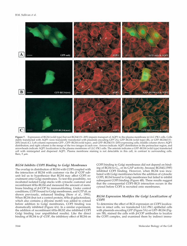
Figure 7. Expression of RGS4 (wild type) but not RGS4(131–205) impairs transport of AQP1 to the plasma membrane in LLC-PK1 cells. Cellsstably transfected with AQP1 were transiently transfected with plasmids encoding GFP (A), GFP–RGS4 (wild type) (B), or GFP–RGS4(131–205) (mut) (C). Left column represents GFP-, GFP–RGS4 (wild type)-, and GFP–RGS4(131–205)-expressing cells; middle column shows AQP1distribution; and right column is the merge of the two images in each row. Arrows indicate AQP1 distribution in the perinuclear region, andarrowheads indicate AQP1 localization in the plasma membrane of LLC-PK1 cells. The asterisk indicates a GFP–RGS4 (wild type) transfectedcell with mistargeted and dispersed AQP1. Plasma membrane staining is not detectable in this cell, in contrast to surrounding cells.Bars, 5 mm.
B.M. Sullivan et al.
Molecular Biology of the Cell3164
nofluorescence microscopy. Control cells showed punctateGolgi b-COP localization, whereas cells expressing RGS4showed a marked loss of perinuclear b-COP staining. Treat-ment of cells with GDP–magnesium aluminum fluoride(AMF), which increased b-COP staining in control cells, didnot stimulate recruitment of b-COP to Golgi membranes incells expressing RGS4 (our unpublished results). To deter-mine if the effect of RGS4 on b-COP localization was due toits ability to bind b9-COP, we transfected cells with a plas-mid encoding GFP–RGS4, which lacks the b9-COP bindingsite (amino acids 131–205), and stained for b-COP (Figure5C). Like GFP alone, GFP–RGS4(131–205) did not have anydetectable effect on the Golgi localization of b-COP.
In contrast to the specific effect of RGS4 expression onCOPI localization, the distribution of endogenous ARF1 orgiantin, an integral membrane protein of the cis-Golgi(Seelig et al., 1994), appeared to be unaffected by RGS4expression (Figure 6). These studies suggest that Golgi mor-phology is not disrupted in RGS4-expressing cells and thatthere is a selective effect of RGS4 on COPI association withGolgi membranes.
RGS4 Impairs Intracellular TransportTo determine whether intracellular transport was altered inRGS4-expressing cells, we examined the localization ofAQP1, which is constitutively targeted to the plasma mem-brane (PM), in stably transfected LLC-PK1 cells (Katsura etal., 1995) (Figure 7). In contrast to the PM localization ofAQP1 in cells transfected with a GFP construct (cell perim-eter staining) (Figure 7A), cells expressing GFP–RGS4 (wild
type) showed accumulation of AQP1 in the cytosolic andperinuclear areas, indicating that constitutive transport ofthe protein to the PM was inhibited in these cells (Figure 7B).This effect was not dependent on the GAP activity of RGS4,because RGS4(L159F) had a similar effect on AQP1 (ourunpublished results). Lastly, the inhibition of AQP1 traffick-ing by RGS4 correlated with its ability to bind b9-COP,because GFP–RGS4(131–205) had no detectable effect on thetransport of AQP1 (Figure 7C).
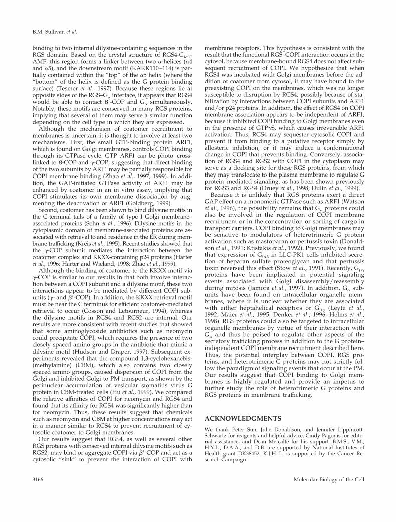
Finally, as an overall measure of transport, we examinedthe SEAP in HEK293T cells by expressing SEAP under thecontrol of a constitutive promoter and measuring alkalinephosphatase protein levels in supernatants and cell lysatesof RGS4-transfected cells, as described previously (Andreevet al., 1999) (Figure 8). Approximately 60% of the alkalinephosphatase was secreted into the medium after 4 h. Com-pared with cells transfected with an empty vector, the se-cretion of SEAP was inhibited ;65% by wild-type RGS4. Asin previous assays, this effect did not depend on GAP activ-ity, because the GAP-dead mutant (L159F) inhibited secre-tion of alkaline phosphatase as well as wild-type RGS4.However, a mutant unable to bind b9-COP (K100E) did notinhibit alkaline phosphatase secretion significantly, suggest-ing that the effect of RGS4 correlated with its ability to bindthe b9-COP in this assay. From these two independent mea-sures of intracellular trafficking, we conclude that RGS4expression impairs intracellular transport.
DISCUSSION
In this study, we have characterized a novel and directinteraction between two members of the RGS family ofsignaling molecules, RGS4 and RGS2, and the coat proteinb9-COP. These RGS proteins are likely to associate with theintact COPI complex, as demonstrated by the ability of RGS4to coimmunoprecipitate with several coatomer subunitsfrom detergent lysates of HEK293T cells and by cofraction-ation of native RGS4 and RGS2 with COPI in two differentcell lines. The most significant result of the RGS–COPI in-teraction is the ability of RGS4 and RGS2 to inhibit cytoplas-mic COPI binding to purified Golgi membranes in vitro andto decrease Golgi-associated COPI in intact LLC-PK1 epithe-lial cells. Moreover, RGS4 protein expression impaired con-stitutive trafficking of AQP1 to the plasma membrane inthese cells and decreased secretion of alkaline phosphatasein HEK293T cells.
Although an established biochemical mechanism of RGSprotein action is to accelerate GTP hydrolysis by Gia and Gqa
by stabilizing the transition state conformation of the Gprotein during the hydrolysis reaction (mimicked by AMF)(Berman et al., 1996a; Druey and Kehrl, 1997; Tesmer et al.,1997; Srinivasa et al., 1998), the binding of RGS4 to b9-COPand its effect on COPI membrane localization is independentof a G protein. A mutation in RGS4, L159F, which abolishesits binding to Gia, does not diminish its ability to interactwith coatomer or its inhibition of COPI Golgi associationboth in vitro and in intact cells. Moreover, neither Golgimembrane stimulation with AMF nor GTPgS appears tohave any bearing on the inhibitory effects of RGS4 on COPIbinding. These results also suggest that the enzymatic GAPactivity of RGS4 is not required for its effect on COPI local-ization. We mapped the region in RGS4 involved in b9-COP
Figure 8. RGS4 inhibits secretion of placental alkaline phospha-tase in HEK293T cells. Cells were transfected with plasmidsencoding SEAP cytomegalovirus–b-galactosidase and a controlvector or various HA–RGS4 plasmids. Forty-eight hours aftertransfection, medium was washed out and replaced with freshmedium for 4 h. Alkaline phosphatase was measured from thesupernatant and cell lysate as luciferase activity and divided byb-galactosidase values to obtain normalized SEAP levels. The bargraph represents means 6 SEM of two independent experiments(assayed in triplicate) expressed as a percentage of control vectoralone (ratios of SEAP in supernatant and the sum of alkalinephosphatase in supernatant and cell lysate).
RGS4 and RGS2 Bind Coatomer
Vol. 11, September 2000 3165
binding to two internal dilysine-containing sequences in theRGS domain. Based on the crystal structure of RGS4-Gia1-AMF, this region forms a linker between two a-helices (a4and a5), and the downstream motif (KAKK110–114) is par-tially contained within the “top” of the a5 helix (where the“bottom” of the helix is defined as the G protein bindingsurface) (Tesmer et al., 1997). Because these regions lie atopposite sides of the RGS–Ga interface, it appears that RGS4would be able to contact b9-COP and Ga simultaneously.Notably, these motifs are conserved in many RGS proteins,implying that several of them may serve a similar functiondepending on the cell type in which they are expressed.
Although the mechanism of coatomer recruitment tomembranes is uncertain, it is thought to involve at least twomechanisms. First, the small GTP-binding protein ARF1,which is found on Golgi membranes, controls COPI bindingthrough its GTPase cycle. GTP–ARF1 can be photo–cross-linked to b-COP and g-COP, suggesting that direct bindingof the two subunits by ARF1 may be partially responsible forCOPI membrane binding (Zhao et al., 1997, 1999). In addi-tion, the GAP-initiated GTPase activity of ARF1 may beenhanced by coatomer in an in vitro assay, implying thatCOPI stimulates its own membrane dissociation by aug-menting the deactivation of ARF1 (Goldberg, 1999).
Second, coatomer has been shown to bind dilysine motifs inthe C-terminal tails of a family of type I Golgi membrane–associated proteins (Sohn et al., 1996). Dilysine motifs in thecytoplasmic domain of membrane-associated proteins are as-sociated with retrieval to and residence in the ER during mem-brane trafficking (Kreis et al., 1995). Recent studies showed thatthe g-COP subunit mediates the interaction between thecoatomer complex and KKXX-containing p24 proteins (Harteret al., 1996; Harter and Wieland, 1998; Zhao et al., 1999).
Although the binding of coatomer to the KKXX motif viag-COP is similar to our results in that both involve interac-tion between a COPI subunit and a dilysine motif, these twointeractions appear to be mediated by different COPI sub-units (g- and b9-COP). In addition, the KKXX retrieval motifmust be near the C terminus for efficient coatomer-mediatedretrieval to occur (Cosson and Letourneur, 1994), whereasthe dilysine motifs in RGS4 and RGS2 are internal. Ourresults are more consistent with recent studies that showedthat some aminoglycoside antibiotics such as neomycincould precipitate COPI, which requires the presence of twoclosely spaced amino groups in the antibiotic that mimic adilysine motif (Hudson and Draper, 1997). Subsequent ex-periments revealed that the compound 1,3-cyclohexanebis-(methylamine) (CBM), which also contains two closelyspaced amino groups, caused dispersion of COPI from theGolgi and inhibited Golgi-to-PM transport, as shown by theperinuclear accumulation of vesicular stomatitis virus Gprotein in CBM-treated cells (Hu et al., 1999). We comparedthe relative affinities of COPI for neomycin and RGS4 andfound that its affinity for RGS4 was significantly higher thanfor neomycin. Thus, these results suggest that chemicalssuch as neomycin and CBM at higher concentrations may actin a manner similar to RGS4 to prevent recruitment of cy-tosolic coatomer to Golgi membranes.
Our results suggest that RGS4, as well as several otherRGS proteins with conserved internal dilysine motifs such asRGS2, may bind or aggregate COPI via b9-COP and act as acytosolic “sink” to prevent the interaction of COPI with
membrane receptors. This hypothesis is consistent with theresult that the functional RGS–COPI interaction occurs in thecytosol, because membrane-bound RGS4 does not affect sub-sequent recruitment of COPI. We hypothesize that whenRGS4 was incubated with Golgi membranes before the ad-dition of coatomer from cytosol, it may have bound to thepreexisting COPI on the membranes, which was no longersusceptible to disruption by RGS4, possibly because of sta-bilization by interactions between COPI subunits and ARF1and/or p24 proteins. In addition, the effect of RGS4 on COPImembrane association appears to be independent of ARF1,because it inhibited COPI binding to Golgi membranes evenin the presence of GTPgS, which causes irreversible ARF1activation. Thus, RGS4 may sequester cytosolic COPI andprevent it from binding to a putative receptor simply byallosteric inhibition, or it may induce a conformationalchange in COPI that prevents binding. Conversely, associa-tion of RGS4 and RGS2 with COPI in the cytoplasm mayserve as a docking site for these RGS proteins, from whichthey may translocate to the plasma membrane to regulate Gprotein–mediated signaling, as has been shown previouslyfor RGS3 and RGS4 (Druey et al., 1998; Dulin et al., 1999).
Because it is unlikely that RGS proteins exert a directGAP effect on a monomeric GTPase such as ARF1 (Watsonet al., 1996), the possibility remains that Ga proteins couldalso be involved in the regulation of COPI membranerecruitment or in the concentration or sorting of cargo intransport carriers. COPI binding to Golgi membranes maybe sensitive to modulators of heterotrimeric G proteinactivation such as mastoparan or pertussis toxin (Donald-son et al., 1991; Ktistakis et al., 1992). Previously, we foundthat expression of Gia3 in LLC-PK1 cells inhibited secre-tion of heparan sulfate proteoglycan and that pertussistoxin reversed this effect (Stow et al., 1991). Recently, Gbg
proteins have been implicated in potential signalingevents associated with Golgi disassembly/reassemblyduring mitosis (Jamora et al., 1997). In addition, Ga sub-units have been found on intracellular organelle mem-branes, where it is unclear whether they are associatedwith either heptahelical receptors or Gbg (Leyte et al.,1992; Maier et al., 1995; Denker et al., 1996; Helms et al.,1998). RGS proteins could also be targeted to intracellularorganelle membranes by virtue of their interaction withGa and thus be poised to regulate other aspects of thesecretory trafficking process in addition to the G protein–independent COPI membrane recruitment described here.Thus, the potential interplay between COPI, RGS pro-teins, and heterotrimeric G proteins may not strictly fol-low the paradigm of signaling events that occur at the PM.Our results suggest that COPI binding to Golgi mem-branes is highly regulated and provide an impetus tofurther study the role of heterotrimeric G proteins andRGS proteins in membrane trafficking.
ACKNOWLEDGMENTS
We thank Peter Sun, Julie Donaldson, and Jennifer Lippincott-Schwartz for reagents and helpful advice, Cindy Pagonis for edito-rial assistance, and Dean Metcalfe for his support. B.M.S., V.M.,H.Y.L., D.A.A., and D.B. are supported by National Institutes ofHealth grant DK38452. K.J.H.-L. is supported by the Cancer Re-search Campaign.
B.M. Sullivan et al.
Molecular Biology of the Cell3166

REFERENCES
Andreev, J., Simon, J.-P., Sabatini, D.D., Kam, J., Plowman, G.,Randazzo, P.A., and Schlessinger, J. (1999). Identification of a newPyk2 target protein with ARF-GAP activity. Mol. Cell. Biol. 19,2338–2350.
Beadling, C., Druey, K.M., Richter, G., Kehrl, J.H., and Smith, K.M.(1999) Regulators of G. protein signaling exhibit distinct patterns ofgene expression and target G protein specificity in human lympho-cytes. J. Immunol. 162, 2677–2682.
Berman, D.M., and Gilman, A.G. (1998). Mammalian RGS proteins:barbarians at the gate. J. Biol. Chem. 273, 1269–1272.
Berman, D.M., Kozasa, T., and Gilman, A.G. (1996a). The GTPase-activating protein RGS4 stabilizes the transition state for nucleotidehydrolysis. J. Biol. Chem. 271, 27209–27212.
Berman, D.M., Wilkie, T.M., and Gilman, A.G. (1996b). GAIP andRGS4 are GTPase activating proteins for the Gi subfamily of Gprotein a subunits. Cell 86, 445–452.
Cosson, P., and Letourneur, F. (1994). Coatomer interaction withdi-lysine endoplasmic reticulum retention motifs. Science 263,1629–1631.
Cosson, P., and Letourneur, F. (1997). Coatomer (COPI)-coated ves-icles: role in intracellular transport and protein sorting. Curr. Opin.Cell Biol. 9, 484–487.
Csukai, M., Chen, C.H., De Matteis, M.A., and Mochly-Rosen, D.(1997). The coatomer protein b9-COP, a selective binding protein(RACK) for protein kinase Ce. J. Biol. Chem. 272, 29200–29206.
Denker, S.P., McCaffery, J.M., Palade, G.E., Insel, P.A., and Far-quhar, M.G. (1996). Differential distribution of a subunits and bgsubunits of heterotrimeric G proteins on Golgi membranes of theexocrine pancreas. J. Cell Biol. 133, 1027–1040.
De Vries, L., Elenko, E., McCaffery, J.M., Fischer, T., Hubler, L.,McQuistan, T., Watson, N., and Farquhar, M.G. (1998). RGS-GAIP, a GTPase-activating protein for Gai heterotrimeric G pro-teins, is located on clathrin-coated vesicles. Mol. Biol. Cell 9,1123–1134.
Donaldson, J.G., Cassel, D., Kahn, R.A., and Klausner, R.D. (1992).ADP-ribosylation factor, a small GTP-binding protein, is requiredfor binding of the coatomer protein b-COP to Golgi membranes.Proc. Natl. Acad. Sci. USA 89, 6408–6412.
Donaldson, J.G., Kahn, R.A., Lippincott-Schwartz, J., and Klausner,R.D. (1991). Binding of ARF and b-COP to Golgi membranes: pos-sible regulation by a trimeric G protein. Science 254, 1197–1199.
Druey, K.M., Blumer, K.J., Kang, V.H., and Kehrl, J.H. (1996). Inhi-bition of G-protein-mediated MAP kinase activation by a new mam-malian gene family. Nature 379, 742–746.
Druey, K.M., and Kehrl, J.H. (1997). Inhibition of regulator of Gprotein signaling function by two mutant RGS4 proteins. Proc. Natl.Acad. Sci. USA 94, 12851–12856.
Druey, K.M., Sullivan, B.M., Brown, D., Fischer, E.R., Watson, N.,Blumer, K.J., Gerfen, C.R., Scheschonka, A., and Kehrl, J.H. (1998).Expression of GTPase-deficient Gia2 results in translocation of cyto-plasmic RGS4 to the plasma membrane. J. Biol. Chem. 273, 18405–18410.
Duden, R., Hosobuchi, M., Hamamoto, S., Winey, M., Byers, B., andSchekman, R. (1994). Yeast b- and b9-coat proteins (COP). J. Biol.Chem. 269, 24486–24495.
Dulin, N.O., Sorokin, A., Reed, E., Elliott, S., Kehrl, J.H., and Dunn,M.J. (1999). RGS3 inhibits G protein-mediated signaling via trans-location to the membrane and binding to Ga11. Mol. Cell. Biol. 19,714–723.
Fiedler, K., Veit, M., Stamnes, M.A., and Rothman, J.E. (1996).Bimodal interaction of coatomer with the p24 family of putativecargo receptors. Science 273, 1396–1399.
Garcia-Higuera, I., Gaitatzes, C., Smith, T.F., and Neer, E.J. (1998).Folding a WD repeat propeller: role of highly conserved asparticacid residues in the G protein b subunit and Sec13. J. Biol. Chem.273, 9041–9049.
Gaynor, E.C., and Emr, S.D. (1997). COPI-independent. anterogradetransport: cargo-selective ER to Golgi protein transport in yeastCOPI mutants. J. Cell Biol. 24, 789–802.
Goldberg, J. (1999). Structural and functional analysis of the ARF1-ARFGAP complex reveals a role for coatomer in GTP hydrolysis.Cell 96, 893–902.
Guo, Q., Penman, M., Trigatti, B.L., and Krieger, M. (1996). A singlepoint mutation in e-COP results in temperature-sensitive, lethaldefects in membrane transport in a Chinese hamster ovary cellmutant. J. Biol. Chem. 271, 11191–11196.
Guo, Q., Vasile, E., and Krieger, M. (1994). Disruptions in Golgistructure and membrane traffic in a conditional lethal mammaliancell mutant are corrected by e-COP. J. Cell Biol. 125, 1213–1224.
Harrison-Lavoie, K.J., Lewis, V.A., Hynes, G.M., Collison, K.S., Nut-land, E., and Willison, K.R. (1993). A102 kDa subunit of a Golgi-associated particle has homology to b subunits of trimeric G pro-teins. EMBO J. 12, 2847–2853.
Harter, C., Pavel, J., Coccia, F., Draken, E., Wegehingel, S., Tschoch-ner, H., and Wieland, F. (1996). Nonclathrin coat protein g, a subunitof coatomer, binds to the cytoplasmic dilysine motif of membraneproteins of the early secretory pathway. Proc. Natl. Acad. Sci. USA93, 1902–1906.
Harter, C., and Wieland, F.T. (1998). A single binding site for dil-ysine retrieval motifs and p23 within the g subunit of coatomer.Proc. Natl. Acad. Sci. USA 95, 11649–11654.
Helms, J.B., Helms-Brons, D., Brugger, B., Gkantiragas, I., Eberle, H.,Nickel, W., Nurnberg, B., Gerdes, H.H., and Wieland, F.T. (1998). Aputative heterotrimeric G protein inhibits the fusion of COPI-coatedvesicles. J. Biol. Chem. 273, 15203–15208.
Hollenberg, S.M., Sternglanz, R., Cheng, P.F., and Weintraub, H.(1995). Identification of a new family of tissue-specific basic helix-loop-helix proteins with a two-hybrid system. Mol. Cell. Biol. 15,3813–3822.
Hu, T., Kao, C.-Y. Hudson, R.T., Chen, A., and Draper, R.K. (1999).Inhibition of secretion by 1,3 cyclohexanebis(methylamine), a diba-sic compound that interferes with coatomer function. Mol. Biol. Cell10, 921–933.
Huang, C., Hepler, J.R., Gilman, A.G., and Mumby, S.M. (1997).Attenuation of Gi-, and Gq-mediated signaling by expression ofRGS4 or GAIP in mammalian cells. Proc. Natl. Acad. Sci. USA 94,6159–6163.
Hudson, R.T., and Draper, R.K. (1997). Interaction of coatomer withaminoglycoside antibiotics: evidence that coatomer has at least twodilysine binding sites. Mol. Biol. Cell 10, 1901–1910.
Jamora, C., Takizawa, P.A., Zaarour, R.F., Denesvre, C., Faulkner,D.J., and Malhotra, V. (1997). Regulation of Golgi structure throughheterotrimeric G proteins. Cell 91, 617–626.
Katsura, T., Verbavatz, J.-M., Farinas, J., Ma, T., Ausiello, D.A.,Verkman, A.S., and Brown, D. (1995). Constitutive and regulatedmembrane expression of aquaporin 1 and aquaporin 2 water chan-nels in stably transfected LLC-PK1 epithelial cells. Proc. Natl. Acad.Sci. USA 92, 7212–7216.
Klausner, R.D., Donaldson, J.G., and Lippincott-Schwartz, J. (1992).Brefeldin A: insights into the control of membrane traffic and or-ganelle structure. J. Cell Biol. 116, 1071–1080.
RGS4 and RGS2 Bind Coatomer
Vol. 11, September 2000 3167
Kreis, T.E., Lowe, M., and Pepperkok, R. (1995). COPs regulatingmembrane traffic. Annu. Rev. Cell Dev. Biol. 11, 677–706.
Ktistakis, N.T., Linder, M.E., and Roth, M.G. (1992). Action of brefel-din A blocked by activation of a pertussis-toxin-sensitive G protein.Nature 356, 344–346.
Letourneur, F., Gaynor, E.C., Hennecke, S., Demolliere, C., Duden,R., Emr, S.D., Riezman, H., and Cosson, P. (1994). Coatomer isessential for retrieval of dilysine-tagged proteins to the endoplasmicreticulum. Cell 79, 1199–1207.
Leyte, A., Barr, F.A., Kehlenbach, R.H., and Huttner, W.B. (1992).Multiple trimeric G-proteins on the trans-Golgi network exert stim-ulatory and inhibitory effects on secretory vesicle formation. EMBOJ. 11, 4795–4804.
Lowe, M., and Kreis, T.E. (1995). In vitro assembly and disassemblyof coatomer. J. Biol. Chem. 270, 31364–31371.
Maier, O., Ehmsen, E., and Westermann, P. (1995). Trimeric Gprotein a subunits of the Gs and Gi families localized at the Golgimembrane. Biochem. Biophys. Res. Commun. 208, 135–143.
Marshansky, V., Bourgoin, S., Londono, I., Bendayan, M., and Vi-nay, P. (1997). Identification of ADP-ribosylation factor-6 in brushborder membrane and early endosomes of human kidney proximaltubules. Electrophoresis 18, 538–547.
Neer, E.J., Schmidt, C.J., Nambudripad, R., and Smith, T.F. (1994).The ancient regulatory-protein family of WD-repeat proteins. Na-ture 371, 297–300.
Palmer, D.J., Helms, J.B., Beckers, C.J.M., Orci, L., and Rothman, J.E.(1993). Binding of coatomer to Golgi membranes requires ADP-ribosylation factor. J. Biol. Chem. 268, 12083–12089.
Pelham, H.R.B. (1994). About turn for the COPs? Cell 79, 1125–1127.
Pepperkok, R., Scheel, J., Horstmann, H., Hauri, H.P., Griffiths, G.,and Kreis, T.E. (1993). b-COP is essential for biosynthetic membranetransport from the endoplasmic reticulum to the Golgi complex invivo. Cell 74, 71–82.
Peter, F., Plutner, H., Zhu, H., Kreis, T.E., and Balch, W.E. (1993).b-COP is essential for transport of protein from the endoplasmicreticulum to the Golgi in vitro. J. Cell. Biol. 122, 1155–1167.
Peters, P.J., Hsu, V.W., Ooi, C.E., Finazzi, D., Teal, S.B., Oorschot,V., Donaldson, J.G., and Klausner, R.D. (1995). Overexpression ofwild-type and mutant ARF1 and ARF6: distinct perturbations ofnonoverlapping membrane compartments. J. Cell Biol. 128, 1003–1017.
Presley, J.F., Cole, N.B., Schroer, T.A., Hirschberg, K., Zaal, K.J.M.,and Lippincott-Schwartz, J. (1997). ER-to-Golgi transport visualizedin living cells. Nature 389, 81–85.
Rohman, M., and Harrison-Lavoie, K.J. (2000). Separation of co-purifying GroEL from GST fusion proteins. Protein Expression andPurification (in press).
Scales, S.J., Pepperkok, R., and Kreis, T.E. (1997). Visualization ofER-to-Golgi transport in living cells reveals a sequential mode ofaction for COPII and COPI. Cell 90, 1137–1148.
Schekman, R., and Mellman, I. (1997). Does COPI go both ways?Cell 90, 197–200.
Schekman, R., and Orci, L. (1996). Coat proteins and vesicle bud-ding. Science 271, 1526–1533.
Seelig, H.P., Schranz, P., Schroter, H. Wiemann, C., Griffiths, G., andRenz, M. (1994). Molecular genetic analyses of a 376-kilodalton
Golgi complex membrane protein (giantin). Mol. Cell. Biol. 14,2564–2576.
Serafini, T., Orci, L., Amherdt, M., Brunner, M., Kahn, R.A., andRothman, J.E. (1991). ADP-ribosylation factor is a subunit of the coatof Golgi-derived COP-coated vesicles: a novel role for a GTP-bind-ing protein. Cell 67, 239–253.
Shima, D.T., Scales, S.J., Kreis, T.E., and Pepperkok, R. (1999). Seg-regation of COPI-rich domains in endoplasmic-reticulum-to-Golgitransport complexes. Curr. Biol. 9, 821–824.
Smith, T.F., Gaitatzes, C., Saxena, K., and Neer, E.J. (1999). The WDrepeat: a common architecture for diverse functions. Trends Bio-chem. Sci. 24, 181–185.
Sohn, K., Orci, L., Ravazzola, M., Amherdt, M., Bremser, M., Lott-speich, F., Fiedler, K., Helms, J.B., and Wieland, F.T. (1996). A majortransmembrane protein of Golgi-derived COPI-coated vesicles in-volved in coatomer binding. J. Cell Biol. 135, 1239–1248.
Srinivasa, S.P., Watson, N., Overton, M.C., and Blumer, K.J. (1998).Mechanism of RGS4, a GTPase-activating protein for G protein asubunits. J. Biol. Chem. 273, 1529–1533.
Stenbeck, G., Harter, C., Brecht, A., Herrmann, D., Lottspeich, F.,Orci, L., and Wieland, F.T. (1993). b9-COP, a novel subunit ofcoatomer. EMBO J. 12, 2841–2845.
Stow, J.L., de Almeida, J.B., Narula, N., Holtzman, E.J., Ercolani,L., and Ausiello, D.A. (1991). A heterotrimeric G protein, Gai-3,on Golgi membranes regulates the secretion of a heparan sulfateproteoglycan in LLC-PK1 epithelial cells. J. Cell Biol. 114, 1113–1124.
Tesmer, J.J.G., Berman, D.M., Gilman, A.G., and Sprang, S.R. (1997).Structure of RGS4 bound to AlF4-activated Gia1: stabilization of thetransition state for nucleotide hydrolysis. Cell 89, 251–261.
Van Der Voorn, L., and Ploegh, H.L. (1992). The WD-40 repeat. FEBSLett. 307, 131–134.
Wall, M.A., Coleman, D.E., Lee, E., Iniguez-Lluhi, J.A., Posner, B.A.,Gilman, A.G., and Sprang, S.R. (1995). The structure of the G proteinheterotrimer Gia1b1g2. Cell 83, 1047–1058.
Waters, M.G., Serafini, T., and Rothman, J.E. (1991). “Coatomer”: acytosolic protein complex containing subunits of non-clathrin-coated Golgi transport vesicles. Nature 349, 248–251.
Watson, N., Linder, M.E., Druey, K.M., Kehrl, J.H., and Blumer, K.J.(1996). RGS family members: GTPase activating proteins for hetero-trimeric G-protein a-subunits. Nature 383, 172–175.
Wylie, F., Heimann, K., Tam, L.L., Brown, D., Rabnott, G., and Stow,J.L. (1999). GAIP, a Gai3 binding protein, is associated with Golgi-derived vesicles and protein trafficking. Am. J. Physiol. 276, C497–C506.
Yan, Y., Chi, P.P., and Bourne, H.R. (1997). RGS4 inhibits Gq-medi-ated activation of mitogen-activated protein kinase and phospho-inositide synthesis. J. Biol. Chem. 272, 11924–11927.
Zhao, L., Helms, J.B., Brugger, B., Harter, C., Martoglio, B., Graf, R.,Brunner, J., and Wieland, F.T. (1997). Direct and GTP-dependentinteraction of ADP ribosylation factor 1 with coatomer subunit b.Proc. Natl. Acad. Sci. USA 94, 4418–4423.
Zhao, L., Helms, J.B., Brunner, J., and Wieland, F.T. (1999). GTP-dependent binding of ADP-ribosylation factor to coatomer in closeproximity to the binding site for dilysine retrieval motifs and p23.J. Biol. Chem. 274, 14198–14203.
B.M. Sullivan et al.
Molecular Biology of the Cell3168
Copyright © 2022 FDOKUMEN