Review: Environmental exposure to mercury and its toxicopathologic implications for public health
49
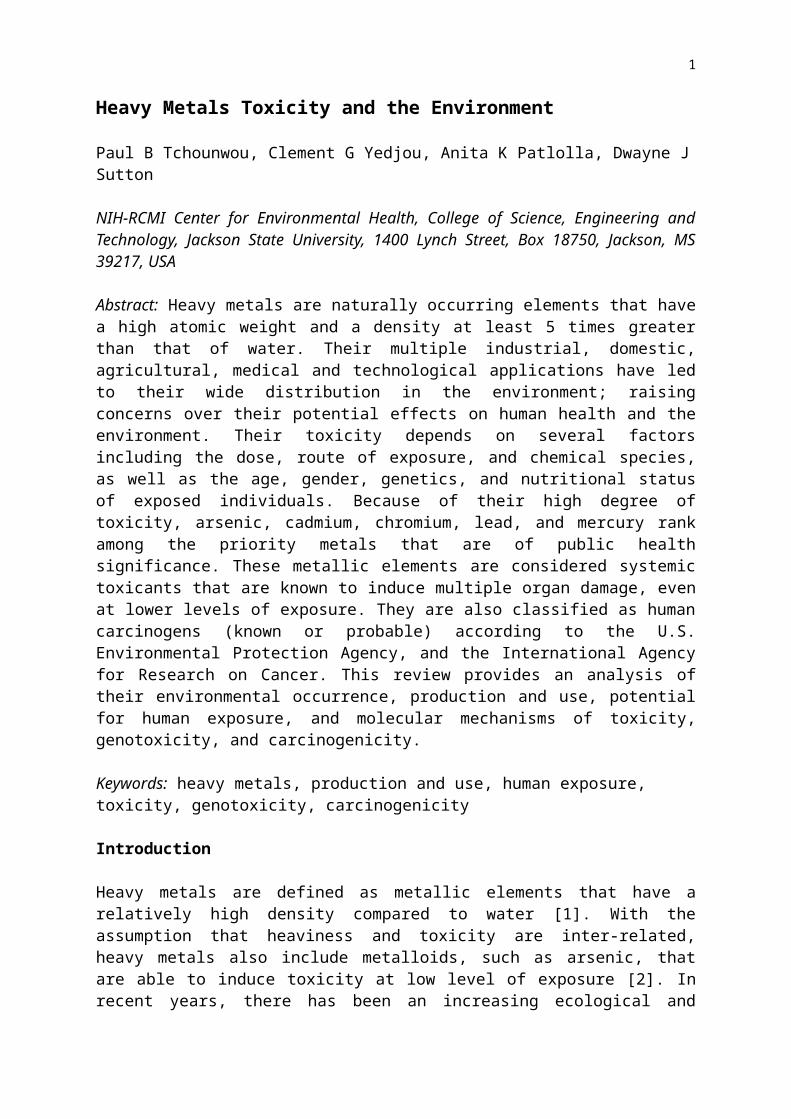
1 Heavy Metals Toxicity and the Environment Paul B Tchounwou, Clement G Yedjou, Anita K Patlolla, Dwayne J Sutton NIH-RCMI Center for Environmental Health, College of Science, Engineering and Technology, Jackson State University, 1400 Lynch Street, Box 18750, Jackson, MS 39217, USA Abstract: Heavy metals are naturally occurring elements that have a high atomic weight and a density at least 5 times greater than that of water. Their multiple industrial, domestic, agricultural, medical and technological applications have led to their wide distribution in the environment; raising concerns over their potential effects on human health and the environment. Their toxicity depends on several factors including the dose, route of exposure, and chemical species, as well as the age, gender, genetics, and nutritional status of exposed individuals. Because of their high degree of toxicity, arsenic, cadmium, chromium, lead, and mercury rank among the priority metals that are of public health significance. These metallic elements are considered systemic toxicants that are known to induce multiple organ damage, even at lower levels of exposure. They are also classified as human carcinogens (known or probable) according to the U.S. Environmental Protection Agency, and the International Agency for Research on Cancer. This review provides an analysis of their environmental occurrence, production and use, potential for human exposure, and molecular mechanisms of toxicity, genotoxicity, and carcinogenicity. Keywords: heavy metals, production and use, human exposure, toxicity, genotoxicity, carcinogenicity Introduction Heavy metals are defined as metallic elements that have a relatively high density compared to water [1]. With the assumption that heaviness and toxicity are inter-related, heavy metals also include metalloids, such as arsenic, that are able to induce toxicity at low level of exposure [2]. In recent years, there has been an increasing ecological and
Transcript of Review: Environmental exposure to mercury and its toxicopathologic implications for public health
1
Heavy Metals Toxicity and the Environment
Paul B Tchounwou, Clement G Yedjou, Anita K Patlolla, Dwayne JSutton
NIH-RCMI Center for Environmental Health, College of Science, Engineering andTechnology, Jackson State University, 1400 Lynch Street, Box 18750, Jackson, MS39217, USA
Abstract: Heavy metals are naturally occurring elements that havea high atomic weight and a density at least 5 times greaterthan that of water. Their multiple industrial, domestic,agricultural, medical and technological applications have ledto their wide distribution in the environment; raisingconcerns over their potential effects on human health and theenvironment. Their toxicity depends on several factorsincluding the dose, route of exposure, and chemical species,as well as the age, gender, genetics, and nutritional statusof exposed individuals. Because of their high degree oftoxicity, arsenic, cadmium, chromium, lead, and mercury rankamong the priority metals that are of public healthsignificance. These metallic elements are considered systemictoxicants that are known to induce multiple organ damage, evenat lower levels of exposure. They are also classified as humancarcinogens (known or probable) according to the U.S.Environmental Protection Agency, and the International Agencyfor Research on Cancer. This review provides an analysis oftheir environmental occurrence, production and use, potentialfor human exposure, and molecular mechanisms of toxicity,genotoxicity, and carcinogenicity.
Keywords: heavy metals, production and use, human exposure, toxicity, genotoxicity, carcinogenicity
Introduction
Heavy metals are defined as metallic elements that have arelatively high density compared to water [1]. With theassumption that heaviness and toxicity are inter-related,heavy metals also include metalloids, such as arsenic, thatare able to induce toxicity at low level of exposure [2]. Inrecent years, there has been an increasing ecological and

2
global public health concern associated with environmentalcontamination by these metals. Also, human exposure has risendramatically as a result of an exponential increase of theiruse in several industrial, agricultural, domestic andtechnological applications [3]. Reported sources of heavymetals in the environment include geogenic, industrial,agricultural, pharmaceutical, domestic effluents, andatmospheric sources [4] . Environmental pollution is veryprominent in point source areas such as mining, foundries andsmelters, and other metal-based industrial operations [1, 3,4].
Although heavy metals are naturally occurring elementsthat are found throughout the earth’s crust, mostenvironmental contamination and human exposure result fromanthropogenic activities such as mining and smeltingoperations, industrial production and use, and domestic andagricultural use of metals and metal-containing compounds [4-7]. Environmental contamination can also occur through metalcorrosion, atmospheric deposition, soil erosion of metal ionsand leaching of heavy metals, sediment re-suspension and metalevaporation from water resources to soil and ground water [8].Natural phenomena such as weathering and volcanic eruptionshave also been reported to significantly contribute to heavymetal pollution [1, 3, 4, 7, 8]. Industrial sources includemetal processing in refineries, coal burning in power plants,petroleum combustion, nuclear power stations and high tensionlines, plastics, textiles, microelectronics, wood preservationand paper processing plants [9-11].
It has been reported that metals such as cobalt (Co),copper (Cu), chromium (Cr), iron (Fe), magnesium (Mg),manganese (Mn), molybdenum (Mo), nickel (Ni), selenium (Se)and zinc (Zn) are essential nutrients that are required forvarious biochemical and physiological functions [12].Inadequate supply of these micro-nutrients results in avariety of deficiency diseases or syndromes [12].
Heavy metals are also considered as trace elementsbecause of their presence in trace concentrations (ppb rangeto less than 10ppm) in various environmental matrices [13].Their bioavailability is influenced by physical factors suchas temperature, phase association, adsorption andsequestration. It is also affected by chemical factors thatinfluence speciation at thermodynamic equilibrium,
3
complexation kinetics, lipid solubility and octanol/waterpartition coefficients [14]. Biological factors such asspecies characteristics, trophic interactions, andbiochemical/physiological adaptation, also play an importantrole [15].
The essential heavy metals play biochemical andphysiological functions in plants and animals. They areimportant constituents of several key enzymes and playimportant roles in various oxidation-reduction reactions [12].Copper for example serves as an essential co-factor forseveral oxidative stress-related enzymes including catalase,superoxide dismutase, peroxidase, cytochrome c oxidases,ferroxidases, monoamine oxidase, and dopamine β-monooxygenase[16-18]. Hence, it is an essential nutrient that isincorporated into a number of metalloenzymes involved inhemoglobin formation, carbohydrate metabolism, catecholaminebiosynthesis, and cross-linking of collagen, elastin, and hairkeratin. The ability of copper to cycle between an oxidizedstate, Cu(II), and reduced state, Cu(I), is used bycuproenzymes involved in redox reactions [16-18]. However, itis this property of copper that also makes it potentiallytoxic because the transitions between Cu(II) and Cu(I) canresult in the generation of superoxide and hydroxyl radicals[16-19]. Also, excessive exposure to copper has been linked tocellular damage leading to Wilson disease in humans [18, 19].Similar to copper, several other essential elements arerequired for biologic functioning, however, excess amount ofsuch metals produces cellular and tissue damage leading to avariety of adverse effects and human diseases. For someincluding chromium and copper, there is a very narrow range ofconcentrations between beneficial and toxic effects [19, 20].Other metals such as aluminium (Al), antinomy (Sb), arsenic(As), barium (Ba), beryllium (Be), bismuth (Bi), cadmium (Cd),gallium (Ga), germanium (Ge), gold (Au), indium (In), lead(Pb), lithium (Li), mercury (Hg), nickel (Ni), platinum (Pt),silver (Ag), strontium (Sr), tellurium (Te), thallium (Tl),tin (Sn), titanium (Ti), vanadium (V) and uranium (U) have noestablished biological functions and are considered as non-essential metals [20].
In biological systems, heavy metals have been reported toaffect cellular organelles and components such as cell membrane,mitochondrial, lysosome, endoplasmic reticulum, nuclei, and some

4
enzymes involved in metabolism, detoxification, and damagerepair [21]. Metal ions have been found to interact with cellcomponents such as DNA and nuclear proteins, causing DNA damageand conformational changes that may lead to cell cyclemodulation, carcinogenesis or apoptosis [20-22]. Several studiesfrom our laboratory have demonstrated that reactive oxygenspecies (ROS) production and oxidative stress play a key rolein the toxicity and carcinogenicity of metals such as arsenic[23, 24, 25], cadmium [26], chromium [27, 28], lead [29, 30],and mercury [31, 32]. Because of their high degree oftoxicity, these five elements rank among the priority metalsthat are of great public health significance. They are allsystemic toxicants that are known to induce multiple organdamage, even at lower levels of exposure. According to theUnited States Environmental Protection Agency (U.S. EPA), andthe International Agency for Research on Cancer (IARC), thesemetals are also classified as known or probable humancarcinogens based on epidemiological and experimental studiesshowing an association between exposure and cancer incidencein humans and animals.
Heavy metal-induced toxicity and carcinogenicity involvesmany mechanistic aspects, some of which are not clearlyelucidated or understood. However, each metal is known to haveunique features that confer to its specific toxicologicalmechanisms of action. This review provides an analysis oftheir environmental occurrence, production and use, potentialfor human exposure, and molecular mechanisms of toxicity,genotoxicity, and carcinogenicity of arsenic, cadmium,chromium, lead and mercury.
Arsenic
Environmental Occurrence, Industrial Production and Use
Arsenic is a ubiquitous element that is detected at lowconcentrations in virtually all environmental matrices [33].The major inorganic forms of arsenic include the trivalentarsenite and the pentavalent arsenate. The organic forms arethe methylated metabolites – monomethylarsonic acid (MMA),dimethylarsinic acid (DMA) and trimethylarsine oxide.Environmental pollution by arsenic occurs as a result ofnatural phenomena such as volcanic eruptions and soil
5
erosion, and anthropogenic activities [33]. Several arsenic-containing compounds are produced industrially, and have beenused to manufacture products with agricultural applicationssuch as insecticides, herbicides, fungicides, algicides, sheepdips, wood preservatives, and dye-stuffs. They have also beenused in veterinary medicine for the eradication of tapewormsin sheep and cattle [34]. Arsenic compounds have also beenused in the medical field for at least a century in thetreatment of syphilis, yaws, amoebic dysentery, andtrypanosomaiasis [34,35]. Arsenic-based drugs are still usedin treating certain tropical diseases such as African sleepingsickness and amoebic dysentery, and in veterinary medicine totreat parasitic diseases, including filariasis in dogs andblack head in turkeys and chickens [35]. Recently, arsenictrioxide has been approved by the Food and Drug Administrationas an anticancer agent in the treatment of acute promeylocyticleukemia [36]. Its therapeutic action has been attributed tothe induction of programmed cell death (apoptosis) in leukemiacells [24].
Potential for Human Exposure
It is estimated that several million people are exposed toarsenic chronically throughout the world, especially incountries like Bangladesh, India, Chile, Uruguay, Mexico,Taiwan, where the ground water is contaminated with highconcentrations of arsenic. Exposure to arsenic occurs via theoral route (ingestion), inhalation, dermal contact, and theparenteral route to some extent [33,34,37]. Arsenicconcentrations in air range from 1 to 3 ng/m3 in remotelocations (away from human releases), and from 20 to 100 ng/m3
in cities. Its water concentration is usually less than10g/L, although higher levels can occur near natural mineraldeposits or mining sites. Its concentration in various foodsranges from 20 to 140 ng/kg [38]. Natural levels of arsenic insoil usually range from 1 to 40 mg/kg, but pesticideapplication or waste disposal can produce much higher values[25].
Diet, for most individuals, is the largest source ofexposure, with an average intake of about 50 g per day.Intake from air, water and soil are usually much smaller, butexposure from these media may become significant in areas of
6
arsenic contamination. Workers who produce or use arseniccompounds in such occupations as vineyards, ceramics, glass-making, smelting, refining of metallic ores, pesticidemanufacturing and application, wood preservation,semiconductor manufacturing can be exposed to substantiallyhigher levels of arsenic [39]. Arsenic has also beenidentified at 781 sites of the 1,300 hazardous waste sitesthat have been proposed by the U.S. EPA for inclusion on thenational priority list [33,39]. Human exposure at these sitesmay occur by a variety of pathways, including inhalation ofdusts in air, ingestion of contaminated water or soil, orthrough the food chain [40].
Contamination with high levels of arsenic is of concernbecause arsenic can cause a number of human health effects.Several epidemiological studies have reported a strongassociation between arsenic exposure and increased risks ofboth carcinogenic and systemic health effects [41]. Interestin the toxicity of arsenic has been heightened by recentreports of large populations in West Bengal, Bangladesh,Thailand, Inner Mongolia, Taiwan, China, Mexico, Argentina,Chile, Finland and Hungary that have been exposed to highconcentrations of arsenic in their drinking water and aredisplaying various clinico-pathological conditions includingcardiovascular and peripheral vascular disease, developmentalanomalies, neurologic and neurobehavioural disorders,diabetes, hearing loss, portal fibrosis, hematologic disorders(anemia, leukopenia and eosinophilia) and carcinoma [25, 33,35, 39]. Arsenic exposure affects virtually all organ systemsincluding the cardiovascular, dermatologic, nervous,hepatobilliary, renal, gastro-intestinal, and respiratorysystems [41]. Research has also pointed to significantlyhigher standardized mortality rates for cancers of thebladder, kidney, skin, and liver in many areas of arsenicpollution. The severity of adverse health effects is relatedto the chemical form of arsenic, and is also time- and dose-dependent [42,43]. Although the evidence of carcinogenicity ofarsenic in humans seems strong, the mechanism by which itproduces tumors in humans is not completely understood [44].
Mechanisms of Toxicity and Carcinogenicity
7
Analyzing the toxic effects of arsenic is complicated becausethe toxicity is highly influenced by its oxidation state andsolubility, as well as many other intrinsic and extrinsicfactors [45]. Several studies have indicated that the toxicityof arsenic depends on the exposure dose, frequency andduration, the biological species, age, and gender, as well ason individual susceptibilities, genetic and nutritionalfactors [46]. Most cases of human toxicity from arsenic havebeen associated with exposure to inorganic arsenic. Inorganictrivalent arsenite (AsIII ) is 2-10 times more toxic thanpentavalent arsenate (AsV) [5]. By binding to thiol orsulfhydryl groups on proteins, As (III) can inactivate over200 enzymes. This is the likely mechanism responsible forarsenic’s widespread effects on different organ systems. As(V) can replace phosphate, which is involved in manybiochemical pathways [5, 47].
One of the mechanisms by which arsenic exerts its toxiceffect is through impairment of cellular respiration by theinhibition of various mitochondrial enzymes, and theuncoupling of oxidative phosphorylation. Most toxicity ofarsenic results from its ability to interact with sulfhydrylgroups of proteins and enzymes, and to substitute phosphorousin a variety of biochemical reactions [48]. Arsenic in vitroreacts with protein sulfhydryl groups to inactivate enzymes,such as dihydrolipoyl dehydrogenase and thiolase, therebyproducing inhibited oxidation of pyruvate and betaoxidation offatty acids [49]. The major metabolic pathway for inorganicarsenic in humans is methylation. Arsenic trioxide ismethylated to two major metabolites via a non-enzymaticprocess to monomethylarsonic acid (MMA), which is furthermethylated enzymatically to dimethyl arsenic acid (DMA) beforeexcretion in the urine [40, 47]. It was previously thoughtthat this methylation process is a pathway of arsenicdetoxification, however, recent studies have pointed out thatsome methylated metabolites may be more toxic than arsenite ifthey contain trivalent forms of arsenic [41].
Tests for genotoxicity have indicated that arseniccompounds inhibit DNA repair, and induce chromosomalaberrations, sister-chromatid exchanges, and micronucleiformation in both human and rodent cells in culture [50-52]and in cells of exposed humans [53]. Reversion assays withSalmonella typhimurium fail to detect mutations that are induced
8
by arsenic compounds. Although arsenic compounds are generallyperceived as weak mutagens in bacterial and animal cells, theyexhibit clastogenic properties in many cell types in vivo and invitro [54]. In the absence of animal models, in vitro celltransformation studies become a useful means of obtaininginformation on the carcinogenic mechanisms of arsenictoxicity. Arsenic and arsenical compounds are toxic to andinduce morphological transformations of Syrian hamster embryo(SHE) cells as well as mouse C3H10T1/2 cells and BALB/3T3cells [55,56].
Based on the comet assay, it has been reported thatarsenic trioxide induces DNA damage in human lymphophytes [57]and also in mice leukocytes [58]. Arsenic compounds have alsobeen shown to induce gene amplification, arrest cells inmitosis, inhibit DNA repair, and induce expression of the c-fosgene and the oxidative stress protein heme oxygenase inmammalian cells [58, 59]. They have been implicated aspromoters and comutagens for a variety of toxic agents [60].Recent studies in our laboratory have demonstrated thatarsenic trioxide is cytotoxic and able to transcriptionallyinduce a significant number of stress genes and relatedproteins in human liver carcinoma cells [61].
Epidemiological investigations have indicated that long-term arsenic exposure results in promotion of carcinogenesis.Several hypotheses have been proposed to describe themechanism of arsenic-induced carcinogenesis. Zhao et al. [62]reported that arsenic may act as a carcinogen by inducing DNAhypomethylation, which in turn facilitates aberrant geneexpression. Additionally, it was found that arsenic is apotent stimulator of extracellular signal-regulated proteinkinase Erk1 and AP-1 transactivational activity, and anefficient inducer of c-fos and c-jun gene expression [63].Induction of c-jun and c-fos by arsenic is associated withactivation of JNK [64]. However, the role of JNK activation byarsenite in cell transformation or tumor promotion is unclear.
In another study, Trouba et al. [65] concluded that long-term exposure to high levels of arsenic might make cells moresusceptible to mitogenic stimulation and that alterations inmitogenic signaling proteins might contribute to thecarcinogenic action of arsenic. Collectively, several recentstudies have demonstrated that arsenic can interfere with cellsignaling pathways (e.g., the p53 signaling pathway) that are

9
frequently implicated in the promotion and progression of avariety of tumor types in experimental animal models, and ofsome human tumors [66, 68]. However, the specific alterationsin signal transduction pathways or the actual targets thatcontribute to the development of arsenic-induced tumors inhumans following chronic consumption of arsenic remainsuncertain.
Recent clinical trials have found that arsenic trioxidehas therapeutic value in the treatment of acute promyelocyticleukemia, and there is interest in exploring its effectivenessin the treatment of a variety of other cancers [69,70]. Inacute promyelocytic leukemia, the specific molecular eventcritical to the formation of malignant cells is known. Thestudy by Puccetti et al. [71] found that forced overexpressionof BCR-ABL susceptibility in human lymphoblasts cells resultedin greatly enhanced sensitivity to arsenic-induced apoptosis.They also concluded that arsenic trioxide is a tumor specificagent capable of inducing apoptosis selectively in acutepromyelocytic leukemia cells. Several recent studies haveshown that arsenic can induce apoptosis through alterations inother cell signaling pathways [72,73]. In addition to acutepeomyelocytic leukemia, arsenic is thought to have therapeuticpotential for myeloma [74]. In summary, numerous cancerchemotherapy studies in cell cultures and in patients withacute promyelocytic leukemia demonstrate that arsenic trioxideadministration can lead to cell-cycle arrest and apoptosis inmalignant cells.
Previous studies have also examined p53 gene expressionand mutation in tumors obtained from subjects with a historyof arsenic ingestion. p53 participates in many cellularfunctions, cell-cycle control, DNA repair, differentiation,genomic plasticity and programmed cell death. Additionalsupport for the hypothesis that arsenic can modulate geneexpression has been provided by several different studies[75,76]. Collectively, these studies provide further evidencethat various forms of arsenic can alter gene expression andthat such changes could contribute substantially to the toxicand carcinogenic actions of arsenic treatment in humanpopulations [77].
Several in vitro studies in our laboratory havedemonstrated that arsenic modulates DNA synthesis, gene andprotein expression, genotoxicity, mitosis and/or apoptotic

10
mechanisms in various cell lines including keratinocytes,melanocytes, dendritic cells, dermal fibroblasts,microvascular endothelial cells, monocytes, and T-cells [78],colon cancer cells [79], lung cancer cells [80], humanleukemia cells [81], Jurkat-T lymphocytes [82], and humanliver carcinoma cells [83]. We have also shown that oxidativestress plays a key role in arsenic induced cytotoxicity, aprocess that is modulated by pro- and/or anti-oxidants such asascorbic acid and n-acetyl cysteine [84-86]. We have furtherdemonstrated that the toxicity of arsenic depends on itschemical form, the inorganic form being more toxic than theorganic one [42].
Various hypotheses have been proposed to explain thecarcinogenicity of inorganic arsenic. Nevertheless, themolecular mechanisms by which this arsenical induces cancerare still poorly understood. Results of previous studies haveindicated that inorganic arsenic does not act through classicgenotoxic and mutagenic mechanisms, but rather may be a tumorpromoter that modifies signal transduction pathways involvedin cell growth and proliferation [68]. Although much progresshas been recently made in the area of arsenic’s possiblemode(s) of carcinogenic action, a scientific consensus has notyet reached. A recent review discusses nine different possiblemodes of action of arsenic carcinogenesis: induced chromosomalabnormalities, oxidative stress, altered DNA repair, alteredDNA methylation patterns, altered growth factors, enhancedcell proliferation, promotion/progression, suppression of p53,and gene amplification [87]. Presently, three modes(chromosomal abnormality, oxidative stress, and altered growthfactors) of arsenic carcinogenesis have shown a degree ofpositive evidence, both in experimental systems (animal andhuman cells) and in human tissues. The remaining possiblemodes of carcinogenic action (progression of carcinogenesis,altered DNA repair, p53 suppression, altered DNA methylationpatterns and gene amplification) do not have as much evidence,particularly from in vivo studies with laboratory animals, in vitrostudies with cultured human cells, or human data from case orpopulation studies. Thus, the mode-of-action studies suggestthat arsenic might be acting as a cocarcinogen, a promoter, ora progressor of carcinogenesis.
Cadmium

11
Environmental Occurrence, Industrial Production and Use
Cadmium is a heavy metal of considerable environmental andoccupational concern. It is widely distributed in the earth'scrust at an average concentration of about 0.1 mg/kg. Thehighest level of cadmium compounds in the environment isaccumulated in sedimentary rocks, and marine phosphatescontain about 15 mg cadmium/kg [88].
Cadmium is frequently used in various industrialactivities. The major industrial applications of cadmiuminclude the production of alloys, pigments, and batteries[89]. Although the use of cadmium in batteries has shownconsiderable growth in recent years, its commercial use hasdeclined in developed countries in response to environmentalconcerns. In the United States for example, the daily cadmiumintake is about 0.4μg/kg/day, less than half of the U.S. EPA’soral reference dose [90]. This decline has been linked to theintroduction of stringent effluent limits from plating worksand, more recently, to the introduction of generalrestrictions on cadmium consumption in certain countries.
Potential for Human Exposure
The main route of exposure to cadmium is via inhalation orcigarette smoke or ingestion of food. Skin absorption is rare.Human exposure to cadmium is possible through a number ofseveral sources including employment in primary metalindustries, eating contaminated food, smoking cigarettes, andworking in cadmium-contaminated work places, with smokingbeing a major contributor [91, 92]. Other sources of cadmiuminclude emissions from industrial activities, includingmining, smelting, and manufacturing of batteries, pigments,stabilizers, and alloys [93]. Cadmium is also present in traceamounts in certain foods such as leafy vegetables, potatoes,grains and seeds, liver and kidney, and crustaceans andmollusks [94]. In addition, foodstuffs that are rich incadmium can greatly increase the cadmium concentration inhuman bodies. Examples are liver, mushrooms, shellfish,mussels, cocoa powder and dried seaweed. An important route ofexposure is the circulatory system whereas blood vessels areconsidered to be main stream organs of cadmium toxicity.
12
Chronic inhalation exposure to cadmium particulates isgenerally associated with changes in pulmonary function andchest radiographs that are consistent with emphysema [95].Workplace exposure to airborne cadmium particulates has beenassociated with decreases in olfactory function [96]. Severalepidemiologic studies have documented an association ofchronic low-level cadmium exposure with decreases in bonemineral density and osteoporosis [97-99].
Exposure to cadmium is commonly determined by measuringcadmium levels in blood or urine. Blood cadmium reflectsrecent cadmium exposure (from smoking, for example). Cadmiumin urine (usually adjusted for dilution by calculating thecadmium/creatinine ratio) indicates accumulation, or kidneyburden of cadmium [100, 101]. It is estimated that about 2.3%of the U.S. population has elevated levels of urine cadmium(>2µg/g creatinine), a marker of chronic exposure and bodyburden [102]. Blood and urine cadmium levels are typicallyhigher in cigarette smokers, intermediate in former smokersand lower in nonsmokers [102, 103]. Because of continuing useof cadmium in industrial applications, the environmentalcontamination and human exposure to cadmium have dramaticallyincreased during the past century [104].
Molecular Mechanisms of Toxicity and Carcinogenicity
Cadmium is a severe pulmonary and gastrointestinal irritant,which can be fatal if inhaled or ingested. After acuteingestion, symptoms such as abdominal pain, burning sensation,nausea, vomiting, salivation, muscle cramps, vertigo, shock,loss of consciousness and convulsions usually appear within 15to 30 min [105]. Acute cadmium ingestion can also causegastrointestinal tract erosion, pulmonary, hepatic or renalinjury and coma, depending on the route of poisoning [105,106]. Chronic exposure to cadmium has a depressive effect onlevels of norepinephrine, serotonin, and acetylcholine [107].Rodent studies have shown that chronic inhalation of cadmiumcauses pulmonary adenocarcinomas [108, 109]. It can alsocause prostatic proliferative lesions includingadenocarcinomas, after systemic or direct exposure [110].
Although the mechanism of cadmium induced toxicity ispoorly understood, it has been speculated that cadmium causesdamage to cells primarily through the generation of reactive
13
oxygen species [111], which causes single-strand DNA damageand disrupts the synthesis of nucleic acids and proteins [112].Studies using two-dimensional gel electrophoresis have shownthat several stress response systems are expressed in responseto cadmium exposure, including those for heat shock, oxidativestress, stringent response, cold shock, and SOS [113- 115]. Invitro studies indicate that cadmium induces cytotoxic effects atthe concentrations 0.1 to 10mM and free radical-dependent DNAdamage [116, 117]. In vivo studies have shown that cadmiummodulates male reproduction in mice model at a concentrationof 1 mg/kg body weight [118]. However, cadmium is a weakmutagen when compared with other carcinogenic metals [119].Previous reports have indicated cadmium affects signaltransduction pathways; inducing inositol polyphosphateformation, increasing cytosolic free calcium levels in variouscell types [120], and blocking calcium channels [121, 122]. Atlower concentrations (1-100 μM), cadmium binds to proteins,decreases DNA repair [123], activates protein degradation, up-regulates cytokines and proto-oncogenes such as c-fos, c-jun,and c-myc [124], and induces expression of several genesincluding metallothioneins [125], heme oxygenases, glutathionetransferases, heat-shock proteins, acute-phase reactants, andDNA polymerase β [126].
Cadmium compounds are classified as human carcinogens byseveral regulatory agencies. The International Agency forResearch on Cancer [91] and the US National Toxicology Programhave concluded that there is adequate evidence that cadmium isa human carcinogen. This designation as a human carcinogen isbased primarily on repeated findings of an association betweenoccupational cadmium exposure and lung cancer, as well as onvery strong rodent data showing the pulmonary system as atarget site [91]. Thus, the lung is the most definitivelyestablished site of human carcinogenesis from cadmiumexposure. Other target tissues of cadmium carcinogenesis inanimals include injection sites, adrenals, testes, and thehemopoietic system [91, 108, 109]. In some studies,occupational or environmental cadmium exposure has also beenassociated with development of cancers of the prostate,kidney, liver, hematopoietic system and stomach [108, 109].Carcinogenic metals including arsenic, cadmium, chromium, andnickel have all been associated with DNA damage through basepair mutation, deletion, or oxygen radical attack on DNA
14
[126]. Animal studies have demonstrated reproductive andteratogenic effects. Small epidemiologic studies have noted aninverse relationship between cadmium in cord blood, maternalblood or maternal urine and birth weight, and length at birth[127, 128].
Chromium
Environmental Occurrence, Industrial Production and Use
Chromium (Cr) is a naturally occurring element present in theearth’s crust, with oxidation states (or valence states)ranging from chromium (II) to chromium (VI) [129]. ElementalChromium [Cr(0)] does not occur naturally. Chromium compoundsare stable in the trivalent [Cr(III)] form and occur in naturein this state in ores, such as ferrochromite. The hexavalent[Cr(VI)] form is the second-most stable state [28]. Chromiumenters into various environmental matrices (air, water, andsoil) from a wide variety of natural and anthropogenic sourceswith the largest release occurring from industrialestablishments. Industries with the largest contribution tochromium release include metal processing, tannery facilities,chromate production, stainless steel welding, and ferrochromeand chrome pigment production. In humans and animals,[Cr(III)] is an essential nutrient that plays a role inglucose, fat and protein metabolism by potentiating the actionof insulin [5]. Hexavalent chromium [Cr(VI)] is a toxicindustrial pollutant that is classified as human carcinogen byseveral regulatory and non-regulatory agencies [130-132]. Thehealth hazard associated with exposure to chromium depends onits oxidation state, ranging from the low toxicity of themetal form to the high toxicity of the hexavalent form. AllCr(VI)-containing compounds were once thought to be man-made,with only Cr(III) naturally ubiquitous in air, water, soil andbiological materials. Recently, however, naturally occurringCr(VI) has been found in ground and surface waters at valuesexceeding the World Health Organization limit for drinkingwater of 50 µg of Cr(VI) per liter [133]. Chromium is widelyused in numerous industrial processes and as a result, is acontaminant of many environmental systems [134]. Commerciallychromium compounds are used in industrial welding, chromeplating, dyes and pigments, leather tanning and wood
15
preservation. Chromium is also used as anticorrosive incooking systems and boilers [135, 136].
Potential for Human Exposure
It is estimated that more than 300,000 workers are exposedannually to chromium and chromium-containing compounds in theworkplace. The increase in the environmental concentrations ofchromium has been linked to air and wastewater release ofchromium, mainly from metallurgical, refractory, and chemicalindustries. Chromium released into the environment fromanthropogenic activity occurs mainly in the hexavalent form[Cr(VI)] [130]. Occupational Cr(VI) exposure has been a majorconcern, as workers in industries that use Cr(VI) compoundsare at higher risk of developing Cr-induced diseases [137].However, the general human population and some wildlife mayalso be at risk. It is estimated that 33 tons of total Cr arereleased annually into the environment [130]. The U.S.Occupational Safety and Health Administration (OSHA) recentlyset a ‘‘safe’’ level of 5µg/m3, for an 8-hr time-weightedaverage, even though this revised level may still pose acarcinogenic risk [138]. For the general human population,atmospheric levels range from 1 to 100 ng/cm3 [139], but canexceed this range in areas that are close to Cr manufacturing.
Non-occupational exposure to the metal occurs viaingestion of chromium containing food and water whereasoccupational exposure occurs via inhalation [140]. Chromiumconcentrations range between 1 and 3000 mg/kg in soil, 5 to800 µg/L in sea water, and 26 µg/L to 5.2 mg/L in rivers andlakes [129]. Chromium content in foods varies greatly anddepends on the processing and preparation. In general, mostfresh foods typically contain chromium levels ranging from <10to 1,300 µg/kg. Present day workers in chromium-relatedindustries can be exposed to chromium concentrations twoorders of magnitude higher than the general population [141].Even though the principal route of human exposure to chromiumis through inhalation, and the lung is the primary targetorgan, significant human exposure to chromium has also beenreported to take place through the skin [142, 143]. Forexample, the widespread incidence of dermatitis noticed amongconstruction workers is attributed to their exposure tochromium present in cement [143]. Occupational and
16
environmental exposure to Cr(VI)-containing compounds is knownto cause multiorgan toxicity such as renal damage, allergy andasthma, and cancer of the respiratory tract in humans [5, 144].
Breathing high levels of chromium (VI) can causeirritation to the lining of the nose, and nose ulcers. Themain health problems seen in animals following ingestion ofchromium (VI) compounds are irritation and ulcers in thestomach and small intestine, anemia, sperm damage and malereproductive system damage. Chromium (III) compounds are muchless toxic and do not appear to cause these problems. Someindividuals are extremely sensitive to chromium(VI) orchromium(III), allergic reactions consisting of severe rednessand swelling of the skin have been noted. An increase instomach tumors was observed in humans and animals exposed tochromium(VI) in drinking water. Accidental or intentionalingestion of extremely high doses of chromium (VI) compoundsby humans has resulted in severe respiratory, cardiovascular,gastrointestinal, hematological, hepatic, renal, andneurological effects as part of the sequelae leading to deathor in patients who survived because of medical treatment[141]. Although the evidence of carcinogenicity of chromium inhumans and terrestrial mammals seems strong, the mechanism bywhich it causes cancer is not completely understood [145].
Mechanisms of Toxicity and Carcinogenicity
Major factors governing the toxicity of chromium compounds areoxidation state and solubility. Cr(VI) compounds, which arepowerful oxidizing agents and thus tend to be irritating andcorrosive, appear to be much more toxic systemically thanCr(III) compounds, given similar amount and solubility [146,147]. Although the mechanisms of biological interaction areuncertain, the variation in toxicity may be related to theease with which Cr(VI) can pass through cell membranes and itssubsequent intracellular reduction to reactive intermediates.Since Cr(III) is poorly absorbed by any route, the toxicity ofchromium is mainly attributable to the Cr(VI) form. It can beabsorbed by the lung and gastrointestinal tract, and even to acertain extent by intact skin. The reduction of Cr(VI) isconsidered to serve as a detoxification process when it occursat a distance from the target site for toxic or genotoxiceffect while reduction of Cr(VI) may serve to activate

17
chromium toxicity if it takes place in or near the cellnucleus of target organs [148]. If Cr(VI) is reduced toCr(III) extracellularly, this form of the metal is not readilytransported into cells and so toxicity is not observed. Thebalance that exists between extracellular Cr(VI) andintracellular Cr(III) is what ultimately dictates the amountsand rates at which Cr(VI) can enter cells and impart its toxiceffects [134].
Cr(VI) enters many types of cells and under physiologicalconditions can be reduced by hydrogen peroxide (H2O2),glutathione (GSH) reductase, ascorbic acid, and GSH to producereactive intermediates, including Cr(V), Cr(IV),thiylradicals, hydroxyl radicals, and ultimately, Cr(III). Anyof these species could attack DNA, proteins, and membranelipids, thereby disrupting cellular integrity and functions[149, 150].
Studies with animal models also found many harmfuleffects of Cr (VI) on mammals. Subcutaneous administration ofCr (VI) to rats caused severe progressive proteinuria, ureanitrogen and creatinine, as well as elevation in serum alanineaminotransferase activity and hepatic lipid peroxide formation[151]. Similar studies reported by Gumbleton and Nicholls[152] found that Cr (VI) induced renal damage in rats whenadministered by single sc injections. Bagchi et aldemonstrated that rats received Cr (VI) orally in waterinduced hepatic mitochondrial and microsomal lipidperoxidation, as well as enhanced excretion of urinary lipidmetabolites including malondialdehyde [153, 154]. Moreover,some adverse health effects induced by Cr (VI) have beenreported in humans. Epidemiological investigations havereported respiratory cancers in workers occupationally exposedto Cr (VI)-containing compounds [142, 148]. DNA strand breaksin peripheral lymphocytes and lipid peroxidation products inurine observed in chromium-exposed workers also support theevidence of Cr (VI)-induced toxicity to humans [155, 156].Oxidative damage is considered to be the underlying cause ofthese genotoxic effects including chromosomal abnormalities[157, 158], and DNA strand breaks [159]. Nevertheless, recentstudies indicate a biological relevance of non-oxidativemechanisms in Cr(VI) carcinogenesis [160].
Carcinogenicity appears to be associated with theinhalation of the less soluble/insoluble Cr(VI) compounds. The
18
toxicology of Cr(VI) does not reside with the elemental form.It varies greatly among a wide variety of very differentCr(VI) compounds [161]. Epidemiological evidence stronglypoints to Cr(VI) as the agent in carcinogenesis. Solubilityand other characteristics of chromium, such as size, crystalmodification, surface charge, and the ability to bephagocytized might be important in determining cancer risk[135].
Studies in our laboratory have indicated that chromium(VI) is cytotoxic and able to induce DNA damaging effects suchas chromosomal abnormalities [162], DNA strand breaks, DNAfragmentation and oxidative stress in Sprague-Dawley rats andhuman liver carcinoma cells [27, 28]. Recently, our laboratoryhas also demonstrated that chromium (VI) induces biochemical,genotoxic and histopathologic effects in liver and kidney ofgoldfish, carassius auratus [163].
Various hypothesis have been proposed to explain thecarcinogenicity of chromium and its salts, however someinherent difficulties exist when discussing metalcarcinogenesis. A metal cannot be classified as carcinogenicper se since its different compounds may have differentpotencies. Because of the multiple chemical exposure inindustrial establishments, it is difficult from anepidemiological standpoint to relate the carcinogenic effectto a single compound. Thus, the carcinogenic risk must oftenbe related to a process or to a group of metal compoundsrather than to a single substance. Differences in carcinogenicpotential are related not only to different chemical forms ofthe same metal but also to the particle size of the inhaledaerosol and to physical characteristics of the particle suchas surface charge and crystal modification [164].
Lead
Environmental Occurrence, Industrial Production and Use
Lead is a naturally occurring bluish-gray metal present insmall amounts in the earth’s crust. Although lead occursnaturally in the environment, anthropogenic activities such asfossil fuels burning, mining, and manufacturing contribute tothe release of high concentrations. Lead has many differentindustrial, agricultural and domestic applications. It is
19
currently used in the production of lead-acid batteries,ammunitions, metal products (solder and pipes), and devices toshield X-rays. An estimated 1.52 million metric tons of leadwere used for various industrial applications in the UnitedStated in 2004. Of that amount, lead-acid batteries productionaccounted for 83 percent, and the remaining usage covered arange of products such as ammunitions (3.5 percent), oxidesfor paint, glass, pigments and chemicals (2.6 percent), andsheet lead (1.7 percent) [165, 166].
In recent years, the industrial use of lead has beensignificantly reduced from paints and ceramic products,caulking, and pipe solder [167]. Despite this progress, it hasbeen reported that among 16.4 million United States homes withmore than one child younger than 6 years per household, 25% ofhomes still had significant amounts of lead-contaminateddeteriorated paint, dust, or adjacent bare soil [168]. Lead indust and soil often re-contaminate cleaned houses [169] andcontributes to elevating blood lead concentrations in childrenwho play on bare, contaminated soil [170]. Today, the largestsource of lead poisoning in children comes from dust and chipsfrom deteriorating lead paint on interior surfaces [171].Children who live in homes with deteriorating lead paint canachieve blood lead concentrations of 20µg/dL or greater [172].
Potential for Human Exposure
Exposure to lead occurs mainly via inhalation of lead-contaminated dust particles or aerosols, and ingestion oflead-contaminated food, water, and paints [173, 174]. Adultsabsorb 35 to 50% of lead through drinking water and theabsorption rate for children may be greater than 50%. Leadabsorption is influenced by factors such as age andphysiological status. In the human body, the greatestpercentage of lead is taken into the kidney, followed by theliver and the other soft tissues such as heart and brain butthe lead in the skeleton represents the major body fraction[175]. The nervous system is the most vulnerable target oflead poisoning. Headache, poor attention spam, irritability,loss of memory and dullness are the early symptoms of theeffects of lead exposure on the central nervous system (CNS).
Since the late 1970’s, lead exposure has decreasedsignificantly as a result of multiple efforts including the
20
elimination of lead in gasoline, and the reduction of leadlevels in residential paints, food and drink cans, andplumbing systems [173, 174]. Several federal programsimplemented by state and local health governments have notonly focused on banning lead in gasoline, paint and solderedcans, but have also supported screening programs for leadpoisoning in children and lead abatement in housing [167].Despite the progress in these programs, human exposure to leadremains a serious health problem [176, 177]. Lead is the mostsystemic toxicant that affects several organs in the bodyincluding the kidneys, liver, central nervous system,hematopoetic system, endocrine system, and reproductive system[173].
Lead exposure usually results from lead in deterioratinghousehold paints, lead in the work place, lead in crystals andceramic containers that leaches into water and food, lead usein hobbies, and lead use in some traditional medicines andcosmetics [167, 174]. Several studies conducted by theNational Health and Nutrition Examination surveys (NHANES)have measured blood lead levels in the U.S. populations andhave assessed the magnitude of lead exposure by age, gender,race, income and degree of urbanization [176]. Although theresults of these surveys have demonstrated a general declinein blood lead levels since the 1970s, they have also shownthat large populations of children continue to have elevatedblood lead levels (> 10g/dL). Hence, lead poisoning remainsone of the most common pediatric health problems in the UnitedStates today [167, 173, 174, 176-179]. Exposure to lead is ofspecial concern among women particularly during pregnancy.Lead absorbed by the pregnant mother is readily transferred tothe developing fetus [180]. Human evidence corroborates animalfindings [181], linking prenatal exposure to Pb with reducedbirth weight and preterm delivery [182], and with neuro-developmental abnormalities in offspring [183].
Molecular Mechanisms of Toxicity and CarcinogenicityThere are many published studies that have documented theadverse effects of lead in children and the adult population.In children, these studies have shown an association betweenblood level poisoning and diminished intelligence, lowerintelligence quotient-IQ, delayed or impaired neurobehavioraldevelopment, decreased hearing acuity, speech and language

21
handicaps, growth retardation, poor attention span, and antisocial and diligent behaviors [178, 179, 184, 185]. In theadult population, reproductive effects, such as decreasedsperm count in men and spontaneous abortions in women havebeen associated with high lead exposure [186, 187]. Acuteexposure to lead induces brain damage, kidney damage, andgastrointestinal diseases, while chronic exposure may causeadverse effects on the blood, central nervous system, bloodpressure, kidneys, and vitamin D metabolism [173, 174, 178,179, 184-187].
One of the major mechanisms by which lead exerts itstoxic effect is through biochemical processes that includelead's ability to inhibit or mimic the actions of calcium andto interact with proteins [173]. Within the skeleton, lead isincorporated into the mineral in place of calcium. Lead bindsto biological molecules and thereby interfering with theirfunction by a number of mechanisms. Lead binds to sulfhydryland amide groups of enzymes, altering their configuration anddiminishing their activities. Lead may also compete withessential metallic cations for binding sites, inhibitingenzyme activity, or altering the transport of essentialcations such as calcium [188]. Many investigators havedemonstrated that lead intoxication induces a cellular damagemediated by the formation of reactive oxygen species (ROS)[189]. In addition, Jiun and Hseien [190] demonstrated thatthe levels of malondialdehyde (MDA) in blood strongly correlatewith lead concentration in the blood of exposed workers. Otherstudies showed that the activities of antioxidant enzymes,including superoxide dismutase (SOD), and glutathioneperoxidase in erythrocytes of workers exposed to lead areremarkably higher than that in non-exposed workers [191]. Aseries of recent studies in our laboratory demonstrated thatlead-induced toxicity and apoptosis in human cancer cellsinvolved several cellular and molecular processes includinginduction of cell death and oxidative stress [29, 192],transcriptional activation of stress genes [30], DNA damage[29], externalization of phosphatidylserine and activation ofcaspase-3 [193].
A large body of research has indicated that lead acts byinterfering with calcium-dependent processes related toneuronal signaling and intracellular signal transduction. Leadperturbs intracellular calcium cycling, altering releasability
22
of organelle stores, such as endoplasmic reticulum andmitochondria [194, 195]. In some cases lead inhibits calcium-dependent events, including calcium-dependent release ofseveral neurotransmitters and receptor-coupled ionophores inglutamatergic neurons [196]. In other cases lead appears toaugment calcium-dependent events, such as protein kinase C andcalmodulin [194, 197].
Experimental studies have indicated that lead ispotentially carcinogenic, inducing renal tumors in rats andmice [198, 199], and is therefore considered by the IARC as aprobable human carcinogen [200]. Lead exposure is also knownto induce gene mutations and sister chromatid exchanges [201,202], morphological transformations in cultured rodent cells[203], and to enhance anchorage independence in diploid humanfibroblasts [204]. In vitro and in vivo studies indicated that leadcompounds cause genetic damage through various indirectmechanisms that include inhibition of DNA synthesis andrepair, oxidative damage, and interaction with DNA-bindingproteins and tumor suppressor proteins. Studies by Roy and hisgroup showed that lead acetate induced mutagenicity at a toxicdose at the E. coli gpt locus transfected to V79 cells [205]. Theyalso reported that toxic doses of lead acetate and leadnitrate induced DNA breaks at the E. coli gpt locus transfected toV79 cells [205]. Another study by Wise and his collaboratorsfound no evidence for direct genotoxic or DNA-damaging effectsof lead except for lead chromate. They pointed out that thegenotoxicity may be due to hexavalent chromate rather thanlead [206].
Mercury
Environmental Occurrence, Industrial Production and Use
Mercury is heavy metal belonging to the transition elementseries of the periodic table. It is unique in that it existsor is found in nature in three forms (elemental, inorganic,and organic), with each having its own profile of toxicity[207]. At room temperature elemental mercury exists as aliquid which has a high vapor pressure and is released intothe environment as mercury vapor. Mercury also exists as acation with oxidation states of +1 (mercurous) or +2(mercuric) [208]. Methylmercury is the most frequently

23
encountered compound of the organic form found in theenvironment, and is formed as a result of the methylation ofinorganic (mercuric) forms of mercury by microorganisms foundin soil and water [209].
Mercury is a widespread environmental toxicant andpollutant which induces severe alterations in the body tissuesand causes a wide range of adverse health effects [210]. Bothhumans and animals are exposed to various chemical forms ofmercury in the environment. These include elemental mercuryvapor (Hg0), inorganic mercurous (Hg+1), mercuric (Hg+2), andthe organic mercury compounds [211]. Because mercury isubiquitous in the environment, humans, plants and animals areall unable to avoid exposure to some form of mercury [212].
Mercury is utilized in the electrical industry (switches,thermostats, batteries), dentistry (dental amalgams), andnumerous industrial processes including the production ofcaustic soda, in nuclear reactors, as antifungal agents forwood processing, as a solvent for reactive and precious metal,and as a preservative of pharmaceutical products [213]. Theindustrial demand for mercury peaked in 1964 and began tosharply decline between 1980 and 1994 as a result of federalbans on mercury additives in paints, pesticides, and thereduction of its use in batteries [214].
Potential for Human Exposure
Humans are exposed to all forms of mercury through accidents,environmental pollution, food contamination, dental care,preventive medical practices, industrial and agriculturaloperations, and occupational operations [215]. The majorsources of chronic, low level mercury exposure are dentalamalgams and fish consumption. Mercury enters water as anatural process of off-gassing from the earth’s crust and alsothrough industrial pollution [216]. Algae and bacteriamethylate the mercury entering the waterways. Methyl mercurythen makes its way through the food chain into fish,shellfish, and eventually into humans [217].
The two most highly absorbed species are elementalmercury (Hg0) and methyl mercury (MeHg). Dental amalgamscontain over 50% elemental mercury [218]. The elemental vaporis highly lipophilic and is effectively absorbed through thelungs and tissues lining the mouth. After it enters the blood,
24
it rapidly passes through cell membranes, which include boththe blood-brain barrier and the placental barrier [219]. Onceit gains entry into the cell, Hg0 is oxidized and becomeshighly reactive Hg2+. Methyl mercury derived from eating fishis readily absorbed in the gastrointestinal tract and becauseof its lipid solubility, can easily cross both the placentaland blood-brain barriers. Once mercury is absorbed it has avery low excretion rate. A major proportion of what isabsorbed accumulates in the kidneys, neurological tissue andthe liver. All forms of mercury are toxic and their effectsinclude gastrointestinal toxicity, neurotoxicity, andnephrotoxicity [213].
Molecular Mechanisms of Mercury Toxicity
The molecular mechanisms of toxicity of mercury are based onits chemical activity and biological features which suggestthat oxidative stress is involved in its toxicity [220].Through oxidative stress mercury has shown mechanisms ofsulfhydryl reactivity. Once in the cell both Hg2+ and MeHgform covalent bonds with cysteine residues of proteins anddeplete cellular antioxidants. Antioxidants enzymes serve as aline of cellular defense against mercury compounds [221]. Theinteraction of mercury compounds suggests the production ofoxidative damage through the accumulation of reactive oxygenspecies which would normally be eliminated by cellularantioxidants.
In eukaryotic organisms the primary site for theproduction of reactive oxygen species occurs in themitochondria through normal metabolism [222]. Inorganicmercury has been reported to increase the production of thesereactive oxygen species through causing defects in oxidativephosphorylation and electron transport at the ubiquinone-cytochrome b5 step [223]. Through the acceleration of the rateof electron transfer in the electron transport chain in themitochondria, mercury induces the premature shedding ofelectrons to molecular oxygen which causes an increase in thegeneration of reactive oxygen species [224].
Oxidative stress appears to also have an effect oncalcium homeostasis. The role of calcium in the activation ofproteases, endonucleases and phospholipases is wellestablished. The activation of phospholipase A2 has been shown

25
to result in an increase in reactive oxygen species throughthe increase generation of arachidonic acid. Arachidonic acidhas also been shown to be an important target of reactiveoxygen species [225]. Both organic and inorganic mercury havebeen shown to alter calcium homeostasis but through differentmechanisms. Organic mercury compounds (MeHg) are believed toincrease intracellular calcium through accelerating the influxof calcium from the extracellular medium and mobilizingintracellular stores, while inorganic mercury (Hg2+) compoundsincrease intracellular calcium stores only through the influxof calcium from the extracellular medium [226]. Mercurycompounds have also been shown to induce increased levels ofMDA in both the livers, kidneys, lungs and testes of ratstreated with HgCl2 [227]. This increase in concentration wasshown to correlate with the severity of hepatotoxicity andnephrotoxicity [228]. HgCl2-induced lipid peroxidation wasshown to be significantly reduced by antioxidant pretreatmentwith selenium. Selenium has been shown to achieve thisprotective effect through direct binding to mercury or servingas a cofactor for glutathione peroxidase and facilitating itsability to scavenge reactive oxygen species [229]. Vitamin Ewas also shown to protect against HgCl2-induced lipidperoxidation in the liver [230].
Molecular Mechanisms of Mercury Carcinogenicity
Metal-induced carcinogenicity has been a research subject ofgreat public health interest. Generally, carcinogenesis isconsidered to have three stages including initiation,promotion, and progression and metastasis. Although mutationsof DNA, which can activate oncogenesis or inhibit tumorsuppression, were traditionally thought to be crucial factorsfor the initiation of carcinogenesis, recent studies havedemonstrated that other molecular events such as transcriptionactivation, signal transduction, oncogene amplification, andrecombination, also constitute significant contributingfactors [231, 232]. Studies have shown that mercury and othertoxic metals effect cellular organelles and adversely affecttheir biologic functions [231, 233]. Accumulating evidencealso suggests that reactive oxygen species play a major rolein the mediation of metal-induced cellular responses andcarcinogenesis [234-236].

26
The connection between mercury exposure andcarcinogenesis is very controversial. While some studies haveconfirmed its genotoxic potential, others have not shown anassociation between mercury exposure and genotoxic damage[237]. In studies implicating mercury as a genotoxic agent,oxidative stress has been described has the molecularmechanism of toxicity. Hence, mercury has been shown to inducethe formation of reactive oxygen species known to cause DNAdamage in cells, a process which can lead to the initiation ofcarcinogenic processes [238, 239]. The direct action of thesefree radicals on nucleic acids may generate genetic mutations.Although mercury-containing compounds are not mutagenic inbacterial assays, inorganic mercury has been shown to inducemutational events in eukaryotic cell lines with doses as lowas 0.5 µM [240]. These free radicals may also induceconformational changes in proteins that are responsible forDNA repair, mitotic spindle, and chromosomal segregation[241]. To combat these effects, cells have antioxidantmechanisms that work to correct and avoid the formation ofreactive oxygen species (free radicals) in excess. Theseantioxidant mechanisms involve low molecular molecules such asvitamins C and E, melatonin, glutathione, superoxidedismutase, catalase, glutathione peroxidase and glutathionereductase. Cells respond to mercury exposures by increasingthe levels of these low molecular weight molecules whichprotect them through their antioxidant capacity and thechelation of mercury [242].
Glutathione levels in human populations exposed tomethylmercury intoxication by eating contaminated fish havebeen shown to be higher than normal. These studies were alsoable to confirm a direct and positive correlation betweenmercury and glutathione levels in blood. They also confirmedan increased mitotic index and polyploidal aberrationsassociated with mercury exposure [243]. Epidemiologicalstudies have demonstrated that enzymatic activity was alteredin populations exposed to mercury; producing genotoxicalterations, and suggesting that both chronic and relativelylow level mercury exposures may inhibit enzyme activity andinduce oxidative stress in the cells [244]. There is no doubtthat the connection between mercury exposure andcarcinogenesis is very controversial. However, in-vitro studiessuggest that the susceptibility to DNA damage exists as a

27
result of cellular exposure to mercury. These studies alsoindicate that metal-induced toxicity and carcinogenicity maybe cell-, organ- and/or species- specific.
Prospects
A comprehensive analysis of published data indicates thatheavy metals such as arsenic cadmium, chromium, lead, andmercury, occur naturally. However, anthropogenic activitiescontribute significantly to environmental contamination. Thesemetals are systemic toxicants known to induce adverse healtheffects in humans, including cardiovascular diseases,developmental abnormalities, neurologic and neurobehavioraldisorders, diabetes, hearing loss, hematologic and immunologicdisorders, and various types of cancer. The main pathways ofexposure include ingestion, inhalation, and dermal contact.The severity of adverse health effects is related to the typeof heavy metal and its chemical form, and is also time- anddose-dependent. Among many other factors, speciation plays akey role in metal toxicokinetics and toxicodynamics, and ishighly influenced by factors such as valence state, particlesize, solubility, biotransformation, and chemical form.Several studies have shown that toxic metals exposure causeslong term health problems in human populations. Although theacute and chronic effects are known for some metals, little isknown about the health impact of mixtures of toxic elements.Recent reports have pointed out that these toxic elements mayinterfere metabolically with nutritionally essential metalssuch as iron, calcium, copper, and zinc [245, 246]. However,the literature is scarce regarding the combined toxicity ofheavy metals. Simultaneous exposure to multiple heavy metalsmay produce a toxic effect that is either additive,antagonistic or synergistic.
A recent review of a number of individual studies thataddressed metals interactions reported that co-exposure tometal/metalloid mixtures of arsenic, lead and cadmium producedmore severe effects at both relatively high dose and low doselevels in a biomarker-specific manner [247]. These effectswere found to be mediated by dose, duration of exposure andgenetic factors. Also, human co-exposure to cadmium andinorganic arsenic resulted in a more pronounced renal damagethan exposure to each of the elements alone [248]. In many
28
areas of metal pollution, chronic low dose exposure tomultiple elements is a major public health concern.Elucidating the mechanistic basis of heavy metal interactionsis essential for health risk assessment and management ofchemical mixtures. Hence, research is needed to furtherelucidate the molecular mechanisms and public health impactassociated with human exposure to mixtures of toxic metals.
Acknowledgement
This research was supported in by the National Institutes ofHealth RCMI Grant No. 2G12RR013459, and in part by theNational Oceanic and Atmospheric Administration ECSC Grant No.NA06OAR4810164 & Subcontract No. 000953.
References
1 Fergusson JE (eds.) (1990) The Heavy Elements: Chemistry,Environmental Impact and Health Effects. Pergamon Press. Oxford
2 Duffus JH (2002) Heavy metals-a meaningless term? Pure ApplChem 74(5): 793-807
3 Bradl H (eds.) (2002) Heavy Metals in the Environment: Origin,Interaction and Remediation Volume 6. Academic Press. London
4 He ZL, Yang XE, Stoffella PJ (2005) Trace elements inagroecosystems and impacts on the environment. J Trace ElemMed Biol 19(2-3):125-140
5 Goyer RA (2001) Toxic effects of metals. In: CD Klaassen(eds.): Cassarett and Doull’s Toxicology: The Basic Science of Poisons.McGraw-Hill Publisher. New York. 811-867
6 Herawati N, Suzuki S, Hayashi K, Rivai IF, Koyoma H(2000) Cadmium, copper and zinc levels in rice and soilof Japan, Indonesia and China by soil type. Bull Env ContamToxicol 64:33–39
7 Shallari S, Schwartz C, Hasko A, Morel JL (1998) Heavymetals in soils and plants of serpentine and industrialsites of Albania. Sci Total Environ 19: 209: 133–142
8 Nriagu JO (1989) A global assessment of natural sourcesof atmospheric trace metals. Nature 338: 47-49
9 Arruti A, Fernández-Olmo I, Irabien A (2010) Evaluationof the contribution of local sources to trace metals

29
levels in urban PM2.5 and PM10 in the Cantabria region(Northern Spain). J Environ Monit 12(7):1451-1458
10 Sträter E, Westbeld A, Klemm O (2010) Pollution incoastal fog at Alto Patache, Northern Chile. Environ SciPollut Res Int. [Epub ahead of print]
11 Pacyna JM (1996) Monitoring and assessment of metalcontaminants in the air. In: Chang LW, Magos L, Suzuli T(eds.): Toxicology of Metals. CRC Press, Boca Raton, FL. 9-28
12 WHO/FAO/IAEA (1996) Trace Elements in Human Nutrition and Health.World Health Organization. Geneva, Switzerland.
13 Kabata-Pendia A (eds.) (2001) Trace Elements in Soils and Plants.3rd ed. CRC Press, Boca Raton, FL
14 Hamelink JL, Landrum PF, Harold BL, William BH, Eds.(1994) Bioavailability: Physical, Chemical, and Biological Interactions.CRC Press Inc. Boca Raton, FL
15 Verkleji JAS (1993) The effects of heavy metals stresson higher plants and their use as biomonitors In: Plant asBioindicators: Indicators of Heavy Metals in the Terrestrial Environment(Ed: B. Markert), VCH, New York. 415-424
16 Stern BR (2010) Essentiality and toxicity in copperhealth risk assessment: overview, update and regulatoryconsiderations. Toxicol Environ Health A 73(2):114-127.
17 Harvey LJ, McArdle HJ (2008) Biomarkers of copper status:a brief update. Br J Nutr 99(S3): S10-S13
18 Agency for Toxic Substances and Disease Registry (ATSDR)(2002) Toxicological Profile for Copper. Centers for DiseaseControl. Atlanta, GA.
19 Tchounwou P, Newsome C, Williams J, Glass K (2008)Copper-induced cytotoxicity and transcriptionalactivation of stress genes in human liver carcinomacells. Metal Ions Biol Med 10: 285-290
20 Chang LW, Magos L, Suzuki T (eds.) (1996) Toxicology ofMetals. CRC Press, Boca Raton. FL, USA
21 Wang S, Shi X (2001) Molecular mechanisms of metaltoxicity and carcinogenesis. Mol Cell Biochem 222: 3-9
22 Beyersmann D, Hartwig A (2008) Carcinogenic metalcompounds: recent insight into molecular and cellularmechanisms. Arch Toxicol 82(8):493-512
23 Yedjou CG, Tchounwou PB (2006) Oxidative stress in humanleukemia cells (HL-60), human liver carcinoma cells(HepG2) and human Jerkat-T cells exposed to arsenictrioxide. Metal Ions Biol Med 9: 298-303
30
24 Yedjou GC, Tchounwou PB (2007) In vitro cytotoxic andgenotoxic effects of arsenic trioxide on human leukemiacells using the MTT and alkaline single cell gelelectrophoresis (comet) assays. Mol Cell Biochem 301: 123-130
25 Tchounwou PB, Centeno JA and Patlolla AK (2004) Arsenictoxicity, mutagenesis and carcinogenesis - a health riskassessment and management approach. Mol Cell Biochem 255: 47-55
26 Tchounwou PB, Ishaque A, Schneider J (2001) Cytotoxicityand transcriptional activation of stress genes in humanliver carcinoma cells (HepG2) exposed to cadmiumchloride. Mol Cell Biochem 222: 21-28
27 Patlolla A, Barnes C, Field J, Hackett D, Tchounwou PB(2009) Potassium dichromate-induced cytotoxicity,genotoxicity and oxidative stress in human livercarcinoma (HepG2) cells. Int J Environ Res Public Health 6: 643-653
28 Patlolla A, Barnes C, Yedjou C, Velma V, Tchounwou PB(2009) Oxidative stress, DNA damage and antioxidantenzyme activity induced by hexavalent chromium in SpragueDawley rats. Environ Toxicol 24(1): 66-73
29 Yedjou GC, Tchounwou PB (2008) N-acetyl-cysteine affordsprotection against lead-induced cytotoxicity andoxidative stress in human liver carcinoma (HepG2) cells.Intl J Environ Res Public Health 4(2), 132-137
30 Tchounwou PB, Yedjou CG, Foxx D, Ishaque A, Shen E (2004)Lead-induced cytotoxicity and transcriptional activationof stress genes in human liver carcinoma cells (HepG2).Mol Cell Biochem 255: 161-170
31 Sutton DJ, and Tchounwou PB (2007) Mercury induces theexternalization of phosphatidylserine in human proximaltubule (HK-2) cells. Intl J Environ Res Public Health 4(2): 138-144
32 Sutton D, Tchounwou PB, Ninashvili N, Shen E (2002)Mercury induces cytotoxicity, and transcriptionallyactivates stress genes in human liver carcinoma cells. IntlJ Mol Sci 3(9): 965-984
33 Agency for Toxic Substances and Disease Registry (ATSDR)(2000) Toxicological Profile for Arsenic TP-92/09. Center for DiseaseControl, Atlanta, Georgia34 Tchounwou PB, Wilson B, Ishaque A (1999) Important
considerations in the development of public health

31
advisories for arsenic and arsenic-containing compoundsin drinking water. Rev Environ Health 14 (4): 211-229
35 Centeno JA, Tchounwou PB, Patlolla AK, Mullick FG,Murakat L, Meza E, Gibb H, Longfellow D, Yedjou CG (2005)Environmental pathology and health effects of arsenicpoisoning: a critical review. In: Naidu R, Smith E, SmithJ, Bhattacharya P (eds.): Managing Arsenic In the Environment:From Soil to Human Health. CSIRO Publishing Corp. Adelaide,Australia
36 Rousselot P, Laboume S, Marolleau JP, Larghero T, NogueraML, Brouet JC, Fermand JP (1999) Arsenic trioxide andmelarsoprol induce apoptosis in plasma cell lines and inplasma cells from myeloma patients. Cancer Res 59: 1041-1048
37 National Research Council Canada (NRCC) (1978) Effects ofArsenic in the Environment. National Research Council ofCanada. 1-349
38 Morton WE, Dunnette DA (1994). Health effects ofenvironmental arsenic. In: Nriagu JO (eds.): Arsenic in theEnvironment Part II: Human Health and Ecosystem Effects. John Wiley & Sons, Inc., New York. 17-34.39 National Research Council (2001) Arsenic in Drinking Water. 2001
Update. On line at: http://www.nap.edu/ books/0309076293/html/
40 Tchounwou PB, Centeno JA (2008) Toxicologic pathology. In:Handbook of Pre-Clinical Development. Gad SC (ed). John Wiley &Sons. New York. NY. pp 551-580
41 Tchounwou PB, Patlolla AK, Centeno JA (2003) Carcinogenicand systemic health effects associated with arsenicexposure-a critical review. Toxicol Pathol 31(6): 575-588
42 Tchounwou PB, Wilson BA, Abdelgnani AA, Ishaque AB,Patlolla AK (2002) Differential cytotoxicity and geneexpression in human liver carcinoma (HepG2) cells exposedto arsenic trioxide and monosodium acid methanearsonate(MSMA). Intl J Mol Sci 3: 1117-1132
43 Yedjou GC, Moore P, Tchounwou PB (2006) Dose and timedependent response of human leukemia (HL-60) cells toarsenic trioxide. Intl J Environ Res Public Health, 3(2):136-140
44 Chappell W, Beck B, Brown K, North D, Thornton I, ChaneyR, Cothern R, Cothern CR, North DW, Irgolic K,Thornton I, Tsongas T (1997) Inorganic arsenic: A

32
need and an opportunity to improve risk assessment. EnvironHealth Perspect 105:1060-106745 Centeno JA, Gray MA, Mullick FG, Tchounwou PB, and Tseng
C (2005) Arsenic in drinking water and health issues. In:TA Moore, A Black, JA Centeno, JS Harding, DA Trumm(eds.): Metal Contaminants in New Zealand. Resolutionz Press,New Zealand. 195-219
46 Abernathy CO, Liu YP, Longfellow D, Aposhian HV, Beck B,Fowler B, Goyer R, Menzer R, Rossman T, Thompson C,Waalkes R (1999) Arsenic: health effects, mechanisms ofactions and research issues. Environ Health Perspect 107: 593-597
47 Hughes MF (2002) Arsenic toxicity and potentialmechanisms of action. Toxicol Lett 133: 1-16
48 Wang Z, Rossman TG (1996). In: Cheng, L.W. (Ed.), TheToxicology of Metals, Vol. 1. CRC Press, Boca Raton, FL.221-24349 Belton JC, Benson NC, Hanna ML, Taylor RT (1985) Growthinhibition and cytotoxic effects of three arseniccompounds on cultured Chinese hamster ovary cells.J Environ Sci Health 20A: 37-7250 Li JH, Rossman TC (1989) Inhibition of DNA ligaseactivity by arsenite: A possible mechanism of itscomutagenesis. Mol Toxicol 2:1-9.51 Jha AN, Noditi M, Nilsson R, Natarajan AT (1992)Genotoxic effects of sodium arsenite on humancells. Mutat Res 284: 215-221.52 Hartmann A, Speit G (1994) Comparative investigations ofthe genotoxic effects of metals in the single cell gelassay and the sister-chromatid exchange test. Environ
Mol Mutagen 23:299-30553 Patlolla A, Tchounwou PB (2005) Cytogenetic evaluation of
arsenic trioxide toxicity in Sprague-Dawley rats. Mut Res– Gen Tox Environ Mutagen 587(1-2): 126-133
54 Basu A, Mahata J, Gupta S, Giri AK (2001) Genetictoxicology of a paradoxical human carcinogen, arsenic: areview. Mutat Res 488: 171- 194
55 Landolph JR (1989) Molecular and cellular mechanisms oftransformation of C3H/10T1/2C18 and diploid humanfibroblasts by unique carcinogenic, non- mutagenicmetal compounds. A review. Biol Trace Elem Res 21: 459-467
33
56 Takahashi M, Barrett JC, Tsutsui T (2002) Transformationby inorganic arsenic compounds of normal Syrian hamsterembryo cells into a neoplastic state in which theybecome anchorage-independent and cause tumors in newbornhamsters, Int J Cancer 99: 629-63457 Anderson D, Yu TW, Phillips BJ, Schemezer P (1994) The
effect of various antioxidants and other modifying agentson oxygen-radical-generated DNA damage in humanlymphocytes in the Comet assay. Mutation Res 307: 261-271
58 Saleha Banu B, Danadevi K, Kaiser Jamil, Ahuja YR,Visweswara Rao K, Ishap M (2001) In vivo genotoxic effectof arsenic trioxide in mice using comet assay, Toxicol 162:171-177
59 Hartmann A, Peit G (1994) Comparative investigations ofthe genotoxic effects of metals in the single cell gelassay and the sister chromatid exchange test. Environ MolMutagen 23: 299-305
60 Barrett JC, Lamb PW, Wang TC, Lee TC (1989) Mechanisms ofarsenic-induced cell transformation. Biol. Trace Ele. Res 21:421-429
61 Tchounwou PB, Yedjou CG, Dorsey WC (2003) Arsenictrioxide – induced transcriptional activation andexpression of stress genes in human liver carcinoma cells(HepG2). Cell Mol Biol 49: 1071-1079
62 Zhao CQ, Young MR, Diwan BA, Coogan TP, Waalkes MP (1997)Association of arsenic-induced malignant transformationwith DNA hypomethylation and aberrant gene expression. Proc Natl Acad Sci USA 94: 10907-1091263 Liu Y, Guyton KZ, Gorospe M, Xu Q, Lee JC, Holbrook NJ(1996) Differential activation of ERK, JNK/SAPK andP38/CSBP/RK map kinase family members during thecellular response to arsenite. Free Rad Biol Med 21:771-78164 Ludwig S, Hoffmeyer A, Goebeler M, Kilian K, Hafner H,Neufeld B, Han J, Rapp UR (1998). The stress inducerarsenite activates mitogen-activated protein kinasesextracellular signal-regulated kinases 1 and 2 via a MAPKkinase 6/p38- dependent pathway. J Biol Chem 273: 1917-192265 Trouba KJ, Wauson EM, Vorce RL (2000) Sodium arsenite-
induced dysregulation of proteins involved inproliferative signaling. Toxicol Appl Pharmacol 164(2): 161-170

34
66 Vogt BL, Rossman TG (2001) Effects of arsenite on p53,p21 and cyclin D expression in normal human fibroblasts-a possible mechanism for arsenite’s comutagenicity.Mutat Res 478(1-2): 159-16867 Chen NY, Ma WY, Huang C, Ding M, Dong Z (2000) Activationof PKC is required for arsenite-induced signaltransduction. J Environ Pathol Toxicol Oncol 19(3): 297-30668 Porter AC, Fanger GR, Vaillancourt RR (1999) Signaltansduction pathways regulated by arsenate andarsenite. Oncogene 18(54): 7794-780269 Soignet SL, Frankel SR, Douer D, Tallman MS, Kantarjian
H, Calleja E, Stone RM, Kalaycio M, Scheinberg DA,Steinherz P, Sievers EL, Coutré S, Dahlberg S, Ellison R,Warrell RP Jr (2001) United States multicenter study ofarsenic trioxide in relapsed acute promyelocyticleukemia. J Clin Oncol 19(18):3852-3860
70 Murgo AJ (2001) Clinical trials of arsenic trioxidein hematologic and solid tumors: overview of theNational Cancer Institute Cooperative Research andDevelopment Studies. Oncologist 6(2): 22-28
71 Puccetti ES, Guller S, Orleth A, Bruggenolte N,Hoelzer D, Ottmann OG, Ruthardt M (2000). BCR-ABLmediates arsenic trioxide-induced apoptosis independently
of its aberrant kinase activity. Cancer Res 60(13): 3409-341372 Seol JG, Park WH, Kim ES, Jung CW, Hyun JM, Kim BK,
Lee YY (1999). Effect of arsenic trioxide on cellcycle arrest in head and neck cancer cell-line PCI-1.Biochem Biophys Res Commun, 265(2): 400-404
73 Alemany M, Levin J (2000) The effects of arsenictrioxide on human Megakaryocytic leukemia cell lines witha comparison of its effects on other cell lineages.Leukemia Lymphoma 38(1-2): 153-16374 Deaglio S, Canella D, Baj G, Arnulfo A, Waxman S,
Malavasi F (2001) Evidence of an immunologicmechanism behind the therapeutic effects of arsenictrioxide on myeloma cells. Leuk Res 25(3): 237-239
75 Tully DB, Collins BJ, Overstreet JD, Smith CS, Dinse GE,Mumtaz MM, Chapin RE (2000) Effects of arsenic,cadmium, chromium and lead on gene expressionregulated by a battery of 13 different promoters in

35
recombinant HepG2 cells. Toxicol Appl Pharmacol168(2): 79-90
76 Lu T, Liu J, LeCluyse EL, Zhou YS, Cheng ML, Waalkes MP(2001) Application of cDNA microarray to the study ofarsenic-induced liver diseases in the populationof Guizhou, China. Toxicol Sci 59(1):185-19277 Harris CC (1991). Chemical and physical carcinogenesis:
advances and perspectives. Cancer Res 51: 5023s-5044s.
78 Graham-Evans B, Colhy HHP, Yu H, Tchounwou PB (2004)Arsenic-induced genotoxic and cytotoxic effects in humankeratinocytes, melanocytes, and dendritic cells. Intl JEnviron Res Public Health 1(2): 83-89
79 Stevens JJ, Graham B, Walker AM, Tchounwou PB, Rogers C(2010) The effects of arsenic trioxide on DNA synthesisand genotoxicity in human colon cancer cells. Intl J EnvironRes Public Health 7(5): 2018-2032
80 Walker AM, Stevens JJ, Ndebele K, Tchounwou PB (2010)Arsenic trioxide modulates DNA synthesis and apoptosis inlung carcinoma cells. Intl J Environ Res Public Health, 7(5): 1996-2007
81 Yedjou CG, Tchounwou PB (2009) Modulation of p53, c-fos,RARE, cyclin A and cyclin D1 expression in human leukemia(HL-60) cells exposed to arsenic trioxide. Mol Cell Biochem331: 207-214
82 Yedjou C, Sutton LM, Tchounwou PB (2008) Genotoxicmechanisms of arsenic trioxide effect in human Jurkat T-lymphoma cells. Metal Ions Biol Med 10: 495-499
83 Brown E, Yedjou C, Tchounwou PB (2008) Cytotoxicty andoxidative stress in human liver carcinoma cells exposedto arsenic trioxide. Metal Ions Biol Med 10: 583-587
84 Yedjou CG, Thuisseu L, Tchounwou C, Gomes M, Howard C,Tchounwou PB (2009) Ascorbic acid potentiation ofarsenic trioxide anticancer activity against acutepromyelocytic leukemia. Arch Drug Inf 2(4): 59-65
85 Yedjou C, Rogers C, Brown E, Tchounwou P (2008)Differential effect of ascorbic acid and n-acetyl-cysteine on arsenic trioxide – mediated oxidative stressin human leukemia (HL-60) cells. J Biochem Mol Tox 22: 85-92.
86 Yedjou GC, Moore P, and Tchounwou PB (2006) Dose- andtime-dependent response of human leukemia (HL-60) cellsto arsenic trioxide treatment. Intl J Environ Res Public Health

36
3(2): 136-14087 Miller WH, Schipper HM, Lee JS, Singer J, Waxman S (2002)Mechanisms of action of arsenic trioxide – review. Cancer Res 62: 3893-390388 Gesamp (1987) IMO/FAO/UNESCO/WMO/WHO/IAEA/UN/UNEP Joint Group
of Experts on the Scientific Aspects of Marine Pollution: Report of theseventeenth session (Reports and Studies No. 31). World HealthOrganization. Geneva, Switzerland
89 Wilson, D.N. (1988) Cadmium - market trends andinfluences. In: Cadmium Association (eds.): Cadmium 87.Proceedings of the 6th International Cadmium Conference. London. 9-16.
90 U.S. Environmental Protection Agency (EPA) (2006)Cadmium Compounds. [accessed 4 March 2009].
91 International Agency for Research on Cancer (IARC) (1993)Monographs – Cadmium. Lyon, France
92 Paschal DC, Burt V, Caudill SP, Gunter EW, Pirkle JL,Sampson EJ, et al. (2000) Exposure of the U.S. populationaged 6 years and older to cadmium: 1988-1994. Arch EnvironContam Toxicol 38:377-383.
93 Agency for Toxic Substances and Disease Registry (ATSDR)(2008) Draft Toxicological Profile for Cadmium. Atlanta,GA
94 Satarug S, Baker JR, Urbenjapol S, Haswell-Elkins M,Reilly PE, Williams DJ, et al. (2003) A globalperspective on cadmium pollution and toxicity in non-occupationally exposed population. Toxicol Lett 137:65-83
95 Davison AG, Fayers PM, Taylor AJ, Venables KM, DarbyshireJ, Pickering CA, et al. (1988) Cadmium fume inhalationand emphysema. Lancet1 (8587): 663-667.
96 Mascagni P, Consonni D, Bregante G, Chiappino G,Toffoletto F (2003) Olfactory function in workers exposedto moderate airborne cadmium levels. Neurotoxicol 24:717-724
97 Åkesson A, Bjellerup P, Lundh T, Lidfeldt J, Nerbrand C,Samsioe G, et al. (2006) Cadmium-induced effects on bonein a population-based study of women. Environ Health Perspect114:830-834
98 Gallagher CM, Kovach JS, Meliker JR (2008) Urinarycadmium and osteoporosis in U.S. women ≥ 50 years of age:NHANES 1988-1994 and 1999-2004. Environ Health Perspect116:1338-1343

37
99 Schutte R, Nawrot TS, Richart T, Thijs L, VanderschuerenD, Kuznetsova T, et al. (2008) Bone resorption andenvironmental exposure to cadmium in women: a populationstudy. Environ Health Perspect 116:777-783
100 Jarup, L, Berglund, M, Elinder, CG, et al. (1998) Health effects of cadmium exposure--a review of the literatureand a risk estimate [published erratum appears in Scand JWork Environ Health 1998 Jun; 24(3):240]. Scand J Work Environ Health 24(1):1.
101 Wittman, R, Hu, H (2002) Cadmium exposure and nephropathy in a 28-year-old female metals worker. Environ Health Perspect 110:1261
102 Becker K, Kaus S, Krause C, Lepom P, Schulz C, Seiwert M,et al. (2002) German Environmental Survey 1998 (GerESIII): environmental pollutants in blood of the Germanpopulation. Intl J Hyg Environ Health 205:297-308
103 Mannino DM, Holguin F, Greves HM, Savage-Brown A, StockAL, Jones RL (2004) Urinary cadmium levels predict lowerlung function in current and former smokers: data fromthe Third National Health and Nutrition ExaminationSurvey. Thorax 59:194-198.
104 Elinder CG, Järup L (1996) Cadmium exposure and healthrisks: Recent findings. Ambio 25:370
105 Baselt RC, Cravey RH (1995) Disposition of Toxic Drugs andChemicals in Man, 4th Edn. Year Book Medical Publishers,Chicago, IL. 105-107
106 Baselt RC (2000) Disposition of Toxic Drugs and Chemicals in Man,5th Ed. Chemical Toxicology Institute. Foster City, CA
107 Singhal RL, Merali Z, Hrdina PD (1976) Aspects of thebiochemical toxicology of cadmium Fed Proc 35(1): 75-80
108 Waalkes MP, Berthan G (Ed.) (1995) Handbook on Metal-Ligand Interactions of Biological Fluids, Marcel Dekker;New York 2:471-482.
109 Waalkes MP, Misra RR, Chang LW (Ed.) (1996) Toxicology ofMetals. CRC Press, Boca Raton, FL 231-244
110 Waalkes MP, Rehm S (1992) Fundam Appl Toxicol 19:512. 111 Stohs, Bagchi (1995) Oxidative mechanisms in the toxicity
of metal ions. Free Radic Biol Med 18:321-336 112 Mitra RS (1984) Protein synthesis in Escherichia coli during
recovery from exposure to low levels of Cd2+. Appl EnvironMicrobiol 47:1012-1016
38
113 Blom A, Harder W, Matin A (1992) Unique and overlappingpollutant stress proteins of Escherichia coli. Appl EnvironMicrobiol 58:331-334
114 Feriance PA, Farewell, Nystrom T (1998) The cadmium-stress stimulon of Escherichia coli K-12. Microbiol144:1045-1050
115 Coogan TP, Bare RM, Waalkes MP (1992) Cadmium-induced DNAstrand damage in cultured liver cells: reduction incadmium genotoxicity following zinc pretreatment. ToxicolAppl Pharmacol 113:227-233
116 Tsuzuki K, Sugiyama M, Haramaki N (1994) DNA single-strand breaks and cytotoxicity induced by chromate (VI),cadmium (II), and mercury (II) in hydrogen peroxide-resistant cell lines. Environ Health Perspect 102: 341-342
117 Mukherjee S, Das SK, Kabiru W, Russell KR, Greaves K,Ademoyero AA, et al. (2002) Acute cadmium toxicity andmale reproduction. Adv Reprod 6: 143-155
118 Rossman TG, Roy NK, Lin WC (1992) Is cadmium genotoxic?IARC Sci Publ 118: 367-375
119 Smith JB, Dwyer SC, Smith L (1989) Lowering extracellularpH evokes inositol polyphosphate formation and calciummobilization. J Biol Chem 264: 8723-8728
120 Th'evenod F, Jones SW (1992) Cadmium block of calciumcurrent in frog sympathetic neurons. Biophys J 63: 162-168
121 Suszkiw J, Toth G, Murawsky M, Cooper GP (1984) Effectsof Pb2+ and Cd2+ on acetylcholine release and Ca2+ movementsin synaptosomes and subcellular fractions from rat brainand Torpedo electric organ. Brain Res 323: 31-46
122 Dally H, Hartwig A (1997) Induction and repair inhibitionof oxidative DNA damage by nickel (II) and cadmium (II)in mammalian cells. Carcinogenesis 18: 1021-1026
123 Abshire MK, Devor DE, Diwan BA, Shaughnessy JD Jr,Waalkes MP (1996) In vitro exposure to cadmium in rat L6myoblasts can result in both enhancement and suppressionof malignant progression in vivo. Carcinogenesis 17: 1349-1356
124 Durnam DM, Palmiter RD (1981) Transcriptional regulationof the mouse metallothionein-I gene by heavy metals. J BiolChem 256: 5712-5716
125 Hwua Y, Yang J (1998) Effect of 3-aminotriazole onanchorage independence and mutagenicity in cadmium- and
39
lead-treated diploid human fibroblasts. Carcinogenesis 19:881-888
126 Landolph J (1994) Molecular mechanisms of transformationof CH3/10T1/2 C1 8 mouse embryo cells and diploid humanfibroblasts by carcinogenic metal compounds. Environ HealthPerspect 102: 119-125
127 Nishijo M, Tawara K, Honda R, Nakagawa H, Tanebe K, SaitoS (2004) Relationship between newborn size and mother'sblood cadmium levels, Toyama, Japan. Arch Environ Health59(1): 22-25
128 Zhang YL, Zhao YC, Wang JX, Zhu HD, Liu QF, Fan YG, etal. (2004) Effect of environmental exposure to cadmium onpregnancy outcome and fetal growth: a study on healthypregnant women in China. J Environ Sci Health B 39: 2507-2515
129. Jacobs JA, Testa SM (2005) Overview of chromium(VI) inthe environment: background and history. In: Guertin J,Jacobs JA, Avakian CP (eds.): Chromium (VI) Handbook. CRCPress. Boca Raton, Fl. 1–22.
130. Agency for Toxic Substances and Disease Registry (ATSDR)(2000) Toxicological Profile for Chromium. U.S. Department ofHealth and Human Services, Public Health Service.Atlanta, GA
131 IARC (1990) Chromium, nickel and welding. In: IARCMonographs on the Evaluation of Carcinogenic Risks to Humans Vol. 49.IARC Scientific Publications, IARC. Lyon, France
132 U.S. EPA (1992) Integrated Risk Information System (IRIS).Environmental Criteria and Assessment Office. UnitedStates Environmental Protection Agency. Cincinnati, OH.
133 Velma V, Vutukuru SS, Tchounwou PB (2009) Ecotoxicologyof hexavalent chromium in freshwater fish: a criticalreview. Rev Environ Health 24(2):129-145
134 Cohen MD, Kargacin B, Klein CB, Costa M (1993).Mechanisms of chromium carcinogenicity and toxicity. CritRev Toxicol 23: 255–281
135 Norseth T (1981) The carcinogenicity of chromium. EnvironHealth Perspect 40:121–130
136 Wang XF, Xing ML, Shen Y, Zhu X, Xu LH (2006). Oraladministration of Cr (VI) induced oxidative stress, DNAdamage and apoptotic cell death in mice. Toxicology. 228: 16–23.
137 Guertin J (2005) Toxicity and health effects of chromium(all oxidation states). In: Guertin J, Jacobs JA, Avakian

40
CP eds.): Chromium (VI) Handbook. CRC Press. Boca Raton, FL.216–234
138 Occupational Safety and Health Administration (OSHA)(2006). Occupational exposure to hexavalent chromium.Washington, DC: Final rule. Federal Registers 71: 10099–10385
139 Singh J, Pritchard DE, Carlisle DL, Mclean JA, MontaserA, Orenstein JM, Patierno SR (1999) Internalization ofcarcinogenic lead chromate particles by cultured normalhuman lung epithelial cells: Formation of intracellularlead-inclusion bodies and induction of apoptosis. ToxicolAppl Pharmacol 161: 240–248
140 Langård S, Vigander T (1983) Occurrence of lung cancer inworkers producing chromium pigments. Br J Ind Med 40(1): 71-74
141 Agency for Toxic Substances and Disease Registry (ATSDR)(2008) Toxicological Profile for Chromium. U.S. Department ofHealth and Human Services, Public Health Service.Atlanta, GA
142 Costa M (1997). Toxicity and carcinogenicity of Cr(VI) inanimal models and humans. Critical Reviews in Toxicology 27: 431–442
143 Shelnutt SR, Goad P, Belsito DV (2007) Dermatologicaltoxicity of hexavalent chromium. Crit. Rev Toxicol 37: 375–387
144 WHO/IPCS (1988) Environmental Health Criteria 61: Chromium. WorldHealth Organization. Geneva, Switzerland
145 Chen TL, Wise SS, Kraus S, Shaffiey F, Levine K, ThompsonDW, Romano T, O’Hara T and Wise JP (2009) Particulatehexavalent chromium is cytotoxic and genotoxic to theNorth Atlantic right whale (Eubalaena glacialis) lung andskin fibroblasts. Environ Mol Mutagenesis 50: 387-393
146 Connett PH, Wetterhahn KE (1983) Metabolism ofcarcinogenic chromate by cellular constituents. StructBonding 54: 93-24
147 De Flora S, Bagnasco M, Serra D, Zanacchi P (1990).Genotoxicity of chromium compounds: a review. Mutat Res238: 99-172
148 Dayan AD, Paine AJ (2001) Mechanisms of chromiumtoxicity, carcinogenicity and allergenicity: review ofthe literature from 1985 to 2000. Hum Exp Toxicol 20(9):439-451
149 De Mattia G, Bravi MC, Laurenti O, De Luca O, Palmeri A,Sabatucci A, Mendico G, Ghiselli A (2004) Impairment of
41
cell and plasma redox state in subjects professionallyexposed to chromium. Am J Ind Med 46(2): 120-125
150 O’ Brien TJ, Ceryak S, Patierno SR (2003) Complexities ofchromium carcinogenesis: role of cellular response,repair and recovery mechanisms. Mutat Res 533: 3-36
151 Kim E, Na KJ (1991) Nephrotoxicity of sodium dichromatedepending on the route of administration. Arch Toxicol 65:537 – 541
152 Gumbleton M, Nicholls PJ (1988) Dose-response and time-response biochemical and histological study of potassiumdichromate-induced nephrotoxicity in the rat. Food ChemToxicol 26:37 – 44
153 Bagchi D, Hassoun EA, Bagchi M, Muldoon D, Stohs SJ(1995) Oxidative stress induced by chronic administrationof sodium dichromate (Cr VI) to rats. Comp Biochem Physiol110C: 281-287
154 Bagchi D, Vuchetich PJ, Bagchi M, Hassoun EA, Tran MX,Tang L, Stohs SJ, (1997) Induction of oxidative stress bychronic administration of sodium dichromate (chromium VI)and cadmium chloride (cadmium II) to rats. Free Rad Biol Med22:471- 478
155 Gambelunghe A, Piccinini R, Ambrogi M, Villarini M,Moretti M, Marchetti C, Abbritti G, Muzi G (2003) PrimaryDNA damage in chrome-plating workers. Toxicology 188(2-3):187-195
156 Goulart M, Batoreu MC, Rodrigues AS, Laires A, Rueff J(2005). Lipoperoxidation products and thiol antioxidantsin chromium-exposed workers. Mutagenesis 20(5): 311-315
157 Wise JP Sr, Wise SS, Little JE (2002) The cytotoxicityand genotoxicity of particulate and soluble hexavalentchromium in human lung cells. Mutat Res 517: 221–229
158 Wise SS, Holmes AL, Ketterer ME, Hartsock WJ, FomchenkoE, Katsifis SP, Thompson WD, Wise JP Sr (2004) Chromiumis the proximate clastogenic species for lead chromate-induced clastogenicity in human bronchial cells. Mutat Res560: 79–89
159 Xie H, Wise SS, Holmes AL, Xu B, Wakeman T, Pelsue SC,Singh NP, Wise JP Sr. (2005) Carcinogenic lead chromateinduces DNA double-strand breaks in human lung cells.Mutat Res 586: 160–172
160 Zhitkovich A, Song Y, Quievryn G, Voitkun V (2001) Non-oxidative mechanisms are responsible for the induction of
42
mutagenesis by reduction of Cr(VI) with cysteine: role ofternary DNA adducts in Cr(III)-dependent mutagenesis.Biochem 40(2):549-60
161 Katz SA, Salem H (1993) The toxicology of chromium withrespect to its chemical speciation: a review. J Appl Toxicol13(3): 217-224
162 Patlolla AK, Armstrong N and Tchounwou PB (2008)Cytogenetic evaluation of potassium dichromate toxicityin bone marrow cells of Sprague-Dawley rats. Metal Ions BiolMed 10: 353-358
163 Velma V, Tchounwou PB (2010) Chromium-inducedbiochemical, genotoxic and histopathologic effects inliver and kidney of goldfish, carassius auratus. Mutat Res698(1-2): 43-51
164 Norseth T (1986) The carcinogenicity of chromium and itssalts. Br J Ind Med 3(10): 649-651
165 Gabby PN (2006) Lead: in Mineral Commodity Summaries. U.S.Geological Survey. Reston, VA. available athttp://minerals.usgs.gov/minerals/ pubs/commodity/lead/lead_mcs05.pdf.
166 Gabby PN (2003) “Lead.” Environmental Defense, “Alternatives to Lead-Acid Starter Batteries,” Pollution Prevention Fact Sheet, available athttp://www.cleancarcampaign.org/FactSheet_BatteryAlts.pdf
167 Centers for Disease control (CDC) (1991) Preventing LeadPoisoning in Young children: A statement by the Centers for Disease Control.Atlanta, GA.
168 Jacobs DE, Clickner RP, Zhou JY, et al (2002) Theprevalence of lead-based paint hazards in U.S. housing.Environ Health Perspect 110: A599-A606
169 Farfel MR, Chisolm JJ Jr. (1991) An evaluation ofexperimental practices for abatement of residential lead-based paint: report on a pilot project. Environ Res 55:199-212
170 Centers for Disease Control and Prevention CDC) (2001)Managing Elevated Blood Lead Levels Among Young Children:Recommendations From the Advisory Committee on Childhood LeadPoisoning Prevention. Atlanta
171 Lanphear BP, Matte TD, Rogers J, et al (1998) Thecontribution of lead-contaminated house dust andresidential soil to children's blood lead levels. Apooled analysis of 12 epidemiologic studies. Environ Res 79:51-68

43
172 Charney E, Sayre J, Coulter M (1980) Increased leadabsorption in inner city children: where does the leadcome from? Pediatrics 6: 226-231
173 Agency for Toxic Substances and Disease Registry (ATSDR(1999) Toxicological Profile for Lead. Public Health Service,U.S. Department of Health and Human Services. Atlanta
174 Agency for Toxic Substances and Disease Registry (ATSDR)(1992) Case Studies in Environmental Medicine - Lead Toxicity. PublicHealth Service, U.S. Department of Health and HumanServices. Atlanta
175 Flora SJS, Flora GJS, Saxena G. (2006) Environmentaloccurrence, health effects and management of leadpoisoning. In: SB Cascas, Sordo J (eds.): Lead: Chemistry,Analytical Aspects, Environmental Impacts and Health Effects. ElsevierPublication, Netherlands. 158- 228
176 Pirkle JL, Brady DJ, Gunter EW, Kramer RA, Paschal DC,Flegal KM, Matte TD (1994) The decline in blood leadlevels in the United States: The National Health andNutrition Examination Surveys (NHANES). J Am Med Assoc 272:284-291
177 Pirkle JL, Kaufmann RB, Brody DJ, Hickman T, Gunter EW,Paschal DC (1998) Exposure of the U.S. population tolead: 1991-1994. Environ Health Perspect 106(11): 745-750
178 United States Environmental Protection Agency (U.S. EPA)(2002) Lead Compounds. Technology Transfer Network- Air ToxicsWebsite. Online at:http://www.epa.gov/cgi-bin/epaprintonly.cgi.
179 Kaul B, Sandhu RS, Depratt C, Reyes F (1999) Follow-upscreening of lead-poisoned children near an auto batteryrecycling plant, Haina, Dominican Republic. Environ HealthPerspect 107(11): 917-920
180 Ong CN, Phoon WO, Law HY, Tye CY, Lim HH (1985)Concentrations of lead in maternal blood, cord blood, andbreast milk. Arch Dis Child 60, 756-759
181 Corpas I, Gaspar I, Martinez S, Codesal J, Candelas S,Antonio MT (1995) Testicular alterations in rats due togestational and early lactational administration of lead.Report Toxicol 9:307-313
182 Andrews KW, Savitz DA, Hertz-Picciotto I (1994) Prenatallead exposure in relation to gestational age and birthweight: a review of epidemiologic studies. Am J Ind Med26:13-32

44
183 Huel G, Tubert P, Frery N, Moreau T, Dreyfus J (1992)Joint effect of gestational age and maternal leadexposure on psychomotor development of the child at sixyears. Neurotoxicol 13, 249-254
184 Litvak P, Slavkovich V, Liu X, Popovac D, Preteni E,Capuni-Paracka S, Hadzialjevic S, Lekic V, Lolacono N,Kline J, Graziano J (1998) Hyperproduction oferythropoietin in nonanemic lead-exposed children. EnvironHealth Perspect 106(6): 361-364
185 Amodio-Cocchieri R, Arnese A, Prospero E, Roncioni A,Barulfo L, Ulluci R, Romano V (1996) Lead in human bloodform children living in Campania, Italy. J Toxicol EnvironHealth 47: 311-320
186 Hertz- Picciotto I (2000). The evidence that leadincreases the risk for spontaneous abortion. Am J Ind Med38: 300-309
187 Apostoli P, Kiss P, Stefano P, Bonde JP, Vanhoorne M(1998) Male reproduction toxicity of lead in animals andhumans. Occup Environ Med 55:364-374
188 Flora SJS, Saxena G, Gautam P, Kaur P, Gill KD (2007)Lead induced oxidative stress and alterations in biogenicamines in different rat brain regions and their responseto combined administration of DMSA and MiADMSA. Chem BiolInterac 170: 209-220
189 Hermes-Lima M, Pereira B, Bechara EJ (1991) Are freeradicals involved in lead poisoning? Xenobiotica 8: 1085-1090
190 Jiun, Y. S., and Hsien, L. T (1994) Lipid peroxidation inworkers exposed to lead. Arch Environ Health 49: 256-259
191 Bechara, E. J., Medeiros, M. H., Monteiro, H. P., Hermes-Lima, M., Pereira, B., and Demasi, M (1993) A freeradical hypothesis of lead poisoning and inbornporphyrias associated with 5-aminolevulinic acidoverload. Quim Nova 16: 385-392
192 Yedjou CG, Steverson M, Paul Tchounwou PB (2006) Leadnitrate-induced oxidative stress in human liver carcinoma(HepG2) cells. Metal Ions Biol Med 9: 293-297
193 Yedjou CG, Milner J, Howard C, and Tchounwou PB (2010)Basic apoptotic mechanisms of lead toxicity in humanleukemia (HL-60) cells. Int. J Environ Res Public Health 7(5):2008-2017
194 Goldstein G (1993) Evidence that lead acts as a calcium

45
substitute in second messenger metabolism. Neurotoxicol14:97-102
195 Simons T (1993) Lead-calcium interactions in cellularlead toxicity. Neurotoxicol 14: 77-86
196 Vijverberg HPM, Oortgiesen M, Leinders T, van Kleef RGDM(1994) Metal interactions with voltage- and receptor-activated ion channels. Environ Health Perspect 102(3): 153-158
197 Schanne FA, Long GJ, Rosen JF (1997) Lead induced rise inintracellular free calcium is mediated through activationof protein kinase C in osteoblastic bone cells. BiochimBiophys Acta 1360(3):247-254
198 Waalkes MP, Hiwan BA, Ward JM, Devor DE, Goyer RA (1995)Renal tubular tumors and a typical hepper plasics inB6C3F, mice exposed to lead acetate during gestation andlactation occur with minimal chronic nephropathy. CancerRes 55: 5265-5271
199 Goyer RA (1993) Lead toxicity: current concerns. EnvironHealth Prospect 100: 177-187
200 International Agency for Research on Cancer (IARC) (1987)Overall Evaluation of Carcinogenicity: An updating ofMonographs. Volumes 1-42, Supplement 7. In: IARCMonographs on the Evaluation of Carcinogenic Risks to Humans. IARC,Lyons, France. 230-232
201 Yang JL, Wang LC, Chamg CY, Liu TY (1999) Singlet oxygenis the major species participating in the induction ofDNA strand breakage and 8-hydrocy-deoxyguanosine adductby lead acetate. Environ Mol Mutagen 33: 194-201
202 Lin RH, Lee CH, Chen WK, Lin-Shiau SY (1994) Studies oncytotoxic and genotoxic effects of cadmium nitrate andlead nitrate in Chinese hamster ovary cells. Environ MolMutagen 23: 143-149
203 Dipaolo JA, Nelson Rh, Casto BC (1978) In-vitro neoplastictransformation of Syrian hamster cell by lead acetate andits relevance to environmental carcinogenesis. Br J Cancer38: 452-455
204 Hwua YS, Yang JL (1998) Effect of 3-amonotriazole onanchorage independence and multgenicity in cadmium-treated and lead-treated diploid human fibroblasts,Carcinogenesis 19: 881-888
205 Roy N, Rossman T (1992) Mutagenesis and comutagenesis bylead compounds. Mutat Res 298: 97-103.
46
206 Wise JP, Orenstein JM, Patierno SR (1993) Inhibition oflead chromate clastogenesis by ascorbate: relationship toparticle dissolution and uptake. Carcinogenesis 14: 429-434
207 Clarkson TW, Magos L, and Myers, GJ (2003) Thetoxicology of mercury-current exposures and clinicalmanifestations. N. Engl. J. Med. 349: 1731– 1737
208 Guzzi G, LaPorta, CAM (2008) Molecular mechanismstriggered by mercury. Toxicol 244: 1–12
209 Dopp E, Hartmann LM, Florea AM, Rettenmier AW,and Hirner AV (2004) Environmental distribution,analysis, and toxicity of organometal (loid) compounds.Crit Rev Toxicol 34: 301-333
210 Sarkar BA (2005) Mercury in the environment: Effectson health and reproduction. Rev Environ Health 20:39-56
211 Zahir A, Rizwi SJ, Haq SK, Khan RH (2005) Low dosemercury toxicity and human health. Environ Toxicol Pharmacol20: 351-360
212 Holmes P, Hames KAF, Levy LS (2009) Is low-level mercury exposure of concern to human health? SciTotal Environ 408: 171-182
213 Tchounwou PB, Ayensu WK, Ninashvilli N, Sutton D(2003) Environmental exposures to mercury and itstoxicopathologic implications for public health. EnvironToxicol 18: 149- 175
214 U.S. EPA (Environmental Protection Agency) (1997)Mercury Study Report to Congress. Available at:http://www.epa.gov/mercury /report.htm.
215 Sarkar BA (2005) Mercury in the environment: Effectson health and reproduction. Rev. Environ. Health 20: 39-56216 Dopp E, Hartmann LM, Florea AM, Rettenmier AW,
Hirner AV (2004) Environmental distribution, analysis,and toxicity of organometal (loid)
compounds. Crit Rev Toxicol 34: 301-333217 Sanfeliu C, Sebastia J, Cristofol R, Rodriquez-Farre
E (2003) Neurotoxicity of organomercurial compounds.Neurotox. Res. 5:283-305
218 Zahir A, Rizwi, SJ, Haq, SK, Khan, RH (2005) Lowdose mercury toxicity and human health. Environ ToxicolPharmacol 20:351-360
219 Guzzi G, and LaPorta, CAM (2008) Molecular mechanismstriggered by mercury. Toxicology 244: 1–12

47
220 Valko M, Morris H, Cronin MTD (2005) Metals,Toxicity, and oxidative Stress. Current Medicinal Chemistry12: 1161–1208
221 Valko M, Rhodes CJ, Monocol J, Izakovic-Mazur M(2006) Free radicals, metals and antioxidants inoxidative stress-induced cancer. Chemico-BiologicalInteractions 160: 1-40
222 Shenker BJ, Guo TL, Shapiro IM (2000) Mercury-induced apoptosis in human lymphoid cells: Evidence thatthe apoptotic pathway is mercurial species dependent.Environmental Research Section A 84: 89 – 99
223 Palmeira CM, Madeira VMC (1997) Mercuric chloridetoxicity in rat liver mitochondria and isolatedhepatocytes. Environ Toxicol Pharmacol 3: 229-235
224 Lund BO, Miller DM, and Woods JS (1991) Mercuryinduced H2O2 formation and lipid peroxidation in vitro inrat kidney mitochondria Biochem Pharmacol 42: S181-S187.
225 Clarkson TW, Magos L (2006) The toxicology ofmercury and its chemical compounds. Crit Rev Toxicol36: 609–662
226 Sunja Kim S, Dayani, L, Rosenberg PA, Li, J (2010)RIP1 kinase mediates arachidonic acid-induced oxidativedeath of oligodendrocyte precursors. Intl Physiol PathophysiolPharmacol 2(2): 137-147
227 Lash, LH, Putt DA, Hueni SE, Payton SG, Zwicki J(2007) Interactive toxicity of inorganic mercury andtrichloroethylene in rat and human proximal tubules(Effects of apoptosis, necrosis, and glutathione status).Toxicol Appl Pharmacol 221(3): 349-362
228 Lund BO, Miller DM, Woods JS (1991) Mercuryinduced H2O2 formation and lipid peroxidation in vitro inrat kidney mitochondria Biochem Pharmacol 42: S181-S187
229 Rooney, JPK (2007) The role of thiols, dithiols,nutritional factors and interacting ligands in thetoxicology of mercury. Toxicol 234: 145-156
230 Agarwal, R, Goel SK, Chandra R, Behari JR (2010)Role of viamin E in preventing acute mercury toxicity inrat. Environ Toxicol Pharmacol 29: 70-78
231 Leaner VD Donninger H, and Birrer MJ (2007)Transcription Factors as Targets for Cancer Therapy: AP-1a Potential Therapeutic Target. Current Cancer Therap Rev 3:1-6

48
232 Marnett LJ (2000) Oxyradicals and DNA damage. Carcinogenesis21(3): 361-370
233 Zalups RK, Koropatnik J (eds.) (2000) Molecular Biology andToxicology of Metals. Taylor & Francis. London
234 Magos L, Clarkson TW (2006) Overview of the clinicaltoxicity of mercury. Ann Clin Biochem 43: 257-268
235 Valko M, Izakovic M, Mazur M, Rhodes CJ, Tesler J(2004) Role of oxygen radicals in DNA damage and cancerincidence. Mol Cell Biochem 266: 79-110
236 Crespo-Lopez MR, Macedo GL, Pereira SID, ArrifanoGPF, Picano-Dinc DLW, doNascimento JLM, Herculano AM(2009) Mercury and human genotoxicity: Criticalconsiderations and possible molecular mechanisms.Pharmacol Res 60: 212-220
237 Valko M, Izakovic M, Mazur M, Rhodes CJ, Tesler J(2004) Role of oxygen radicals in DNA damage and cancerincidence. Mol Cell Biochem 266: 79-110
238 Valko M, Rhodes CJ, Monocol J, Izakovic-Mazur M(2006) Free radicals, metals and antioxidants inoxidative stress-induced cancer. Chemico-Biological Interactions160: 1-40
239 Ogura H, Takeuchi T, Morimoto KA (1996) Acomparison of the 8-hydroxyl-deoxyguanosine, chromosomeaberrations and micronucleus techniques for theassessment of the genotoxicity of mercury compounds inhuman blood
lymphocytes. Mut Res 340: 175-182240 Inoue M, Sato EF, Nishikawa M, Park AM, Kari Y,
Imada I, Utsumi K, (2003) Mitochondrial generation ofreactive oxygen species and its role in aerobic life.Curr Med Chem 10: 2495-2505
241 Valko M, Rhodes CJ, Monocol J, Izakovic-Mazur M(2006) Free radicals, metals and antioxidants inoxidative stress-induced cancer. Chem Biol Interac 160: 1-40
242 Pinheiro MCN, Macchi BM, Vieira JLF, Oikawa T,Amoras WW, Santos EO (2008) Mercury exposure andantioxidant defenses in women: a comparative study in theAmazon. Environ Res 107: 53-59
243 Amorim MI, Mergler D, Bahia MO, Miranda H, Lebel J(2000) Cytogenetic damage related to low levels ofmethylmercury contamination in theBrazilian Amazon. Ann Acad Bras Cienc 72: 497-507

49
244 Rana SVS (2008) Metals and apoptosis: recentdevelopments. J Trace Elem Med Biol 22: 262-284
245 López Alonso M, Prieto Montaña F, Miranda M, Castillo C,Hernández J, Luis Benedito J (2004) Interactions betweentoxic (As, Cd, Hg and Pb) and nutritional essential (Ca,Co, Cr, Cu, Fe, Mn, Mo, Ni, Se, Zn) elements in thetissues of cattle from NW Spain. Biometals 17(4): 389-97
246 Abdulla M, Chmielnicka J (1990) New aspects on thedistribution and metabolism of essential trace elementsafter dietary exposure to toxic metals. Biol Trace Elem Res23: 25-53
247 Wang G, Fowler BA (2008) Roles of biomarkers inevaluating interactions among mixtures of lead, cadmiumand arsenic. Toxicol Appl Pharmacol 233(1): 92-99
248 Nordberg GF, Jin T, Hong F, Zhang A, Buchet JP, Bernard A(2005) Biomarkers of cadmium and arsenic interactions.Toxicol Appl Pharmacol 206(2): 191-197