Response of Human Myeloid Leukemia Cells to Various Sources of Colony stimulating Activity and...
9
1983;43:2350-2357. Cancer Res Raymond Taetle, Alendry Caviles and James Koziol Phytohemagglutinin-conditioned Medium of Colony-stimulating Activity and Response of Human Myeloid Leukemia Cells to Various Sources Updated version http://cancerres.aacrjournals.org/content/43/5/2350 Access the most recent version of this article at: E-mail alerts related to this article or journal. Sign up to receive free email-alerts Subscriptions Reprints and . [email protected] Department at To order reprints of this article or to subscribe to the journal, contact the AACR Publications Permissions . [email protected] Department at To request permission to re-use all or part of this article, contact the AACR Publications Research. on October 8, 2013. © 1983 American Association for Cancer cancerres.aacrjournals.org Downloaded from Research. on October 8, 2013. © 1983 American Association for Cancer cancerres.aacrjournals.org Downloaded from Research. on October 8, 2013. © 1983 American Association for Cancer cancerres.aacrjournals.org Downloaded from Research. on October 8, 2013. © 1983 American Association for Cancer cancerres.aacrjournals.org Downloaded from Research. on October 8, 2013. © 1983 American Association for Cancer cancerres.aacrjournals.org Downloaded from Research. on October 8, 2013. © 1983 American Association for Cancer cancerres.aacrjournals.org Downloaded from Research. on October 8, 2013. © 1983 American Association for Cancer cancerres.aacrjournals.org Downloaded from Research. on October 8, 2013. © 1983 American Association for Cancer cancerres.aacrjournals.org Downloaded from Research. on October 8, 2013. © 1983 American Association for Cancer cancerres.aacrjournals.org Downloaded from
-
Upload
independent -
Category
Documents
-
view
2 -
download
0
Transcript of Response of Human Myeloid Leukemia Cells to Various Sources of Colony stimulating Activity and...
1983;43:2350-2357. Cancer Res Raymond Taetle, Alendry Caviles and James Koziol Phytohemagglutinin-conditioned Mediumof Colony-stimulating Activity and Response of Human Myeloid Leukemia Cells to Various Sources
Updated version
http://cancerres.aacrjournals.org/content/43/5/2350
Access the most recent version of this article at:
E-mail alerts related to this article or journal.Sign up to receive free email-alerts
Subscriptions
Reprints and
To order reprints of this article or to subscribe to the journal, contact the AACR Publications
Permissions
To request permission to re-use all or part of this article, contact the AACR Publications
Research. on October 8, 2013. © 1983 American Association for Cancercancerres.aacrjournals.org Downloaded from
Research. on October 8, 2013. © 1983 American Association for Cancercancerres.aacrjournals.org Downloaded from
Research. on October 8, 2013. © 1983 American Association for Cancercancerres.aacrjournals.org Downloaded from
Research. on October 8, 2013. © 1983 American Association for Cancercancerres.aacrjournals.org Downloaded from
Research. on October 8, 2013. © 1983 American Association for Cancercancerres.aacrjournals.org Downloaded from
Research. on October 8, 2013. © 1983 American Association for Cancercancerres.aacrjournals.org Downloaded from
Research. on October 8, 2013. © 1983 American Association for Cancercancerres.aacrjournals.org Downloaded from
Research. on October 8, 2013. © 1983 American Association for Cancercancerres.aacrjournals.org Downloaded from
Research. on October 8, 2013. © 1983 American Association for Cancercancerres.aacrjournals.org Downloaded from

[CANCER RESEARCH 43, 2350-2357, May 1983]0008-5472/83/0043-0000$02.00
Response of Human Myeloid Leukemia Cells to Various Sources of Colony-stimulating Activity and Phytohemagglutinin-conditioned Medium1
Raymond Taetle,2 Alendry Caviles, and James Koziol
Departments of Pathology [R.T.], Medicine [R. T., A.C.] and Mathematics [J.K.], the University of California Medical Center, San Diego, California 92103
ABSTRACT
The response of human myeloid leukemia cells to varioussources of colony-stimulating activity (CSA) and media conditioned by phytohemagglutinin-stimulated mononuclear cells(PHA-LCM) was investigated in liquid and colony culture. PHA-LCM, placenta-conditioned medium, GCT cell line-conditionedmedium, leukocyte-conditioned medium, and partially purified
CSA for human and murine cells were tested for ability to supportgrowth of granulocyte-macrophage colonies from adherent cell-
depleted human bone marrow. This activity was correlated withability to support leukemia colony growth in methylcellulose, and[3H]thymidine incorporation in liquid culture by normal bonemarrow cells, leukemia cells, and the KG-1 myeloid leukemia cell
line. For normal cells, growth and liquid culture responses werehighly correlated for various sources of CSA (r = 0.92), andaddition of data using PHA-LCM changed results only slightly (r= 0.89). [3H]thymidine incorporation by leukemia cells from pa
tients without a prior history of a myeloproliferative disorder wasalso highly correlated with normal CSA (r = 0.97) for sourcesother than PHA-LCM. Responses of leukemia blasts and KG-1cells in liquid culture to PHA-LCM appeared in excess of its CSA
for normal cells. Colony growth by leukemia cells was not clearlycorrelated with either liquid culture activity for leukemia cells orCSA for normal cells. PHA-LCM was also not statistically superior to placenta-conditioned medium as stimulus for leukemiacolony growth, but was superior to placenta-conditioned medium
for some patients. Differentiation in culture did not appear todepend on CSA source. We conclude that normal myeloid cellsrespond to CSA in a highly correlated fashion in both colony andliquid cultures. The majority of myeloid leukemia cells respondto either PHA-LCM or CSA, but the ability of PHA-LCM to
support leukemia cell growth is greater than its CSA content.The possibility exists that overlapping populations responsive toCSA and to PHA-LCM are present simultaneously in patients
with myeloid leukemia.
INTRODUCTION
Progenitors of normal granulocytes and macrophages formcolonies in semisolid media under the influence of glycoproteins,termed CSF3 or CSA (5, 30). Under the influence of these
Received August 30, 1982; accepted February 8. 1983.'This investigation was supported in part by USPHS Grant CA32094-01
awarded by the National Cancer Institute, by American Cancer Society GrantRD136, and by the Cancer Research Coordinating Committee.
2Junior Faculty Clinical Fellow (No. 583) of the American Cancer Society. To
whom requests for reprints should be addressed, at University Hospital H720, 225Dickinson St., San Diego, Calif. 92103-9981.
3The abbreviations used are: CSF, colony-stimulating factor; CSA, colony-
stimulating activity; PHA, phytohemagglutinin; ANLL, acute nonlymphocytic leukemia; ALL, acute lymphoblastic leukemia; TdT, terminal deoxynudeotidyl transfer-ase; MEM, minimum essential media; FCS, fetal bovine serum; E-rosette, sheeperythrocyte rosette; SRBC, sheep red blood cells; LCM, leukocyte-conditioned
proteins, normal cells undergo both proliferation and functionaldifferentiation in vitro (5, 6, 30, 37, 42, 50). Conditioned mediacontaining CSF, and leukocyte feeders, which elaborate theseproteins, also stimulate the formation of aggregates of undiffer-
entiated acute myeloid leukemia cells in vitro (4, 19, 29, 44).Some murine and human myeloid leukemia cell lines also respondto CSF(s) with increased colony growth, and a small subset ofthese cell lines undergo differentiation (17, 31,41).
In these studies we have examined the effects of varioussources of CSF on growth and differentiation of human myeloidleukemia cells, and have contrasted the response of these cellsin a liquid culture system to their response in semisolid media.Our studies indicate that responses by leukemia cells to conditioned media containing CSF span a spectrum of events, including colony formation and proliferation in liquid culture, but thatthese activities are not as closely linked as in normal cells.Further, media conditioned by PHA-stimulated mononuclear cells
(1) may stimulate leukemia cells by means other than normalCSA.
MATERIALS AND METHODS
Patients. Peripheral blood or bone marrow was collected from patientswith acute leukemia at initial diagnosis or when off all therapy for at least2 weeks. The patient's leukemia was classified according to the French-
American-British system, using standard morphological and histochemi-
cal criteria (13). Seventeen patients with ANLL were studied, including 4patients experiencing blast transformation of chronic myelogenous leukemia or myeloid metaplasia. Initial diagnoses and white blood cell countsare given in Table 1. Three patients with ALL were also studied. Thesepatients were classified according to French-American-British criteria,
and the diagnosis was confirmed by surface antigen typing for commonacute lymphocyte leukemia and la antigens, and by immunofluorescencestaining for TdT (Table 1). Surface antigen typing was provided by Dr.Ivor Royston as a core service of the University of California-San Diego
Cancer Center, using previously described techniques (39). Commonacute lymphocytic leukemia antigen was detected using the monoclonalJ5 antibody (38) (kindly provided to Dr. Royston by Dr. Jerome Ritz); laantigen was detected by a rat monoclonal antibody (Hybritech, Inc., LaJolla, Calif.) (12); and TdT was detected by rabbit anti-TdT (Bethesda
Research Laboratories, Gaithersburg, Md.) (11).Processing of Blood and Bone Marrow Specimens. Peripheral blood
and bone marrow were collected in heparin from leukemia patients,normal donors, or nonleukemic patients with histologically normal bonemarrows. The cells were separated on Ficoll-Hypaque gradients (Ficol-Paque, Pharmacia Fine Chemicals, Piscataway, N. J.; specific gravity =
1.077), as previously described (46). The interface cells were collectedby aspiration and were washed 3 times in Iscove's modification of
Dulbecco s MEM (Grand Island Biological Co., Grand Island, N. Y.).Normal cells and bone marrow specimens were used fresh. Leukemiablood cells were either used fresh, or frozen in 10% dimethyl sulfoxide
media; PLCM, placenta-conditioned media; CSF-H, human colony-stimulating factorfrom pancreatic cell line; CFU-GM, colony-forming units-granulocyte-macrophage;dThd. tntiated thymidme: SI, stimulation index.
2350 CANCER RESEARCH VOL. 43

Myeloid Leukemia Response to CSA
Table 1
Characteristics of leukemia patient specimens
Patient1234567891011121314151617AABBccDiagnosisM4M2M4M4M2M2M1MlM2M4M1M1M4M2M2M2M2L1L2L2Bloodor
marrowBloodBloodMarrowMarrowMarrowBloodBloodBloodBloodBloodBloodBloodBloodMarrowBloodBloodBloodBloodBloodBloodBloodBloodBloodWBC128.727.033.3119.046.060.952.334476960.0303394995100.094.5285.0103.0%of blasts
(plated)Comments>9995918040827899999990878995999267518960949991Monosomy?Myeloid
metaplasiaCML,"26%basophilsCML,
49%basophilsCML,blastcrisisCML,blastcrisisCALLA
positive, lapositive,TdTpositiveCALLA
negative, la negative, TdTpositiveCALLA
positive, lapositive,TdTpositive
* CML, chronic myetocytic leukemia; CALLA, common acute lymphocytic leu
kemia antigen.
and 10% PCS (Microbiologie Associates, San Diego, Calif.) at -80°, as
previously described (46).Fresh leukemia specimens and normal bone marrows to be used for
liquid cultures were depleted of E-rosette-positive cells by sedimentationon Ficoll-Hypaque, as described by Minden ef al. (34). Leukemia cells (5x 10^/ml) or normal bone marrow cells (5x10'/ml) were mixed with a
final 1% suspension of 2-aminoethylisothiouronium bromide hydrobrom-ide-treated (26) SRBC. The SRBC and target cells were gently pelleted
(150 x g for 10 min), the supernatant was reduced to 10% volume, andthe pellet was overiayed with 0.5 ml of SRBC-absorbed FCS. Afterincubation at 4°for at least 1 hr, the cell pellet was gently layered onto
Ficoll-Hypaque, and centrifuged. The SRBC-rosette-depleted (T-lympho-cyte-depleted) cells were recovered from the interface and washed twicein Iscove's MEM. After separation, E-rosette-positive cells constituted 3
±4% (S.D.; range, 0 to 10) of plated cells.Sources of CSF. CSF for use in colony or liquidcultures was provided
by conditioned media and partially purified reagents. Media conditionedby normal, peripheral blood mononuclear cells immobilized in agar (LCM)was prepared as described by Iscove et al. (15). PLCM was preparedaccording to the procedure of Schlunk and Schleyer (43). Partially purifiedCSF active on human (CSF-H) or murine cells was a gift from Dr. AdelYunis, University of Miami (52). GCT cell line-conditioned medium waspurchased from Grand Island Biological Co. (8). PHA-LCM was preparedfrom medium conditioned by PHA-stimulated normal peripheral blood
mononuclear cells for 7 days, as described by Aye ef al. (1). Two batcheswere prepared from normal donors (PHA-LCM Batches 1 and 2), and athird (PHA-LCM Batch 3) was a gift from Dr. Carlos Izaguirre, Universityof Toronto, Ontario Cancer Institute, prepared from a patient with he-
machromatosis.Reagents were used at optimal CSA concentrations for normal cells,
including: LCM, 20%; PLCM, 20%; CSF-H, 100 to 150 units/ml; CSFacting on murine cells, 100 units/ml; GCT, 15%; and PHA-LCM, 10%.Two sources (PLCM and CSF-H) could be titered to plateau concentra
tions; others contained inhibitory materials at higher concentrations.Assay for CSA for Normal Granulocyte-Macrophage Progenitors.
CSA of these preparations for progenitors was assessed as previouslydescribed (49), by ability to support the growth of granulocyte-macro-phage colonies from adherent cell-depleted, nonleukemic, human bone
marrow. Bone marrow cells were incubated in tissue culture flasks at 1x 10⁶ cells/ml in Iscove's media and 10% prescreened PCS for at least
2 hr. The nonadherent cells were removed, washed twice, and plated intriplicate at 2 x 10⁵/rnl in a single layer of 0.3% agar. The final culturemixture contained 15% PCS, Iscove's MEM, and a source of CSA at
optimal titer. Cultures were incubated in 7.5% CO2 at 37°for 14 days.
Aggregates of 10 to 40 cells were scored as clusters, and >40 cellswere scored as colonies after 7 and 14 days of incubation. Using 4 to 6separate marrows, CFU-GM growth for all reagents was compared to a
plateau concentration (20%) of a single lot of PLCM; CSA was expressedas percentage of colony or cluster growth in the presence of PLCM(Table 2).
Liquid Culture Techniques. Liquid cultures of normal marrow andleukemia peripheral blood or bone marrow, were performed as previouslydescribed (47), using a modification of the technique of Aye et al. (1).Fresh E-rosette-depleted leukemia or normal marrow cells (1x10⁶/ml)were cultured in Iscove's MEM and 15% PCS with various sources of
CSA in triplicate or quadruplicate 0.2-ml aliquots in microtiter plates
(Falcon Plastics, Cockeysville, Md.). On Day 3 and/or 6, cells were pulsedfor 4 hr with [3H]dThd (0.4 ¿iCiin 10 p\ media), harvested onto glass
filters, and counted in a scintillation counter. Slides for cell characterization were prepared as cytospins on Day six.
KG-1 myeloid leukemia cells (25), a gift from Dr. Izaguirre, were
maintained in liquid culture in Roswell Park Memorial Institute mediumwith 10% FCS. Cells for colony and liquid culture were obtained duringgrowth (log) phase. Liquid cultures for assay of CSA were performed asdescribed by Lusis and Koeffler (25). KG-1 cells (5 x lO'/ml) werecultured in triplicate or quadruplicate 0.2 ml-aliquots in Iscove's MEM
and 10% PCS for 24 hr, with or without PLCM or PHA-LCM. Cells werethen pulsed with [3H]dThd for 4 hr, harvested onto glass filters, and
counted in scintillation vials.
Normal peripheral blood mononuclear cells were cultured in quintuplicate 0.2-ml aliquots in a final mixture containing 5 x 10⁵ cells/ml,Dulbecco's MEM, 10% PCS, and 2 ^g PHA per ml (Burroughs Wellcome
and Co., Research Triangle Park, N. C.). Cells were pulsed with [3H]-
dThd on Days 3 and 6, harvested onto glass filters, and counted inscintillation vials.
In order to facilitate comparison of experiments with different baseline levels of [3H]dThd incorporation in PCS, a SI was calculated for each
experiment
SI = cpm (stimulated specimen)
cpm (PCS control)
Colony-forming Assays. The colony-forming assay for normal, gran-ulocyte-macrophage progenitors has been described above. Culture ofblast colony-forming cells from the peripheral blood or bone marrow of
patients with ANLL was performed, as described by Minden ef al. (34).E-rosette-depleted leukemia blood cells (4x10'yml) were cultured in a
final mixture containing 1.0% methylcellulose, Iscove's MEM, 15% PCS,
and various sources of CSA. Triplicate 1-ml aliquots were cultured in 35-mm tissue culture dishes (Lux Plastics), or quintuplicate 0.1-ml aliquots
were cultured in microtiter plates. Aggregates of greater than 20 cellswere scored as colonies after 5 to 7 days of incubation in 7.5% CO? at37°.
Colonies of KG-1 cells were grown by a modification of previoustechniques (47). Triplicate 1-ml aliquots containing 20,000 KG-1 cells perml, Iscove's MEM and 15% PCS with or without a source of CSA were
suspended in a single layer of 0.3% agar. Aggregates of greater than 40cells were scored as colonies after 7 to 10 days in 7.5% COi at 37°.
Characterization of Plated and Cultured Cells. Cytospin preparations of leukemia specimens were prepared after E-rosette depletion,
and differential cell counts on 500 cells were performed. The meanpercentage of blast cells in leukemia specimens was 86 ±16% (S.D.;Table 1). Cells in blast colonies and leukemia liquid cultures wereprepared as cytospins and were stained with Wright-Giemsa on the last
day of culture. For some studies, cells were also stained with nonspecific
MAY 1983 2351
R.Taetleetal.
esterase and chloroacetate esterase to identify cells of monocytic orgranuiocytic lineage, respectively. Cells in liquid cultures and blast colonycultures were also assessed for rosette formation with 2-aminoethyliso-thiouronium bromide hydrobromide-treated SRBC; 3 ±2% (S.D.), of
cells in these cultures formed SRBC rosettes.Statistical Methods. To detect differences in CSA, comparison of
responses to various sources of CSA in liquid culture was performed fornormal marrow and leukemia cells, using a 3-way mixed model analysis
of variance (3). In those instances where differences were found, amultiple comparisons procedure (32) was used to ascertain which agentsdiffered. For correlation analysis between Day 7 CSA for normal granu-locyte-macrophage progenitors, and Sis in liquid culture for normal and
leukemia cells, both data sets were normalized (as percentage of activity)to the most active preparation (PLCM). Correlation analysis was performed using the standard product-moment formula. Differences in response by KG-1 cells in liquid culture to sources of CSA were assessed,using the Student's f test for paired observations.
RESULTS
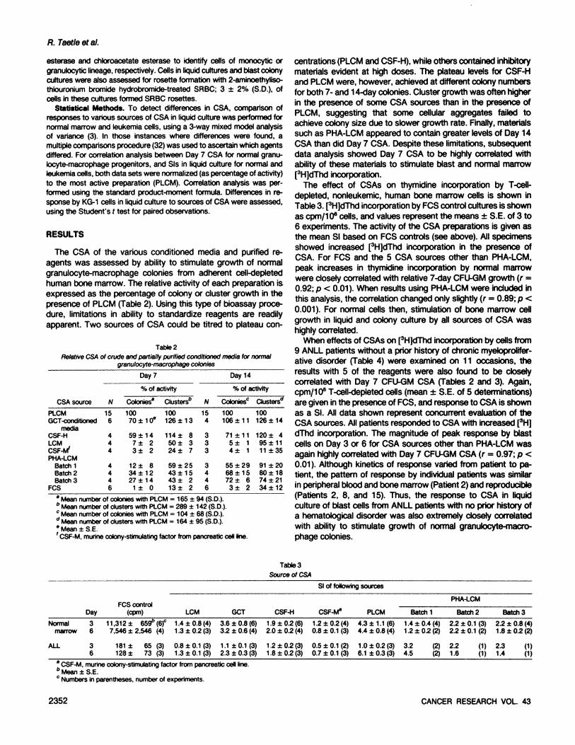
The CSA of the various conditioned media and purified reagents was assessed by ability to stimulate growth of normalgranulocyte-macrophage colonies from adherent cell-depleted
human bone marrow. The relative activity of each preparation isexpressed as the percentage of colony or cluster growth in thepresence of PLCM (Table 2). Using this type of bioassay procedure, limitations in ability to standardize reagents are readilyapparent. Two sources of CSA could be titred to plateau con-
Tabte2RelativeCSAof crude and partially purified conditioned media for normal
granulocyte-macrophagecolonies
Day7%
ofactivityCSA
sourcePLCMGCT-condittonedmediaCSF-HLCMCSF-NfPHA-LCMBalchiBatch
2Batch3PCSN1564444446Colonies"10070
±10e59
±147±23±
212±
834±1227±141
± 0Clusters"100126
±13114±
850+324
±759
±2543±1543±
213± 2N1543333446Day
14%
ofactivityColonies0100106±1171
±115±14±
155
±2968±1572
±63±2Clusters"100126±14120
±495±1111±3591
±2080-1874
±2134±12
•Mean number of colonies with PLCM = 165 ±94 (S.D.).6 Mean number of clusters with PLCM = 289 ±142 (S.D.).c Mean number of colonies with PLCM = 104 ±68 (S.D.)." Mean number of clusters with PLCM = 164 ±95 (S.D.)." Mean ±S.E.' CSF-M.murine colony-stimulatingfactor from pancreatic cell line.
centrations (PLCM and CSF-H), while others contained inhibitorymaterials evident at high doses. The plateau levels for CSF-H
and PLCM were, however, achieved at different colony numbersfor both 7- and 14-day colonies. Cluster growth was often higher
in the presence of some CSA sources than in the presence ofPLCM, suggesting that some cellular aggregates failed toachieve colony size due to slower growth rate. Finally, materialssuch as PHA-LCM appeared to contain greater levels of Day 14
CSA than did Day 7 CSA. Despite these limitations, subsequentdata analysis showed Day 7 Ã’SA to be highly correlated withability of these materials to stimulate blast and normal marrow[3H]dThd incorporation.
The effect of CSAs on thymidine incorporation by T-cell-
depleted, nonleukemic, human bone marrow cells is shown inTable 3. [3H]dThd incorporation by FCS control cultures is shownas cpm/106 cells, and values represent the means ±S.E. of 3 to
6 experiments. The activity of the CSA preparations is given asthe mean SI based on FCS controls (see above). All specimensshowed increased [3H]dThd incorporation in the presence of
CSA. For FCS and the 5 CSA sources other than PHA-LCM,
peak increases in thymidine incorporation by normal marrowwere closely correlated with relative 7-day CFU-GM growth (r =0.92; p < 0.01). When results using PHA-LCM were included inthis analysis, the correlation changed only slightly (r = 0.89; p <
0.001). For normal cells then, stimulation of bone marrow cellgrowth in liquid and colony culture by all sources of CSA washighly correlated.
When effects of CSAs on [3H]dThd incorporation by cells from
9 ANLL patients without a prior history of chronic myeloprolifer-
ative disorder (Table 4) were examined on 11 occasions, theresults with 5 of the reagents were also found to be closelycorrelated with Day 7 CFU-GM CSA (Tables 2 and 3). Again,cpm/106 T-cell-depleted cells (mean ±S.E. of 5 determinations)
are given in the presence of FCS, and response to CSA is shownas a SI. All data shown represent concurrent evaluation of theCSA sources. All patients responded to CSA with increased [3H]
dThd incorporation. The magnitude of peak response by blastcells on Day 3 or 6 for CSA sources other than PHA-LCM wasagain highly correlated with Day 7 CFU-GM CSA (r = 0.97; p <
0.01). Although kinetics of response varied from patient to patient, the pattern of response by individual patients was similarin peripheral blood and bone marrow (Patient 2) and reproducible(Patients 2, 8, and 15). Thus, the response to CSA in liquidculture of blast cells from ANLL patients with no prior history ofa hematological disorder was also extremely closely correlatedwith ability to stimulate growth of normal granulocyte-macro
phage colonies.
TablesSource of CSASI
of followingsourcesNormal
marrowALLDay363
6FCS
control(cpm)11,312±
659" (6)c
7,546 ±2,546(4)181
± 65 (3)128± 73 (3)LCM1.4
±0.8(4)1.3 ±0.2(3)0.8
±0.1 (3)1.3 + 0.1(3)GCT3.6
±0.8 (6)3.2 ±0.6(4)1.1
±0.1(3)2.3 ±0.3 (3)CSF-H1.9
±0.2(6)2.0 ±0.2(4)1.2
±0.2(3)1.8 ±0.2(3)CSF-M81
.2 ±0.2 (4)0.8 ±0.1(3)0.5
±0.1 (2)0.7 ±0.1 (3)PLCM4.3
±1.1(6)4.4 ±0.8(4)1.0
±0.2(3)6.1 ±0.3(3)Batch
11.4
±0.4(4)1.2 ±0.2(2)3.2
(2)4.5 (2)PHA-LCMBatch22.2
±0.1 (3)2.2 ±0.1(2)2.2
(1)1.6 (1)Batch
32.2
±0.8 (4)1.8 ±0.2(2)2.3
(1)1.4 (1)
" CSF-M, murine colony-stimulatingfactor from pancreatic cell line.
Mean ±S.E.' Numbers in parentheses, number of experiments.
2352 CANCER RESEARCH VOL 43

Myeloid Leukemia Response to CSA
Table 4[3H]TdR incorporation by T-cell-depleted leukemia cells in the presence of various sources of CSA
SI with following CSFsourcePatientANLL1.
Blood2.
Blood2.
Marrow3.
Blood4.
Marrow5.Blood6.
Blood7.
Blood8.Blood8.
Blood13.
Blood14.Blood15.
Blood16.
Blood17.
BloodDay36363636335363363633535353PCS
control(cpm)1,1
04±277°2,747
±196781±30953±65873
±84844±591,670±426,784±1,8791
,967 ±578713±131,436
±99534±122,486±1182,595±1,3991,426±502,377
±601,026±39524±12605
±20015,797 ±1,OX934±502,134±1003,539
±59056,677±84948,443±95336,433±1,333LCM1.92.44.71.54.92.49.41.3m1.81.61.01.22.91.63.01.64.7NT.8.3.0.1.1.1NTGCT22.85.015.42.616.84.714.41.93.22.63.91.75.06.93.03.1NT6.6NT1.61.01.31.21.01.0NTCSF-H2.32.513.52.610.74.122.61.96.33.15.41.01.24.51.81.52.12.0NT1.31.21.00.7NT1.1NTCSF-M"1.21.22.31.11.51.310.11.51.11.20.91.11.3NT0.50.80.40.5NTNTNTNTNTNT1.0NTPLCM26.814.319.43.825.84.321.02.8NT6.38.62.97.65.93.05.93.911.837.01.91.01.30.81.21.01.1PHA-LCM20.911.614.80.34.40.718.20.411.13.70.41.13.24.46.28.56.817261.11.10.91.81.00.81.00.9PHA-LCM
+PLCM26.711.012.30.44.64.620.00.5NT7.41.02.07.34.92.69.1NTNTNT1.80.71.11.1NTNTNT
* CSF-M, murine colony-stimulating factor from pancreatic cell line; NT, not tested.15Mean ±S.E.
The response of myeloid leukemia cells in liquid culture toPHA-LCM, however, seemed out of proportion to the Day 7 CSA
of these preparations (Table 4). While PLCM was superior toPHA-LCM as a stimulus for normal cells in liquid or colony culture
(p < 0.05), it was not superior to PLCM as a stimulus for leukemiacells (p > 0.10). Further, when data for PHA-LCM in liquid culture
were included in the correlation analysis between Day 7 CSA fornormal cells and activity for ANLL cells, the correlation was lowerand nonsignificant (r = 0.59; p > 0.10), indicating that for PHA-LCM, blast-stimulating activity was poorly correlated with CSA
for normal cells. When values for sources of CSA other thanPHA-LCM were plotted, these points lay along a line of regres
sion, as expected from the high degree of correlation (data notshown). The points representing PHA-LCM were displaced from
this line, showing much greater activity for myeloid leukemiacells in liquid culture than for normal CFU-GM. A disparity,therefore, existed in the ability of PHA-LCM to stimulate leukemia
blast cells in liquid culture, and its ability to stimulate growth ofnormal granulocyte-macrophage progenitors. Despite this observation, additive effects of PHA-LCM and PLCM were rarely
observed in liquid cultures of blast cells (data not shown), suggesting that overlapping target cell populations were present.The response of myeloid leukemia cells to PHA-LCM was not
duplicated by addition to culture of PHA at a concentrationapproximating the maximum amount which might remain in thisconditioned medium (0.2 ¿tg/ml);PHA either inhibited or showedno effect on ANLL blasts, and showed no additive effect withPLCM.
These observations on fresh leukemia blast cells were confirmed by examination of effects of PLCM and PHA-LCM on themyeloid leukemia cell line KG-1. Two subclones of KG-1 havebeen described. KG-1 requires a source of CSA to form colonies
in agar and responds to CSA after 24 hr of liquid culture with
increased [3H]dThd incorporation (25). The magnitude of this
response is proportional to the titer of the CSA, and thus formsa model for interaction of myeloid leukemia cells with CSA. KG-1a does not require CSA to clone in agar (25). Our KG-1 cells
had properties of both described lines. These cells formed colonies in soft agar without CSA, and CSA did not increase thenumber of colonies formed (data not shown). However, dose-dependent, significant increases in [3H]dThd incorporation were
observed in the presence of CSA after 24 hr in liquid culture.This increase was slightly, but consistently, greater in the presence of PHA-LCM than with PLCM. In 4 experiments, incorporation of [3H]dThd by KG-1 cells in the presence of PCS averaged42,474 ±3,259 cpm/5 x 10s cells (S.E.). The SI for PLCM was
1.41 ±0.2 (S.E.; p < 0.05 greater than control); for PHA-LCMBatch 2 it was 1.60 ±0.4; and for PHA-LCM Batch 3 it was
1.57 ±0.2 (Batches 2 and 3, p < 0.05). This observation mustbe interpreted in light of the modest Day 7 CSA of PHA-LCM fornormal cells (27 to 34% of PLCM). Thus, PHA-LCM was slightlysuperior or equal to PLCM in ability to stimulate KG-1, myeloid
leukemia blasts, similar to effects on ANLL cells in liquid culture.Again, PHA alone did not duplicate this effect.
Relative specificity of CSA for myeloid cells was demonstratedin liquid cultures of ALL cells and normal PHA-stimulated mono-
nuclear cells. ALL cells did not respond with significant increasesin [3H]dThd incorporation to PHA-LCM or other CSA; stimulated
cpm never exceeded 500 on Day 3 or Day 6 (Table 3). PHA-
stimulated normal peripheral blood mononuclear cells also failedto increase [3H]dThd incorporation in the presence of either PHA-
LCM or other sources of Ã’SA (data not shown).Although the stimulatory activity for normal cells of CSA was
highly correlated in liquid and colony culture, and leukemia cellsin liquid culture appeared similar, this relationship unraveled asstudies were expanded to include patients with blast transfor-
MAY 1983 2353

R. Taetle et al.
mation of chronic myeloproliferative disorders. [3H]dThd incor
poration by leukemia cells from patients with blast transformationof chronic myelocytic leukemia or myeloid metaplasia was examined on 4 occasions. Very little stimulation of thymidine incorporation was noted in these patients (Table 4), and patterns ofresponse were again reproducible for individual patients. Baseline thymidine incorporation was higher than in other patientswith ANLL, despite a lower percentage of blasts in some preparations (Table 1). No response to CSA in liquid culture wasobserved in a patient (Patient 15) whose Wood cells were completely dependent on CSA for colony formation (Table 5). Thus,in these patients with blast transformation, a dissociation wasapparent between response to CSA in colony culture and inliquid culture, and responses to CSA in liquid culture appearedmuted at best.
A dissociation between responses to CSA in colony and liquidculture was confirmed by blast colony-forming assays from other
patients with ANLL (Table 5). No relationship was apparentbetween liquid culture response and either spontaneous or CSA-
stimulated colony growth by these cells. Patient 1, who showeda dramatic response to PLCM and PHA-LCM (SI 27 and 21,
respectively) in liquid culture, showed a high level of spontaneouscolony formation and practically no response to CSF or PHA-
LCM in colony culture. Patients 8 and 13, who also respondedto PLCM and PHA-LCM in liquid culture, failed to form colonies
in response to either PHA-LCM or other sources of CSA.
Whereas ability of various sources of CSA to stimulate normalcolony growth and proliferation in liquid culture was highly correlated, these phenomena were not consistently related in ANLLpatients.
We examined relative activity of PHA-LCM and PLCM in colony
culture in 11 ANLL patients (Table 5). All values shown wereobtained in concurrent studies. PHA-LCM was not consistently
or statistically superior to PLCM in ability to stimulate growth ofleukemia colonies. Results for individual patients demonstratedthat PHA-LCM was superior to PLCM for some patients (Patients
2, 6, 9, and 11), a finding which must again be interpreted inlight of its rather modest CSA for normal cells. Additive effectsby PHA-LCM and PLCM were uncommon and generally of small
magnitude in blast colony culture (Table 5), italicized values),again suggesting overlapping target cell populations in mostpatients.
Finally, we examined effects of the various CSA sources onmorphological differentiation by leukemia cells from four patientsin liquid and colony cultures (Table 6). As previously noted byourselves and others (19,33,48), in 4 of 5 patients studied, littlemorphological differentiation was observed in the presence orabsence of CSA or PHA-LCM. Culture with several sources of
CSA was associated with appearance of increased numbers ofmacrophages, and while it is not clear that these macrophages
TabteSGrowth of leukemiacolony-forming cells in the presence of PHA-LCMand CSA
CSAPatientANLL12678910111213Blast
transformation151617PCS99*141165004102701302052LCM11833249006NTNTNTNT0330NTGCT100451691008NTNT45NT0310NTCSF-H10511246200822NT0360540NTCSF-M"88914NT007NTNTNTNT0620NTPLCM1116225730023NT594041044088PHA-LCM1242703200065384113831078861073LCM742431900031NTNTNTNT0NTNT3NTCSA
in presenceofPHA-LCMGCT1193386000064NTNT255NT0NT1005NTCSF-H120265390006929NT140310NT955NTCSF-M972803900048NTNTNTNT0NTNT13NTPLCM145?2407200069NT66260410NT1091NTPHA-LCM
Batchno.1112221111132313
* Colonies/2 x 104 T, cell depleted cells.* CSF-M, murine colony-stimulating factor from pancreatic cell line; NT, not tested." Italicized numbers, additive effects of PHA-LCM and PLCM.
Table6Morphological differentiation by ANLL leukemiacells in the presence of CSA
PCSGCTCSF-HPLCMPHA-LCMNo.
ofStudies54554Blasts65(18-93)"49(16-99)59(21-96)52(11-96)61(31-96)Promyetocyte12(0-55)9(0-36)7(0-36)12(0-56)8(0-27)Mean
%Myelocyte-Band-Poly14(0-63)20(0-66)12(0-43)16(0-71)15(0-50)Macro
phage7(0-14)21(0-32)19(1-33)20(0-37)17(0-33)Lymphocyte1(0-6)1(0-4).12(0-7)1(0-3)1(0-4)Eosinophil0(0-1)0(0-1)1(0-7)00
" Numbers in parentheses, range.
2354 CANCER RESEARCH VOL. 43

Myeloid Leukemia Response to CSA
originated from leukemia clones, we have observed such cellularmaturation in previous studies in patients with acute monocyticleukemia in response to PHA-LCM (48). Cells from Patient 1
underwent extensive differentiation in liquid culture (percentageof mature granulocytic forms, 43 to 71%), but no definite differences were apparent between cells from PCS control and CSA-
stimulated cultures. Histochemical staining for chloroacetate esterase or a-naphthylacetate esterase confirmed the morpholog
ical differentiation of these cells. Differentiation in liquid culture,therefore, appeared to be more a function of the cells understudy rather than the stimulus although macrophage formationmay have been favored by CSA.
DISCUSSION
Glycoproteins which induce in vitro growth of colonies ofgranulocytes and macrophages (CFU-GM) from human or murine
bone marrow and peripheral blood have been termed CSF (5).The aggregate in vitro activity of these proteins in conditionedmedia is sometimes referred to as CSA. The in vivo role of thesematerials is unclear (5, 20), but normal CFU-GM exhibit an
absolute requirement for their presence in vitro (5, 30). Even incrude form, such as PLCM, the activity of some CSA preparations appears specific for granulocyte precursors. Under theinfluence of CSA, normal cells undergo both proliferation anddifferentiation in culture (5, 6, 30,37). Recently, multiple classesof CSFs have been described (22,35), which may have differenttargets within marrow. The role and chemistry of CSFs haverecently been reviewed (5).
Human myeloid leukemia cells also form colonies in responseto conditioned media containing CSA for normal cells (PHA-
LCM), or to feeder layers of normal blood cells, which elaborateCSA (4,19,29, 44). However, cells from only a fraction of humanleukemia cells form colonies in response to CSA, and cells withincolonies do not undergo differentiation (19, 29, 51). Disparitieshave been described between ability of conditioned media tostimulate colony growth of leukemia cells and normal cells (10).Certain myeloid leukemia cell lines respond to CSA for normalcells with increased proliferation (25, 40), while others do not(18). PHA and medium conditioned by normal PHA-stimulated
mononuclear cells also induce growth of myeloid blast colonies(4, 7, 24); however, PHA-conditioned medium (PHA-LCM) con
tains variable, but usually low titers of CSFs (31), and thepossibility that biological activities other than CSA stimulate blastgrowth may be raised.
By using in vitro colony and liquid culture techniques, theexpected activity of a wide variety of sources of CSA, includingPHA-LCM, on growth of normal myeloid cells was demonstrated
in these studies. Activities in liquid and colony culture wereextremely highly correlated for normal cells. In liquid culture, cellsfrom 9 patients with ANLL also showed a proliferative responsesimilar to that of normal cells to CSA. It is tempting to suggestthat some myeloid blast cells were even more responsive thanwere normal cells to CSA, since Sis of up to 60 were observedin some patients. Base-line [3H]dThd incorporation in bone mar
row specimens was significantly higher (p < 0.05) than in leukemia specimens. This probably reflects ongoing proliferation ofnonmyeloid, non-CSF-responsive cells in normal marrow, and
may have obscured the magnitude of response by normal myeloid cells. Others, however, have noted exceptional responsiveness of some leukemia cells to CSF (2). Although the number of
patients studied was limited, these results suggest that themajority of ANLL cells respond to CSA by proliferating.
In liquid culture, responses of leukemia cells to PHA-LCM were
out of proportion to ability of this conditioned medium to stimulatenormal CFU-GM or bone marrow thymidine incorporation. Although PHA-LCM was only 15% as active as PLCM for normal7-day CFU-GM, it was nearly as active as PLCM in liquid culturesof leukemia cells. This increased blast-stimulating activity forPHA-LCM was confirmed by studies of the leukemia cell line KG-1. Stimulation of [3H]dThd incorporation by KG-1 cells correlates
with CSA for normal cells (25), and PHA-LCM was as good asor superior to PLCM in stimulation of [3H]TdR incorporation by
KG-1 cells, consistent with results with fresh leukemia cells.Further, PHA-LCM was clearly superior to PLCM in ability to
stimulate growth of leukemia colonies from several patients.Kubota ef al. (19) have also compared the ability of severalsources of CSA to induce colonies from leukemia peripheralblood. These authors concluded that there was no difference inthe ability of PHA-LCM, GCT, or media conditioned by the T-cellline, Mo-1, in ability to stimulate formation of leukemia colonies.
However, if one examines their data for patients growing morethan 16 colonies/105 cells, one notes a pattern similar to our
results. Despite having only 50% of the activity of Mo-1 -conditioned medium for normal cells, PHA-LCM stimulated nearly asmany leukemia colonies as did Mo-1-conditioned medium, andboth appeared superior to GCT-conditioned medium. Consistent
with these observations, Swart ef al. (45) have recently demonstrated that leukemia colony-forming cells which respond to
leukocyte feeders can be separated in some patients from cellsresponding to PHA-stimulated peripheral blood cells; these pop
ulations, however, showed substantial overlap.Iscove ef al. (16) have recently proposed a model relevant to
these observations for responses by normal murine progenitorcells to humoral agents. After extensive attempts they wereunable to separate lineage-specific growth factors for myeloid-erythroid blasts, megakaryocyte-erythroid blasts, granulocyte-
macrophage colonies, and granulocyte colonies from lymphocyte-conditioned medium. They postulated the existence of alineage-nonspecific growth factor to which a variety of progenitor
cells respond. As cells mature, they also display receptors for,and respond to, a lineage-nonspecific factor and lineage-specific
factors, such as CSF (myeloid progenitors) or erythropoietin(erythroid progenitors).
It has been suggested that leukemia cells are "dyssynchron-ous" when compared to normal cells, with respect to display of
membrane proteins or enzyme markers (14,27). ANLL cells thencould also show dyssynchrony with respect to receptors for agrowth factor in PHA-LCM and the lineage-specific growth factor
CSF. Whereas normal cells show an orderly change in receptordisplay, and therefore highly correlated responses to CSF bycolony-forming and liquid culture cells, the leukemia cells mightshow variable and simultaneous display of receptors for multiplehumoral stimuli. Leukemia colony-forming and liquid culture cells
from individual patients would, therefore, show variable responses to CSA and PHA-LCM, but since many cells display
receptors for both stimuli, most responses would show considerable overlap. Unusual clones showing additive effects of PLCMand PHA-LCM presumably represent instances when distinctPHA-LCM and CSF-responsive colony-forming cell populations
exist, consistent with observations by Swart ef al. (45). Althoughstimuli used in this study were crude, using a partially purified
MAY 1983 2355

R. Taef/e ef al.
CSF source, (CSF-H), the results were similar. Admittedly this
explanation is speculative, but is consistent with the lack ofcorrelation of normal CSA and leukemia colony growth withPHA-LCM, and lack of correlation between leukemia liquid culture and colony culture results.
In murine systems, myeloid leukemia cell lines show variabledependence on the presence of CSF-like molecules for prolifer
ation; some are independent, others show partial responses incolony cultures, and still others undergo differentiation (22, 41).Our observations suggest that, within human leukemia cell populations, these same multiple cell types may be present simultaneously, and may be detected by various assay techniques.Cell populations responsive to humoral stimuli other than CSF,or simultaneously responsive to CSF and other less specificmolecules, may also be present. Alternatively, the responsesobserved could be a composite of abnormal and residual normalcells. This seems unlikely, since leukemia blasts are usuallymonoclonal and dominant at presentation (9), and normal colony-
forming populations containing differentiating cells are usuallynot detected (28).
Induction of macrophage differentiation by all CSAs testedwas seen in 4 patients. One patient showed extensive myeloiddifferentiation in FCS control cultures, leading one to questionwhy this process failed to take place in vivo. As in prior studies(19, 33, 48), the vast majority of leukemia cells in liquid andcolony cultures remained primitive. Lotem ef al. (22), and Nicolaand Metcalf (36), have shown that humoral factors inducingmurine leukemia cell differentiation can be separated from otherCSFs, and these differentiation factors may be absent from ourreagents. Our results are also consistent with studies by Lieber-
mann ef al. (21) in murine systems, showing that CSA inducesproliferation in normal cells (MGI-1 ), but induces a separate factorcausing differentiation (MGI-2); murine leukemia cells fail to produce MGI-2, but proliferate in response to MGI-1. Human cells
in our studies behaved similarly; leukemia cells failed to differentiate in the presence of multiple CSA sources, but displayedproliferative responses in liquid and colony culture. This point isfar from trivial, since induction of differentiation of murine leukemia clones by CSA results in loss of self-replicative capacity by
the leukemia cells (23). Cells from our patients either lacked thecapacity to produce or respond to stimuli to differentiation, orsuch stimuli for human cells are largely absent from our CSAsources and need to be sought elsewhere.
REFERENCES1. Aye. M. T., Till, J. E., and McCulloch, E. A. Growth of leukemic cells in culture.
Blood, 40.: 806-811, 1972.2. Brennan, J. K., DiPersio, J. F., Abboud, C. N., and Uchtman, M. A. The
exceptional responsiveness of certain human myeloid leukemia cells to colony-stimulating activity. Blood, 55: 1230-1239, 1979.
3. Brownlee, K. A. Statistical Theory and Methodology in Science and Engineering, Ed. New York: John Wiley & Sons, 1965.
4. Buick, R. N., Till, J. E., and McCulloch, E. A. Colony assay for proliferativeblast cells circulating in myeloblastic leukemia. Lancet, 7: 862-963, 1977.
5. Burgess, A. W., and Metcalf, D. Review: the nature and action of granulocyte-macrophage colony-stimulating factors. Blood, 56. 947-978,1980.
6. Cline. M. J., and Golde, D. W. Controlling the production of blood cells. Blood,53: 157-163,1979.
7. Dicke, K. A., Spitzer, G., and Aheam, M. J. Colony formation in vitro byleukaemic cells in acute myelogenous leukaemia with phytohemagglutinin asstimulating factor. Nature (Lond.), 259: 129-130, 1976.
8. DiPersio, J. F., Brennan, J.K., Lichtman, M. A., Abboud, C. N., and Kirkpatrick,F. H. The fractionation, characterization and subcellular localization of colony-stimulating activities released by the human monocyte-like cell line GCT. Blood,56:717-727,1980.
9. Fialkow, P. J., Singer, J. W., Adamson, J. W., Vaidyak, Dow, L. W., Ochs, J.,
and Moohr, J. W. Acute non-lymphocytic leukemia: heterogeneity of stem cellorigin. Blood, 57: 1068-1073, 1981.
10. Francis, G. E., Berney. J. J., Tuma, G. A., Wing, M. A., and Hoffbrand, A. V.Divergent sensitivities of leukaemic cells to human placenta! conditionedmedium and leukocyte feeder layers. Leuk. Res., 4: 531-536, 1980.
11. Froelich, T. W., Buchanan, G. R., Cornet, J. A. M., Sartain, P. A., and Smith,R. G. Terminal deoxynucleotidyl transferase-containing cells in peripheralblood: implications for the surveillance of patients with lymphoblastic leukemiaor lymphoma in remission. Blood, 58: 214-220, 1981.
12. Gasser, D. L., Winters, B. A., Haas, J. B., McKeam, T. J., and Kennet, R. H.Monoclonal antibody directed to a B-cell antigen present in rats, mice andhumans. Proc. Nati. Acad. Sei. U. S. A. 76:4636-4640,1979.
13. Gralnick, H. R., Galton, D. A. G., Catovsky, D., Sultan, C., and Bennett, J. M.Classification of acute leukemia. Ann. Intern. Med., 87: 740-753,1977.
14. Greaves, M. F. "Target" cells, cellular phenotypes, and lineage fidelity in
human leukaemia. J. Cell. Physiol. (Suppl.), 1: 113-125,1982.15. Iscove, N. N., Senn, J. S., Till, J. E., and McCulloch, E. A. Colony formation
by normal and leukemic human cells in culture: effect of conditioned mediumfrom human leukocytes. Blood, 37: 1-5, 1971.
16. Iscove, N. N., Roitsch, C. A., Williams, N., and Guilbert, L J. Moleculesstimulating early red cell, granulocyte, macrophage, and megakaryocyte precursors in culture: similarity in size, hydrophobicity, and charge. J. Cell.Physiol., 1: 65-78,1982.
17. Koeffler, H. P., and Golde, D. W. Acute myelogenous leukemia: a human cellline responsive to colony-stimulating activity. Science (Wash. D. C.), 200:1153-1154,1978.
18. Kubota, K., Preisler, H. D., Lok, M. S., and Minowada, J. Lack of effect ofcolony stimulating activity on human myeloid leukemia cell line (ML-2) cells.Leuk. Res., 5:311-320, 1981.
19. Kubota, K., Preisler, H. D., Sagawa, K., and Minowada, J. Comparison betweenagar and methylcellulose cultures of human leukemic cells. Cancer Res., 41:3052-3057, 1981.
20. Levitt, L. J., and Quesenberry, P. J. Further studies on the mechanisms ofmarrow granulocytic hyperplasia in mice chronically injected with endotoxin.Br. J. Haematol., 50: 269-281, 1982.
21. Uebermann, D., Hoffmann-Liebermann, B., and Sachs, L. Regulation and roleof different macrophage and granulocyte-inducing proteins in normal andleukemic myeloid cells. Int. J. Cancer, 29: 159-161,1982.
22. Lotem, J., Lipton, J. H., and Sachs, L. Separation of different molecular formsof macrophage and granulocyte inducing proteins for normal and leukemicmyeloid cells. Int. J. Cancer, 25: 763-771, 1980.
23. Lotem, J., and Sachs, L. In vivo inhibition of the development of myeloidleukemia by injection of macrophage and granulocyte inducing protein. Int. J.Cancer, 28: 375-386, 1981.
24. Lowenberg, B., Swart, K., and Hagemeiher, A. PHA-induced colony formationin acute non-lymphocytic and chronic myeloid leukemia. Leuk. Res., 4: 143-
149, 1980.25. Lusis, A. J., and Koeffler, H. P. Action of granulocyte/macrophage colony
stimulating factors: studies using a human leukemic cell line. Proc. Nati. Acad.Sei. U. S. A., 7: 5346-5350, 1980.
26. Madsen, M., and Johnsen, H. E. A methodological study of E-rosette formationusing AET-treated sheep red blood cells. J. Immunol. Methods, 27: 61-74,1979.
27. Marie, J. P., Izaguirre, C. A., Civin, C. J., Mirro, J., and McCulloch, E. A.Granulopoietic differentiation in AML blasts in culture. Blood, 58: 670-677,1981.
28. McCulloch, E. A., and Till, J. E. Blast cells in acute myeloblastic leukemia: amodel. Blood Cells, 7: 63-77, 1981.
29. Mertelsmann, R., Moore, M. A. S., Broxmeyer, H. E., Cirrincione, C., andClarkson, B. Diagnostic and prognostic significance of the CFU-c assay inacute nonlymphoblastic leukemia. Cancer Res., 47: 4844-4848, 1981.
30. Metcalf, D. Hemopoietic Colonies. New York: Springer-Verlag, 1977.31. Metcalf, D. Clonal analysis of the action of GM-CSF on the proliferation and
differentiation of myelomonocytic leukemic cells. Int. J. Cancer, 24: 616-623,1979.
32. Miller, R. G. Simultaneous Statistical Inference, Chap. 2. New York: McGraw-Hill Book Co., 1966.
33. Minden, M. D., Buick, R. N., and McCulloch, E. A. Proliferative state of blastcell progenitors in acute myeloblastic leukemia. Blood, 52: 592-600,1978.
34. Minden, M. D., Buick, R. N., and McCulloch, E. A. Separation of blast cell andT-lymphocyte progenitors in the blood of patients with acute myeloblasticleukemia. Blood, 54: 186-195, 1979.
35. Morstyn, G., Nicola, N. A., and Metcalf, D. Separate actions of different colonystimulating factors from human placental conditioned medium on humanhemopoietic progenitor cell survival and proliferation. J. Cell. Physiol., 709:133-142,1981.
36. Nicola, N. A., and Metcalf, D. Biochemical properties of differentiation factorsfor murine myelomonocytic leukemic cells in organ conditioned media—separation from colony-stimulating factors. J. Cell. Physiol., 709: 253-264, 1981.
37. Quesenberry, P., and Levitt, L. Hemopoietic stem cells. N. Engl. J. Med., 307:755-759, 819-823, 868-872, 1979.
38. Ritz, J., Pesando, J. M., Sallan, S. E., Clavell, L. A., Salan, S. E., andSchlossman, S. F. Serotherapy of acute lymphoblastic leukemia with mono-
2356 CANCER RESEARCH VOL. 43

donai antibody. Blood, 58: 141-152,1981.39. Royston, l., Majda, J. A., Baird, S. A., Meserve, B. L, and Griffiths, J. C. The
65,000 dalton antigen of T-cells (T65) is also found on chronic lymphocyticleukemia cells bearing surface immunoglobulin. J. Immunol., 725: 725-731,1980.
40. Ruscetti, F. W., Collins, S. J., Woods, A. M., and Gallo, R. C. Clonal analysisof the response of human myeloid leukemic cell lines to colony stimulatingactivity. Blood, 58: 285-292,1981.
41. Sachs, L. Regulation of normal cell differentiation and malignancy in myeloidleukemia. In: B. Clarkson, P. A. Marks, and J. E. Till. (eds.). Differentiation ofNormal and Neoplastic Hematopoietic Cells. Cold Spring Harbor, N. Y.: ColdSpring Harbor Laboratory, 1978.
42. Sachs, L. Constitutive gene expression and restoration of the normal pheno-
type in malignant cells: a model for the origin of the evolution of leukemia.Blood Cells, 7.-31-44,1981.
43. Schlunk, T., and Schleyer, M. The influence of culture conditions on theproductivity of colony stimulating activity by human placenta. Exp. Hematol.(Copenh.), 8: 179-184,1980.
44. Spitzer, G., Verma, D. S., Dicke, K. A., and McCredie, K. B. Culture studies invitro in human leukemia. Semin. Hematol., 75: 352-378.1978.
Myeloid Leukemia Response to CSA
45. Swart, K., Hagemeijer, A., and Lowenberg, B. Acute myeloid leukemia growthIn vitro: differences of colony-forming cells in PHA supplemented and standardleukocyte feeder cultures. Blood, 58: 816-822, 1982.
46. Taetle, R. Acidic isoferritins (leukemia-associated inhibitory activity) fail to inhibitblast proliferation in acute myelogenous leukemia. Blood, 58. 653-657, 1981.
47. Taetle, R., and Guittard, J. P. Modulation of leukaemia blast colony growth bysteroid hormones. Br. J. Haematol., 50: 247-255, 1982.
48. Taetle, R., and Ivor, L. In vitro evidence for an unusual progenitor cell in acutemonocytic leukemia. Cancer (Phila.), 46: 1602-1607,1980.
49. Taetle, R., and Koessler, A. Effects of cyclic nucleotides and prostaglandinson normal and abnormal myeloid progenitor proliferation. Cancer Res., 40:1223-1229,1980.
50. Till, J. E., and McCulloch, E. A. Hemopoietic cell differentiation. Biochim.Biophys. Acta, 605: 431-459, 1980.
51. Vincent, P. C., Sutherland, R., Bradley, M., Und, D., and Gunz, F. W. Marrowculture studies in adult acute leukemia at presentation and remission. Blood,49:903-917,1977.
52. Wu, M. C., and Yunis, A. A. Common pattern of 2 distinct types of colonystimulating factor in human tissues and cultured cells. J. Clin. Invest., 65: 772-775, 1980.
MAY 1983 2357