Resource recovery from septic tank effluent using duckweed-based tilapia aquaculture
11
World Applied Sciences Journal 19 (2): 265-275, 2012 ISSN 1818-4952; © IDOSI Publications, 2012 DOI: 10.5829/idosi.wasj.2012.19.02.6485 Corresponding Author: Fagr Khamis Abdel Gawad, Center of Excellence for Advanced Science (CEAS), National Research Centre (NRC), Water Pollution Research Department, Gizza, 12622, Egypt. Tel: +20-1001542413. 265 Fish as Bio Indicators in Aquatic Environmental Pollution Assessment: A Case Study in Abu-Rawash Area, Egypt Mohamed R. Lasheen, Fagr Kh. Abdel-Gawad, Aly A. Alaneny and Hassan M.H. Abd El bary 1 1 1 2 Water Pollution Research Department, National Research Centre, 12311 Dokki, Giza, Egypt 1 Chemistry Department, Faculty of Science, Al-Azhar University, Nasr city, Cairo, Egypt 2 Abstract: The objective of this study was to evaluate the toxic impact of wastewater discharge effluent on aquatic environment. Lead, Zinc, Copper and Cadmium were determined in some tissues of sharp toothed African Catfish (Clarias garipinus) and Nile tilapia (Oreochromis niloticus) collected from only two sites and water samples from all six sites. The concentration of heavy metals in water was within the permissible limits for discharge drain water into River Nile (Egyptian law 48/1982). A significant difference between the four metals in different / similar tissues of different / similar species (p<0.05), with the exception of lead / cadmium metal concentration in fish edible part tissues were higher than the permissible levels according to Egyptian Organization for Standardization and Quality Control. Randomly amplified polymorphic DNA technology and Micronucleus test were applied to evaluate the genotoxic effects of wastewater effluent on (Clarias garipinus and Oreochromis niloticus). Results of RAPD and Micronucleus test showed that both wastewater effluents and agriculture drains were genotoxic to fish. Key words: Heavy Metal Concentration Water Quality Pollution Agricultural Drains Genotoxicity RAPD Micronucleus Test INTRODUCTION essential metals since they play an important role in Heavy metals pollution in aquatic environment has essential metals, as they are toxic, even in trace amounts become a worldwide problem during past few decades. [7]. El-Naggar et al. [8] determined the concentration of This fact is mainly attributed to their persistent stability heavy metals in fish samples collected from River Nile and toxic effect to aquatic as well as terrestrial creatures and its two branches (Damietta and Rossetta) including [1]. Among environmental pollutants, metals are of El-Oqsur, El-Menia, El-Hawamdia, Shoubra El-Khema, particular concern, due to their potential toxic effect and El-Rahawy drain, Kom Hamada, Talkha, El-Serw and ability to bioaccumulate in aquatic ecosystems [2]. Heavy Faraskourn and found that the concentration of Cu, Pb metal concentrations in aquatic ecosystems are usually and Cd were at high level in all regions. Bahnasawy et al. monitored by measuring their concentrations in water, [9] determined the concentration of Zn, Cu, Pb and Cd in sediments and biota [3] which generally exist in low levels samples of two fish species (Mugil cephalus and Liza in water and attain considerable concentration in ramada) from five locations in Lake Manzala. The highest sediments and biota [4]. Fish muscle is commonly concentrations of metals were in gills tissue and the analyzed to determine contaminant concentrations and to lowest were in muscles tissue. Metals in the fish muscles assess the health risks because it is the main part were within the permissible limits according to EOSQC consumed by humans. Fish can be considered as one of [10]. Elnimr [11] studied the concentration of heavy metals the most significant indicators in freshwater systems for in fish samples (Tilapia nilotica and cat fish) collected the impact of metal pollution [5]. The commercial and from Kafer-El-Zayat and found that the concentration edible species have been widely investigated in order to of Pb, Zn and Cd were 1.2, 1.03 and 0.17 mg kg check for those hazardous to human health [6]. Heavy respectively, these results are higher than the permissible metals such as copper, iron, chromium and nickel are limits of EOSQC [10] except Zn. biological systems, where cadmium and lead are non- 1
-
Upload
independent -
Category
Documents
-
view
3 -
download
0
Transcript of Resource recovery from septic tank effluent using duckweed-based tilapia aquaculture
World Applied Sciences Journal 19 (2): 265-275, 2012ISSN 1818-4952;© IDOSI Publications, 2012DOI: 10.5829/idosi.wasj.2012.19.02.6485
Corresponding Author: Fagr Khamis Abdel Gawad, Center of Excellence for Advanced Science (CEAS), National Research Centre (NRC), Water Pollution Research Department, Gizza, 12622, Egypt. Tel: +20-1001542413.
265
Fish as Bio Indicators in Aquatic Environmental PollutionAssessment: A Case Study in Abu-Rawash Area, Egypt
Mohamed R. Lasheen, Fagr Kh. Abdel-Gawad, Aly A. Alaneny and Hassan M.H. Abd El bary1 1 1 2
Water Pollution Research Department, National Research Centre, 12311 Dokki, Giza, Egypt1
Chemistry Department, Faculty of Science, Al-Azhar University, Nasr city, Cairo, Egypt2
Abstract: The objective of this study was to evaluate the toxic impact of wastewater discharge effluent onaquatic environment. Lead, Zinc, Copper and Cadmium were determined in some tissues of sharp toothedAfrican Catfish (Clarias garipinus) and Nile tilapia (Oreochromis niloticus) collected from only two sites andwater samples from all six sites. The concentration of heavy metals in water was within the permissible limitsfor discharge drain water into River Nile (Egyptian law 48/1982). A significant difference between the four metalsin different / similar tissues of different / similar species (p<0.05), with the exception of lead / cadmium metalconcentration in fish edible part tissues were higher than the permissible levels according to EgyptianOrganization for Standardization and Quality Control. Randomly amplified polymorphic DNA technology andMicronucleus test were applied to evaluate the genotoxic effects of wastewater effluent on (Clarias garipinusand Oreochromis niloticus). Results of RAPD and Micronucleus test showed that both wastewater effluentsand agriculture drains were genotoxic to fish.
Key words: Heavy Metal Concentration Water Quality Pollution Agricultural Drains Genotoxicity RAPD Micronucleus Test
INTRODUCTION essential metals since they play an important role in
Heavy metals pollution in aquatic environment has essential metals, as they are toxic, even in trace amountsbecome a worldwide problem during past few decades. [7]. El-Naggar et al. [8] determined the concentration ofThis fact is mainly attributed to their persistent stability heavy metals in fish samples collected from River Nileand toxic effect to aquatic as well as terrestrial creatures and its two branches (Damietta and Rossetta) including[1]. Among environmental pollutants, metals are of El-Oqsur, El-Menia, El-Hawamdia, Shoubra El-Khema,particular concern, due to their potential toxic effect and El-Rahawy drain, Kom Hamada, Talkha, El-Serw andability to bioaccumulate in aquatic ecosystems [2]. Heavy Faraskourn and found that the concentration of Cu, Pbmetal concentrations in aquatic ecosystems are usually and Cd were at high level in all regions. Bahnasawy et al.monitored by measuring their concentrations in water, [9] determined the concentration of Zn, Cu, Pb and Cd insediments and biota [3] which generally exist in low levels samples of two fish species (Mugil cephalus and Lizain water and attain considerable concentration in ramada) from five locations in Lake Manzala. The highestsediments and biota [4]. Fish muscle is commonly concentrations of metals were in gills tissue and theanalyzed to determine contaminant concentrations and to lowest were in muscles tissue. Metals in the fish musclesassess the health risks because it is the main part were within the permissible limits according to EOSQCconsumed by humans. Fish can be considered as one of [10]. Elnimr [11] studied the concentration of heavy metalsthe most significant indicators in freshwater systems for in fish samples (Tilapia nilotica and cat fish) collectedthe impact of metal pollution [5]. The commercial and from Kafer-El-Zayat and found that the concentrationedible species have been widely investigated in order to of Pb, Zn and Cd were 1.2, 1.03 and 0.17 mg kgcheck for those hazardous to human health [6]. Heavy respectively, these results are higher than the permissiblemetals such as copper, iron, chromium and nickel are limits of EOSQC [10] except Zn.
biological systems, where cadmium and lead are non-
1
World Appl. Sci. J., 19 (2): 265-275, 2012
266
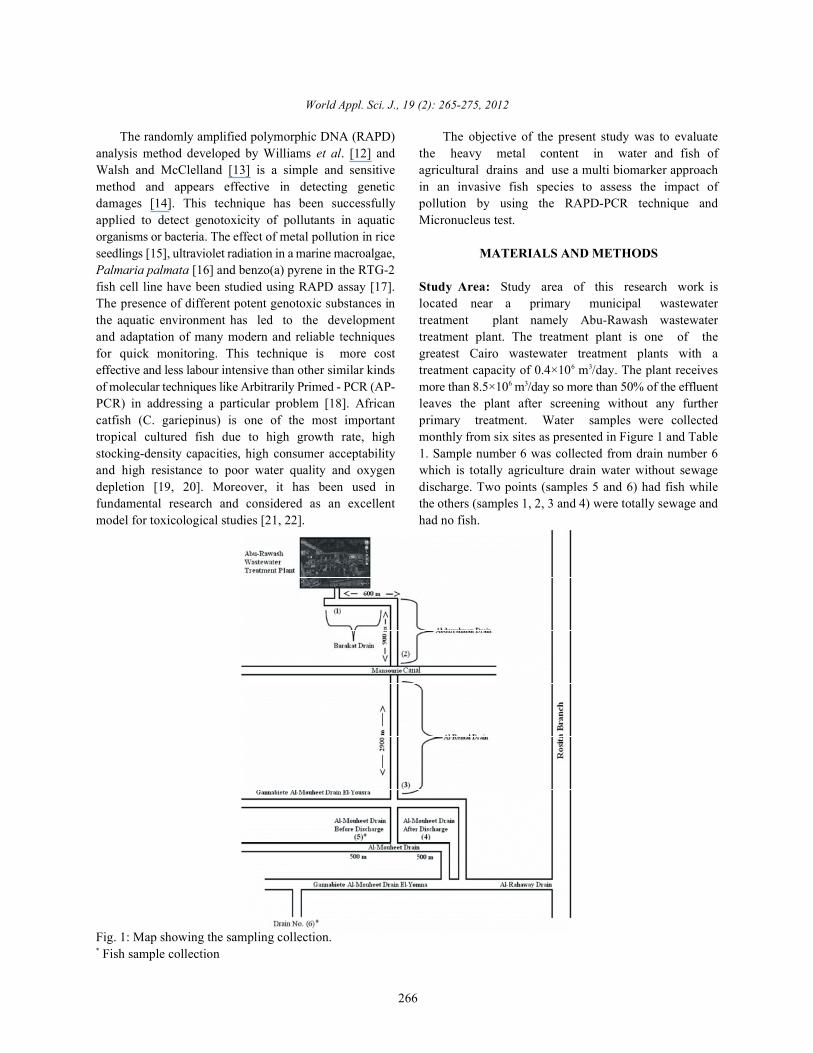
The randomly amplified polymorphic DNA (RAPD) The objective of the present study was to evaluateanalysis method developed by Williams et al. [12] and the heavy metal content in water and fish ofWalsh and McClelland [13] is a simple and sensitive agricultural drains and use a multi biomarker approachmethod and appears effective in detecting genetic in an invasive fish species to assess the impact ofdamages [14]. This technique has been successfully pollution by using the RAPD-PCR technique andapplied to detect genotoxicity of pollutants in aquatic Micronucleus test.organisms or bacteria. The effect of metal pollution in riceseedlings [15], ultraviolet radiation in a marine macroalgae, MATERIALS AND METHODSPalmaria palmata [16] and benzo(a) pyrene in the RTG-2fish cell line have been studied using RAPD assay [17]. Study Area: Study area of this research work isThe presence of different potent genotoxic substances in located near a primary municipal wastewaterthe aquatic environment has led to the development treatment plant namely Abu-Rawash wastewaterand adaptation of many modern and reliable techniques treatment plant. The treatment plant is one of thefor quick monitoring. This technique is more cost greatest Cairo wastewater treatment plants with aeffective and less labour intensive than other similar kinds treatment capacity of 0.4×10 m /day. The plant receivesof molecular techniques like Arbitrarily Primed - PCR (AP- more than 8.5×10 m /day so more than 50% of the effluentPCR) in addressing a particular problem [18]. African leaves the plant after screening without any furthercatfish (C. gariepinus) is one of the most important primary treatment. Water samples were collectedtropical cultured fish due to high growth rate, high monthly from six sites as presented in Figure 1 and Tablestocking-density capacities, high consumer acceptability 1. Sample number 6 was collected from drain number 6and high resistance to poor water quality and oxygen which is totally agriculture drain water without sewagedepletion [19, 20]. Moreover, it has been used in discharge. Two points (samples 5 and 6) had fish whilefundamental research and considered as an excellent the others (samples 1, 2, 3 and 4) were totally sewage andmodel for toxicological studies [21, 22]. had no fish.
6 3
6 3
Fig. 1: Map showing the sampling collection. Fish sample collection*
World Appl. Sci. J., 19 (2): 265-275, 2012
267
Table 1: Sampling location and the distance from Abu-Rawash wastewater treatment plant and fish sample collectionLocation Cu Pb Cd ZnBarakat drain 0.012±0.01 0.002±0.01 0.009±0.1 0.1±0.07a a a a
Abdul-Rahman 0.05±0.02 0.002±0.01 0.010±0.1 0.13±0.17a a a a
Al-Remal drain 0.04±0.02 0.002±0.01 0.026±0.1 0.12±0.14a a a a
Al-Moheet drain( after ) 0.02±0.01 0.02±0.01 0.011±0.1 0.06±0.05a a a a
Al-Moheet drain (before) 0.05±0.01 0.02±0.01 0.006±0.02 0.06±0.07a a a a
agricultural drains 0.01±0.01 0.03±0.05 0.008±0.1 0.04±0.01a a a a
Egyptian law 48/1982 0.20 5 0.01 5* Fish sample collection, after WWTP discharge, before WWTP discharge1 2
Sampling: Sub-Surface water samples were collected A12: 5'-TCGGCGATAG-3', A17: 5'-GACCGCTTGT-3', A20:using acid washed, distilled water rinsed polyethylene 5-GGG TAA CGC C-3, A19: 5-CAA ACG TCG G-3 andbottles. Samples were collected at monthly interval A20: 5'-GTTGCGATCC-3'. The primers were obtained fromduring one year's period from April 2008 to March 2009. the Operon Technology. DNA amplification reactionsFish samples (Clarias garipinus, Oreochromis niloticus) were performed under conditions reported by Williamswere collected from El-Moheet drain and agriculture et al. [12] and Plotsky et al. [28]. PCR amplification wasdrains (Fig. 1). The collected samples were transported conducted in 50µl reaction volume containing 100 ngalive in a large plastic water container supplied with genomic DNA, 100 mM dNTPs, 40 nM primer (Bio Basicbattery aerators as a source of air and washed with Inc. Canada), 2.5 units of Taq DNA polymerase. The PCRdistilled water. reactions were carried out in a thermocycler (Bioer-Xp
Heavy metals Analysis: Water samples were preserved min at 94°C, followed by 45 cycles of 1 min denaturationimmediately after collection by acidifying with at 95°C,1 min annealing at 36°C and 2 min extension atconcentrated HNO to pH < 2; by using 5 ml for 1 liter of 72°C. Final extension at 72°C, for 5 min was allowed before3
water sample. Heavy metals were extracted from the holding the reaction at 4°C for 10 min. Reaction productscollected water samples using nitric acid digestion method were stored at 4°C prior to electrophoresis.according to APHA [22]. Fish samples (1.0 g dry weight)were digested with 6 ml of HNO (65%) and 2 ml of H O Gel Electrophoresis: Each sample of RAPD products (103 2 2
(30%) at 280°C on a hot plate for 4 h. Finally, 2 ml of 1 N µl) was mixed with gel loading buffer and loaded onto anHNO was added to the residue and the solution agarose gel (2%, w/ v) for electrophoresis. Amplification3
evaporated again on the hot plate, continuing until every products separated by gels were visualized andsample was completely digested. After cooling, a further documented using the Gel Documentation system,10 ml of 1 N HNO was added. The solution was then Syngene Bio Imaging model: InGenius LHR (Made in UK).3
diluted and filtered through a 0.45-µ nitrocellulosemembrane filter [24]. The metal concentrations were Micronucleus Test: A drop of blood from blood and gillsmeasured using Atomic Absorption spectrometer Varian was mixed with a drop of fetal calf serum on a glass slideSpectr AA (220) with graphite furnace accessory and and air-dried. The slide was fixed in methyl alcohol for 5equipped with deuterium arc background corrector. min and stained with 5% Giemsa for 7 min. Two thousands
Random Amplified Polymorphic DNA (RAPD) determination of erythrocytes percentage that containedDNA Extraction and RAPD Analysis: The genomic DNA micronuclei [28]. For micronuclei analysis, erythrocyteswas extracted from liver tissue according to the method were examined using an optical microscope equippeddescribed by Sharma et al. [25] with minor modification. Labo Americal, (USA) with an oil-immersion lens at aThe integrity of extracted genomic DNA was checked by 1000x magnificationelectrophoresis in 0.8% agarose gels. The RAPD reactiondescribed by Becerril et al. [26] and Ferrero et al. [27] was Statistical Analysis: One way analysis of varianceperformed using 5 ng DNA templates in a total volume of (ANOVA) and level of probability below p<0.05 was25ìl. From the twenty one primers used, only six primers considered as statistically significant. Values wereselected because of their good RAPD profiles were used expressed as the means ± standard error. All statisticalin this study. Their sequences are, A06:5 -GGTCCTGAC-3 tests were performed with the use of SPSS for Windows- -
A07 5'-GAAACGGGTG-3', A09: 5'-GTGATCGCAG-3', Version 10.1., :
thermal, China) programmed with a first denaturation of 5
erythrocytes were examined for each fish for the
World Appl. Sci. J., 19 (2): 265-275, 2012
268
Table 2: heavy metals mean concentrations (±SD) mg/ L in water samples from different drains 1
Site Distance(m) from WWTP LocationBarakat Drain Zero Out of Abu-Rawash wastewater treatment plantAbdul-Rahman 1500 Extension of Barakat drainAl-Remal Drain 2900 Extension of Abdul-Rahman drainAl-Mohet Drain(1) 3400 after the discharge point of Al-Remal drainAl-Mohet Drain (2) 3200 before the discharge point of Al-Remal drain*
Ibrahim drain ------ agriculture drain water without sewage discharge and discharge to Al-Mohet*
Means with the same letter in the same column are not significantly different (P>0.05).
Table 3: Metal concentrations mg kg in the muscle and liver of Cat fish and Nile tilapia.1
Location Fish species No. of fish metals concentration mg/kg-------------------- --------------- -------------- --------------------------------------------------------------------------------------------El-Moheet drain Cat fish 35 Origen Lead zinc copper cadmium
Liver 2.2±1.2 85±15 23±9.4 0.8±0.3a b b a
Muscle 2.1±2.4 46±12 0.5±0.2 0.8±0.3a b a a
Agriculture drains Nile tilapia 45 Liver 1.4±1.3 36±17 47±16 4.2±3.1a b b a
Muscle 0.87±0.8 1.2±1.01 20±6.5 1.2±1.3a a b a
EOSQC 1993 0.1 50 20 0.1*
= non significant, = significant a b
* EOSQC, Egyptian Organization for Standardization and Quality Control [10].
RESULTS AND DISCUSSION and 3.1±6.5 mg kg respectively. In Nile tilapia the means
Metals in Water: Table 2 shows that all the metals 38±42 and 1.8±1.01 mg kg and muscle 0.87±0.8, 1.2±1.01,(Pb, Zn, Cu and Cd) are within the permissible limits for 20±6.5, 1.2±1.3, mg kg respectively. The maximum leaddischarge drain water into River Nile [30]. These results level permitted for fishes was 0.1 mg kg according toare in agreement with El-Araby [31], who stated that the Egyptian Organization for Standardization and Qualityaverage concentrations of heavy metals in different Control [10]. Generally, lead levels in the analyzed fishlocations in El-Moheet drain are within the permissible samples were found to be higher than legal limits.range according to the Egyptian law [30], also Authman The high accumulation of zinc in studied fish liver agrees[32] carried out a study on the heavy metals in Sabal with Hamed [34]. Ibrahim and Mahmoud [35] revealed thatdrainage and found that the concentrations of Cd, Cu, Pb this increase is anticipated to industrial effluents fromand Zn were 0.06, 0.18, 1.48 and 0.67 mg/l respectively, Talkha Electricity station and sewage from El-Rahawythese results are higher than the permissible limits of drain respectively. Zinc contents of the fish samples inEgyptian law [30]. El Bouraie et al. [33] studied heavy this study ranged from 1.2–85 mg kg for Nile tilapiametals in five drain outfalls (El-Rahway, Sabal, El-Tahreer, muscle and Cat fish liver respectively. In addition zincZawyat El-Bahr and Tala), also found that the level of contents had been reported by Aucoin et al. [36] to bemetals is within the permissible limits of both Egyptian 4.62–14.6 mg kg in different fish species. These resultslaws [30]. are lower than our study. The maximum zinc level
Metals in Fish: The concentration of metals (Pb, Cd, Zn copper accumulation in this study might be due toand Cu mg kg ) in the dry tissues of muscle and liver of industrial and sewage wastes. These results agree with1
Catfish and Nile tilapia were summarized in Table 3. those obtained by El-Naggar et al. [8] who mentioned thatComparison of the extent of heavy metal accumulation in values for Cu were 18– 55 mg kg and added that thisthe analyzed tissues showed that the differences in increase is anticipated to industrial, drainage and sewagedistribution among different tissues were statistically effluents. Concentrations of Cu in the fish samples weresignificant for all assessed metals (p < 0.05). The between 0.5– 47 mg kg . The maximum copper level wasaccumulated metal concentration in liver was higher thanits level in the muscles.
For Catfish, mean concentrations of Pb, Zn, Cu andCd, in the liver were 2.2±1.2, 85±15, 23±9.4 and 0.8±0.3 mgkg respectively. For muscle were 2.1±2.4, 8.4±12, 23±231
1
of Pb, Zn, Cu and Cd in the liver were 1.4±1.3, 47±16,1
1
1
-1
1
permitted for fishes is 50 mg kg [10]. The elevation of1
1
1
observed in Nile tilapia and the minimum copper level inCat fish. The copper values are similar to the values ofBahnasawy et al. [9] (3.8–5.2 mg kg ). The maximum1
copper level permitted for fishes is 20 mg kg according1
to Egypt Food Codex [10]. Copper levels in analyzed
World Appl. Sci. J., 19 (2): 265-275, 2012
269
fish samples were found to be higher than legal limits. (TWW) has genotoxic effects on fish. The different bandThe values of cadmium accumulation in present study profile (appearance/disappearance) between polluted(0.8-4.2 mg kg dry weight), for Cat fish liver and muscle fishes and control fishes was shown. A total of 257 bands1
and Nile tilapia liver respectively, were higher than with an average of 5.3 bands per primer were detected.those obtained by Yacoub [37]. Moustafa et al. [38] found Our results are similar to that obtained by Yoon and Kimthat the concentration values for Cd were 09–1.9 mg kg , [40]. They used oligodecamers (20 - mer random primers)1
while the maximum Cadmium level permitted for fishes is to study genetic similarity and diversity between two0.1 mg kg according to EOSQC [10]. Cadmium levels in populations of Korean catfish (Silurus asotus) and the1
some fish species were found to be higher than legal average of bands obtained were 8.2–13.6 band per primer.limits. Additionally, the length of all the bands obtained in our
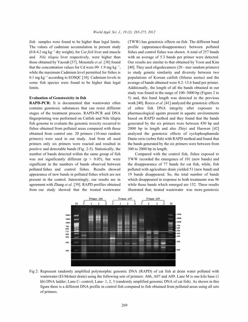
Evaluation of Genotoxicity in fish 5) and, this band length was detected in the previousRAPD-PCR: It is documented that wastewater often work [40]. Rocco et al. [41] analyzed the genotoxic effectscontains genotoxic substances that can resist different of zebra fish DNA integrity after exposure tostages of the treatment process. RAPD-PCR and DNA pharmacological agents present in aquatic environmentsfingerprinting was performed on Catfish and Nile tilapia based on RAPD method and they found that the bandsfish genome to evaluate the genomic toxicity occurred to generated by the six primers were between 450 bp andfishes obtained from polluted areas compared with those 2000 bp in length and also Zhiyi and Haowen [42]obtained from control one. 20 primers (10-mer random analyzed the genotoxic effects of cyclophosphamideprimers) were used in our study. And from all used Danio rerio (zebra fish) with RAPD method and found thatprimers only six primers were reacted and resulted in the bands generated by the six primers were between frompositive and detectable bands (Fig. 2-5). Statistically, the 300 to 2000 bp in length.number of bands detected within the same group of fish Compared with the control fish, fishes exposed towas not significantly different (p > 0.05), but were TWW recorded the emergence of 101 (new bands) andsignificant in the numbers of bands observed between the disappearance of 77 bands for cat fish, while, fishpolluted fishes and control fishes. Results showed polluted with agriculture drain yielded 51 (new band) andappearance of new bands in polluted fishes which are not 19 bands disappeared. So, the total number of bandspresent in the control. Interestingly, our results are in which disappeared in response to both treatments was 96agreement with Zhang et al. [39]. RAPD profiles obtained while those bands which emerged are 152. These resultsfrom our study showed that the treated wastewater illustrated that; treated wastewater was more genotoxic
study was found in the range of 100–3000 bp (Figure 2 to
Fig 2: Represent randomly amplified polymorphic genomic DNA (RAPD) of cat fish at drain water polluted withwastewater (El-Moheet drain) using the following sets of primers: A06, A07 and A09. Lane M is one kilo base (1kb) DNA ladder; Lane C- control; Lane- 1, 2, 3 (randomly amplified genomic DNA of cat fish). As shown in thisfigure there is a different DNA profile in control fish compared to fish obtained from polluted areas using all setsof primers.
World Appl. Sci. J., 19 (2): 265-275, 2012
270
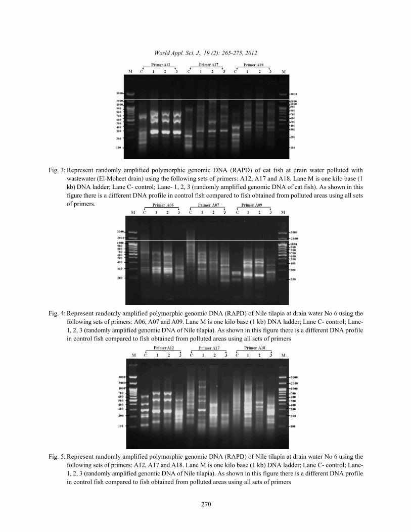
Fig. 3: Represent randomly amplified polymorphic genomic DNA (RAPD) of cat fish at drain water polluted withwastewater (El-Moheet drain) using the following sets of primers: A12, A17 and A18. Lane M is one kilo base (1kb) DNA ladder; Lane C- control; Lane- 1, 2, 3 (randomly amplified genomic DNA of cat fish). As shown in thisfigure there is a different DNA profile in control fish compared to fish obtained from polluted areas using all setsof primers.
Fig. 4: Represent randomly amplified polymorphic genomic DNA (RAPD) of Nile tilapia at drain water No 6 using thefollowing sets of primers: A06, A07 and A09. Lane M is one kilo base (1 kb) DNA ladder; Lane C- control; Lane-1, 2, 3 (randomly amplified genomic DNA of Nile tilapia). As shown in this figure there is a different DNA profilein control fish compared to fish obtained from polluted areas using all sets of primers
Fig. 5: Represent randomly amplified polymorphic genomic DNA (RAPD) of Nile tilapia at drain water No 6 using thefollowing sets of primers: A12, A17 and A18. Lane M is one kilo base (1 kb) DNA ladder; Lane C- control; Lane-1, 2, 3 (randomly amplified genomic DNA of Nile tilapia). As shown in this figure there is a different DNA profilein control fish compared to fish obtained from polluted areas using all sets of primers
World Appl. Sci. J., 19 (2): 265-275, 2012
271
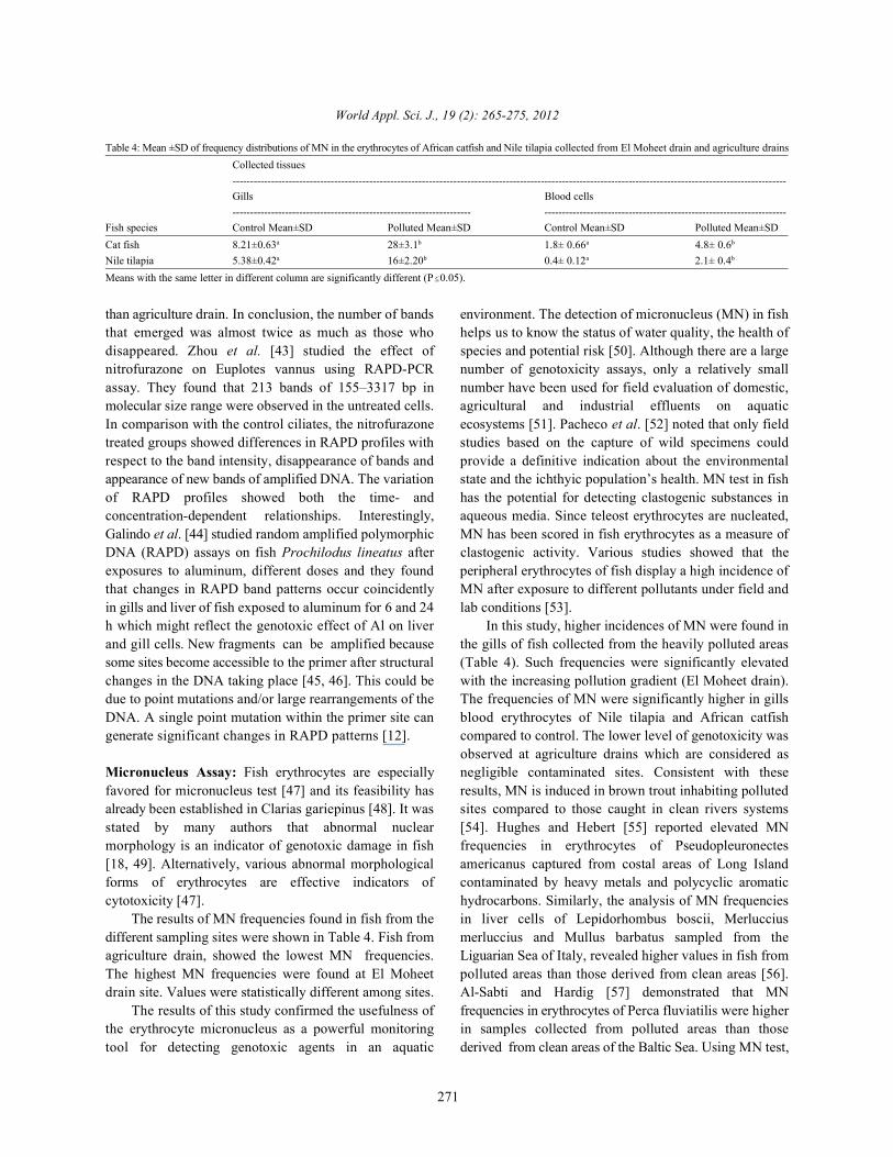
Table 4: Mean ±SD of frequency distributions of MN in the erythrocytes of African catfish and Nile tilapia collected from El Moheet drain and agriculture drainsCollected tissues--------------------------------------------------------------------------------------------------------------------------------------------------------------Gills Blood cells-------------------------------------------------------------------- ---------------------------------------------------------------------
Fish species Control Mean±SD Polluted Mean±SD Control Mean±SD Polluted Mean±SDCat fish 8.21±0.63 28±3.1 1.8± 0.66 4.8± 0.6a b a b
Nile tilapia 5.38±0.42 16±2.20 0.4± 0.12 2.1± 0.4a b a b
Means with the same letter in different column are significantly different (P 0.05).
than agriculture drain. In conclusion, the number of bands environment. The detection of micronucleus (MN) in fishthat emerged was almost twice as much as those who helps us to know the status of water quality, the health ofdisappeared. Zhou et al. [43] studied the effect of species and potential risk [50]. Although there are a largenitrofurazone on Euplotes vannus using RAPD-PCR number of genotoxicity assays, only a relatively smallassay. They found that 213 bands of 155–3317 bp in number have been used for field evaluation of domestic,molecular size range were observed in the untreated cells. agricultural and industrial effluents on aquaticIn comparison with the control ciliates, the nitrofurazone ecosystems [51]. Pacheco et al. [52] noted that only fieldtreated groups showed differences in RAPD profiles with studies based on the capture of wild specimens couldrespect to the band intensity, disappearance of bands and provide a definitive indication about the environmentalappearance of new bands of amplified DNA. The variation state and the ichthyic population’s health. MN test in fishof RAPD profiles showed both the time- and has the potential for detecting clastogenic substances inconcentration-dependent relationships. Interestingly, aqueous media. Since teleost erythrocytes are nucleated,Galindo et al. [44] studied random amplified polymorphic MN has been scored in fish erythrocytes as a measure ofDNA (RAPD) assays on fish Prochilodus lineatus after clastogenic activity. Various studies showed that theexposures to aluminum, different doses and they found peripheral erythrocytes of fish display a high incidence ofthat changes in RAPD band patterns occur coincidently MN after exposure to different pollutants under field andin gills and liver of fish exposed to aluminum for 6 and 24 lab conditions [53].h which might reflect the genotoxic effect of Al on liver In this study, higher incidences of MN were found inand gill cells. New fragments can be amplified because the gills of fish collected from the heavily polluted areassome sites become accessible to the primer after structural (Table 4). Such frequencies were significantly elevatedchanges in the DNA taking place [45, 46]. This could be with the increasing pollution gradient (El Moheet drain).due to point mutations and/or large rearrangements of the The frequencies of MN were significantly higher in gillsDNA. A single point mutation within the primer site can blood erythrocytes of Nile tilapia and African catfishgenerate significant changes in RAPD patterns [12]. compared to control. The lower level of genotoxicity was
Micronucleus Assay: Fish erythrocytes are especially negligible contaminated sites. Consistent with thesefavored for micronucleus test [47] and its feasibility has results, MN is induced in brown trout inhabiting pollutedalready been established in Clarias gariepinus [48]. It was sites compared to those caught in clean rivers systemsstated by many authors that abnormal nuclear [54]. Hughes and Hebert [55] reported elevated MNmorphology is an indicator of genotoxic damage in fish frequencies in erythrocytes of Pseudopleuronectes[18, 49]. Alternatively, various abnormal morphological americanus captured from costal areas of Long Islandforms of erythrocytes are effective indicators of contaminated by heavy metals and polycyclic aromaticcytotoxicity [47]. hydrocarbons. Similarly, the analysis of MN frequencies
The results of MN frequencies found in fish from the in liver cells of Lepidorhombus boscii, Merlucciusdifferent sampling sites were shown in Table 4. Fish from merluccius and Mullus barbatus sampled from theagriculture drain, showed the lowest MN frequencies. Liguarian Sea of Italy, revealed higher values in fish fromThe highest MN frequencies were found at El Moheet polluted areas than those derived from clean areas [56].drain site. Values were statistically different among sites. Al-Sabti and Hardig [57] demonstrated that MN
The results of this study confirmed the usefulness of frequencies in erythrocytes of Perca fluviatilis were higherthe erythrocyte micronucleus as a powerful monitoring in samples collected from polluted areas than thosetool for detecting genotoxic agents in an aquatic derived from clean areas of the Baltic Sea. Using MN test,
observed at agriculture drains which are considered as
World Appl. Sci. J., 19 (2): 265-275, 2012
272
Wirzinger et al. [58] detected a more pronounced relation 5. Begüm, A., M.D.N. Amin, S. Kaneco and K. Ohta,between the amount of sewage and level of genotoxicdamage in field-collected three-spined sticklebacks.Bagdonas et al. [59] detected higher frequencies of MNin different fish species collected from the polluted sitesof Nemunas River, Lithuania. In finally Ali et al. [18] andOsman et al. [60] suggested that the micronucleus test infish erythrocytes as a sensitive monitor for aquaticpollution. A study carried out by Ergene et al. [61]suggested that the genotoxic effects observed inerythrocytes of fishes exposed to the Goksu Delta(Turkey) are generally correlated with heavy metal contentof water samples. Therefore, we can suggest that theinduction of cytogenetic damage can be due, at least forthis analysis, to the presence of heavy metals in therefinery effluent. The results of this study confirm theusefulness of the erythrocyte MN and RAPD-PCR aspowerful monitoring tools for detecting genotoxic agentsin water environment.
It can be concluded that Abu rawash wastewatertreatment plant effluent and agriculture drain aregenotoxic. Wastewater effluent is much more genotoxicthan agriculture drain. African catfish was more sensitiveto genotoxic pollutants than Nile tilapia which can beattributed to their feeding peculiarities. Metal levels inwater were in the permissible limit according to theEgyptian low.
REFERENCES
1. MacFarlane, G.B. and M.D. Burchettt, 2000. Cellulardistribution of Cu, Pb and Zn in the Grey MangroveAvicemnia marina (Forsk.). Vierh Aquatic Botanic,68: 45-59.
2. Censi, P., S.E. Spoto, F. Saiano, M. Sprovieri,S. Mazzola, G. Nardone, S.I.D. Geronimo, R. Punturoand D. Ottonello, 2006. Heavy metals in coastal watersystems. A case study from the northwestern Gulf ofThailand. Chemosphere, 64: 1167-1176. Camusso M.,L. Viganoand R. Baitstrini, 1995. Bioaccumulation oftrace metals in rainbow trout. Ecotox. Environ. Safe.,31: 133-141.
3. Namminga H. N. and J. Wilhm, 1976. Effects of highdischarge and an oil refinery cleanup operation bonheavy metals in water and sediments in SkeletonCreek. Proceedings of the Oklahoma Academy ofScience, 56: 133-138.
4. Rashed, M.N., 2001. Monitoring of environmentalheavy metals in fish from Nasser lake. Environ. Int.,27: 27-33.
2005. Selected elemental composition of the muscletissue of three species of fish, Tilapia nilotica,Cirrhina mrigala and Clarius batrachus, from the freshwater Dhanmondi Lake in Bangladesh. FoodChemistry, 93: 439-443.
6. Fernandes, C., A. Fontaínhas-Fernandes, D. Cabraland M.A. Salgado, 2008. Heavy metals in water,sediment and tissues of Liza saliens from Esmoriz-Paramos lagoon, Portugal. Environ. Monit. Assess.,136: 267-275.
7. El-Naggar, A.M., S.A. Mahmoud and S.I. Tayel, 2009.Bioaccumulation of Some Heavy Metals andHistopathological Alterations in Liver ofOreochromis niloticus in Relation to Water Quality atDifferent Localities along the River Nile, Egypt.World J. Fish Marine Sci., 1: 105-114.
8. Bahnasawy, M., A. Khidr and N. Dheina,2009. Seasonal Variations of Heavy MetalsConcentrations in Mullet, Mugil Cephalus andLiza Ramada (Mugilidae) from Lake Manzala,Egypt. Journal of Applied Sciences Research,5(7): 845-852.
9. EOSQC (Egyptian Organization for Standardizationand Quality Control), 1993. Maximum level for heavymetal contamination in food, E.S. No. 2360. Makereferences like this style.
10. Elnimr, T., 2011. Evaluation of some heavy metals inPangasius hypothalmus and Tilapia nilotica and therole of acetic acid in lowering their levels.International Journal of Fisheries and Aquaculture,3(8): 151-157.
11. Williams, J., A.R. Kubelik, K.J. Livak, J.A. Rafalskiand S.V. Tinger, 1990. DNA polymorphisms amplifiedby arbitrary primers are useful as genetic markers.Nucleic Acid Res., 18: 6531-6535.
12. Walsh, J. and M. McClelland, 1990. Genomicfingerprinting using arbitrary primed PCR and a matrixof pairwise combination of primers. Nucleic AcidsResearch, 18: 7213-18.
13. Rong, Z. and H. Yin, 2004: A method for genotoxicitydetection using random amplified polymorphismDNA with Danio rerio. Ecotoxicology andEnvironmental Safety, 58: 96-103.
14. Le, T.X., Y. Munekage and S. Kato, 2005.Antibiotic resistance in bacteria from shrimpfarming in mangrove areas. Sci. Total Environ.,349: 95-105.
World Appl. Sci. J., 19 (2): 265-275, 2012
273
15. Atienzar, A.F., B. Cordi, M.E. Donkin, A.J. Evenden, 25. Becerril, C., M. Ferrero, F. Sanz and A. Castano, 1999.A.N. Jha and M.H. Depledge, 2000. Comparison of Detection of mitomycin C-induced genetic damage inultraviolet- induced genotoxicity detected by random fish cells by use of RAPD. Mutagenesis, 14: 449-456.amplified polymorphic DNA with chlorophyll 26. Ferrero, M., A. Castano, A. Gonzalez, F. Sanz andfluorescence and growth in a marine macroalgae, C. Becerril, 1998. Characterization of RTG-2 fish cellPalmaria palmate. Aquatic Toxicology, 50(1-2): 1-12. line by random amplified polymorphic DNA.
16. Castano, A. and C. Becerril, 2004. In vitro assessment Ecotoxicology and Environmental Safety,of DNA damage after short- and long-term exposure 40(1-2): 56-64.to benzo(a)pyrene using RAPD and the RTG-2 fish 27. Plotsky, Y., M.G. Kaiser and S.J. Lamont, 1995.cell line, Mutat. Res., 552: 141-151. Genetic characterization of highly inbred chicken
17. Atienzar, F.A. and A.N. Jha, 2006. The random lines by two DNA methods: DNA fingerprinting andamplified polymorphic DNA (RAPD) assay and polymerase chain reaction using arbitrary primers.related techniques alied to genotoxicity and Anim. Genet., 26: 163-170.carcinogenesis studies: A critical review. Mutation 28. De Flora, S., D. Vigano, D. D’Agostini, A. Camoirano,Research, 613: 76-102. C. Bennicelli and A. Arillo, 1993. Multiple
18. Ali F. Kh., A.M. El-Shehawi and M.A. Seehy, 2008. genotoxicity biomarkers in fish exposed in situ toMicronucleus test in fish genome: A sensitive polluted river water. Mutat. Res., 319: 162-177.monitor for aquatic pollution. African Journal of 29. Egyptian Law (48/1982). The Implementer RegulationsBiotechnology, 7(5): 606-612. for law 48/1982 regarding the protection of the River
19. Karami, A., A.Christianus, Z. Ishak, S.C. Courtenay, Nile and water ways from pollution. Map. PeriodicalM.A. Syed, Noor, M. Azlina and H. Noorshinah, Bull., 3-4: 12-35.2010. Effect of triploidization on juvenile African 30. El-Araby, M.M., 2006. Heavy metals in main drains atcatfish (Clarias gariepinus). Aquacult. Int., great Cairo and correlation with polyamines,18: 851-858. glutathione and lipid peroxidation in
20. Osman, A.G., I.A.A. Mekkawy, J. Verreth and hyperaccumulating plants. Act botanica Hungarica,F. Kirschbaum, 2007. Effects of lead nitrate on the 48(3-4): 291-309.activity of metabolic enzymes during early 31. Authman, M.M.N., 2008. Oreochromis niloticus as adevelopmental stages of the African catfish, Clarias Biomonitor of Heavy Metal Pollution with Emphasisgariepinus (Burchell, 1822). Fish Physiol. Biochem., on Potential Risk and Relation to Some Biological33: 1-13. Aspects. Global Veterinaria, 2(3): 104-109.
21. Mekkawy, I.A.A., U.M. Mahmoud, A.G. Osman and 32. El Bouraie, M.M., A.A. El Barbary, M.M. Yehia andA.H. Sayed, 2010. Effects of ultraviolet A on the E.A. Motawea, 2010. Heavy metal concentrations inactivity of two metabolic enzymes, DNA damage and surface river water and bed sediments at Nile Deltalipid peroxidation during early developmental stages in Egypt. Suoseura Finnish Peatland Society,of the African catfish, Clarias gariepinus (Burchell, 61(1): 1-12.1822). Fish Physiol. Biochem., 36: 605-626. 33. Hamed, M.A., 1998. Distribution of trace metals in the
22. APHA, 2005. Standard Methods for the Examination River Nile ecosystem Damietta Branch betweenof Water and Wastewater, 21 ed., American Public Mansoura city and Damietta Province. J. Egypt. Ger.st
Health Association, Washington, DC. Soc. Zool. (A) Comparative physiology, 27: 399-415.23. Alam, M.G.M., A. Tanaka, G. Allinson, 34. Ibrahim, S.A. and S.A. Mahmoud, 2005. Effect of
L.J.B. Laurenson, F. Stagnitti and E. Snow, 2002. A heavy metals accumulation on enzyme activity andcomparison of trace element concentrations in histology in liver of some Nilefish in Egypt. J. Aquatcultured and wild carp (Cyprinus carpio) of lake Biol. and Fish, 9: 203-219.Kasumigaura, Japan. Ecotoxicology and 35. Aucoin, J., R. Blanchard, C. Billiot, C. Partridge,Environmental Safety, 53: 348-354. D. Schultz, K. Mandhare, M.J. Beck and J.N. Beck,
24. Sharma, D., K.B.C. Appa Rao and S.M. Totey, 2000. 1999. Trace metals in fish and sediments fromMeasurement of within and between population lake Boeuf, Southeastern Louisiana. Microchem. J.,genetic variability in quails. Br. Poult. Sci., pp: 41. 62: 299-307.
World Appl. Sci. J., 19 (2): 265-275, 2012
274
36. Yacoub, A.M., 2007. Study on some heavy metals 47. Bahari, I.B., F.M. Noor and N.M. Daud, 1994.accumulated in some organs of three River Nile fishes Micronucleated erythrocytes as an assay to assessfrom Cairo and Kalubia governorates. African J. Biol. actions by physical and chemical genotoxic agents inSci., 3: 9-21. Clarias gariepinus. Mutat. Res., 313: 1-5.
37. Moustafa, M.M., M. Abd El Aziz, A.Z. Abd El 48. Talapatra, S.N. and S.K. Banerjee, 2007. Detection ofMeguid and A.M. Hussien, 2011. Evaluation of some micronucleus and abnormal nucleus in erythrocytesheavy metals pollution on Oreochromis niloticus in from the gill and kidney of Labeo bata cultivatedRiver Nile and Ismailia Canal. Researcher, 3(2): 75-79. in sewage-fed fish farms. Food Chem. Toxicol.,
38. Zhang, X., Z. Zhang, X. Zhang and B. Wu, 2010. 45: 210-215.Effects of Yangtze River source water on genomic 49. Al-Sabti, K. and C.D. Metcalfe, 1995. Fish micronucleipolymorphisms of male mice detected by RAPD. for assessing genotoxicity in water. Mutat. Res.,Human and experimental Toxicology, 29(2): 113-120. 343: 121-135.
39. Yoon, J.M. and G.W. Kim, 2001. Randomly amplified 50. Claxton, L.D., V.S. Houk and T.J. Hughes, 1998.polymorphic DNA polymerase chain reaction Genotoxicity of industrial wastes and effluents.analysis of two different populations of cultured Mutation Research, 410: 237-43.Korean catfish Silurus sotus. J. Biosci., 26(5): 641-647. 51. Pacheco, M., M. Santos, M. Teles, M. Oliveira,
40. Rocco, L., G. Frenzill, D. Fusco, C. Peluso and J. Rebelo and L. Pombo, 2005. Biotransformation andV. Stingo, 2010. Evaluation of zebrafish DNA genotoxic biomarkers in mullet species (Liza sp.) fromintegrity after exposure to pharmacological agents a contaminated coastal lagoon (Ria de Aveiro,present in aquatic environments. Ecotoxicology and Portugal). Environmental Monitoring andEnvironmental Safety, 73: 1530-1536. Assessment, 107: 133-53.
41. Zhiyi, R. and Y. Haowen, 2004. A method for 52. Osman, A. and A. Harabawy, 2010. Hematotoxic andgenotoxicity detection using random amplified genotoxic potential of ultraviolet-A radiation on thepolymorphism DNA with Danio rerio. Ecotoxicology African catfish Clarias gariepinus (Burchell, 1822).and Environmental Safety, 58: 96-103. Journal of Fisheries International, 5: 44-53.
42. Zhou, L., J. Li, X. Lin and Al-Rasheid, Kh., 2011. Use 53. Rodriguez-CeaA., F. Ayllon and E. Garcia-Vazquez,of RAPD to detect DNA damage induced by 2003. Micronucleus test in freshwater fish species:nitrofurazone in marine ciliate, Euplotes vannus An evaluation of its sensitivity for application in field(Protozoa, Ciliophora). Aquatic Toxicology, surveys. Ecotoxicology and Environmental Safety,103: 225-232. 56: 442-8.
43. Galindo, B., G. Troilo, I. Cólus, C. Martinez and 54. Hughes, J.B. and A.T. Hebert, 1991. ErythrocyteS. Sofia, 2010. Genotoxic Effects of Aluminum on the micronuclei in winter flounder (PseudopleuronectesNeotropical Fish Prochilodus lineatus. Water Air Soil americanus): Results of field surveys during 1980-Pollut., 212: 419-428. 1988 from Virginia to Nova Scotia and in Long Island
44. Pietrasanata, L.I., B.L. Smith and M.C. MacLeod, Sound. Archives of Environment Contamination and2000. A novel approach for analyzing the structure of Toxicology, 20: 474-9.DNA modified by benzo[a]pyrine diol epoxide at 55. Pietripiana, D., M. Modena, P. Guidetti, C. Falugisingle-molecule resolution. Chem. Res. Toxicol., and M. Vacchi, 2002. Evaluating the genotoxic13: 351-355. damage and hepatic tissue alterations in demersal
45. Enan, M.R., 2006. Application of random amplified fish species: A case study in the Liguarian Seapolymorphic DNA (RAPD) to detect the genotoxic (NW Mediterranean). Marine Pollution Bulletin,effect of heavy metals. Biotechnol. Appl. Biochem., 44: 238-43.43: 147-154. 56. Al-Sabti, K. and J. Hardig, 1990. Micronucleus test in
46. Bushra, A., M. Abul farah, M.A. Niamat and fish for monitoring the genotoxic effects of theA. Waseem, 2002. Induction of micronuclei and industrial waste products in the Baltic Sea, Sweden.erythrocyte alterations in the catfish Clarias Comparative Biochemistry and Physiology Partbatrachus by 2, 4-dichlorophenoxyacetic acid and C, Comparative Pharmacology and Toxicology,butachlor. Mutat. Res., 518: 135-144. 97: 179-82.
World Appl. Sci. J., 19 (2): 265-275, 2012
275
57. Wirzinger, G., L. Weltje, J. Gercken and H. Sordyl, 60. Ergene, S., T. Çava , A. Çelik, N. Köleli, F. Kaya and2007. Genotoxic damage in field-collected three- A. Karahan, 2007. Monitoring of nuclearspined sticklebacks (Gasterosteus aculeatus L.): A abnormalities in peripheral erythrocytes of three fishsuitable biomonitoring tool. Mutation Research, species from the Goksu Delta (Turkey): genotoxic628: 19-30. damage in relation to water pollution. Ecotoxicology,
58. Bagdonas, E., E. Dukelskis and J. Lazutka, 2003. 16(4): 385-391.Frequency of micronuclealted erythrocytes in wildfish from natural freshwater body. Ekologija, 1: 67-71.
59. Osman, A.G.M., A.M. Abd El Reheem,M.A. Moustafa, U.M. Mahmoud, K.Y. Abuel-Fadland W. Kloas, 2011. In situ evaluation of thegenotoxic potential of the river Nile: I. Micronucleusand nuclear lesion tests of erythrocytes ofOreochromis niloticus niloticus (Linnaeus, 1758) andClarias gariepinus (Burchell, 1822). Toxicological &Environmental Chemistry, 93(5): 1002-1017.