Appraisal of Sarajevo Air Pollution Control Project Yugoslavia
Upload
khangminh22Category
view
0download
0
E K I Z A N D D E M I R A S L A N E M B A L M I N G S O L U T I O N S W I T H H O N E Y 229
1Research and Application Hospital, Suleyman Demirel University, Isparta, Turkey2Department of Anatomy, Faculty of Veterinary Medicine, Burdur Mehmet Akif Ersoy University, Burdur, Turkey
*Corresponding author:Assoc. Prof. Dr. Yasin DemiraslanDepartment of Anatomy, Faculty of Veterinary Medicine, Burdur Mehmet Akif Ersoy University Burdur, TurkeyAdress: Degirmenler Neighborhood, Istiklal Campus, Faculty of Veterinary Medicine, Burdur Mehmet Akif Ersoy University, 15100, Burdur, TurkeyPhone: +90 248 213 2162ORCID: 0000-0003-3612-6142Email: [email protected]
Original Submission: 20 December 2020
Revised Submission: 16 February 2021
Accepted: 02 March 2021
How to cite this article:Ekiz R, Demiraslan Y. 2021. Investigation of cadaver preservative properties of solutions obtained with a mixture of honey and ethyl alcohol in different proportions: a pilot study on sheep heart. Veterinaria, 70(2), 229-43.
ABSTRACT
The aim of this study is to evaluate the cadaver embalming effects of the honey-based solutions used as an alternative to formaldehyde which is extremely hazardous for human health. In the study, 42 sheep hearts were used. Tissue and swap samples were taken from different honey solution groups on the 3rd, 75th, 150th, and 225th day to perform histological, microbiological, texture and color analyses. In the macroscopic examination, there was a more significant hardness in the hearts kept in the solution I in A and B groups than the solutions II and III. General fixation was found to be positive in the histological analysis. However, the histological structure became deformed later. The microbiological analysis showed no bacterial or mycotic growth. Moreover, according to the comparative results of the hardness scale applied to the groups, the heart tissues which were kept in AI, BII, and BIII solutions on the 3rd day, in AII and BIII solutions on the 150th
day, in BIII solutions on the 225th day, in AIII on all days were statistically found to be similar to the fresh heart tissue. Based on the results of the color analysis, the heart tissues of the experimental groups were not statistically similar to the fresh heart tissue in within-group and between-group comparisons of L value. Consequently, it is thought that the positive effect of honey solutions on the sheep heart tissue should be evaluated in organs with different parenchyma tissue and properties and whole cadaver.
Keywords: Embalming, formaldehyde, heart, honey
INVESTIGATION OF CADAVER PRESERVATIVE PROPERTIES OF SOLUTIONS OBTAINED WITH A MIXTURE OF HONEY AND ETHYL ALCOHOL IN DIFFERENT PROPORTIONS: A PILOT STUDY ON SHEEP HEARTRuhsar Ekiz1, Yasin Demiraslan2*
RESEARCH ARTICLE
doi.org/10.51607/22331360.2021.70.2.229

V E T E R I N A R I A VO L . 7 0 • I S S U E 2 • 2 0 2 1230
Table 1 The percentage of solution contents by the groups
Groups HoneyEthyl alcohol
(96%)a
Citric acidb Glycerinc Distilled water Lavender essence
Ist 50 35 7 7 - 1IInd 40 27 6 6 20 1IIIrd 30 20 5 4 40 1
a; Limko®, b; Tekkim®, c; Tekkim®
INTRODUCTION
Cadaver is a dead human or animal body that is used by veterinary medicine, nursing and medical students for observation and research. It is stated in the literature (Erbay et al. 2015) that the use of cadaver is one of the essential elements for education and is of prime importance for students to learn individually, touch cadaver and perform manipulative procedures. Modern cadaver preservation technique emerged after a chemist, August Wilhelm von Hofmann, discovered formaldehyde in 1868 (Brenner, 2014). In this sense, the mixtures containing formaldehyde, phenol, thymol, glycerin, ethyl alcohol, and distilled water at different rates are among the most common cadaver fixation solutions used today (Kalanjati et al. 2012; Whitehead and Savoia, 2008). Recently, some researchers (Patil et al. 2015; Piatek-Koziej et al. 2019) have examined the fixative effects of honey or sweeteners. However, upon the literature review, any study presenting the embalming effect of honey-based solutions from all aspects (color, tissue, odor, microbial situation etc.) was not found. The aim of the study was to assess the cadaver embalming effect of honey solutions at different rates on the sheep heart.
MATERIALS AND METHODS
For the study, an approval was obtained from Burdur Mehmet Akif Ersoy University Animal Experiments Local Ethics Committee (18.10.2017-332). In the study, the hearts of 42 healthy sheep older than 6 months were used. These hearts were obtained from a meat facility. After the sheep were slaughtered, the hearts were perfused with 0.9% saline to prevent autolysis. Following this procedure, the hearts were brought to the laboratory. Tissue samples were taken from 6 of the hearts to determine fresh cadaver characteristics. The other hearts were perfused with 10% benzalkonium chloride (Dermosept, Zefiran), and they were primarily divided into 3 groups with 12 samples. Then the groups were divided into groups with 6 samples according to + 4ºC (group A) and room temperature (group B) storage conditions. Thus, a total of 6 experimental groups were obtained. The solutions to be used were prepared as in Table 1. On the other hand, the flower honey (Aksu Vital®, Turkey) to be used in the solutions was analyzed in terms of its basic contents (Table 2). Based on the related group, the hearts were perfused using the prepared solutions, and they were placed into the seven-liter containers containing the embalming solutions.
For histological, microbiological, texture, and color analyses, the samples were taken from the experimental groups on the days 3, 75, 150, and 225, when they were subjected to the solution.

E K I Z A N D D E M I R A S L A N E M B A L M I N G S O L U T I O N S W I T H H O N E Y 231
Histological Analysis
Atrial and ventricular tissue samples of 1 cm3
were taken from the hearts for the control and experimental groups. The tissue samples obtained from the control group were fixed in 10% buffered formaldehyde for 24 hours. Then, they were rinsed with tap water for 24 hours. Then, both fresh and experimental tissues were kept in graduated alcohol (70%, 80%, 96%) for 1 hour and in absolute alcohol for 2 hours. They were blocked with paraplast after being passed through methyl benzoate and benzol series. 3 5-µm thick sections were taken from each paraffin block (Leica RM 2155, Germany). Hematoxylin-eosin staining was performed to the tissue sections in order to assess the histological quality. These histological sections were assessed under the light microscope (BX51, Olympus, Tokyo, Japan), and the photos were taken in the requested regions using a digital camera integrated into the microscope (DP74, Olympus, Tokyo, Japan). The tissue samples obtained from each group were examined in terms of the quality of the fixations. The histological quality was assessed semi-quantitatively in terms of nuclear morphology, cytoplasm, and staining quality. Each assessment criteria were scored as +; Bad, ++; Medium, +++; Good, and ++++; Excellent.
Accordingly, for nuclear morphology, determined were as follows; “++++: Apparent nuclear chromatin, no pyknosis +++: Unclear nuclear chromatin in some cells, ++: Pyknosis in the nuclei of some cells, and +: Nuclei are fully pyknotic”; for cytoplasmic determination, “++++: Certain cell
lines, no cytoplasmic shrinkage, striation in striated muscles, +++: Partial cytoplasmic shrinkage, striation loss in striated muscles,++: Cellular swelling, ambiguous cell lines, +: Ambiguous cell lines, autolytic changes in tissues”.
Microbiological Analysis
The swap samples taken based on the experimental groups and days were inoculated in 5% sheep blood agar (Oxoid), MacConkey agar (Oxoid) and Sabouraud Dextrose agar (Oxoid) supplemented with chloramphenicol (0.05 mg/ml). While the blood agar and MacConkey agar petri dishes were incubated at 37°C under aerobic conditions for 24-48 hours, Sabouraud Dextrose agar petri dishes were incubated at 25°C under aerobic conditions for 7 days. At the end of incubation, the post-gram staining microscopic morphologies of the bacterial colonies growing in blood and MacConkey agars were examined, and the isolated bacteria were identified with the standard bacteriological methods revealing their catalase, coagulase and hemolysis properties. Sabouraud Dextrose agar was checked during incubation as from the 48th
hour in terms of growth of fungal colony (Quinn et al. 2004).
Texture Analysis
For texture analysis, the tissue samples of 1 cm3
were taken from the wall of the left ventricle. Texture analysis was performed on these samples based on the groups using TexturePro CT software in CT3 Texture Analyzer Brookfield device.
Table 2 The basic properties of the honey used in the study
Dry matter (%) Acidity (meq acid kg / honey) pH Glucose (%) Fructose (%)
Value 89.6 40 5.3 29.7 39.21
V E T E R I N A R I A VO L . 7 0 • I S S U E 2 • 2 0 2 1232
Color Analysis
The color analysis was performed from the wall of the right ventricle using a Konica Minolta device. In this device, L (blackness-whiteness, lightness value), a (redness-greenness) and b (yellowness-blueness) values were determined quantitatively by performing 3 parallel measurements in each sample.
Odor Analysis
The odor analysis was applied to voluntary students (n:50) from the faculty of veterinary medicine with the survey (Ari and Cinaroglu, 2011), and the frequency values of these assessments were calculated. The odor survey consisted of a scale of 0–10, ranging from no odor (0–1) to little odor (2–4), heavy odor (5–7) and disgusting odor (8–10) (Turan et al. 2017).
Statistical Analysis
The presence of any within-group and between-group difference for the texture and color data of all the hearts was determined using repeated-
measurement analysis of variance (ANOVA) and post-hoc Tukey tests in MINITAP (Statistical Package Program, 20.0 version, Minitab Inc., England) software.
RESULTS
Macroanatomical
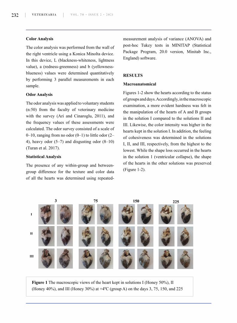
Figures 1-2 show the hearts according to the status of groups and days. Accordingly, in the macroscopic examination, a more evident hardness was felt in the manipulation of the hearts of A and B groups in the solution I compared to the solutions II and III. Likewise, the color intensity was higher in the hearts kept in the solution I. In addition, the feeling of cohesiveness was determined in the solutions I, II, and III, respectively, from the highest to the lowest. While the shape loss occurred in the hearts in the solution 1 (ventricular collapse), the shape of the hearts in the other solutions was preserved (Figure 1-2).
Figure 1 The macroscopic views of the heart kept in solutions I (Honey 50%), II (Honey 40%), and III (Honey 30%) at +4ºC (group A) on the days 3, 75, 150, and 225

E K I Z A N D D E M I R A S L A N E M B A L M I N G S O L U T I O N S W I T H H O N E Y 233
Histological
The fixation-related findings observed in tissues of all groups in the histological examinations were summarized in a table (Table 3). Accordingly, the nuclear and cytoplasmic morphology was good
(+++) until the day 75. But, after 75 days, it was medium (++) for all solutions. The staining quality was in all days and groups normal as in the control group.
Table 3 The semi-quantitative data of the histomorphological quality of the hearts kept in solutions I, II, and III at +4 ºC and room temperature (+22oC) on the days
Sol. Nuclear morphology Cytoplasm Staining quality
Days of preserv.
Temp. (ºC) C ++++ ++++ Normal
3rd
+4
I +++ +++ Normal
II +++ +++ Normal
III +++ +++ Normal
room
I +++ +++ Normal
II +++ +++ Normal
III +++ +++ Normal
Figure 2 The macroscopic views of the heart kept in solutions I (Honey 50%), II (Honey 40%), and III (Honey 30%) at room temperature (group B) on the days 3, 75, 150, and 225

V E T E R I N A R I A VO L . 7 0 • I S S U E 2 • 2 0 2 1234
Sol. Nuclear morphology Cytoplasm Staining quality
Days of preserv.
Temp. (ºC) C ++++ ++++ Normal
75th
+4
I +++ +++ Normal
II +++ +++ Normal
III +++ +++ Normal
room
I ++ ++ Normal
II ++ ++ Normal
III ++ ++ Normal
150th
+4
I ++ ++ Normal
II ++ ++ Normal
III ++ ++ Normal
room
I ++ ++ Normal
II ++ ++ Normal
III ++ ++ Normal
225th
+4
I ++ ++ Normal
II ++ ++ Normal
III ++ ++ Normal
room
I ++ ++ Normal
II ++ ++ Normal
III ++ ++ NormalC; control group (10% formaldehyde)
Figures 3-4 show the histological appearance of the hearts fixed in different honey solutions.

E K I Z A N D D E M I R A S L A N E M B A L M I N G S O L U T I O N S W I T H H O N E Y 235
Microbiological
In the study, the bacterial or mycotic growth did not generally occur. Besides, Bacillus sp. and Staphylococcus aureus grew initially in the samples taken from the hearts kept in the solutions I and II
in the B group for 3 days. However, no growth was observed in the microbiological inoculation of the parallel swap sample taken simultaneously. This was evaluated as the contamination of the swap sample for which growth was determined.
Figure 3 Histomorphological views of the heart tissue kept in solutions I, II, and IIIrd at +4 ºC (group A) and room temperature (group B) on the days 3 and 75, HE. Asterisk: loss of striation in myofibrils, black arrow: uncertain cell boundaries, blue arrow: pyknosis in nuclei, a. atrial tissue, v. ventricular tissue, Bar: 20µm
Figure 4 Histomorphological views of the heart tissue kept in solutions I, II, and III at +4 ºC (group A) and room temperature (group B) on the days 150 and 225. HE. Asterisk: loss of striation in myofibrils, black arrow: uncertain cell boundaries, blue arrow: pyknosis in nuclei. a. atrial tissue, v. ventricular tissue, Bar: 20µm

V E T E R I N A R I A VO L . 7 0 • I S S U E 2 • 2 0 2 1236
Texture
The mean and standard deviation values of the hardness degree of the fresh heart tissue were determined to be 1.8 and 0.31, respectively. Table 4 shows the findings related to the hardness degree obtained from experimental groups. Accordingly,
statistically significant results were determined among the groups, days, and solutions (P<0.01). The heart tissues kept in AI, BII, and BIII solutions for the day 3, in AII and BIII solutions for the day 150, in BIII solution for the day 225, and in AIII solution for the all days were statistically similar to the fresh heart tissue.
Table 4 The means and standard deviations (SD) of hardness degree in terms of groups
3rd 75th 150th 225th
Temp (oC) Solutions Mean±SD Mean±SD Mean±SD Mean±SD P
+ 4I 2.75±0.55Cbcd 3.83±017Cb 11.2±1.66Aa 4.96±0.56Bb 0.000II 4.96±0.67Ab 4±0.15Bab 3.43±0.63Bcd 4.75±0.43Ab 0.000III 1.21±0.06BCd 1.56±0.61Bc 0.92±0.05Ce 2.44±0.49Ac 0.000
roomI 9.16±2.65Aa 4.34±0.3Ba 6.35±.06Bb 6.50±0.23ABa 0.002II 2.34±0.3ABcd 1.1±0.2Bd 4.36±2.1Abc 2.79±0.26ABc 0.005III 3.07±0.31Abc 0.5±0.02Ce 1.16±0.08Bde 1.22±0.16Bd 0.000
P 0.000 0.000 0.000 0.000
The values are shown in Newton. The uppercase letters show the similarities of different sampling periods of a solution. The lowercase letters show the similarities of different solution groups for a sampling period.
Color
The mean and standard deviation values of the “L” parameter of the fresh heart tissue were determined as 93.75 and 0.82, respectively. Table 5 shows the values of “L” parameter of experimental groups. The heart tissues of the experimental group were not statistically similar to the fresh heart tissue.
The mean and standard deviation values of the “a” parameter of the fresh heart tissue were found to be 0.01 and 0.00, respectively. Table 5 shows the values of the “a” parameter of experimental groups. The heart tissues kept in AI solution on
the day 150, in AIII, BII, and BIII solutions on the day 3, in BI solution on the day 75 were similar to fresh heart tissue.
The mean and standard deviation values of the “b” parameter of the fresh heart tissue were determined to be 2.6 and 0.04, respectively. Table 5 shows the values of “b” parameter of the experimental groups. The heart tissues kept in AI solution on the days 75, 150, and 225, in AII solution on the days 3 and 150, in AIII solution on the day 150, in BI solution on the days 150 and 225, and in BII solution on the days 75 and 150 were similar to the fresh heart tissue.

E K I Z A N D D E M I R A S L A N E M B A L M I N G S O L U T I O N S W I T H H O N E Y 237
Table 5 The mean and standard deviation (SD) of the color parameters in terms of groups
Days of Preservation
3 75 150 225
Parameters Temp (oC) Soluti-ons Mean±SD Mean±SD Mean±SD Mean±SD P
L
4
I 35.53± 3.53Aa 27.24±2.78Be 26.37±2.68Cd 26.4±2.68Cd 0.000
II 27.19±5.04Abc 44.78±3.86Ba 26.53±2.71Ad 30.86±3.15Ac 0.000
III 33.61±2.28Bab 37.14±3.59Cb 35.61±3.51ABa 38.67±3.67Ab 0.000
room
I 25.84±0.97Ac 23.23±2.27Bf 27.29±2.79Ac 26.85±2.74Ad 0.000
II 30.51±2.15Aabc 29.09±2.98Bd 26.6±2.71Acd 30.17±3.09Ac 0.000
III 34.82±3.09Ba 32.98±3.33Cc 32.08±3.25Bb 53.29±3.87Aa 0.000
P 0.000 0.000 0.000 0.000
A
4
I -0.87±0.23Dbcd 1.03±0.24Bc 0.09±0.02Ce 2.18±0.52Ab 0.000
II -2.04±1.15Bd 1.20±0.17Ac 1.81±0.43Ac 2.31±0.48Ab 0.000
III 0.38±0.64Cab 3.87±0.66Aa 3.56±0.63ABa 3.06±0.5Ba 0.000
room
I -1.35±0.83Ccd 0.26±0.06Bd 1.07±0.25Ad 0.79±0.18ABd 0.000
II -0.33±0.59Babc 2.64±0.58Ab 2.44±0.57Ab 3.20±0.69Aa 0.000
III 0.8±0.53Ba 2.78±0.54Ab 3.6±0.71Aa 1.44±0.15Ac 0.000
P 0.000 0.000 0.000 0.000
B
4
I 5.21±0.29Ac 3.01±0.85Bcd 3.64±1.08Bb 3.45±1.01Bc 0.000
II 4.14±1.75Bc 8.87±1.09Ab 2.80±0.8Bc 8.52±2.62Ab 0.000
III 7.98±1.35BCab 11.62±3.31ABa 4.84±1.61Ca 15.66±4.83Aa 0.000
room
I 4.76±0.53Ac 1.31±0.38Cd 2.07±0.57BCc 3.02±0.86Bc 0.000
II 7.68±1.71ABb 4.66±1.32Bc 4.89±1.5ABa 7.9±2.43Ab 0.013
III 10.21±0.72Aa 7.79±2.21Bb 5.55±1.49Ca 10.26±1.83Ab 0.000
P 0.000 0.000 0.000 0.000
The uppercase letters show the similarities of different sampling periods of a solution. The lowercase letters show the similarities of different solution groups for a sampling period. L: blackness-whiteness, lightness value, a: redness-greenness, b: yellowness-blueness.
Odor
Table 6 shows the results of the odor survey. Accordingly, the percentage values of the odor survey of the hearts kept at +4 ºC were more
positive compared to the hearts kept at room temperature. The same situation was also valid for the solution compared to the other solutions.

V E T E R I N A R I A VO L . 7 0 • I S S U E 2 • 2 0 2 1238
Table 6 The frequencies of odor survey in terms of groups
Temperature (oC)+4 +22
SolutionsDay Odor Level I II III I II III
3rd
No odor 83 67 56 71 62 60A little odor 15 29 27 21 21 15Heavy odor 0 2 15 6 15 15
Disgusting odor 2 2 2 2 2 10
75th
No odor 84 68 48 32 9 23A little odor 14 21 39 55 52 27Heavy odor 2 11 11 11 32 32
Disgusting odor 0 0 2 2 7 18
150th
No odor 75 66 55 76 43 57A little odor 21 29 31 20 44 25Heavy odor 4 5 14 4 13 16
Disgusting odor 0 0 0 0 0 2
225th
No odor 82 59 57 50 56 59A little odor 18 41 32 37 39 36Heavy odor 0 0 11 11 5 5
Disgusting odor 0 0 0 2 0 0
DISCUSSION AND CONCLUSION
In the study, a solution containing honey, ethyl alcohol, glycerin, citric acid, distilled water, and essence of lavender at different rates was used, and the cadaver embalming effect of this solution on the sheep heart was assessed generally with objective analyses. Upon the literature review, no study assessing the cadaver embalming effect of this solution has been encountered. In this respect, the present study is the first trial. In addition, the number of studies comparing the objective findings are very limited. At this point, it is
considered useful to mention the study limitations and some issues before the analysis of the findings. Firstly, the study was conducted on the heart, a muscular organ since it reflects a great majority of body tissue. In addition, the study was designed considering the period of one academic year (32 weeks).
Brenner (2014) describes an “ideal” fixation solution as a bacteriostatic solution that can keep tissues for a long time by preserving their natural structure without drying, hardening, and discoloring them. Also, Brenner (2014) classifies the chemicals used in the preparation

E K I Z A N D D E M I R A S L A N E M B A L M I N G S O L U T I O N S W I T H H O N E Y 239
of cadaver as disinfectants, fixatives, buffers, surfactants, anticoagulants, colorants, carrier substances, softeners, and perfumes. Additionally, antioxidants, which preserve the natural structures of tissues for a longer time and help store them without impairment, are another chemical used for the cadaver fixation (Friker et al., 2007; Janczyk et al., 2011). These solutions should not only prevent drying and fungal and bacterial growth, but also reveal the least harm for environment and the least potential danger in the laboratory (Brenner, 2014). Soviet scientists Levina and Tsarlin (Mladenov, 1967) stated that they could preserve living human tissue in honey solution for about 4-6 months. Mladenov, 1967 reported that kidney and liver tissues kept in honey did not impair for 4 years and they had a fresh tissue-like color. Besides, the author (Mladenov, 1967) indicated that there was no microbiological activity. The researcher emphasized that negative results were obtained in artificial honey in terms of preservative effect. Due to these results, the researcher concluded that honey had a significant preservative effect on animal tissues (Mladenov, 1967). The other studies on the tissue preservation effect of honey have been conducted by Subrahmanyam (1993) and Sharquie and Najim (2001). In these studies, they concluded that honey was a good tissue fixative. Especially in the study conducted by Sharquie and Najim (2001) with 6 Swiss albino mice, 6 rabbits and two human fetuses, they reported that the cadaver- preserving effect lasted for up to 3 years. Ethyl alcohol is used commonly as a solvent and anti-infective substance in the content of cadaver preservation solution (Correia et al., 2014). In addition, ethyl alcohol may be used in fixation, which is the first stage of the histological preparation, instead of formaldehyde (Buesa, 2008). Hammer et al. (2012) stated that ethyl alcohol and glycerin used for fixation, did not have harmful effects, while tissues remained similar to their natural states,
muscles and cavernous organs keeping their elasticity. Citric acid is used as a preservative-antioxidant generally in foods as it forms an antimicrobial effect by increasing ambient acidity (He et al., 2015). For these reasons, in the study, the characteristics of the solution prepared by mixing ethyl alcohol for fixation, honey for antimicrobial effect, glycerin for softening, citric acid for preventing oxidation and sugar crystallization, essence of lavender for forming odor quality and distilled water for providing economy at different rates was investigated in terms of “being ideal”.
Some researchers (Patil et al. 2015; Sabarinath et al. 2014; Ozkan et al. 2012) have suggested the use of honey solutions at different rates for fixative effect on small tissue samples. Besides, Piatek-Koziej et al. (2019) did not confirm honey solutions as a fixative for large tissues. However, based on the histological findings of the study, our honey solutions can be recommended as a fixative for large tissues like the sheep heart.
Cadaver fixation methods should include disinfection of cadaver to reduce the risk of infection-contamination (Davidson and Benjamin, 2006; Trompette and Lemonnier, 2009). When cadavers are used for dissection, they may be a source of infection for students and instructors (Shoja et al., 2013). For this purpose, benzalkonium chloride is a biocide that may be added into solutions to prevent microbial activity. Having antiseptic and fungicide characteristics, benzalkonium chloride may be used as anti-mould (Brenner, 2014; MacDonald and MacGregor, 1997). In the present study, benzalkonium chloride was used in heart perfusion and then as bath following the sampling in the next stages. This antiseptic was not added into the solutions.
Turan et al. (2017) used soap, ethyl alcohol, citric acid, and benzalkonium chloride as fixative solution in whole goat cadaver. Turan et al. (2017)

V E T E R I N A R I A VO L . 7 0 • I S S U E 2 • 2 0 2 1240
reported that the general histological view of quadriceps femoris muscle was similar to fresh cadaver in the first sampling, and the general histological view got increasingly impaired at the end of the study. It was reported that there was no statistically significant difference in the stiffness degree of muscle in terms of the sampling periods. In the present study, no difference was observed in the macro-anatomic view of the sheep hearts in terms of the periods. In the general histological view of the hearts, an increasing impairment was observed. Some statistically different results were observed in the within- or between-group comparison performed for the stiffness degree in the texture analysis (in results section).
As they have antemortem color characteristics, cadaver fluids should enable muscles and organs to keep their color if possible (Coleman and Kogan, 1998). But, cadaver fixation fluids and techniques may cause color changes in tissue and organs (Silva et al., 2007). It has been stated that color analysis devices may be used in order to assess these changes (Ari and Cinaroglu, 2011). In our study, statistically different results were obtained in terms of fresh and experimental groups (in results section).
The embalmed cadavers fixed with formaldehyde have disadvantages such as malodor (Brenner, 2014). In their soapy water-related study, Turan et al. (2017) reported that total percentage of the participants who gave “no odor” or “a little odor” answer ranged from 70% to 80%. In the present study, the essence of lavender was used to eliminate this malodor effect, and survey was performed in order to observe the results. According to the odor survey results, the results of the hearts kept at +4ºC were more positive compared to the hearts kept at room temperature. In addition, the total rate of the “no odor” and “a little odor” was over 90% in all the sampling periods in the solution I.
Mold on the skin and organ surface is a common problem in the cadavers fixed with formaldehyde. If there is contamination in cadavers, one of the most important issues that may threaten health is the presence of bacteria (Mladenov, 1967). Turan et al. (2017) stated that there was no growth other than Bacillus sp. in microbiological analysis. In the present study, swap samples were taken from the solutions based on their temperature groups on the days 3, 75, 150, and 225 in order to determine bacterial, fungal and mycotic growth. It was determined based on the microbiological inoculation results that the bacterial or mycotic growth did not occur generally. But, Bacillus sp. and Staphylococcus aureus grew initially in the samples taken from solutions I and II in group B on the day 3. However, no growth was observed in the microbiological inoculation of the swap samples taken simultaneously. This was evaluated as the contamination of the swap sample for which growth was determined.
Janczyk et al. (2011) stated that a mixture containing brine salt, ethyl alcohol, polyethylene glycol, oil of marjoram and tap water had better results compared to 6% formaldehyde solution. Keeping human cadavers in a saturated salt solution has been tested commonly, especially for surgical education, and its advantages have been emphasized. But, noxious chemicals such as formaldehyde and phenol, even in small amounts, have been used in these studies as well (Hayashi et al. 2014).
In conclusion, the present study was designed to prepare a solution as an alternative to chemicals known to have harmful effects. This study was conducted on the heart, which is a muscular organ, by considering both scientific facts and economic aspect. The data obtained from subjective and objective analyses indicated that studies conducted to form “ideal” fixation solution have progressed

E K I Z A N D D E M I R A S L A N E M B A L M I N G S O L U T I O N S W I T H H O N E Y 241
in the right direction. From this point, it is thought that it would be useful to assess the honey solutions on the organs with different parenchymal tissue and whole cadavers. In the study, when all the findings and economic situation were evaluated, it was concluded that the “ideal solution” was the IInd solution.
ACKNOWLEDGEMENTS
The authors would like to thank Assist. Prof. Dr. M. Ozbek, Assist. Prof. Dr. O. Sahan and Assist. Prof. Dr. A. Simsek. This study is summarized from the master thesis. This research (Project No: 0484-YL-17) was supported by Scientific Research Projects Coordinator of Burdur Mehmet Akif Ersoy University.
CONFLICTS OF INTEREST
The authors do not have any conflicts of interest to declare.
REFERENCES
Ari HH, Cinaroglu S. 2011. A new approach to preservation of some organs using alkyd resin. Res Vet Sci, 90, 16-9. doi.org/10.1016/j.rvsc. 2010.05.2017
Brenner E. 2014. Human body preservation-old and new techniques. J Anat, 224, 316-44.
Buesa RJ. 2008. Histology without formalin? Ann Diagn Pathol, 12, 387-96. doi.org/10.1111/joa.12160
Coleman R, Kogan I. 1998. An improved low-formaldehyde embalming fluid to preserve cadavers for anatomy teaching. J Anat, 192, 443-46. doi.org/10.1046/j.1469-7580.1998.19230443.x
Correia JC, Steyl JL, De Villiers HC. 2014. Assessing the survival of Mycobacterium tuberculosis in unembalmed and embalmed human remains. Clin Anat, 27, 304-7. doi.org/10.1002/ca.22355
Davidson SS, Benjamin Jr. WH. 2006. Risk of infection and tracking of work-related infectious diseases in the funeral industry. Am J Infect Control, 34, 655-60. doi.org/10.1016/j.ajic.2006.05.290
Erbay H, Bilir A, Gonul Y, Turamanlar O, Songur A. 2015.Medical students’ perception of cadaver and their attitudes towards using cadaver in education. TJOB, 2, 63-72.
Friker J, Zeiler E, McDaniel BJ. 2007. Vom Formalin zum Salz – Entwicklung und Einführung einer Konservierungslösung auf Salzbasis für anatomische Unterrichtspräparate, Tierarztl Prax Ausg K Kleintiere Heimtiere, 35, 243-8.
Hammer N, Loffler S, Feja C, Sandrock M, Schmidt W, Bechmann I, et al. 2012. Ethanol–glycerin fixation with thymol conservation: a potential alternative to formaldehyde and phenol embalming. Anat Sci Educ, 5, 225-33. doi.org/10.1002/ase.1270
Hayashi S, Homma H, Naito M, Oda J, Nishiyama T, Kawamoto A, et al. 2014. Saturated salt solution method: a useful cadaver embalming for surgical skills training. Medicine, 93, e196. doi.org/10.1097/MD.0000000000000196
He FY, Kim HW, Hwang KE, Song DH, Kim YJ, Ham YK, et al. 2015. Effect of ginger extract and citric acid on the tenderness of duck breast muscles. Korean J Food Sci An, 35, 721-30. doi.org/10.5851/kosfa.2015.35.6.721
Janczyk P, Weigner J, Luebke-Becker A, Kaessmeyer S, Plendl J. 2011. Nitrite pickling salt as an alternative to formaldehyde for embalming in veterinary anatomy-A study based on histo- and microbiological analyses. Ann Anat, 193, 71-5. doi.org/10.1016/j.aanat.2010.08.003
Kalanjati VP, Prasetiowati L, Alimsardjono H. 2012. The use of lower formalin-containing embalming solution for anatomy cadaver preparation. Med J Indones, 21, 203-7. doi.org/10.13181/mji.v21i4.505

V E T E R I N A R I A VO L . 7 0 • I S S U E 2 • 2 0 2 1242
Macdonald GJ, MacGregor DB. 1997. Procedures for embalming cadavers for the dissecting laboratory. Proc Soc Exp Biol Med, 215, 363-5. doi.org/10.3181/00379727-215-44144
Mladenov S. 1967. The preservative effect of honey. Pchelarstvo Magazine, Issue, 12.
Ozkan N, Salva E, Cakalagaoglu F, Tuzuner B. 2012. Honey as a substitute for formalin? Biotech Histochem, 87, 148-53. doi.org/10.3109/10520295.2011.590155
Patil S, Rao RS, Ganavi BS, Majumbar B. 2015. Natural sweeteners as fixatives in histopathology: A longitudinal study. J Nat Sc Biol Med, 6, 67-70. doi.org/10.4103/0976-9668.149089
Piątek-Koziej K, Holda J, Koziej M, Tyrak K, Jasinska KA, Bonczar A, et al. 2019. Fixative properties of honey solutions as a formaldehyde substitute in cardiac tissue preservation. Folia Med Cracov, 59, 101-14. doi.org/10.24425/fmc.2019.128029
Quinn OJ, Markey BK, Carter ME, Donnely WJ, Leonard FC. 2004. Streptococci. In: Veterinary Microbiology and Microbial Disease. Blackwell Science Ltd., Oxford; pp: 49-54.
Sabarinath B, Sivapathasundharam B, Sathyakumar M. 2014. Fixative properties of honey in comparison with formalin. J Histotechnol, 37, 21-5.
doi.org/10.1179/2046023613Y.0000000037
Sharquie K, Najim RA. 2001. Honey as a new tissue preservative. J PALD, 12, 49-54.
Shoja MM, Benninger B, Agutter P, Loukas M, Tubbs RS. 2013. A historical perspective: infection from cadaveric dissection from the 18th to 20th centuries. Clin Anat, 26, 154-60.doi.org/10.1002/ca.22169
Silva RM, Matera JM, Ribeiro AA. 2007. New alternative methods to teach surgical techniques for veterinary medicine students despite the absence of living animals. Is that an academic paradox.Anat Histol Embryol, 36, 220-4. doi.org/10.1111/j.1439-0264.2007.00759.x
Subrahmanyam M. 1993. Storage of skin graft in honey. Lancet, 341, 63-4. doi.org/10.1016/0140-6736(93)92547-7
Trompette P, Lemonnier M. 2009. Funeral embalming: the transformation of a medical innovation. Sci Stud, 22, 9-30.
Turan E, Gules O, Kilimci FS, Kara ME, Dilek OG, Sabanci SS, et al. 2017. The mixture of liquid foam soap, ethanol and citric acid as a new fixative-preservative solution in veterinary anatomy. Ann Anat, 209, 11-7. doi.org/10.1016/j.aanat.2016.09.002
Whitehead MC, Savoia MC. 2008. Evaluation of methods to reduce formaldehyde levels of cadavers in the dissection laboratory. Clin Anat, 21, 75-81. doi.org/10.1002/ca.20567
E K I Z A N D D E M I R A S L A N E M B A L M I N G S O L U T I O N S W I T H H O N E Y 243
ISTRAŽIVANJE SVOJSTAVA OTOPINA MEDA I ETIL ALKOHOLA U RAZLIČITIM OMJERIMA ZA PREZERVACIJU LEŠEVA: PILOT STUDIJA NA OVČIJIM SRCIMA
SAŽETAK
Cilj našeg istraživanja jeste procijeniti učinke na balzamiranje leševa korištenjem otopina na bazi meda koje se koriste kao alternativa formaldehidu koji je izrazito opasan za ljudsko zdravlje. U istraživanju su korištena 42 ovčija srca. Uzorci tkiva su uzeti iz različitih grupa otopina meda 3., 75., 150. i 225. dan kako bi se obavila histološka, mikrobiološka i teksturalna analiza, kao i analiza boje. Makroskopskim pregledom je uočena izrazitija tvrdoća srca prezerviranih u otopini I u grupama A i B u odnosu na otopine II i III. Ukupna fiksacija je histološkom analizom bila pozitivna. Međutim, histološka struktura se u kasnijoj fazi deformirala. Mikrobiološka analiza nije pokazala znakove bakterijskog ili mikotičnog rasta. Prema komparativnim rezultatima primjene skale tvrdoće na grupe, srčana tkiva koja su prezervirana u otopinama AI, BII i BIII 3. dan, AII i BIII otopinama 150. dan, BIII otopinama
225. dan i AIII svih dana su statistički utvrđena sličnima svježem srčanom tkivu. Prema rezultatima analize boja, srčana tkiva iz eksperimentalnih grupa statistički nisu bila slična svježem srčanom tkivu niti unutar grupa ni među grupama usporedbom L vrijednosti. Smatra se da pozitivni učinci otopina meda na ovčije srčano tkivo trebaju biti evaluirani u organima sa različitim parenhimskim tkivom i svojstvima, kao i u cijelim leševima.
Ključne riječi: Balzamiranje, formaldehid, med, srce
Copyright © 2022 FDOKUMEN