Repurposing of bisphosphonates for the prevention and therapy of nonsmall cell lung and breast...
6
Repurposing of bisphosphonates for the prevention and therapy of nonsmall cell lung and breast cancer Agnes Stachnik a,1 , Tony Yuen a,1 , Jameel Iqbal a , Miriam Sgobba b , Yogesh Gupta a , Ping Lu a , Graziana Colaianni a,c , Yaoting Ji a,d , Ling-Ling Zhu a,d , Se-Min Kim a , Jianhua Li a , Peng Liu a , Sudeh Izadmehr a , Jaya Sangodkar a , Thomas Scherer a , Shiraz Mujtaba a , Matthew Galsky a , Jorge Gomez a , Solomon Epstein a , Christoph Buettner a , Zhuan Bian d , Alberta Zallone c , Aneel K. Aggarwal a , Shozeb Haider b , Maria I. New a,2 , Li Sun a,3 , Goutham Narla a,e,3 , and Mone Zaidi a,2,3 a Departments of Medicine, Pediatrics, and Chemical and Structural Biology, and the Tisch Cancer Institute, Icahn School of Medicine at Mount Sinai, New York, NY 10029; b Department of Pharmaceutical and Biological Chemistry, School of Pharmacy, University College, London WC1N 1AX, United Kingdom; c Department of Histology, University of Bari, Bari 70121, Italy; d Department of Research, School of Stomatology, Wuhan University, Wuhan 430079, China; and e Department of Medicine and Institute for Transformative Molecular Medicine, Case Western Reserve University, Cleveland, OH 44106 Contributed by Maria I. New, November 11, 2014 (sent for review October 2, 2014; reviewed by Wafik El-Deiry and H. Michael Shepard) A variety of human cancers, including nonsmall cell lung (NSCLC), breast, and colon cancers, are driven by the human epidermal growth factor receptor (HER) family of receptor tyrosine kinases. Having shown that bisphosphonates, a class of drugs used widely for the therapy of osteoporosis and metastatic bone disease, reduce cancer cell viability by targeting HER1, we explored their potential utility in the prevention and therapy of HER-driven cancers. We show that bisphosphonates inhibit colony formation by HER1 ΔE746-A750 -driven HCC827 NSCLCs and HER1 wt -expressing MB231 triple negative breast cancers, but not by HER low -SW620 colon cancers. In parallel, oral gavage with bisphosphonates of mice xenografted with HCC827 or MB231 cells led to a significant reduction in tumor volume in both treatment and prevention pro- tocols. This result was not seen with mice harboring HER low SW620 xenografts. We next explored whether bisphosphonates can serve as adjunctive therapies to tyrosine kinase inhibitors (TKIs), namely gefitinib and erlotinib, and whether the drugs can target TKI- resistant NSCLCs. In silico docking, together with molecular dynamics and anisotropic network modeling, showed that bis- phosphonates bind to TKIs within the HER1 kinase domain. As predicted from this combinatorial binding, bisphosphonates en- hanced the effects of TKIs in reducing cell viability and driving tumor regression in mice. Impressively, the drugs also overcame erlotinib resistance acquired through the gatekeeper mutation T790M, thus offering an option for TKI-resistant NSCLCs. We sug- gest that bisphosphonates can potentially be repurposed for the prevention and adjunctive therapy of HER1-driven cancers. drug repurposing | cancer therapy | cancer prevention B isphosphonates are the most commonly used class of thera- peutics for osteoporosis and cancer bone disease, with a proven record of efficacy and safety in people (1, 2). There is in- creasing evidence, however, that bisphosphonates can directly kill cancer cells (3). We recently showed that aminobisphosphonates can inactivate human epidermal growth factor receptor (HER) family of receptor tyrosine kinases (RTKs) (4). We found that the drugs directly bind the HER1/2 kinase domain and, by inhibiting downstream signaling, reduce the cell viability in HER-driven lung, breast, and colon cancers (4). Knocking down the four HER iso- forms abrogate bisphosphonate action, proving a selective action through this pathway (4). Indeed, this new action might explain the reduced disseminated tumor cell burden and increased disease- and recurrence-free survival documented in early breast cancer patients (5–8). The process may also explain epidemiologic observations in which patients on oral bisphosphonates for their os- teoporosis had a lower incidence of colon and breast cancer (9–11). Lung cancer is responsible for the largest number of deaths worldwide. About 80% of lung cancers are nonsmall cell cancers (NSCLCs), of which ∼30% are driven by two activating mutations in the HER1 kinase domain (HER1 ΔE746-A750 and HER1 L868R ) (12). Since their introduction, tyrosine kinase inhibitors (TKIs), most notably erlotinib and gefitinib, have dramatically improved survival of NSCLC patients (13). However, prolonged therapy for over 3 y invariably results in resistance (14). About 50% of the resistance arises from a second site mutation of the gatekeeper residue T790 to a methionine (15, 16). This mutation results in reduced affinity to TKIs, poor drug efficacy, and a rapid downhill clinical course. In addition, HER1/2 gene amplification and over- expression drive a significant number of breast and colon cancers. Here, we report that bisphosphonates attenuate tumor growth in nude mice xenografted with HER1 ΔE746-A750 -driven NSCLCs or HER1 wt -expressing MB231 breast cancer cells. Impressively, tumor growth was profoundly reduced with treatment begun at the time of grafting (prevention protocol), whereas mice har- boring HER low -SW620 colon cancers remained resistant. We also provide evidence for combinatorial binding of bisphospho- nates and TKIs to the HER1 kinase domain, resulting in additive effects on tumor regression in HER1 ΔE746-A750 -grafted mice. We suggest that the two drugs could potentially be used in concert in Significance Small molecules to target oncogenic signaling cascades in cancer have achieved success in molecularly defined patient subsets. The path to approval is often protracted and plagued with failures. Repositioning Food and Drug Administration- approved drugs with known side effects has become a major focus. Bisphosphonates are a commonly prescribed therapy for osteoporosis and skeletal metastases. The drugs have also been associated with reduced tumor burden in some patients, but the mechanism is unknown. Here we provide evidence that bisphosphonates inhibit the human EGFR (HER) receptor tyro- sine kinase, including the commonly mutated forms that drive nonsmall cell lung cancer, as well as a resistance mutation. This new mechanism lays the basis for the future use of bisphosphonates for the prevention and therapy of HER family-driven cancers. Author contributions: T.Y., A.K.A., S.H., M.I.N., L.S., G.N., and M.Z. designed research; A.S., T.Y., J.I., M.S., Y.G., P. Lu, G.C., Y.J., L.-L.Z., S.-M.K., J.L., S.I., J.S., T.S., and L.S. performed research; A.S., T.Y., J.I., M.S., Y.G., P. Lu, G.C., S.-M.K., P. Liu, S.I., S.M., M.G., J.G., S.E., C.B., Z.B., A.Z., A.K.A., S.H., L.S., G.N., and M.Z. analyzed data; and A.S., T.Y., M.I.N., G.N., and M.Z. wrote the paper. Reviewers: W.E.-D., Fox Chase Cancer Center; and H.M.S., Halozyme Therapeutics, Inc. Conflict of interest statement: M.Z., J.I., and G.N. are named inventors of a pending patent application related to the work described. 1 A.S. and T.Y. contributed equally to this work. 2 To whom correspondence may be addressed. Email: [email protected] or mone. [email protected]. 3 L.S., G.N., and M.Z. contributed equally to this work. This article contains supporting information online at www.pnas.org/lookup/suppl/doi:10. 1073/pnas.1421422111/-/DCSupplemental. www.pnas.org/cgi/doi/10.1073/pnas.1421422111 PNAS | December 16, 2014 | vol. 111 | no. 50 | 17995–18000 MEDICAL SCIENCES
Transcript of Repurposing of bisphosphonates for the prevention and therapy of nonsmall cell lung and breast...
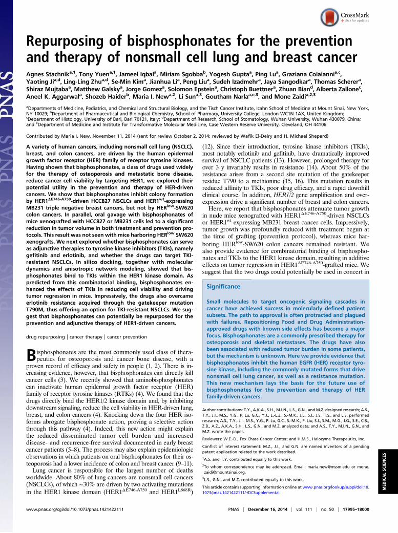
Repurposing of bisphosphonates for the preventionand therapy of nonsmall cell lung and breast cancerAgnes Stachnika,1, Tony Yuena,1, Jameel Iqbala, Miriam Sgobbab, Yogesh Guptaa, Ping Lua, Graziana Colaiannia,c,Yaoting Jia,d, Ling-Ling Zhua,d, Se-Min Kima, Jianhua Lia, Peng Liua, Sudeh Izadmehra, Jaya Sangodkara, Thomas Scherera,Shiraz Mujtabaa, Matthew Galskya, Jorge Gomeza, Solomon Epsteina, Christoph Buettnera, Zhuan Biand, Alberta Zallonec,Aneel K. Aggarwala, Shozeb Haiderb, Maria I. Newa,2, Li Suna,3, Goutham Narlaa,e,3, and Mone Zaidia,2,3
aDepartments of Medicine, Pediatrics, and Chemical and Structural Biology, and the Tisch Cancer Institute, Icahn School of Medicine at Mount Sinai, New York,NY 10029; bDepartment of Pharmaceutical and Biological Chemistry, School of Pharmacy, University College, London WC1N 1AX, United Kingdom;cDepartment of Histology, University of Bari, Bari 70121, Italy; dDepartment of Research, School of Stomatology, Wuhan University, Wuhan 430079, China;and eDepartment of Medicine and Institute for Transformative Molecular Medicine, Case Western Reserve University, Cleveland, OH 44106
Contributed by Maria I. New, November 11, 2014 (sent for review October 2, 2014; reviewed by Wafik El-Deiry and H. Michael Shepard)
A variety of human cancers, including nonsmall cell lung (NSCLC),breast, and colon cancers, are driven by the human epidermalgrowth factor receptor (HER) family of receptor tyrosine kinases.Having shown that bisphosphonates, a class of drugs used widelyfor the therapy of osteoporosis and metastatic bone disease,reduce cancer cell viability by targeting HER1, we explored theirpotential utility in the prevention and therapy of HER-drivencancers. We show that bisphosphonates inhibit colony formationby HER1ΔE746-A750-driven HCC827 NSCLCs and HER1wt-expressingMB231 triple negative breast cancers, but not by HERlow-SW620colon cancers. In parallel, oral gavage with bisphosphonates ofmice xenografted with HCC827 or MB231 cells led to a significantreduction in tumor volume in both treatment and prevention pro-tocols. This result was not seen with mice harboring HERlow SW620xenografts. We next explored whether bisphosphonates can serveas adjunctive therapies to tyrosine kinase inhibitors (TKIs), namelygefitinib and erlotinib, and whether the drugs can target TKI-resistant NSCLCs. In silico docking, together with moleculardynamics and anisotropic network modeling, showed that bis-phosphonates bind to TKIs within the HER1 kinase domain. Aspredicted from this combinatorial binding, bisphosphonates en-hanced the effects of TKIs in reducing cell viability and drivingtumor regression in mice. Impressively, the drugs also overcameerlotinib resistance acquired through the gatekeeper mutationT790M, thus offering an option for TKI-resistant NSCLCs. We sug-gest that bisphosphonates can potentially be repurposed for theprevention and adjunctive therapy of HER1-driven cancers.
drug repurposing | cancer therapy | cancer prevention
Bisphosphonates are the most commonly used class of thera-peutics for osteoporosis and cancer bone disease, with a
proven record of efficacy and safety in people (1, 2). There is in-creasing evidence, however, that bisphosphonates can directly killcancer cells (3). We recently showed that aminobisphosphonatescan inactivate human epidermal growth factor receptor (HER)family of receptor tyrosine kinases (RTKs) (4). We found that thedrugs directly bind the HER1/2 kinase domain and, by inhibitingdownstream signaling, reduce the cell viability in HER-driven lung,breast, and colon cancers (4). Knocking down the four HER iso-forms abrogate bisphosphonate action, proving a selective actionthrough this pathway (4). Indeed, this new action might explainthe reduced disseminated tumor cell burden and increaseddisease- and recurrence-free survival documented in early breastcancer patients (5–8). The process may also explain epidemiologicobservations in which patients on oral bisphosphonates for their os-teoporosis had a lower incidence of colon and breast cancer (9–11).Lung cancer is responsible for the largest number of deaths
worldwide. About 80% of lung cancers are nonsmall cell cancers(NSCLCs), of which ∼30% are driven by two activating mutationsin the HER1 kinase domain (HER1ΔE746-A750 and HER1L868R)
(12). Since their introduction, tyrosine kinase inhibitors (TKIs),most notably erlotinib and gefitinib, have dramatically improvedsurvival of NSCLC patients (13). However, prolonged therapy forover 3 y invariably results in resistance (14). About 50% of theresistance arises from a second site mutation of the gatekeeperresidue T790 to a methionine (15, 16). This mutation results inreduced affinity to TKIs, poor drug efficacy, and a rapid downhillclinical course. In addition, HER1/2 gene amplification and over-expression drive a significant number of breast and colon cancers.Here, we report that bisphosphonates attenuate tumor growth
in nude mice xenografted with HER1ΔE746-A750-driven NSCLCsor HER1wt-expressing MB231 breast cancer cells. Impressively,tumor growth was profoundly reduced with treatment begun atthe time of grafting (prevention protocol), whereas mice har-boring HERlow-SW620 colon cancers remained resistant. Wealso provide evidence for combinatorial binding of bisphospho-nates and TKIs to the HER1 kinase domain, resulting in additiveeffects on tumor regression in HER1ΔE746-A750-grafted mice. Wesuggest that the two drugs could potentially be used in concert in
Significance
Small molecules to target oncogenic signaling cascades incancer have achieved success in molecularly defined patientsubsets. The path to approval is often protracted and plaguedwith failures. Repositioning Food and Drug Administration-approved drugs with known side effects has become a majorfocus. Bisphosphonates are a commonly prescribed therapy forosteoporosis and skeletal metastases. The drugs have alsobeen associated with reduced tumor burden in some patients,but the mechanism is unknown. Here we provide evidence thatbisphosphonates inhibit the human EGFR (HER) receptor tyro-sine kinase, including the commonly mutated forms that drivenonsmall cell lung cancer, as well as a resistance mutation.This new mechanism lays the basis for the future use ofbisphosphonates for the prevention and therapy of HERfamily-driven cancers.
Author contributions: T.Y., A.K.A., S.H., M.I.N., L.S., G.N., and M.Z. designed research; A.S.,T.Y., J.I., M.S., Y.G., P. Lu, G.C., Y.J., L.-L.Z., S.-M.K., J.L., S.I., J.S., T.S., and L.S. performedresearch; A.S., T.Y., J.I., M.S., Y.G., P. Lu, G.C., S.-M.K., P. Liu, S.I., S.M., M.G., J.G., S.E., C.B.,Z.B., A.Z., A.K.A., S.H., L.S., G.N., and M.Z. analyzed data; and A.S., T.Y., M.I.N., G.N., andM.Z. wrote the paper.
Reviewers: W.E.-D., Fox Chase Cancer Center; and H.M.S., Halozyme Therapeutics, Inc.
Conflict of interest statement: M.Z., J.I., and G.N. are named inventors of a pendingpatent application related to the work described.1A.S. and T.Y. contributed equally to this work.2To whom correspondence may be addressed. Email: [email protected] or [email protected].
3L.S., G.N., and M.Z. contributed equally to this work.
This article contains supporting information online at www.pnas.org/lookup/suppl/doi:10.1073/pnas.1421422111/-/DCSupplemental.
www.pnas.org/cgi/doi/10.1073/pnas.1421422111 PNAS | December 16, 2014 | vol. 111 | no. 50 | 17995–18000
MED
ICALSC
IENCE
S

NSCLC patients. Finally, bisphosphonates retain their ability toinhibit the viability of cells harboring the HER1T790M gatekeepermutation, a prelude to their use in overcoming TKI resistance.
ResultsWe found that zoledronic acid inhibited colony formation byHER1ΔE746-A750-driven HCC827 NSCLCs or HER1wt-expressingMB231 triple negative breast cancer cells, without effects onHERlow-SW620 colon cancer (Fig. 1A). We next testedbisphosphonate effects on tumor growth in vivo by xenograftingBALB/c nu/nu mice with HCC827, MB231 or SW620 cells. Se-quential measurement of tumor volume before and after dailygastric gavage with risedronate (1.42 μg/kg) or zoledronic acid(1.36 μg/kg) (Table S1), begun when HCC827 and MB231 tumorsbecame palpable, showed significant reductions in tumor volumeas early as 6 d postinitiation (Fig. 1B). In a parallel preventionprotocol, when drug was given from the time of transplant,HER1wt-expressing MB231 tumors showed a dramatic and pro-gressive reduction in volume, whereas HERlow SW620 coloncancers showed no such response to bisphosphonate (Fig. 1C).The latter finding buttresses our in vitro data (4) establishing thedependence of bisphosphonate action on HER1 expression.Based on initial observations of additivity between gefitinib and
zoledronic acid (17), we investigated whether bisphosphonatesand TKIs display combinatorial binding to the HER kinase do-main. Docking protonated or deprotonated zoledronic acid intoHER1wt crystal structure in complex with erlotinib (PDB ID code1M17) revealed that there was enough space in the kinase pocketto accommodate both drugs (Fig. 2A). Furthermore, when thebisphosphonate is protonated at physiologic pH values, it bindsTKIs via a conserved water bridge (Fig. 2A). Similar predictedbinding modes are shown for the zoledronic acid/gefitinib andminodronic acid/erlotinib pairs (Fig. 2A). Of note, tiludronate didnot display dual binding as it lacks an N-atom to make an H-bondwith erlotinib and its p-chlorophenyl ring clashes with erlotinib(Fig. 2A).Molecular docking further revealed that the HER1L858R mu-
tant conformation was similar to the active state of HER1wt inthe presence of erlotinib and zoledronic acid (Fig. 2B). We usedanisotropic network modeling (ANM) of the HER1L858R mutantwith the two drugs to study conformational fluctuations in theCα-helix and activation loop. We found that despite the presenceof erlotinib and zoledronic acid, the Cα-helix remained in a col-lapsed conformation (Fig. 2B). Molecular dynamics (MD) con-firmed our ANM findings showing that, in the presence of bothdrugs, the interaction between R858 and Y891 locked the acti-vation loop allowing the Cα-helix to collapse (Fig. 2B). Thus, wespeculate combinatorial binding of erlotinib and zoledronic acidwill inhibit the HER1 kinase not by inhibiting Cα-helix collapse,but instead by preventing downstream phosphorylation becauseof the absence of a hydrolysable ɤ-phosphate.We hypothesized that the biologic action of the drug pairs
exceeds that of either drug; in other words, the two drugs be-haved as “one big drug.” When submaximal concentrations ofzoledronic acid (10 μM) and erlotinib (10 nM) were combined,the inhibition of colony formation in HCC827 cells was greaterthan that with either drug alone (Fig. S1A). Isobologramsshowed evidence of additivity (CI or combination index ∼1)
Fig. 1. Bisphosphonates inhibit HER-driven tumor growth in preventionand treatment protocols. (A) Colony formation assays performed withHCC827, MB231, and SW620 cells to study the effect of zoledronic acid (ZA;mean colony counts ± SEM; three experiments with two or three replicatewells pooled). (B and C) Tumor volume was measured sequentially followingtransplant of HCC827 (lung), MB231 (breast), or SW620 (colon) cancer cellsinto BALB/c nu/nu mice. Drugs were begun daily by oral gavage once tumors
became palpable (treatment; B) or at the time of graft (prevention; C).HCC827 and MB231 tumors showed evidence of reduced growth with Ris(1.42 μg/kg, daily, gavage) or ZA (1.36 μg/kg, daily, gavage), whereas HERlow
SW620 cells did not. Change (Δ) in tumor volume plotted for single mice oras group means ± SEM; statistics: ANOVA with Bonferroni’s correction;bisphosphonate- vs. vehicle-treated mice; *P < 0.05; number of mice used forthe analysis corresponds to the number of animals shown in the plot forindividual tumor volumes, e.g., n = 12 mice in B, Upper, control.
17996 | www.pnas.org/cgi/doi/10.1073/pnas.1421422111 Stachnik et al.

when erlotinib and zoledronic acid were combined in bothHCC827 and H3255 cells (Fig. S1A). Similar results were notedwith the minodronic acid/erlotinib combination, but not withtiludronate/erlotinib (Fig. S1B).We sought to probe the cellular mechanism underlying the
observed additivity. HCC827 cells treated with erlotinib showeda dramatic increase in apoptosis, as evidenced by increases incells in the sub-G1 phase (FACS) and poly(ADP-ribose) poly-merase (PARP) cleavage (Western blot), and reductions inpAKT (Fig. S1C). In contrast, zoledronic acid caused a markedcell-cycle arrest, noted as reduced cyclin B1/D1 and proliferatingcell nuclear antigen (PCNA) expression (Western blot) (Fig.S1C). The two drugs together caused profoundly greater apo-ptosis, as evidenced by more marked increases in cells in the sub-G1 phase, as well as PARP cleavage (Fig. S1C). Importantly, thetwo bisphosphonates zoledronic acid and risedronate did notdisplay additivity, either in viability inhibition (MTT) or cell-cycle arrest (FACS) (Fig. S1D). This result would not be un-expected because they bind to the same site within the HER1kinase domain.To provide proof-of-concept that bisphosphonates enhance
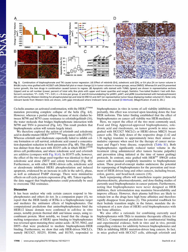
the tumor suppressive action of erlotinib in vivo, we xenograftedHCC827 cells into BALB/c nu/nu mice. Tumor volumes plottedfor individual mice show that, whereas erlotinib and zoledronicacid each attenuated tumor growth (Fig. 1), combining the twodrugs resulted in tumor regression (Fig. 3A). Nine days following
treatment initiation, whereas each drug triggered a significantreduction in tumor volume, the effect of the two drugs surpassedthe effect of either agent (Fig. 3A). Representative photomicro-graphs of TUNEL-labeled tumor tissue from mice displayinga median tumor volume are shown (Fig. 3B). The induction ofapoptosis is both qualitatively and quantitatively greater in tissueobtained from mice treated with both drugs than with one drugalone (Fig. 3B). We also labeled tissue for phosphorylated HER1(pHER1) and downstream molecules, pERK and pAKT. Again,whereas erlotinib and zoledronic acid both reduced phosphory-lation, the effect of the two agents appeared more marked thateither drug alone (Fig. 3 C and D).Although TKIs result in objective treatment responses in
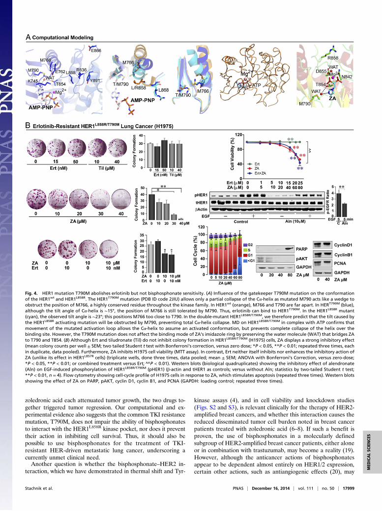
HER1 mutation-driven lung cancers (13), resistance to therapydevelops invariably, the most common mechanism being a sec-ond site mutation at the gatekeeper residue T790 (15). Wetherefore sought to determine, both computationally and experi-mentally, whether HER1-mutation–driven, TKI-resistant NSCLCsretain bisphosphonate sensitivity. If so, bisphosphonates couldbecome a viable option for the therapy of resistant NSCLCs,which otherwise display a downhill course.Computationally, we note that the HER1T790M mutation (PBD
ID code 2JIU) causes only a partial collapse of the Cα-helix asmutant M790 acts as a wedge and interferes with the spatial po-sitioning of the side chain of the highly conserved M766 (Fig. 4A).MD shows that, during movement of the activation loop, the
Fig. 2. Combinatorial binding of bisphosphonates and TKIs. (A) Docking of ZA in the HER1 kinase crystal structure that was cocrystallized with erlotinib (Ert)(PDB ID code 1M17). The phosphate backbone of ZA interacts with Mg2+, the removal of which prevents ZA docking. ZA also interacts with the NH groupbetween the two aromatic rings in Ert either by itself (as a deprotonated tautomer) or via a structural water (WAT) (as a protonated tautomer). Ert associateswith T790 via WAT. Fluid phase: binding mode of ZA and Ert observed from the solvent. Tiludronate (Til) does not have an imidazole ring to make an H-bondwith Ert. It instead contains a p-chlorophenyl that results in severe steric clashes (green sphere) with Ert. ZA docked together with gefitinib (Gef) (PDB ID code2ITY) and binds in a similar orientation to that observed in the ZA–Ert complex, as a deprotonated (pink dashed line) or protonated (orange dashed line)tautomer. Minodronic acid (MA) can likewise bind with Ert as a deprotonated or protonated tautomer. (B) Molecular docking reveals that the HER1L858R
mutant conformation (cyan) is similar to the active state of HER1wt (yellow) in presence of Ert and ZA. However, the simultaneous binding of Ert and ZAinhibits the kinase by preventing the downstream phosphorylation because of the absence of a hydrolysable ɤ-phosphate. ANM of the HER1L858R mutant withErt and ZA. Eigenvectors highlighting conformational fluctuations in the Cα-helix and activation loop (A loop) are shown. Of note is that, despite the presenceof Ert and ZA, the Cα-helix is in a collapsed conformation. MD confirms ANM findings showing that, in presence of Ert and ZA, the interaction between R858and Y891 locks the activation loop allowing the Cα-helix to collapse.
Stachnik et al. PNAS | December 16, 2014 | vol. 111 | no. 50 | 17997
MED
ICALSC
IENCE
S
Cα-helix assumes an activated conformation, with the HER1T790M
mutation preventing complete collapse of the helix (Fig. 4A).However, whereas a partial collapse because of steric clashes be-tween M790 and M793 cause resistance to erlotinib/gefitinib (16),the water molecule that bridges bisphosphonate interaction withM790 and T854 is preserved (Fig. 4A). This result predicts thatbisphosphonate action will remain largely intact.We therefore explored the action of erlotinib and zoledronic
acid in double-mutant HER1L858R/T790M lung cancer cells (H1975).Whereas erlotinib and tiludronate expectedly failed to inhibit col-ony formation or cell survival, zoledronic acid caused a concentra-tion-dependent reduction in both parameters (Fig. 4B). This effectwas distinct from that seen with H3255 cells in which HER1L858R
drives cell proliferation, and where zoledronic acid and erlotinibare additive (compare with Fig. S1A). In H1975 cells, however,the effect of the two drugs used together was identical to that ofzoledronic acid alone (MTT and colony formation) (Fig. 4B).Furthermore, as with other HER1-driven cells, zoledronic acidor alendronate inhibited HER1 phosphorylation and inducedapoptosis, evidenced by an increase in cells in the sub-G1 phase,as well as enhanced PARP cleavage. There were minimal/noeffects on cell cycle protein expression (Fig. 4B). Taken together,the data suggest that bisphosphonates could potentially be usedto overcome TKI resistance.
DiscussionIt has been unclear why only certain cancers respond to bis-phosphonates and others do not. In a companion paper (4), wereport that the HER family of RTKs is a bisphosphonate targetand mediates the antitumor effects of bisphosphonates. Ourcomputational predictions that aminobisphosphonates bind theHER1/2 kinase domain were confirmed in cell-free in vitroassays, notably protein thermal shift and kinase assays, using re-combinant protein. Most notably, we found that the change inmelting temperature of HER1 upon binding to bisphosphonateswas reversed upon mutating three amino acids—K745, N842, andD855—that were predicted to mediate bisphosphonate-HERbinding. Furthermore, we show that only HER-driven NSCLCs,namely HCC827, H3255, H1666, and H1703, responded to
bisphosphonates in vitro in terms of cell viability inhibition; im-portantly, this effect was reversed upon knocking down the fourHER isoforms. This latter finding established that the effect ofbisphosphonates on cancer cell viability was HER-mediated.Here, we report the effect of the two most commonly used,
Food and Drug Approved-approved bisphosphonates, zole-dronic acid and risedronate, on tumor growth in mice xeno-grafted with HCC827 NSCLCs or HER1-driven MB231 breastcancer cells. The daily doses of the respective drugs (1.42 and1.36 mg/kg) translate to approximately twice their annual cu-mulative exposure when used for the therapy of cancer metas-tases and Paget’s bone disease, respectively (Table S1). Bothbisphosphonates significantly reduced tumor volume in thetreatment (drug administered after tumors became palpable)and prevention (drug initiated at the time of tumor grafting)protocols. In contrast, mice grafted with HERlow SW620 coloncancer cells remained completely insensitive to bisphosphonateaction. These proof-of-concept studies underscore the potentialfor repurposing of bisphosphonates for the prevention and treat-ment of HER-driven lung and other cancers, including breast,colon, gastric, and head/neck cancers (18).Such repurposing efforts will nonetheless require purposeful
pharmacokinetic, pharmacodynamic, and dose-finding studies ofcurrently used bisphosphonates in cancer patients. Furthermore,noting that bisphosphonates were never designed as HERinhibitors, their reformulation may maximize bioavailability andimprove efficacy. However, their plasma half-lives are likely toremain low as the drugs have high bone affinities and thereforerapidly disappear from plasma (1). This potential roadblock fortheir bedside transition might, in the future, mandate the de-velopment of a new class of HER1-avid bisphosphonates withlow bone affinities.We also offer a rationale for combining currently used
bisphosphonates with TKIs to maximize therapeutic efficacy forNSCLCs. Computational predictions for combinatorial bindingand experimental evidence, both in vitro and in vivo, providecompelling evidence for additivity between bisphosphonates andTKIs in inhibiting HER1 mutation-driven lung cancers. In fact,in mice grafted with HCC827 cells, although erlotinib and
Fig. 3. Combination of bisphosphonate and TKI causes tumor regression. (A) Effect of relotinib (Ert), zoledronic acid (ZA), or Ert plus ZA on tumor volume inBALB/c nu/numice grafted with HCC827 cells [Waterfall plot or mean change (Δ) in tumor volume in mouse groups, versus DMSO]. Whereas Ert and ZA preventedtumor growth, the two drugs in combination caused tumors to regress. (B) Apoptotic cells stained with TUNEL (green) are shown in representative sections(Upper) and as cell number (Lower; percent of total cells; Box plots with upper and lower quartiles and range). Statistics: Two-tailed Student t test with Bon-ferroni’s correction; *P < 0.05, **P < 0.01; n = 8 mice per group. (C and D) Immunolabeling for pHER1, pAKT, and pERK (counterstained with hematoxylin/eosin)(D) confirmed by Western blotting for phosphorylated (p) and total (t) ERK1/2 and AKT (on representative tumor tissue displaying median volume) (C). Note: onlyrelevant bands from Western blots are shown, with gaps introduced where irrelevant lanes are excised (SI Methods). (Magnification: B and D, 20×.)
17998 | www.pnas.org/cgi/doi/10.1073/pnas.1421422111 Stachnik et al.
zoledronic acid each attenuated tumor growth, the two drugs to-gether triggered tumor regression. Our computational and ex-perimental evidence also suggests that the common TKI resistancemutation, T790M, does not impair the ability of bisphosphonatesto interact with the HER1L858R kinase pocket, nor does it preventtheir action in inhibiting cell survival. Thus, it should also bepossible to use bisphosphonates for the treatment of TKI-resistant HER-driven metastatic lung cancer, underscoring acurrently unmet clinical need.Another question is whether the bisphosphonate–HER2 in-
teraction, which we have demonstrated in thermal shift and Tyr-
kinase assays (4), and in cell viability and knockdown studies(Figs. S2 and S3), is relevant clinically for the therapy of HER2-amplified breast cancers, and whether this interaction causes thereduced disseminated tumor cell burden noted in breast cancerpatients treated with zoledronic acid (6–8). If such a benefit isproven, the use of bisphosphonates in a molecularly definedsubgroup of HER2-amplified breast cancer patients, either aloneor in combination with trastuzumab, may become a reality (19).However, although the anticancer actions of bisphosphonatesappear to be dependent almost entirely on HER1/2 expression,certain other actions, such as antiangiogenic effects (20), may
Fig. 4. HER1 mutation T790M abolishes erlotinib but not bisphosphonate sensitivity. (A) Influence of the gatekeeper T790M mutation on the conformationof the HER1wt and HER1L858R. The HER1T790M mutation (PDB ID code 2JIU) allows only a partial collapse of the Cα-helix as mutated M790 acts like a wedge toobstruct the position of M766, a highly conserved residue throughout the kinase family. In HER1wt (orange), M766 and T790 are far apart. In HERT790M (blue),although the tilt angle of Cα-helix is ∼15°, the position of M766 is still tolerated by M790. Thus, erlotinib can bind to HER1T790M. In the HER1L858R mutant(cyan), the observed tilt angle is ∼23°; this positions M766 too close to T790. In the double-mutant HER1L858R/T790M, we therefore predict that the tilt caused bythe HER1L858R activating mutation will be obstructed by M790, preventing total Cα-helix collapse. MD on HER1L858R/T790M in complex with ATP confirms thatmovement of the mutated activation loop allows the Cα-helix to assume an activated conformation, but prevents complete collapse of the helix over thebinding site. However, the T790M mutation does not affect the binding mode of ZA’s imidazole ring by preserving the water molecule (WAT) that bridges ZAto T790 and T854. (B) Although Ert and tiludronate (Til) do not inhibit colony formation in HER1L858R/T790M (H1975) cells, ZA displays a strong inhibitory effect(mean colony counts per well ± SEM; two tailed Student t test with Bonferroni’s correction, versus zero dose; *P < 0.05, **P < 0.01; repeated three times, eachin duplicate, data pooled). Furthermore, ZA inhibits H1975 cell viability (MTT assay). In contrast, Ert neither itself inhibits nor enhances the inhibitory action ofZA (unlike its effect in HER1L857R cells) (triplicate wells, done three times, data pooled; mean ± SEM; ANOVA with Bonferroni’s Correction, versus zero-dose;*P < 0.05, **P < 0.01; or combined treatment versus Ert; ^̂ P < 0.01). Western blots (biological quadruplicates) showing the inhibitory effect of alendronate(Aln) on EGF-induced phosphorylation of HER1L858R/T790M (pHER1) (β-actin and tHER1 as controls; versus without Aln; statistics by two-tailed Student t test;**P < 0.01, n = 4). Flow cytometry showing cell-cycle profile of H1975 cells in response to ZA, which stimulates apoptosis (repeated three times). Western blotsshowing the effect of ZA on PARP, pAKT, cyclin D1, cyclin B1, and PCNA (GAPDH: loading control; repeated three times).
Stachnik et al. PNAS | December 16, 2014 | vol. 111 | no. 50 | 17999
MED
ICALSC
IENCE
S
indeed be exerted via other RTKs, such as the VEGFR. This willdepend on whether or not, as our data indicate, a bisphospho-nate can fit into the kinase domain of a given RTK (4).
Materials and MethodsComputational docking, MD, and ANM studies were performed using HERcrystal structures from the Protein Database. Cancer cell lines were subjectto the MTT or colony formation assays as detailed in SI Methods (21). Forcell-cycle assays, cells treated with bisphosphonate and erlotinib weresubject to flow cytometry. For the in vivo studies, cells were injected in theflank of BALB/c nu/nu mice, with tumor sizes measured sequentially by
calipers (21, 22), followed by TUNEL staining, immunohistochemistry, andWestern blotting.
ACKNOWLEDGMENTS. This work was supported in part by NationalInstitutes of Health Grants DK80459 (to M.Z. and L.S.), AG40132 (to M.Z.),AG23176 (to M.Z.), AR06592 (to M.Z.), and AR06066 (to M.Z.); the ItalianSpace Agency (A.Z.); a grant from National Science Foundation of China,Ministry of China (International Collaborative Grant to Z.B. and M.Z.); andthe National Center for Advancing Translational Sciences, National Institutesof Health, through Icahn School of Medicine at Mount Sinai’s Clinical andTranslational Science Award (to S.I.). G.N., formerly recipient of a HowardHughes Medical Institute Physician-Scientist Early Career Award, is a namedHarrington Scholar.
1. Russell RG (2011) Bisphosphonates: The first 40 years. Bone 49(1):2–19.2. Wilson C, Holen I, Coleman RE (2012) Seed, soil and secreted hormones: Potential
interactions of breast cancer cells with their endocrine/paracrine microenvironment
and implications for treatment with bisphosphonates. Cancer Treat Rev 38(7):
877–889.3. Coleman R, Gnant M, Morgan G, Clezardin P (2012) Effects of bone-targeted agents
on cancer progression and mortality. J Natl Cancer Inst 104(14):1059–1067.4. Yuen T, et al. (2014) Bisphosphonates inactivate human EGFRs to exert antitumor
actions. Proc Natl Acad Sci USA 111:17989–17994.5. Gnant M, et al. (2009) Endocrine therapy plus zoledronic acid in premenopausal
breast cancer. N Engl J Med 360(7):679–691.6. Coleman RE, et al. (2011) Breast-cancer adjuvant therapy with zoledronic acid. N Engl
J Med 365(15):1396–1405.7. Gnant M, et al. (2011) Long-term follow-up in ABCSG-12: Significantly improved
overall survival with adjuvant zoledronic acid in premenopausal patients withendocrine-receptor–positive early breast cancer. Cancer Res 71(24, Suppl):S1–S2.
8. Coleman R, et al. (2013) Zoledronic acid (zoledronate) for postmenopausal womenwith early breast cancer receiving adjuvant letrozole (ZO-FAST study): Final 60-month
results. Ann Oncol 24(2):398–405.9. Pazianas M, Abrahamsen B, Eiken PA, Eastell R, Russell RG (2012) Reduced colon cancer
incidence andmortality in postmenopausal women treatedwith an oral bisphosphonate—
Danish National Register Based Cohort Study. Osteoporos Int 23(11):2693–2701.10. Rennert G, Pinchev M, Rennert HS, Gruber SB (2011) Use of bisphosphonates and
reduced risk of colorectal cancer. J Clin Oncol 29(9):1146–1150.
11. Sendur MA, et al. (2012) Demographic and clinico-pathological characteristics ofbreast cancer patients with history of oral alendronate use. Med Oncol 29(4):2601–2605.
12. Ding L, et al. (2008) Somatic mutations affect key pathways in lung adenocarcinoma.Nature 455(7216):1069–1075.
13. Ciardiello F, Tortora G (2008) EGFR antagonists in cancer treatment. N Engl J Med358(11):1160–1174.
14. Nishino M, et al. (2011) Imaging of lung cancer in the era of molecular medicine. AcadRadiol 18(4):424–436.
15. Sequist LV, et al. (2011) Genotypic and histological evolution of lung cancers ac-quiring resistance to EGFR inhibitors. Sci Transl Med 3(75):75ra26.
16. Yun CH, et al. (2008) The T790M mutation in EGFR kinase causes drug resistance byincreasing the affinity for ATP. Proc Natl Acad Sci USA 105(6):2070–2075.
17. Chang JW, et al. (2009) Bisphosphonate zoledronic acid enhances the inhibitory effectsof gefitinib on EGFR-mutated non-small cell lung carcinoma cells. Cancer Lett 278(1):17–26.
18. Krasinskas AM (2011) EGFR signaling in colorectal carcinoma. Pathol Res Int 2011:932932.19. Romond EH, et al. (2005) Trastuzumab plus adjuvant chemotherapy for operable
HER2-positive breast cancer. N Engl J Med 353(16):1673–1684.20. Di Salvatore M, et al. (2011) Anti-tumour and anti-angiogenetic effects of zoledronic
acid on human non-small-cell lung cancer cell line. Cell Prolif 44(2):139–146.21. Sangodkar J, et al. (2012) Targeting the FOXO1/KLF6 axis regulates EGFR signaling
and treatment response. J Clin Invest 122(7):2637–2651.22. Hemmi H, et al. (2012) Treml4, an Ig superfamily member, mediates presentation of
several antigens to T cells in vivo, including protective immunity to HER2 protein.J Immunol 188(3):1147–1155.
18000 | www.pnas.org/cgi/doi/10.1073/pnas.1421422111 Stachnik et al.