
Ultra-high field parallel imaging of the superior parietal lobule during mental maze solving

Remapping Attentional Priorities:Differential Contribution of SuperiorParietal Lobule and Intraparietal Sulcus
Pascal Molenberghs1, Marsel M. Mesulam2,3, Ronald Peeters4
and Rik R.C. Vandenberghe1,5
1Cognitive Neurology Laboratory, Experimental Neurology
Section, Katholieke Universiteit Leuven, Belgium 2Cognitive
Neurology and Alzheimer’s Disease Center and 3Department
of Neurology, Feinberg School of Medicine, Northwestern
University, Chicago, IL 60611, USA, 4Radiology Department
and 5Neurology Department, University Hospital
Gasthuisberg, Leuven, Belgium
Seeking and selectively attending to significant extrapersonalstimuli in a dynamic environment requires the updating of anattentional priority map. Using functional magnetic resonanceimaging, we investigated the role of posterior parietal cortex insuch remappings of attentional priorities where the configuration,location, and significance of stimuli were systematically varied. Ourdata revealed a functional dissociation between 2 juxtaposedposterior parietal regions: one in the superior parietal lobule(SPL) and another in the intraparietal sulcus (IPS). SPL waspreferentially activated in all conditions where a spatial displace-ment occurred in the location of the target, the location of thedistracter, or the focus of attention (exogenous and endogenousshifts of spatial attention). Shifts of the attentional focus alsoactivated the IPS but principally if they were guided endogenouslyby internal rules of relevance rather than stimulus displacement perse (endogenous attention shifts). Only the IPS region was activatedby transient resetting of target significance when the stimulusconfiguration changed but the attentional focus remained spatiallyfixed (feature attention shifts). These 2 components of the large-scale frontoparietal spatial attention network therefore havecommon and distinctive functions. In specific, the IPS componentis more closely related to the compilation of an attentional prioritymap, including the endogenous recalibration of attentional weights.The SPL component, on the other hand, is more closely related tothe modification of spatial coordinates linked to attentionalpriorities (spatial shifting). Collectively, these 2 areas allowposterior parietal cortex to dynamically encode extrapersonalevents according to their spatial coordinates and valence.
Keywords: endogenous control, fMRI, perietal, shifting
Introduction
Posterior parietal cortex is a major component of a distributed
spatial attention network (Mesulam 1981; Corbetta and
Shulman 2002; Husain and Rorden 2003). The posterior parietal
contribution to spatial attention emanates from multiple areas
including the superior parietal lobule (SPL) (Vandenberghe
et al. 2001a; Yantis et al. 2002; Pollmann et al. 2003), upper and
lower banks of the intraparietal sulcus (IPS) (Corbetta et al.
1993; Nobre et al. 1997; Gitelman et al. 1999; Corbetta and
Shulman 2002; Woldorff et al. 2004), angular gyrus (Mort et al.
2003; Husain and Rorden 2003; Hillis et al. 2005), and
temporoparietal junction (TPJ) (Corbetta et al. 2000; Corbetta
and Shulman 2002; Kincade et al. 2005; Astafiev et al. 2006). SPL
and IPS are often activated together, for example, when subjects
attentively track moving targets compared with passive viewing
(Culham et al. 1998), when subjects attend to motion compared
with color (Shulman et al. 2002), or during spatial cueing
(Gitelman et al. 1999; Simon et al. 2002). More recently, several
attempts have been made to characterize the specializations of
these parietal areas, and distinctive roles in endogenous control
(Hopfinger et al. 2000; Corbetta and Shulman 2002; Kincade
et al. 2005; Vandenberghe et al. 2005), shifting (Corbetta et al.
2000; Vandenberghe et al. 2001a; Yantis et al. 2002), and
sustaining attention (Vandenberghe et al. 2001b; Husain and
Rorden 2003) have been attributed to these areas.
The current study of selective attention focused on the
remapping of attentional priorities. Remapping refers to tran-
sitions from one attentional priority map (Koch and Ullman
1985) to another. We studied such transitions against a baseline
of sustained attention (Vandenberghe et al. 2001a; Yantis et al.
2002; Liu et al. 2003; Serences et al. 2004; Shomstein and Yantis
2004). This approach departs from a classical trial-by-trial
approach with blank intertrial intervals and allows us to subtract
out processes related to the maintenance of attention. We also
removed effects related to detection and motor responses
(Shulman et al. 1999; Woldorff et al. 2004) by temporally
separating the occurrence of transient dimmings that had to be
detected from the remapping events to which functional
magnetic resonance imaging (fMRI) responseswere time locked.
In the current study, we attempted to tease apart 2 processes
that are involved in remapping attentional priorities. In everyday
life, spatial attention is dynamically redistributed in response to
changes in the location, perceptual features, and relevance of
extrapersonal stimuli. A first process consisted of the recalibra-
tion of attentional weights (Bundesen 1990). Within the
framework of the theory of visual attention (TVA), an atten-
tional weight is computed for each perceptual unit (object in
the visual field) and used for allocation of attention (i.e., visual
processing resources) (Bundesen et al. 2005). The attentional
weight of a perceptual unit (wx) is the product of the sensory
eidence [g(x, j)] that object x has a feature j multiplied by the
behavioral pertinence of that feature (variable pj). Within TVA, j
is defined broadly and can refer to, for example, a certain color,
a shape, a spatial position, or membership of a category
(Bundesen et al. 2005). The rate at which a perceptual unit is
encoded in visual short-term memory is determined by its
attentional weight relative to the attentional weights of the
other units (biased competition [Desimone and Duncan 1995]).
In a dynamic world, sensory evidence g(x, j) or behavioral
pertinence pj continuously change. As a consequence, the
attentional weights must be continuously updated. We refer
to the updating of weights as the ‘‘recalibration of attentional
weights.’’ On the basis of previous human brain mapping
(Vandenberghe et al. 2005) and monkey electrophysiology
studies (Gottlieb and Goldberg 1999; Wardak et al. 2002; Bisley
and Goldberg 2003; Treue 2003), we hypothesized that the
horizontal segment of the IPS plays a critical role in this process
Cerebral Cortex November 2007;17:2703--2712
doi:10.1093/cercor/bhl179
Advance Access publication January 30, 2007
� The Author 2007. Published by Oxford University Press. All rights reserved.
For permissions, please e-mail: [email protected]
by guest on August 22, 2014
http://cercor.oxfordjournals.org/D
ownloaded from

(Vandenberghe et al. 2005), especially when stimulus selection
is under high endogenous control (Corbetta and Shulman 2002).
The second process of interest was spatial shifting. On the
basis of previous work (Vandenberghe et al. 2001a; Yantis et al.
2002), we hypothesized that SPL would be activated by the need
to shift the spatial focus of attention regardless of what
triggered the shift and that, conversely, a feature change would
activate SPL more when the feature change triggered a spatial
displacement of the attentional focus than when the spatial
focus of attention remained locked to the same location.
We used the following strategy to separate these 2 processes.
Subjects viewed a display containing 2 stimuli pulled from 2 sets
of shapes. Each set contained one relevant and one irrelevant
stimulus, and this was defined beforehand. The task was to press
a button when the relevant stimulus dimmed but not when the
irrelevant stimulus dimmed. By changing either the shape or the
spatial location of stimuli, we manipulated the currently
attended location. A feature change required endogenous
recalibration of attentional weights but did not necessarily
change the relevant stimulus site. Conversely, a change of
stimulus position to a previously unoccupied location elicited
a spatial attentional shift without much endogenous control.
Our purpose was not to compare feature-based with location-
based attention: This has already been studied in the past by
comparing spatial with feature cueing (Vandenberghe et al.
2001b; Giesbrecht et al. 2003) or interdimensional shifts with
a sustained attention baseline (Liu et al. 2003). In our study,
shape always defined which stimulus was relevant. The type of
spatial shifting we studied also differed from shifts evoked by
invalid cueing as studied before (Corbetta et al. 2000). In our
study, a shift was not associated with a breach of expectancy
(Corbetta et al. 2000; Corbetta and Shulman 2002; Kincade et al.
2005; Astafiev et al. 2006). The position to which a stimulus
shifted could not be predicted.
We hypothesized that endogenous recalibration of atten-
tional weights and spatial shifting were dissociable processes
that differentially involved IPS and SPL, respectively.
Methods
SubjectsWe conducted 4 event-related fMRI experiments. Sixteen subjects
(9 women, 7 men) participated in the main experiment, 4 subjects
(1 woman, 3 men) in each of 2 control experiments, and 8 subjects
(4 women, 4 men) in a second, ‘‘auditory switching’’ experiment. All
participants were between 18 and 30 years of age and strictly right
handed (Oldfield 1971), free of psychotropic or vasoactive medication,
without neurological or psychiatric history, and had a normal structural
brain magnetic resonance imaging (MRI) scan. They gave written
informed consent in accordance with the Declaration of Helsinki. The
ethical commission, University Hospital Gasthuisberg, Leuven, approved
the experiment.
Stimuli and TasksThe fMRI experiments were conducted using Superlab for PC version
2.0 (Cedrus, Phoenix, AR). Visual stimuli were projected from a Barco
6400i LCD projector (1024 3 768 pixels) onto a screen (40 by 30 deg)
36 cm in front of the subjects’ eyes.
In the main experiment, 2 stimuli occupied 2 out of 6 possible
locations on the horizontal meridian (Fig. 1A) at 3.18, 9.46, or 15.5 deg
eccentricity to the left or to the right. There were 2 possible, yoked
stimulus pairs: A square together with a triangle or a circle together with
a diamond (stimulus surface 1.59 deg2; luminance 89 cd/m2) (Fig. 1A).
At any given time, only one pair of stimuli was presented. Each pair
contained a relevant and an irrelevant stimulus. Subjects were instructed
and trained beforehand which of the 2 stimuli within each pair was
relevant. This was constant within subjects and counterbalanced
between subjects. When the relevant stimulus dimmed, subjects had
to respond (Fig. 1C) but not when the irrelevant stimulus dimmed. A
dimming consisted of a change of luminance from 89 to 45 cd/m2 with
a duration of 100 ms. Within each run (duration 8 min 6 s), the relevant
stimulus dimmed 30 times (Fig. 1C) and the irrelevant stimulus dimmed
30 times. Half of the subjects responded with their left hand, half with
their right hand.
This sustained attention baseline was transiently interrupted by
stimulus changes of 4 different types (events), 36 of each type. Event
Figure 1. Stimuli and tasks. For each of 2 experiments, we show as an example onerandomly chosen segment of an event sequence. In reality, the order of events waspseudorandomized throughout the run. The labels ‘‘rel. stim.’’ (relevant stimulus) and‘‘irrel. stim.’’ (irrelevant stimulus) are for clarification only and were not presented tothe subjects. (A) Main experiment. Which stimulus within each of the 2 pairs wasrelevant was constant within a subject (in this example, the square and the circle,respectively). (A1) Feature change that leads to a spatial attentional shift. (A2) Featurechange but target and distracter stay in place (no spatial shift). (A3) Relevant stimulusmoves to a previously unoccupied position. In this example, the stimulus shifts to theopposite hemifield, but throughout a run, the number of between- versus within-hemifield shifts was matched as well as the number of leftward versus rightward shifts.(A4) Irrelevant stimulus moves to a previously unoccupied position. Again, the numberof between- versus within-hemifield shifts was matched as well as the number ofleftward versus rightward shifts. (B) Auditory switching experiment. In this example,tone I signals a change in stimulus relevance, tone II signals that stimulus relevancedoes not change. (B1) Auditory tone signals a change in relevance between stimuli. Thisleads to a spatial attentional shift. No visual changes. (B2) Nothing changes. (B3)Relevant and irrelevant stimulus swap positions leading to a spatial attentional shift.(B4) Relevant and irrelevant stimulus swap positions. Auditory tone signals a change ofrelevance between stimuli. (C) Example of a dimming target. Subjects had to press a keyfor a dimming of the relevant stimulus but not when the irrelevant stimulus dimmed.
2704 Remapping Attentional Priorities d Molenberghs et al.
by guest on August 22, 2014
http://cercor.oxfordjournals.org/D
ownloaded from
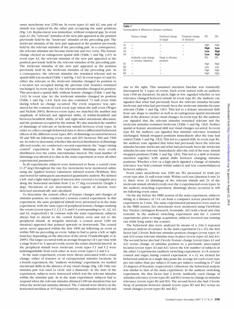
onset asynchrony was 2250 ms. In event types A1 and A2, one pair of
stimuli was replaced by the other pair occupying the same positions
(Fig. 1A). Replacement was immediate, without temporal gap. In event
type A1, the ‘‘relevant’’ stimulus of the new pair appeared at the position
previously held by the ‘‘irrelevant’’ stimulus of the preceding pair. The
irrelevant stimulus of the new pair appeared at the position previously
held by the relevant stimulus of the preceding pair. As a consequence,
the relevant stimulus site became irrelevant and vice versa. The feature
change elicited an endogenous spatial shift (Table 1 and Fig. 1A1). In
event type A2, the relevant stimulus of the new pair appeared at the
position previously held by the relevant stimulus of the preceding pair.
The irrelevant stimulus of the new pair appeared at the position
previously held by the irrelevant stimulus of the preceding pair. As
a consequence, the relevant stimulus site remained relevant and no
spatial shift was needed (Table 1 and Fig. 1A2). In event types A3 and A4,
either the relevant or the irrelevant stimulus changed its position to
a location not occupied during the previous trial. Features remained
unchanged. In event type A3, the relevant stimulus changed its position.
This provoked a spatial shift, without feature changes (Table 1 and Fig.
1A3). In event type A4, the irrelevant stimulus changed its position
(Table 1 and Fig. 1A4). Each run also contained 72 null event trials
during which no change occurred. The event sequence was opti-
mized for the contrast of each event type minus the null event (Wager
and Nichols 2003). Between event types, we matched the number and
amplitude of leftward and rightward shifts, of within-hemifield and
between-hemifield shifts, of left- and right-sided attentional allocation,
and the positions occupied by the stimuli. We also matched the number
of dimmings of relevant or irrelevant stimuli between event types. In
order to collect enough behavioral data to detect differential behavioral
effects of the different event types, 80% of dimmings occurred between
50 and 500 ms following event onset and 20% between 500 and 1500
ms. In order to determinewhether this skewed distribution of dimmings
affected results, we conducted a second experiment, the ‘‘target timing
control’’ experiment. In this experiment, dimmings were evenly
distributed over the entire interstimulus interval. The total number of
dimmings was identical to that in the main experiment as were all other
experimental parameters.
In all experiments, subjects were instructed to fixate a central cross
(1.27 deg) throughout the run. Eye movements were monitored online
using the Applied Science Laboratory infrared system (Waltham, MA)
and stored for subsequent automatized quantitative analysis. We defined
a left- and a right-sided region of interest that covered a rectangular area
along the horizontal meridian from 2 to 17 deg eccentricity (height 4
deg). Deviations of eye movements into regions of interest were
detected automatically and calculated.
To determine the sensory effect of feature changes and changes of
stimulus position, we conducted a sensory control experiment. In this
experiment, the same peripheral stimuli were presented as in the main
experiment, with the same types of peripheral sensory changes marking
the events (event types C1, C2, C3, and C4 corresponding to A1, A2, A3,
and A4, respectively). In contrast with the main experiment, subjects
always had to attend to the central fixation cross and not to the
peripheral stimuli. At irregular intervals, a central arrow (100 ms
duration) appeared that pointed upwards or downwards. The central
arrow never appeared within the first 1000 ms following an event or
within 500 ms preceding an event. Subjects had to press a left or right
hand key depending on the direction of the arrow (Vandenberghe et al.
2005). The target occurred with an average frequency of 1 per trial, with
a range from 0 to 3, spread evenly across the entire intertrial interval. As
the peripheral stimuli were irrelevant, event types C1 and C2 were
indistinguishable from each other as were event types C3 and C4.
In the main experiment, events were always associated with a visual
change, either of features or of extrapersonal stimulus locations. In
a fourth experiment, the ‘‘auditory switching’’ experiment, we studied
attentional shifts in the absence of any visual change (Fig. 1B). Only one
stimulus pair was used (a circle and a diamond). At the start of the
experiment, subjects were instructed which was the relevant stimulus
within the stimulus pair. As in the main experiment, subjects had to
respond when the relevant stimulus dimmed (100 ms duration) but not
when the irrelevant stimulus dimmed. The 2 stimuli were shown on the
horizontal meridian at 10.5 deg eccentricity, one stimulus to the left and
one to the right. This sustained attention baseline was transiently
interrupted by 4 types of events. Each event started with an auditory
tone (100 ms duration). Its pitch, high or low, signaled whether or not
relevance changed between stimuli. In event type B1, the auditory cue
signaled that what had previously been the relevant stimulus became
irrelevant and what had previously been the irrelevant stimulus became
relevant (Table 1 and Fig. 1B1). This led to a feature attentional shift
from one shape to another as well as an endogenous spatial attentional
shift, in the absence of any visual changes. In event type B2, the auditory
cue signaled that the relevant stimulus remained relevant and the
irrelevant stimulus remained irrelevant (Table 1 and Fig. 1B2). Neither
spatial or feature attentional shift nor visual changes occurred. In event
type B3, the auditory cue signaled that stimulus relevance remained
unchanged. Stimuli swapped positions immediately after the tone had
ended (Table 1 and Fig. 1B3). This led to a spatial shift. In event type B4,
the auditory tone signaled that what had previously been the relevant
stimulus became irrelevant and what had previously been the irrelevant
stimulus became relevant. Immediately after the end of the tone, stimuli
swapped positions (Table 1 and Fig. 1B4). This led to a shift in feature
attention together with spatial shifts between changing stimulus
positions. Whether a low or a high pitch signaled a change of stimulus
relevance was held constant within subjects and was counterbalanced
between subjects.
Event onset asynchrony was 3200 ms. We presented 24 trials per
event type plus 24 null event trials. Within each run (duration 6 min 24
s), there were 24 dimmings of the relevant stimuli and 24 of the
irrelevant stimuli, divided evenly over the 4 experimental event types. In
the auditory switching experiment, dimmings always occurred at 400
ms following event onset.
On the day before the fMRI session of the main experiment, subjects
sitting at a distance of 114 cm from a computer screen practiced the
experiment in 3 runs. The same experimental parameters were used as
in the fMRI session. Eye movements were monitored using ViewPoint
Eye Tracker (Arrington Research, Scottsdale, AZ) with a head and chin
restraint. In the auditory switching experiment and the 2 control
experiments, prior to image acquisition, subjects received one training
run while lying under the scanner.
The behavioral data were analyzed by means of a 2-by-2 repeated
measures analysis of variance. In the main experiment (n = 16), the first
factor had 2 levels: Relevant stimulus position changes (event types A1
and A3) versus relevant stimulus stays in place (event types A2 and A4).
The second factor also had 2 levels: Feature change (event types A1 and
A2) versus change of stimulus position to a previously unoccupied
location (event types A3 and A4). Given the low number of subjects in
the other 3 experiments (auditory switching experiment: n = 8, sensory
control and target timing control experiment: n = 4), we treated for
behavioral analysis as a single data point the average for each event type
per run rather than per subject (6 runs per subject, resulting in 47 and
23 degrees of freedom, respectively). Otherwise the behavioral analysis
was similar to that of the main experiment. In the auditory switching
experiment, the first factor had 2 levels: Auditorily cued change of
stimulus relevance (event types B1 and B4) versus no change in stimulus
relevance (event types B2 and B3). The second factor also had 2 levels:
Swap of positions between stimuli (event types B3 and B4) versus no
visual changes (event types B1 and B2).
Table 1Commonalities & differences between conditions
Feature change Positional changeof target
Positional changeof distracter
IPS SPL
A1 þ þ þ High HighA2 þ � � High LowA3 � þ � Low HighA4 � � þ Low High
Auditorily cued changein stimulus relevance
Swap in stimuluspositions
IPS SPL
B1 þ � High HighB2 � � Low LowB3 � þ High HighB4 þ þ High High
Cerebral Cortex November 2007, V 17 N 11 2705
by guest on August 22, 2014
http://cercor.oxfordjournals.org/D
ownloaded from

Image AcquisitionA 3-T Philips Intera system (Best, Netherlands) equipped with an 8-
channel head volume coil provided 3D T1 anatomical volume images
(time repetition [TR] = 9.6 ms, time echo [TE] = 4.6 ms, turbo field echo
shot interval = 1748 ms, in-plane resolution = 1 mm, slice thickness = 1.2
mm) and T2* echoplanar images (EPIs) with blood oxygenation level--
dependent (BOLD) contrast. EPIs (TR = 2 s, TE = 30 ms, SENSE parallel
imaging factor = 2) comprised 36 axial slices acquired continuously in
ascending order (voxel size = 2.75 3 2.75 3 3.75 mm3). In the main
experiment and the 2 control experiments, we acquired a total of 250
volumes per run and in the auditory switching experiment 198 volumes
per run. The first 6 volumes were discarded to allow the MRI signal to
reach steady state. In the main experiment and the 2 control experi-
ments, each subject underwent 6 runs each of 216 trials. In the auditory
switching experiment, each subject underwent 6 runs each of 120 trials.
Image AnalysisImage processing analysis was performed using Statistical Parametric
Mapping version 2002 (Wellcome Department of Imaging Neurosci-
ence, London, UK). All experiments were preprocessed in the sameway.
Following correction for differences in timing of slice acquisition
within a volume, EPI volumes were realigned and resliced using sinc
interpolation. A mean EPI volume was obtained during realignment, and
the structural MRI was coregistered with that mean volume. The
structural scan was normalized to the Montreal Neurological Institute
T1 template in Talairach space (Talairach and Tournoux 1988; Friston
et al. 1995) using nonlinear basis functions. The same deformation
parameters were applied to the EPI volumes. The EPI volumes were
spatially smoothed using a 5 3 5 3 7 mm3 filter. Data from different runs
were proportionally scaled to a grand mean of 100 arbitrary units to
account for overall differences in the intensity of whole-brain volumes
across the time series. The time series for each voxel were high-pass
filtered to (1/128) Hz. The event-related response, synchronized with
the acquisition of the top slice, was modeled by a canonical hemody-
namic response function (HRF) consisting of a mixture of 2 gamma
functions that emulate the early peak at 5 s and the subsequent
undershoot (Friston et al. 1999). The temporal derivative of the HRF
was also included in the model. Statistical inference was corrected for
intrinsic autocorrelations. A statistical parametric map of the t statistic
for the parameter estimates was generated and subsequently trans-
formed to a Z map. Data were analyzed using a random effects general
linear model. One contrast image per individual was calculated. At the
second level of analysis, we examined for each of the contrasts whether,
on average, the contrast images revealed significant differences (1-
sample t-test).
The significance map for the group random effects analysis was
thresholded at P < 0.001 for voxel-level inference with a cluster-level
threshold of P < 0.05 corrected for the whole brain search volume.
The auditory switching experiment and the 2 control experiments
were analyzed by means of a fixed-effects analysis due to the low
number of subjects. We restricted the analysis of the auditory switching
experiment and of the 2 control experiments to volumes of interest that
were defined on the basis of the main experiment. These volumes of
interest consisted of the clusters that were significantly activated in the
main experiment. The significance threshold in the control experiments
and the auditory switching experiment was set at P < 0.05 corrected for
the volume of interest.
Results
Behavioral Data
Performance parameters are listed in Tables 2 and 3. When
subjects pressed for a dimming of an irrelevant stimulus, this
was counted as a false alarm.
In the main experiment, subjects responded significantly
more slowly when the relevant stimulus position changed (A1
and A3) compared with when the relevant stimulus stayed in
place (A2 and A4) (F1,15 = 4.21, P < 0.05) (Table 2). Subjects
missed significantly more targets when the relevant or the
irrelevant stimulus moved to a previously unoccupied position
(A3 and A4) than when features changed (A1 and A2) (F1,15 =17.99, P < 0.0001). There was no interaction effect (reaction
times F1,15 = 0.66, P = 0.42; accuracies F1,15 = 0.73, P = 0.40; false
alarms F1,15 = 0.07, P = 0.79). The average number of eye
movements did not differ significantly between the different
event types (P > 0.9) (Table 2).
In the target timing control experiment, subjects responded
significantly more slowly when the relevant stimulus position
changed (A1 and A3) compared with when the relevant
stimulus stayed in place (A2 and A4) (F1,23 = 4.25, P = 0.043).
Subjects missed significantly more targets when the relevant or
the irrelevant stimulus moved to a previously unoccupied
position (A3 and A4) than when features changed (A1 and A2)
(F1,23 = 11.78, P < 0.001). There were no interaction effects
(reaction times F1,23 = 0.00, P = 0.996; accuracies F1,23 = 0.87, P =0.35; false alarms F1,23 = 0.82, P = 0.37).
In the auditory switching experiment, reaction times (F1,47 =158, P > 0.20), true hit rate (F1,47 = 0.81, P = 0.49), and false
alarms (F1,47 = 0.38, P = 0.77) did not differ significantly between
events (Table 3). Subjects tended to respond faster when
nothing changed (event type B2) than in any of the other 3
event types (Table 3).
In the sensory control experiment, reaction times (F3,69 =1.27, P = 0.29) and true hit rate (F3,69 = 1.08, P = 0.37) did not
differ significantly between events (Table 3).
Neuroimaging Data
Superior Parietal Lobule
Overall, SPL activity was determined by the occurrence of
spatial shifts, regardless of whether they were elicited by
Table 2Behavioral parameters [mean (standard deviation)]
A1 A2 A3 A4 Null
Main experimentReaction times (ms) 678 (79) 666 (90) 674 (94) 646 (68) 631 (85)True hit rate (%) 67.3 (23.2) 66.4 (23.8) 56.4 (22.0) 53.6 (17.1) 64.6 (26.5)False alarm rate (%) 5.6 (4.9) 4.6 (6.6) 3.9 (4.9) 3.5 (5.2) 3.7 (4.3)Number of saccades 0.46 (0.31) 0.51 (0.29) 0.58 (0.35) 0.46 (0.30) 0.50 (0.21)Target timing control experimentReaction times (ms) 590 (140) 545 (117) 596 (182) 551 (149) 525 (93)True hit rate (%) 77.8 (26.8) 84.0 (19.9) 66.7 (25.1) 64.6 (27.3) 70.8 (21.0)False alarm rate (%) 1.4 (4.8) 4.9 (10.4) 1.4 (6.7) 2.1 (7.4) 4.2 (10.1)Number of saccades 0.67 (0.96) 0.58 (0.65) 0.75 (0.74) 0.63 (1.01) 0.50 (0.83)
Note: False alarm rate: number of key responses to dimmings of the irrelevant stimulus divided by total number of irrelevant dimmings.
2706 Remapping Attentional Priorities d Molenberghs et al.
by guest on August 22, 2014
http://cercor.oxfordjournals.org/D
ownloaded from

a feature change (A1), by a change in position of the relevant
stimulus (A3), the irrelevant stimulus (A4), or both (B3, B4), or
by a change in stimulus relevance (B1, B4) (Table 1 and Figs 2C
and 3B). When features changed but the relevant and irrelevant
stimuli stayed in place (A2), SPL was significantly less active
(Table 1 and Fig. 2C). Conditions of high SPL activity were those
associated with a spatial shift (A1, A3, A4, B1, B3, B4), whereas
a feature change without spatial shift activated SPL much less
(A2). Details are presented below.
In SPL, a feature change led to a strong response if the feature
change led to an endogenous spatial shift but not when features
changed but the relevant and irrelevant stimuli stayed in place
(A1 – A2: 3, –51, 54, Z = 4.94, extent 251, corrected P < 0.001)
(Fig. 2A,C). When a feature change led to a spatial shift (A1), SPL
was as active as when the relevant stimulus moved to a pre-
viously unoccupied position without feature change (A3) (A3 –
A1: uncorrected P > 0.05). Moreover, in SPL, it did not make
a difference whether it was the relevant or the irrelevant
stimulus that moved to a new position (A3 – A4: uncorrected P >
0.05) (Fig. 2C): When either the relevant or the irrelevant
stimulus moved to a new position, SPL was significantly more
active than when target and distracter stayed in place and only
the features changed (A3 – A2: 6, –51, 57, Z = 5.01, extent 218,
corrected P < 0.001; A4 – A2: 6, –51, 54, Z = 5.94, extent 306,
corrected P < 0.001). A feature change without spatial shift was
associated with lowest activity of all conditions (Fig. 2C). In
comparison with baseline, however, activity was increased even
during that condition (A2 – baseline: 0, –57, 51, Z = 4.32, extent
113, corrected P = 0.043). To summarize, SPL was predomi-
nantly activated when a spatial shift occurred that was triggered
either by a feature change or by a change in stimulus positions
(A1, A3, A4) (Fig. 2C).
We obtained identical results in the target timing control
experiment as in the main experiment (A1 minus A2: –3, –60, 57,
Z = 3.54, corrected P = 0.040; A3 minus A2: 3, –48, 45, Z = 3.50,
corrected P = 0.046; A4 minus A2: 3, –45, 51, Z = 3.63, corrected
P = 0.031).
We defined an SPL volume of interest based on the contrast
between feature changes with versus without endogenous
spatial shift (A1 – A2). Within this volume of interest, we
Figure 2. Main experiment. (A) Red: contrast between a feature change that leads to an endogenous spatial attentional shift and a feature change that does not lead to a spatialattentional shift (A1 � A2). Green: contrast between a feature change and a change of stimulus positions to a previously unoccupied location [(A1 þ A2) � (A3 þ A4)]. Z mapsthresholded at P\ 0.001 (voxel-level inference) and projected onto transverse and coronal brain sections. (B, C, D) Main experiment. Event-related response averaged over allvoxels belonging to the left IPS (B) (extent [ext.] 78 voxels), SPL (C) (ext. 251), and right IPS (D) (ext. 32) cluster, respectively, and over all 16 subjects. X axis: time to event onset inseconds. Red: feature change that leads to a spatial attentional shift. Blue: feature change but relevant stimulus stays in place. Black: relevant stimulus moves to a previouslyunoccupied position. Magenta: irrelevant stimulus moves to a previously unoccupied position.
Table 3Behavioral parameters of the 2 control experiments [mean (standard deviation)]
B1 B2 B3 B4
Auditory switching experimentReaction times (in ms) 845 (298) 781 (174) 853 (245) 836 (251)True hit rate (%) 70.5 (29.0) 73.0 (28.6) 76.0 (27.5) 76.7 (24)False alarm rate (%) 22.0 (28.7) 19.1 (29.4) 20.5 (30.8) 20.5 (24)Number of saccades 0.58 (0.79) 0.52 (0.80) 0.60 (0.82) 0.63 (0.87)
Sensory control experimentC1 C2 C3 C4
Reaction times (ms) 514 (44) 522 (38) 512 (27) 530 (39)True hit rate (%) 84.1 (10.4) 88.4 (9.7) 82.6 (10.5) 85.2 (11.0)Number of saccades 0.21 (0.41) 0.29 (0.62) 0.25 (0.61) 0.33 (0.56)
Cerebral Cortex November 2007, V 17 N 11 2707
by guest on August 22, 2014
http://cercor.oxfordjournals.org/D
ownloaded from

examined whether, in the main experiment, the direction of
a voluntary shift (A1 and A3: leftward vs. rightward) or the
hemifield where voluntary shifts took place (A1 and A3: within
left vs. within right hemifield) affected SPL responses: In the left
hemispheric part of the SPL volume, activity was significantly
higher when the shifts occurred within the right hemifield as
opposed to the left hemifield (–27, –78, 42, Z = 3.91, corrected
P = 0.011). A trend in the opposite direction was found in the
right hemispheric part of the SPL volume (left hemifield shift
minus right hemifield shift: 12, –78, 54, Z = 2.66, uncorrected P =0.004). The effect of the direction of the shift, leftward versus
rightward, remained far below significance (uncorrected P >
0.01).
In the sensory control experiment, activity significantly
decreased in response to each of the event types. The activity
decrease was less pronounced during positional changes than
during feature changes [(C3 + C4) – (C1 + C2): –3, –60, 57, Z =4.89, corrected P < 0.001] (Fig. 3E).
When an auditory tone signaled a change of relevance
between stimuli, SPL was significantly more active than when
the tone signaled no change (B1 – B2) (15, –75, 57, Z = 4.69,
corrected P < 0.001; –21, –72, 54, Z = 4.47, corrected P < 0.01)
(Fig. 3B). The SPL response to a change of stimulus relevance in
the absence of visual changes (B1) was as high as when stimuli
swapped positions (B3, B4) (Fig. 3B).
Intraparietal Sulcus
Overall, IPS activity was high when features changed, and a high
level of endogenous control was needed (A1, A2, B1, B3, B4).
Obvious changes in the position of target or distracter (A3, A4)
activated IPS to a much lesser degree (Table 1 and Fig. 2B,D).
Details are provided below.
In the main experiment, when features changed (A1 + A2),
the IPS was significantly more active than when stimuli moved
to a previously unoccupied position (A3 + A4) (Fig. 2A,B,D)
[(A1 + A2) – (A3 + A4): –30, –57, 45, Z = 4.11, extent 78, correctedP < 0.001; 33, –66, 42, Z = 3.96, extent 32, corrected P < 0.01].
This was the case when the feature change led to a spatial shift
(A1 – A3: –24, –75, 48, Z = 4.47, extent 116, corrected P < 0.001;
33, –69, 54, Z = 3.95, extent 40, corrected P < 0.005) but also
when it did not lead to a spatial shift (A2 – A4: –30, –60, 48, Z =3.47, uncorrected P < 0.001; 33, –66, 45, Z = 3.30, uncorrected
P < 0.001) (Fig. 2B,D). When a feature change led to an
endogenous spatial shift, activity was slightly higher than
when the feature change did not lead to a spatial shift (A1 – A2:
36, –66, 51, Z = 3.60, corrected P = 0.005; A1 – A2: –27, –69,
45, Z = 2.51, corrected P = 0.22).
Stronger IPS activation during feature compared with posi-
tional changes was confirmed by the target timing control
experiment [(A1 + A2) – (A3 + A4): left IPS: –27, –63, 45, Z = 5.83,
corrected P < 0.001; right IPS: 33, –69, 48, Z = 2.83, corrected P =0.040].
Within the left and right IPS volume, we examined whether
the direction of a voluntary shift (A1 and A3: leftward vs.
rightward) and the hemifield in which a voluntary shift
occurred (A1 and A3: left sided vs. right sided) affected
responses: The left IPS volume showed higher activity during
rightward versus leftward shifts (–30, –57, –54, Z = 3.22,
corrected P = 0.048) and during shifts within the right hemifield
versus shifts within the left hemifield (–30, –57, 42, Z = 3.22,
Figure 3. Event-related response averaged over all voxels belonging to the left IPS (A, D) (ext. 78), SPL (B, E) (ext. 251), and right IPS (C, F) (ext. 32) clusters that are shown inFigure 2A. (A, B, C) Auditory switching experiment. Responses are averaged over all 8 participants. X axis: time to event onset in seconds. Y axis: percentage of fMRI signal change.Orange: auditory tone signals a change in stimulus relevance that leads to a feature attention shift and a spatial attentional shift. No visual changes. Cyan: no visual changes, nochange in stimulus relevance, or spatial shifts. Gray: stimuli swap positions leading to a spatial attentional shift. No changes in stimulus relevance. Magenta: stimuli swap positionsand relevance changes between stimuli. (D, E, F) Sensory control experiment. Responses are averaged over all 4 subjects.
2708 Remapping Attentional Priorities d Molenberghs et al.
by guest on August 22, 2014
http://cercor.oxfordjournals.org/D
ownloaded from

P = 0.049). In the right IPS volume, there was no effect of the
hemifield where a shift occurred (left-sided or right-sided), and
no effect of the direction of shift, even when the threshold was
lowered to uncorrected P < 0.05.
In the sensory control experiment, responses to each of the
event types were significantly decreased compared with base-
line (Fig. 3D,F). There were no differences between feature
changes and positional changes, even when the threshold was
lowered to uncorrected P < 0.01 (Fig. 3D,F).
When an auditory signal indicated that stimulus relevance
changed, in the absence of visual changes, IPS was significantly
more active than when stimulus relevance and positions
remained unchanged (B1 – B2) (–27, –60, 54, Z = 4.76, corrected
P < 0.001) (Fig. 3A). The right IPS showed a trend in the same
direction (B1 – B2) (36, –66, 51, Z = 2.72, corrected P = 0.051)
(Fig. 3C). The IPS response to a change of stimulus relevance
(B1) was as high as its response to a change of stimulus positions
(B3, B4) (Fig. 3A,C).
Other Activations
In the main experiment, apart from IPS, several other areas were
significantly activated when stimulus features changed (A1 +A2) than when stimuli moved to a previously unoccupied
position (A3 + A4) (Fig. 4): The upper bank of the cingulate
sulcus (–3, 12, 54, Z = 4.36, extent 163, corrected P < 0.0001)
(Fig. 4), the crossing between the left inferior frontal sulcus and
the precentral sulcus (–48, 6, 39, Z = 4.41, extent 143, corrected
P < 0.0001) (Fig. 4), and the left posterior occipitotemporal
sulcus (–45, –72, –15, Z = 4.31, extent 113, corrected P < 0.0001)(Fig. 4). The right inferior frontal gyrus (36, 27, 0, Z = 4.00,
extent 40, corrected P < 0.005) and the right middle frontal
gyrus (36, 51, 18, Z = 3.77, extent 26, corrected P < 0.05) were
also more active during a feature change than when stimuli
moved to a previously unoccupied location. This was partially
due to a differential activity decrease.
Apart from SPL, no areas showed higher activity in the
presence of a voluntary spatial shift compared with its absence
[(A1 + A3) – (A2 + A4)].
For each of the contrasts of the main experiment, we
specifically probed the TPJ (Corbetta et al. 2000; Kincade
et al. 2005). This region is reliably activated during the target
phase of invalidly cued trials (Corbetta et al. 2000; Kincade et al.
2005). We defined a spherical volume of interest centered at the
coordinates provided by Kincade et al. (2005) (51, –51, 26; 54,
–48, 30; –57, –43, 31) and a radius of 3 voxels. In the main
experiment, the left TPJ (–54, –42, 30) responded significantly
more strongly to positional changes of the relevant or the
irrelevant stimulus than to feature changes (–54, –42, 30, Z =3.06, P = 0.008). This was mainly due to a differential decrease
compared with baseline.
Discussion
Two juxtaposed parietal regions, the SPL (Figs 2A [red] and 4)
and the horizontal segment of the IPS (Fig. 2A green),
contribute to the remapping of attentional priorities, each in
a different way. Activity in SPL was mainly related to spatial
shifting, regardless of what triggered the spatial shift: A feature
change, a change in position of relevant or irrelevant stimuli, or
a change in relevance of stimuli (Fig. 2C). In contrast, activity in
IPS was mainly related to feature attention shifts and endoge-
nous control (Fig. 2B,D), even when the spatial focus of
attention remained fixed.
A sensory effect cannot account for our findings. In the main
experiment, a significant difference was found in SPL between 2
event types that were strictly matched sensorially: Feature
changes with versus without endogenous spatial attentional
shifts (Fig. 2C). This was not the case for IPS in the main
experiment that was more active in response to feature changes
versus positional changes. We therefore conducted a sensory
control experiment (Fig. 3D,F): In the sensory control exper-
iment, feature changes did not cause differential activity
compared with positional changes, excluding a sensory account
for the IPS effect. Third, in the auditory switching experiment,
when the relevance of stimuli changed without visual changes,
IPS and SPL were as strongly activated as when visual features
changed (Fig. 3A,B,C). The source of our SPL and IPS findings
must therefore be extraretinal.
SPL was activated not only when the relevant stimulus but
also when the irrelevant stimulus moved to a previously
unoccupied location (Fig. 2C). Activity in SPL actually was
highest during that condition. The abrupt onset of a stimulus at
a previously unoccupied location is likely to capture attention
and trigger an exogenous spatial shift (Jonides and Yantis 1988).
When the irrelevant stimulus moves, the exogenous shift may
be followed by a corrective endogenous shift back to the
relevant stimulus. When the relevant stimulus moves to a pre-
viously unoccupied location, the spatial shift to the sudden-
onset stimulus most probably has both an exogenous and an
endogenous component. Purely endogenous spatial shifts that
were elicited by a feature change activated the same SPL region
as when stimuli moved to a previously unoccupied location (Fig.
2C). Our data provide strong evidence that SPL is involved in
spatial shifting, both exogenous and endogenous. The common
condition that led to SPL activation was a spatial displacement in
the location of stimuli or in the spatial focus of attention (Fig.
2C). When stimulus features changed but attentional priorities
kept their locations (Fig. 2C), SPL was significantly less active.
Our findings concerning the SPL are in keeping with previous
observations (Vandenberghe et al. 2001a; Yantis et al. 2002)
(Fig. 5). Involvement of SPL in spatial shifting may also explain
why SPL is activated during visual marking in a visual search
Figure 4. Main experiment. Red: contrast between a feature change that leads toa spatial attentional shift and a feature change that does not lead to a spatialattentional shift (A1 � A2). Green: contrast between a feature change and a positionalchange [(A1 þ A2) � (A3 þ A4)]. Z maps are thresholded at an uncorrected P\0.001 and superposed onto a standard brain using CARET software (http://brainmap.wustl.edu).
Cerebral Cortex November 2007, V 17 N 11 2709
by guest on August 22, 2014
http://cercor.oxfordjournals.org/D
ownloaded from

paradigm (Pollmann et al. 2003). Visual marking is a process that
keeps old stationary items out of search and favors processing of
new items coming up at previously unoccupied locations
(Pollmann et al. 2003) (Fig. 5). Under all these circumstances
(Vandenberghe et al. 2001a; Yantis et al. 2002; Pollmann et al.
2003) spatial coordinates associated with attentional weights
have to be modified within the attentional priority map.
SPL activation was stronger in the presence of a spatial
attentional shift than when features changed without spatial
shift. At first sight, our findings seem to be at odds with a set of
previous studies implicating SPL in shifts not only between
locations but also between feature dimensions (Liu et al. 2003)
or between overlapping objects (Serences et al. 2004). In our
experiments, SPL was also activated by feature shifts in the
absence of spatial shifts (Fig. 2C), but its response to a spatial
shift was much stronger comparatively (Fig. 2C). When a feature
shift caused a spatial shift or when a spatial shift occurred
without feature shift, responses more than doubled that
obtained when a feature shift occurred without spatial shift.
Because of this substantial difference in response, we put
forward that SPL is particularly involved in spatial rather than
feature attention shifts. Second, in our experiments, the feature
attention shifts were always within the same dimension, that is,
a change in which shape was relevant. It is possible that shifts
between feature dimensions, such as color versus motion (Liu
et al. 2003), activate SPL more strongly than intradimensional
feature shifts. Third, in previous studies, it is hard to exclude
that subtle spatial shifts may possibly have confounded the
contrasts used to isolate feature attention shifts, especially
because online eye monitoring was not always used (Liu et al.
2003). At the moment when subjects shift between color and
direction of motion of a moving dot pattern, it is possible that
subjects refixate new elements of the random dot pattern (Liu
et al. 2003). Likewise, when they shift between overlapping
faces and houses (Serences et al. 2004) or male and female
voices (Shomstein and Yantis 2006), the strategies applied by
the brain to solve these unusual problems may be partly spatial.
For instance, the stimuli may be processed as distinct spatial
layers. We demonstrate within the same study a much stronger
responses to spatial shifts in SPL than to feature shifts. For this
reason, we propose that SPL is more closely related to spatial
shifting than to feature attention shifts.
In monkeys, the dorsal bank of IPS and SPL has been
implicated in visuomanual coordination, for example, during
delayed reach paradigms (Galletti et al. 1996; Snyder et al. 1997;
Eskandar and Assad 1999; Battaglia-Mayer et al. 2000; Ferraina
et al. 2001). In humans, SPL is activated during reversal of
conditional-associative manual responses (Rushworth et al.
2001) (Fig. 5) and during spatial matching-to-sample trials,
with pointing as effector (Astafiev et al. 2003) (Fig. 5). The
accuracy of reaching movements in a dynamic environment
strongly depends on how well an individual adapts to spatial
changes within the environment. The robust effect of a change
of spatial coordinates is in agreement with SPL involvement in
visuomotor coordination and manual reaching.
The decrease in IPS and SPL activity compared with baseline
during the sensory control experiment (Fig. 3D,E,F) can be
explained by the fact that a central target never occurred in the
period immediately before or after a peripheral stimulus change
(Corbetta and Shulman 2002).
As for IPS, our activity focus overlaps with the activation
observed in previous studies of selective attention and endog-
enous or top--down control (e.g., Woldorff et al. 2004; Kincade
et al. 2005; Vandenberghe et al. 2005; Serences et al. 2005;
Summerfield et al. 2006) (Fig. 5). It overlaps with a region that is
activated by memory-guided saccades in a retinotopic manner,
a putative human homologue of the lateral intraparietal (LIP)
area in monkeys (Sereno et al. 2001; Silver et al. 2005). Even
when the spatial focus of attention remained fixed, feature
changes activated IPS more strongly than when stimulus
positions changed (Table 1 and Fig. 2B,D). This is opposite to
the pattern seen in SPL (Table 1 and Fig. 2C). It indicates that IPS
is involved in endogenous attention shifts and feature attention
shifts and less in exogenous shifts. Within the framework of the
TVA (Bundesen 1990; Bundesen et al. 2005), we attribute this
strong IPS response to the need to recalibrate attentional
weights when recalibration needs to be guided endogenously
without more obvious changes of external coordinates (Cor-
betta and Shulman 2002; Vandenberghe et al. 2005). According
to TVA, attentional weights depend on sensory evidence g(x, j)and on behavioral pertinence pj. Because in our experiments,
shape defines pertinence, the potential effect of a change in
shape upon the attentional weights is much stronger than that
of a change in location. Interpreting a change of a significance-
defining feature demands more endogenous recalibration than
when a stimulus moves to a previously unoccupied location and
retains its pertinent features. Likewise, in the auditory switch-
ing experiment, an auditorily cued change in pertinence of
features requires more recalibration than when the auditory
Figure 5. Superposition of activity peaks obtained in previous imaging studies ontothe SPL (red) and IPS (green) activations obtained in our experiment. Z maps andpeaks are projected onto the lateral and medial surface of a human standard brainusing CARET software (http://brainmap.wustl.edu).
2710 Remapping Attentional Priorities d Molenberghs et al.
by guest on August 22, 2014
http://cercor.oxfordjournals.org/D
ownloaded from

cue indicates that the pertinence of features remains the same.
In the auditory switching experiment, IPS was also activated
when stimuli swapped positions and an auditory cue indicated
whether or not feature relevance changed. Under such con-
ditions, more endogenous recalibration is needed than when
a stimulus moves to a previously unoccupied location, keeps its
features, and retains its relevance. This higher need for
endogenous control is manifested by the longer reaction times
when stimuli swap positions in the auditory switching exper-
iment than when a stimulus shifts to a previously unoccupied
location in the main experiment (Tables 2 and 3). The IPS
profile provides strong evidence for its role in endogenous
attention shifts and feature attention shifts rather than exoge-
nous shifts (Corbetta and Shulman 2002).
Spatial selectivity, for example, for direction of shifts
(Serences and Yantis 2006) or for attended locations (Sereno
et al. 2001; Silver et al. 2005), is present in IPS. Similar effects of
shifting direction and hemifield were confirmed in our study,
especially in the left hemisphere (Weintraub and Mesulam
1987). In a spatially selective way, the saliency map in IPS can
exert a top--down modulation (Pessoa et al. 2003; Kastner and
Pinsk 2004) of visual and oculomotor areas to determine the
focus of attention and the goal of the next saccade if a saccade is
appropriate (Ipata et al. 2006). In response to extrapersonal or
internal changes, recalibration of a saliency map is needed
regardless of whether or not as an outcome of attentional
weights retain their spatial distribution. In both instances, IPS
will be more active. In contrast, SPL is specifically activated
when the spatial distribution of attentional weights changes.
To conclude, 2 juxtaposed parietal structures, the horizontal
IPS segment, and SPL, contribute differently to remapping. IPS is
involved in endogenous spatial shifts and feature attention shifts
and less involved in exogenous shifts. Within our theoretical
framework, we propose that the horizontal IPS segment
mediates the recalibration of attentional weights, especially
when endogenous control is high, regardless of whether the
attentional weights retain their spatial distribution. In contrast,
SPL is more closely related to spatial shifting, both exogenous
and endogenous. SPL is preferentially involved when spatial
coordinates within an attentional priority map are altered
during remapping.
Notes
This work was supported by grant G.0076.02 from the Fund for
Scientific Research, Flanders, Belgium (RV), KU Leuven Research grant
OT/04/41 (RV), and National Institutes of Health grant NS030863. RV is
a Clinical Investigator of the Fund for Scientific Research (FW0),
Flanders. Conflict of Interest: None declared.
Address correspondence to Rik R.C. Vandenberghe, Neurology
Department, University Hospital Gasthuisberg, Herestraat 49, 3000
Leuven, Belgium. Email: [email protected].
References
Astafiev S, Shulman G, Corbetta M. 2006. Visuospatial reorienting signals
in the human temporo-parietal junction are independent of
response selection. Eur J Neurosci. 23:591--596.
Astafiev S, Shulman G, Stanley C, Snyder A, Van Essen D, Corbetta M.
2003. Functional organization of human intraparietal and frontal
cortex for attending, looking and pointing. J Neurosci. 23:
4689--4699.
Battaglia-Mayer A, Ferraina S, Mitsuda T, Marconi B, Genovesio A, Onorati
P, Lacquanti F, Caminiti R. 2000. Early coding of reaching in the
parietooccipital cortex. J Neurophysiol. 83:2374--2391.
Bisley J, Goldberg M. 2003. Neuronal activity in the lateral intraparietal
area and spatial attention. Science. 299:81--86.
Bundesen C. 1990. A theory of visual attention. Psychol Rev. 97:
523--547.
Bundesen C, Habekost T, Kyllingsbaek S. 2005. A neural theory of visual
attention: bridging cognition and neurophysiology. Psychol Rev.
112:291--328.
Corbetta M, Kincade J, Ollinger J, McAvoy M, Shulman G. 2000.
Voluntary orienting is dissociated from target detection in human
posterior parietal cortex. Nat Neurosci. 3:292--297.
Corbetta M, Miezin F, Shulman G, Petersen S. 1993. A PET study of
visuospatial attention. J Neurosci. 13:1202--1226.
Corbetta M, Shulman G. 2002. Control of goal-directed and stimulus-
driven attention in the brain. Nat Rev Neurosci. 3:201--215.
Culham J, Brandt S, Cavanagh P, Kanwisher N, Dale A, Tootell R. 1998.
Cortical fMRI activation produced by attentive tracking of moving
targets. J Neurophysiol. 80:2657--2670.
Desimone R, Duncan J. 1995. Neural mechanisms of selective visual
attention. Annu Rev Neurosci. 18:193--222.
Eskandar E, Assad J. 1999. Dissociation of visual, motor and predictive
signals in parietal cortex during visual guidance. Nat Neurosci.
2:88--93.
Ferraina S, Battaglia-Mayer A, Genovesio A, Marconi B, Onorati P,
Caminiti R. 2001. Early coding of visuomanual coordination during
reaching in parietal area PEc. J Neurophysiol. 85:462--467.
Friston K, Ashburner J, Frith C, Poline J, Heather J, Frackowiak R. 1995.
Spatial realignment and normalization of images. Hum Brain Mapp.
2:165--189.
Friston K, Zarahn E, Josephs O, Henson R, Dale A. 1999. Stochastic
designs in event-related fMRI. Neuroimage. 10:607--619.
Galletti C, Fattori P, Battaglini P, Shipp S, Zeki S. 1996. Functional
demarcation of a border between areas V6 and V6A in the superior
parietal gyrus of the macaque monkey. Eur J Neurosci. 8:30--52.
Giesbrecht B, Woldorff M, Song A, Mangun G. 2003. Neural mechanisms
of top-down control during spatial and feature attention. Neuro-
image. 19:496--512.
Gitelman D, Nobre A, Parrish T, LaBar K, Kim Y, Meyer J, Mesulam M.
1999. A large-scale distributed network for covert spatial attention.
Brain. 122:1093--1106.
Gottlieb J, Goldberg M. 1999. Activity of neurons in the lateral
intraparietal area of the monkey during an antisaccade task. Nat
Neurosci. 2:906--912.
Grefkes C, Weiss P, Zilles K, Fink G. 2002. Crossmodal processing of
object features in human anterior intraparietal cortex: an fMRI study
implies equivalencies between humans and monkeys. Neuron.
35:173--184.
Hillis A, Newhart M, Heidler J, Barker P, Herskovits EH, Degaonkar M.
2005. Anatomy of spatial attention: insights from perfusion
imaging and hemispatial neglect in acute stroke. J Neurosci. 25:
3161--3167.
Hopfinger J, Buonocore M, Mangun G. 2000. The neural mechanisms of
top-down attentional control. Nat Neurosci. 3:284--291.
Husain M, Rorden C. 2003. Non-spatially lateralized mechanisms in
hemispatial neglect. Nat Rev Neurosci. 4:26--36.
Ipata A, Gee A, Goldberg M, Bisley J. 2006. Activity in the Lateral
Intraparietal area predicts the goal and latency of saccades in a free-
viewing visual search task. J Neurosci. 26:3656--3661.
Jonides J, Yantis S. 1988. Uniqueness of abrupt visual onset in capturing
attention. Percept Psychophys. 43:346--354.
Kastner S, Pinsk M. 2004. Visual attention as a multilevel selection
process. Cognit Affect Behav Neurosci. 4:483--500.
Kincade J, Abrams R, Astafiev S, Shulman G, Corbetta M. 2005. An event-
related functional magnetic resonance imaging study of voluntary
and stimulus-driven orienting of attention. J Neurosci. 25:
4593--4604.
Koch C, Ullman S. 1985. Shifts in selective visual attention: towards the
underlying neural circuitry. Hum Neurobiol. 4:219--227.
Liu T, Slotnick S, Serences J, Yantis S. 2003. Cortical mechanisms
of feature-based attentional control. Cereb Cortex. 13:1334--1343.
Mesulam M. 1981. A cortical network for directed attention and
unilateral neglect. Ann Neurol. 10:309--325.
Cerebral Cortex November 2007, V 17 N 11 2711
by guest on August 22, 2014
http://cercor.oxfordjournals.org/D
ownloaded from

Mort D, Malhotra P, Mannan S, Rorden C, Pambakian A, Kennard C,
Husain M. 2003. The anatomy of visual neglect. Brain.
126:1986--1997.
Nobre A, Sebestyen G, Gitelman D, Mesulam M, Frackowiak R, Frith C.
1997. Functional localization of the system for visuospatial attention
using positron emission tomography. Brain. 120:515--533.
Oldfield R. 1971. The assessment and analysis of handedness: the
Edinburgh inventory. Neuropsychologia. 9:97--113.
Pessoa L, Kastner S, Ungerleider L. 2003. Neuroimaging studies of
attention: from modulation of sensory processing to top-down
control. J Neurosci. 23:3990--3998.
Pollmann S, Weidner R, Humphreys G, Olivers C, M¨uller K, Lohmann G,
Wiggins C, Watson D. 2003. Separating distractor rejection and
target detection in posterior parietal cortex: an event-related fMRI
study of visual marking. Neuroimage. 18:310--323.
Rushworth M, Paus T, Sipila P. 2001. Attention systems and the
organization of the human parietal cortex. J Neurosci. 21:5262--5271.
Serences J, Schwarzback J, Courtney S, Golay X, Yantis S. 2004. Control
of object-based attention in human cortex. Cereb Cortex.
14:1346--1357.
Serences J, Shomstein S, Leber A, Golay X, Egeth H, Yantis S. 2005.
Coordination of voluntary and stimulus-driven attentional control in
human cortex. Psychol Sci. 16:114--122.
Serences J, Yantis S. Forthcoming 2007. Spatially selective representa-
tions of voluntary and stimulus-driven attentional priority in human
occipital, parietal and frontal cortex. Cereb Cortex. 17:284--293.
Sereno M, Pitzalis S, Martinez A. 2001. Mapping of contralateral space in
retinotopic coordinates by a parietal cortical area in humans.
Science. 294:1350--1354.
Shomstein S, Yantis S. 2004. Control of attention shifts between vision
and audition in human cortex. J Neurosci. 47:10702--10706.
Shomstein S, Yantis S. 2006. Parietal cortex mediates voluntary control
of spatial and nonspatial auditory attention. J Neurosci. 26:435--439.
Shulman G, d’Avossa G, Tansy A, Corbetta M. 2002. Two attentional
processes in the parietal lobe. Cereb Cortex. 12:1124--1131.
Shulman G, Tansy A, Kincade M, Petersen S, McAvoy M, Corbetta M.
1999. Areas involved in encoding and applying directional expect-
ations to moving objects. J Neurosci. 19:9480--9496.
Silver M, Ress D, Heeger D. 2005. Topographic maps of visual spatial
attention in human parietal cortex. J Neurophysiol. 94:1358--1371.
Simon O, Mangin J, Cohen L, Le Bihan D, Dehaene S. 2002. Topograph-
ical layout of hand, eye, calculation and language-related areas in the
human parietal lobe. Neuron. 33:475--487.
Snyder L, Batista A, Andersen R. 1997. Coding of intention in the
posterior parietal cortex. Nature. 386:167--170.
Summerfield J, Lepsien J, Gitelman D, Mesulam M, Nobre A. 2006.
Orienting attention based on long-term memory experience.
Neuron. 49:905--916.
Talairach J, Tournoux P. 1988. Co-planar stereotaxic atlas of the human
brain. New York: Thieme Medical Publishers, Inc.
Treue S. 2003. Visual attention: the where, what, how and why of
saliency. Curr Opin Neurobiol. 13:428--432.
Vandenberghe R, Geeraerts S, Molenberghs P, Lafosse C, Vandenbulcke
M, Peeters K, Peeters R, Van Hecke P, Orban G. 2005. Attentional
responses to unattended stimuli in human parietal cortex. Brain.
128:2843--2857.
Vandenberghe R, Gitelman D, Parrish T, Mesulam M. 2001a. Functional
specificity of superior parietal mediation of spatial shifting. Neuro-
image. 14:661--673.
Vandenberghe R, Gitelman D, Parrish T, Mesulam M. 2001b. Location- or
feature-based targeting of peripheral attention. Neuroimage.
14:34--47.
Wager T, Nichols T. 2003. Optimization of experimental design in fMRI:
a general framework using a genetic algorithm. Neuroimage.
18:293--309.
Wardak C, Olivier E, Duhamel J. 2002. Saccadic target selection deficits
after lateral intraparietal area inactivation in monkeys. J Neurosci.
22:9877--9884.
Weintraub S, Mesulam M. 1987. Right cerebral dominance in spatial
attention. Arch Neurol. 44:621--624.
Woldorff M, Hazlett C, Fichtenholtz H, Weisman D, Dale A, Song A. 2004.
Functional parcellation of attentional control regions of the brain. J
Cognit Neurosci. 16:149--165.
Yantis S, Schwarzbach J, Serences J, Carlson R, Steinmetz M, Pekar J,
Courtney S. 2002. Transient neural activity in human parietal cortex
during spatial attention shifts. Nat Neurosci. 5:995--1003.
2712 Remapping Attentional Priorities d Molenberghs et al.
by guest on August 22, 2014
http://cercor.oxfordjournals.org/D
ownloaded from
Copyright © 2022 FDOKUMEN