a phylogenetic comparative study of parietal-eye size as an ...

Gastrointestinal, Hepatobiliary and Pancreatic Pathology
Inefficient Chronic Activation of Parietal Cells inAe2a,b
�/� Mice
Sergio Recalde,*† Francisco Muruzabal,‡
Norbert Looije,* Cindy Kunne,* Marıa A. Burrell,‡
Elena Saez,† Eduardo Martınez-Anso,†
January T. Salas,† Pablo Mardones,*Jesus Prieto,† Juan F. Medina,† andRonald P.J. Oude Elferink*From the Laboratory of Experimental Hepatology,* Academic
Medical Center Liver Center, Amsterdam, The Netherlands; the
Laboratory of Molecular Genetics,† Division of Gene Therapy and
Hepatology, University of Navarra School of Medicine, Clınica
Universitaria and Center for Applied Medical Research,
Pamplona, Spain; and the Department of Histology and
Pathology,‡ University of Navarra School of Medicine, Pamplona,
Spain
In parietal cells, basolateral Ae2 Cl�/HCO3� exchanger
(Slc4a2) appears to compensate for luminal H� pump-ing while providing Cl� for apical secretion. In mouseand rat, mRNA variants Ae2a, Ae2b1, Ae2b2, and Ae2c2are all found in most tissues (although the latter at verylow levels), whereas Ae2c1 is restricted to the stomach.We studied the acid secretory function of gastric mucosain mice with targeted disruption of Ae2a, Ae2b1, andAe2b2 (but not Ae2c) isoforms. In the oxyntic mucosa ofAe2a,b
�/� mice, total Ae2 protein was nearly undetect-able, indicating low gastric expression of the Ae2c iso-forms. In Ae2a,b
�/� mice basal acid secretion was nor-mal, whereas carbachol/histamine-stimulated acidsecretion was impaired by 70%. These animals showedincreased serum gastrin levels and hyperplasia of Gcells. Immunohistochemistry and electron microscopyrevealed baseline activation of parietal cells with fusionof intracellular H�/K�-ATPase-containing vesicles withthe apical membrane and degenerative changes (but notsubstantial apoptosis) in a subpopulation of these cells.Increased expression of proliferating cell nuclear anti-gen in the oxyntic glands suggested enhanced Ae2a,b
�/�
parietal cell turnover. These data reveal a critical role ofAe2a-Ae2b1-Ae2b2 isoforms in stimulated gastric acidsecretion whereas residual Ae2c isoforms could accountto a limited extent for basal acid secretion. (Am J Pathol
2006, 169:165–176; DOI: 10.2353/ajpath.2006.051096)
HCl is secreted at the apical membrane of the gastricparietal cell in response to gastrin,1 histamine, andacetylcholine.1–3 Parietal cell stimulation results in in-sertion of tubulovesicular membranes harboring theH�/K�-ATPase pump in the apical plasma mem-brane.4,5 Cl� secretion via chloride channels ensureselectroneutrality and completes the apical aspect ofacid secretion.5,6 The massive proton secretion by pa-rietal cells has to be balanced by basolateral import ofprotons or extrusion of bicarbonate, to maintain a nearneutral intracellular pH (pHi). Na�-independent elec-troneutral Cl�/HCO3
� exchange seems to be particu-larly relevant for bulk transport because it both sup-plies Cl� influx for its apical secretion and extrudesHCO3
�, so as to acidify the cytosol and thereby allowapical proton secretion.7–11 Previous studies have of-fered indirect evidence for a role of the basolateral Ae2Cl�/HCO3
� exchanger (Slc4a2) in this process,12–14
although another basolateral Cl�/HCO3� exchanger,
Slc26a7, has been identified in parietal cells, and amajor role for this exchanger in gastric secretion waspostulated.15 However, mice with disruption of the Ae2gene were recently reported to have a stomach contentwith neutral pH on stimulation with histamine, suggest-ing that gastric acid secretion entirely depends on Ae2function.16
The Ae2 gene, like other members of the AE family,may drive alternative transcription from different pro-moter sequences. Thus far, five N-terminal variants ofthe Ae2 mRNA (the complete message Ae2a and al-ternative Ae2b1, Ae2b2, Ae2c1, and Ae2c2) have beendescribed to be driven in the mouse stomach.17 Alter-native exons 1b1 and 1b2 are transcribed from pro-moter sequences within intron 2, and each one is
Supported by grants from the Netherlands Organization for ScientificResearch (NWO, program 912-02-073), the Spanish Fondo de Investiga-ciones Sanitarias (project 01/0777), and the Spanish Instituto de SaludCarlos III (C03/02); and through the Union Temporal de Empresas for theProject Centro de Investigacion Medica Aplicada (University of Navarra).
Accepted for publication March 30, 2006.
Address reprint requests to Juan F. Medina, Division of Gene Therapyand Hepatology, University of Navarra School of Medicine, Clınica Uni-versitaria and Center for Applied Medical Research (CIMA); Avda. Pio XII55, E-31008 Pamplona, Spain. E-mail: [email protected].
American Journal of Pathology, Vol. 169, No. 1, July 2006
Copyright © American Society for Investigative Pathology
DOI: 10.2353/ajpath.2006.051096
165

spliced to exon 3 in corresponding 5�-variants Ae2b1and Ae2b2. The 5� diversity of these messages leadsto small changes, the initial 17 amino acids of 1237residues of Ae2a being replaced by three residues(MTQ) in Ae2b1 and by eight residues (MDFLLRPQ) inAe2b2. The pattern of alternative transcription fromintron 2 is conserved between mouse, rat, and hu-mans,18 although expression of variants type b in hu-mans seems to be more tissue-specific than in rodentspecies.17,19,20 In mouse and rat, but not in humans,19
additional alternative exons 1c1 and 1c2 are both tran-scribed from overlapping sequences in intron 5 (withsome differences between these species). Exon 1c1 isspliced to exon 6 in variant Ae2c1, and the more down-stream exon 1c2 proceeds directly with exon 6 in vari-ant Ae2c2.14,17,20 –22 Expression of variant Ae2c1 israther stomach-specific,20,22 whereas that of Ae2c2occurs in most tissues (although at very low levels).Recent systematic functional characterization in Xeno-pus oocytes showed that, like the well-characterizedAe2a protein,23–25 all variant polypeptides exceptAe2c2 can mediate anion exchange. Interestingly,however, Ae2c1 differs from Ae2a, Ae2b1, and Ae2b2in its alkaline-shifted pHo(50) sensitivity.26 WhetherAe2c transcripts are translated into proteins in thestomach remains to be determined, as no immunocy-tochemical detection of resultant Ae2c polypeptideshas yet been performed in this tissue.
Previously, we reported the generation of mice with atargeted disruption of the more widely distributed vari-ants Ae2a, Ae2b1, and Ae2b2 but not Ae2c1 and Ae2c2isoforms,27 hereby referred to as Ae2a,b
�/� mice. Thisanimal model enables us to assess the relative impor-tance of the c isoforms of Ae2. We show that Ae2a-Ae2b1-Ae2b2 disruption causes near complete absence of allAe2 protein isoforms from the gastric mucosa, indicatingthat translation of the Ae2c isoforms in the stomach isvery low (less than 3% of total Ae2 protein). Whereasgastric acid secretion is completely abrogated in micewith a disruption of the entire Ae2 gene (Ae2�/� mice),16
we demonstrate here that gastric acid secretion inAe2a,b
�/� mice is impaired on stimulation. Although un-der basal conditions Ae2a,b
�/� mice have a normal gas-tric acid secretion they show hypergastrinemia as well asa pattern of chronic activation and increased turnover ofparietal cells. Altogether, our findings highlight an impor-tant role of Ae2a-Ae2b1-Ae2b2 polypeptides in stimu-lated gastric acid secretion. Under basal conditions, vir-tually undetectable levels of Ae2c1 protein in the parietalcells might contribute in some measure to intracellularhomeostasis.
Materials and Methods
Animal Breeding and Surgical Procedures
Ae2a,b-targeted mice were generated as described.27
Animals were bred in heterozygous couples againstFVB/N background. All procedures with animals wereapproved by the Institutional Animal Care and Use Com-
mittee. Gastric perfusion experiments were performedwith 4-month-old female Ae2a,b
�/� mice and femaleAe2�/� and Ae2a,b
�/� littermates as controls, essentiallyas described.28 Briefly, the stomach was perfusedthrough an inflow catheter with 0.9% NaCl (0.3 ml/minute), and the effluent from an outflow catheter wascollected in fractions of 15 minutes. After 60 minutes,carbachol was added to the perfusate (20 �g/ml) andhistamine was intravenously injected (23 �g/100 g). pHwas measured in effluent fractions, the concentration ofH� also being accurately determined by titration withNaOH.
Histopathological Analysis of the Stomach
After sacrifice of the animals, gastric tissue was dis-sected, fixed, and embedded in paraffin. Identificationof cell types in the gastric glands was performed bystaining of sections (3 to 4 �m) with the Cook meth-od,29 without the aurantia stain. For immunohistochem-istry, deparaffinized sections were washed with dis-tilled water for 5 minutes, placed in citrate buffer (10mmol/L, pH 6.0), and heated in a microwave oven forantigen retrieval (15 minutes at 750 W and 15 minutesat 375 W). After blocking with goat serum (Dako,Glostrup, Denmark), we used the following antibodies:mouse monoclonal antibodies against H�/K�-ATPase�-subunit (either clone 2B6 from Research DiagnosticInc., Flanders, NJ, or clone 2G11 from Sigma, St.Louis, MO), gastrin (CURE/Digestive Diseases Re-search Center, Los Angeles, CA), and proliferating cellnuclear antigen (PCNA, from Dako), as well as a rabbitpolyclonal antibody against the C-terminal sequence ofall five Ae2 isoforms (obtained in our laboratory afterrabbit immunization with the Ae2 C-terminal peptide1223-1237; this polyclonal antibody may also cross-react with the C-terminus of Ae1, the major integraltransmembrane protein in the erythrocyte plasmamembrane). Immunoperoxidase staining was per-formed by the avidin-biotin-peroxidase complex pro-cedure.30 Sections were washed in Tris-buffered salinefor 5 minutes and then in 100 mmol/L of sodium ace-tate/acetic acid buffer, pH 6.0, for an additional 5minutes. The peroxidase activity was revealed with3,3�-diaminobenzidine (Sigma) as described.31 Fordouble-immunofluorescence staining, OCT-frozen sec-tions fixed with cold acetone and further hydrated andblocked [with phosphate-buffered saline (PBS)-3%bovine serum albumin] were incubated for 1 hour withthe H�/K�-ATPase antibody from Sigma (diluted1:50,000). Then, some sections were sequentially in-cubated with our rabbit Ae2 antiserum (diluted 1:100; 2hours) followed by incubation with fluorescent second-ary antibodies: Alexa Fluor 488 goat anti-mouse IgG(H�L) from Molecular Probes, Eugene, OR, and fluo-rescein isothiocyanate-labeled goat anti-rabbit IgGfrom Sigma (reference no. F9887). Alternatively, sec-tions incubated with the H�/K�-ATPase antibody werefurther treated with phalloidin-tetramethyl-rhodamineisothiocyanate (TRITC) labeled from Sigma (reference
166 Recalde et alAJP July 2006, Vol. 169, No. 1

no. P1951) in PBS supplemented with 0.1% TritonX-100 and 3% bovine serum albumin (20 minutes),followed by incubation with the Alexa-labeled anti-mouse secondary antibody. After washing with PBS,sections were mounted and visualized at specific ex-citation-emission wavelengths. Terminal dUTP nick-end labeling (TUNEL) assay was performed on OCT-frozen sections with the In Situ Cell Death Detection kit,POD (Roche Diagnostics, Penzberg, Germany), ac-cording to the manufacturer’s instructions. Beforemounting the samples for light microscopy visualiza-tion, sections were sequentially incubated with anti-fluorescein-POD conjugate and substrate solution.
For electron microscopy, small pieces (�1 mm3) ofthe gastric corpus were fixed with 4% glutaraldehydein 100 mmol/L sodium cacodylate buffer, pH 7.3, at 4°Cfor 3 hours. The material was postfixed in 1% phos-phate-buffered osmium tetroxide, pH 7.3, at 4°C for 1hour. Ultrathin sections (30 nm) were cut, double-stained with uranyl acetate and lead hydroxide, andexamined with a Zeiss 10 CR transmission electronmicroscope.
Relative Quantification of mRNAs in GastricMucosa by Reverse Transcriptase-PolymeraseChain Reaction (RT-PCR)
Corpus mucosa was extracted as below for Westernblot analysis for further isolation of total RNA with theTri Reagent (T9424; Sigma), aliquots of total RNA beingreverse-transcribed. For relative quantitation of mRNAexpression levels, resultant cDNAs were subjected toreal-time PCR with specific primer pairs, using the iQSYBR Green Supermix and the iCycler iQ Apparatus(both from Bio-Rad Laboratories, Hercules, CA). Reac-tion specificities were assessed through detection ofexpected peaks in the melt curve graphs and electro-phoresis of PCR products on agarose gels. To calcu-late the relative mRNA levels, standard curves wereobtained for each primer pair using known dilutions ofa control template (a cDNA from wild-type mousestomach total RNA). The log of relative concentrationsin diluted samples (x axis) was plotted versus resultantthreshold cycles (y axis), giving the equation of eachstandard curve. The slope of resultant standard curvesreflects the efficiency of each reaction, which maydiffer depending on the used primer pair. The relativevalues obtained for each message were normalizedwith corresponding relative values for GAPDH. MouseAe2 isoform-specific forward primers were 5�-GCTAA-GATTTGGCCATGAGC-3� for Ae2a (in exon 2), 5�-GT-GCTGTCAGCTCCTGCACT-3� for Ae2b1 (in exon 1b1),5�-TTCACCCCTGCCGCCATGGACTT-3� for Ae2b2 (inexon 1b2), 5�-TAGTGTCTCTGAGGGGC-AAAGCA-3�for Ae2c1 (in exon 1c1), and 5�-TCCAGGAGTGG-AAGTCAGGT-3� for Ae2c2 (in exon 1c2). Moreover, theforward primer 5�-CGGGACACGAAATCTA-GAGC-3�in exon 1 of Ae2a was used to assess for a possiblemutant transcript in targeted mice due to splicing ofexon 1 to exon 3. The common reverse primer 5�-
CGGTGGTATTCAAAGTCTTCC-3� (in exons 3 and 4)was used for Ae2a, Ae2b1, Ae2b2, and mutant tran-script. 5�-TCATGAGGTCAAGGTCGGCT-3� was the re-verse primer for Ae2c1 (in exon 6). 5�-CCTCCT-CGAATTTTATCCAGC-3� and 5�-CCAACAGCAGCT-CATTCAGC-3� (both in exon 8) as well as 5�-CC-AGGCAGGGTCTGCTGATC-3� (in exon 9) were alltested as reverse primers for Ae2c2. Forward 5�-GAC-TACTACAGGAGGGAATC-3� and reverse 5�-AACTCG-GATAAGCCCAACAC-3�) were the used primers forSlc26a7 Cl�/HCO3
� exchanger. Forward 5�-CTTC-TAGCTCAGACCCCGATG-3� and reverse 5�GTCTCG-GCTGCTAAGTAAGACAG-3� were the primers previ-ously reported for the Na�/H� exchanger Nhe4,32
whereas forward 5�-ACTGGAAGGACAAGCTCAAC-3�and reverse 5�-TGTGTCTGTTGTAGGACCGC-3� werethe primers used for NheI. The H�/K�-ATPase �-sub-unit mRNA was also quantitated by using the forward5�-GTGCATCTATGTGCTGATGC-3� and the reverse5�-TGCACACGATGCTGACTTGC-3� primers. GAPDHmRNA was the normalizing control (with forward 5�-CCAAGGTCATCCATGACAA-C-3� and reverse 5�-TGT-CATACCAGGAAATGAGC-3�).
Measurements of Serum Gastrin Levels
The serum levels of gastrin were measured in fastingmice by radioimmunoassay with the 125I-Gastrin RIA kit(IBL, Hamburg, Germany).
Western Blot Analysis
After decapitation of the animals and stomach dissection,the corpus mucosa was extracted with a razor blade,washed, and briefly centrifuged. Protein extracts wereprepared by adding RIPA buffer (50 mmol/L Tris-HCl, pH7.4, 150 mmol/L KCl, 1% IGEPAL, 0.5% sodium deoxy-cholate, and 0.1% sodium dodecyl sulfate) and a cocktailof protease inhibitors (Complete; Roche Diagnostics,Mannheim, Germany) followed by sonication. After cen-trifugation, supernatants were mixed with loading bufferand either boiled (95°C for 5 minutes) or not. Samples runin 0.1% sodium dodecyl sulfate 4 to 20% gradient poly-acrylamide gel electrophoresis were electrotransferred tonitrocellulose membranes. After blocking with 10% milkproteins for 1 hour at room temperature, membraneswere incubated with our rabbit antibody against Ae2C-terminal peptide 1223–1237 (diluted 1:500), followedby incubation with peroxidase-conjugated secondarygoat anti-rabbit antibody (Cell Signaling Technology,Beverly, MA) diluted 1:5000 and further visualization ofbands with a Western Lightning chemiluminescence kit(Perkin-Elmer Life Sciences, Boston, MA).
Statistical Analysis
Data are given as mean � SD. For analysis of a normaldistribution of quantitative values we used the KS varLilliefors and Shapiro Wilks tests. Variables with a normaldistribution were analyzed with the one-way analysis of
Altered Parietal Cells in Ae2a,b�/� Mice 167
AJP July 2006, Vol. 169, No. 1

variance followed by the Student’s t-test (number ofPCNA-positive cells in the oxyntic gastric glands andserum gastrin levels) or by the Student-Newmann-Keuls(SNK) post hoc test (proton secretion values). A variablewith no normal distribution (number of G cells in theantrum) was analyzed with the Mann-Whitney test. All Pvalues were two-tailed.
Results
Transcriptional Expression of the Ae2 Gene andOther Acid Secretion-Related Genes in theStomachs of Targeted Mice
As mentioned in the Introduction, five variants of Ae2mRNA (Ae2a, Ae2b1, Ae2b2, Ae2c1, and Ae2c2) are
transcribed in mouse stomach from three different pro-moter sequences of the Ae2 gene.17 Because our tar-geting construct disrupted only Ae2a, Ae2b1, andAe2b2 messages,27 one could expect a possible com-pensatory increase in the expression of Ae2c tran-scripts, and more in particular of Ae2c1, which inmouse and rat has been reported to be stomach-spe-cific.14,17,20,22,26,33 However, quantitation by real-timePCR revealed no increase in the gastric expressionlevel of type c transcripts in the Ae2a,b
�/� mice. In fact,the levels of Ae2c1 variant showed lower values intargeted mice compared with wild-type and heterozy-gous mice (both P � 0.05; see Figure 1). On the otherhand, quantitation of Ae2c2 transcript expression gaveno detectable signals in several samples of the threegenotypes (not shown). This was the case despitetrying the highly sensitive procedure of real-time PCR
Figure 1. Quantitative real-time RT-PCR analysis of gastric expression levels of Ae2a, Ae2b1, Ae2b2, and Ae2c1 mRNAs, as well as those for theH�/K�-ATPase �-subunit, Slc26a7, Nhe4, and Nhe1 in unstimulated Ae2�/�, Ae2a,b
�/�, and Ae2a,b�/� mice. For each message, mean values in the
wild-type mice are referred to as 100% of corresponding mRNA expression. Relative values obtained for each message were normalized with correspondingrelative values for GAPDH. The top right diagram shows additional analysis of Ae2a mRNA but using a forward primer for exon 1 (see Materials andMethods) to detect mutant mRNA (exon 1/exon 3 splicing, without exon 2) in Ae2a,b
�/� and Ae2a,b�/� samples. The inset shows the two products that
may result from RT-PCR depending on the genotype: the expected 310-bp band derived from the complete mRNA for Ae2a in Ae2�/� and Ae2a,b�/� gastric
samples, and a lower 196-bp band derived from mutant mRNA without exon 2 in Ae2a,b�/� and Ae2a,b
�/� samples. Shown bands correspond toamplifications in the linear phase after 30 cycles. RT-PCR for GAPDH mRNA was used as a normalizing control. In the 100-bp ladder to the left, the thickupper band is 600-bp long.
168 Recalde et alAJP July 2006, Vol. 169, No. 1

with three different primer pairs, ie, a common forwardprimer specific for exon-1c2 (which together with areverse primer for Ae2 exon 6 is able to amplify tracesof mouse genomic DNA) and three different reverseprimers (see Materials and Methods). These results arein agreement with recently published data indicatingthat expression of Ae2c2 transcript is extremely low inmost tissues.26
When we amplified transcripts for Ae2a, Ae2b1, andAe2b2 variants, PCR products with the expected sizes(235 bp for Ae2a, 207 bp for Ae2b1, and 208 bp forAe2b2) were obtained in wild-type and heterozygousanimals but not in Ae2a,b
�/� mice (not shown). Quantita-tive data revealed that the expression levels of thesethree isoforms in heterozygous mice were each 75 to80% (instead of 50%) of those in the wild type, suggest-ing an increased transcription from the single wild-typeallele in Ae2a,b
�/� mice (Figure 1).In Ae2a,b
�/� mice, amplification with a particularprimer set (see Materials and Methods) could detect amutant-transcript band (196-bp versus the 310-bpwild-type band) due to exon 1/exon 3 splicing in theabsence of exon 2 (Figure 1, inset). In Ae2a,b
�/� miceboth the 196-bp and 310-bp bands could be detected.The efficiency of the exon 1/exon 3 splicing is �25% ofthe wild-type splicing, as indicated by densitometry ofthe two bands detected in heterozygous specimens aswell as by the values in real-time PCR quantitationbetween Ae2a,b
�/� and Ae2a,b�/� samples (ie, in sam-
ples with single peaks). Sequence analysis of the mu-tant band showed that initial ATG codons are not inframe for Ae2 polypeptide, thus precluding Ae2translation.
Real-time PCR quantitation of the levels of mRNAs forother proteins that are (or might be) involved in acidsecretion such as the H�/K�-ATPase �-subunit, Slc26a7(a Cl�/HCO3
� exchanger previously localized at the ba-solateral membrane of parietal cells),15 and Nhe4 (aNa�/H� exchanger proposed as possibly coupled withAe2 in the parietal cells)32 as well as Nhe1, was alsoperformed. As shown in Figure 1, expression levels ofthese messages showed no significant differences be-tween the three genotypes. Ae2a,b
�/� mice had a ten-dency to higher mean values than those observed in thewild-type animals (although with no statistical signifi-cance). This tendency in Ae2a,b
�/� mice is slightly incontrast with the relative decreases (40 to 70%) in theexpression levels of H�/K�-ATPase �-subunit andSlc26a7 mRNAs reported in the complete Ae2-nullmice.16
Assessment of Ae2 Variant Proteins in theStomachs of Targeted Mice
To analyze whether any Ae2 polypeptide is translatedfrom the remaining Ae2c1 transcript in the Ae2a,b
�/�
mice, we performed Western blot analysis and immu-nofluoresce using a polyclonal rabbit antibody againstthe C-terminal sequence 1223 to 1237 conserved in allAe2 isoforms. In gastric tissue preparations from both
wild-type and heterozygous mice boiled in sodium dode-cyl sulfate sample buffer, a major band (�300 kd) wasdetected; this band was virtually absent in Ae2a,b
�/�
boiled preparations, with no additional bands of differentsize (Figure 2A). Nonboiled wild-type samples showed amuch lower amount of the 300-kd molecular weight band,and instead two bands of �165 kd and 150 kd appeared,which again were virtually absent in stomach prepara-tions from Ae2a,b
�/� mice (Figure 2B). Densitometricanalysis of blots from either procedure revealed that thetotal residual amount of protein in Ae2a,b
�/� mice repre-sented at the most 3% of the amount observed in wild-type stomachs.
The 300-kd band detected with our Ae2 antibody hasalso been observed with different Ae2 antibodies byother investigators, who speculated that this most likelyreflects Ae2 dimers.22,34,35 One hundred sixty-five kd isin turn the predicted size for glycosylated monomericvariants Ae2a, Ae2b1, and Ae2b2, whereas 150 kd isslightly greater than the size of 140 kd estimated forglycosylated monomeric Ae2c isoforms.35 It is unlikelythat the 150-kd band exclusively corresponds to Ae2cproteins because both the 150-kd and 160-kd bands arebasically absent in the Ae2a,b
�/� mice. We thus hypoth-esize that the 150-kd band in wild-type samples primarilyrepresents a proteolytic breakdown product or differentlyglycosylated Ae2a-Ae2b1-Ae2b2 polypeptides. Still, wecannot exclude completely that this band may alsocontain a very small amount of the Ae2c1 polypeptideresulting from translation of residual Ae2c1 variant tran-script. We therefore estimate that translation of Ae2 pro-tein from this transcript should amount to 3% or less of thetotal Ae2 protein. In line with our quantitative data on theexpression of H�/K�-ATPase �-subunit mRNA, Westernblot analysis with an antibody against H�/K�-ATPaseshowed no differences at the protein level between miceof the three genotypes (Figure 2B).
Figure 2. Western blot analysis of corpus mucosa extracts from unstimu-lated mice. A: Immunoblot of boiled extracts from Ae2�/�, Ae2a,b
�/�, andAe2a,b
�/� mice with a rabbit polyclonal antibody against an Ae2 C-terminalpeptide 1223-1237. B: Immunoblot of nonboiled extracts from Ae2�/� andAe2a,b
�/� mice visualized with the Ae2 antibody. C: Immunoblot of non-boiled extracts from Ae2�/� and Ae2a,b
�/� mice using a monoclonal anti-body against the H�/K�-ATPase �-subunit (Research Diagnostic Inc.,Flanders, NJ).
Altered Parietal Cells in Ae2a,b�/� Mice 169
AJP July 2006, Vol. 169, No. 1

Double-labeling immunofluorescence of wild-typespecimens with our polyclonal antibody to the C-termi-nal Ae2 peptide and the H�/K�-ATPase antibody lo-calized Ae2 at the basolateral plasma membrane of theparietal cells (Figure 3), which concurs with a formerreport.15 Parietal cells from Ae2a,b
�/� mice showedlabeling of H�/K�-ATPase, but there was no detectableAe2 immunoreactivity (Figure 3). In Ae2a,b
�/� mucosaour Ae2 antibody only stained a few erythrocytes, be-cause it can also recognize the C-terminus in erythro-cyte Ae1 due to its high homology with the Ae2 C-terminus. The different staining pattern of Ae2a,b
�/�
and Ae2a,b�/� samples authenticates that the Ae2 an-
tibody recognizes Ae2 and confirms that if there is anyexpression of Ae2c isoforms in the Ae2a,b
�/� oxyntic
mucosa it occurs at very low and undetectable levels inimmunofluorescence.
Gastric Acid Secretion in Ae2a,b�/� Mice
To assess parietal cell function we performed gastricperfusion of wild-type, heterozygous, and Ae2a,b
�/�
mice under basal conditions and on stimulation withhistamine and carbachol. Titration of the effluentshowed similar basal H� secretion rate in all genotypes(�0.6 �Eq H�/15 minutes; Figure 4). Histamine/carba-chol stimulation resulted in a sixfold increase in H�
secretion in wild-type mice whereas a significantlylower increase (2.5-fold) was found in Ae2a,b
�/� ani-mals (Figure 4, left); the stimulation pattern in heterozy-gotes was similar to that in wild-type mice (Figure 4,right). The differences in stimulated proton secretionrates were highly significant (P � 0.001 for Ae2a,b
�/�
versus Ae2�/� and Ae2a,b�/�). Finally, time-course
analysis of intragastric pH after histamine/carbacholstimulation showed that Ae2a,b
�/� mice took muchlonger (60 minutes after stimulation) to reach their min-imal pH of 4.0 compared with wild-type and heterozy-gous mice, which reached pH 3.5 at 30 minutes.
Hypergastrinemia and G-Cell Hyperplasia inAe2a,b
�/� Mice
Serum gastrin levels in Ae2a,b�/� were significantly
higher than in wild types (144 � 48 pmol/L in Ae2a,b�/�
mice versus 89 � 52 pmol/L in wild-type mice; P �0.01). Gastrin levels of heterozygotes were not differentfrom those in wild-type animals (68 � 47 pmol/L).Staining of the antral gastric mucosa for gastrin re-vealed a threefold increased number of G cells inAe2a,b
�/� mice compared to wild-type animals (an av-erage of 35 � 7.6 Ae2a,b
�/� cells/field versus 12.7 �3.2 wild-type cells/field, P � 0.0011; Figure 5). Thenumber of G cells in heterozygous mice was similar tothat in wild-type animals (not shown).
Morphological Alterations of Parietal Cells
The observation of hypergastrinemia and hyperplasiaof G cells in Ae2a,b
�/� mice suggested that the gastrictissue in Ae2a,b
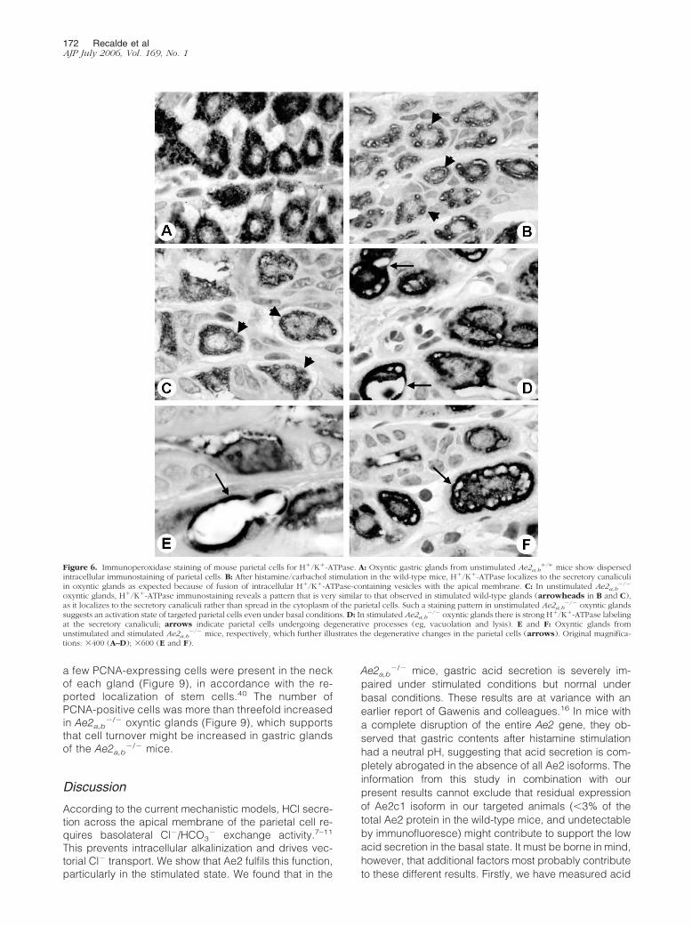
�/� mice is in an increased state ofactivation as an attempt to compensate for impairedacid secretion. Immunohistochemical staining with an-tibodies against H�/K�-ATPase, the proton pump di-rectly responsible for the gastric acid secretion, con-firmed this (Figures 6 and 7). Staining of parietal cellsin Ae2a,b
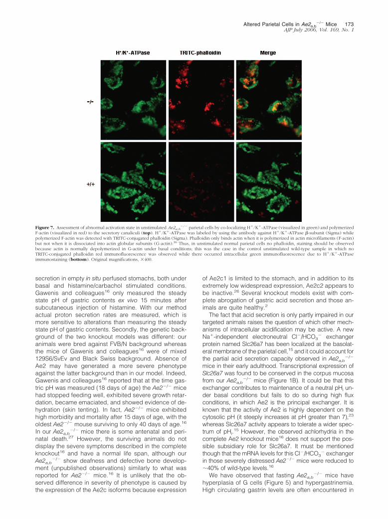
�/� mice indicated that there is a fusion ofintracellular H�/K�-ATPase-containing vesicles withthe apical membrane of the parietal cells both with andwithout histamine/carbachol stimulation, whereas inwild-type mice this fusion was only observed on stim-ulation (Figure 6). The occurrence of such a fusion inunstimulated Ae2a,b
�/� parietal cells was further con-firmed through co-labeling with H�/K�-ATPase anti-
Figure 3. Double-immunofluorescence staining of the parietal cells in thegastric glands of the oxyntic mucosa from unstimulated wild-type and Ae2a,b
�/�
mice, by using a monoclonal antibody against the H�/K�-ATPase �-subunit(visualized in green; Sigma, St. Louis, MO) and a rabbit polyclonal antibodyagainst the Ae2 C-terminal peptide 1223-1237 (visualized in red). Intracellularimmunostaining for H�/K�-ATPase is observed in both wild-type andAe2a,b
�/� parietal cells. However, the Ae2 antibody stains the basolateral mem-brane of the parietal cells in the wild-type parietal cells (arrowheads in corre-sponding merged images), but not the parietal cells of the Ae2a,b
�/� gastricglands. In both wild-type and Ae2a,b
�/� specimens, the Ae2 antibody can alsorecognize the trapped red blood cells (arrows in middle images) because of itscross-reactivity with Ae1. Original magnifications: 200 in the top rows for eachgenotype and 1000 in corresponding bottom rows.
170 Recalde et alAJP July 2006, Vol. 169, No. 1

body and TRITC-conjugated phalloidin (a toxin thatbinds essentially to actin microfilaments, ie, to F-actinor polymerized actin,36 but not to the depolymerized orglobular G-actin). F-actin is associated with the mem-branes of the secretory canaliculus but not with theresting tubulovesicles.37,38 Thus, unstimulated normalparietal cells have no polymerized F-actin, but ratherdepolymerized G-actin, and are expected not to bindlabeled phalloidin. Certainly, this was the case in ourunstimulated wild-type parietal cells (Figure 7). How-ever, unstimulated Ae2a,b
�/� parietal cells showedapical immunofluorescence signals from TRITC-conju-gated phalloidin, which co-localized with the H�/K�-ATPase immunofluorescence, characteristic of parietalcell activation (Figure 7). In addition to these featuresin the unstimulated Ae2a,b
�/� mice, immunoperoxidasestaining for H�/K�-ATPase suggested that a fraction of
their parietal cells undergo degenerative processes(Figure 6D). Because Ae2 is thought to function in pHi
correction during apical proton secretion, it is possiblethat, in the absence of this function, parietal cells aredamaged because of deficient pHi homeostasis duringapical proton secretion.
Electron microscopy of gastric mucosa (Figure 8) pro-vided a striking difference in morphology betweenAe2a,b
�/� and wild-type parietal cells. UnstimulatedAe2a,b
�/� parietal cells showed well-developed tubulo-vesicular system and collapsed canaliculi with no lumen(Figure 8A), that resemble recycling early secretory ele-ments39; moreover, mitochondria appeared concen-trated at the cell periphery in these unstimulatedAe2a,b
�/� parietal cells (Figure 8A). On stimulation,Ae2a,b
�/� parietal cells showed alterations of the tubu-lovesicular system, which varied in severity from col-lapsed canaliculi and decreased number of tubu-lovesicles (Figure 8B) to highly distorted canaliculi(Figure 8C). Also, stimulated Ae2a,b
�/� parietal cellsshowed a lower electrodensity, the periphery of the cellbecoming rather free from organelles (see Figure 8, Band C, for stimulated targeted cell and compare withFigure 8E for stimulated wild-type cell). Additionally, mi-tochondria in stimulated Ae2a,b
�/� parietal cells ap-peared more dispersed than in basal conditions, andsome showed intramitochondrial holes and distorted cris-tae (Figure 8G). On the other hand, both unstimulatedand stimulated Ae2a,b
�/� parietal cells contained ahigher number of dense lysosomes than the wild-typecontrols.
H�/K�-ATPase immunohistochemistry indicated thatthe number and localization of parietal cells within theglands in the oxyntic mucosa were rather similar inAe2a,b
�/� and wild-type mice (not shown). Althoughthere were some parietal cells undergoing degenera-tive processes in the Ae2a,b
�/� mucosa (Figure 6D), aTUNEL assay did not display increased apoptosis (notshown). In any case, the maintenance of a normalnumber of parietal cells despite increased degenera-tive changes suggested an increased turnover of thesecells in the Ae2a,b
�/� mice. We therefore stained gas-tric mucosa for PCNA. In wild-type oxyntic glands only
Figure 4. Gastric proton secretion rate in Ae2�/�, Ae2a,b�/�, and Ae2a,b
�/� mice. Left: Wild-type mice (filled circles) and Ae2a,b�/� mice (open circles).
Right: Wild-type mice (filled circles) and Ae2a,b�/� mice (open triangles). Fractions of the gastric fluid were collected during 15 minutes and titrated to
neutrality to determine proton content. Both histamine and carbachol were administered after 60 minutes from the initiation of the study. Proton secretion rates(y axis) are expressed as �Eq. H�/15 minutes; asterisks, P � 0.001; statistical differences between Ae2a,b
�/� and Ae2a,b�/� (not shown) were like those obtained
when comparing Ae2a,b�/� versus Ae2a,b
�/� (left).
Figure 5. Antral mucosa of the stomach from unstimulated wild-type (A)and Ae2a,b
�/� (B) mice were stained with an antibody to gastrin. For eachanimal, cell count was performed in five serial sections of the antrum; five toseven fields of the antral mucosa, in which the pyloric glands are viewedlongitudinally, were selected and the average number of gastrin-positive cells(G-cells) per field was estimated. A significant increase in the number of Gcells was found in the Ae2a,b
�/� antral mucosa. Original magnifications,200.
Altered Parietal Cells in Ae2a,b�/� Mice 171
AJP July 2006, Vol. 169, No. 1
a few PCNA-expressing cells were present in the neckof each gland (Figure 9), in accordance with the re-ported localization of stem cells.40 The number ofPCNA-positive cells was more than threefold increasedin Ae2a,b
�/� oxyntic glands (Figure 9), which supportsthat cell turnover might be increased in gastric glandsof the Ae2a,b
�/� mice.
Discussion
According to the current mechanistic models, HCl secre-tion across the apical membrane of the parietal cell re-quires basolateral Cl�/HCO3
� exchange activity.7–11
This prevents intracellular alkalinization and drives vec-torial Cl� transport. We show that Ae2 fulfils this function,particularly in the stimulated state. We found that in the
Ae2a,b�/� mice, gastric acid secretion is severely im-
paired under stimulated conditions but normal underbasal conditions. These results are at variance with anearlier report of Gawenis and colleagues.16 In mice witha complete disruption of the entire Ae2 gene, they ob-served that gastric contents after histamine stimulationhad a neutral pH, suggesting that acid secretion is com-pletely abrogated in the absence of all Ae2 isoforms. Theinformation from this study in combination with ourpresent results cannot exclude that residual expressionof Ae2c1 isoform in our targeted animals (�3% of thetotal Ae2 protein in the wild-type mice, and undetectableby immunofluoresce) might contribute to support the lowacid secretion in the basal state. It must be borne in mind,however, that additional factors most probably contributeto these different results. Firstly, we have measured acid
Figure 6. Immunoperoxidase staining of mouse parietal cells for H�/K�-ATPase. A: Oxyntic gastric glands from unstimulated Ae2a,b�/� mice show dispersed
intracellular immunostaining of parietal cells. B: After histamine/carbachol stimulation in the wild-type mice, H�/K�-ATPase localizes to the secretory canaliculiin oxyntic glands as expected because of fusion of intracellular H�/K�-ATPase-containing vesicles with the apical membrane. C: In unstimulated Ae2a,b
�/�
oxyntic glands, H�/K�-ATPase immunostaining reveals a pattern that is very similar to that observed in stimulated wild-type glands (arrowheads in B and C),as it localizes to the secretory canaliculi rather than spread in the cytoplasm of the parietal cells. Such a staining pattern in unstimulated Ae2a,b
�/� oxyntic glandssuggests an activation state of targeted parietal cells even under basal conditions. D: In stimulated Ae2a,b
�/� oxyntic glands there is strong H�/K�-ATPase labelingat the secretory canaliculi; arrows indicate parietal cells undergoing degenerative processes (eg, vacuolation and lysis). E and F: Oxyntic glands fromunstimulated and stimulated Ae2a,b
�/� mice, respectively, which further illustrates the degenerative changes in the parietal cells (arrows). Original magnifica-tions: 400 (A–D); 600 (E and F).
172 Recalde et alAJP July 2006, Vol. 169, No. 1
secretion in empty in situ perfused stomachs, both underbasal and histamine/carbachol stimulated conditions.Gawenis and colleagues16 only measured the steadystate pH of gastric contents ex vivo 15 minutes aftersubcutaneous injection of histamine. With our methodactual proton secretion rates are measured, which ismore sensitive to alterations than measuring the steadystate pH of gastric contents. Secondly, the genetic back-ground of the two knockout models was different: ouranimals were bred against FVB/N background whereasthe mice of Gawenis and colleagues16 were of mixed129S6/SvEv and Black Swiss background. Absence ofAe2 may have generated a more severe phenotypeagainst the latter background than in our model. Indeed,Gawenis and colleagues16 reported that at the time gas-tric pH was measured (18 days of age) the Ae2�/� micehad stopped feeding well, exhibited severe growth retar-dation, became emaciated, and showed evidence of de-hydration (skin tenting). In fact, Ae2�/� mice exhibitedhigh morbidity and mortality after 15 days of age, with theoldest Ae2�/� mouse surviving to only 40 days of age.16
In our Ae2a,b�/� mice there is some antenatal and peri-
natal death.27 However, the surviving animals do notdisplay the severe symptoms described in the completeknockout16 and have a normal life span, although ourAe2a,b
�/� show deafness and defective bone develop-ment (unpublished observations) similarly to what wasreported for Ae2�/� mice.16 It is unlikely that the ob-served difference in severity of phenotype is caused bythe expression of the Ae2c isoforms because expression
of Ae2c1 is limited to the stomach, and in addition to itsextremely low widespread expression, Ae2c2 appears tobe inactive.26 Several knockout models exist with com-plete abrogation of gastric acid secretion and those an-imals are quite healthy.2
The fact that acid secretion is only partly impaired in ourtargeted animals raises the question of which other mech-anisms of intracellular acidification may be active. A newNa�-independent electroneutral Cl�/HCO3
� exchangerprotein named Slc26a7 has been localized at the basolat-eral membrane of the parietal cell,15 and it could account forthe partial acid secretion capacity observed in Ae2a,b
�/�
mice in their early adulthood. Transcriptional expression ofSlc26a7 was found to be conserved in the corpus mucosafrom our Ae2a,b
�/� mice (Figure 1B). It could be that thisexchanger contributes to maintenance of a neutral pHi un-der basal conditions but fails to do so during high fluxconditions, in which Ae2 is the principal exchanger. It isknown that the activity of Ae2 is highly dependent on thecytosolic pH (it steeply increases at pH greater than 7),23
whereas Slc26a7 activity appears to tolerate a wider spec-trum of pHi.
15 However, the observed achlorhydria in thecomplete Ae2 knockout mice16 does not support the pos-sible subsidiary role for Slc26a7. It must be mentionedthough that the mRNA levels for this Cl�/HCO3
� exchangerin those severely distressed Ae2�/� mice were reduced to�40% of wild-type levels.16
We have observed that fasting Ae2a,b�/� mice have
hyperplasia of G cells (Figure 5) and hypergastrinemia.High circulating gastrin levels are often encountered in
Figure 7. Assessment of abnormal activation state in unstimulated Ae2a,b�/� parietal cells by co-localizing H�/K�-ATPase (visualized in green) and polymerized
F-actin (visualized in red) to the secretory canaliculi (top). H�/K�-ATPase was labeled by using the antibody against H�/K�-ATPase �-subunit (Sigma) whilepolymerized F-actin was detected with TRITC-conjugated phalloidin (Sigma). Phalloidin only binds actin when it is polymerized in actin microfilaments (F-actin)but not when it is dissociated into actin globular subunits (G-actin).36 Thus, in unstimulated normal parietal cells no phalloidin, staining should be observedbecause actin is normally depolymerized in G-actin under basal conditions; this was the case in the control unstimulated wild-type sample in which noTRITC-conjugated phalloidin red immunofluorescence was observed while there occurred intracellular green immunofluorescence due to H�/K�-ATPaseimmunostaining (bottom). Original magnifications, 400.
Altered Parietal Cells in Ae2a,b�/� Mice 173
AJP July 2006, Vol. 169, No. 1

animal models with low gastric acid secretion,2 whichmay represent a compensatory mechanism. It seems thatthe hypergastrinemia observed in our targeted micecauses parietal cell stimulation even in the basal state.
Immunoperoxidase labeling for H�/K�-ATPase, the pro-ton pump directly responsible for the gastric acid secre-tion, as well as immunofluorescent co-labeling for H�/K�-ATPase and microfilaments of polymerized actin bothrevealed that unstimulated Ae2a,b
�/� parietal cells exhibita fusion of the tubulovesicular system and pericanalicularredistribution of H�/K�-ATPase-containing vesicles (Fig-ures 6 and 7). In the wild-type parietal cell, it has beendescribed that these changes only occur on stimulationand constitute a characteristic pattern of the secretingstatus.37,41 Electron microscopy confirmed the activa-tion-like pattern in the unstimulated Ae2a,b
�/� parietalcell, but with a well-developed tubulovesicular systemand collapsed canaliculi (Figure 8A) that resemble recy-cling early secretory elements.39 These changes wereaccompanied by mitochondrial alterations with intramito-chondrial holes and distorted cristae and increasednumbers of lysosomes. Subcellular alterations in theAe2a,b
�/� parietal cell worsened with secretory stimula-tion. These alterations together with the previouslymentioned degenerative features observed in targetedparietal cells by immunoperoxidase staining for H�/K�-ATPase (but with no evidence for substantial apoptosis)pointed toward severe damage and death of a fraction ofparietal cells by necrosis. Despite that, the total number
Figure 8. Electron microscopy of the parietal cells in the oxyntic mucosa from unstimulated Ae2a,b�/� mice (A), stimulated Ae2a,b
�/� mice (B–C), unstimulatedwild-type mice (D), and stimulated wild-type mice (E). Unstimulated Ae2a,b
�/� parietal cells (A) show a well-developed tubulovesicular system (TV) but alsocollapsed canaliculi with no lumen (arrows) and mitochondria located at the periphery of the cell (MT). In stimulated Ae2a,b
�/� mice, there are parietal cells(B) with collapsed canaliculi (arrows), decreased tubulovesicles, and peripheral space somewhat free from organelles (arrowheads). There are also parietalcells in stimulated Ae2a,b
�/� oxyntic glands (C) in which canaliculi are broken and rather disorganized (dotted circles). The dotted circle in E indicates awell-organized canaliculi in a stimulated wild-type parietal cell. F and G: Higher magnification of stimulated wild-type and Ae2a,b
�/� parietal cells, respectively.In the latter, some mitochondria (MT) with intramitochondrial holes and distorted cristae are shown. Original magnifications, 3350.
Figure 9. PCNA staining of gastric oxyntic glands from unstimulated wild-type (A) and Ae2a,b
�/� (B) mice. For each animal, cell count was performedin five serial sections of the corpus of the stomach; five to seven fields of theoxyntic mucosa, in which the oxyntic glands are viewed longitudinally, wereselected and the average number of PCNA-positive cells per field wasestimated. The number of PCNA-positive cells was more than threefoldincreased in Ae2a,b
�/� oxyntic glands, indicating increased cell division andproliferation in these glands. Original magnifications, 200.
174 Recalde et alAJP July 2006, Vol. 169, No. 1
of parietal cells seemed to be maintained. Most probablythis could be achieved by a compensatory proliferation ofparietal cells, as suggested by increased PCNA stainingof the Ae2a,b
�/� oxyntic gastric glands.In conclusion, our data show that Ae2 is relevant for
pH homeostasis under conditions of increased protonflux in parietal cells and that under stimulated condi-tions this function is primarily achieved by the Ae2aand Ae2b isoforms, whereas minute levels of theAe2c1 isoform and maintained expression of theSlc26a7 Cl�/HCO3
� exchanger both might be partiallysufficient to counterbalance pH alkalinization underbasal conditions. The shortage in the cytosolic acidifi-cation system may lead to the enhanced parietal celldegeneration that might be compensated by enhancedparietal cell proliferation.
Acknowledgments
We thank P. Garces and B. Irigoyen for their excellenttechnical assistance and Drs. M. Zaratiegui and J.M.Banales for their valuable help with statistics.
References
1. Dockray GJ, Varro A, Dimaline R, Wang T: The gastrins: their produc-tion and biological activities. Annu Rev Physiol 2001, 63:119–139
2. Samuelson LC, Hinkle KL: Insights into the regulation of gastric acidsecretion through analysis of genetically engineered mice. Annu RevPhysiol 2003, 65:383–400
3. Schmidt WE, Schmitz F: Genetic dissection of the secretory machin-ery in the stomach. Gastroenterology 2004, 126:606–609
4. Agnew BJ, Duman JG, Watson CL, Coling DE, Forte JG: Cytologicaltransformations associated with parietal cell stimulation: critical stepsin the activation cascade. J Cell Sci 1999, 112:2639–2646
5. Yao X, Forte JG: Cell biology of acid secretion by the parietal cell.Annu Rev Physiol 2003, 65:103–131
6. Sherry AM, Malinowska DH, Morris RE, Ciraolo GM, Cuppoletti J:Localization of ClC-2 Cl� channels in rabbit gastric mucosa. Am JPhysiol 2001, 280:C1599–C1606
7. Muallem S, Blissard D, Cragoe Jr EJ, Sachs G: Activation of theNa�/H� and Cl�/HCO3
� exchange by stimulation of acid secretion inthe parietal cell. J Biol Chem 1988, 263:14703–14711
8. Seidler U, Carter K, Ito S, Silen W: Effect of CO2 on pHi in rabbitparietal, chief, and surface cells. Am J Physiol 1989, 256:G466–G475
9. Paradiso AM, Townsley MC, Wenzl E, Machen TE: Regulation ofintracellular pHi in resting and in stimulated parietal cells. Am JPhysiol 1989, 257:C554–C561
10. Thomas HA, Machen TE: Regulation of Cl�/HCO3� exchange in
gastric parietal cells. Cell Regul 1991, 2:727–73711. Seidler U, Hubner M, Roithmaier S, Classen M: pHi and HCO3
�
dependence of proton extrusion and Cl�-base exchange rates inisolated rabbit parietal cells. Am J Physiol 1994, 266:G759–G766
12. Stuart-Tilley A, Sardet C, Pouyssegur J, Schwartz MA, Brown D, AlperSL: Immunolocalization of anion exchanger AE2 and cation ex-changer NHE-1 in distinct adjacent cells of gastric mucosa. Am JPhysiol 1994, 266:C559–C568
13. Jons T, Warrings B, Jons A, Drenckhahn D: Basolateral localization ofanion exchanger 2 (AE2) and actin in acid-secreting (parietal) cells ofthe human stomach. Histochemistry 1994, 102:255–263
14. Rossmann H, Alper SL, Nader M, Wang Z, Gregor M, Seidler U: Three5�-variant mRNAs of anion exchanger AE2 in stomach and intestine ofmouse, rabbit, and rat. Ann NY Acad Sci 2000, 915:81–91
15. Petrovic S, Ju X, Barone S, Seidler U, Alper SL, Lohi H, Kere J,Soleimani M: Identification of a basolateral Cl�/HCO3
� exchangerspecific to gastric parietal cells. Am J Physiol 2003, 284:G1093–G1103
16. Gawenis LR, Ledoussal C, Judd LM, Prasad V, Alper SL, Stuart-TilleyA, Woo AL, Grisham C, Sanford LP, Doetschman T, Miller ML, ShullGE: Mice with a targeted disruption of the AE2 Cl�/HCO3
� exchangerare achlorhydric. J Biol Chem 2004, 279:30531–30539
17. Lecanda J, Urtasun R, Medina JF: Molecular cloning and genomicorganization of the mouse AE2 anion exchanger gene. BiochemBiophys Res Commun 2000, 276:117–124
18. Aranda V, Martinez I, Melero S, Lecanda J, Banales JM, Prieto J,Medina JF: Shared apical sorting of anion exchanger isoforms AE2a,AE2b1, and AE2b2 in primary hepatocytes. Biochem Biophys ResCommun 2004, 319:1040–1046
19. Medina JF, Lecanda J, Acın A, Ciesielczyk P, Prieto J: Tissue-specificN-terminal isoforms from overlapping alternate promoters of the hu-man AE2 anion exchanger gene. Biochem Biophys Res Commun2000, 267:228–235
20. Wang Z, Schultheis PJ, Shull GE: Three N-terminal variants of the AE2Cl�/HCO3
� exchanger are encoded by mRNAs transcribed fromalternative promoters. J Biol Chem 1996, 271:7835–7843
21. Stuart-Tilley AK, Shmukler BE, Brown D, Alper SL: Immunolocalizationand tissue-specific splicing of AE2 anion exchanger in mouse kidney.J Am Soc Nephrol 1998, 9:946–959
22. Alper SL, Rossmann H, Wilhelm S, Stuart-Tilley AK, Shmukler BE,Seidler U: Expression of AE2 anion exchanger in mouse intestine.Am J Physiol 1999, 277:G321–G332
23. Stewart AK, Chernova MN, Shmukler BE, Wilhelm S, Alper SL:Regulation of AE2-mediated Cl� transport by intracellular or byextracellular pH requires highly conserved amino acid residues ofthe AE2 NH2-terminal cytoplasmic domain. J Gen Physiol 2002,120:707–722
24. Chernova MN, Stewart AK, Jiang L, Friedman DJ, Kunes YZ, AlperSL: Structure-function relationships of AE2 regulation by Cai
2�-sen-sitive stimulators NH4
� and hypertonicity. Am J Physiol 2003,284:C1235–C1246
25. Stewart AK, Kerr N, Chernova MN, Alper SL, Vaughan-Jones RD:Acute pH-dependent regulation of AE2-mediated anion exchangeinvolves discrete local surfaces of the NH2-terminal cytoplasmic do-main. J Biol Chem 2004, 279:52664–52676
26. Kurschat CE, Shmukler BE, Jiang L, Wilhelm S, Kim EH, ChernovaMN, Kinne RK, Stewart AK, Alper SL: Alkaline-shifted pHo sensitivityof AE2c1-mediated anion exchange reveals novel regulatory deter-minants in the AE2 N-terminal cytoplasmic domain. J Biol Chem 2006,281:1885–1896
27. Medina JF, Recalde S, Prieto J, Lecanda J, Saez E, Funk CD, VecinoP, van Roon MA, Ottenhoff R, Bosma PJ, Bakker CT, Oude ElferinkRPJ: Anion exchanger 2 is essential for spermiogenesis in mice. ProcNatl Acad Sci USA 2003, 100:15847–15852
28. Wang KS, Komar AR, Ma T, Filiz F, McLeroy J, Hoda K, Verkman AS,Bastidas JA: Gastric acid secretion in aquaporin-4 knockout mice.Am J Physiol 2000, 279:G448–G453
29. Cook HC: Periodic acid-Schiff-toluidine blue-aurantia: a stain for thegland cells of the stomach. Stain Technol 1962, 37:317–319
30. Hsu SM, Raine L, Fanger H: The use of antiavidin antibody andavidin-biotin-peroxidase complex in immunoperoxidase technics.Am J Clin Pathol 1981, 75:816–821
31. Shu SY, Ju G, Fan LZ: The glucose oxidase-DAB-nickel method inperoxidase histochemistry of the nervous system. Neurosci Lett 1988,85:169–171
32. Gawenis LR, Greeb JM, Prasad V, Grisham C, Sanford LP,Doetschman T, Andringa A, Miller ML, Shull GE: Impaired gastric acidsecretion in mice with a targeted disruption of the NHE4 Na�/H�
exchanger. J Biol Chem 2005, 280:12781–1278933. Chen D, Zhao CM, Hakanson R, Rehfeld JF: Gastric phenotypic
abnormality in cholecystokinin 2 receptor null mice. Pharmacol Toxi-col 2002, 91:375–381
34. Zolotarev AS, Shmukler BE, Alper SL: AE2 anion exchanger polypep-tide is a homooligomer in pig gastric membranes: a chemical cross-linking study. Biochemistry 1999, 38:8521–8531
35. Frische S, Zolotarev AS, Kim YH, Praetorius J, Alper S, Nielsen S, WallSM: AE2 isoforms in rat kidney: immunohistochemical localizationand regulation in response to chronic NH4Cl loading. Am J Physiol2004, 286:F1163–F1170
36. Cooper JA: Effects of cytochalasin and phalloidin on actin. J Cell Biol1987, 105:1473–1478
37. Mercier F, Reggio H, Devilliers G, Bataille D, Mangeat P: Membrane-
Altered Parietal Cells in Ae2a,b�/� Mice 175
AJP July 2006, Vol. 169, No. 1

cytoskeleton dynamics in rat parietal cells: mobilization of actin andspectrin upon stimulation of gastric acid secretion. J Cell Biol 1989,108:441–453
38. Nguyen NV, Gleeson PA, Courtois-Coutry N, Caplan MJ, Van Driel IR:Gastric parietal cell acid secretion in mice can be regulated inde-pendently of H/K ATPase endocytosis. Gastroenterology 2004,127:145–154
39. Forte JG, Black JA, Forte TM, Machen TE, Wolosin JM: Ultrastructuralchanges related to functional activity in gastric oxyntic cells. Am JPhysiol 1981, 241:G349–G358
40. Karam SM: Lineage commitment and maturation of epithelial cells inthe gut. Front Biosci 1999, 4:D286–D296
41. Forte JG, Hanzel DK, Urushidani T, Wolosin JM: Pumps and path-ways for gastric HCl secretion. Ann NY Acad Sci 1989, 574:145–158
176 Recalde et alAJP July 2006, Vol. 169, No. 1
Copyright © 2022 FDOKUMEN