NEUROPSIKOLOGI RESUME LOBUS PARIETAL MAGISTER PROFESI PSIKOLOGI
Upload
independentCategory
view
1download
0
Neuron, Vol. 32, 985–995, December 20, 2001, Copyright 2001 by Cell Press
Case StudyUnilateral Right Parietal DamageLeads to Bilateral Deficitfor High-Level Motion
level system that appears to require attention (Cava-nagh, 1992; Lu and Sperling 1996). Evidence of twoseparate motion systems was first presented by Wert-heimer (1912), and modern research (e.g., Verstraten etal., 2000) continues to confirm this notion. Recent stud-
Lorella Battelli,1,5 Patrick Cavanagh,1
James Intriligator,2 Mark J. Tramo,3
Marie-Anne Henaff,4 Francois Michel,4
and Jason J.S. Barton2
1Department of Psychologyies have shown that this attention based mechanism isHarvard Universitylimited in both spatial and temporal resolution (Lu andCambridge, Massachusetts 02138Sperling, 1996; He et al., 1996; Verstraten et al., 2000;2 Department of Neurology and OpthalmologyIntriligator and Cavanagh, 2001). In contrast, low-levelBeth Israel Deaconess Medical Centermechanisms can signal motion even when a stimulus isBoston, Massachusetts 02215not attentively tracked and responds to motion at much3 Department of Neurologyhigher temporal frequencies. This low-level passive sys-Harvard Medical School andtem is putatively mediated by directionally selectiveMassachusetts General Hospitalcells in the early visual cortices (Hubel and Wiesel, 1968)Boston, Massachusetts 02114and is velocity based, whereas the high-level system is4 INSERMposition based and depends on attention (Seiffert andUnite 280Cavanagh, 1998, 1999). Since parietal lesions are asso-Lyon, Franceciated with deficits in attention, we hypothesized thatparietal lesions ought to impair high-level motion per-ception. In the present report, we examine two high-Summarylevel motion tasks, namely multiple object tracking andapparent motion. Similarities and differences in the im-Patients with right parietal damage demonstrate a va-pairments for these two high-level motion tasks shouldriety of attentional deficits in their left visual field con-reveal similarities and differences in the attention mech-tralateral to their lesion. We now report that patientsanisms they may call on.with right lesions also show a severe loss in the per-
Selective attention mechanisms were tested with aception of apparent motion in their “good” right visualtask of divided attention in which the subject had tofield ipsilateral to their lesion. Three tests of attentionreport a letter presented among three different letterswere conducted, and losses were found only in thefor 66 or 300 ms. At the offset of the display, the subjectcontralesional fields for a selective attention and awas required to identify a letter based on its position inmultiple object tracking task. Losses in apparent mo-the letter string. The cued report for brief visual displaystion, however, were bilateral in all cases. The deficitwas first developed by Sperling (1960), and we used ain apparent motion in the parietal patients supportsmodified version of his task. Recent studies have shownprevious claims that this relatively effortless percepthow patients with parietal lobe lesions can be affectedis mediated by attention. However, the bilateral deficitin their ability to perform this type of task (Duncan etsuggests that the disruption is due to a bilateral loss inal., 1999). In contrast to the tasks of Duncan et al., wethe temporal resolution of attention to transient eventsasked our subjects to report only one letter among four,that drive the apparent motion percept.and the subject never knew which letter until briefly afterthe letters were presented. With only four items, normalIntroductionsubjects could attend to and select the indicated targetwith ease.
Many studies have indicated the importance of parietalPerception of motion-defined rectangles was used as
cortex in attention (cf. Corbetta et al., 1998). In particular, a test for deficits in low-level motion perception wherefollowing damage to the right parietal region, patients no tracking was involved. Subjects had to judge if theoften exhibit hemilateral neglect, including serious defi- rectangles were horizontally or vertically oriented. Previ-cits in attentional processing in the contralesional visual ous studies have shown a variety of motion deficits infield (Posner et al., 1984). In this paper, we contrast the patients with lesions in extrastriate motion areas, suchperformance of parietal patients to normal subjects on as MT, V3, or V3A (Zihl et al., 1983; Plant and Nakayama,two high-level motion tasks in order to determine the 1993; Vaina and Cowey, 1996; Greenlee and Smith, 1997;extent and nature of attentional processing underlying Vaina et al., 1998). These studies provide evidence thatthese tasks. MT is located in the lateral temporo-occipital cortex. On
Much of our perception of motion appears to be ef- the other hand, Greenlee et al. (1995) examined fourfortless, as if little in the way of attention is required. parietal lobe patients and found no deficit in motionHowever, there are two well established motion systems perception (speed judgments in particular). Spinelli and(Julesz, 1971; Braddick, 1974; Anstis, 1980; Cavanagh, Zoccolotti (1992) found that the perception of moving1992; Lu and Sperling, 1996): a low-level system (or gratings in patients with unilateral spatial neglect andsystems, according to Lu and Sperling, 1996) that is parietal lesions was normal. Therefore, it appears thatindeed effortless, passive, and preattentive, and a high- the temporo-occipital region, but not the parietal lobes,
is involved in direction or speed judgments of low-levelmotion.5 Correspondence: [email protected]
Neuron986
One important role of attention is to select objects of Unlike attentive tracking, perceiving apparent motioninterest in the environment and keep track of them as seems relatively effortless. No instructions are required,they move (Pylyshyn and Storm, 1988). A single object and the motion percept is uniformly seen by all normalcan be tracked with eye movements, but the eyes cannot observers. It is this ease of perception, coupled withfollow more than one object; therefore, additional atten- the possibility of use as a probe of attention, that hastional mechanisms are required to track multiple moving focused our interest on apparent motion as a test forobjects. A typical multiple object tracking test (Pylyshyn patient populations. In our apparent motion task, theand Storm, 1988) is constructed as follows: nine identi- perception of motion depends on an accurate analysiscal disks are set in random motion in a display. Four of not only of the location of the flashes but also of theirthe disks are identified as targets by flashing briefly timing. The evidence for a role of attention in apparentbefore again becoming identical to all of the other disks. motion (Dick et al., 1991; Verstraten et al., 2000) impliesThe observers keep track of the targets while all nine that a loss of either spatial or temporal attention coulddisks move about randomly. After 5 s, all of the items disrupt motion perception. The evidence for losses instop moving and the observer reports which disks were spatial attention following parietal damage is wellthe four targets. With concentrated effort, most people known; additionally, there is evidence for losses in tem-can successfully track four targets. This task, like many poral attention. For example, patients affected by exten-video games, is engaging but tiring. Although attention is sive right hemisphere lesions are impaired in auditorycentral to the performance of this task, the continuously tasks requiring time perception (Harrington et al., 1998;moving disks all generate low-level motion responses Cusak et al., 2000) as well as in the orienting of attentionas well. It is not yet clear the extent to which these low- in time (Husain et al., 1997).level signals contribute to the accuracy of tracking. One note of caution, however: if the two flashes of the
Several neuroimaging and TMS studies have shown apparent motion display are sufficiently close together,that the parietal cortex is important for visual attention they will trigger both a low-level response by fallingin general (Corbetta et al.1993, 2000; Pascual-Leone et within the receptive field of a directionally selective unital., 1994; Ashbridge et al., 1997; Hilgetag et al., 2001). and the presumed high-level response. Conversely, ifIn addition, patient studies show that visuospatial and the step size between the stimuli is sufficiently largeattentive problems are often seen after damage to the (more than 2�) (Anstis, 1980), low-level motion makesparietal lobes (Arguin et al., 1994; Robertson et al., 1997; no contribution to apparent motion (Anstis, 1980; Boul-Robertson and Manly, 1999). ton and Baker, 1993).
A recent fMRI study (Culham et al., 1998) found that We tested seven patients: three with unilateral rightthe parietal areas were significantly more active during parietal lesions (cases DS, JR, and JL), three with bilat-the multiple object tracking task than in passive viewing eral parietal lesions (cases WGD, AT, and LF), and oneof the same stimuli. However, tracking did not differen- control patient (case IB) with a more posterior lesion totially activate other regions involved in motion percep- the visual areas sparing the parietal cortex. Patients’tion (like the MT region). This lack of MT activation con- performance was compared to three age-matched con-trasts with prior reports of attentional modulation of trol subjects. All of our unilateral right parietal patientslow-level motion signals in this area. Thus, the results had some signs of visual neglect.of Culham et al. (1998) would appear to be specific tohigh-level motion and not a general effect of attention Resultsupon motion signals (O’Craven et al., 1997). Based onthese results (as would be expected), a preliminary re-
Experiment 1: Static Letter Detectionport has found that parietal lesions disrupt attentive
This task measured attentional selection in the absencetracking in contralesional fields (F. Michel et al., 1997,
of any stimulus motion. We used a modified version ofCognit. Neurosci. Soc., abstract).the partial report first used by Sperling (1960) to testA stimulus does not have to move in a continuousselection from a brief visual display of multiple stimuli.manner to be seen to move. When a single light is flashedWe presented four letters in a horizontal array in onebriefly at one location followed shortly thereafter by afield or the other for an exposure time of 66 and 300 mssecond light at another location, a clear impression ofand asked subjects to report a letter from a particularmotion is produced. Whether this motion is low-level orposition in the array, with a different position specifiedhigh-level and how the two separate events are linkedon each trial (Figure 1A).into one motion percept (the correspondence problem)
Patients JR and JL were tested in this task. The resultshas been debated for many years (Ullman, 1978; Cava-are shown in Figure 2. As expected, they both showednagh and Mather, 1989).a selective impairment in the hemifield contralateral toAlthough the perception of motion in the flashed stim-the lesion site (Figure 2). Their failure to perform thisulus seems effortless, a number of studies have sug-task was not due to impaired visual resolution or lettergested that attention is involved (Dick et al., 1991; Ver-recognition as the patients were both able to read thesestraten et al., 2000; see Mather, 1994, for a review).letters when presented singly.Wertheimer (1912) was the first to suggest that apparent
motion might involve attentive tracking: the result of theExperiment 2: Motion-Defined Rectanglesinvoluntary dragging of attention from the first flash toThis task examined the patients’ ability to detect low-the second. Attention is “grabbed by” the first stimuluslevel motion. The monitor presented a field of randomlyand then the second stimulus, and this is referred to asflickering black and white dots (see Experimental Proce-an exogenous shift of attention (Yantis, 1993) or “passive
sensorial attention” (James, 1890). dures). In the center of one quadrant, a region of dots

High-Level Motion in Right Parietal Patients987
Figure 1. Examples of the Four Tasks
(A) Example of the stimuli used in experiment 1.(B) Example of the stimuli used in experiment 2. The 90% coherence difference is shown. The arrows indicate motion, and they were notpresent on the display. The vertically oriented rectangle did not have a line contour as the shape was made visible solely by the differencein motion direction coherence.(C) Example of the stimulus used in experiment 3. The subject had to track two out of five disks. In the actual task, the target disks wereindicated by turning red (here depicted as black) (Time 1) for a brief interval after which they reverted to the same green as the others, whilethe other remained green (here depicted as white) (Time 2).(D) Example of stimuli in experiment 4. In the apparent motion stimulus, two dots in diagonally opposite corners are flashed simultaneouslyand then switched off (Frame 1) and replaced by two dots appearing on the remaining two corners (Frame 2). The frames are alternated in acontinuous cycle. In the flicker stimulus, all four dots are switched on simultaneously and remain on for the same duration as in the apparentmotion case. At high-cycle rates, the apparent motion stimulus generates no motion impressions and appears to be four flashing dots. Atthese rates, the discrimination performance drops to chance.
moved together in a semicoherent fashion (Figure 1B). al., 1998; Newsome and Pare, 1988). Patients affectedby MT lesions fail in low-level motion tasks where theyThis coherent region was rectangular in shape and ori-
ented either horizontally or vertically. The subject’s task are asked to detect motion of a small number of dotscoherently moving within a dynamic background. How-was to indicate the orientation. This type of test is used
widely in psychophysical and neurophysiological litera- ever, they perform normally in motion segmentationtasks when the background is stationary (Vaina et al.,ture to examine low-level motion mechanisms (Vaina et1990) or when there is only a low-level of backgroundnoise (Rizzo et al., 1995) at very long exposure time(5.4 s). In our task, there was always a high proportionof background noise in which the target shape was em-bedded, and the subjects had to detect the global mo-tion to perceive the target shape correctly. The shapewas made more visible by increasing the percentage ofdots coherently moving in the same direction. Trackingthe moving dots within the rectangle was not necessaryto make the shape judgment, and the tracking of anyindividual dot did not improve the performance (the rect-angular shape is a property of the overall motion, andit cannot be recovered from the motion of any individualdot). Attention is required in order to notice and report
Figure 2. Static Letter Detectionthe shape of the rectangle, but the motion that defines
The percentage of correct responses in the static letter selectionthe shape does not require attention for the shape totask. Results for left (LVF) and right (RVF) visual fields are reportedbecome visible. This has been demonstrated in a visualseparately for patients JR, JL, and control subjects. The dotted line
indicates chance (25%). search task (Cavanagh et al., 1990) where the speed to

Neuron988
Figure 3. Motion-Defined Rectangles
The percent of dot coherence at which the subjects perform 75%correct are reported for each unilateral patient: DS, JR, and JL, anda group of three age-matched controls. Lower coherence thresholdindicates better performance. Average threshold was 40 (�10.5) forage-matched control and 39.4 (�9.6) for the patients. The dottedline indicates the average performance of control subjects. On they axis, the arrows indicate good and bad performance.
detect a vertical, motion-defined rectangle in a field ofhorizontal, motion-defined rectangles (distractors) was
Figure 4. Multiple Object Trackingunaffected by the number of distractors.(A) Results display tracking for two out of five disks. Percentage ofFigure 3 depicts the results for the motion-definedcorrect responses are represented as a function of visual field (leftrectangle judgments. The percent coherence thresholdand right) for each unilateral patient (DS, JR, and JL), a lesion control
(at which the patients could report orientation at 75% patient (IB), and a group of three age-matched controls. The dottedcorrect) is shown for each patient and for age-matched line indicates chance level (40%).
(B) Results display tracking for one out of three disks. Percentagecontrols (this threshold was obtained with a smoothedof correct responses are represented as a function of visual fieldfunction-fitting procedure, and the function derives a(left and right) for each patient and a group of three age-matchedpoint from each coherence level that is weighted by itscontrols. The dotted line indicates chance level (33%). Higher per-total number of observations). There was no systematiccent correct indicates better performance.
difference in performance between the left and rightvisual fields. Therefore, the data in the graph for left andright visual fields have been collapsed. Thus, we confirm in their ipsilesional fields (in the portions of visual space
represented in their unaffected hemisphere). However,previous reports (Spinelli and Zoccolotti, 1992) that low-level motion perception is normal in both fields in pa- performance in the tracking task was severely degraded
in the contralesional field for the three patients. Thetients with right parietal damage.Fisher’s exact test confirmed that the contralateral fieldsof the patients significantly differed from the hemifieldsExperiment 3: Multiple Object Tracking
The next question we asked was whether the right pari- of age-matched control subjects (p � 0.001 for eachcomparison). When tracking only one disk, DS and JRetal patients would also be able to perform a task that
relies on the high-level (attentive) motion system. We showed some loss in the contralesional field (Figure 4B)but not as severe as when tracking two. JL was moreinvestigated this by having the patients perform a multi-
ple object tracking task. Subjects were asked to track severely impaired in single object tracking in the lefthemifield. Because these subjects performed normallyeither one or two items out of a field of five identical
moving items. This task required continuous attentional on the low-level motion task, we consider these lossesa manifestation of disrupted attentional tracking rathermonitoring of moving stimuli (Figure 1C). Assuming that
the patients could see the low-level motion of these than a loss in low-level motion processing. The partiallyspared ability of DS and JR to track a single movingitems, this second experiment added an attentional
component of keeping track of the items as they moved. object in the contralesional field confirmed that the stim-uli were visible, their motions could be discerned, andIn Figures 4A and 4B, the percentage of successful
tracking responses for each visual field is reported for that the task instructions were understood. With onlyone target on one side of fixation, there is a concerneach patient and a group of age-matched controls. Re-
sults are reported for tracking two out of five (Figure 4A) that the patients may make eye movements to bring thesingle target into their good field. This might also applyand one out of three disks (Figure 4B). Note that when
two disks had to be tracked, they were always displayed to the two target display, although it is less likely be-cause if the patient had moved his eyes to the contrale-one in each hemifield. In such a situation, the best
tracking strategy was to maintain fixation on the bull’s- sional target, then he would have lost track of the ipsile-sional one. Results show that this was not the case. Weeye and follow both targets with attention. A Fisher’s
exact test was used to compare each hemifield of each did ask the patients to maintain fixation, and they didnot report making any eye movements to the singlesubject with the age-matched controls. The patients
could track as proficiently as the age-matched controls target. Nevertheless, some of their ability to track in the

High-Level Motion in Right Parietal Patients989
of 9.8 Hz and 8.5 Hz in the left and right visual fields,respectively, whereas all of the patients reached thresh-old at much lower alternation frequencies (Figure 5).Compared to the tracking task, we observed a remark-able difference in the pattern of loss (Figure 4). Whileobject tracking was only impaired in the contralesionalhemifield, all three unilateral patients (DS, JR, and JL)showed bilateral loss.
How Do Bilateral Parietal Patients Performon High-Level Motion TasksWe were able to test three bilateral parietal patients (LF,WGD, and AT) to see whether their deficits for apparentFigure 5. Apparent Motionmotion would be similar to those of the right parietalMotion threshold for each unilateral parietal patient (DS, JR, andpatients or more severe. They all showed selective atten-JL), for the lesion control patient (IB), and three age-matched con-
trols. The threshold is expressed as the cycle rate at which the tion impairments in both hemifields (Table 1) and bilat-subject could discriminate flicker from motion with 75% accuracy. eral losses in the apparent motion task. The patientsThe lower the threshold, the worse the performance. The dotted were all able to read letters singly presented, and theirline indicates the average performance of the group of age-matched
linguistic abilities were preserved.control subjects.Low-level motion performance (experiment 2) was
normal (patients LF and AT) or better than normal (WGD,the youngest patient). Because LF found it difficult tocontralesional field may be attributable to uncontrolledfocus on the fixation mark, he was tested with the mo-eye movements.tion-defined stimulus at screen center, and he per-On the other hand, the impairment in tracking oneformed normally. Patient AT’s low-level motion wasitem in the contralesional field when there was a secondtested with a slightly different task. She was presentedtarget in the other field confirms the expected loss forwith two squares, one with dots moving in random tra-spatial attention in the contralesional field. This resultjectories and another with a mixture of randomly movingcould be explained in terms of visual extinction (Vallardots and coherently moving dots, the proportions ofet al., 1994), an inability to detect a target during simulta-which were varied. The task was a same/different judge-neous presentation of another similar target in the oppo-ment as a function of the coherence ratio. Her perfor-site hemifield. The patients could perceive a single stim-mance was in the normal range.ulus in either hemifield when presented alone. Extinction
All three patients showed a deficit in tracking two ofoften persists after recovery from more severe signs offive items in the multiple item tracking. AT performedneglect (Vuilleumier and Rafal, 2000).the visual tracking with a slightly different stimuli presen-tation as the target disks were displayed in one singleExperiment 4: Apparent Motionregion, whereas in all other cases, the disks were pre-We next asked our patients to participate in a simplesented within two gray regions centered from fixation.apparent motion task. Subjects reported whether they
Finally, the bilateral parietal patients also showed sig-saw motion or static flickering in a display of four dots.nificant losses in the apparent motion task in both fieldsWith an appropriate temporal offset between the dots,(experiment 4). WGD did retain better sensitivity to ap-there was a compelling motion illusion (Ternus, 1938;parent motion in his left field than in his right field. TheRamachandran and Anstis, 1983), either horizontally orrange of thresholds for the bilateral patients was veryvertically. In an attempt to prevent the subject fromsimilar to that of the right parietal patients (4–6 Hz).moving his/her eyes toward the target stimulus, the
quadrant of presentation was unpredictable across tri-als. The display is schematized in Figure 1D. The fre- Discussionquency of alternation was varied from 1.7 to 14.9 Hzacross trials. On each trial, the observers saw either an This study provides important findings about the tempo-
ral dynamics of attention in left hemispatial neglect. Pa-alternating quartet or a display in which all four dotsappeared and disappeared simultaneously. Finally, to tients affected by right parietal lesion are impaired at
performing an apparent motion task in both left andprevent judgements based on the first display frame(e.g., “did I just see two dots or four?”), there was a right visual fields. Our results show that the impairment
is not due to problems in low-level motion, spatial atten-pretrial delay of 40 ms before each trial during whichthe four dots were flashed simultaneously. tion, selection, or tracking as all of these are at normal
levels in the right (ipsilesional) field for these patients.The results are plotted in Figure 5. Note that in thistask, low thresholds (slower critical rates) indicate poor Having ruled out these factors, we are left with only
one aspect that distinguishes apparent motion from theperformance. Motion thresholds were taken as the alter-nation frequency at which subjects could discriminate other forms of motion that we tested: the rapid deploy-
ment of transient attention to the discrete, sequentialmovement from flickering 75% of the time. The thresholdwas obtained by fitting the data with a smoothing func- flashes of the stimulus. We claim that the patients’ diffi-
culty with apparent motion results from the inability oftion. The thresholds for both left and right hemifieldsare reported separately. A group of three age-matched transient attention to resolve the successive onsets and
offsets of adjacent flashes, preventing their integrationcontrol subjects could perceive motion at a threshold
Neuron990
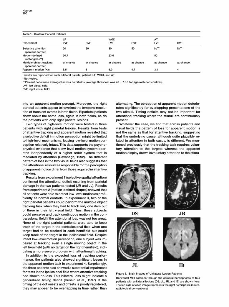
Table 1. Bilateral Parietal Patients
LF WGD ATExperiment LVF RVF LVF RVF LVF RVF
Selective attention 20 30 30 50 N/T* N/T(percent correct)
Motion-defined 50.7 23.6 50rectangles (**)
Multiple object tracking at chance at chance at chance at chance at chance at chance(percent correct)
Apparent motion (Hz) 5.5 6 6.9 4.7 3.1 4
Results are reported for each bilateral parietal patient: LF, WGD, and AT.* Not tested.** Percent coherence averaged across hemifields (average threshold was 40 � 10.5 for age-matched controls).LVF, left visual field.RVF, right visual field.
into an apparent motion percept. Moreover, the right alternating. The perception of apparent motion deterio-rates significantly for overlapping presentations of theparietal patients appear to have lost the temporal resolu-
tion of transient events in both fields. Biparietal patients two stimuli. Timing deficits may not be important forattentional tracking where the stimuli are continuouslyshow about the same loss, again in both fields, as do
the patients with only right parietal lesions. present.Whatever the case, we find that across patients andTwo types of high-level motion were tested in three
patients with right parietal lesions. Results from tests visual fields the pattern of loss for apparent motion isnot the same as that for attentive tracking, suggestingof attentive tracking and apparent motion revealed that
a selective deficit in motion perception might be limited that the underlying cause, although quite plausibly re-lated to attention in both cases, is different. We men-to high-level mechanisms, leaving low-level motion per-
ception relatively intact. This data supports the psycho- tioned previously that the tracking task requires volun-tary attention to the targets whereas the apparentphysical evidence that a low-level motion system oper-
ates independently of a higher order system that is motion display draws involuntary attention to the stimu-mediated by attention (Cavanagh, 1992). The differentpattern of loss in the two visual fields also suggests thatthe attentional resources responsible for the perceptionof apparent motion differ from those required in attentivetracking.
Results from experiment 1 (selective spatial attention)confirmed the attentional deficit resulting from parietaldamage in the two patients tested (JR and JL). Resultsfrom experiment 2 (motion-defined shapes) showed thatall patients were able to detect low-level motion as profi-ciently as normal controls. In experiment 3, two of theright parietal patients could perform the multiple objecttracking task when they had to track only one item outof three in their left visual field. Thus, these subjectscould perceive and track continuous motion in the con-tralesional field if the attentional load was not too great.None of the right parietal patients were able to keeptrack of the target in the contralesional field when onetarget had to be tracked in each hemifield but couldkeep track of the target in the ipsilesional field. Despiteintact low-level motion perception, one subject was im-paired at tracking even a single moving object in theleft hemifield (with no target on the right hemifield), indi-cating a more severe problem with attentional tracking.
In addition to the expected loss of tracking perfor-mance, the patients also showed significant losses inthe apparent motion task in experiment 4. Surprisingly,the three patients also showed a substantial impairmentfor tests in the ipsilesional field where attentive tracking Figure 6. Brain Images of Unilateral Lesion Patientshad shown no loss. This bilateral loss might indicate a
Horizontal MRI sections through the cerebral hemispheres of fourgeneralized timing deficit (Husain et al., 1997). If the patients with unilateral lesions (DS, JL, JR, and IB) are shown here.timing of the dot onsets and offsets is poorly registered, The left side of each image represents the right hemisphere (neuro-
radiological conventions).they may appear to be overlapping in time rather than

High-Level Motion in Right Parietal Patients991
Table 2. Distribution of Lesions
Superior Lateral Middle InferiorParietal Angular Supramarginal Occipital Lingual Temporal Temporal
Patient Lobule Gyrus Gyrus Precuneus Gyri Cuneus Gyrus Gyrus Gyrus
DS R R R RJR R R R R R RJL R R R R R RIB L L L LLF R,L R,L R,L R,L RWGD R,L R,L R RAT R,L R,L R,L R,L L
Distribution of lesions involving visual cortex and multimodal cortex in the parietal, occipital, and temporal lobes.R, right hemisphere.L, left hemisphere.
lus. Thus, one possible explanation of the dissociation get stimulus. Hence, it may be the conjunction of bothspatial and temporal attention that creates the rightin outcomes for the two tasks is that it reflects variations
in the contributions of the two hemispheres to endoge- hemispheric bias in our apparent motion task.Interestingly, a recent single unit study in macaquenous and exogenous attention (Corbetta et al., 2000).
In an fMRI study, Corbetta and coworkers found that monkeys (J. Assad, personal communication) supportsour findings. Direction-selective neurons in parietal cor-the temporo-parietal junction and the precuneus in the
right hemisphere were active when a visual target ap- tex were tested with a visual stimulus consisting ofevenly spaced columns of dots that could be displacedpeared at an unattended location, while both intraparie-
tal sulci were active during voluntary orienting to a tar- at fixed intervals by a fraction of the intercolumn spacingto create a uniform apparent motion. The animals wereget. Although the size of the lesion varies across our
right patients (Figure 6), they all had lesions involving trained to report the direction of the apparent motion. Onsome presentations, the dots were displaced by exactlythe angular gyrus (which includes the area called the
temporo-parietal junction by Corbetta et al.) and precu- half of their intercolumn spacing such that the perceiveddirection of motion was perceptually bistable. On theseneus (see Table 2 for a synthesis of the anatomical
distribution of the lesion for each patient) which Corbetta bistable presentations, many neurons in the lateral intra-parietal area (LIP) were more active when the animalet al. implicate in the orientation of attention toward
sudden stimuli in an unattended area like those in our reported perceiving the neuron’s preferred direction ofmotion than the opposite direction of motion. In MT andapparent motion task. Alternatively, a combined PET
and fMRI study (Coull and Nobre, 1998) showed activa- MST, far fewer neurons were modulated by the animal’sreport of direction on perceptually bistable trials. There-tion of the right parietal cortex when subjects were re-
quired to attend to both spatial and temporal properties fore, higher parietal cortical areas appear to be some-how involved in a representation that reflects subjectiveof a cue. Our apparent motion task requires orienting
of attention in space to sudden onsets together with a perception.Finally, it is interesting to notice that all three rightprecise registration of temporal intervals within the tar-
Figure 7. Brain Images of Bilateral Patients
Horizontal and sagittal MRI sections throughthe cerebral hemispheres of patients with bi-lateral lesions (LF, WGD, and AT) are shownhere, showing their left (LH) and right (RH)hemispheres.

Neuron992
parietal patients lost the perception of apparent motion temporal resolution deficit of visual perception whichat alternation rates higher than 6 Hz, which corresponds could probably affect other modalities. A recent studyto an SOA of about 200 ms. This is similar to the results (Harrington et al., 1998) confirms this hypothesis andreported by Rorden et al. (1997), where the authors pre- supports the idea of a role of the right inferior parietalsented right parietal patients with a temporal order cortex in time perception task with auditory stimuli.judgement (TOJ) task. They presented two unconnected Right parietal patients were unable to judge the differ-bars, one in each visual field with different time intervals ence in duration of two tone pairs, while left parietalbetween them, and the patients were asked to report patients could perform the task as well as normal con-which bar appeared first. The authors assumed that trols.when two stimuli were physically simultaneous, the bar The data collected with bilateral parietal patients (seethat had the subject’s attention would be seen first. Table 1 and Figure 7) shows that, as expected, perfor-When normal subjects were asked to maintain fixation, mance on the selective attention, tracking, and apparentthey correctly reported simultaneous stimuli as simulta- motion tasks was poor in both fields, whereas low-levelneous. In contrast, Rorden et al. needed to present the motion was preserved in both fields. Most importantly,contralesional bar 200 ms in advance in order for the for the apparent motion task, the loss in performanceparietal patients to judge it as simultaneous with the bar was no worse than for the patients with only right parietalin the ipsilesional field. With anything less than 200 ms lesions. We are currently investigating left parietal pa-advanced presentation, the patients always judged the tients to determine whether the bilateral deficit we foundipsilesional bar as coming first. They explained the re- is specific to the right parietal patients. We ran threesults in terms of a disruption in the ability to judge the unilateral left parietal patients, and preliminary dataorder of events displaced in space (one in each hemi- showed preserved ability to perform low-level motionfield) and time. This impairment causes a severe bias perception. In the attention-based motion tasks, twoto the right (ipsilesional side) and a consequent delay patients were impaired at tracking the disks in the fieldin visual awareness for contralesional events. We dem- contralateral to the lesion (right visual field) confirmingonstrated that a timing deficit can occur within each the attentional deficit while the third patient performedfield, a result that rules out a simple differential delay like normal controls in this task. Furthermore, two of thebetween the two hemifields or an alertness problem left parietal patients performed normally in the apparent(Robertson et al., 1998) as the only underlying cause of motion task, while one showed losses, more severelythe deficit in the apparent motion task. in the left visual field. This last patient had additional
Furthermore, although our patients could all discrimi- lesions in the right basal ganglia that might have contrib-nate synchronous from alternating flashing at rates in uted separately to a deficit in visual spatial attentionthe range of 2–4 Hz, performance abruptly fell to chance (Vallar, 1993; Bellmann et al., 2001).again at lower rates for some of them. These results In conclusion, our data support two different roles forcontrast sharply with those found with normal observers attention in high-level motion, and these are affectedwho can see motion up to SOAs of 700 ms (1.4 Hz). differently by parietal lesions. Active tracking tasks ap-Patient IB, who had a left occipital lesion, performed the pear to call on voluntary, sustained attention, and this istask as well as age-matched controls in all conditions. impaired in the field contralateral to the lesion. Apparent
In sum, we found that right parietal lesions can cause motion tasks appear to rely on involuntary, transienta visual tracking deficit in the contralesional field, while attention, and the resolution of events picked up by thisperformance in the ipsilesional field remains intact. Fur- attention system appears to be much reduced followingthermore, we found that apparent motion can also be right parietal damage. This loss then degrades apparentdisrupted by right parietal lesions, whereas the percep- motion performance in both fields.tion of low-level motion remains intact. Performance ofour parietal patients in this task was different from that
Experimental Proceduresof the patients affected by lesion to the motion area MT(Vaina et al., 1990; Rizzo et al., 1995). Furthermore, the
Case Historiesfeatures of our low-level motion task ruled out the possi- We formally tested seven stroke patients. Three of the patients, DS,bility that we were testing independent motion pro- JR, and JL had unilateral, right parietal lobe lesions with somecessing areas such as KO (Dupont et al., 1997) or early extension into surrounding structures. Three of the other patients,visual areas such as V1, which has been demonstrated WGD, LF, and AT, had bilateral lesions involving parietal cortex
and surrounding structures. In one patient, IB, the lesion principallyto be significantly active during motion segmentationinvolved the left occipital cortex with limited extension into adjacenttasks (Reppas et al., 1997). In our task, the percentageparietal cortex. Magnetic resonance images (MRIs) acquired forof dots coherently moving in the same direction in theclinical indications were analyzed to define the anatomical distribu-
target shape varies randomly across trials between six tion of the lesions using the Damasio atlas (1995). Three subjects,levels (see Experimental Procedures for more details), two males and one female, with no history of neurological diseasewhile in the tasks used in the fMRI studies mentioned (mean age 65.6), served as age-matched normal controls.above the dots density was constant. In a coherence Patient DS, a 65-year-old right-handed man, was admitted to the
hospital in September 1998 with left hemispatial neglect and mild leftmotion task (similar to our low-level motion task) usedhemiparesis. Computerized tomography (CT) showed a hemorrhagein a recent fMRI study (Shulman et al., 1999), patternsinvolving deep and superficial right parietal lobe structures. Twoof strong activation were observed in MT. This datamonths later, MRI showed signal abnormalities involving the right
supports our finding that visual areas subserving low- superior parietal lobule, angular gyrus, supramarginal gyrus, andlevel motion were preserved in our patients. precuneus (Figure 6). When we first tested DS 6 months after his
Finally, the different attentional deficits we found in stroke, he felt fully recovered and was back to his usual activities,including golf. However, he complained that he consistently droveour patients could indicate a more general spatial and

High-Level Motion in Right Parietal Patients993
the ball off to the right of the fairway. On the Sunnybrook neglect bilaterally (Figure 7). MRI showed signal abnormalities in the rightand left superior parietal lobule, angular gyrus, supramarginal gyrus,battery (Black et al., 1990), DS scored 14/100, indicating mild resid-
ual left hemispatial neglect. lateral occipital gyri, and postcentral gyrus plus the left cuneus.When we tested AT 15 years later, signs of Balint’s syndrome werePatient JR, a 70-year-old right-handed man, was admitted to the
hospital in October 1998 with left hemispatial neglect, left superior still present. Visual field testing revealed homonymous paracentralscotomas in the right inferior quadrant.quadrantanopia, normal visual acuity, and left hemiparesis. CT re-
vealed a hemorrhage involving deep and superficial right parietal Table 2 reports a synthesis of the anatomical distribution of thelesion for each patient.lobe and temporal lobe structures. Ten months later, MRI showed
extensive signal abnormalities involving the right superior parietal Ethical committee approval from Harvard University and from BethIsrael Deaconess Medical Center were granted for all procedures.lobule, angular gyrus, supramarginal gyrus, precuneus, lateral oc-
cipital gyri, and middle temporal and superior temporal gyri. Theanterior extent of the lesion also involved the transverse gyrus of EquipmentHeschl, parahippocampal gyrus, temporal pole, postcentral and pre- The experiments were conducted on a PowerMac 120 computer.central gyri, and the inferior frontal gyrus. Deeper involvement in- Software for experiments 2, 3, and 4 were written in Think C usingcluded the insula, putamen, and posterior limb of the internal cap- programming routines (Shell) created by Raynald Comtois (http://sule (Figure 6). We first tested JR 8 months after his stroke. He www.kagi.com/visionshell/). Experiment 1 was programmed oncomplained of strange visual phenomena, such as an ant changing PsychLab 2.2.6 for Macintosh. An Apple Hi-Res color monitor waslocation on the floor without actually having seen it move from one used for all experiments for all subjects except JR, JL, and LF,position to another. On the Sunnybrook neglect battery (Black et where a Powerbook 1400c/166 computer and Apple Studio Displayal., 1990), JR scored 10/100, indicating mild residual left hemispatial were used. The monitors were calibrated for linearity before theneglect. experiments were conducted. The same basic equipment was used
Patient JL, a 62-year-old right-handed man, suffered a left occipi- in all experiments.tal lobe hemorrhage in 1998 and a right parietal hemorrhage in1999. After the first stroke, the patient exhibited symptoms of right Stimuli and Procedurehemianopia, which gradually recovered. After the second stroke, In experiments 2 and 4, the stimuli were always presented centeredthe patient exhibited symptoms of left hemianopia, which have not 10� from the fixation point in one of the four quadrants: upper left,improved. Nine months after his second stroke, MRI revealed right- upper right, lower left, or lower right. The presentation of the stimulisided signal abnormalities involving the superior parietal lobule, was in one of the four quadrants randomly across trials. We empha-angular gyrus, supramarginal gyrus, precuneus, cuneus, and lateral sized to the observers that because the stimulus position was uncer-occipital gyri (Figure 6). There was no imaging evidence of his prior tain, it was important for them to maintain fixation. There were equalleft-sided lesion. A clinically occult lacunar infarct was also seen in numbers of presentations in each of the four quadrants, and stimulusthe left pons. We first tested JL 11 months after his second stroke. conditions were selected such that there were equal numbers ofHe complained that his vision looked “watery” and that stationary each. All experiments were conducted in a dimly lit room at 57 cmvisual objects were jumping and moving. Examination revealed a viewing distance from the display. When the subject presented aleft inferior quadrantopia. Visual acuity with correction was 20/40 field defect diagnosed with the neuro-opthalmological exam, thein his right eye and 20/25 in his left eye. On the Sunnybrook neglect experiment was modified in order to avoid testing the impairedbattery (Black et al., 1990), JL scored 40/100, indicating severe left portions of the visual field. The subject’s vocal response was enteredhemispatial neglect. by the experimenter on the keyboard.
Patient IB, a 70-year-old right-handed man, was admitted to thehospital in 1998 with a right homonymous hemianopia. MRI on ad-
Experiment 1: Static Letter Detectionmission revealed ischemic infarction of the left cuneus, lingual gyrus,
We used a letter detection task in which four black letters wereand underlying white matter with extension into small portions of
presented to either the left or right of a fixation cross placed in thethe superior parietal lobule and precuneus (Figure 6). When we
center of an all white background. On each trial, the four letterstested IB 6 months later, his visual fields were full and his perfor-
were centered 2� from fixation, were written in uppercase, and hadmance on the Sunnybrook neglect battery (2/100) was normal.
no meaning (i.e., they were not words). Each letter subtended 1� �Patient LF, a 73-year-old man, was admitted to the hospital in
1� of visual angle (Figure 1A). They were presented for 66 or 300 ms,1999 with Balint’s syndrome (simultanagnosia, oculomotor apraxia,
and no masking was used. The following procedure was used: theand optic ataxia). MRI revealed ischemic infarcts involving the rightfixation cross was presented for unlimited time until the space barand left superior parietal lobule, angular gyrus, supramarginal gy-was pressed (to make sure that the patient was correctly fixatingrus, and lateral occipital gyri plus the right middle temporal gyrus,before each trial). After 2 s, the four letters were presented, andleft postcentral gyrus, and left precentral gyrus (Figure 7). Whenimmediately after each trial the experimenter instructed the subjectwe tested LF 5 months later, he complained of “scrambled vision.”to name one of the four letters (e.g.: “tell me the third letter” orHe misreached for objects, had difficulty locating leftward and“tell me the first letter”). The percentage of correct responses wasrightward targets on a figure-cancellation task, copied only frag-measured as a function of the side of presentation (left or right). Thements of drawings (e.g., the petals of a daisy without the leaves,experiment consisted of 20 trials, ten for each side of presentation,stem, and pot), and had difficulty reading because words appearedrandomly distributed across trials. Before beginning the experiment,fragmented. Goldmann perimetry disclosed bilateral inferior visualsix practice trials were run.field defects that were worse on the left, and his visual acuity was
20/30 using the right eye and 20/70 using the left eye. On the Sun-Experiment 2: Motion-Defined Rectanglesnybrook test battery, he omitted items on both sides of space,In this task, we measured the ability of the subject to perceive two-without a lateralized bias.dimensional shapes generated by a difference in motion coherenceWGD, a 22-year-old right-handed man, was admitted to the hospi-of the target dots compared to the background (Figure 1B). Thetal in November 1998 with a headache, Balint’s syndrome, and leftbackground consisted of randomly moving black and white pixelface and hand numbness. MRI revealed ischemic infarction of thedots of 50% density and a mean luminance of 60 cd/m2. The dotsright and left superior parietal lobule, angular gyrus, and lateralmoved at a velocity of 3�/s. On each trial, the subject had to identifyoccipital gyri plus the right middle and inferior temporal gyri (Figurethe orientation of a rectangle (subtending 7.5� � 4.3�) presented for7). When we first tested him 6 months after his strokes, WGD’s450 ms in one of the four quadrants randomly across trials. TheBalint’s syndrome was much improved; there was a left inferiorrectangle could be oriented either horizontally or vertically (a two-quadrantopia, normal visual acuity, and no evidence of hemispatialalternative forced choice procedure). The difference (the percentageneglect (5/100 on Sunnybrook test battery). WGD reported difficultyof dots coherently moving in the same direction) between the shapeplaying video games and following action sequences in movies (e.g.,and the background was varied randomly across trials. The percent-sword fights) because they appeared fragmented.age of coherence differences tested were: 20%, 35%, 50%, 65%,Patient AT, a 45-year-old woman, was admitted to the hospital
with eclampsia and hemorrhages into the parietal and occipital lobes 80%, and 95%. The stimuli were presented in blocks of 96 trials for
Neuron994
each visual quadrant (16 trials for each level of coherence, for a see any motion at all. Ten practice trials were also run precedingthe beginning of the experiment.total of 384 trials) randomly ordered, with 15 practice trials preceding
the beginning of the experiment.Acknowledgments
Experiment 3: Multiple Object TrackingThis research was supported by a doctoral fellowship from the Uni-To ensure that the observers maintained fixation during tracking,versity of Trieste (Italy) to L.B. and by NEI EY09258 to P.C. We thanktwo gray regions on a black background were simultaneously pres-the patients for their time and interest, especially JR and his valuableent on the screen during each trial, each one centered 4.5� fromobservations on his visual deficits. We wish to thank Adriane Seiffertfixation in the right and left visual fields, respectively. The two grayfor her insightful comments on a previous draft of this paper.regions consisted of two squares 3.5 cm per side. Within the regions,
five identical green (20 cd/m2) disks (0.5 cm diameter) moved in aReceived June 22, 2001; revised October 16, 2001.semirandom fashion. The velocity of the disks was 2�/s (Figure 1C).
Although all motions were approximately linear, very small randomReferencesvariations in the path of each disk were introduced every 45 ms
producing unpredictable paths. These disks “bounced” off the edgeAnstis, S. (1980). The perception of apparent movement. Philos.of the square and repelled one another, never colliding or occludingTrans. R. Soc. Lond. B Biol. Sci. 290, 153–168.one another. During each trial, one or two of the disks were changedArguin, M., Cavanagh, P., and Joanette, Y. (1994). Visual featureto red for 1.2 s (or more if necessary for the patient) and then turnedintegration with an attentional deficit. Brain Cogn. 24, 44–56.back to green. The observer’s task was to keep track of the disks
while keeping the gaze steady on a bull’s-eye which appeared in Ashbridge, E., Walsh, V., and Cowey, A. (1997). Temporal aspectsthe center of the display to provide a fixation point. After 5 s, all of visual search studied by transcranial magnetic stimulation. Neu-five disks stopped moving, and the observer indicated (via a mouse) ropsychologia 35, 1121–1131.which one (or two disks) was the one that had turned red at the Bellmann, A., Meuli, R., and Clarke, S. (2001). Two types of auditorybeginning. The target to be tracked could have been presented as neglect. Brain 124, 676–687.follows: (1) one target disk presented in the left or right of the visual
Black, S.E., Vu, B., Martin, D., and Szalai, J. (1990). Evaluation of afield among four distractors, two for each side, or (2) two targetbedside battery for hemispatial neglect in acute stroke. J. Clin. Exp.disks which could be one on the left and one on the right visualNeuropsychologia 12, 102–110.field, with the remaining three distractors distributed between theBoulton, J.C., and Baker, C.L. (1993). Dependence on stimulus onsettwo hemifields. The stimuli were presented in blocks of 20 trials forasynchrony in apparent motion: evidence for two mechanisms. Vi-each condition (one disk or two disks) for a total of 40 trials, randomlysion Res. 33, 2013–2019.ordered, with ten practice trials preceding the beginning of the ex-
periment. Before each trial, the experimenter checked the observ- Braddick, O. (1974). A short-range process in apparent motion. Vi-er’s eye position visually to make sure he/she was always fixating sion Res. 14, 519–527.the bull’s-eye. Cavanagh, P. (1992). Attention-based motion perception. Science
257, 1563–1565.Experiment 4: Apparent Motion Cavanagh, P., and Mather, G. (1989). Motion: the long and the shortWe used a variant of the simple (two dots) apparent motion display of it. Spat. Vis. 4, 103–129.called the “quartet display” (Ternus, 1938). This display involved the Cavanagh, P., Arguin, M., and Treisman, A. (1990). Effect of surfacealternation of two visual frames. On the first frame, two white dots medium on visual search for orientation and size features. J. Exp.(each measuring 0.5� in diameter) were arrayed on diagonally op- Psychol. Hum. Percept. Perform. 16, 479–491.posed vertices of a square (measuring 2 � 2 cm), and on the next
Corbetta, M., Miezin, F., Shulman, G.L., and Petersen, S.E. (1993).frame the dots were arrayed on the opposite pair of vertices (FigureA PET study of visuospatial attention. J. Neurosci. 13, 1202–1226.1D). If the interval between the successive frames was appropriate,Corbetta, M., Akbudak, E., Conturo, T.E., Snyder, A.Z., Ollinger, J.M.,observers perceived apparent motion of two dots in either a hori-Drury, H.A., Linenweber, M.R., Petersen, S.E., Raichle, M.E., Vanzontal or vertical direction. The frequency of alternation was variedEssen, D.C., et al. (1998). Common network of functional areas forfrom 1.7–14.9 Hz across trials. The disks were white (luminance ofattention and eye movements. Neuron 21, 761–773.70 cd/m2 on a gray background, with a mean luminance of 25 cd/m2).
Before each trial, there was a pretrial delay of 40 ms, during which Corbetta, M., Kinkade, J.M., Ollinger, J.M., McAvoy, M.P., and Shul-the four dots were flashed simultaneously. This was done in order man, G.L. (2000). Voluntary orienting is dissociated from target de-to prevent the observer from giving a response based only on the tection in human posterior parietal cortex. Nat. Neurosci. 3, 292–297.cue given by the first frame (two dots in case of motion or four in Coull, J.T., and Nobre, A.C. (1998). Where and when to pay attention:case of flashing). The aspect ratio of the imaginary square upon the neural systems for directing attention to spatial locations andwhich the dots were placed was equal to one. to time intervals as revealed by both PET and fMRI. J. Neurosci. 18,
For the comparison stimulus, four dots were presented in syn- 7426–7435.chrony with equivalent timing.
Culham, J.C., Brandt, S.A., Cavanagh, P., Kanwisher, N.G., Dale,Within a two-alternative forced choice procedure, the observerA.M., and Tootell, R.B. (1998). Cortical fMRI activation produced byhad to report whether he or she saw two moving or four flashingattentive tracking of moving targets. J. Neurophysiol. 80, 2657–2670.dots. Percentage of correct responses was measured as a functionCusak, R., Carlyon, R.P., and Robertson, I.H. (2000). Neglect be-of the cycle rate. At all cycle rates, each dot was present for 25%tween but not within auditory objects. J. Cogn. Neurosci. 12, 1056–of the overall cycle duration (25% duty cycle). At very rapid rates1065.the subjects could not distinguish between flashing and moving
dots, whereas at slower rates the apparent motion was not visible Damasio, H. (1995). Human Brain Anatomy in Computerized Images.any longer, and subjects reported alternation of the dots instead (Oxford: Oxford University Press).of motion. A threshold-to-motion perception was calculated which Dick, M., Ullman, S., and Sagi, D. (1991). Short- and long-rangecorresponded to 75% of correct responses. Exposure time for each processes in structure-from-motion. Vision Res. 31, 2025–2028.trial was set at 500 ms. The stimuli were presented in blocks of 32
Duncan, J., Bundesen, C., Chavda, S., Olson, A., Humphreys, G.,trials for each SOA level (eight levels: 14.9 Hz, 9.7 Hz, 6.3 Hz, 4.1 Hz,
and Shibuya, H. (1999). Systematic analysis of deficits in visual3.1 Hz, 2.5 Hz, 2.1 Hz, and 1.7 Hz), and each visual quadrant was
attention. J. Exp. Psychol. 128, 450–478.randomly ordered. Each subject ran a total of 1024 trials. Before
Dupont, P., De Bruyn, B., Vandenberghe, R., Rosier, A.M., Michiels,the beginning of the experiment, we gave each of the subjectsJ., Marchal, G., Moertelmans, L., and Orban, G.A. (1997). The kineticexamples of flashing and moving dots, changing the cycle rateoccipital region in human visual cortex. Cereb. Cortex 7, 283–292.manually from very rapid (30 Hz) to very slow (2 Hz) in order to
familiarize subjects with the task and to test whether they could Greenlee, M.W., and Smith, A.T. (1997). Detection and discrimination

High-Level Motion in Right Parietal Patients995
of first- and second-order motion in patients with unilateral brain Visual extinction and prior entry: impaired perception of temporalorder with intact motion perception after unilateral parietal damage.damage. J. Neurosci. 17, 804–818.Neuropsychologia 35, 421–433.Greenlee, M.W., Lang, H.J., Mergner, T., and Seeger, W. (1995).Seiffert, A.E., and Cavanagh, P. (1998). Position, not velocity, is theVisual short-term memory of stimulus velocity in patients with unilat-cue to motion detection of second-order stimuli. Vision Res. 38,eral posterior brain damage. J. Neurosci. 15, 2287–2300.3569–3582.Harrington, D.L., Haaland, K.Y., and Knight, R.T. (1998). CorticalSeiffert, A.E., and Cavanagh, P. (1999). Position-based motion per-networks underlying mechanisms of time perception. J. Neurosci.ception for color and texture stimuli: effects of contrast and speed.18, 1085–1095.Vision Res. 39, 4172-4185.He, S., Cavanagh, P., and Intriligator, J. (1996). Attentional resolutionShulman, G.L., Ollinger, J.M., Akbudak, E., Conturo, T.E., Snyder,and the locus of visual awareness. Nature 383, 334–337.A.Z., Petersen, S.E., and Corbetta, M. (1999). Areas involved in en-Hilgetag, C.C., Theoret, H., and Pascual-Leone, A. (2001). Enhancedcoding and applying directional expectations to moving objects. J.visual spatial attention ipsilateral to rTMS-induced ‘virtual lesions’Neurosc. 19, 9480–9496.of human parietal cortex. Nat. Neurosci. 4, 953–957.Sperling, G. (1960). The information available in brief visual presenta-Hubel, D.H., and Wiesel, T.N. (1968). Receptive fields and functionaltions. Psycholog. Monographs Gen. Appl. 74, 1–29.architecture of monkey striate cortex. J. Physiol. 195, 215–243.Spinelli, D., and Zoccolotti, P. (1992). Perception of moving andHusain, M., Shapiro, K., Martin, J., and Kennard, C. (1997). Abnormalstationary gratings in brain damaged patients with unilateral spatialtemporal dynamics of visual attention in spatial neglect patients.neglect. Neuropsychologia 30, 393–401.Nature 385, 154–156.Ternus, J. (1938). The problem of phenomenal identity. In A SourceIntrilligator, J., and Cavanagh, P. (2001). The spatial resolution ofBook of Gestalt Psychology, W.D. Ellis, ed. (London: Rotledge &visual attention. Cognit. Psychol. 43, 171-216.Kegan Paul).
James, W. (1890). The Principles of Psychology, Vol. 1 (ReprintedUllman, S. (1978). Two dimensionality of the correspondence pro-1950, New York: Dover).cess in apparent motion. Perception 7, 683–693.
Julesz, B. (1971). Foundations of Cyclopean Perception. (Chicago:Vaina, L.M., and Cowey, A. (1996). Impairment of the perception ofUniversity of Chicago Press).second order motion but not first order motion in a patient with
Lu, Z.-L., and Sperling, G. (1996). Three systems for visual motion unilateral focal brain damage. Proc. R. Soc. Lond. B Biol. Sci. 263,perception. Curr. Dir. Psychol. Sci. 5, 44–53. 1225–1232.Mather, G. (1994). Motion detector models: psychophysical evi- Vaina, L., Makris, N., Kennedy, D., and Cowey, A. (1998). The selec-dence. In Visual Detection of Motion, A.T. Smith and R.J. Snowden, tive impairment of the perception of first-order motion by unilateraleds. (London: Academic Press). cortical brain damage. Vis. Neurosci. 15, 333–348.Newsome, W.T., and Pare, E.B. (1988). A selective impairment of Vaina, L.M., Lemay, M., Bienfang, D., Choi, A.Y., and Nakayama,motion perception following lesions of the middle temporal visual K. (1990). Intact “biological motion” and “structure from motion”area (MT). J. Neurosci. 8, 2201–2211. perception in a patient with impaired motion mechanisms: a caseO’Craven, K.M., Rosen, B.R., Kwong, K.K., Treisman, A., and Savoy, study. Vis. Neurosci. 5, 353–369.R.L. (1997). Voluntary attention modulates fMRI activity in human Vallar, G. (1993). The anatomical basis of spatial hemineglect inMT-MST. Neuron 18, 591–598. humans. In Unilateral Neglect: Clinical and Experimental Studies,Pascual-Leone, A., Gomez-Tortosa, E., Grafman, J., Always, D., I.H. Robertson and J.C. Marshall, eds. (Hove, UK: Lawrence ErlbaumNichelli, P., and Hallett, M. (1994). Induction of visual extinction by Associates), pp. 27–59.rapid-rate transcranial magnetic stimulation of parietal lobe. Neurol- Vallar, G., Rusconi, M.L., Bignamini, L., Geminiani, G., and Perani,ogy 44, 494–498. D. (1994). Anatomical correlates of visual and tactile extinction inPlant, G.T., and Nakayama, K. (1993). The characteristics of residual humans: a clinical CT study. J. Neurol. Neurosurg. Psychiatry 57,motion perception in the hemifield contralateral to lateral occipital 464–470.lesions in humans. Brain 116, 1337–1353. Verstraten, F.A.J., Cavanagh, P., and Labianca, A. (2000). Limits ofPosner, M.I., Walker, J.A., Friedrich, F.J., and Rafal, R. (1984). Effects attentive tracking reveal temporal properties of attention. Vis. Res.of parietal injury on covert orienting of visual attention. J. Neurosci. 40, 3651–3664.4, 1863–1874. Vuilleumier, P.O., and Rafal, R.D. (2000). A systematic study of visualPylyshyn, Z.W., and Storm, R.W. (1988). Tracking multiple indepen- extinction. Between- and within-field deficits of attention in hemi-dent targets: evidence for a parallel tracking mechanism. Spat. Vis. spatial neglect. Brain 123, 1263–1279.3, 179–197. Wertheimer, M. (1912). Experimentelle studien uber das sehen vonRamachandran, V.S., and Anstis, S.M. (1983). Displacement thresh- bewegung. Zeitschrift fur Psychologie 61, 161–265.old for coherent apparent motion in random dot-patterns. Vision Yantis, S. (1993). Stimulus-driven attentional capture and attentionalRes. 23, 1719–1724. control settings. J. Exp. Psychol. Hum. Percept. Perform. 19,Reppas, J.B., Niyogi, S., Dale, A.M., Sereno, M.I., and Tootell, R.B.H. 676–681.(1997). Representation of motion boundaries in retinotopic human Zihl, J., von Cramon, D., and Mai, N. (1983). Selective disturbancevisual cortical areas. Nature 388, 175–178. of movement vision after bilateral brain damage. Brain 106, 313–340.Rizzo, M., Nawrot, M., and Zihl, J. (1995). Motion and shape percep-tion in cerebral akinetopsia. Brain 118, 1105–1117.
Robertson, I.H., and Manly, T. (1999). Sustained attention deficits intime and space. In Attention, Space and Action: Studies in CognitiveNeuroscience, G.W. Humphreys, J. Duncan, and A. Treisman, eds.(Oxford: Oxford University Press), pp. 297–310.
Robertson, L.C., Treisman, A., Friedman-Hill, S., and Grabowecky,M. (1997). A possible connection between spatial deficits and featurebinding in a patient with parietal damage. J. Cogn. Neurosci. 9,295–317.
Robertson, I.H., Mattingley, J.B., Rorden, C., and Driver, J. (1998).Phasic alerting of neglect patients overcomes their spatial deficitin visual awareness. Nature 395, 169–172.
Rorden, C., Mattingley, J.B., Karnath, H.O., and Driver, J. (1997).
Copyright © 2022 FDOKUMEN