Accurate c-myc-olfactory receptor quantification on ... - Hal Inrae

Schizophrenia Research 153 (2014) 18–24
Contents lists available at ScienceDirect
Schizophrenia Research
j ourna l homepage: www.e lsev ie r .com/ locate /schres
Altered depth of the olfactory sulcus in ultra high-risk individuals andpatients with psychotic disorders
Tsutomu Takahashi a,⁎, Stephen J. Wood b,c, Alison R. Yung d,e, Barnaby Nelson d, Ashleigh Lin c,Murat Yücel b,f, Lisa J. Phillips g, Yumiko Nakamura a, Michio Suzuki a, Warrick J. Brewer d,Tina M. Proffitt d, Patrick D. McGorry d, Dennis Velakoulis b, Christos Pantelis b
a Department of Neuropsychiatry, University of Toyama, Toyama, Japanb Melbourne Neuropsychiatry Centre, Department of Psychiatry, University of Melbourne and Melbourne Health, Victoria, Australiac School of Psychology, University of Birmingham, Birmingham, UKd Orygen Youth Health Research Centre, Centre for Youth Mental Health, University of Melbourne, Victoria, Australiae Institute of Brain, Behaviour and Mental Health, University of Manchester, Manchester, UKf Monash Clinical and Imaging Neuroscience (MCIN) Laboratory, School of Psychological Sciences, Monash University, Victoria, Australiag Department of Psychology, University of Melbourne, Victoria, Australia
⁎ Corresponding author at: Department of NeuropsychiaSugitani, Toyama 930-0194, Japan. Tel.: +81 76 434 2281
E-mail address: [email protected] (T. Taka
0920-9964/$ – see front matter © 2014 Elsevier B.V. All rihttp://dx.doi.org/10.1016/j.schres.2014.01.041
a b s t r a c t
a r t i c l e i n f oArticle history:Received 15 November 2013Received in revised form 10 January 2014Accepted 27 January 2014Available online 14 February 2014
Keywords:Olfactory sulcusMagnetic resonance imagingNeurodevelopmentHigh-riskSchizophreniaPsychosis
A shallow olfactory sulcus has been reported in schizophrenia, possibly reflecting abnormal forebrain develop-ment during early gestation. However, it remains unclear whether this anomaly exists prior to the onset of psy-chosis and/or differs according to illness stage. In the current study, magnetic resonance imaging was used toinvestigate the length and depth of the olfactory sulcus in 135 ultra high-risk (UHR) individuals [of whom 52later developed psychosis (UHR-P) and 83 did not (UHR-NP)], 162 patients with first-episode psychosis (FEP),89 patients with chronic schizophrenia, and 87 healthy controls. While there was no group difference in thelength of the sulcus, UHR-P subjects had significantly shallower olfactory sulcus at baseline as compared withUHR-NP and control subjects. The depth of this sulcus became increasingly more superficial as one movedfrom UHR-P subjects to FEP patients to chronic schizophrenia patients. Finally, the depth of the olfactory sulcusin the UHR-P subjects was negatively correlated with the severity of negative symptoms. These findings suggestthat the altered depth of the olfactory sulcus, which exists before psychosis onset, could be predictive of transi-tion to psychosis, but also suggest ongoing changes of the sulcus morphology during the course of the illness.
© 2014 Elsevier B.V. All rights reserved.
1. Introduction
The olfactory sulcus appears during fetal development at around16 weeks of gestation (Chi et al., 1977) and its depth is considered to re-late to olfactory function in healthy subjects (Hummel et al., 2003).Given the evidence that schizophrenia patients exhibit olfactory dys-function as a possible vulnerability marker (Brewer et al., 2001, 2003;Turetsky et al., 2009b; Kamath et al., in press), as well as the fetal stageof the sulcus formation at which neurodevelopmental disruption couldincrease the risk for schizophrenia (Fatemi and Folsom, 2009), olfactorysulcus morphology might be a potential early neurodevelopmentalmarker of schizophrenia. However, magnetic resonance imaging (MRI)studies of the olfactory sulcus in schizophrenia have yielded inconsistentresults. We demonstrated an abnormally shallow olfactory sulcus in
try, University of Toyama, 2630; fax: +81 76 434 5030.hashi).
ghts reserved.
first-episode psychosis patients (Takahashi et al., 2013a), while bothnormal (Nguyen et al., 2011) and shallow (Turetsky et al., 2009a) sulcusdepths were reported in chronic patients. This inconsistency maybe partly due to methodological issues such as different sample char-acteristics (e.g., illness stage, medication) and tracing methodologies.Although a recent MRI study found no progressive changes in the sul-cus depth in first-episode schizophrenia (Takahashi et al., 2013a), thenature of the sulcus morphology in the course of the illness remains un-clear. In addition, to our knowledge, no MRI studies have investigatedwhether the olfactory sulcus abnormalities in schizophrenia are diag-nostically specific or common to various psychotic disorders (e.g., affec-tive psychosis).
Our previous MRI studies in ultra high-risk (UHR) individuals (Yunget al., 2004a), 35% of whom made the transition to psychosis accordingto their long-term outcome (Nelson et al., 2013), revealed abnormali-ties in sulcus/gyral folding in the anterior cingulate cortex (ACC)(Yücel et al., 2003) or in the size of the adhesio interthalamica (AI)(Takahashi et al., 2008a) regardless of later transition status, whichcould represent pre-existing vulnerability to psychopathology as a
19T. Takahashi et al. / Schizophrenia Research 153 (2014) 18–24
consequence of early neurodevelopmental insult (Pantelis et al., 2005).On the other hand, we found no abnormality in the cavum septumpellucidum (CSP) that is also related to fetal neurodevelopment (Rakicand Yakovlev, 1968) in UHR individuals (Takahashi et al., 2008b), sug-gesting different biological processes responsible for these gross brainabnormalities. A recent MRI study demonstrated decreased olfactorysulcus depth in a Japanese high-risk sample (Takahashi et al., 2013b),supporting the notion that olfactory impairment appears to be a prom-ising vulnerability marker of the psychosis risk status especially forthose who subsequently develop schizophrenia (Brewer et al., 2003;Turetsky et al., 2012). However, that preliminary MRI study could nottake account of sample outcome (e.g., with andwithout later transition)due to small sample size and needs replication in a larger well-definedhigh-risk cohort.
The present study aimed to investigate the olfactory sulcusmorphol-ogy in a large sample of patients at various stages of psychotic illnessand with various diagnoses [first-episode psychosis (FEP), chronicschizophrenia, and ultra high-risk individuals who did (UHR-P) anddid not (UHR-NP) develop psychosis] compared with healthy controls.On the basis of a possible role of the olfactory sulcus depth as an earlyneurodevelopmental marker, as well as previous MRI (Turetsky et al.,2009a; Takahashi et al., 2013a,b) and olfactory ability (Brewer et al.,2001, 2003; Turetsky et al., 2009b, 2012) findings, we predicted thatthe UHR-P subjects, FEP patients, and chronic schizophrenia patientswould have a shallower olfactory sulcus to a similar degree as comparedwith controls. We also investigated the association between the olfac-tory sulcus morphology and clinical features (clinical variables anddiagnosis) as well as other brain structures potentially related to earlyneurodevelopment (i.e., ACC folding, AI length, and CSP length).
2. Methods
2.1. Subjects
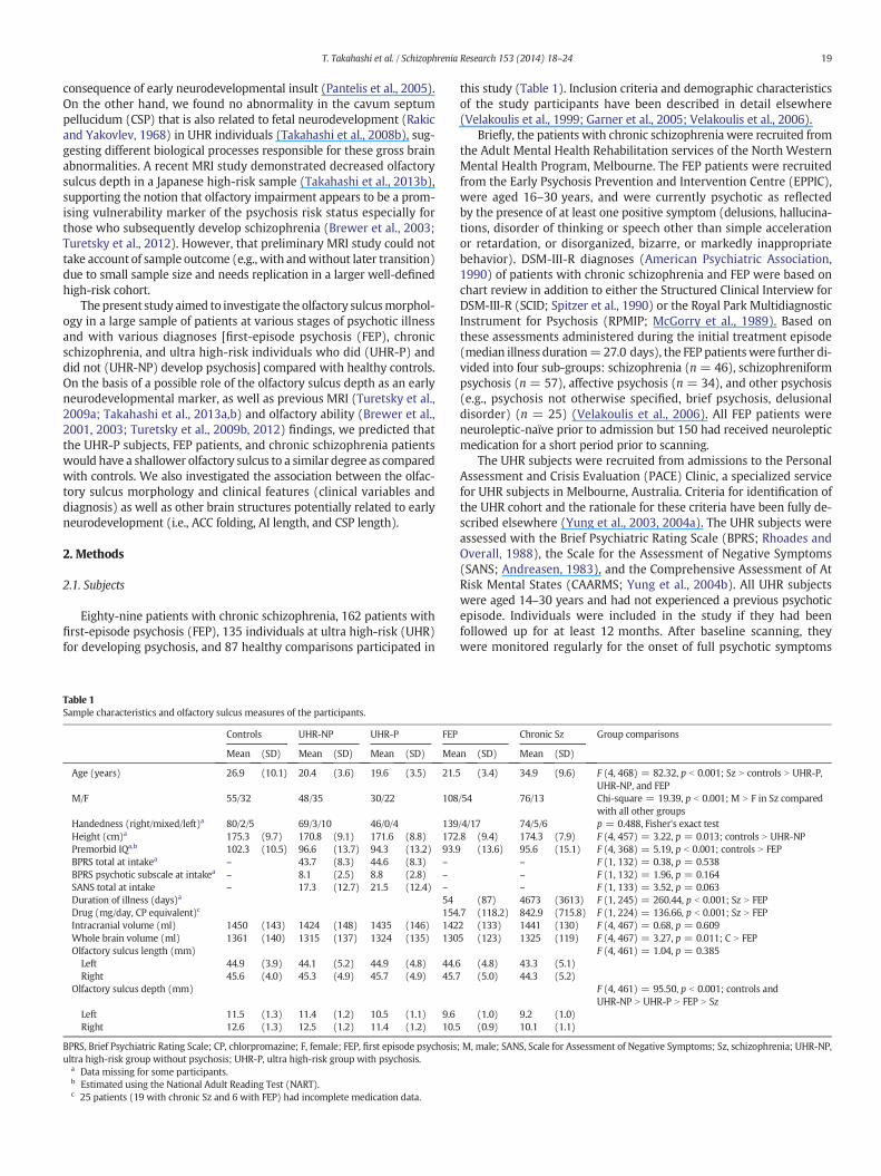
Eighty-nine patients with chronic schizophrenia, 162 patients withfirst-episode psychosis (FEP), 135 individuals at ultra high-risk (UHR)for developing psychosis, and 87 healthy comparisons participated in
Table 1Sample characteristics and olfactory sulcus measures of the participants.
Controls UHR-NP UHR-P FEP
Mean (SD) Mean (SD) Mean (SD) Mea
Age (years) 26.9 (10.1) 20.4 (3.6) 19.6 (3.5) 21.5
M/F 55/32 48/35 30/22 108
Handedness (right/mixed/left)a 80/2/5 69/3/10 46/0/4 139Height (cm)a 175.3 (9.7) 170.8 (9.1) 171.6 (8.8) 172Premorbid IQa,b 102.3 (10.5) 96.6 (13.7) 94.3 (13.2) 93.9BPRS total at intakea – 43.7 (8.3) 44.6 (8.3) –
BPRS psychotic subscale at intakea – 8.1 (2.5) 8.8 (2.8) –
SANS total at intake – 17.3 (12.7) 21.5 (12.4) –
Duration of illness (days)a 54Drug (mg/day, CP equivalent)c 154Intracranial volume (ml) 1450 (143) 1424 (148) 1435 (146) 142Whole brain volume (ml) 1361 (140) 1315 (137) 1324 (135) 130Olfactory sulcus length (mm)Left 44.9 (3.9) 44.1 (5.2) 44.9 (4.8) 44.6Right 45.6 (4.0) 45.3 (4.9) 45.7 (4.9) 45.7
Olfactory sulcus depth (mm)
Left 11.5 (1.3) 11.4 (1.2) 10.5 (1.1) 9.6Right 12.6 (1.3) 12.5 (1.2) 11.4 (1.2) 10.5
BPRS, Brief Psychiatric Rating Scale; CP, chlorpromazine; F, female; FEP, first episode psychosisultra high-risk group without psychosis; UHR-P, ultra high-risk group with psychosis.
a Data missing for some participants.b Estimated using the National Adult Reading Test (NART).c 25 patients (19 with chronic Sz and 6 with FEP) had incomplete medication data.
this study (Table 1). Inclusion criteria and demographic characteristicsof the study participants have been described in detail elsewhere(Velakoulis et al., 1999; Garner et al., 2005; Velakoulis et al., 2006).
Briefly, the patients with chronic schizophrenia were recruited fromthe Adult Mental Health Rehabilitation services of the North WesternMental Health Program, Melbourne. The FEP patients were recruitedfrom the Early Psychosis Prevention and Intervention Centre (EPPIC),were aged 16–30 years, and were currently psychotic as reflectedby the presence of at least one positive symptom (delusions, hallucina-tions, disorder of thinking or speech other than simple accelerationor retardation, or disorganized, bizarre, or markedly inappropriatebehavior). DSM-III-R diagnoses (American Psychiatric Association,1990) of patients with chronic schizophrenia and FEP were based onchart review in addition to either the Structured Clinical Interview forDSM-III-R (SCID; Spitzer et al., 1990) or the Royal Park MultidiagnosticInstrument for Psychosis (RPMIP; McGorry et al., 1989). Based onthese assessments administered during the initial treatment episode(median illness duration=27.0 days), the FEP patientswere further di-vided into four sub-groups: schizophrenia (n = 46), schizophreniformpsychosis (n = 57), affective psychosis (n = 34), and other psychosis(e.g., psychosis not otherwise specified, brief psychosis, delusionaldisorder) (n = 25) (Velakoulis et al., 2006). All FEP patients wereneuroleptic-naïve prior to admission but 150 had received neurolepticmedication for a short period prior to scanning.
The UHR subjects were recruited from admissions to the PersonalAssessment and Crisis Evaluation (PACE) Clinic, a specialized servicefor UHR subjects in Melbourne, Australia. Criteria for identification ofthe UHR cohort and the rationale for these criteria have been fully de-scribed elsewhere (Yung et al., 2003, 2004a). The UHR subjects wereassessed with the Brief Psychiatric Rating Scale (BPRS; Rhoades andOverall, 1988), the Scale for the Assessment of Negative Symptoms(SANS; Andreasen, 1983), and the Comprehensive Assessment of AtRisk Mental States (CAARMS; Yung et al., 2004b). All UHR subjectswere aged 14–30 years and had not experienced a previous psychoticepisode. Individuals were included in the study if they had beenfollowed up for at least 12 months. After baseline scanning, theywere monitored regularly for the onset of full psychotic symptoms
Chronic Sz Group comparisons
n (SD) Mean (SD)
(3.4) 34.9 (9.6) F (4, 468) = 82.32, p b 0.001; Sz N controls N UHR-P,UHR-NP, and FEP
/54 76/13 Chi-square = 19.39, p b 0.001; M N F in Sz comparedwith all other groups
/4/17 74/5/6 p = 0.488, Fisher's exact test.8 (9.4) 174.3 (7.9) F (4, 457) = 3.22, p = 0.013; controls N UHR-NP
(13.6) 95.6 (15.1) F (4, 368) = 5.19, p b 0.001; controls N FEP– F (1, 132) = 0.38, p = 0.538– F (1, 132) = 1.96, p = 0.164– F (1, 133) = 3.52, p = 0.063
(87) 4673 (3613) F (1, 245) = 260.44, p b 0.001; Sz N FEP.7 (118.2) 842.9 (715.8) F (1, 224) = 136.66, p b 0.001; Sz N FEP2 (133) 1441 (130) F (4, 467) = 0.68, p = 0.6095 (123) 1325 (119) F (4, 467) = 3.27, p = 0.011; C N FEP
F (4, 461) = 1.04, p = 0.385(4.8) 43.3 (5.1)(5.0) 44.3 (5.2)
F (4, 461) = 95.50, p b 0.001; controls andUHR-NP N UHR-P N FEP N Sz
(1.0) 9.2 (1.0)(0.9) 10.1 (1.1)
; M, male; SANS, Scale for Assessment of Negative Symptoms; Sz, schizophrenia; UHR-NP,

20 T. Takahashi et al. / Schizophrenia Research 153 (2014) 18–24
based on operationalized criteria (Yung et al., 2004a) and werethen divided into subgroups according to their long-term outcome[2–14 years later (Nelson et al., 2013)]. 52 UHR subjects (38.5%) devel-oped psychosis (UHR-P) and 83 (61.4%) did not (UHR-NP). DSMdiagnoses were available for 34 patients in the psychosis group.The predominant diagnosis was schizophrenia spectrum (n = 20),but there were also diagnoses of affective psychosis (n = 7), andother psychoses (n = 7). All UHR subjects were neuroleptic naïve atthe time of the brain scan. After the brain scan, 21 subjects startedlow-dose risperidone therapy and cognitive behavior therapy as partof a double-blind randomized study examining a 6-month therapeuticintervention to reduce the risk of progression to psychosis (McGorryet al., 2002). Most of the remaining UHR participants received casemanagement and supportive therapy for at least six months.
Healthy volunteers were recruited from similar socio-demographicareas as the patients by approaching ancillary hospital staff and throughadvertisements. These controls did not have any personal or familyhistory of psychiatric illness.
All subjects were physically healthy, and none had a lifetime historyof serious head trauma, neurological illness, serious medical or surgicalillness, steroid or substance abuse. The sizes of the AI and CSP of theparticipants in this study have been examined previously (Takahashiet al., 2008a,b). ACC folding pattern data (Yücel et al., 2002, 2003)were available for 354 of 473 subjects in this study (111 FEP, 71 chronicschizophrenia, 97 UHR, and 75 control subjects). This study wasapproved by the regional ethics committee while written informedconsent was obtained from all subjects prior to study participation.
2.2. Magnetic resonance imaging procedures
MRI scans were acquired with a 1.5-T GE Signa scanner (GeneralElectric Medical Systems, Milwaukee, Wisconsin). A 3D volumetricspoiled gradient recalled echo in the steady state sequence gener-ated 124 contiguous 1.5 mm coronal slices (TR = 14.3 ms, TE =3.3 ms, flip = 30°, FOV = 24 × 24 cm, matrix = 256 × 256, voxeldimension = 0.938 × 0.938 × 1.5 mm). The intracranial volume (ICV)and whole brain volume were measured to correct for differences inhead size as previously described (Velakoulis et al., 2006); thefive groups (healthy controls, UHR-NP, UHR-P, FEP, and chronic schizo-phrenia) did not differ significantly in their ICV volumes but the con-trols had a larger whole brain volume than FEP patients (Table 1).
2.3. Olfactory sulcus measurements
For the assessment of the olfactory sulcus, the images were pro-cessed on a Linux PC (Fujitsu Limited, Tokyo, Japan) using Dr. Viewsoftware (AJS, Tokyo, Japan). Brain images were realigned in threedimensions and reconstructed into contiguous coronal images witha 0.938-mm thickness, perpendicular to the anterior commissure–
B
Rt
A
LtRt
Fig. 1.Olfactory sulci on coronal (A), axial (B), and sagittal (C, right hemisphere) views, whichwshow the plane of the posterior tangent through the eyeballs (PPTE).
posterior commissure line. As described in detail elsewhere (Takahashiet al., 2013a), one rater (TT), who was blind to the subjects' identity,measured the depth of the olfactory sulcus in all coronal slices wherethe sulcus was clearly seen (Fig. 1). On each coronal slice, the olfactorysulcus was traced beginning with the deepest point of the sulcus andending inferiorly with a tangent line connecting the top surfaces of thegyrus rectus andmedial orbital gyrus (Rombaux et al., 2009). The lengthof the sulcus in the anterior–posterior direction (mm) was determinedby the multiplication of the number of these coronal slices by 0.938.Intra- and inter-rater (TT and YN) reliabilities of the sulcus measure-ments were assessed using intraclass correlation coefficients (ICCs) in10 randomly selected brains. Intra-rater ICCs were 0.91 (left depth),0.94 (left length), 0.91 (right depth), and 0.98 (right length). Inter-rater ICCs were 0.92 (left depth), 0.92 (left length), 0.83 (right depth),and 0.93 (right length).
2.4. Statistical analysis
Clinical and demographic differences between groups were exam-ined with one-way analysis of variance (ANOVA) or chi-square test.The average depth (sum of the depth in all slices containing thesulcus/slice number) and length of the olfactory sulcus were analyzedusing the repeated measures analysis of covariance (ANCOVA), withage and ICV as covariates, diagnosis (healthy controls, UHR-NP, UHR-P, FEP, and chronic schizophrenia) and gender as between-subject fac-tors, and hemisphere as a within-subject variable. Post-hoc Scheffé'stests were used to follow-up these analyses.
Since there were significant group differences in age and gender(Table 1), the healthy controls were also divided into two subgroups;older controls [28 males and 9 females, mean (SD) = 35.5 (9.7)years, matched to chronic schizophrenia for age (F = 0.09, df = 1,124, p = 0.765) and gender (chi-square = 1.71, p = 0.191)] andyounger controls [27 males and 23 females, mean (SD) = 20.5 (3.2)years, matched to FEP (age, F = 3.39, df = 1, 210, p = 0.067; gender,chi-square = 2.65, p = 0.104) and both UHR groups (age, F = 1.18,df = 2, 182, p = 0.309; gender, chi-square = 0.21, p = 0.899)]. Therelationships between the olfactory sulcus measures and clinicalvariables as well as the length of AI/CSP were examined by Pearson'spartial correlation coefficients controlling for ICV. The length ofthe CSP was log-transformed because of their skewed distribution(Takahashi et al., 2008b). In order to examine the relationship be-tween the olfactory sulcus measures and ACC folding pattern, theolfactory sulcus length and depth were analyzed by ANCOVAs withage and ICV as covariates, with diagnosis and ACC sulcal patternfor each hemisphere (prominent, present, and absent) as between-subject factors. The relationship between the olfactory sulcus measuresand paracingulate asymmetry index (leftward, symmetric, and right-ward) was also analyzed using the same model. Statistical significancewas defined as p b 0.05 (two-tailed).
C
Lt
ere colored on 0.938-mm consecutive coronal slices. Panel A and the dotted line on panel B

21T. Takahashi et al. / Schizophrenia Research 153 (2014) 18–24
The findings presented here, including those of correlational analy-ses, remained essentially the same even when we used UHR diagnosesaccording to the outcome at 12 months after intake (rather than thelonger term follow up data) as in our early studies (Yücel et al., 2003;Takahashi et al., 2008a,b). There was a group difference in the wholebrain volume (controls N FEP; Table 1), but the present results of theolfactory sulcus did not change even when we used the whole brainvolume instead of ICV as a covariate for the ANCOVAs.
3. Results
3.1. Demographic characteristics
Comparison of the groups revealed no difference in handedness andICV, but there were significant group differences in age, gender, height,and premorbid IQ (Table 1). The two UHR groups (UHR-P versus -NP)did not differ with respect to global psychopathological state accordingto the BPRS or negative symptoms according to the SANS.
3.2. Depth and length of the olfactory sulcus
ANCOVA of the olfactory sulcus length revealed no significant effectinvolving diagnosis (Table 1), but that for depth showed significantmain effects of diagnosis [F (4, 461)= 95.50, p b 0.001] andhemisphere[F (1, 463) = 531.51, p b 0.001] and an interaction between these fac-tors [F (4, 463) = 2.60, p = 0.036]. Post-hoc analyses showed that theolfactory sulcus depth was shallower in the UHR-P subjects (p b 0.001for both hemispheres), but not inUHR-NP subjects (p= 1.000), as com-pared with healthy controls and that the patients with later illnessstages had increasingly shallower depth of the olfactory sulcus (i.e., con-trols and UHR-NP N UHR-P N FEP N chronic schizophrenia; all p b 0.001for both hemispheres) (Table 1, Fig. 2). The olfactory sulcus depth wassignificantly deeper in the right hemisphere for all groups (p b 0.001).The sulcus depth did not differ among four FEP subgroups (schizophre-nia, schizophreniform psychosis, affective psychosis, and other psycho-sis patients) [F (3, 152) = 0.52, p = 0.672] or three UHR-P subgroups(subjects who later developed schizophrenia spectrum, affective psy-chosis, and other psychoses) [F (2, 26) = 0.17, p = 0.846]. There wasno significant effect involving gender in any of these analyses.
The present results of the sulcus depth did not change even whenwe separately analyzed the older groups (older controls and chronicschizophrenia patients) and younger groups (younger controls, FEP,and UHR subjects). Briefly, the olfactory sulcus was significantly deeper
Controls
Lt Rt
UHR-NP
Olf
acto
ry s
ulcu
s de
pth
(mm
)
5
10
15
20
*
UH
Lt Rt Lt
*
*
**
Fig. 2. Scatterplots of olfactory sulcus depth in healthy controls, ultra high-risk nonpsychotic (psychosis (FEP), and patients with chronic schizophrenia (Sz). Horizontal bars indicate means
in the older controls than in chronic schizophrenia patients [ANCOVA,F (1, 120) = 73.65, p b 0.001; post-hoc tests, p b 0.001 for both hemi-spheres] and the sulcus depth became increasingly more superficial atlater illness stages [ANCOVA, F (3, 337) = 101.20, p b 0.001; youngercontrols and UHR-NP N UHR-P N FEP (post hoc tests, all p b 0.001 forboth hemispheres)].
3.3. Correlational analysis
There was a negative correlation between the olfactory sulcusdepth and age only for the controls (left, r = −0.376, p b 0.001; right,r=−0.399, p b 0.001). For the FEP and chronic schizophrenia patients,the olfactory sulcus length and depth did not correlate with illnessduration or daily medication dosage. Total SANS score in the UHR-Psubjects was negatively correlated with the sulcus depth in the righthemisphere (r = −0.426, p = 0.002), but no correlations were foundbetween the sulcus measures and clinical variables in the UHR-NPsubjects.
3.4. Relationship between the olfactory sulcus measures and otherstructures
The olfactory sulcus depth correlated with whole brain volume onlyfor the controls (right, r = 0.330, p = 0.002). However, the sulcuslength and depth did not significantly correlate with morphologicchanges in the AI and CSP in either diagnostic group. For the relationshipbetween the olfactory sulcus measures (depth and length) and ACCfolding pattern [sulcal pattern (left, right) and asymmetry index],ANCOVAs revealed no main effects of ACC sulcal features or diagnosis-by-ACC interactions.
4. Discussion
To our knowledge, this is the first MRI study to report morpho-logic changes of the olfactory sulcus across various illness stages in-cluding those of a sample at clinical high-risk of psychosis that wasfollowed-up longitudinally. The UHR subjects who developed psychosis(UHR-P) had a significantly shallower olfactory sulcus as comparedwith both UHR who did not develop psychosis (UHR-NP) and controlsubjects, suggesting that such anomaly already exists prior to theonset as a possible risk marker of transition to psychosis. On the otherhand, although this cross-sectional study cannot directly address pro-gressive brain changes, the sulcus depth in various stages of psychosis
**
R-P FEP Chronic Sz
Rt Lt Rt Lt Rt
* * *
**
*
*
UHR-NP) subjects, ultra high-risk psychotic (UHR-P) subjects, patients with first episodeof each group. *p b 0.001.

22 T. Takahashi et al. / Schizophrenia Research 153 (2014) 18–24
(UHR-P N FEP N chronic schizophrenia) is an indication that these pa-tients may also manifest progressive changes in the sulcus morphologyduring the course of the illness.
The present study replicated previous findings in patients with first-episode (Takahashi et al., 2013a) and chronic (Turetsky et al., 2009a)schizophrenia as well as in a small sample of clinical high-risk indi-viduals (Takahashi et al., 2013b) in showing that patients withpsychotic disorders had abnormally shallow olfactory sulci beforepsychosis onset. Our findings also demonstrated that altered depth ofthe olfactory sulcus, which could be due to an embryonic disruption ofthe olfactory system (Abolmaali et al., 2002; Hummel et al., 2003),might be a neurobiological predictor of future transition to psychosis.Medication with antipsychotics might affect brain morphology inschizophrenia (Lieberman et al., 2005; Andreasen et al., 2013), but wefound no significant relationship between the sulcus morphology anddosage of antipsychotic medication in our FEP and chronic schizophre-nia patients. In addition, we also found altered depth of the olfactorysulcus in antipsychotic-naïve UHR-P subjects. A negative finding ofolfactory sulcus in chronic schizophrenia by Nguyen et al. (2011)might partly be due to technical issues, as they measured the sulcusdepth using a single slice based on external landmarks [i.e., the planeof the posterior tangent through the eyeballs (PPTE)], whereas ourresults and those of Turetsky et al. (2009a) were based on themeasure-ment of the entire structure. TheseMRI findings are consistent with thenotion that olfactory dysfunction, which exists in the first-episodeor prodromal phase of schizophrenia (Brewer et al., 2001, 2003) aswell as in the patients' first-degree relatives (Kamath et al., in press),may be a sensitive indicator of schizophrenia pathology and mayeven serve as an early warning sign of disease vulnerability or onset(Turetsky et al., 2009b).
A series of our MRI studies of possible neurodevelopmental markersin various illness stages may provide a clue to the timing of neuro-developmental abnormalities underlying psychosis. Our group hasshown that the UHR cohort shares abnormalities in the olfactory sulcusdepth and ACC folding in patients with florid psychosis (Yücel et al.,2003), suggesting neurodevelopmental disturbance by the third trimes-ter of gestation, as these sulco-gyral patterns develop around 16 to25 weeks' gestation (Chi et al., 1977; Garel et al., 2001). In the sameUHR and FEP subjects as in this study, we also found abnormally smallAI (Takahashi et al., 2008a), which suggested an abnormal neurode-velopment around 13 to 14 weeks of gestation (Rosales et al., 1968).However, these patients had a CSP with a normal size (Takahashiet al., 2008b), which is related to fusion of the septum pellucidiwithin 3–6 months of birth (Shaw and Alvord, 1969), supporting theidea that psychotic disorders are more closely related to aberrantneurodevelopment early in gestation. Interestingly, only olfactory sul-cus depth among these possible neurodevelopmental markers was pre-dictive of future transition into psychosis, suggesting different biologicalprocesses responsible for these gross brain abnormalities. Discrepantfindings such as increased prevalence of a large CSP have been also re-ported in a clinical high-risk sample (Choi et al., 2008), although 37%(11/30) of them were receiving antipsychotics at the time of scanning.Since our findings suggested a mild relation between the olfactory sul-cus depth and prodromal symptomatology, it seemsworthwhile to fur-ther evaluate the relation between these potential neurodevelopmentalmarkers and clinical features of high-risk subjects (e.g., medication sta-tus, future transition, and symptom severity).
On the other hand, the current results of an increasingly moresuperficial sulcus in subjects with later stages of psychosis (UHR-P N FEP N chronic schizophrenia) imply that the olfactory sulcusabnormalities in psychosis cannot be fully explained by abnormalneurodevelopment. Illness duration in the current FEP and chronicschizophrenia patients did not correlate with the olfactory sulcus mea-sures, but our results in healthy subjects raise the possibility that thesulcal depth changes with age. Childhood maltreatment, which hasbeen shown to elevate the risk of psychiatric disorders, could also affect
subsequent brain development including the orbitofrontal cortex (Kellyet al., 2013). Interestingly, a shallow olfactory sulcus (Wang et al., 2011)and olfactory dysfunction (Mesholam et al., 1998) have also been re-ported in neurodegenerative diseases such as Parkinson's disease, al-though the pathological mechanism is unknown. Thus, our findingsof the olfactory sulcus in psychosis may also reflect ongoing changespossibly due to the illness itself and/or other factors (e.g., effect of anti-psychotics), andmay be associatedwith or even be consequent on otherprogressive brain structural changes. Indeed, dynamic brain changes,including excessive cortical thinning (Sun et al., 2009a,b; van Harenet al., 2011) or gray matter reduction (Mané et al., 2009) over time inthe frontal area, may occur during or after the onset of schizophrenia(Pantelis et al., 2007). Previous longitudinal analyses demonstratedthat the olfactory sulcus depth remained stable over time in first-episode schizophrenia with mean inter-scan interval of 2.7 years(Takahashi et al., 2013a), but further longitudinal follow-up of first-episode and additional prodromal/chronic patients would be requiredto examine the nature of the olfactory sulcus changes associated withpsychosis.
Consistent with the results of olfactory identification ability inneuroleptic-naïve FEP patients (Brewer et al., 2001), which demon-strated that olfactory identification deficits were not specific to schizo-phrenia among various psychotic conditions, the current study did notidentify any difference in the olfactory sulcusmeasures between the FEPsubgroups, suggesting that olfactory sulcus malformation is present in arather diverse population with psychotic symptoms, such as affectivepsychosis or other psychoses. We also found no difference betweenUHR-P subgroups, although this comparison was limited by small sam-ple size especially for the prodromal state of affective and other psycho-ses. Given that olfactory deficits might be specific to high-risk subjectsof schizophrenia spectrum (Brewer et al., 2003) and there have beenonly a few MRI studies of brain abnormalities predating the onset ofaffective psychoses (Bechdolf et al., 2012; Dazzan et al., 2012), diseasespecificity of the olfactory sulcus abnormalities before psychosis onsetshould be further tested in a larger sample.
Several limitations of the current study should be taken intoaccount. First, although our findings of altered depth of the olfactorysulcus may at least partly reflect embryonic disruption of the olfactorysystem, we did not assess olfactory function or other olfactory struc-tures. Reduced olfactory bulb volume in schizophrenia patients(Turetsky et al., 2000; Nguyen et al., 2011) and in first-degree rela-tives (Turetsky et al., 2003) suggests its significant role in the neuro-developmental pathology of schizophrenia. The olfactory bulb can bewell identified on T2-weighted MR images (Rombaux et al., 2009;Duprez and Rombaux, 2010), but our T1-weighted images did notallow reliable measurement of the bulb. Second, the participants inthis study were not matched for age and gender between the groups.The olfactory sulcus morphology has been implicated in earlyneurodevelopment, but age may affect its depth as demonstrated inthis study. However, statistical conclusions of the present studyremained the same when we separately analyzed the older groups(older controls and chronic schizophrenia patients) and youngergroups (younger controls, FEP, and UHR subjects) using two age-and gender-matched control subgroups. Moreover, there was no sig-nificant effect involving gender in any of the analyses of this study.Third, detailed clinical data of the patients with FEP and chronicschizophrenia such as the symptomatology at the time of scanningwere not available. Given that impairment of olfactory identificationhas been associated with negative symptoms in schizophrenia (Breweret al., 1996, 2001), the possible relation between the olfactory sulcusmorphology and clinical features of psychotic disorders is worthy of fur-ther examination. Finally, as olfactory deficits are also reported in otherpsychiatric disorders such as major depression (Burón and Bulbena,2013) and reduced global sulcation appears to be a feature of schizo-phrenia (Penttilä et al., 2008), the disease and regional specificities ofour findings should be further examined.

23T. Takahashi et al. / Schizophrenia Research 153 (2014) 18–24
In conclusion, we found a shallow olfactory sulcus in high-risk in-dividuals prior to the onset of psychosis, implicating that such mor-phological anomalies could be an early neurodevelopmental markerrelated to future transition to psychosis. However, current results ofprominent sulcus changes in patients with later illness stages suggestthat olfactory sulcus abnormalities in psychotic disorders may alsoreflect progressive brain changes during and after the onset of illness.Additional longitudinal studies would be required for the under-standing of the nature of olfactory sulcus changes in the course ofpsychosis.
Role of funding sourceThis research was supported in part by Grants-in-Aid for Scientific Research (C) (Nos.
22591275 and, 24591699) and Grants-in-Aid for Scientific Research (B) (No. 24390281)from the Japanese Society for the Promotion of Science, Health and Labour Sciences Re-search Grants (Comprehensive Research on Disability, Health and Welfare, H23-Seishin-Ippan-002 and H23-Seishin-Ippan-009), and Research Grants from the JSPS Asian CoreProgram and the Research Group For Schizophrenia, Japan. This research was also sup-ported by NHMRC Program Grants (IDs: 350241, 566529). Professor Wood and AssociateProfessor Brewer were supported by Clinical Career Development Awards fromthe NHMRC, and A/Prof Brewer was additionally supported by the Colonial Foundation.Professor Yücel was supported by an NHMRC Senior Research Fellowship (ID: 1021973).Professor Pantelis was supported by an NHMRC Senior Principal Research Fellowship(ID: 628386) and a NARSAD Distinguished Investigator Award. The funding agencieshad no further role in design and conduct of the study; collection, management, analysisand interpretation of the data; and preparation, review, or approval of the manuscript.
ContributorsIn this study, Drs. Suzuki, Pantelis, and Brewer conceived the idea and methodology
of the study. Dr. Takahashi conducted the statistical analyses and wrote the manuscript.Drs. McGorry, Yung, Brewer, Nelson, Lin, Phillips, and Proffitt recruited subjects, andwere involved in clinical and diagnostic assessments. Drs. Takahashi and Nakamuraanalyzed the MRI data. Drs. Suzuki, Pantelis, Velakoulis, Wood, and Yücel contributed towriting and editing of the manuscript. All authors contributed to and have approved thefinal manuscript.
Conflict of interestAll authors declare that they have no conflicts of interest.
AcknowledgementsThe authors are grateful to the clinical staff of the Personal Assessment and Crisis
Evaluation (PACE) Clinic, Early Psychosis Prevention and Intervention Centre (EPPIC),and Adult Mental Health Rehabilitation services of the North Western Mental HealthProgram for their assistance in diagnostic and psychopathological assessments of thestudy participants.
References
Abolmaali, N.D., Hietschold, V., Vogl, T.J., Hüttenbrink, K.B., Hummel, T., 2002. MR evalu-ation in patients with isolated anosmia since birth or early childhood. Am.J. Neuroradiol. 23, 157–164.
American Psychiatric Association, 1990. Diagnostic and Statistical Manual of Mental Dis-orders, Revised Third edition. American Psychiatric Press, Washington, DC.
Andreasen, N.C., 1983. The Scale for the Assessment of Negative Symptoms (SANS). TheUniversity of Iowa, Iowa City.
Andreasen, N.C., Liu, D., Ziebell, S., Vora, A., Ho, B.C., 2013. Relapse duration, treatment in-tensity, and brain tissue loss in schizophrenia: a prospective longitudinal MRI study.Am. J. Psychiatry 170, 609–615.
Bechdolf, A., Wood, S.J., Nelson, B., Velakoulis, D., Yücel, M., Takahashi, T., Yung, A.R., Berk,M., Wong, M.T., Pantelis, C., McGorry, P.D., 2012. Amygdala and insula volumes priorto illness onset in bipolar disorder: a magnetic resonance imaging study. PsychiatryRes. 201, 34–39.
Brewer, W.J., Edwards, J., Anderson, V., Robinson, T., Pantelis, C., 1996. Neuropsychologi-cal, olfactory, and hygiene deficits in men with negative symptom schizophrenia.Biol. Psychiatry 40, 1021–1031.
Brewer, W.J., Pantelis, C., Anderson, V., Velakoulis, D., Singh, B., Copolov, D.L., McGorry,P.D., 2001. Stability of olfactory identification deficits in neuroleptic-naive patientswith first-episode psychosis. Am. J. Psychiatry 158, 107–115.
Brewer, W.J., Wood, S.J., McGorry, P.D., Francey, S.M., Phillips, L.J., Yung, A.R., Anderson, V.,Copolov, D.L., Singh, B., Velakoulis, D., Pantelis, C., 2003. Impairment of olfactory iden-tification ability in individuals at ultra-high risk for psychosis who later developschizophrenia. Am. J. Psychiatry 160, 1790–1794.
Burón, E., Bulbena, A., 2013. Olfaction in affective and anxiety disorders: a review of theliterature. Psychopathology 46, 63–74.
Chi, J.G., Dooling, E.C., Gilles, F.H., 1977. Gyral development of the human brain. Ann.Neurol. 1, 86–93.
Choi, J.K., Kang, D.H., Park, J.Y., Jung, W.H., Choi, C.H., Chon, M.W., Jung, M.H., Lee, J.M.,Kwon, J.S., 2008. Cavum septum pellucidum in subjects at ultra-high risk for
psychosis: compared with first-degree relatives of patients with schizophrenia andhealthy volunteers. Prog. Neuropsychopharmacol. Biol. Psychiatry 32, 1326–1330.
Dazzan, P., Soulsby, B., Mechelli, A., Wood, S.J., Velakoulis, D., Phillips, L.J., Yung, A.R.,Chitnis, X., Lin, A., Murray, R.M., McGorry, P.D., McGuire, P.K., Pantelis, C., 2012. Volu-metric abnormalities predating the onset of schizophrenia and affective psychoses: anMRI study in subjects at ultrahigh risk of psychosis. Schizophr. Bull. 38, 1083–1091.
Duprez, T.P., Rombaux, P., 2010. Imaging the olfactory tract (cranial nerve #1). Eur.J. Radiol. 74, 288–298.
Fatemi, S.H., Folsom, T.D., 2009. The neurodevelopmental hypothesis of schizophrenia,revisited. Schizophr. Bull. 35, 528–548.
Garel, C., Chantrel, E., Brisse, H., Elmaleh, M., Luton, D., Oury, J.F., Sebag, G., Hassan, M.,2001. Fetal cerebral cortex: normal gestational landmarks identified using prenatalMR imaging. AJNR Am. J. Neuroradiol. 22, 184–189.
Garner, B., Pariante, C.M., Wood, S.J., Velakoulis, D., Phillips, L., Soulsby, B., Brewer, W.J.,Smith, D.J., Dazzan, P., Berger, G.E., Yung, A.R., van den Buuse, M., Murray, R., McGorry,P.D., Pantelis, C., 2005. Pituitary volume predicts future transition to psychosis in indi-viduals at ultra-high risk of developing psychosis. Biol. Psychiatry 58, 417–423.
Hummel, T., Damm, M., Vent, J., Schmidt, M., Theissen, P., Larsson, M., Klussmann, J.P.,2003. Depth of olfactory sulcus and olfactory function. Brain Res. 975, 85–89.
Kamath, V., Turetsky, B.I., Calkins, M.E., Kohler, C.G., Conroy, C.G., Borgmann-Winter, K.,Gatto, D.E., Gur, R.E., Moberg, P.J., 2014. Olfactory processing in schizophrenia, non-ill first-degree family members, and young people at-risk for psychosis. WorldJ. Biol. Psychiatry. http://dx.doi.org/10.3109/15622975.2011.615862 (in press).
Kelly, P.A., Viding, E., Wallace, G.L., Schaer, M., De Brito, S.A., Robustelli, B., McCrory, E.J.,2013. Cortical thickness, surface area, and gyrification abnormalities in children ex-posed to maltreatment: neural markers of vulnerability? Biol Psychiatry 74, 845–852.
Lieberman, J.A., Tollefson, G.D., Charles, C., Zipursky, R., Sharma, T., Kahn, R.S., Keefe, R.S.,Green, A.I., Gur, R.E., McEvoy, J., Perkins, D., Hamer, R.M., Gu, H., Tohen, M., HGDHStudy Group, 2005. Antipsychotic drug effects on brain morphology in first-episodepsychosis. Arch. Gen. Psychiatry 62, 361–370.
Mané, A., Falcon, C., Mateos, J.J., Fernandez-Egea, E., Horga, G., Lomeña, F., Bargalló, N.,Prats-Galino, A., Bernardo, M., Parellada, E., 2009. Progressive gray matter changesin first episode schizophrenia: a 4-year longitudinal magnetic resonance studyusing VBM. Schizophr. Res. 114, 136–143.
McGorry, P.D., Kaplan, I., Dossetor, C., Herrman, H., Copolov, D., Singh, B., 1989. Royal ParkMultidiagnostic Instrument for Psychosis. National Health and Medical ResearchCouncil, Melbourne, Australia.
McGorry, P.D., Yung, A.R., Phillips, L.J., Yuen, H.P., Francey, S., Cosgrave, E.M., Germano, D.,Bravin, J., McDonald, T., Blair, A., Adlard, S., Jackson, H., 2002. Randomized controlledtrial of interventions designed to reduce the risk of progression to first-episode psy-chosis in a clinical sample with subthreshold symptoms. Arch. Gen. Psychiatry 59,921–928.
Mesholam, R.I., Moberg, P.J., Mahr, R.N., Doty, R.L., 1998. Olfaction in neurodegenerativedisease: a meta-analysis of olfactory functioning in Alzheimer's and Parkinson'sdiseases. Arch. Neurol. 55, 84–90.
Nelson, B., Yuen, H.P., Wood, S., Lin, A., Spiliotacopoulos, D., Bruxner, A., Broussard, C.,Simmons, M., Foley, D.L., Brewer, W.J., Francey, S.M., Amminger, G.P., Thompson, A.,McGorry, P.D., Yung, A.R., 2013. Long-term follow-up of a group at ultra high risk(“prodromal”) for psychosis: the PACE 400 study. JAMA Psychiatry 70, 793–802.
Nguyen, A.D., Pelavin, P.E., Shenton, M.E., Chilakamarri, P., McCarley, R.W., Nestor, P.G.,Levitt, J.J., 2011. Olfactory sulcal depth and olfactory bulb volume in patients withschizophrenia: an MRI study. Brain Imaging Behav. 5, 252–261.
Pantelis, C., Yücel, M., Wood, S.J., Velakoulis, D., Sun, D., Berger, G., Stuart, G.W., Yung, A.,Phillips, L., McGorry, P.D., 2005. Structural brain imaging evidence for multiple path-ological processes at different stages of brain development in schizophrenia.Schizophr. Bull. 31, 672–696.
Pantelis, C., Velakoulis, D., Wood, S.J., Yücel, M., Yung, A.R., Phillips, L.J., Sun, D.Q., McGorry,P.D., 2007. Neuroimaging and emerging psychotic disorders: the Melbourne ultra-high risk studies. Int. Rev. Psychiatry 19, 371–381.
Penttilä, J., Paillére-Martinot, M.L., Martinot, J.L., Mangin, J.F., Burke, L., Corrigall, R.,Frangou, S., Cachia, A., 2008. Global and temporal cortical folding in patients withearly-onset schizophrenia. J. Am. Acad. Child. Adolesc. Psychiatry 47, 1125–1132.
Rakic, P., Yakovlev, P.I., 1968. Development of the corpus callosum and cavum septi inman. J. Comp. Neurol. 132, 45–72.
Rhoades, H.M., Overall, J.E., 1988. The semistructured BPRS interview and rating guide.Psychopharmacol. Bull. 24, 101–104.
Rombaux, P., Grandin, C., Duprez, T., 2009. How to measure olfactory bulb volume andolfactory sulcus depth? B-ENT 5 (Suppl. 13), 53–60.
Rosales, R.K., Lemay, M.J., Yakovley, P.I., 1968. The development and involution of massaintermedia with regard to age and sex. J. Neuropathol. Exp. Neurol. 27, 166.
Shaw, C.M., Alvord Jr., E.C., 1969. Cava septi pellucidi et vergae: their normal and patho-logical states. Brain 92, 213–223.
Spitzer, R.L., Williams, J.B., Gibbon, M., First, M.B., 1990. Structured Clinical Interview forDSM-III-R, Patient Edition (SCID-P). American Psychiatric Press, Washington, DC.
Sun, D., Phillips, L., Velakoulis, D., Yung, A., McGorry, P.D., Wood, S.J., van Erp, T.G.,Thompson, P.M., Toga, A.W., Cannon, T.D., Pantelis, C., 2009a. Progressive brain struc-tural changes mapped as psychosis develops in ‘at risk’ individuals. Schizophr. Res.108, 85–92.
Sun, D., Stuart, G.W., Jenkinson, M., Wood, S.J., McGorry, P.D., Velakoulis, D., van Erp, T.G.,Thompson, P.M., Toga, A.W., Smith, D.J., Cannon, T.D., Pantelis, C., 2009b. Brain surfacecontraction mapped in first-episode schizophrenia: a longitudinal magnetic reso-nance imaging study. Mol. Psychiatry 14, 976–986.
Takahashi, T., Yücel, M., Yung, A.R.,Wood, S.J., Phillips, L.J., Berger, G.E., Ang, A., Soulsby, B.,McGorry, P.D., Suzuki, M., Velakoulis, D., Pantelis, C., 2008a. Adhesio interthalamica inindividuals at high-risk for developing psychosis and patients with psychotic disor-ders. Prog. Neuropsychopharmacol. Biol. Psychiatry 32, 1708–1714.

24 T. Takahashi et al. / Schizophrenia Research 153 (2014) 18–24
Takahashi, T., Yung, A.R., Yücel, M., Wood, S.J., Phillips, L.J., Harding, I.H., Soulsby, B.,McGorry, P.D., Suzuki, M., Velakoulis, D., Pantelis, C., 2008b. Prevalence of largecavum septi pellucidi in ultra high-risk individuals and patients with psychotic disor-ders. Schizophr. Res. 105, 236–244.
Takahashi, T., Nakamura, Y., Nakamura, K., Ikeda, E., Furuichi, A., Kido, M., Kawasaki, Y.,Noguchi, K., Seto, H., Suzuki, M., 2013a. Altered depth of the olfactory sulcus in first-episode schizophrenia. Prog. Neuropsychopharmacol. Biol. Psychiatry 40, 167–172.
Takahashi, T., Nakamura, Y., Nakamura, K., Nishiyama, S., Ikeda, E., Furuichi, A., Kido, M.,Noguchi, K., Suzuki, M., 2013b. Altered depth of the olfactory sulcus in subjects atrisk of psychosis. Schizophr. Res. 149, 186–187.
Turetsky, B.I., Moberg, P.J., Yousem, D.M., Doty, R.L., Arnold, S.E., Gur, R.E., 2000. Reducedolfactory bulb volume in patients with schizophrenia. Am. J. Psychiatry 157, 828–830.
Turetsky, B.I., Moberg, P.J., Arnold, S.E., Doty, R.L., Gur, R.E., 2003. Low olfactory bulbvolume in first-degree relatives of patients with schizophrenia. Am. J. Psychiatry160, 703–708.
Turetsky, B.I., Crutchley, P., Walker, J., Gur, R.E., Moberg, P.J., 2009a. Depth of the olfactorysulcus: a marker of early embryonic disruption in schizophrenia? Schizophr. Res. 115,8–11.
Turetsky, B.I., Hahn, C.G., Borgmann-Winter, K., Moberg, P.J., 2009b. Scents and nonsense:olfactory dysfunction in schizophrenia. Schizophr. Bull. 35, 1117–1131.
Turetsky, B.I., Kamath, V., Calkins, M.E., Brewer, W.J., Wood, S.J., Pantelis, C., Seidman, L.J.,Malaspina, D., Good, K.P., Kopala, L.C., Moberg, P.J., 2012. Olfaction and schizophreniaclinical risk status: just the facts. Schizophr. Res. 139, 260–261.
van Haren, N.E., Schnack, H.G., Cahn, W., van den Heuvel, M.P., Lepage, C., Collins, L.,Evans, A.C., Hulshoff Pol, H.E., Kahn, R.S., 2011. Changes in cortical thickness duringthe course of illness in schizophrenia. Arch. Gen. Psychiatry 68, 871–880.
Velakoulis, D., Pantelis, C., McGorry, P.D., Dudgeon, P., Brewer, W., Cook, M., Desmond, P.,Bridle, N., Tierney, P., Murrie, V., Singh, B., Copolov, D., 1999. Hippocampal volumein first-episode psychoses and chronic schizophrenia: a high-resolution magneticresonance imaging study. Arch. Gen. Psychiatry 56, 133–141.
Velakoulis, D., Wood, S.J., Wong, M.T., McGorry, P.D., Yung, A., Phillips, L., Smith, D.,Brewer, W., Proffitt, T., Desmond, P., Pantelis, C., 2006. Hippocampal and amygdalavolumes according to psychosis stage and diagnosis: a magnetic resonance imagingstudy of chronic schizophrenia, first-episode psychosis, and ultra-high-risk indi-viduals. Arch. Gen. Psychiatry 63, 139–149.
Wang, J., You, H., Liu, J.F., Ni, D.F., Zhang, Z.X., Guan, J., 2011. Association of olfactory bulbvolume and olfactory sulcus depth with olfactory function in patients with Parkinsondisease. AJNR Am. J. Neuroradiol. 32, 677–681.
Yücel, M., Stuart, G.W., Maruff, P., Wood, S.J., Savage, G.R., Smith, D.J., Crowe, S.F., Copolov,D.L., Velakoulis, D., Pantelis, C., 2002. Paracingulate morphologic differences in maleswith established schizophrenia: a magnetic resonance imaging morphometric study.Biol. Psychiatry 52, 15–23.
Yücel, M.,Wood, S.J., Phillips, L.J., Stuart, G.W., Smith, D.J., Yung, A., Velakoulis, D., McGorry,P.D., Pantelis, C., 2003. Morphology of the anterior cingulate cortex in young men atultra-high risk of developing a psychotic illness. Br. J. Psychiatry 182, 518–524.
Yung, A.R., Phillips, L.J., Yuen, H.P., Francey, S.M., McFarlane, C.A., Hallgren, M., McGorry,P.D., 2003. Psychosis prediction: 12-month follow up of a high-risk (“prodromal”)group. Schizophr. Res. 60, 21–32.
Yung, A.R., Phillips, L.J., Yuen, H.P., McGorry, P.D., 2004a. Risk factors for psychosis in an ultrahigh-risk group: psychopathology and clinical features. Schizophr. Res. 67, 131–142.
Yung, A.R., Phillips, L.J., McGorry, P.D., 2004b. Treating Schizophrenia in the ProdromalPhase. Taylor & Francis, London.
Copyright © 2022 FDOKUMEN