
Reliability and relationship of colposcopical, cytological and hystopathological findings in the...
108
YU ISSN 0042-8450 VOJNOSANITETSKI PREGLED ^asopis lekara i farmaceuta Vojske Srbije Military Medical and Pharmaceutical Journal of Serbia Vojnosanitetski pregled Vojnosanit Pregl 2012; October Vol. 69 (No. 10): p. 827-934.
-
Upload
independent -
Category
Documents
-
view
5 -
download
0
Transcript of Reliability and relationship of colposcopical, cytological and hystopathological findings in the...

YU ISSN 0042-8450
VOJNOSANITETSKI PREGLED^asopis lekara i farmaceuta Vojske Srbije
Military Medical and Pharmaceutical Journal of Serbia
Vojnosanitetski pregledVojnosanit Pregl 2012; October Vol. 69 (No. 10): p. 827-934.

YU ISSN 0042-8450 vol. 69, br. 10, 2012.
Štampa Vojna štamparija, Beograd, Resavska 40 b.
VOJNOSANITETSKI PREGLEDPrvi broj Vojnosanitetskog pregleda izašao je septembra meseca 1944. godine
asopis nastavlja tradiciju Vojno-sanitetskog glasnika, koji je izlazio od 1930. do 1941. godine
IZDAVAUprava za vojno zdravstvo MO Srbije
IZDAVA KI SAVET
prof. dr sc. med. Boris Ajdinoviprof. dr sc. pharm. Mirjana Antunovi
doc. dr Radovan ekanac, puk.prof. dr sc. med. Dragan Din i , puk.
prof. dr sc. med. Zoran Hajdukovi , puk.prof. dr sc. med. Nebojša Jovi , puk.
prof. dr sc. med. Marijan Novakovi , brigadni generalprof. dr sc. med. Zoran Popovi , puk. (predsednik)
prof. dr sc. med. Predrag Romi , puk.prim. dr Stevan Sikimi , puk.
ME UNARODNI URE IVA KI ODBOR
Prof. Andrej Aleksandrov (Russia)Assoc. Prof. Kiyoshi Ameno (Japan)
Prof. Rocco Bellantone (Italy)Prof. Hanoch Hod (Israel)
Prof. Abu-Elmagd Kareem (USA)Prof. Hiroshi Kinoshita (Japan)
Prof. Celestino Pio Lombardi (Italy)Prof. Philippe Morel (Switzerland)
Prof. Kiyotaka Okuno (Japan)Prof. Stane Repše (Slovenia)
Prof. Mitchell B. Sheinkop (USA)Prof. Hitoshi Shiozaki (Japan)
Prof. H. Ralph Schumacher (USA)Prof. Miodrag Stojkovi (UK)
Assist. Prof. Tibor Tot (Sweden)
URE IVA KI ODBOR
Glavni i odgovorni urednikprof. dr sc. pharm. Silva Dobri
Urednici:
prof. dr sc. med. Bela Balintprof. dr sc. stom. Zlata Brkiprof. dr sc. med. Snežana Ceroviakademik Miodrag oli , brigadni generalakademik Radoje oloviprof. dr sc. med. Aleksandar urovi , puk.doc. dr sc. med. Branka uroviprof. dr sc. med. Borisav Jankovidoc. dr sc. med. Lidija Kandolf-Sekuloviakademik Vladimir Kanjuhakademik Vladimir Kostiprof. dr sc. med. Zvonko Magiprof. dr sc. med. oko Maksi , puk.doc. dr sc. med. Gordana Mandi -Gajiprof. dr sc. med. Dragan Miki , puk.prof. dr sc. med. Darko Mirkoviprof. dr sc. med. Slobodan Obradovi , potpukovnikakademik Miodrag Ostojiprof. dr sc. med. Predrag Peško, FACSakademik or e Radakprof. dr sc. med. Ranko Rai evi , puk.prof. dr sc. med. Predrag Romi , puk.prof. dr sc. med. Vojkan Stani , puk.prof. dr sc. med. Dara Stefanoviprof. dr sc. med. Dušan Stefanovi , puk.prof. dr sc. med. Vesna Šuljagiprof. dr sc. stom. Ljubomir Todoroviprof. dr sc. med. Milan Višnjiprof. dr sc. med. Slavica Vu ini
Tehni ki sekretari ure iva kog odbora:dr sc. Aleksandra Gogi , dr Snežana JankoviREDAKCIJAGlavni menadžer asopisa:dr sc. Aleksandra Gogi
Stru ni redaktorimr sc. med. dr Sonja Andri -Krivoku a, dr Maja Markovi ,dr Snežana Jankovi
Tehni ki urednik: Milan PerovanoviRedaktor za srpski i engleski jezik:Dragana Mu ibabi , prof.Korektori: Ljiljana Milenovi , Brana SaviKompjutersko-grafi ka obrada:Vesna Toti , Jelena Vasilj, Snežana uji
Adresa redakcije: Vojnomedicinska akademija, Institut za nau ne informacije, Crnotravska 17, poštanski fah 33–55, 11040 Beograd, Srbija. Telefoni:glavni i odgovorni urednik 3609 311, glavni menadžer asopisa 3609 479, pretplata 3608 997. Faks 2669 689. E-mail (redakcija): [email protected] objavljene u „Vojnosanitetskom pregledu“ indeksiraju: Science Citation Index Expanded (SCIE), Journal CitationReports/Science Edition, Index Medicus (Medline), Excerpta Medica (EMBASE), EBSCO, Biomedicina Serbica. Sadržajeobjavljuju Giornale di Medicine Militare i Revista de Medicina Militara. Prikaze originalnih radova i izvoda iz sadržajaobjavljuje International Review of the Armed Forces Medical Services.
asopis izlazi dvanaest puta godišnje. Pretplate: žiro ra un kod Uprave za javna pla anja u Beogradu br. 840-941621-02 – VMA (zaVojnosanitetski pregled), poziv na broj 1962-1. Za pretplatu iz inostranstva obratiti se službi pretplate na tel. 3608 997. Godišnja pretplata:4 000 dinara za gra ane Srbije, 8 000 dinara za ustanove iz Srbije i 150 € (u dinarskoj protivvrednosti na dan uplate) za pretplatnike izinostranstva. Kopiju uplatnice dostaviti na gornju adresu.

YU ISSN 0042-8450 vol. 69 No. 10, 2012
Printed by: Vojna štamparija, Beograd, Resavska 40 b.
VOJNOSANITETSKI PREGLEDThe first issue of Vojnosanitetski pregled was published in September 1944
The Journal continues the tradition of Vojno-sanitetski glasnik which was published between 1930 and 1941
PUBLISHERMilitary Health Department, Ministry of Defence, Serbia
PUBLISHER’S ADVISORY BOARD
Assoc. Prof. Boris Ajdinovi , MD, PhDAssoc. Prof. Mirjana Antunovi , BPharm, PhDCol. Assist Prof. Radovan ekanac, MD, PhD
Col. Assoc. Prof. Dragan Din i , MD, PhDCol. Assoc. Prof. Zoran Hajdukovi , MD, PhD
Col. Prof. Neboša Jovi , MD, PhDBrigadier General Assoc. Prof. Marijan Novakovi , MD, PhD
Col. Prof. Zoran Popovi , MD, PhD (Chairman)Col. Prof. Predrag Romi , MD, PhD
Col. Stevan Sikimi , MD
INTERNATIONAL EDITORIAL BOARD
Prof. Andrej Aleksandrov (Russia)Assoc. Prof. Kiyoshi Ameno (Japan)
Prof. Rocco Bellantone (Italy)Prof. Hanoch Hod (Israel)
Prof. Abu-Elmagd Kareem (USA)Prof. Hiroshi Kinoshita (Japan)
Prof. Celestino Pio Lombardi (Italy)Prof. Philippe Morel (Switzerland)
Prof. Kiyotaka Okuno (Japan)Prof. Stane Repše (Slovenia)
Prof. Mitchell B. Sheinkop (USA)Prof. Hitoshi Shiozaki (Japan)
Prof. H. Ralph Schumacher (USA)Prof. Miodrag Stojkovi (UK)
Assist. Prof. Tibor Tot (Sweden)
EDITORIAL BOARDEditor-in-chiefProf. Silva Dobri , BPharm, PhD
Co-editors:Prof. Bela Balint, MD, PhDAssoc. Prof. Zlata Brki , DDM, PhDAssoc. Prof. Snežana Cerovi , MD, PhDBrigadier General Prof. Miodrag oli , MD, PhD, MSAASProf. Radoje olovi , MD, PhD, MSAASCol. Assoc. Prof. Aleksandar urovi , MD, PhDAssoc. Prof. Branka urovi , MD, PhDProf. Borisav Jankovi , MD, PhDAssist. Prof. Lidija Kandolf-Sekulovi , MD, PhDProf. Vladimir Kanjuh, MD, PhD, MSAASProf. Vladimir Kosti , MD, PhD, MSAASProf. Zvonko Magi , MD, PhDCol. Prof. oko Maksi , MD, PhDAssoc. Prof. Gordana Mandi -Gaji , MD, PhDCol. Assoc. Prof. Dragan Miki , MD, PhDProf. Darko Mirkovi , MD, PhDAssoc. Prof. Slobodan Obradovi , MD, PhDProf. Miodrag Ostoji , MD, PhD, MSAASProf. Predrag Peško, MD, PhD, FACSProf. or e Radak, MD, PhD, MSAASCol. Prof. Ranko Rai evi , MD, PhDCol. Prof. Predrag Romi , MD, PhDCol. Prof. Vojkan Stani , MD, PhDAssoc. Prof. Dara Stefanovi , MD, PhDCol. Prof. Dušan Stefanovi , MD, PhDProf. Milan Višnji , MD, PhDAssoc. Prof. Slavica Vu ini , MD, PhDAssoc. Prof. Vesna Šuljagi , MD, PhD.Prof. Ljubomir Todorovi , DDM, PhD
Main Journal ManagerAleksandra Gogi , PhD
Editorial staff
Sonja Andri -Krivoku a, MD, MSc; Snežana Jankovi , MD;Maja Markovi , MD; Dragana Mu ibabi , BA
Technical editorMilan Perovanovi
ProofreadingLjiljana Milenovi , Brana Savi
Technical editingVesna Toti , Jelena Vasilj, Snežana uji
Editorial Office: Military Medical Academy, INI; Crnotravska 17, PO Box 33–55, 11040 Belgrade, Serbia. Phone:Editor-in-chief +381 11 3609 311; Main Journal Manager +381 11 3609 479; Fax: +381–11–2669–689; E-mail: [email protected] published in the Vojnosanitetski pregled are indexed in: Science Citation Index Expanded (SCIE), Journal CitationReports/Science Edition, Index Medicus (Medline), Excerpta Medica (EMBASE), EBSCO, Biomedicina Serbica. Contentsare published in Giornale di Medicine Militare and Revista de Medicina Militara. Reviews of original papers and abstractsof contents are published in International Review of the Armed Forces Medical Services.The Journal is published monthly. Subscription: Account in Uprava za javna pla anja in Belgrade. Giro Account No. 840-941621-02 – VMA (za Vojnosanitetski pregled), refer to number 1962-1. To subscribe from abroad phone to+381 11 3608 997. Subscription prices per year: individuals 4,000.00 Din, institutions 8,000.00 Din in Serbia, and foreignsubscribers 150 €.
Volumen 69, Broj 10 VOJNOSANITETSKI PREGLED Strana DCCCXXIX
SADRŽAJ / CONTENTS
UVODNIK / EDITORIALTihomir V. IliMyths about stroke – on the road to changeMitovi o moždanom udaru – na putu ka promenama .................................................................................. 831
ORIGINALNI LANCI / ORIGINAL ARTICLESTatjana Pavlica, Verica Boži -Krsti , Rada Raki , Dejan SakaPrevalencija prekomerne telesne mase i gojaznosti kod odrasle seoske populacije Ba ke i BanataPrevalence of overweight and obesity in adult rural population of the northern part of Ba ka and Banat . 833Dragana Stanojevi , Svetlana Apostolovi , Ružica Jankovi -Tomaševi , Sonja Šalinger-Martinovi ,Milan Pavlovi , Milan Živkovi , Nenad Božinovi , Dušanka Kutleši -KurtoviPrevalence of renal dysfunction and its influence on functional capacity in elderly patients withstable chronic heart failureU estalost bubrežne disfunkcije i njen uticaj na funkcionalni kapacitet kod starijih bolesnika sahroni nom stabilnom insuficijencijom srca................................................................................................. 840Jasmina Kari , Siniša Risti , Snežana Medenica,Vaska Tadi , Svetlana SlavniKarakteristike itanja gluvih i nagluvih u enikaReading characteristics of deaf and hard-of-hearing pupils ........................................................................ 846Mirko Resan, Miroslav Vukosavljevi , Milorad MilivojeviFotorefraktivna keratektomija u korekciji miopije – naše jednogodišnje iskustvoPhotorefractive keratectomy for correction of myopia – our one-year experience ..................................... 852Dragana Bosi -Živanovi , Milica Medi -Stojanoska, Branka Kova ev-ZavišiKvalitet života obolelih od dijabetesa melitusa tipa 2The quality of life in patients with diabetes mellitus type 2........................................................................ 858Lazar Stijak, Zoran Blagojevi , Marko Kadija, Gordana Stankovi ,Vuk Djuleji ,Darko Milovanovi ,Branislav FilipoviUloga zadnjeg tibijalnog nagiba u rupturi prednje ukrštene vezeThe role-share-influence of the posterior tibial slope on rupture of the anterior cruciate ligament ............ 864Vera Milenkovi , Radmila Spari , Jelena Dotli , Lidija Tuli , Ljiljana Mirkovi , Svetlana Milenkovi ,Jasmina AtanackoviReliability and relationship of colposcopical, cytological and hystopathological findings in thediagnostic processPouzdanost i odnos kolposkopskih, citoloških i histopatoloških nalaza u dijagnosti kom procesu ........... 869
AKTUELNE TEME / CURRENT TOPICSJelenka Nikoli , Vanja Ni kovi , Danilo A imoviContemporary aspects of the diagnostics of alcoholic liver diseaseSavremeni aspekti dijagnostikovanja alkoholnog ošte enja jetre ............................................................... 874Dejan Milenkovi , Marina Jovanovi Milenkovi , Vladimir Vujin, Aca Aleksi , Zoran Radoji iElectronic health system – development and implementation into the health system of theRepublic of SerbiaElektronski zdravstveni sistem – razvoj i uvo enje u zdravstveni sistem Republike Srbije ....................... 880

Strana DCCCXXX VOJNOSANITETSKI PREGLED Volumen 69, Broj 10
OPŠTI PREGLEDI / GENERAL REVIEWSTihomir V. Ili , Nela V. IliPlasti na reorganizacija ljudskog motornog korteksaPlastic reorganisation of human motor cortex ............................................................................................. 891Jasna Mihailovi , Jasna TrifunoviRadionuclide treatment of metastatic disease in patients with differentiated thyroid carcinomaLe enje metastatske bolesti radionuklidom kod bolesnika sa diferenciranim karcinomom tiroideje ......... 899
KAZUISTIKA / CASE REPORTSVukašin Badža, Vojin Jovan evi , Franja Fratri , Goran Rogli ,Nenad SudarovPossibilities of thermovision application in sport and sport rehabilitationMogu nosti primene termovizije u sportu i sportskoj rehabilitaciji ............................................................ 904Milan R. Radovanovi , Dragan R. Milovanovi , Dragana Ignjatovi -Risti , Mirjana S. RadovanoviHeroin addict with gangrene of the extremities, rhabdomyolysis and severe hyperkalemiaHeroinski zavisnik sa gangrenom ekstremiteta, rabdomiolizom i teškom hiperkalemijom ........................ 908Rada Vu i , Slavko Kneževi , Zorica Lazi , Olivera Andreji ,Dragan Din i , Violeta Iri - upi ,Vladimir ZdravkoviElevation of troponin values in differential diagnosis of chest pain in view of pulmonarythromboembolismOdre ivanje vrednosti troponina pri diferencijalnom dijagnostikovanju bola u grudima sa stanovištapulmonarne tromboembolije ....................................................................................................................... 913
ISTORIJA MEDICINE / HISTORY OF MEDICINEMilorad RadusinThe Spanish Flu – Part II: the second and third waveŠpanska groznica – II deo: drugi i tre i talas............................................................................................... 917
LETTER TO THE EDITOR / PISMO UREDNIKUBrana DimitrijeviThe Spanish Flu (A coment on the article: Radusin M. The Spanich Flu – Part I: the first wave.Vojnosanit Pregl 2012; 69(9): 812–7.Španska groznica (Komentar o lanku: Radusin M. Španska groznica – I deo: prvi talas. VojnosanitPregl 2012; 69(9): 812–7............................................................................................................................. 928
ERRATUM.................................................................................................................................................. 929
UPUTSTVO AUTORIMA / INSTRUCTIONS TO THE AUTHORS ...................................................... 931
Ovogodišnje Olimpijske i Paraolimpijske igre još jednom su potvrdile da je za posti-zanje vrhunskih rezultata u sportu, kao i u životu uopšte, neophodno o uvanje ifizi kog i mentalnog zdravlja. U oktobru, u sklopu obeležavanja Svetskog dana protivosteoporoze (20. oktobar) i Svetskog dana protiv moždanog udara (29. oktobar), ek-sperti Svetske zdravstvene organizacije poseban akcent stavi e na zna aj prevencije zaostavrenje tog cilja (vidi Uvodnik, str. 831–2).
The London 2012 Olympic and Paralympic Games confirmed once again the necessityto maintain both physical and mental health for achieving top results not only in sportbut in life generally. On October 20 and 29, within marking World Osteoporosis Dayand World Stroke Day, respectively, the World Health Organisation experts will spe-cially emphasize the significance of prevention in realizing this goal (see Editorial, p.831–2).

Vojnosanit Pregl 2012; 69(10): 831–832. VOJNOSANITETSKI PREGLED Strana 831
Correspondence to: Tihomir V. Ili , Medical Faculty of the Military Medical Academy, University of Defense. Crnotravska 17, 11 000Belgrade, Serbia. Phone: +381 11 3609 064. E-mail: [email protected]
E D I T O R I A L
Myths about stroke – on the road to changeMitovi o moždanom udaru – na putu ka promenama
Tihomir V. Ili
Medical Faculty of the Military Medical Academy, University of Defense,Belgrade, Serbia
Editorials that are written at the time when the worldmarks the fight against certain diseases always aim to attractthe attention of general public to the importance of certainmedical problems. Stroke is certainly one of such issues, sin-ce unlike myocardial infarction, it has long lagged behind inthe shadow of therapeutic nihilism. Starting with the 2006,World Stroke Organization, with the help of national organi-zations around the world, through promotional events, on 29October each year tries to raise awareness and educationallevel on this difficult but treatable and preventable disease.
As every year, the campaign will have its slogan, thistime "Because I care ...", refering to the understanding ofpersonal risk factors, the need for physical activity, healthynutrition and to recognize the warning signs of stroke andhow to take action against it.
So, what has changed in recent years about this diseasethat particularly drew attention to the entire population?
Stroke is a very important area that is growing rapidly.Considering the fact that this disease affects about 15 millionpeople worldwide annually, along with a high mortality rateof about 6 million deaths every year, stroke creates a hugeeconomic burden on society 1. Interestingly, at the same ti-me, the economic aspect of this burden relates more to de-veloping countries, where the incidence of stroke in the last 4decades has increased by about 2 times, as opposed to the re-ducing incidence in developed countries which is about 40%.These changes in the economically well-off societies are be-lieved to be the consequence of improved medical educationof the entire population and raising awareness of the impor-tance of risk factors strict control 2.
For year, the essential role of medicine in this area wasreflected in the claim "that the stroke does not happen" and ifit still happens, what remained were almost exclusively sup-portive measures. However, over the past two decades, thepicture is gradually changing. Several innovative proceduressubstantially changed the landscape of the disease, as well asthe future of its victims.
The first major breakthrough occurred in the mid-nineties after FDA approval of the first thrombolytictherapy for acute ischemic stroke, recombinant intravenoustissue plasminogen activator (rTPA) 3. This treatment appro-ach has begun very cautiously, due to the concern about highrate intracranial bleeding, as already seen in some previousattempts to use thrombolysis in acute stroke. However, withnumerous success stories and the dramatic beneficial chan-ges that rTPA brings to stroke victims, the effect was named– the Lazarus Effect (after the biblical story, Jesus is said tohave raised Lazarus from the dead). A new approach to thetreatment is accomplished on health effects and cost savingsto the society of enormous proportions, so that the USA ac-hieved a total savings in the amount of 363 mill. $ per theyear 2010, although it is estimated that rTPA applies in only1%–8% of people with acute stroke 4.
However, along with this change in the form of effecti-ve therapeutic interventions, in the same decade in the widerworld begans the organization of specific units for the treat-ment of this category of patients – so-called stroke units.Doubt about the contribution of the specific organization ofhealth care and treatment through stroke units, wascompletely overcome by analyzing more than 7,000 patients,treated in this way, by the Cohrane Collaboration group. Ac-cording to this analysis the specific organization of health ca-re and patient care fundamentally has changed the outcome,both in terms of survival and by the functional improvementand independence 5. Yet, stroke units are still more often lo-cated in academic institutions even in developed countries,assuming that the application of rTPA or certain neuroradi-ological interventional procedures (thrombectomy,angioplasty and stenting of the carotid and other cerebral ar-teries) need experience and expertise.
Therefore, in some countries, an attempt was made toorganize regional telemedical networks with the so-calledvirtual stroke units, located in university hospitals. Neurolo-gists with special expertise in the treatment of stroke in these

Strana 832 VOJNOSANITETSKI PREGLED Volumen 69, Broj 10
departments via videoconferencing perform the evaluation ofneurologic status in patients in smaller district hospitals andrural areas, such as the Telemedi Pilot Project for IntegrativeStroke Care (TEMPiS) Network, the program which isparticularly successfully organized in Germany, Bavaria 6.
In addition to these highlights, many other proceduresand approaches contributing to a greater or lesser extent, tothe prevention and treatment of this disease, lead the tran-
sformation of this area of modern neurology orphan, to thegrowing wave of optimism and enthusiasm, which requiresincreased levels of public awareness of what stroke is andhow it can be successfully treated.
So, let us continue to keep an eye on the news to coverthis area and…tell everyone “Stroke is no longer – the Strokeof God's Hand.”
R E F E R E N C E S
1. World Health Organization. Preventing chronic diseases: avital investment: Geneva: World Health Organization;2005.
2. Feigin VL, Lawes CM, Bennett DA, Barker-Collo SL, Parag V.Worldwide stroke incidence and early case fatality reported in56 population-based studies: a systematic review. Lancet Neu-rol 2009; 8(4): 355–69.
3. The National Institute of Neurological Disorders and Stroke rt-PAStroke Study Group. Tissue plasminogen activator for acute is-chemic stroke. N Engl J Med 1995; 333(24): 1581–7.
4. Johnston SC. The economic case for new stroke thrombolytics.Stroke 2010; 41(10 Suppl): S59–62.
5. Organised inpatient (stroke unit) care for stroke. Stroke UnitTrialists' Collaboration. Cochrane Database Syst Rev 2007; (4):CD000197.
6. Audebert HJ, Schultes K, Tietz V, Heuschmann PU, Bogdahn U,Haberl RL, et al. Long-term effects of specialized stroke carewith telemedicine support in community hospitals on behalf ofthe Telemedical Project for Integrative Stroke Care (TEMPiS).Stroke 2009; 40(3): 902–8.

Vojnosanit Pregl 2012; 69(10): 833–839. VOJNOSANITETSKI PREGLED Strana 833
Correspondence to: Tatjana Pavlica, Departman za biologiju i ekologiju, Prirodnomatemati ki fakultet, Univerzitet Novi Sad, TrgDositeja Obradovi a 21, 21 000 Novi Sad, Srbija. Tel.: +381 21 485 2687. E-mail: [email protected]
O R I G I N A L N I L A N C I UDC: 314.144:[616-071.3:613.25(497.113)DOI: 10.2298/VSP1210833P
Prevalencija prekomerne telesne mase i gojaznosti kod odrasle seoskepopulacije Ba ke i Banata
Prevalence of overweight and obesity in adult rural population of the northernpart of Ba ka and Banat
Tatjana Pavlica*, Verica Boži -Krsti *, Rada Raki *, Dejan Saka †
*Departman za biologiju i ekologiju, Prirodnomatemati ki fakultet, Univerzitet NoviSad, Novi Sad, Srbija; †Institut za kardiovaskularne bolesti Vojvodine, Sremska
Kamenica, Srbija
Apstrakt
Uvod/Cilj. Gojaznost danas predstavlja est hroni nizdravstveni problem razvijenih zemalja. Povezana je sakardiovaskularnim bolestima, dijabetesom i mnogim obli-cima malignih bolesti. Cilj rada bio je da se utvrdi preva-lencija prekomerne telesne mase i gojaznosti kod odraslepopulacije Ba ke i Banata. Metode. Na osnovu višeetap-nog stratifikovanog uzorka, metodom slu ajnog uzorkaodabrane su 4 504 odrasle osobe prose ne starosti40,61 ± 11,29 godina. Ispitivanje je ura eno u 46 ruralnihnaselja. Prevalencija prekomerne telesne mase i gojaznostiizra unata je na osnovu antropometrijskih pokazatelja: in-deksa telesne mase (body mass index – BMI), obima struka(waist circumference – WC) i odnosa izme u obima struka iobima kukova (waist to hip ratio – WHR). Korelacija izme uBMI, WC i WHR utvr ena je Pirsonovim koeficijentomkorelacije, a multiplom regresionom analizom utvr ena jepovezanost sociodemografskih parametara i pokazateljagojaznosti. Rezultati. Utvr ena je statisti ki zna ajna po-zitivna korelacija za sve antropometrijske parametre kodoba pola. Podaci pokazuju da 66,32% muškaraca i 49,68%žena ima problem pove ane uhranjenosti. U proseku, kodoba pola, 38,52% ima prekomernu telesnu masu dok jegojaznih 19,48%. Faktori koji su najviše uticali na pove a-ne vrednosti BMI, WC i WHR bili su kod muškaraca sta-rost, a kod žena starost, obrazovanje i poreklo. Obrazova-nje žena bilo je u negativnoj korelaciji sa stepenom uhra-njenosti. Zaklju ak. Prevalencije prekomerno uhranjenih igojaznih osoba koje su dobijene na osnovu antropometrij-skih parametara razlikuju se. Me utim, bez obzira na pri-menjenu metodu prevalencija osoba pove ane uhranjeno-sti veoma je visoka i jedna je od najviših u Evropi. Rezul-tati ukazuju na potrebu za obukom stanovništva i redov-nom kontrolom njihovog zdravstvenog stanja.
Klju ne re i:gojaznost; prevalenca; srbija; demografija; socijalnifaktori; pol, faktor.
Abstract
Background/Aim. Obesity represents one of the frequenthealth problems in developed countries today. It is relatedto cardiovascular diseases, diabetes and various cancerforms. The aim of the study was to determine the preva-lence of overweight and obesity in adult population of thenorthern Ba ka and Banat. Methods. On the basis of amultistage stratified random sampling, 4,505 individuals ofthe age 40.61 ± 11.29 years took part in the study. Thestudy included 46 rural settlements. The overweight andobesity prevalence was obtained using the anthropometricindicators of body mass index (BMI), waist circumference(WC) and the waist to hip ratio (WHR). The correlationsamong BMI, WC and WHR were determined by the Pear-son's correlation coefficient while the multiple regressionanalysis was used for correlating sociodemographic parame-ters and the obesity index. Results. A significant positive cor-relation was found in relation to all anthropometric parame-ters in both sexes. The data indicated that 66.32% of malesand 49.68% of females had an overweight problem. On aver-age, approximately 38.52% of subjects of both sexes wereoverweight, while 19.48% were obese. The factors that largelycontributed to higher values of the obesity index were the ageof male subjects and the age, education and origin in females.Ragarding the female subjects, the level of education nega-tively correlated with the level of nutritional condition. Con-clusion. The prevalence values of the overweight and obesesubjects, obtained on the basis of the anthropometric pa-rameters, vary. However, regardless methods applied, thepercentage of the overweight and obese persons is very high,being among the highest recorded in European populations.The obtained results indicate the necessity of introducingbetter education programmes and conducting regular healthcontrols among citizens in these regions.
Key words:obesity; prevalence; serbia; demography; socialconditions; gender.

Strana 834 VOJNOSANITETSKI PREGLED Volumen 69, Broj 10
Pavlica T, et al. Vojnosanit Pregl 2012; 69(10): 833–839.
Uvod
Gojaznost danas predstavlja est hroni ni zdravstveniproblem koji snižava kvalitet života i zna ajno uti e na mor-biditet i ukupni mortalitet. Prema podacima Svetske zdravs-tvene organizacije (SZO) 1, prevalencija prekomerne telesnemase i gojaznosti u stalnom je porastu u razvijenim zemlja-ma od 1980-te godine. Procenjuje se da je danas u svetu okomilijardu ljudi prekomerno uhranjeno, dok je gojaznih oko300 miliona 2. Prekomerna uhranjenost i gojaznost ranije subili problem razvijenih zemalja, me utim, novija istraživanjazemalja u razvoju pokazuju da je u nekim zemljama preva-lencija gojaznosti visoka, ili ak i ve a od one u razvijenimzemljama 3. Nedavna istraživanja kod odraslih u Šri Lanki 4
ustanovila su visoku u estalost prekomerne telesne mase igojaznosti, posebno abdominalne gojaznosti. Istraživanjakoja su ra ena pre 1989. godine pokazuju da je u zemljama urazvoju gojaznost povezana sa višim socioekonomskim sta-tusom i da predstavlja bolest bogatijih slojeva, dok je u raz-vijenim zemljama trend obrnut 5. Novija istraživanja u ze-maljama u razvoju pokazuju druga iju povezanost socioeko-nomskog statusa i gojaznosti. Zna ajne socioekonomskepromene kroz koje prolaze zemlje u razvoju doprinose pro-menama u na inu i stilu života. To je uslovilo manju zastup-ljenost gojaznosti kod osoba koje pripadaju višim društvenimslojevima, što je karakteristika razvijenih zemalja 6, 7. Preva-lencija gojaznosti može se zna ajno razlikovati izme u regi-ona iste države 8. Istraživanja u nekim zemljama Evrope 9 i uAmerici 10 utvrdila su ve u prevalenciju gojaznosti kod sta-novništva seoskih krajeva u odnosu na stanovništvo iz grad-skih sredina. Ove razlike naro ito su izražene u zemljama saniskim bruto društvenim proizvodom 8.
Prema podacima Instituta za javno zdravlje Srbije 11 vi-še od polovine odraslog stanovništva Srbije (54%) ima pove-anu telesnu masu, pri emu je 36,7% sa prekomernom tele-
snom masom, dok je 17,3% gojazno 12. Gojaznost je poveza-na sa kardiovaskularnim bolestima, dijabetesom i mnogimformama malignih bolesti 2. Prema zvani nim podacima Mi-nistarstva zdravlja Republike Srbije 12, više od 46% odraslepopulacije u Srbiji ima hipertenziju, a 56,8% smrtnih ishodaje prouzrokovano bolestima srca i krvnih sudova. U popula-ciji Beograda starosti od 30 do 69 godina, od 1990. do 2002.godine, skoro svaka druga osoba umrla je od neke kardiova-skularne bolesti 13. Poznavanje telesnog sastava zbog toga jeveoma zna ajno i esto se istražuje u epidemiološkim, klini-kim i populacionim studijama 14. Koli ina i distribucija ma-
snog tkiva danas se naj eš e odre uju pomo u antropološkihindeksa. Prednost antropometrijskih metoda je što su oneneinvazivne, jednostavne za koriš enje, ne iziskuju velikamaterijalna ulaganja i vrlo su pogodne za terenska istraživa-nja. Indeks telesne mase (Body mass index – BMI kg/m2)prema preporuci SZO veoma je korisna mera za procenuuhranjenosti i indikator rizika od bolesti uslovljenih telesnommasom. Pokazatelji distribucije masnog tkiva su obim strukai kukova, kao i (weist hip ratio – WHR) indeks koji se dobijaiz njihovog me usobnog odnosa. Istraživanja 15, 16 su ukazalana važnost distribucije masnog tkiva u proceni gojaznosti ipojavi zdravstvenog rizika. Utvr eno je da gomilanje mas-
nog tkiva u abdominalnom regionu (androidna gojaznost)ima ve i zdravstveni rizik od bolesti od akumulacije masti upredelu kukova. Androidna gojaznost povezana je sa hiper-tenzijom, pove anim nivoom triglicerida i dijabetesom 17, 18.Kako bi se potencijalni zdravstveni rizik otkrio u ranom sta-dijumu, zna ajno je poznavati distribuciju masnog tkiva, aki kod osoba sa normalnom telesnom masom.
Vojvodina je region Srbije koja je po broju gojaznih,obolelih i umrlih od raznih kardiovaskularnih bolesti bila nasamom vrhu još u bivšoj Jugoslaviji, a situacija nije manjealarmantna ni danas. Stoga, cilj ovog rada bio je da se pomo-u antropometrijskih indeksa utvrdi zastupljenost prekomer-
ne telesne mase i gojaznosti kod odrasle ruralne populacijeBa ke i Banata.
Metode
Transverzalno antropološko istraživanje izvršeno je uperiodu od 2001. do 2007. godine u skladu sa preporukamaInternacionalnog biološkog programa (IBP) 18 i SZO 2.
Na osnovu višeetapnog stratifikovanog uzorka meto-dom slu ajnog uzorka odabrane su 4 504 odrasle osobe(1 965 muškaraca, 2 539 žena) prose ne starosti40,61 ± 11,29 godina iz 46 seoskih naselja koja su ravno-merno raspore ena na celokupnoj teritoriji Ba ke i Banata iobuhvataju ve inu ovih oblasti. Prema humano-biološkojklasifikaciji stanovništvo je bilo podeljeno na starosedeoce,doseljenike i stanovništvo mešanog porekla. Starosedeoci suoni iji su praroditelji (babe i dede) ro eni na teritoriji Voj-vodine. Tu spadaju etni ke grupe Srba, Ma ara, Slovaka,Rumuna, Rusina, Roma i drugih manje brojnih etni kih gru-pa. Doseljeno stanovništvo obuhvata one iji su praroditeljisa obe strane ro eni van teritorije Vojvodine, a doseljeni suiz Crne Gore, Bosne, Hercegovine, Hrvatske, Like i Srbije.Mešanom stanovništvu pripadaju oni iji su praroditelji sajedne strane ro eni u Vojvodini, a sa druge strane van Voj-vodine. Za svakog ispitanika izra unate su decimalne godinena osnovu datuma ispitivanja i datuma ro enja, a na osnovudecimalne starosti stanovništvo je bilo podeljeno na pet uzra-snih kategorija. Prema stepenu obrazovanja ispitanici su bilipodeljeni na tri grupe: sa osnovnim (do 8 godina školovanja),srednjim (12 godina školovanja) i višim i visokim obrazova-njem (14 i 16 godina školovanja).
Koriš enjem standardnih antropometrijskih instrume-nata (Sieber Hegner, Switzerland) izmerene su visina tela,masa tela, obim struka i obim kukova. Sva merenja vršena suu prepodnevnim asovima, a ispitanici su bili lako obu eni ibez obu e. Visina tela merena je pomo u antropometra sa ta-noš u 0,1 cm. Masa tela merena je na digitalnoj vagi sa ta -
noš u od 100 g. Obim struka meren je pomo u merne trakena središnjoj liniji izme u najniže ta ke rebarnog luka i naj-više ta ke bedrenog grebena karli ne kosti, na kraju normal-ne ekspiracije. Obim kukova meren je u nivou velikog tro-hantera butne kosti.
Uhranjenost je utvr ena pomo u formule za BMI[BMI = masa tela/visina2 (kg/m2); pothranjeni < 18,5 kg/m2;normalno uhranjeni 18,5–24,9 kg/m2; prekomerno uhranjeni25,0–29,9 kg/m2; gojazni 30 kg/m2]. Abdominalna gojaz-
Volumen 69, Broj 10 VOJNOSANITETSKI PREGLED Strana 835
Pavlica T, et al. Vojnosanit Pregl 2012; 69(10): 833–839.
nost utvr ena je preko obima struka (WC) i indeksa distribu-cije masnog tkiva (Waist circumference – WHR). Usvojenegrani ne vrednosti za obim struka kod muškaraca bile su WC
94 cm za prekomernu uhranjenost i WC 102 cm za goja-znost, a kod ženskog pola WC 80 cm za prekomernu uhra-njenost i WC 88 cm za gojaznost. Kod muškog pola grani-na vrednost koja ozna ava prekomernu uhranjenost bila je
za WHR 0,95, a za gojaznost WHR 1,0; kod ženskogpola vrednosti su bile 0,80 za prekomernu uhranjenost, a
0,85 za gojaznost 2.Podaci su obra eni koriš enjem programa SPSS 10. Iz-
ra unate su prevalencije prekomerne uhranjenosti i gojazno-sti na osnovu sva tri antropološka parametra, a razlike izme-u grupa testirane su 2 testom. Korelacija izme u BMI, WC
i WHR utvr ena je Pirsonovim koeficijentom korelacije nanivou zna ajnosti od p < 0,01. Multiplom regresionom anali-zom utvr ena je povezanost sociodemografskih parametara iindeksa gojaznosti.
Rezultati
U tabeli 1 predstavljeni su podaci o sociodemografskimkarakteristikama ispitanika. Uo ava se da je ve ina ispitanika
bila mla a od 60 godina, ve inom su živeli u naseljima do5000 stanovnika, najviše su starosedela kog porekla i sa sre-dnjim obrazovanjem.
Pirsonovim koeficijentom korelacije utvr ena je statis-ti ki zna ajna pozitivna korelacija za sve antropometrijskeparametre kod oba pola. Kod muškaraca korelacija je izno-sila: BMI i WHR R = 0,63, p < 0,01; BMI i WC = 0,87, p <0,01; WHR i WC = 0,83, p < 0,01, a kod žena koeficijentikorelacije bile su: BMI i WHR R = 0,56, p < 0,01; BMI iWC = 0,89, p < 0,01; WHR i WC = 0,89, p < 0,01.
U tabeli 2 predstavljena je prevalencija muškaraca saprekomernom telesnom masom i gojaznoš u na osnovu BMI,obima struka i WHR. Kod muškaraca, na osnovu BMI, pre-
valencija prekomerne telesne mase bila je, 45,09% a gojaz-nosti 21,23%. Procenat prekomerno teških i gojaznih pove-avao se sa uzrastom i uo ena je zna ajna povezanost gojaz-
nosti sa godinama života (p < 0,001). Muškarci su samo unajmla oj grupi naj eš e bili normalno uhranjeni, a u svimostalim uzrasnim grupama preovla ivala je prekomernauhranjenost. Muškarci razli itog porekla imali su sli nu pre-valenciju prekomerno teških i gojaznih osoba. Obrazovanjemuškaraca nije bio zna ajan faktor koji uti e na BMI(p > 0,05), a prevalencija prekomerno teških i gojaznih osobabila je sli na u sve tri obrazovne grupe. Prema obimu strukaznatan broj muškaraca (57,55%) imao je pove ane vrednostii centralnu gojaznost. Najmanju prevalenciju gojaznostiimali su muškarci u najmla oj uzrasnoj kategoriji, a zatim seona pove avala, te je u ostalim grupama gojaznost bila zas-tupljena kod više od polovine muškaraca. Nije utvr ena zna-ajnija povezanost izme u pove anog obima struka i porekla
(p > 0,05) i obrazovanja (p > 0,05). Nešto manji procenatcentralne gojaznosti (45,59%) uo en je na osnovu WHR.Kao u slu aju prethodne dve karakteristike, uzrast ispitanikapredstavljao je najzna ajniji faktor koji je uticao na vrednostiWHR (p < 0,001).
Kod ženskog pola (tabela 3) na osnovu BMI utvr en jezna ajno manji procenat (p < 0,01) prekomerno uhranjenih(31,95%) i gojaznih (17,73%) osoba. Zna ajan uticaj na vre-dnosti BMI imao je uzrast ispitanica (p < 0,001), a sa stare-njem se pove avao procenat prekomerno teških i gojaznihžena. Na vrednosti BMI zna ajno su uticali poreklo(p < 0,001) i obrazovanje (p < 0,001). Žene razli itog pore-kla me usobno se nisu razlikovale u frekvenciji prekomernouhranjenih, me utim, kod žena starosedelaca utvr en je zna-ajno ve i procenat gojaznih (p < 0,01). Obrazovanje je bio
zna ajan faktor koji je uticao na vrednosti BMI. Sa pove a-njem stepena obrazovanja opadao je procenat prekomerneuhranjenosti, a u grupi sa višim i visokim obrazovanjem nije
Tabela 1Sociodemografske karakteristike ispitanika
Muškarci (n = 1 865, 43,91%) Žene (n = 2 382; 56,09%)Karakteristike n % n %Godine života
20–29 448 24,02 423 17,7630–39 446 23,91 577 24,2240–49 511 27,40 799 33,5450–59 386 20,70 529 22,21> 60 74 3,97 54 2,27
Broj stanovnikado 5,000 1007 53,99 1205 50,595,000–10,000 491 26,33 668 28,0410,000–20,000 367 19,68 509 21,37
Poreklostarosedeoci 1112 59,62 1437 60,33doseljenici 675 36,19 863 36,23mešano 78 4,18 82 3,44
Obrazovanjeosnovno 264 14,15 532 22,33srednje 1245 66,76 1283 53,86više 356 19,09 567 23,80

Strana 836 VOJNOSANITETSKI PREGLED Volumen 69, Broj 10
Pavlica T, et al. Vojnosanit Pregl 2012; 69(10): 833–839.
Tabe
la 2
Prek
omer
na u
hran
jeno
st i
goja
znos
t u r
azli
itim
gru
pam
a m
uška
raca
pre
ma
inde
ksu
tele
sne
mas
e (b
ody
mas
s ind
ex –
BM
I), o
bim
u st
ruka
(wai
stci
rcum
fere
nce
– W
C) i
inde
ksu
dist
ribu
cije
mas
nog
tkiv
a (w
aist
to h
ip ra
tio –
WH
R)
BM
I, n
(%)
WC
, n (%
)W
HR
, n (%
)So
ciod
emog
rafs
keka
rakt
erist
ike
prek
omer
nate
lesn
a m
asa
goja
znos
tuk
upno
(%)
prek
omer
nate
lesn
a m
asa
goja
znos
tuk
upno
(%)
prek
omer
nate
lesn
a m
asa
goja
znos
tuk
upno
(%)
Uku
pni u
zora
k (n
)88
6 (4
5,09
)41
7 (2
1,23
)66
,32
499
(25,
39)
632
(32,
16)
57,5
547
7 (2
4,27
)41
9 (2
1,32
)45
,59
God
ine
živo
ta20
–29
179
(37,
84)
44 (9
,30)
47,1
490
(19,
03)
65 (1
3,74
)32
,77
53 (1
1,20
)21
(4,4
4)15
,64
30–3
920
3 (4
3,38
)10
2 (2
1,8)
65,1
812
0 (2
5,64
)13
4 (2
8,63
)54
,27
114
(24,
36)
68 (1
4,53
)38
,89
40–4
924
3 (4
5,68
)14
5 (2
7,26
)72
,94
143
(26,
88)
208
(39,
10)
65,9
815
6 (2
9,32
)13
8 (2
5,94
)55
,26
50–5
921
8 (5
3,30
)10
4 (2
5,43
)78
,73
114
(27,
87)
189
(46,
21)
74,0
812
7 (3
1,05
)15
1 (3
6,92
)67
,97
> 60
43 (5
1,81
)22
(26,
51)
78,3
232
(38,
55)
36 (4
3,37
)81
,92
27 (3
2,53
)41
(49,
40)
81,9
3Po
rekl
osta
rose
deoc
i50
4 (4
2,62
)23
4 (1
9,78
)62
,428
6 (2
4,17
)35
5 (3
0,00
)54
,17
274
(23,
16)
255
(21,
55)
44,7
1do
selje
nici
335
(47,
99)
164
(23,
49)
71,4
818
7 (2
6,79
)25
2 (3
6,10
)62
,89
174
(24,
93)
152
(21,
78)
46,7
1m
ešan
o42
(50,
00)
14 (1
6,67
)66
,67
26 (3
0,95
)25
(29,
76)
60,7
129
(34,
52)
12 (1
4,29
)48
,81
Obr
azov
anje
osno
vno
115
(40,
21)
48 (1
6,78
)56
,99
58 (2
0,21
)83
(28,
92)
49,1
357
(19,
86)
69 (2
4,04
)43
,9sr
ednj
e54
2 (4
4,14
)28
2 (2
2,96
)67
,10
304
(24,
76)
410
(33,
39)
58,1
531
1 (2
5,32
)25
8 (2
1,01
)46
,33
više
194
(50,
52)
65 (1
6,93
)67
,45
121
(31,
51)
115
(29,
95)
61,4
688
(22,
92)
79 (2
0,57
)43
,49
Tabe
la 3
Prek
omer
na u
hran
jeno
st i
goja
znos
t u r
azli
itim
gru
pam
a že
na p
rem
a in
deks
u te
lesn
e m
ase
( bod
y m
ass i
ndex
– B
MI)
, obi
mu
stru
ka (w
aist
circ
umfe
renc
e–
WC
) i in
deks
u di
stri
buci
je m
asno
g tk
iva
(wai
st to
hip
ratio
– W
HR
)B
MI,
n (%
)W
S, n
(%)
WH
R, n
(%)
Soci
odem
ogra
fski
para
met
ripr
ekom
erna
tele
sna
mas
ago
jazn
ost
ukup
no (%
)pr
ekom
erna
tele
sna
mas
ago
jazn
ost
ukup
no (%
)pr
ekom
erna
tele
sna
mas
ago
jazn
ost
ukup
no (%
)
Uku
pni u
zora
k81
1 (3
1,95
)45
0 (1
7,73
)49
,68
579
(22,
80)
813
(32,
02)
54,8
269
5 (2
7,37
)61
4 (2
4,18
)51
,55
God
ine
20–2
958
(12,
98)
24 (5
,37)
18,3
557
(12,
75)
42 (9
,40)
22,1
568
(15,
18)
37 (8
,28)
23,4
630
–39
151
(24,
47)
63 (1
0,21
)34
,68
145
(23,
50)
116
(18,
80)
42,3
153
(24,
80)
103
(16,
70)
41,5
40–4
932
0 (3
7,78
)17
3 (2
0,43
)58
,21
240
(28,
30)
295
(34,
79)
63,0
927
7 (3
2,67
)21
1 (2
4,88
)57
,55
50–5
926
0 (4
5,86
)16
6 (2
9,27
)75
,13
126
(22,
22)
315
(55,
55)
77,7
718
6 (3
2,80
)22
5 (3
9,68
)72
,48
> 60
25 (4
1,67
)29
(48,
33)
90,0
011
(18,
33)
45 (7
5,00
)93
,33
11 (1
8,33
)38
(63,
33)
81,6
6Po
rekl
ost
aros
edeo
c48
4 (3
1,25
)30
6 (1
9,75
)51
,00
341
(22,
90)
495
(33,
24)
55,5
044
9 (2
8,99
)38
6 (2
4,91
)53
,90
dose
ljeni
k30
2 (3
3,55
)13
4 (1
4,89
)48
,44
190
(22,
22)
268
(31,
34)
53,7
822
6 (2
5,11
)21
3 (2
3,67
)48
,78
meš
ano
25 (2
7,78
)10
(11,
11)
38,8
919
(23,
75)
19 (2
3,75
)47
,50
20 (2
2,22
)15
(16,
67 )
38,8
9O
braz
ovan
jeos
novn
o21
1 (3
7,02
)16
1 (2
8,25
)65
,27
120
(21,
05)
274
(48,
07)
69,1
225
0 (4
3,86
)14
0 (2
4,56
)68
,42
sred
nje
399
(30,
86)
210
(16,
24)
47,1
029
8 (2
3,03
)37
2 (2
8,75
)51
,78
485
(37,
48)
146
(11,
28)
4876
više
69 (1
1,42
)- (
-)11
,42
142
(23,
51)
150
(24,
83)
48,3
420
2 (3
3,44
)56
(9,2
7)42
,71
uo ena nijedna gojazna osoba. Zna ajan procenat žena seodlikovao pove anim vrednostima obima struka (54,82%).Prevalencija centralne gojaznosti bila je povezana sa uzras-tom (p < 0,001), pa je u najstarijoj kategoriji više od 90% že-
na imala pove ane vrednosti obima struka. Kod muškaracado 40. godine uo ena je ve a ukupna prevalencija centralnegojaznosti. Od 40. do 59. godine oba pola imala su sli nu za-stupljenost osoba sa pove anim vrednostima obima struka, a

Volumen 69, Broj 10 VOJNOSANITETSKI PREGLED Strana 837
Pavlica T, et al. Vojnosanit Pregl 2012; 69(10): 833–839.
u najstarijoj kategoriji prevalencija je bila ve a kod žena.Obrazovanje je prepoznato kao zna ajan faktor koji uti e napove ane vrednosti obima struka (p < 0,001). Najviše ženasa zna ajno pove anim vrednostima obima struka ( 88 cm)bilo je u kategoriji sa osnovnim obrazovanjem, a sa pove a-njem stepena obrazovanja prevalencija centralne gojaznostise smanjivala. Sli an procenat centralne gojaznosti uo en je ina osnovu WHR. U ukupnom uzorku 51,55% ispitanicaimala je pove ane vrednosti WHR. Stariji uzrast ispitanica(p < 0,001), starosedela ko poreklo (p < 0,001) i niže obra-zovanje (p < 0,001) uticali su na pove ane vrednosti WHR.
Diskusija
Podaci istraživanja ukazuju na to da 66,32% muškaracai 49,68% žena ima problem pove ane uhranjenosti. U prose-ku, kod oba pola oko 38,52% ima prekomernu telesnu masudok je gojaznih oko 19,48%. Ovi podaci sli ni su podacimaMinistarstva zdravlja Republike Srbije 11, koji ukazuju da uSrbiji najve u prevalenciju prekomerne telesne mase(35,5%) i gojaznosti (23%) ima Vojvodina. Sli na prevalen-cija prekomerne telesne mase (35,7%) i gojaznosti (21,7%)uo ena je i u drugim ispitivanjima vojvo anskog stanovniš-tva 19.
Ve a frekvencija osoba sa prekomernom telesnom ma-som uo ena je kod muškaraca (45,05%) nego žena (31,95%),što je uo eno i ranije u našoj zemlji 19 i svetu 14. U odnosu napodatke SZO 20 o u estalosti osoba sa prekomernom teles-nom masom i gojaznih u razli itim delovima sveta, ispitaniuzorak vojvo anskog stanovništva nalazi se u samom vrhuevropskih zemalja.
Faktori koji su najviše uticali na pove ane vrednostiBMI, bili su starost kod muškaraca, a kod žena starost, obra-zovanje i poreklo. Rezultati su pokazali progresivno pove a-nje mase tela sa godinama života, s posledi nim pove animvrednostima BMI u starijim uzrastima. Razlog ovome je štosa starenjem dolazi do sniženja utroška energije, što je prouz-rokovano opadanjem bazalnog metabolizma i smanjenom fi-zi kom aktivnoš u. Kod muškaraca najve i porast mase telai BMI je izme u dve najmla e uzrasne kategorije, a od 40.godine vrednosti se ne menjaju zna ajnije, što se ogleda usli nim frekvencijama prekomerne telesne mase i gojaznih ustarijim uzrastima. Muškarci su samo u najmla oj kategoriji(20–29 godina) ve inom normalno uhranjeni, a u svim osta-lim uzrastima preovla uje prekomerna uhranjenost. Porastmase tela i BMI kod žena je linearan, te se procenat pove a-ne uhranjenosti pove ava u svakoj uzastopnoj dekadi. Do 50.godine najve i broj žena je normalno uhranjen, ali njihovprocenat opada sa starenjem. Nakon 50. godine one su ve i-nom sa prekomernom telesnom masom i gojazne. Obrazova-nje žena je u negativnoj povezanosti sa stepenom uhranjeno-sti. Najve i procenat žena sa prekomernom telesnom masomi gojaznoš u uo en je u grupi sa osnovnom školom, a sa po-ve anjem stepena obrazovanja njihov procenat opada, te ugrupi sa višim i visokim obrazovanjem nije na ena nijednagojazna žena. Negativna povezanost obrazovanja sa stepe-nom uhranjenosti uo ena je i u drugim ispitivanjima u našojzemlji 21, 19 i svetu 22–24. Razlike izme u ovih grupa žena ve-
rovatno su uzrokovane kako finansijskom situacijom, tako irazli itim stilom života, higijenskim i nutritivnim navikama.Han i sar. 22 navode da obrazovni nivo može uticati na nedo-statak znanja ili interesa za zdravim na inom života, kao i naishranu hranom lošijeg kvaliteta. Nešto ve a frekvencija go-jaznih žena uo ena je kod starosedelaca. Ista tendencija za-beležena je i u ranijim istraživanjima u Vojvodini 25, kada su,tako e, uo ene ve e prose ne vrednosti BMI kod starosede-laca.
Iako je danas BMI preporu en od strane SZO i naj eš ese koristi u antropološkim i epidemiološkim studijama kaopokazatelj uhranjenosti, on ima i nedostataka, s obzirom nato da se na osnovu njega ne može razdvojiti masna i miši nakomponenta. Stoga, njegovo koriš enje nije preporu ljivokod izrazito miši avih osoba, male dece i starijih osoba kodkojih je miši na masa zamenjena masnom 14. Osobe kojeimaju sli ne vrednosti BMI mogu se zna ajno razlikovati ukoli ini abdominalnog masnog tkiva. Zbog toga se poredBMI danas esto koriste i WHR i obim struka koji ukazujukako na ukupnu, tako i na centralnu gojaznost. Distribucijamasnog tkiva, tj, veli ina intraabdominalnog masnog tkiva,zna ajan je faktor, koji može da ukaže na predispoziciju odnastanka kardiovaskularnih i metaboli kih oboljenja 26–29.Kako bi se potencijalni zdravstveni rizik otkrio u ranom sta-dijumu zna ajno je poznavati distribuciju masnog tkiva, ak ikod osoba sa normalnom masom tela 17. Iako su BMI, obimstruka i WHR u zna ajno visokoj korelaciji, prevalencijeprekomerne mase i gojaznosti dobijene na osnovu ovih pa-rametara mogu zna ajno varirati. Najve i procenat preko-merno uhranjenih muškaraca i žena dobijen je na osnovuBMI (25,0–29,9 kg/m2), a najmanji kod muškaraca sa WHR
0,95 i žena sa obimom struka 80 cm. Kod oba pola naj-ve a zastupljenost gojaznih (32%) utvr ena je na osnovuobima struka (WC 102 cm muškarci; WC 88 cm žene).Indeksi BMI i WHR pokazuju isti procenat gojaznih muška-raca (21%), a najmanje gojaznih žena dobijeno je na osnovuBMI (17,73%). Istraživanja u drugim zemljama 14 pokazalasu da prevalencija prekomerne uhranjenosti može zna ajnoda varira me u populacijama kada se odre uje na osnovuobima struka ( 94 cm za muškarce, 80 cm za žene) iliBMI (25,0–29,9 kg/m2). Autori isti u da razlike u prevalen-cijama koje su dobijene na osnovu ovh parametara kod muš-karaca mogu biti i više od 20%, a kod žena oko 10%. Kodmuškaraca, tako e, uo ili su ve u prevalenciju prekomernouhranjenih, na osnovu BMI u odnosu na obim struka, što jesli no rezultatima ovog istraživanja.
Na pove anje abdominalne gojaznosti pozitivno uti ugodine života kod muškaraca, a kod žena godine života, nizaknivo obrazovanja i poreklo. Do 40. godine muškarci imaju ve-u u estalost centralne gojaznosti. Nakon 50. godine prevalen-
cija raste kod žena, ali razlike u odnosu na muški pol nisu zna-ajne. Porast abdominalne gojaznosti kod žena posledica je
menopauze koja dovodi do promena u telesnoj kompoziciji ipromena u adipoznom tkivu 30. Kao u slu aju BMI kod žen-skog pola uo ena je zna ajna negativna povezanost pokazate-lja abdominalne gojaznosti sa obrazovanjem, dok kod muška-raca ova povezanost nije utvr ena, sli no drugim istraživanji-ma 31. Najve u prevalenciju abdominalne gojaznosti, koja je

Strana 838 VOJNOSANITETSKI PREGLED Volumen 69, Broj 10
Pavlica T, et al. Vojnosanit Pregl 2012; 69(10): 833–839.
izražena preko obima struka i WHR, imale su žene sa osnov-nim obrazovanjem, a najmanju žene sa višim obrazovanjem.Rezultati ovog istraživanja pokazuju da je stepen uhranjenostii distribucija masnog tkiva kod žena u znatnoj meri uslovljenaobrazovanjem. U mnogim istraživanjima 22, 32, 33 utvr eno je dasu osobe sa najnižim stepenom obrazovanja imale pove anevrednosti WHR i BMI. Manje obrazovane žene naj eš e suprekomerno uhranjene. U ve ini zemalja bolje obrazovanjeuslovljava odre eni stil života, koji podrazumeva racionalnijuishranu, bolje higijenske uslove, manje konzumiranje alkoholai duvana, pravilnu fizi ku aktivnost i više preventivnih zdrav-stvenih pregleda, što su elementi na koje obrazovanje možeimati zna ajnog uticaja. Svi pokazatelji gojaznosti imali su ve-e vrednosti kod žena starosedelaca. Razlike izme u starose-
dela kih i doseljeni kih žena danas su manje izražene nego uranijim istraživanjima 25, što je verovatno posledica prilago a-vanja, zajedni kog života i menjanja navika u ishrani. Me u-tim, razlike izme u ove dve grupe i žena mešanog porekla su
ve e. Ovi rezultati su možda posledica manjeg broja ispitanicau toj grupi. Drugi razlog je možda manja prose na vrednostdecimalnih godina, koja kod žena mešanog porekla iznosi44,04 ± 9,16, a kod starosedelaca i doseljenica 44,96 ± 9,84 i47,14 ± 8,37.
Zaklju ak
Prevalencije prekomerno uhranjenih i gojaznih osoba,dobijene na osnovu BMI, obima struka i WHR razlikuju se.Me utim, bez obzira na to, dobijena prevalencija pove aneuhranjenosti veoma je visoka i jedna je od najviših u Evropi.Ovi rezultati su u skladu sa rezultatima drugih sli nih istraži-vanja vojvo anskog stanovništva, i ukazuju na potrebu edu-kacije stanovništva o zdravom na inu života i usvajanjuzdravih životnih navika. Potrebna su stalna antropološka is-traživanja, kako bi se na vreme otkrile osobe sa stvarnim ipotencijalnim zdravstvenim problemima.
L I T E R A T U R A
1. World Health Organization. Obesity: preventing and managingthe global epidemic. Report of a WHO consultation. WorldHealth Organ Tech Rep Ser 2000; 894: i xii, 1 253.
2. World Health Organization. Obesity: preventing and managingthe global epidemic. Report of a WHO Consultation (WHOTechnical Report Series 894). Geneva: World Health Organi-zation; 2000.
3. Filozof C, Gonzalez C, Sereday M, Mazza C, Braguinsky J. Obesityprevalence and trends in Latin-American countries. Obes Rev2001; 2(2): 99 106.
4. Katulanda P, Jayawardena MA, Sheriff MH, Constantine GR, Mat-thews DR. Prevalence of overweight and obesity in Sri Lankanadults. Obes Rev 2010; 11(11): 751 6.
5. Monteiro CA, Moura EC, Conde WL, Popkin BM. Socioeconomicstatus and obesity in adult populations of developing coun-tries: a review. Bull World Health Organ 2004; 82(12): 940 6.
6. Kain J, Vio F, Albala C. Obesity trends and determinant factorsin Latin America. Cad Saude Publica 2003; 19 Suppl 1:S77 86.
7. Mendez MA, Cooper RS, Luke A, Wilks R, Bennett F, Forrester T.Higher income is more strongly associated with obesity thanwith obesity-related metabolic disorders in Jamaican adults. IntJ Obes Relat Metab Disord 2004; 28(4): 543 50.
8. Peytremann-Bridevaux I, Faeh D, Santos-Eggimann B. Prevalence ofoverweight and obesity in rural and urban settings of 10 Euro-pean countries. Prev Med 2007; 44(5): 442 6.
9. Boehm BO, Claudi-Boehm S, Yildirim S, Haenle MM, Hay B, MasonRA, et al. Prevalence of the metabolic syndrome in southwestGermany. Scand J Clin Lab Invest Suppl 2005; 240: 122 8.
10. Jackson JE, Doescher MP, Jerant AF, Hart LG. A national studyof obesity prevalence and trends by type of rural county. J Ru-ral Health 2005; 21(2): 140 8.
11. Ministarstvo zdravlja Republike Srbije. Nacionalni vodi za lekare uprimarnoj zdravstvenoj zaštiti. Gojaznost. Beograd: Medicinski fa-kultet Univerziteta u Beogradu, CIBID - Centar za izdava ku,bibliote ku i informacionu delatnost; 2004. (Serbian)
12. Ministarstvo zdravlja Republike Srbije. Uredba o Nacionalnomprogramu, prevencije le enja i kontrole kardiovaskularnihbolesti u Republici Srbiji do 2020. godine. „Službeni glasnikRS“ br. 11/2010. (Serbian)
13. Sipeti S, Vlajinac H, Stefanovi V, Stanisavljevi D. Cardiovasculardiseases mortality in the population of Belgrade aged 30-69
years for 1990-2002 period. Vojnosanit Pregl 2005; 62(5):343 8. (Serbian)
14. Akpinar E, Bashan I, Bozdemir N, Saatci E. Which is the bestanthropometric technique to identify obesity: body mass in-dex, waist circumference or waist-hip ratio? Coll Antropol2007; 31(2): 387 93.
15. Langenberg C, Hardy R, Kuh D, Brunner E, Wadsworth M. Centraland total obesity in middle aged men and women in relation tolifetime socioeconomic status: evidence from a national birthcohort. J Epidemiol Community Health 2003; 57(10): 816 22.
16. Heitmann BL, Frederiksen P, Lissner L. Hip circumference andcardiovascular morbidity and mortality in men and women.Obes Res 2004; 12(3): 482 7.
17. Stokic E. Gojaznost je bolest koja se leci. Novi Sad: Medicinskifakultet Univerziteta u Novom Sadu; 2004. (Serbian)
18. Weiner JS, Lourie J.A. Human biology; a guide to field methods.Oxford, Edinburg: Blackwell Scientific; 1969.
19. Gruji V, Cvejin MM, Nikoli EA, Dragni N, Jovanovi VM,Kvrgi S, et al. Association between obesity and socioeconomicfactors and lifestyle. Vojnosanit Pregl 2009; 66(9): 705 10.
20. World Health Organisation. WHO Global Infobase.Stop theglobal epidemic of chronic disease [updated 2011 January 20].Available from: infobase.who.int/
21. Gruji V, Martinov Cvejin M, A Nikoli E, Ni iforovi Šurkovi O.Epidemiologija gojaznosti odraslog stanovništva Vojvodine.Med Pregl 2005; 58(5 6): 292–5. (Serbian)
22. Han TS, Bijnen FC, Lean ME, Seidell JC. Separate associationsof waist and hip circumference with lifestyle factors. Int J Epi-demiol 1998; 27(3): 422 30.
23. Lipowicz A, Gronkiewicz S, Malina RM. Body mass index, over-weight and obesity in married and never married men andwomen in Poland. Am J Hum Biol 2002; 14(4): 468 75.
24. Andreenko E. Variations in the body mass index (BMI) of mid-dle-aged men: Effects of the occupation, age and social class.Acta Morphol Anthropol 2005; 10: 203 6.
25. Gavrilovi , Ž. Antropološke karakteristike stanovništva FruškeGore. Novi Sad: Matica srpska; 1978. (Serbian)
26. Lahti-Koski M, Pietinen P, Männistö S, Vartiainen E. Trends inwaist-to-hip ratio and its determinants in adults in Finlandfrom 1987 to 1997. Am J Clin Nutr 2000; 72(6): 1436 44.
27. Onat A, Sansoy V, Uysal O. Waist circumference and waist-to-hip ratio in Turkish adults: interrelation with other risk factors

Volumen 69, Broj 10 VOJNOSANITETSKI PREGLED Strana 839
Pavlica T, et al. Vojnosanit Pregl 2012; 69(10): 833–839.
and association with cardiovascular disease. Int J Cardiol 1999;70(1): 43 50.
28. Snijder MB, Dekker JM, Visser M, Bouter LM, Stehouwer CD,Kostense PJ, et al. Associations of hip and thigh circumferencesindependent of waist circumference with the incidence of type2 diabetes: the Hoorn Study. Am J Clin Nutr 2003; 77(5):1192 7.
29. Skrzypczak M, Szwed A, Pawlinska-Chmara R, Skrzypulec V. As-sessment of the BMI, WHR and W/Ht in pre-and postmeno-pausal women. Anthropol Rev 2007; 70: 3 13.
30. Skrzypczak M, Szwed A. Assessment of the body mass indexand selected physiological parameters in pre- and post-menopausal women. Homo 2005; 56(2): 141 52.
31. Ishizaki M, Morikawa Y, Nakagawa H, Honda R, Kawakami N,Haratani T, et al. The influence of work characteristics on bodymass index and waist to hip ratio in Japanese employees. IndHealth 2004; 42(1): 41 9.
32. Chen R, Tunstall-Pedoe H. Socioeconomic deprivation and waistcircumference in men and women: The Scottish MONICAsurveys 1989--1995. Eur J Epidemiol 2005; 20(2): 141 7.
33. Sarlio-Lähteenkorva S, Silventoinen K, Lahti-Koski M, LaatikainenT, Jousilahti P. Socio-economic status and abdominal obesityamong Finnish adults from 1992 to 2002. Int J Obes (Lond)2006; 30(11): 1653 60.
Primljen 28. X 2010.Revidiran 9. XII 2010.
Prihva en 21. XII 2010.

Strana 840 VOJNOSANITETSKI PREGLED Vojnosanit Pregl 2012; 69(10): 840–845.
Correspondence to: Dragana Stanojevi , Bulevar dr Zorana in i a 48, 18 000 Niš, Serbia. Phone: +381 18 422 16 74.E-mail: [email protected]
O R I G I N A L A R T I C L E UDC: 616.12-008.46:616.61]::616-053.9DOI: 10.2298/VSP1210840S
Prevalence of renal dysfunction and its influence on functionalcapacity in elderly patients with stable chronic heart failure
U estalost bubrežne disfunkcije i njen uticaj na funkcionalni kapacitet kodstarijih bolesnika sa hroni nom stabilnom insuficijencijom srca
Dragana Stanojevi , Svetlana Apostolovi , Ružica Jankovi -Tomaševi , SonjaŠalinger-Martinovi , Milan Pavlovi , Milan Živkovi , Nenad Božinovi ,
Dušanka Kutleši -Kurtovi
Clinic for Cardiovascular Diseases, Clinical Center Niš, Niš, Serbia
Abstract
Bacground/Aim. Chronic heart failure (CHF) is highlyprevalent and constitutes an important public health prob-lem around the world. In spite of a large number of phar-macological agents that successfully decrease mortality inCHF, the effects on exercise tolerance and quality of life aremodest. Renal dysfunction is extremely common in patientswith CHF and it is strongly related not only to increasedmortality and morbidity but to a significant decrease in ex-ercise tolerance, as well. The aim of our study was to inves-tigate the prevalence and influence of the renal dysfunctionon functional capacity in the elderly CHF patients. Meth-ods. We included 127 patients aged over 65 years in a stablephase of CHF. The diagnosis of heart failure was based onthe latest diagnostic principles of the European Society ofCardiology. The estimated glomerular filtration rate (eGRF)was determined by the abbreviated Modification of Diet inRenal Disease (MDRD2) formula, and patients were catego-rized using the Kidney Disease Outcomes Quality Initiative(K/DOQI) classification system. Functional capacity wasdetermined by the 6 minute walking test (6MWT). Results.Among 127 patients, 90 were men. The average age was72.5 ± 4.99 years and left ventricular ejection fraction(LVEF) was 40.22 ± 9.89%. The average duration of CHFwas 3.79 ± 4.84 years. Ninty three (73.2%) patients were inNew York Heart Association (NYHA) class II and 34
(26.8%) in NYHA class III. Normal renal function (eGFR 90 mL/min) had 8.9% of participants, 57.8% had eGFR
between 60–89 mL/min (stage 2 or mild reduction in GFRaccording to K/DOQI classification), 32.2% had eGFRbetween 30–59 mL/min (stage 3 or moderate reduction inGFR) and 1.1% had eGFR between 15–29 mL/min (stage 4or severe reduction in GFR). We found statistically signifi-cant correlation between eGFR and 6 minute walking dis-tance (6MWD) (r = 0.390, p < 0.001), LVEF (r = 0.268,p < 0.05), NYHA class ( = -0.269, p < 0.05) and age (r = -0.214, p < 0.05). In multiple regression analysis only pa-tients’ age was a predictor of decreased 6MWD < 300 m(OR = 0.8736, CI = 0.7804 - 0.9781, p < 0.05). Conclu-sion. Renal dysfunction is highly prevalent in the elderlyCHF patients. It is associated with decreased functional ca-pacity and therefore with poor prognosis. This study cor-roborates the use of eGFR not only as a powerful predictorof mortality in CHF, but also as an indicator of the func-tional capacity of cardiopulmonary system. However, clini-cians underestimate a serial measurement of eGFR while itshould be the part of a routine evaluation performed inevery patient with CHF, particularly in the elderly popula-tion.
Key words:heart failure; aged; kidney failure; glomerular filtrationrate; quality of life.
Apstrakt
Uvod/Cilj. Sr ana insuficijencija (SI) je zna ajan zdravs-tveni problem koji ima epidemijske razmere. U terapiji SIkoriste se mnogobrojni lekovi koji uti u na smanjenje mor-taliteta. Me utim, oni ne popravljaju zna ajno funkcionalnikapacitet i kvalitet života. Bubrežna disfunkcija, esto pri-sutna kod bolesnika sa SI, zna ajno je udružena sa pove a-nim morbiditetom i mortalitetom, ali i sa smanjenim funk-cionalnim kapacitetom. Cilj istraživanja bio je da se utvrdiu estalost bubrežne disfunkcije i njen uticaj na funkcionalni
kapacitet kod starijih bolesnika sa stabilnom SI. Metode. Uistraživanje je bilo uklju eno 127 bolesnika, starijih od 65godina u stabilnoj fazi SI. Funkcionalni kapacitet odre ivanje pomo u 6-minutnog testa hodom, glomerularne filtracije(GFR) pomo u skra ene Modification of Diet in Renal Disease(MDRD) formule. Rezultati. Od ukupno 127 bolesnika ustabilnoj fazi SI (prose nog trajanja 3,79 ± 4,84 godina),funkcionalne New York Heart Association (NYHA) klase II iliIII, 90 je bilo muškog pola. Njihova prose na starost izno-sila je 72,5 ± 4,99 godina, a prose na ejekciona frakcija (EF)40,2 ± 9,9%. Normalnu bubrežnu funkciju imalo je 8,9%

Volumen 69, Broj 10 VOJNOSANITETSKI PREGLED Strana 841
Stanojevi D, et al. Vojnosanit Pregl 2012; 69(10): 840–845.
ispitanika, 57,8% bolesnika imalo je lako smanjenu GFR[stadijum 2 prema Kidney Disease Outcomes Quality Initiative(K/DOQI klasifikaciji), 32,2% imalo je umereno redukova-nu GFR (stadijum 3) dok je 1,1% bolesnika imalo teško re-dukovanu GFR (stadijum 4)]. Na ena je zna ajna korelacijaizme u GFR i rastojanja pre enog tokom 6-minutnog testahodom (r = 0,390, p < 0,01), EF (r = 0,268, p < 0,05),NYHA klase ( = -0,269, p < 0,05) i životnog doba (r = -0,214, p < 0,05). Jedini prognosti ki pokazatelj rastojanjapre enog tokom 6-minutnog testa hodom ispod 300 m bila
je starost bolesnika (OR = 0,8736, CI = 0,7804 – 0,9781,p < 0,05). Zaklju ak. Najve i broj ispitanika imao je bubre-žnu disfunkciju koja je bila udružena sa smanjenim funkcio-nalnim kapacitetom koji je u SI udružen sa lošom progno-zom. Serijsko odre ivanje GFR trebalo bi da bude rutinskideo klini kog pregleda svih bolesnika sa SI, posebno starijih.
Klju ne re i:srce, insuficijencija; stare osobe; bubreg,insuficijencija; glomerulska filtracija; kvalitet života.
Introduction
Chronic heart failure (CHF) is a disorder associatedwith high mortality and persistent and prolonged hospitali-zations, and affects over 10 million people in the countriesrepresented by the European Society of Cardiology. The pre-valence of CHF increases markedly with age when the tre-atment is often complicated by the presence of multiple co-morbidities 1. A significant portion, up to 39% of patientswith CHF, also has renal insufficiency, and the prevalenceincreases with the age. Renal insufficiency is directlyassociated with morbidity and mortality independent onestablished risk factors such as New York Heart Association(NYHA) class and left ventricle ejection fraction (LVEF) 2, 3.Chronic heart failure is now seen not only as a cardiac disor-der but rather a cardiorenal and neurohumoral syndromewhich affects quality of life more profoundly than many ot-her chronic diseases 4, 5.
Survival in heart failure is closely related to functionalcapacity and some studies suggest that quality of life is apredictor of CHF course 6, 7. Patients' functional status andultimately quality of life are impaired because the heart isunable to meet the demands of skeletal musculature, andsymptoms manifest as signs of fatigue and dyspnea even inpatients with guideline-based optimized therapy. Abnormali-ties in central hemodynamic function are not sufficient tofully explain exercise intolerance in CHF because indices ofresting ventricular function such as LVEF are poorly corre-lated with peak exercise capacity 1, 8, 9.
However, not only heart failure, but advanced age andrenal dysfunction both closely related, have influence onfunctional capacity 10. The HF-ACTION trial showed thatreduced renal filtration is associated with impairedcardiorespiratory fitness and a clustering of high-risk featu-res in systolic heart failure patients which portend a morecomplicated course and higher all-cause mortality 11.
The ‘gold standard’ method for the assessment of func-tional capacity is the cardiopulmonary exercise test (CPET).However, CPET equipment is expensive and cumbersome,and availability of trained staff is limited. A simple, self-paced, and submaximal alternative is the 6 minute walk test(6MWT) 12. It is commonly used, and is both reproducibleand cheap to administer 1.
The aim of our study was to assess the prevalence ofrenal dysfunction and its influence on functional capacity inelderly patients with stable CHF.
Methods
In our study we included 127 consecutive patients agedover 65 years in a stable phase of CHF (2 weeks withoutworsening of cardiovascular symptoms and without changes inmedical treatment or need for intravenous inotropic support)who were recruited during the scheduled visit to the cardiolo-gist. The diagnosis of heart failure was based on the latest dia-gnostic principles of the European Society of Cardiology(ESC) 13. We enrolled patients with systolic (LVEF 45%)and diastolic heart failure with NYHA function class II and IIIwith at least of one episode of acute cardiac decompensation.Echocardiography measurements were performed on the Vivid4, GE ultrasound system and LVEF was measured accordingto the Simpson’s biplane method. Serum creatinine concentra-tion was measured by the Jaffé method (alkaline picrate reac-tion) with laboratory referent values 53–115 μmol/L. It is of-ten quoted as a barometer of renal impairment, but it isactually a poor indicator of renal function. Therefore, estimati-on of the glomerular filtration rate (eGFR) is preferred for theaccurate assessment of renal function 4, 10. The eGFR was de-termined by the abbreviated Modification of Diet in Renal Di-sease (MDRD) formula and patients were categorized usingthe Kidney Disease Outcomes Quality Initiative (K/DOQI)classification system 14. The abbreviated MDRD formula pro-vides valid estimation of glomerular filtration rate (GFR) andaccording to recent findings it is superior to the Cockcroft-Gault formula 4, 15–18. According to the World HealthOrganization (WHO) anemia is defined as hemoglobin (Hb)concentration < 13.0 g/dL in men and < 12.0 g/dL in women 19.The presence of chronic obstructive pulmonary disease(COPD) was assessed according to the definition of GlobalInitiative for COPD – ,,GOLD” criteria: spirometrically asses-sed ratio of a post-dilatory forced expiratory volume in thefirst second divided by forced vital capacity (FEV1/FVC), theso-called Tiffno index, less than 70% 20. The functionalcapacity was determined by the 6MWT. 6MWT was perfor-med on flat floor, being 25 meters on straight line. The patientswere instructed to walk at their own pace while attempting tocover as much ground as possible in 6 minutes. After 6 minu-tes the distance walked was measured to the nearest meter.The study was performed in Clinic for Cardiovascular Disease,Clinical Center Niš. The study complied with the Declarationof Helsinki. The Medical Ethical Committee of the Faculty ofMedicine, University of Niš, Niš, Serbia, approved the studyprotocol. All participants submitted written informed consent.

Strana 842 VOJNOSANITETSKI PREGLED Volumen 69, Broj 10
Stanojevi D, et al. Vojnosanit Pregl 2012; 69(10): 840–845.
Data are presented as mean standard deviation (SD)for continuous measures and as a proportion for categoricalvariables. We used Pearson’s correlation coefficient (r) forvariables with normal distribution and Spearman’s rho cor-relation coefficient ( ) for ordinal variables. Multiple regres-sion analysis was used to determine predictors of reduced6MWD and therefore, decreased functional capacity. All thevalues of p were two-tailed and statistical significance wasestablished as p < 0.05. Statistical analysis was completedusing the SPSS software, version 17.0 for Windows.
Results
Among 127 patients, 90 (70.9%) were men. The baselinepatients’ characteristics are presented in Table 1. The averageage was 72.5 ± 4.99 years and LVEF was 40.22 ± 9.89%. Theaverage duration of CHF was 3.79 ± 4.84 years. Ninty three(73.2%) patients were in NYHA class II and 34 (26.8%) inNYHA class III. Only 17 (13.38%) patients had diastolic heartfailure (heart failure with preserved LVEF). Comorbidities andprevious medical procedures are presented in Table 2. Theetiology of heart failure is presented in Table 3. Twenty four(18.9%) patients had anemia (the lowest value of hemoglobinwas 98 g/L) and 17 (13.38%) patients had COPD (the averagevalue of Tiffno index was 0.61). They were on stable medicaltherapy during at least 2 weeks before inclusion in the study(Table 4). Only 8.9% of the participants had eGFR 90mL/min, 57.8% had eGFR between 60–89 mL/min (stage 2 ormild reduction in GFR according to K/DOQI classification),32.2% had eGFR between 30–59 mL/min (stage 3 or moderate
reduction in GFR) and 1.1% had eGFR between 15–29mL/min (stage 4 or severe reduction in GFR) (Figure 1). Figu-re 2 shows a scattered diagram with creatinin values groupedin the adequate K/DOQI class. We correlated all the relevantfactors with the 6MWD and eGFR including LVEF, NYHAclass, age, gender, body mass index (BMI), heart rate (HR),
Table 3Etiology of heart failure in the patients included in the
studyEtiology of CHF n (%) of patientsIschemic heart disease 86 (67.6)Arterial hypertension 30 (23.8)Dilated cardiomyopathy 9 (7.4)Valvular disease 2 (1.2)
Table 4Cardiovascular medical treatment of the patients included
in the studyMedication n (%) of patientsACE inhibitors 107 (84.5)Beta blockers 127 (100)Antiplatelet agents 105 (82.5)Anticoagulants (vitamin K antagonist) 41 (32)Digoxin 33 (25.8)Diuretics 90 (71.1)Spironolacton 60 (47.4)Nitrates 63 (49.5)Calcium antagonists 26 (20.6)Statins 43 (34)Amiodaron 14 (11.3)
Table 1The baseline characteristics of the patients included in the study
Characteristics Mean ± SD Min– MaxAge (years) 72.5 ± 4.996 63–86Duration of CHF (years) 3.79 ± 4.836 0–11LVEF (%) 40.22 ± 9.887 20–756MWD (m) 307.31 ± 100.055 60–513Serum creatinine (μmol/L) 102.26 ± 31.820 66–281eGFR (mL/min) 66.17 ± 18.451 15–109Haemoglobin (g/L) 137.41 ± 14.948 98–172BMI (kg/m2) 26.55 ± 3.720 19–38HR (/min) 72.77 ± 12.190 53–129FEV1/VC (Tiffno index) 0.74 ± 0.094 0.43–0.96CHF – chronic heart failure, LVEF – left ventricle ejection fraction,6MWD – six minute walk distance, eGFR – estimated glomerular filtration rate, BMI – body massindex, HR – heart rate
Table 2Comorbidities and previous medical procedures in patients included in the study
Comorbidities and previous medical procedures n (%) of patientsDiabetes mellitus 38 (30.1)Arterial hypertension 92 (72.6)DyslipidemiaCOPD
54 (42.5)17 (13.38)
Coronary artery disease 96 (75.7)Previous myocardial infarction 76 (60.2)Previous percutaneus coronary intervention 10 (8)Previous coronary artery bypass surgery 18 (14.2)
COPD – chronic obstructive pulmonary disease

Volumen 69, Broj 10 VOJNOSANITETSKI PREGLED Strana 843
Stanojevi D, et al. Vojnosanit Pregl 2012; 69(10): 840–845.
hemoglobin level, and the presence of COPD, systolic or dia-stolic heart failure. A statistically significant correlation wasfound between eGFR and 6MWD (r = 0.390, p < 0.01) (Figure3), LVEF (r = 0.268, p < 0.05), NYHA class ( = -0.269,p < 0.05), age (r = -0.214, p < 0.05) and female gender ( = -0.388, p < 0.01). However, there was no significant correlationbetween 6MWD and creatinine (r = -1.75, p = 0.1; linear re-gression, R2 = 0.03, p = 0.1) (Figure 4). Six-minute walk dis-
tance correlated with NYHA class ( = -0.231, p < 0.05),age (r = -0.245, p < 0.01) and female gender ( = - 0.446,p < 00.01), as well. In multiple regression analysis (wherewe included age, gender, the presence of COPD, anaemia,eGFR, NYHA class, LVEF) only patients’ age was a pre-dictor of decreased 6MWD < 300 m (OR = 0.8736, 95%,CI = 0.7804 - 0.9781, p < 0.05). The probability of redu-cing the 6MWD < 300 m increased by 8.7% with everyyear of age.
Discussion
Heart failure plays an important role in public health,being one of the major complications of heart disease anda leading cause of death in developed countries. The natu-ral history of this illness constitutes impairment of functi-onal capacity, physical performance, and ability to per-form daily activities, finally becoming detrimental to thequality of life 9.
Concomitant and significant renal dysfunction is com-mon in patients with heart failure 21, 22. In the study ofWaldum et al., 23 44.9% of the elderly outpatients with heartfailure had eGFR lower than 60 mL/min. Accordingly, in ourstudy 91.1% of patients had renal dysfunction and amongthem 33.3% had moderate or severe renal dysfunction (eG-FR < 60 mL/min). For adequate interpretation of epidemio-logical studies it is essential to use the universal definitionsof renal dysfunction (GFR = 60–89 mL/min), renal impair-ment and chronic kidney disease (GFR below 60 mL/min forat least 3 months, among other criteria) 14. As patients withadvanced heart failure are also likely to have an element ofrenal dysfunction, it is of interest to know howprognostically relevant this is. Renal impairment is often as-sociated with CHF owing to renal hypoperfusion, diuretictreatment, disease-modifying heart failure therapy (angioten-sin-converting enzyme inhibitors, angiotensin II receptorantagonists, aldosterone antagonists), as well as other con-comitant medication and comorbidities 4. Scardovi et al. 24
showed that renal dysfunction in elderly CHF patients is amain independent prognostic predictor across the spectrumof ventricular impairment indices. Although the pathogenesisof reduced GFR may differ between patients and even over
Fig. 1 – Percentage of patients with different stages of renaldysfunction
eGFR – estimated glomerular filtration rate
0 1 2 3 4
300
285
270
255
240
225
210
195
180
165
150
135
120
105
90
75
60
K/DOQI classification
crea
tinin
e K/DOQI class1->90 μmol/L2- 60-89 μmol/L3- 30-59 μmol/L4- 15-29 μmol/L
Fig. 2 – Scattered diagram with creatinine values groupedin the adequate Kidney Disease Outcomes Quality Initiative
(K/DOQI) class
0 20 40 60 80 100 120
600
500
400
300
200
100
0
eGFR
6MW
D r=0.39p<0.001
50 100 150 200 250 300
600
500
400
300
200
100
0
creatinine
6MW
D
Fig. 3 – Correlation between eGFR and 6MWDeGFR – estimated glomerular filtration rate
6MWD – 6-minute walk distance
Fig. 4 – Linear regression line [influence of creatinine on 6-minute walk distance (6MWD)]

Strana 844 VOJNOSANITETSKI PREGLED Volumen 69, Broj 10
Stanojevi D, et al. Vojnosanit Pregl 2012; 69(10): 840–845.
time within an individual, the result is the same: reducedGFR is strongly related to increased mortality and morbidityand impaired functional capacity in CHF.
In our study we found a significantly positive correlati-on between eGFR and 6MWD, while NYHA class, femalegender and age inversely correlated with eGFR which is inaccordance with previous reports 11, 25, 26. A positive correla-tion was found between LVEF and eGFR. Although a greaterproportion of patients with low eGFR have a worse NYHAclass, no evidence of association between LVEF and eGFRcan be consistently demonstrated. Thus, patients withsystolic and diastolic CHF appear to have similar eGFR 27.
Functional capacity provides a strong independent in-sight into the prognosis of patients with heart failure. Infor-mation on peak oxygen consumption (PvO2) duringcardiopulmonary exercise testing is used extensively to eva-luate cardiovascular performance. Several studies have sup-ported PvO2 as an independent prognostic index of survivalin patients with heart failure 28.
The 6MWT has been proposed as a simple,inexpensive, reproducible alternative to CPET. The testshowed a good reproducibility and a significant relationbetween the distance walked during the test and the PvO2
and, respectively, survival has been demonstrated. The6MWT reproduces the activity of daily life and this isparticularly relevant in elderly patients who usually developsymptoms below their theoretical maximal exercise capacity 6.We determined a significant inverse correlation between6MWD, NYHA class, age, female gender and renaldysfunction (eGFR) which is in accordance with findings ofIngle et al. 12. However, hemoglobin concentration, BMI, re-sting heart rate and the presence of COPD did not correlatewith 6MWD which is not in line with previous reports 12, 29.However, the lowest value of hemoglobin was 98 g/L, whichmeans that our patients had very mild anemia. Furthermore,our patients with COPD had the average Tiffno index of 0.61and therefore mild COPD, which could partially explain theeffect of those comorbidities on functional capacity. In ourstudy 6MWD significantly correlated with LVEF, and therewas no significant difference between patients with systolicor diastolic heart failure, which is in line with published lite-rature 27, 30. It has been demonstrated that a walking distanceless than 300 m during the 6MWT is an independent predic-tor of long-term mortality in patients with mild-to-moderateheart failure 28, 31. In our study, multiple regression analysisshowed that only patients’ age was a predictor of reduced6MWD < 300 m. However, only 47.8% of our patients had6MWD below 300 m, so more than 50% of our patients werenot included in this analysis. In the HF-ACTION study agewas the strongest predictor of PvO2 and a significant predic-tor of exercise capacity. Age-dependent comorbidities, suchas renal dysfunction, do not explain changes in PvO2. Age-related changes in cardiovascular physiology, potentially
magnified by the CHF, should be considered as a contributorto the pathophysiology and a target for more effectivetherapy in older patients with CHF 32.
A number of biologically diverse chronic illnesses suchas renal failure, COPD and congestive heart failure result ina significant decrease in exercise tolerance. In each of thesedisease states, treatments aimed at the primary pathology ha-ve provided powerful palliative effects. However, in general,improvements in exercise tolerance following such interven-tions are delayed and incomplete. This has led to an increa-sing awareness that secondary treatment strategies, such asprescribed exercise, designed to restore some level ofphysical performance and quality of life can be beneficial 33.The American Heart Association has recently taken the posi-tion that exercise rehabilitation has an important place in thetreatment of heart failure 34. The potential mechanisms bywhich CHF and renal dysfunction may negatively impactskeletal muscle are complex, resulting from alterations inmuscle perfusion, substrate delivery, and catabolic state me-diated by various factors such as metabolic acidosis, cortico-steroids, proinflammatory cytokines, and decreased physicalactivity, among others. In summary, numerous disorderspromote skeletal myopathy developement, impaired exercisetolerance, and hence a sedentary lifestyle in CHF patients.Reduction in physical activity, in turn, leads to further decli-ne in muscle mass, progressive disability, and various otheruntoward consequences. Regular exercise regimens can in-terrupt this vicious cycle and improve physical condition.Thus the inclusion of exercise as a standard component ofcare appears to be warranted in the overall management ofthese patients 33, 35. Nevertheless, pharmacological manage-ment, focused on kidney protection to prevent or slow pro-gression of renal impairment, may improve outcome inelderly HF patients 24.
Conclusion
Renal dysfunction is highly prevalent in elderly CHFpatients. It is associated with decreased functional capacityand, therefore, poor prognosis. This study corroborates theuse of eGFR not only as a powerful predictor of mortality inCHF, but also as an indicator of the functional capacity ofcardiopulmonary system. Large, prospective studies remainto be performated for understanding the etiology of reducedeGFR in patients with CHF. Inclusion of patients with renalinsufficiency in heart failure studies and the published gui-delines for medication, device, and interventional therapieswould likely improve therapeutic outcomes. This will lead tonovel therapeutic strategies not only in reducing mortalitybut also in improving life quality in CHF. However, clinici-ans underestimate serial measurement of eGFR while it sho-uld be a part of routine evaluation initially performed inevery patient with CHF, particularly in elderly population.

Volumen 69, Broj 10 VOJNOSANITETSKI PREGLED Strana 845
Stanojevi D, et al. Vojnosanit Pregl 2012; 69(10): 840–845.
R E F E R E N C E S
1. Ingle L. Prognostic value and diagnostic potential of cardiopul-monary exercise testing in patients with chronic heart failure.Eur J Heart Fail 2008; 10(2): 112 8.
2. Saltzman HE, Sharma K, Mather PJ, Rubin S, Adams S, Whellan DJ.Renal dysfunction in heart failure patients: what is the evidence?Heart Fail Rev 2007; 12(1): 37 47.
3. Kazory A, Ross EA. Anemia: the point of convergence or diver-gence for kidney disease and heart failure? J Am Coll Cardiol2009; 53(8): 639 47.
4. Gardner RS, Chong KS, O'Meara E, Jardine A, Ford I, McDonaghTA. Renal dysfunction, as measured by the modification of dietin renal disease equations, and outcome in patients with ad-vanced heart failure. Eur Heart J 2007; 28(24): 3027 33.
5. Alehagen U, Rahmqvist M, Paulsson T, Levin LA. Quality-adjustedlife year weights among elderly patients with heart failure. Eur JHeart Fail 2008; 10(10): 1033 9.
6. Rostagno C, Gensini GF. Six minute walk test: a simple and usefultest to evaluate functional capacity in patients with heart failure.Intern Emerg Med 2008; 3(3): 205 12.
7. Peters-Klimm F, Müller-Tasch T, Schellberg D, Gensichen J, Muth C,Herzog W, et al. Rationale, design and conduct of a randomisedcontrolled trial evaluating a primary care-based complex inter-vention to improve the quality of life of heart failure patients:HICMan (Heidelberg Integrated Case Management). BMC Car-diovasc Disord 2007; 7: 25.
8. Nogueira ID, Servantes DM, Nogueira PA, Pelcerman A, Salvetti XM,Salles F, et al. Correlation between quality of life and functionalcapacity in cardiac failure. Arq Bras Cardiol 2010; 95(2): 238 43.(English, Portuguese)
9. Bocalini DS, dos Santos L, Serra AJ. Physical exercise improves thefunctional capacity and quality of life in patients with heart fail-ure. Clinics (Sao Paulo) 2008; 63(4): 437 42.
10. Odden MC, Shlipak MG, Tager IB. Serum creatinine and func-tional limitation in elderly persons. J Gerontol A Biol Sci MedSci 2009; 64(3): 370 6.
11. McCullough PA, Franklin BA, Leifer E, Fonarow GC. Impact of re-duced kidney function on cardiopulmonary fitness in patientswith systolic heart failure. Am J Nephrol 2010; 32(3): 226 33.
12. Ingle L, Rigby AS, Carroll S, Butterly R, King RF, Cooke CB, et al.Prognostic value of the 6 min walk test and self-perceivedsymptom severity in older patients with chronic heart failure.Eur Heart J 2007; 28(5): 560 8.
13. Dickstein K, Cohen-Solal A, Filippatos G, McMurray JJ, Ponikowski P,Poole-Wilson PA, et al. ESC guidelines for the diagnosis andtreatment of acute and chronic heart failure 2008: the TaskForce for the diagnosis and treatment of acute and chronic heartfailure 2008 of the European Society of Cardiology. Developedin collaboration with the Heart Failure Association of the ESC(HFA) and endorsed by the European Society of Intensive CareMedicine (ESICM). Eur J Heart Fail 2008; 10(10): 933 89.
14. National Kidney Foundation. K/DOQI clinical practice guidelinesfor chronic kidney disease: evaluation, classification, and stratifi-cation. Am J Kidney Dis 2002; 39(2 Suppl 1): S1 266.
15. Botev R, Mallié JP, Couchoud C, Schück O, Fauvel JP, Wetzels JF, etal. Estimating glomerular filtration rate: Cockcroft-Gault andModification of Diet in Renal Disease ormulas compared to re-nal inulin clearance. Clin J Am Soc Nephrol 2009; 4(5):899 906.
16. Michels WM, Grootendorst DC, Verduijn M, Elliott EG, Dekker FW,Krediet RT. Performance of the Cockcroft-Gault, MDRD, andnew CKD-EPI formulas in relation to GFR, age, and body size.Clin J Am Soc Nephrol 2010; 5(6): 1003 9.
17. O'Meara E, Chong KS, Gardner RS, Jardine AG, Neilly JB,McDonagh TA. The Modification of Diet in Renal Disease(MDRD) equations provide valid estimations of glomerular fil-tration rates in patients with advanced heart failure. Eur J HeartFail 2006; 8(1): 63 7.
18. Smilde TD, van Veldhuisen DJ, Navis G, Voors AA, Hillege HL.Drawbacks and prognostic value of formulas estimating renal
function in patients with chronic heart failure and systolic dys-function. Circulation 2006; 114(15): 1572 80.
19. Tang YD, Katz SD. The prevalence of anemia in chronic heartfailure and its impact on the clinical outcomes. Heart Fail Rev2008; 13(4): 387 92.
20. Pauwels RA, Buist AS, Ma P, Jenkins CR, Hurd SS. Global strategyfor the diagnosis, management, and prevention of chronic ob-structive pulmonary disease: National Heart, Lung, and BloodInstitute and World Health Organization Global Initiative forChronic Obstructive Lung Disease (GOLD): executive sum-mary. Respir Care 2001; 46(8): 798 825.
21. Liang KV, Williams AW, Greene EL, Redfield MM. Acute decom-pensated heart failure and the cardiorenal syndrome. Crit CareMed 2008; 36(1 Suppl): S75 88.
22. Lang CC, Mancini DM. Non-cardiac comorbidities in chronicheart failure. Heart 2007; 93(6): 665 71.
23. Waldum B, Westheim AS, Sandvik L, Flønaes B, Grundtvig M,Gullestad L, et al. Renal function in outpatients with chronicheart failure. J Card Fail 2010; 16(5): 374 80.
24. Scardovi AB, De Maria R, Celestini A, Perna S, Coletta C, Feola M, etal. Additive prognostic value of cardiopulmonary exercise testingin elderly patients with heart failure. Clin Sci (Lond) 2009;116(5): 415 22.
25. Damman K, Kalra PR, Hillege H. Pathophysiological mechanismscontributing to renal dysfunction in chronic heart failure. J RenCare 2010; 36 Suppl 1: 18 26.
26. Stanojevi D, Apostolovi S, Jankovi R, or evi -Radojkovi D, Šalin-ger-Martinovi S. Impact of renal function on functional capacityin elderly patients with chronic heart failure. Facta Univers 2007;14(2): 71 4.
27. Ronco C, Haapio M, House AA, Anavekar N, Bellomo R. Cardiore-nal syndrome. J Am Coll Cardiol 2008; 52(19): 1527 39.
28. Sharma VK, Chan BP. The prognostic value of early transcranialDoppler ultrasound following cardiopulmonary resuscitation.Ultrasound Med Biol 2008; 34(1): 166.
29. Apostolovic S, Jankovic-Tomasevic R, Salinger-Martinovic S, Djordjevic-Radojkovic D, Stanojevic D, Pavlovic M, et al. Frequency and signifi-cance of unrecognized chronic obstructive pulmonary disease inelderly patients with stable heart failure. Aging Clin Exp Res2011; 23(5 6): 337 42.
30. Ingle L, Cleland JG, Clark AL. Perception of symptoms is out ofproportion to cardiac pathology in patients with "diastolic heartfailure". Heart 2008; 94(6): 748 53.
31. Pollentier B, Irons SL, Benedetto CM, Dibenedetto AM, Loton D, SeylerRD, et al. Examination of the six minute walk test to determinefunctional capacity in people with chronic heart failure: a sys-tematic review. Cardiopulm Phys Ther J 2010; 21(1): 13 21.
32. Forman DE, Clare R, Kitzman DW, Ellis SJ, Fleg JL, Chiara T, et al.Relationship of age and exercise performance in patients withheart failure: the HF-ACTION study. Am Heart J 2009; 158(4Suppl): S6 S15.
33. Adams GR, Vaziri ND. Skeletal muscle dysfunction in chronicrenal failure: effects of exercise. Am J Physiol Renal Physiol2006; 290(4): F753 61.
34. Balady GJ, Williams MA, Ades PA, Bittner V, Comoss P, Foody JM,et al. Core components of cardiac rehabilitation/secondary pre-vention programs: 2007 update: a scientific statement from theAmerican Heart Association Exercise, Cardiac Rehabilitation,and Prevention Committee, the Council on Clinical Cardiology;the Councils on Cardiovascular Nursing, Epidemiology andPrevention, and Nutrition, Physical Activity, and Metabolism;and the American Association of Cardiovascular and PulmonaryRehabilitation. Circulation 2007; 115(20): 2675 82.
35. Middlekauff HR. Making the case for skeletal myopathy as themajor limitation of exercise capacity in heart failure. Circ HeartFail 2010; 3(4): 537 46.
Received on November 3, 2010.Revised on February 9, 2011.
Accepted on February 21, 2011.

Strana 846 VOJNOSANITETSKI PREGLED Vojnosanit Pregl 2012; 69(10): 846–851.
Correspondence to: Jasmina Kari , Fakultet za specijalnu edukaciju i rehabilitaciju, Visokog Stevana 2, Beograd, Srbija.E-mail: [email protected]
O R I G I N A L N I L A N A K UDC: 616.28-008.14:376.1-056.263DOI: 10.2298/VSP1210846K
Karakteristike itanja gluvih i nagluvih u enikaReading characteristics of deaf and hard-of-hearing pupils
Jasmina Kari *, Siniša Risti †, Snežana Medenica†,Vaska Tadi †, Svetlana Slavni *
*Fakultet za specijalnu edukaciju i rehabilitaciju, Beograd, Srbija;†Medicinski fakultet, Univerzitet Isto no Sarajevo,
Fo a, Bosna i Hercegovina
Apstrakt
Uvod/Cilj. Motorni mehanizmi artikulacije imaju presudnuulogu u demutazicionom procesu, jer obuhvataju sve ele-mente sukcesivnog nastajanja govornih pokreta koji dovodedo formiranja govora (tzv. govorna kinestezija). Cilj ovograda bila je procena uticaja perceptivno motornog akta nasaznajni proces itanja kod 130 u enika redovnih škola iškola za gluvu i nagluvu decu na teritoriji Republike Srbije.Metode. Za procenu brzine itanja koriš en je test Kosti a iVladisavljevi eve prema težini od deset nivoa. Za procenurazumevanja pro itanog teksta prema verbalnim odgovori-ma, koriš en je adaptirani trodimenzionalni test itanja He-lene Sax. Rezultati. Na trijažnom artikulacionom testu zaprocenu brzine itanja Kosti a i Vladisavljevi eve prema te-žini od deset nivoa, utvr eno je da su u enici redovnih školau statisti ki zna ajnoj meri brže itali tekstove od gluvih inagluvih u enika. Rezultati dobijeni na adaptiranom trodi-menzionalnom testu itanja Helene Sax pokazali su da nau-ene re i kod gluvog deteta egzistiraju izolovano u njegovoj
svesti, ta nije ako kod gluvog ne postoji etalon akusti kepredstave za grafi ku sliku, svaka re , štampana ili napisanasamo je zbir slova bez zna enja. Zaklju ak. Postoji zna aj-na razlika u brzini itanja teksta, kao i u razumevanju pro i-tanog teksta izme u dece koja uju i gluve i nagluve dece.Neophodno je da se u surdopedagoškoj praksi, pored radana razvoju govora, uporedo radi na semanti koj obradipojma kako bi svaka re dobila puno u svog sadržaja i mo-gu nost širenja njenog zna enja u raznim upotrebnim vred-nostima.
Klju ne re i:gluvo a; sluh, parcijalni gubitak; artikulacija,poreme aji; deca; itanje; upitnici; škole.
Abstract
Background/Aim. Speech motor mechanisms play a crucialrole in the process of demutization, due to the fact that theycover all the elements of the successive development of spechproduction movements leading to speech formation (so-called kinesthesia in speach). The aim of this study was to es-timate the impact of perceptual motor actions on the cogni-tive process of reading in 130 students in regular schools andschools for the deaf and hard-of-hearing children in the Re-public of Serbia. Methods. Kosti and Vladisavljevi testconsisted of the ten levels weight was used for the assessmentof reading speed. To assess understanding of text read byverbal responses, we used three-dimensional adapted readingtest of Helene Sax. Results. The triage-articulation test forassessing reading speed (Kosti and Vladisavljevi ’s test ac-cording to the weight of ten levels, revealed that students inregular schools statistically significantly faster read texts ascompared to the deaf students. The results of the three-dimensional adapted reading test of Helena Sax, show thatthe words learned by deaf children exist in isolation in theirmind, ie, if there is no standard of acoustic performance forgraphic image, in deaf child every word, printed or written, isjust the sum of letters without meaning. Conclusion. Thereis a significant difference in text reading speed and its under-standing among the children who hear and the deaf and hard-of-hearing children. It is essential that in deaf and heard-of-hearing children education, apart from the development ofspeech, parallelly use the concept of semantic processing inorder to get each word by the fullness of its content and thepossibility of expanding its meaning in a variety of assets.
Key words:deafness; hearing loss, functional; articulationdisorders; child; reading; questionnaires; schools.

Volumen 69, Broj 10 VOJNOSANITETSKI PREGLED Strana 847
Kari J, et al. Vojnosanit Pregl 2012; 69(10): 846–851.
Uvod
itanje je kompleksna aktivnost koja podrazumeva vi-še raznih aspekata kao što su vizuelna percepcija grafeme,transformacija grafeme u fonemu, razumevanje pro itanere i, razumevanje suštine neuroloških procesa aktivnostikorteksa u procesu saznavanja, funkcionisanje kognitivnihpolja, itd 1–5.
Do savremenih psiholingvisti kih prou avanja procesaitanja, itanje je smatrano i vezivano, uglavnom, za nastavu
jezika, 4 a u fiziološkom pogledu za vidnu percepciju grafe-ma. Ova istraživanja pokazala su da proces itanja krije u se-bi mnogo složenije procese koji uklju uju aktivnost nekolikopodru ja mozga, pa je u neurobiološkom smislu teško objas-niti kako odre enu kombinaciju slova na papiru povezujemou smislenu poruku koju mozak dešifruje 1. Teško e i greškekoje se javljaju u procesu itanja, u prepoznavanju grafi kihznakova, a posebno u shvatanju zna enja pro itanih grafemau re i, poti u iz znatno dubljih i složenijih neuropsihološkihizvora od onih na koje nastavnici obra aju pažnju 4. Akt ita-nja možemo podeliti na tri dela: upoznavanje grafema i nji-hovo me usobno diferenciranje; transformacija grafema ufonemu i njihova me usobna katenizacija u re i, razumeva-nje pro itanog – ideacija pojma 1–3, 6, 7.
Razumevanje bioloških osnova itanja gotovo da nijebilo mogu e sve do upotrebe savremenih tehnologija u istra-živanju kao što je funkcionalno neurosnimanje, koje obu-hvata pozitronsku emisijsku tomografiju (PET), funkcional-nu magnetnu rezonancu (fMRI) i transkranijalnu magnetnustimulaciju (TMS). Primenom funkcionalnog snimanja upot-punjena su brojna ranija istraživanja funkcije itanja, elektro-fiziološke i neuropsihološke studije, kao i studije deficitafunkcionalnih ošte enja mozga nakon hirurškog odstranjenjaili traume odre enog njegovog dela. Na ovaj na in, modernaistraživanja funkcije itanja ujedinjuju saznanja iz moleku-larne biologije, neuronauka, kognitivnih nauka i bioinforma-tike 6, 8.
Mnogi istraživa i došli su do zaklju aka da greške uitanju nisu rezultat poreme aja relativno jednostavnog
mehanizma koji upravlja fonetsko-grafemskom analizom,ve posledice poreme aja u semanti kim poljima i gramati-kim kategorijama 1, 5, 9, 10, 11. itanje naglas nije prosto de-
kodiranje jednog grafi kog sklopa u odgovaraju e glasove,ve je to prenošenje i primanje zna enja iz teksta. Pre negošto do e do oralnog glasovnog izgovaranja fonema u sklo-pu grafeme, re se kao celina bira iz jednoopšteg kognitiv-no-semanti kog depozita koji se nalazi u svesti i saznanjuitaoca 1, 6, 10.
Motorni mehanizmi artikulacije imaju presudnu ulogu udemutizacionom procesu, jer obuhvataju sve elemente suk-cesivnog nastajanja govornih pokreta, koji dovode do formi-ranja govora (tzv. govorna kinestezija) 11, 12. U enje govoragluvog deteta ne odvija se prirodnim putem i nije bazirano naistim neuropsihološkim dispozicijama feed-backa koji pos-toji kod deteta koje uje, niti se stvara u periodu u kome jošnije došlo do pre-mijelinizacije nervnih puteva. Govor detetaurednog sluha stvara se automatski, dok se kod gluvog detetaautomatizam stvara praksom i velikom aktivnoš u razli itih
segmenata centralnog nervnog sistema (CNS) 7, 11, 12. Pri to-me se svaki artikulacioni pokret pamti u formi engrama kojise u datim uslovima reprodukuju, što daje njihove posebnos-ti 4, 10, 13–16.
Cilj ovog istraživanja bio je da proverimo uticaj per-ceptivno-motornog akta na saznajni proces itanja, kao i daustanovimo da li brzina itanja uti e na razumevanje pro ita-nog kod gluvih i nagluvih u enika, te tako objasnimo i pot-vrdimo neke surdopedagoške postavke u praksi u cilju nje-nog unapre enja.
Metode
Uzorak za istraživanje inilo je 130 u enika redovnih iškola za gluvu i nagluvu decu, od 4. do 8. razreda, sa terito-rije Republike Srbije (63 u enika škola za gluvu i nagluvudecu i 67 u enika redovnih škola). Kako bi se mogli poreditirezultati dobijeni na testu brzine itanja, u enici redovnih iškola za gluvu i nagluvu decu ujedna eni su prema slede imfaktorima: školskom uzrastu, polu, oceni iz srpskog jezikakao i intelektualnim sposobnostima. Za procenu brzine ita-nja koriš en je test Kosti a i Vladisavljevi eve prema težiniod deset nivoa (I – pojedine re i; II – proste re enice; III –proširene re enice; IV – složene re enice; V – tekst putopisa;VI – tekst studije; VII – filozofski tekst; VIII – tekst iz pra-vopisa; IX – tekst iz fiziologije i X – tekst iz Hegelove dija-lektike) 9, 10.
Za procenu razumevanja pro itanog teksta prema ver-balnim odgovorima, koriš en je adaptirani trodimenzionalnitest itanja Helene Sax. 9, 10. Trodimenzionalni test itanjaprimenjen je za ispitivanje razumevanja pro itanog samo nadelu uzorka gluvih i nagluvih u enika. Ovaj test meri stepenrazumljivosti itanja jer zahteva da ispitanik pobroji odre enibroj injenica iz teksta koji je pro itao. Na osnovu našeg is-kustva u radu sa gluvom i nagluvom decom i njihovih ogra-ni enih sposobnosti verbalnog izražavanja, zahteve testaprilagodili smo na taj na in što smo pružili mogu nost glu-vim ispitanicima da, ako ne mogu verbalno da se izraze (po-broje odre eni broj injenica), iste sem verbalnog na ina iz-raze gestom, slikom, ili da sadržaj prepoznaju me u prikaza-nim slikama sli nog sadržaja. Tekst za itanje bio je kratkapri a iz itanke za osnovnu školu sa re ima koje su deci poz-nate iz svakodnevnog života. Zadatak se sastojao u tome dadeca prepri aju pri u re ima, da kažu pet bitnih injenica, dapri u prepri aju gestom, da je nacrtaju kao i da prepoznajuodgovaraju u sliku koja se odnosi na sadržaj teksta.
Pri statisti koj analizi podataka koriš eni su 2 i t-test.
Rezultati
Karakteristike ispitanika ujedna enih prema školskomuzrastu, polu i oceni iz srpskog jezika dati su u tabeli 1.
Brzina itanja teksta prema težini sadržaja
Rezultati dobijeni na ovom testu poslužili su nam dasagledamo kako sadržaj re i uti e na brzinu itanja jer seovim testom ne meri samo brzina itanja, ve i semanti kiaspekt u brzini itanja 9, 10.

Strana 848 VOJNOSANITETSKI PREGLED Volumen 69, Broj 10
Kari J, et al. Vojnosanit Pregl 2012; 69(10): 846–851.
Nakon merenja vremena potrebnog za itanje teksta, iz-ra unate su prose ne vrrednosti izražene u sekundama, a re-zultati su prikazani u tabeli 2. Testiranjem dobijenih rezultatat-testom utvr eno je da su u enici redovnih škola statisti kizna ajno brže itali tekstove od gluvih i nagluvih u enika.Rezultati su prikazani u tabeli 3.
Trodimenzionalni test itanja Helene Sax
Trodimenzionalni test itanja primenjen je za ispitiva-nje razumevanja pro itanog samo na delu uzorka gluvih inagluvih u enika. Zahteve testa prilagodili smo na taj na inšto smo pružili mogu nost gluvim ispitanicima, ako ne mogu
Tabela 1Karakteristike ispitanika ujedna enih prema školskom uzrastu, polu
i oceni iz srpskog jezikaU enici škola za
gluvu i nagluvu decuU enici
redovnih škola UkupnoParametrin % n % n %
Razred 2 = 1,381; df = 4, p = 0,847 IV 13 50,0 13 50,0 26 100 V 13 46,4 15 53,6 28 100 VI 11 44,0 14 56,0 25 100 VII 12 44,4 15 55,6 27 100 VIII 14 58,3 10 41,7 24 100Ukupno 63 48,5 67 51,5 130 100Pol 2 = 0,889; df = 1, p = 0,346 muški 39 52,0 38 48,0 75 100 ženski 24 43,6 31 56,4 55 100Ukupno 63 48,5 67 51,5 130 100Ocena izsrpskog jezika
2 = 2,058; df = 3; p = 0,560
dovoljan (2) 7 43,8 9 56,3 16 100 dobar (3) 17 53,1 15 46,9 32 100 vrlo dobar (4) 20 41,7 28 58,3 48 100 odli an (5) 19 55,9 15 44,1 34 100Ukupno 63 48,5 67 61,5 130 100
Tabela 2Prose na brzina itanja teksta razli ite težine sadržaja kod u enika ošte enog sluha (OS)
i u enika sa urednim sluhom (US) izražena u sekundamaBrzina itanja (sek)
RazredTežinasadržajateksta*
U eniciIV V VI VII VIII
± SD(sek)
p(t-test)
OS 87,0 47,15 44,64 47,92 45,21 54,65 ± 18,02I US 30,0 31,4 21,86 26,53 24,0 26,94 ± 5,26 0,001
OS 64,46 38,69 35,64 35,92 33,79 41,86 ± 13,52II US 22,69 20,0 15,57 16,73 17,0 18,42 ± 4,48 0,001
OS 69,38 37,92 35,64 36,50 32,36 42,51 ± 15,67III US 18,15 16,80 14,36 16,73 15,20 16,30 ± 3,78 0,001
OS 78,46 43,46 42,91 42,58 36,43 48,86 ± 17,48IV US 21,31 18,0 16,71 16,87 14,50 17,60 ± 3,99 0,001
OS 72,08 47,62 47,27 38,00 36,29 48,25 ± 15,65V US 26,46 20,27 20,64 17,47 15,50 20,21 ± 5,07 0,001
OS 72,46 47,46 52,45 44,67 39,07 51,10 ± 14,47VI US 31,0 23,80 24,21 20,53 20,80 24,10 ± 5,08 0,001
OS 75,77 45,23 44,45 42,67 37,36 49,16 ± 16,35VII US 26,77 20,47 20,71 18,40 16,70 20,72 ± 4,94 0,001
OS 86,85 53,23 52,91 47,00 43,36 56,73 ± 18,79VIII US 33,69 23,67 24,50 20,53 19,30 24,43 ± 6,02 0,001
OS 103,31 77,62 78,36 64,17 66,64 77,60 ± 19,40IX US 50,31 37,60 41,64 36,40 33,60 40,04 ± 8,16 0,001
OS 83,54 52,08 54,36 46,58 41,36 55,54 ± 17,95X US 31,62 24,73 23,14 22,93 19,50 24,55 ± 5,08 0,001*Test Kosti a i Vladisavljevi eve: I – pojedine re i; II – proste re enice; III – proširene re enice;IV – složene re enice; V – tekst putopisa; VI – tekst studije; VII – filozofski tekst; VIII – tekst iz pravopisa;IX – tekst iz fiziologije i X – tekst iz Hegelove dijalektike 9, 10

Volumen 69, Broj 10 VOJNOSANITETSKI PREGLED Strana 849
Kari J, et al. Vojnosanit Pregl 2012; 69(10): 846–851.
verbalno da se izraze (pobroje odre eni broj injenica), iste,sem verbalnog na ina, izraze gestom, slikom, ili da sadržajprepoznaju me u prezentiranim slikama sli nog sadržaja.Tekst za itanje bio je kratka pri a iz itanke za osnovnuškolu sa re ima koje su deci poznate iz svakodnevnog života.
Diskusija
Rezultati koje smo dobili ispitivanjem brzine itanja naosnovu testa Kosti a i Vladisavljevi eve 9, 10 prema težini oddeset nivoa 6, 7 pokazuju da brzina itanja nije bila srazmernatežini teksta, kao ni da se brzina itanja sukcesivno ne sma-njuje, ve stvara odre ene skokove, bilo prema školskom uz-rastu, bilo prema težini teksta. Brzina itanja pove ava se saporastom školskog uzrasta. Ovakva distribucija brzine ita-nja prema težini ukazuje da gluvi u enici ne razmišljaju osadržaju teksta koji itaju.
Prose ne brzine itanja u enika redovnih škola poka-zuju da oni brže itaju od gluvih i nagluvih u enika; da je di-stribucija vremena ravnomernija u odnosu na težinu teksta ida kod u enika svih školskih uzrasta težina teksta usporavaitanje; da je tekst iz Gajtonove Fiziologije znatno teži za sve
uzraste od Hegelove Logike, zbog specifi nosti medicinskihizraza; da su najlakši tekst kod svih uzrasta najsporije itali(pojedina ne izolovane re i svrstane u I stepen teško a u i-tanju), što pokazuje da deca nisu nau ila da itaju izolovanere i bez logi kog smisla, ve da su itala sa razumevanjem,što je ve drugi nivo itanja; da se brzina itanja svih nivoatežine teksta pove ava sa školskim uzrastom.
Sli ni rezultati dobijeni su i u drugim studijama 13–16.
Na osnovu rezultata brzine itanja u našem istraživanju,možemo zaklju iti da misaona komponenta nema nikakvogudela u brzini itanja. Za gluvo dete slova su samo grafi kiznaci iza kojih ne stoji sadržaj 4, 12. Tu je suština problemakoji za sobom povla i delimi no ili potpuno ošte enje sluha,posebno ukoliko je nastalo u prelingvalnom uzrastu. Kakogluvo dete samo ne može da nau i govor, ve mora da budegovoru nau eno od strane drugog lica to je dominantno pita-nje – koje re i treba dati gluvom detetu da bi one bile eleme-nat misli pomo u kojih se uslovljava mišljenje. Dakle, kojere i u pogledu frekventne upotrebne vrednosti treba dati glu-vom detetu i što je još važnije, na koji e na in ono biti njimanau eno?
Na testu razumevanja pro itanog teksta Helene Sax 9, 10,dobijeni rezultati pokazali su da je samostalno verbalno pre-pri avanje – razumevanje pro itanog teksta, bilo veoma loše.Deca mnoge pojmove ne znaju (ne razumeju), a re je opojmovima za koje smatramo da su laki i deci pristupa ni.Prilikom ispitivanja zapaženo je da je prepri avanje bilo pra-eno gestovnim na inom izražavanja. Ve ina u enika nije
bila u stanju da prepri a pri u svojim re ima, ve su ponav-ljali re i iz pri e. Pri u su uspeli da prepri aju tri u enika Vrazreda sa kohlearnim implantom i po jedan u enik iz starijihrazreda sa umernim ili težim ošte enjem sluha.
Dobijeni rezultati ukazuju, prvo, na na in u enja ita-nja u školama za gluvu i nagluvu decu, koji se svodi naverbalno memorisanje re i, bez pravog razumevanja njiho-ve semantike, i drugo, da u enici veoma slabo aktivirajusvoj verbalni depozit i da isti ne umeju funkcionalno da ko-riste. Rezultati ukazuju i na neke od specifi nosti u radu sa
Tabela 3Rezultati trodimenzionalnog testa itanja kod gluve i nagluve dece (test Helene Sax)
RazredNa in izlaganja pro itanogteksta IV
n (%)V
n (%)VI
n (%)VII
n (%)VIII
n (%)Ukupnon (%)
Prepri avanje re ima 2= 9,452; df = 2; p = 0,306- dobro 3 (23,1) 1 (9,1) 1 (8,3) 2 (14,3) 7 (11,1)- delimi no dobro 3 (23,1) 4 (30,8) 3 (27,3) 4 (33,3) 8 (57,1) 22 (34,9)- loše 10 (76,9) 6 (46,2) 7 (63,6) 7 (58,3) 4 (28,6) 34 (54,0)
Ukupno 13 (100) 13 (100) 11 (100) 12 (100) 14 (100) 63 (100)Izdvaj nje bitnih injenica 2= 11,549; df = 2; p = 0,172
- dobro 3 (23,1) 2 (18,2) 5 (41,7) 6 (42,9) 16 (25,4)- delimi no dobro 4 (30,8) 3 (23,1) 1 (9,1) 2 (16,7) 4 (28,6) 14 (22,2)- loše 9 (69,2) 7 (53,8) 8 (72,7) 5 (41,7) 4 (28,6) 33 (52,4)
Ukupno 13 (100) 13 (100) 11 (100) 12 (100) 14 (100) 63 (100)Gestovno izlaganje 2= 6,792; df = 2; p = 0,559
- dobro 2 (15,4) 4 (30,8) 3 (27,3) 6 (50,0) 3 (21,4) 18 (28,6)- delimi no dobro 5 (38,5) 4 (30,8) 6 (54,5) 3 (25,0) 7 (50,0) 25 (39,7)- loše 6 (46,2) 5 (38,5) 2 (18,2) 3 (25,0) 4 (28,6) 20 (31,7)
Ukupno 13 (100) 13 (100) 11 (100) 12 (100) 14 (100) 63 (100)Ilustrovanje pri e 2= 18,769; df = 2; p = 0,016
- dobro 3 (23,1) 4 (30,8) 3 (27,3) 8 (66,7) 8 (57,1) 26 (41,3)- delimi no dobro 3 (23,1) 7 (53,8) 7 (63,6) 2 (16,7) 5 (35,7) 24 (38,1)- loše 7 (53,8) 2 (15,4) 1 (16,7) 2 (16,7) 1 (7,1) 13 (20,6)
Ukupno 13 (100) 13 (100) 11 (100) 12 (100) 14 (100) 63 (100)Prepoznavanje sadržajapri e na slici
2 = 3,925; df = 1; p = 0,416
- dobro 2 (15,4) 4 (30,8) 2 (18,2) 5 (41,7) 6 (42,9) 19 (30,2)- loše 11 (84,6) 9 (69,2) 9 (81,8) 7 (58,3) 8 (57,1) 44 (69,8)
Ukupno 13 (100) 13 (100) 11 (100) 12 (100) 14 (100) 63 (100)

Strana 850 VOJNOSANITETSKI PREGLED Volumen 69, Broj 10
Kari J, et al. Vojnosanit Pregl 2012; 69(10): 846–851.
gluvom i nagluvom decom, pre svega, da svaku re kojudete pro ita, treba proveriti kako ju je ono razumelo, kakvozna anje ima i da li je shvata u kontekstu ili izolovano – repo re . Jedna od specifi nosti u razvoju mišljenja gluve inagluve dece, a koja se javlja kao uzrok slabijeg školskoguspeha, jeste teško a u izdvajanju bitnog od nebitnog. ak iprilikom posmatranja slike, deca e izdvajati manje važnekarakteristike od onih suštinskih. Dobijeni rezultati u ovomistraživanju potkrepljuju izneto mišljenje. Iako u starijimrazredima raste i broj ta nih odgovora, ne postoji statisti -ki zna ajna razlika izme u odgovora u enika u odnosu narazred. Najviše neta nih odgovora dali su u enici IV i Vrazreda, me u kojima su bile i re i koje nemaju nikakveveze sa pro itanim tekstom.
Ni kod prepri avanja pri e uz gestovno izražavanje nisudobijeni o ekivani rezultati. Iako su koristili ve i broj poj-mova, u enici nisu bili u stanju da samostalno prepri aju pri-u, ve su, kao i kod verbalnog izražavanja, te re i koristili
izolovano.U enici IV razreda gestom su izražavali nekoliko poj-
mova kao što su: drvo, ptica, ku a. U enici V razreda su, po-red ovih pojmova, koristili još i pojmove: gusenica, opasno,šteta, krov. U enici VI razreda dodavali su: gusenica šteto i-na (odraz znanja iz poznavanja prirode i biologije), pla e,upomo i sli no. U enici VII razreda ve su mogli da izrazeviše detalja: leto, sunce, vru ina, pala kiša, gusenica, lepodrvo, ružno. Sli ne odgovore dali su i u enici VIII razreda,ali su samo tri u enika mogla da sastave pri u.
Ovakvi rezultati govore da su gluvi u enici, razumeva-ju i pojedine re i, njih prevodili na gestovni izraz, a da pritome nisu shvatili pravo zna enje re i.
etvrti oblik provere shvatanja pro itanog je ilustrova-nje pri e. Kao parametre za ocenjivanje uspešnosti, koriš e-no je istih pet elemenata na crtežu (drvo, ptica, gusenica,sunce, kiša). Za razliku od ostalih rezultata, koji su ispodo ekivanih za uzrast, na ovom zadatku u enici su bili najus-pešniji. Na osnovu prikazanih frekvencija i procenata možese zaklju iti da deca iz viših razreda bolje ilustruju tekst pri-e, što je znak da su ga i bolje shvatili, a ak je bilo odgovora
i u formi strip ilustracije. Treba napomenuti da je bilo odgo-vora, posebno u IV razredu koji nisu imali nikakve veze sasadržajem pri e.
Kako su na ovom zadatku u enici bili najuspešniji, mo-že se zaklju iti i da im ovakav na in provere najviše odgova-ra, a ujedno je ovo i najbolji na in za prijem novih informa-cija u toku obrade novog gradiva.
Kao poslednju mogu nost razumevanja pro itane pri e,pripremili smo pet slika, od kojih su tri bile vezane za tekst ikoje je trebalo sukcesivno pore ati, dok su druge dve bile
bez ikakve sadržajne povezanosti sa pri om. Odgovori u e-nika svrstavani su u dve kategorije.
Rezultati dobijeni ovakvim na inom provere razume-vanja pri e bili su ponovo niži od o ekivanih. Ve ina u e-nika IV i V razreda davala je loše odgovore, dok su starijiu enici bili uspešniji, ali ne i u dovoljnoj meri. Prilikom is-pitivanja je uo eno da su deca postupala po istom obrascu,naime, iako su individualno ispitivani, svi su pravili istegreške prilikom raspore ivanja slika i pri tome su koristilisve slike. Pokušavaju i da razumemo prirodu ove greške,tražili smo objašnjenje od u enika i kao odgovor dobili dase i na drugim slikama nalazi neki od elemenata koje supro itali u tekstu.
Dobijeni rezultati na testu razumevanja pro itanog tek-sta pokazuju da re i koje nau i gluvo dete egzistiraju izolo-vano u njegovoj svesti ili u kontekstu u kojem su nau ene,dok se me usobno same ne povezuju u istu kategoriju. Fo-nološka predstava re i ne postaje leksi ka memorija koja seprepoznaje u itanju, niti se širi pobu enost radijalno na su-sedne memorije (teorija lingvisti kih polja). To zna i da gra-femska predstava re i nije podržana odgovaraju om fonem-skom predstavom (posebno akusti kom, jer je dete nije pret-hodno ulo), te i ne dovodi do pobu enja engrama u svesti, asamim tim ni do povratne veze preko semantike natrag umemoriju. Zato, ako kod gluvog ne postoji etalon (engram,memorija, adresa) akusti ke predstave za grafi ku sliku, sva-ka re , štampana ili napisana, samo je zbir slova bez zna e-nja 12.
Zaklju ak
Dobijeni rezultati pokazuju da perceptivno-motorniaspekt nije dominantan u saznajnom procesu iako ima vrloveliku funkciju. Kod u enika ošte enog sluha primarni aktu itanju je ist perceptivno-motori an postupak bez kom-ponente saznavanja. Deca su jednakom brzinom itala i re ikoje su razumela, kao i one koje nisu. To zna i da je domi-nantan bio isklju ivo mehani ki momenat. Gluva deca u ere i izolovano, a ne u kontekstu misaone celine, pa one ta-kve ostaju i u svesti – izolovane jedna od druge. Dakle,problem formiranja pojma i njegova sadržajna strana domi-nantne su u surdopedagoškoj nastavi, a da njima nije biloposve eno dovoljno pažnje može se zaklju iti na osnovudobijenih rezultata. Stoga, pored rada na razvoju govora,treba uporedo raditi i na semanti koj obradi pojma kako bisvaka re dobila puno u svog sadržaja koji e joj omogu itidalji razvoj po principu radijalnog asocijativnog efekta istalnog širenja njenog zna enja u raznim upotrebnim vred-nostima.
L I T E R A T U R A
1. Savi Lj. Methodology of work with young children with hear-ing impairment. Belgrade: Zavod za udžbenike; 1982. (Serbian)
2. Wang Y, Trezek BJ, Luckner JL, Paul PV. The role of phonologyand phonologically related skills in reading instruction for stu-dents who are deaf or hard of hearing. Am Ann Deaf 2008;153(4): 396 407.
3. Savic Lj. Methodology of teaching deaf children speech. Bel-grade: Faculty of Defectology; 1986. (Serbian)
4. Allen TE, Clark MD, del Giudice A, Koo D, LiebermanA, Mayberry R, et al. Phonology and reading: a response toWang, Trezek, Luckner, and Paul. Am Ann Deaf 2009;154(4): 338 45.

Volumen 69, Broj 10 VOJNOSANITETSKI PREGLED Strana 851
Kari J, et al. Vojnosanit Pregl 2012; 69(10): 846–851.
5. Ristic S, Kozomara R, Medenica S, Rajkovaca Z. Modem visualisa-tion techniques in brain functions estimation.Vojnosanit Pregl2009; 66(8): 663 6. (Serbian)
6. Karic J. Meaning, role and understanding of a read text by stu-dents with hearing impairment in class-subject teaching. NoviSad: Pedagoška stvarnost, Savez pedagoških društava Vo-jvodine; 2004. (Serbian)
7. Savi Lj, Kari J. Reading and understanding of what is read bydeaf students. Portorož: Strokovno posavetovanje o aktuelnimprasanjem na daljnega razvoja društvene skrbi za slušno pri-zadete na slovenskom; 1986. (Serbian)
8. Kushalnagar P, Mathur G, Moreland CJ, Napoli DJ, Osterling W,Padden C, et al. Infants and children with hearing loss needearlu language access. J Clin Ethices 2010; 21(2): 143 54.
9. Kyle FE, Harris M. Predictors of reading development in deafchildren: a 3-year longitudinal study. J Exp Child Psychol 2010;107(3): 229 43.
10. Luckner JL, Cooke C. A summary of the vocabulary researchwith students who are deaf or hard of hearing. Amm AnnDeaf 2010; 155(1): 38 67.
11. Freed J, Adams C, Lockton E. Literacy skills in primary school-aged children with pragmatic language impairment: a compari-
son with children with specific language impairment. Int JLang Commun Disord 2011; 46(3): 334 47.
12. Dimic ND. Specificities in reading of deaf children. Belgrade:Faculty of Special Education and Rehabilitation; 1997. (Ser-bian)
13. Dimic ND. Audio-lingual deficits in deaf and half-deaf children– essays on the language of deaf and half-deaf children. Bel-grade: Society of special education teachers of Serbia andMontenegro; 2003. (Serbian)
14. Dimic ND. Deaf and half-deaf children’s problems in languageexpression. Belgrade: Society of special education teachers ofSerbia and Montenegro, 2004. (Serbian)
15. Dimic ND. The importance and roll of reading for deaf chil-dren. Belgrade: School of Special Education and Rehabilita-tion; 1997. (Serbian)
16. Dimic ND. Contemporary approaches to deaf and half-deafchildren’s problem of reading. Belgrade: School of SpecialEducation and Rehabilitation; 1998. (Serbian)
Primljen 11. IX 2011.Revidiran 11. VII 2011.Prihva en 20. VII 2011.

Strana 852 VOJNOSANITETSKI PREGLED Vojnosanit Pregl 2012; 69(10): 852–857.
Correspondence to: Mirko Resan, Klinika za o ne bolesti, Vojnomedicinska akademija, Crnotravska 17, Beograd, Srbija. Mob.: +381 62372 159. E-mail: [email protected]
O R I G I N A L N I L A N A K UDC: 617.753.29-089DOI: 10.2298/VSP1210852R
Fotorefraktivna keratektomija u korekciji miopije – našejednogodišnje iskustvoPhotorefractive keratectomy for correction of myopia – our one-year experience
Mirko Resan*, Miroslav Vukosavljevi *†, Milorad Milivojevi *
*Klinika za o ne bolesti, Vojnomedicinska akademija, Beograd, Srbija,†Medicinski fakultet Vojnomedicinske akademije, Univerzitet odbrane,
Beograd, Srbija
Apstrakt
Uvod/Cilj. Fotorefraktivna keratektomija (PRK) posle la-ser in situ keratomileuze (LASIK) po u estalosti izvo enjadruga je refraktivna hirurška metoda u svetu. Cilj studije bioje da se ispita uspešnost i bezbednost metode PRK u korek-ciji razli itih dioptrijskih ja ina miopije, kao i da se procenikoliko se tkiva rožnja e uklanja pri korekciji 1 dioptrije sfere(Dsph) koriš enjem razli itih opti kih zona (OZ). Metode.Izvedena je prospektivna studija sa periodom pra enja odšest meseci u koju je bilo uklju eno 55 bolesnika kod kojihje metodom PRK operisano 100 miopnih o iju (kod 10bolesnika u studiju je bilo uklju eno jedno oko). Analiziranesu miopne o i s preoperativno najbolje korigovanom vid-nom oštrinom (NKVO) = 1.0 (20/20). U cilju procene us-pešnosti PRK metode operisane miopne o i podelili smo uetiri grupe zavisno od dioptrijske ja ine: 1) -1,75 Dsph
(n = 26); 2) od - 2 do - 3,75 Dsph (n = 44); 3) od -4 do -6,75 Dsph (n = 23); i 4) - 7 Dsph (n = 7). Iz analize suisklju ene miopne o i s preoperativnom NKVO 0,9 (am-bliopne o i), kao i o i s astigmatizmom > - 1,5 Dcyl. Us-pešnost smo procenjivali na osnovu procenta o iju u okvirugore navedenih grupa, kod kojih je šest meseci nakon iz-vedene intervencije nekorigovana vidna oštrina (NVO) bila:a) NVO = 1,0 (20/20) i b) NVO 0,5 (20/40). U cilju pro-cene bezbednosti PRK metode ispitali smo u estalost intra-operativnih i postoperativnih komplikacija, dok smo za pro-cenu koliko se tkiva rožnja e uklanja pri korekciji 1 Dsphkoriš enjem razli itih OZ koristili preoperativne i post-operativne (nakon šest meseci) vrednosti centralne pahime-trije izražene u μm i volumena rožnja e (centralnih 7 mm)izraženog u mm³. U ovoj analizi koristili smo samo miopneo i sa preoperativno istom sfernom refrakcijom. Ukupanbroj takvih o iju bio je 27, i od toga broja 16 o iju je treti-rano koriš enjem OZ 6,5 mm, a 11 o iju koriš enjem OZ 7mm. Rezultati. Refraktivni sferni ekvivalent (RSE) sviho iju bio je u rasponu od - 0,75 do -8,75 Dsph, a preopera-
tivna srednja vrednost RSE sa standardnom devijacijom(srednja RSE ± SD) bila je -3,32 ± 1,83 Dsph. Šest mesecinakon PRK 91% o iju imalo je NVO = 20/20, a 99% o ijuNVO 20/40. U okviru prve grupe ( -1,75 Dsph) preop-erativna srednja RSE ± SD bila je -1,34 ± 0,32 Dsph, šestmeseci nakon PRK 96% o iju imalo je NVO = 20/20, a100% o iju NVO 20/40. U okviru druge grupe (od -2 do-3,75 Dsph) preoperativna srednja RSE ± SD bila je -2,95 ±0,57 Dsph, šest meseci nakon PRK 89% o iju imalo jeNVO = 20/20, a 100% o iju NVO 20/40. U okviru tre egrupe (od -4 do -6,75 Dsph) preoperativna srednja RSE ±SD bila je -4,93 ± 0,70 Dsph, šest meseci nakon PRK 100%o iju imalo je NVO = 20/20. U okviru etvrte grupe ( -7Dsph) preoperativna srednja RSE ± SD bila je -7,71 ± 0,67Dsph, šest meseci nakon PRK 57% o iju imalo je NVO =20/20, a 86% o iju NVO 20/40. Intraoperativnih kom-plikacija nije bilo, dok su se postoperativne komplikacijejavile kod dva bolesnika, i to kod oba bolesnika na jednomoku (2%). Radilo se o defektima epitela rožnja e. U grupio iju koje su tretirane koriš enjem OZ 6,5 mm srednja RSE± SD bila je -2,45 ± 0,99 Dsph, dubina ablacije po 1 Dsphbila je 17,54 ± 5,58 μm i ablatirani volumen centralnih 7mm rožnja e po 1 Dsph bio je 0,43 ± 0,18 mm³. U grupio iju koje su tretirane koriš enjem OZ 7 mm srednja RSE ±SD bila je -3,32 ± 2,26 Dsph, dubina ablacije po 1 Dsph bilaje 23,73 ± 6,91 μm i ablatirani volumen centralnih 7 mmrožnja e po 1 Dsph bio je 0,61 ± 0,31 mm³. Zaklju ak.Metoda PRK je uspešna i bezbedna refraktivna hirurškametoda za korekciju miopije do -8,75 Dsph. Veli ina OZ jeglavni faktor koji odre uje dubinu excimer laser ablacije ivolumen utrošenog tkiva rožnja e po 1 Dsph. Što je OZve a, to je i utrošak tkiva rožnja e ve i.
Klju ne re i:fotorefraktivna keratektomija; miopija; postoperativnekomplikacije; le enje, ishod.

Volumen 69, Broj 10 VOJNOSANITETSKI PREGLED Strana 853
Resan M, et al. Vojnosanit Pregl 2012; 69(10): 852–857.
Abstract
Background/Aim. Photorefractive keratectomy (PRK),after laser in situ keratomileusis (LASIK), is commonlyperformed refractive surgical method worldwide. The aimof this study was to examine the effectiveness and safetyof PRK in correction of various strengths of myopia andto assess how much corneal tissue is being removed withone diopter sphere (Dsph) correction by using differentoptical zones (OZ). Methods. A prospective study with afollow-up period of 6 months included 55 patients ofwhich 100 myopic eyes were treated by PRK method (oneeye was included in 10 patients). Myopic eyes with a preo-perative best corrected visual acuity (BCVA) = 1.0 (20/20)were analysed. In order to assess the effectiveness of PRKoperated myopic eyes were divided into four groups ac-cording to the dioptric power: 1) -1.75 Dsph (n = 26);2) from -2 to -3.75 Dsph (n = 44); 3) from -4 to -6.75Dsph (n = 23), and 4) -7 Dsph (n = 7). Myopic eyeswith preoperative BCVA 0.9 (amblyopic eyes) wereexcluded from the study, as well as eyes with astigmatism> -1.5 Dcyl. To assess the effectiveness of PRK weexamined the percentage of eyes in the mentioned groups,which derived uncorrected visual acuity (UCVA) 6 monthsafter the intervention to the following: a) UCVA = 1.0(20/20) and b) UCVA 0.5 (20/40). To assess the safetyof PRK we examined the frequency of intraoperative andpostoperative complications. To estimate how much cor-neal tissue was removed with one Dsph correction byusing different OZ, we used preoperative and postoperati-ve (after 6 months) central pachymetry values expressed inμm and volume of cornea (central 7 mm) expressed inmm³. In that sense, we used only the myopic eyes withclear preoperative spherical refraction. The total numberof these eyes was 27, of which 16 eyes were treated using a6.5 mm OZ and 11 eyes using a 7 mm OZ. Results. Ref-ractive spherical equivalent (RSE) for all eyes was in the
range from -0.75 to -8.75 Dsph, and preoperative meanvalue of RSE with standard deviation (mean RSE ± SD)was -3.32 ± 1.83 Dsph. Six months after PRK, 91% ofeyes had UCVA = 20/20, and 99% of eyes had UCVA 20/40. In the first group ( -1.75 Dsph) preoperative me-an RSE ± SD was -1.34 ± 0.32 Dsph, six months afterPRK, 96% of eyes had UCVA = 20/20, and 100% of eyeshad UCVA 20/40. In the second group (from -2 to-3.75 Dsph) preoperative mean RSE ± SD was - 2.95 ±0.57 Dsph, six months after PRK, 89% of eyes had UCVA= 20/20, and 100% of eyes had UCVA 20/40. In thethird group (from -4 to -6.75 Dsph) preoperative meanRSE ± SD was - 4.93 ± 0.70 Dsph, six months after PRK,100% of eyes had UCVA = 20/20. In the fourth group (- 7 Dsph) preoperative mean RSE ± SD was -7.71 ± 0.67Dsph, six months after PRK, 57% of eyes had UCVA =20/20, and 86% of eyes had UCVA 20/40. There wereno intraoperative complications while postoperative com-plications occurred in 2 patients – in both cases in one eye(2%). In that cases, epithelial defects were detected. In thegroup of eyes that were treated by 6.5 mm OZ mean RSE± SD was -2.45 ± 0.99 Dsph, the ablation depth per 1Dsph was 17.54 ± 5.58 μm and ablated volume of central7 mm cornea by 1 Dsph was 0.43 ± 0.18 mm³. In the gro-up of eyes that were treated by 7 mm OZ mean RSE ± SDwas -3.32 ± 2.26 Dsph, the ablation depth per 1 Dsph was23.73 ± 6.91 μm and ablated volume of central 7 mm cor-nea by 1 Dsph was 0.61 ± 0.31 mm³. Conclusion. PRK iseffective and safe refractive surgical method for correctingmyopia up to -8 .75 Dsph. OZ size is the main factor de-termining the depth of the excimer laser ablation of thecorneal tissue volume consumed by 1 Dsph. Higher OZvalue determines higher consumption of cornea tissue.
Key words:photorefractive keratectomy; myopia; postoperativecomplications; treatment, outcome.
Uvod
Tokom poslednje tri decenije refraktivna hirurgija jebrzo evoluirala od radijalne keratotomije do femtosekundnelaser in situ keratomileuze (LASIK). Metoda fotorefraktiv-ne keratektomije (PRK) je posle LASIK-a po u estalostiizvo enja druga refraktivna hirurška metoda u svetu. Onapredstavlja uspešnu metodu u odre enim indikacijama idanas se širom sveta primenjuje kao PRK ili u obliku svo-jih modifikacija – LASEK i EpiLASIK 1. Sve metode zasvoje izvo enje zahtevaju koriš enje excimer lasera.Excimer (skra enica od excited dimer) laser je ultraljubi a-sti gasni laser (argon-fluorid, ArF) talasne dužine 193 nmkojim se ostvaruje fotoablativni efekat na tkivo strome rož-nja e 2.
Metoda PRK je površinska ablativna metoda jer seexcimer laserom tanji i remodelira prednji deo strome rož-nja e neposredno ispod Bowman-ove membrane. Ovim seobezbe uje ve a rezidualna debljina strome i time ja a bi-omehani ka snaga rožnja e. Me utim, ablacija prednjegdela strome, a posebno ablacija kroz sloj Bowman-ove
membrane, dovodi do izraženijeg odgovora pri zarastanjurane, što može rezultirati eš om pojavom subepitelnogzamagljenja (haze) i ožiljaka u pore enju sa LASIK meto-dom. Oporavak nakon PRK metode je sporiji i bolniji u od-nosu na LASIK metodu. Ve ina bolesnika 1–4 dana nakonintervencije ima prolazne pojave bola manjeg intenziteta.Postoperativna rehabilitacija vida je nešto duža i traje ne-koliko nedelja 2, 3.
Metoda PRK je pogodna kod osoba sa rizikom od pov-rede oka, distrofijom epitelne bazalne membrane, tanjomrožnja om (kod kojih bi debljina strome nakon ablacije bilamanja od 300 μm), duboko usa enim o ima, manjim palpeb-ralnim otvorom, umereno suvim okom, ravnijom (< 41D) ilistrmijom rožnja om (> 48D), ili stanjima koja su rizi na zajatrogeno pove anje intraokularnog pritiska tokom LASIK-a,kao što su glaukom ili degenerativne promene na o nom dnutipa farinate, palissadice i microcystoides 3.
Metoda PRK izvodi se tako što se prvo ukloni epitelrožnja e (mehani ki sljušti noži em-hokejom, hemijskomabrazijom 20% rastvorom etanola, ili abrazijom rotacionometkicom), potom se stroma rožnja e izloži dejstvu excimer

Strana 854 VOJNOSANITETSKI PREGLED Volumen 69, Broj 10
Resan M, et al. Vojnosanit Pregl 2012; 69(10): 852–857.
lasera ime se ona tanji i remodelira shodno vrsti ametro-pije i njenoj vrednosti, a nakon toga se u cilju reepiteliza-cije postavi terapeutsko meko kontaktno so ivo koje se no-si pet dana.
Prema preporuci Ameri ke oftalmološke akademije[American Academy of Ophthalmology (AAO)] indikacije zaPRK metodu su: miopija do -8,0 dioptrija (Dsph), hipermet-ropija do +4,0 Dsph i astigmatizam do 4 Dcyl 4.
Cilj studije bio je da se ispita uspešnost i bezbednostmetode PRK u korekciji razli itih dioptrijskih ja ina miopije,kao i da se proceni koliko se tkiva rožnja e uklanja pri kore-kciji 1 Dsph koriš enjem razli itih OZ na Wavelight Alle-gretto (400 Hz) excimer laseru.
Metode
U ovu prospektivnu studiju, koja je izvedena u perioduod septembra 2008. do septembra 2009. godine sa periodompra enja od šest meseci, bilo je uklju eno 55 bolesnika kodkojih je PRK metodom operisano 100 miopnih o iju (kod 10pacijenta operisano je jedno oko). Operisano je 35 muškara-ca i 20 žena. U analizu su bile uklju ene miopne o i s preo-perativnom najbolje korigovanom vidnom oštrinom(NKVO) = 1.0 (20/20). Iz analize su bile isklju ene miopneo i s preoperativnom NKVO 0.9 (ambliopne o i), kao i o is astigmatizmom > -1,5 Dcyl.
U cilju procene uspešnosti PRK metode u korigovanjumiopije operisane o i smo podelili u 4 grupe zavisno od di-optrijske ja ine: 1) -1,75 Dsph (n = 26); 2) od -2 do -3,75Dsph (n = 44); 3) od -4 do -6,75 Dsph (n = 23); 4) -7 Dsph(n = 7).
Uspešnost smo procenili na osnovu procenta o iju uokviru gorenavedenih grupa, kod kojih je šest meseci nakonizvedene intervencije nekorigovana vidna oštrina (NVO) bi-la: a) NVO = 1.0 (20/20) i b) NVO 0.5 (20/40) (vidna oš-trina dovoljna za upravljanje motornim vozilom).
Za manifestni refraktivni sferni ekvivalent (MRSE),koji predstavlja postoperativnu sfernu refrakciju merenu au-tomatskom refraktometrijom (ARK), a koja odstupa od ciljnerefrakcije = 0 Dsph uzeli smo ± 1.0 Dsph.
U cilju procene bezbednosti metode PRK ispitali smou estalost intraoperativnih i postoperativnih komplikacija.
U cilju procene koliko se tkiva rožnja e uklanja pri ko-rekciji 1 Dsph koriš enjem razli itih OZ na Wavelight Alle-gretto (400Hz) excimer laseru koristili smo podatke sa Alle-gro Oculyzera i to preoperativnu i postoperativnu (nakon šestmeseci) centralnu pahimetriju izraženu u μm i volumen rož-nja e (centralnih 7 mm) izražen u mm³. U tu svrhu analizira-ne su samo miopne o i sa preoperativnom istom sfernomrefrakcijom. Ukupan broj takvih o iju bio je 27, i od togabroja 16 o iju je tretirano koriš enjem OZ 6,5 mm, a 11o iju koriš enjem OZ 7 mm.
Svaki kandidat za izvo enje PRK metode morao jebiti stariji od 21 godine i morao je imati stabilnu dioptrijuu periodu od godinu dana. Bolesnici koji su nosili mekakontaktna so iva pravili su pauzu u nošenju od 7 dana, aoni koji su nosili tvrda ili tvrda gas propustljiva kontaktnaso iva, 30 dana pre pregleda i 30 dana pre PRK interven-
cije. Preoperativna procena svakog kandidata podrazume-vala je opštu i oftalmološku anamnezu, ARK (KR 8100 P,Topcon, Japan), NVO i NKVO, Schirmer-ov test; pregledprednjeg segmenta oka na slit lampi (SL-D8Z, Topcon, Ja-pan), aplanacionu tonometriju (AT 900 Haag Streit,Swiss), pregled o nog dna na široku zenicu, pregled pred-njeg segmenta oka na Allegro Oculyzer-u i AllegroAnalyzer-u (Wavelight, Germany). Na dobijenim snimci-ma, izme u ostalog, sagledavali smo: kornealnu topogra-fiju (prednje i zadnje površine rožnja e) sa vrednostimakeratometrije (K1 i K2) i postoje ih elevacija; kornealnupahimetriju (od limbusa do limbusa) sa centralnom pahi-metrijom (central corneal thickness, CCT); krivu progre-sije kornealnog istanjenja (krivu keratokonusa); volumenrožnja e (centralnih 7 mm).
Kao grani nu vrednost rezidualne debljine strome rož-nja e (residual stromal bed), nakon uklanjanja epitela i dej-stva excimer lasera, uzimali smo 300 μm i kao kriti ne vred-nosti strmijeg meridijana rožnja e (steep K) uzimali smo <39 D ili > 47 D. Prilikom korigovanja miopije za izra una-vanje vrednosti dioptrije koju treba ukloniti excimer laserablacijom pridržavali smo se nomograma Wellington.
U sklopu preoperativne pripreme i postoperativne tera-pije, kao i pri izvo enju same PRK intervencije pridržavalismo se protokola „bezbolne“ PRK procedure. Za uklanjanje(abraziju) epitela rožnja e tokom izvo enja PRK intervencijekoristili smo rotacionu etkicu (marke Amoils). Excimer la-ser ablaciju izvodili smo na Wavelight Allegretto (400Hz)excimer laseru.
Protokol „bezbolne“ PRK procedure izvodi se u ciljukontrole bola, brže epitelizacije rožnja e, kao i kontrole nas-tanka subepitelnog zamagljenja (haze), a podrazumeva:
1. Preoperativnu pripremu – uzimanje per os vitaminaC i omega-3-masnih kiselina 1 nedelju preoperativno, ibup-rofena 2 dana preoperativno, prednizolona i anksiolitika 30min pre intervencije; i ukapavanje kapi sa kombinacijom de-ksametason/tobramicin neposredno pre intervencije.
2. Intraoperativnu primenu smrznutog balanced saltsolution (BSS) radi usporenja metabolizma rožnja e i ukla-njanja citokina; 0,02% rastvora mitomicin-C samo ako abla-ciona debljina prelazi 100 μm; i kapi deksameta-son/tobramicina i diklofenaka, uz postavljanje mekog tera-peutskog kontakntog so iva koje se nosi 5–7 dana.
3. Postoperativnu primenu: kapi deksameta-son/tobramicin prvih mesec dana, a potom kapi prednizolonadrugi i tre i mesec; diklofenak kapi, na dan intervencije i dvadana posle intervencije; rashla enih kapi vešta kih suza(kombinacija hijaluronske kiseline sa dekspantenolom) zalubrikaciju i ispiranje svakih 10 min tokom ostatka dana na-kon intervencije, a posle toga po potrebi; per os vitamina C iomega-3-masnih kiselina 2 nedelje, i ibuprofena i anksioliti-ka 3 dana nakon intervencije.
Bolesnici su redovno kontrolisani 1, 5, 15. i 30. posto-perativnog dana, kao i 2, 3 i 6 meseci nakon PRK interven-cije. Kontrolni pregledi na Oculyzer-u vršeni su 3. i 6. mese-ca nakon intervencije.
U obradi rezultata koriš ene su deskriptivne statisti kemetode.

Volumen 69, Broj 10 VOJNOSANITETSKI PREGLED Strana 855
Resan M, et al. Vojnosanit Pregl 2012; 69(10): 852–857.
Rezultati
U prikazanoj studiji RSE svih o iju bio je u rasponu od-0,75 do -8,75 Dsph, a preoperativna srednja vrednost RSEsa standardnom devijacijom (srednja RSE ± SD) bila je -3,32± 1,83 Dsph. Šest meseci nakon PRK 91% o iju imalo jeNVO = 20/20, a 99% o iju NVO 20/40 (slika 1 i 2).
U okviru prve grupe ( -1,75 Dsph) preoperativna sred-nja RSE ± SD bila je -1,34 ± 0,32 Dsph, šest meseci nakonPRK 96% o iju imalo je NVO = 20/20, a 100% o iju NVO 20/40 (slika 1 i 2).
U okviru druge grupe (od -2 do -3,75 Dsph) preoperati-vna srednja RSE ± SD bila je -2,95 ± 0,57 Dsph, šest mesecinakon PRK 89% o iju imalo je NVO = 20/20, a 100% o ijuNVO 20/40 (slika 1 i 2).
U okviru tre e grupe (od -4 do -6,75 Dsph) preoperativnasrednja RSE ± SD bila je -4,93 ± 0,70 Dsph, šest meseci nakonPRK 100% o iju imalo je NVO = 20/20 (slika 1 i 2).
U okviru etvrte grupe ( -7 Dsph) preoperativna sred-nja RSE ± SD bila je -7,71 ± 0,67 Dsph, šest meseci nakonPRK 57% o iju imalo je NVO = 20/20, a 86% o iju NVO 20/40 (slika 1 i 2).
Intraoperativnih komplikacija nije bilo, dok su se pos-toperativne komplikacije javile kod dva bolesnika, i to kodoba samo na jednom oku (u estalost 2%). Radilo se o defek-tima epitela rožnja e.
Kod prvog bolesnika, muškarac star 51 godinu, preope-rativna NKVO bila je VOD = sa -3,75/-0,5 ax 107º = 1,0 iVOS = sa -3,75/0,25 ax 72º = 1,0. Na oba oka uspešno je iz-vedena PRK nakon koje je uvedena terapija u skladu sa pro-tokolom „bezbolne“ PRK procedure. Mesec i po dana nakonizvedene intervencije došlo je do pojave defekata epitelarožnja e (slika 3) i pada vida na desnom oku: VOD = 0,5 s.c.
i VOS = 1,0 s.c. Isklju ena je lokalna kortikosteroidna tera-pija za oba oka i u terapiju su uvedene samo kapi sa vešta -kim suzama na bazi Na-hijaluronata i dekspantenola, bezkonzervansa (preparat Hylocare®) 6 x dnevno. Par dana na-
25 2639 44
23 23
4 6
9199
0
20
40
60
80
100
do - 1,75 - 2 do - 3,75 - 4 do - 6,75 preko - 7 ukupno
NVO = 20/20
NVO 20/40
(Dsph)
Bro
j oiju
(%)
Sl. 1 – Broj o iju prema dioptrijskim grupama i ukupno kod kojih je šest meseci nakon izvedene fotorefraktivnekeratektomije (PRK) nekorigovana vidna oštrina (NVO) = 20/20, odnosno NVO 20/40.
96100
89
100 100100
57
8691
99
50
60
70
80
90
100
do - 1,75 - 2 do - 3,75 - 4 do - 6,75 preko - 7 ukupno
NVO = 20/20
NVO 20/40
Bro
j oiju
(%
)
(Dsph)Sl. 2 – Procenat o iju prema dioptrijskim grupama i ukupno kod kojih je šest meseci nakon izvedene fotorefraktivne kerate-
ktomije (PRK) nekorigovana vidna oštrina (NVO) = 20/20, odnosno NVO 20/40.
Sl. 3 – Defekti epitela rožnja e lokalizovani centralno(foto špalt desnog oka)

Strana 856 VOJNOSANITETSKI PREGLED Volumen 69, Broj 10
Resan M, et al. Vojnosanit Pregl 2012; 69(10): 852–857.
kon toga rožnja a desnog oka je epitelizovala i uspostavilasvoju transparentnost i glatko u. Na kontroli nakon šest me-seci dobijene su vrednosti: ARK o.dex. +0,25/-0,5 ax 175º iARK o. sin. -0,5 Dsph, a VOD = 0,9–1,0 s.c. i VOS = 1,0s.c.
Kod drugog bolesnika, muškarca starog 30 godina, pre-operativna NKVO bila je VOD = sa -2,25 Dsph = 1.0 i VOS= sa -2,25/-0,50 ax 88 º = 1,0. Na oba oka uspešno je izvede-na PRK nakon koje je uvedena terapija u skladu sa proto-kolom „bezbolne“ PRK procedure. Šest dana nakon izvedeneintervencije, a jedan dan nakon uklanjanja terapeutskog so i-va, bolesnik se zbog ose aja suvo e i blagog pada vida naoba oka javio na pregled na kome je konstatovano da se radi
o punktatnim defektima epitela rožnja e oba oka, te je kori-govana terapija i smanjena u estalost ukapavanja deksameta-zon/tobramicin (Tobradex®) kapi sa 4 puta na 2 puta dnevnoi u terapiju su uklju ene kapi sa hijaluronskom kiselinom idekspantenolom, (Hylocare®), 8 puta dnevno. Sutradan jedošlo do nestanka gore navedenih tegoba i uspostavljanjadobre vidne oštrine na oba oka, VOD = 1,0 s.c i VOS = 1,0s.c, te je nastavljeno sa korigovanom terapijom. Nakon me-sec dana, zbog iznenadnog bola u levom oku bolesnik se ja-vio na pregled na kome je na en ve i defekt epitela rožnja e(slika 4), sa dobrom vidnom oštrinom na oba oka, VOD =
1,0 s.c i VOS = 1,0 s.c. Isklju ena je lokalna kortikosteroidnaterapija za oba oka, za desno oko je data terapija – samo kapiHylocare® 4 puta dnevno, a levo oko je uz mast sa hloramfe-nikolom 1%, zatvoreno poga icom dva dana. Nakon dva da-na rožnja a levog oka se epitelizovala i uspostavila svojutransparentnost i glatko u, te je u terapiju uvedena mast Hlo-ramfenikol 1% pre spavanja, kapi NaCl 5% 4 puta dnevno,
dve nedelje, i kapi Hylocare®, 4 puta dnevno. Na kontrolinakon šest meseci dobijene su vrednosti: ARK o.dex. -0,50/-0,50 ax 113º i ARK o.sin. -0,25/-0,25 ax 110º, a VOD = 1,0s.c. i VOS = 1,0 s.c.
U grupi o iju koje su tretirane OZ 6,5 mm srednja RSE± SD bila je -2,45 ± 0,99 Dsph, dubina ablacije po 1 Dsphbila je 17,54 ± 5,58 μm i ablatirana zapremina centralnih 7mm rožnja e po 1 Dsph bila je 0,43 ± 0,18 mm³ (tabela 1). Ugrupi o iju koje su tretirane OZ 7 mm srednja RSE ± SD iz-nosila je -3,32 ± 2,26 Dsph, dubina ablacije po 1 Dsph bila je23,73 ± 6,91 μm, a ablatirana zapremina centralnih 7 mmrožnja e po 1 Dsph 0,61 ± 0,31 mm³ (tabela 1).
Diskusija
Analiziraju i raspon RSE u našoj studiji (od -0,75 do-8,75 Dsph) možemo re i da se uklapa u preporuku koju jedala AAO za indikacije za PRK metodu 4. U našoj studijišest meseci nakon PRK 91% o iju imalo je NVO = 20/20, a99% o iju NVO 20/40. Sakimoto i sar. 2 u svome revijal-nom radu iznose kumulativnu analizu velikog broja studijakoje ispituju uspešnost PRK u korekciji miopije (rasponRSE od -1 do -13 Dsph) i navode da je 61,1% o iju imaloNVO = 20/20, dok je 94,3% o iju imalo NVO 20/40. Popi Payette 5 u svojoj studiji u kojoj porede uspešnost metodaPRK i LASIK u korekciji miopije (raspon RSE od -1 do-9,5 Dsph) dobijaju da je godinu dana nakon PRK 86%o iju imalo NVO = 20/20. U studiji u kojoj porede uspeš-nost metoda LASIK i PRK u korekciji miopije do -6,5Dsph (srednja RSE ± SD = -3,44 ± 1,13 Dsph) Hashemi isar. 6 navode da je 3 meseca nakon PRK 82% o iju imaloNVO 20/20, a 97% NVO 20/40. Lee i sar. 7 u svojojstudiji, u kojoj porede uspešnost metoda LASIK i PRK ukorekciji miopije do -6 Dsph (srednja RSE ± SD = -4,54 ±0,80 Dsph), nasli su da je šest meseci nakon PRK 77,8%o iju imalo NVO 20/20.
U cilju pore enja uspešnosti metode PRK u odnosu nametodu LASIK u korekciji razli itih dioptrijskih ja ina mio-pije možemo navesti studiju u kojoj Vukosavljevi i sar. 8
daju svoje iskustvo sa metodom LASIK u korekciji miopije ihipermetropije. Oni navode da je preoperativni RSE miopniho iju bio u rasponu od - 0,75 do - 12 Dsph, i dobijaju da je ugrupi o iju sa preoperativnom refrakcijom -1,75 Dsph sre-dnja RSE ± SD bila -1,39 ± 0,36 Dsph i šest meseci nakonLASIK-a 100% o iju imalo je NVO = 20/20. U grupi o ijusa preoperativnom refrakcijom od -2 do -3,75 Dsph srednjaRSE ± SD bila je -2,85 ± 0,5 Dsph i šest meseci nakon LA-SIK-a 93% o iju imalo je NVO = 20/20, a 100% o iju NVO
20/40. U grupi o iju sa preoperativnom refrakcijom od -4do -6,75 Dsph mean RSE ± SD bila je -5,03 ± 0,75 Dsph i
Tabela 1Uticaj veli ine opti ke zone (OZ) na dubinu ablacije i volumen ablatiranog tkiva rožnja e po 1 Dsph
Parametri OZ 6,5 mm (n = 16) OZ 7 mm (n = 11)Srednja RSE (Dsph), ± SD -2,45 ± 0,99 -3,32 ± 2,26Dubina ablacije po 1 Dsph, (μm), ± SD 17,54 ± 5,58 23,73 ± 6,91Ablatirana zapremina po 1 Dsph (mm³), ± SD 0,43 ± 0,18 0,61 ± 0,31
RSE – refraktivni sferni ekvivalent
Sl. 4 – Ve i defekt epitela rožnja e lokalizovan periferno(foto špalt levog oka)
Volumen 69, Broj 10 VOJNOSANITETSKI PREGLED Strana 857
Resan M, et al. Vojnosanit Pregl 2012; 69(10): 852–857.
šest meseci nakon LASIK-a 90% o iju imalo je NVO =20/20, a 100% o iju NVO 20/40. U grupi o iju sa preope-rativnom refrakcijom -7 Dsph srednja RSE ± SD bila je-7,68 ± 1,03 Dsph i šest meseci nakon LASIK-a 96% o ijuimalo je NVO = 20/20, a 100% o iju NVO 20/40.
Postoperativne komplikacije u našoj studiji javile su sekod dva oka (u estalost 2%) u vidu defekata epitela rožnja e,tj. erozije rožnja e. Edmison 9 u svome revijalnom radu oPRK komplikacijama navodi da je u estalost rekurentniherozija rožnja e nakon PRK 0,5% i isti e da se one mogu ja-viti nakon bilo koje tehnike uklanjanja epitela (mehani ki,alkoholom ili rotacionom etkicom), osim tehnike uklanjanjaepitela laserom.
U našoj studiji u grupi o iju koje su tretirane OZ 6,5mm dubina ablacije po 1 Dsph bila je 17,54 ± 5,58 μm, dokje u grupi o iju koje su tretirane OZ 7 mm dubina ablacije po1 Dsph bila je 23,73 ± 6,91 μm. Wirbelauer i sar. 10, u svojojstudiji u kojoj su merili centralnu pahimetriju kornealnomopti kom koherentnom tomografijom pre intervencije i posleexcimer laser ablacije metodom PRK, koriš enjem ablativnezone od 6 do 6,5 mm dobili su da je srednja RSE ± SD treti-ranih o iju bila -6,7 ± 3,6 Dsph pri emu je planirana srednjavrednost ablatiranog tkiva strome rožnja e bila 91 ± 38 μm.Wirbelauer i sar.11, u studiji u kojoj su tokom LASIK inter-vencije merili centralnu pahimetriju primenom online opticalcoherence pachymetry (OCP) posle kreiranja flapa i posle
excimer laser ablacije dobili su da je u grupi miopnih o ijudubina ablacije po 1 Dsph bila 24,63 ± 7,81 μm, što je za29,2% više od vrednosti dobijenih kalkulacijom na excimerlaseru (srednja RSE ± SD tretiranih o iju bila je -5,52 ± 2,29Dsph).
U našoj studiji u grupi o iju koje su tretirane primenomOZ 6,5 mm ablatirana zapremina centralnih 7 mm rožnja epo 1 Dsph bila je 0,43 ± 0,18 mm³, dok je u grupi o iju kojesu tretirane primenom OZ 7 mm ablatirani volumen central-nih 7 mm rožnja e po 1 Dsph iznosio 0,61 ± 0,31 mm³. Gati-nel i sar. 12 u studiji u kojoj procenjuju volumen tkiva ablati-ranog excimer laserom po 1 Dsph dobijaju da se pri koriš e-nju OZ 6,5 mm ablatira 0,2794 mm³, a pri koriš enju OZ 7mm 0,3883 mm³ tkiva rožnja e miopnih o iju.
Zaklju ak
Naši rezultati pokazuju da je metoda PRK uspešna ibezbedna refraktivna hirurška metoda u korekciji miopije do-8,75 Dsph. Intraoperativnih komplikacija nije bilo, dok jeu estalost postoperativnih komplikacija niska (u našem ispi-tivanju defekti epitela rožnja e javili su se kod dva bolesni-ka, kod svakog na jednom oku). Veli ina OZ je glavni faktorkoji odre uje dubinu excimer laser ablacije i zapreminu utro-šenog tkiva rožnja e po 1 Dsph. Što je OZ ve a to je i utro-šak tkiva rožnja e ve i.
L I T E R A T U R A
1. Grabner G. Die entwicklung der refraktiven chirurgie. Spek-trum Augenheilkd 2009; 23(3): 187–92.
2. Sakimoto T, Rosenblatt MI, Azar DT. Laser eye surgery for re-fractive errors. Lancet 2006; 367(9520): 1432 47.
3. Reynolds A, Moore JE, Naroo SA, Moore CBT, Shah S. Excimerlaser surface ablation – a review. Clin Experiment Ophthalmol2010; 38(2): 168 82.
4. American Academy of Ophthalmology. Patient Evaluation. In:American Academy of Ophthalmology, editor. Refractive surgery.Section 13, 2010-2011. Singapore: American Academy ofOphthalmology; 2010. p. 41 58.
5. Pop M, Payette Y. Photorefractive keratectomy versus laser insitu keratomileusis: a control-matched study. Ophthalmology2000; 107(2): 251 7.
6. Hashemi H, Fotouhi A, Foudazi H, Sadeghi N, Payvar S. Prospec-tive, randomized, paired comparison of laser epithelial kerato-mileusis and photorefractive keratectomy for myopia less than-6.50 diopters. J Refract Surg 2004; 20(3): 217 22.
7. Lee JB, Kim JS, Choe C, Seong GJ, Kim EK. Comparison of twoprocedures: photorefractive keratectomy versus laser in situkeratomileusis for low to moderate myopia. Jpn J Ophthalmol2001; 45(5): 487 91.
8. Vukosavljevi M, Milivojevi M, Resan M, Cerovi V. Laser in situkeratomyleusis (LASIK) for correction of myopia and hyper-metropia – our one year experience. Vojnosanit Pregl 2009;66(12): 979–84. (Serbian)
9. Edmison DR. Complications of photorefractive keratectomy.Int Ophthalmol Clin 1997; 37(1): 83 94.
10. Wirbelauer C, Scholz C, Hoerauf H, Engelhardt R, Birngruber R,Laqua H. Corneal optical coherence tomography before andimmediately after excimer laser photorefractive keratectomy.Am J Ophthalmol 2000; 130(6): 693 9.
11. Wirbelauer C, Aurich H, Pham DT. Online optical coherencepachymetry to evaluate intraoperative ablation parameters inLASIK. Graefes Arch Clin Exp Ophthalmol 2007; 245(6):775 81.
12. Gatinel D, Hoang-Xuan T, Azar DT. Volume estimation of ex-cimer laser tissue ablation for correction of spherical myopiaand hyperopia. Invest Ophthalmol Vis Sci 2002; 43(5):1445 9.
Primljen 17. XI 2010.Revidiran 6. IV 2011.
Prihva en 26. IV 2011.

Strana 858 VOJNOSANITETSKI PREGLED Vojnosanit Pregl 2012; 69(10): 858–863.
Correspondence to: Dragana Bosi -Živanovi , Naselje Igralište L7/22, 22 400 Ruma, Srbija. E-mail: [email protected]
O R I G I N A L N I R A D UDC: 616.379-008.64-05/-06DOI: 10.2298/VSP1210858B
Kvalitet života obolelih od dijabetesa melitusa tipa 2The quality of life in patients with diabetes mellitus type 2
Dragana Bosi -Živanovi *, Milica Medi -Stojanoska†,Branka Kova ev-Zaviši †
*Dom zdravlja Ruma, Ruma, Srbija; †Klinika za endokrinologiju, dijabetes i bolestimetabolizma, Klini ki centar Vojvodine, Novi Sad, Srbija
Apstrakt
Uvod/Cilj. Svetska zdravstvena organizacija (SZO) svo-jom delatnoš u doprinela je porastu razumevanja konceptakvaliteta života. Kvalitet života je bolesnikova percepcijauticaja bolesti i odgovaraju e terapije na njegovu fizi ku iradnu sposobnost, psihološko stanje, socijalnu komunika-ciju i somatsko zdravlje. Osobe sa dijabetesom imaju lošijikvalitet života od ljudi bez hroni nih bolesti. Cilj istraživa-nja bio je da se ispitaju razlike u kvalitetu života poveza-nog sa zdravljem kod obolelih od dijabetesa melitusa (DM)tipa 2 prema starosnim grupama, polu i vrsti primenjeneterapije. Metode. Sprovedena je studija preseka u Dijabe-tološkoj dnevnoj bolnici Klini kog centra Vojvodine uNovom Sadu i Domu Zdravlja Ruma – Služba opšte me-dicine. Bilo je uklju eno 90 bolesnika sa DM tipa 2. Ispita-nici su bili starosti od 40 do 80 godina, podeljeni u etirigrupe prema desetogodišnjim starosnim intervalima. Kori-š en je upitnik za procenu kvaliteta života SZO(WHOQOL – BREF), koji se sastoji od etiri domena: fi-zi ko zdravlje, psihološko zdravlje, socijalne relacije i ži-votno okruženje. Opšti upitnik je sadržao pitanja o soci-jalnodemografskim podacima, dužini trajanja DM, posled-njoj vrednosti glikemije našte i glikoziliranog (HbA1C) he-moglobina, obu enost i za samokontrolu glikemije i njenosprovo enje, informisanosti bolesnika o svojoj bolesti, te-rapiji i njenom uticaju na svakodnevne aktivnosti, kao i oprisustvu komorbiditeta. Rezultati. Prose na dužina tra-janja DM tip 2 bila je 11,2 ± 9,2 godine. Ve ina bolesnika(76%) bili su obu eni za samokontrolu, a 91% dobilo je
dovoljno informacija o svojoj bolesti. Oralne hipoglikemij-ske preparate koristilo je 49%, insulin 21%, oralnu terapijui insulin 29%, a 1% ispitanika bilo je na terapiji dijetetskimrežimom ishrane. Bez teško e svakodnevne aktivnostiobavljalo je njih 29%, nešto teže 41%, a 30% nije mogloda obavlja svakodnevne aktivnosti. Naši ispitanici dali sunižu ocenu kvaliteta života, a najniži nivo bio je u oblastifizi kog zdravlja (51,31). Najniži nivo kvaliteta života bioje kod osoba sa nižom stru nom spremom u oblasti men-talnog zdravlja, socijalnih relacija i životnih uslova(p < 0,01; p < 0,05; p < 0,05) redom. Komorbiditet jeimalo 83% ispitanika i oni su imali niži kvalitet života uodnosu na grupu bez komorbiditeta. Naj eš i komorbidi-teti bili su: arterijska hipertenzija kod 63%, hroni ne kar-diovaskularne bolesti kod 46%, neuropatija kod 23%, oš-te enje vida kod 24%, povišene masno e u krvi kod 39% iamputacije prstiju ili stopala kod 2,2% obolelih od DM.Prose na vrednost HbA1C u grupi sa komorbiditetom bilaje 8,47%, a u grupi bez komorbiditeta 6,46% i ova razlikabila je statisti ki zna ajna (t = 12,37; p < 0,01). Nije bilostatisti ki zna ajne razlike u oceni kvaliteta života premastarosti, polu obolelih, kao ni prema vrsti primenjene tera-pije. Zaklju ak. DM tipa 2 ima negativan uticaj na kvalitetživota. Lošija ocena kvaliteta života bila je u grupi bolesni-ka sa komorbiditetima. Kod bolesnika sa DM uo en je padradne sposobnosti i sposobnosti za obavljanje svakodnev-nih aktivnosti.
Klju ne re i: dijabetes melitus tip 2; kvalitet života;upitnici; komorbiditet.
Abstract
Background/Aim. Through its vrious activities, WorldHealth Organization (WHO) contributed to increasing theunderstanding of the concept of quality of life. People withdiabetes have a lower quality of life than people withoutchronic illnesses. The aim of this study was to examine thedifferences in the quality of life, related to health, in patientswith diabetes mellitus (DM) type 2 by age, gender and typeof therapy. Methods. We performed a cross-sectional studyat the outpatient department of the Clinical Center in Novi
Sad and the Health Center Ruma – General Practice. Thegroup consisted of 90 patients with DM type 2, 41 men and49 women. The age of respondents was from 40 to 80 yearsand they were classifed into four groups according to theten-year age intervals. We applied WHO Quality of lifequestinnaire – BREF 100 composed of four domains:physical health, psychological health, social relationshipsand environment. The general questionnaire asks questionsabout socio-demographic data, duration of diabetes, the lastvalue of blood glucose and glycosylated hemoglobin, train-ing for self-control and its implementation, informing pa-

Volumen 69, Broj 10 VOJNOSANITETSKI PREGLED Strana 859
Bosi -Živanovi D, et al. Vojnosanit Pregl 2012; 69(10): 858–863.
tients about their disease, therapy and its impact on dailyactivities and the presence of comorbidity. In statisticalanalysis the following tests were used: Student’s t-test, F-test, ANOVA (one way). Results. The average duration ofDM type 2 was 11.2 ± 9.2 years. Most of the patients (76%)were trained to self-control and 91% received enough in-formation about their disease. Oral hypoglycemic prepara-tions were used by 49%, insulin by 21%, and oral drugs andinsulin by 29% patients while 1% were on a special regimeof a diet therapy. Daily activities were performed withoutdifficulties by over 29%, with some difficulties by 41% and30% of patients who could not perform daily activities. Thepatients with DM type 2 had significantly lower scors in all4 domains of quality of life (physical health, psychologicalhealth, social relations, environment). The biggest influencewas on physical domains (51.31). Education level had animact on physical and psychological domains. Comorbiditywas found in 83% of the respondents. The most commonwere: arterial hypertension (63%), chronic cardiovasculardisease (46%), neuropathy (23%), impaired vision 24%, ele-
vated blood lipids (39%) and amputation of toes or feet(2.2%). The average value HbA1C in the group with comor-bidity was 8.47% and in the group without comorbidity6.46%. The subjects with comorbidity had low quality of lifeassessment in relation to the group without comorbidity:the domain of physical health (45.64 vs 79.66), psychologicalhealth (50.3 vs 76.86), social relations (52.97 vs 75.46) andenvironment (52.7 vs 75.06). Conclusion. Diabetes mellitustype 2 has negative influence on the quality of life. It con-tributes to the presence of comorbidity. The occurrence ofcomorbidity was associated with higher glucosylated HbA1Cvalues. There was no difference in the assessment of qualityof life regarding gender, age, or the type of therapy used.The quality of life was assessed as low in patients withcomorbidity. However, certain personality characteristicsplay a decisive role in self-evaluation.
Key words:diabetes mellitus type 2; quality of life; questionaires;comorbidity.
Uvod
Svetska zdravstvena organizacija (SZO) svojom delat-noš u doprinela je porastu razumevanja koncepta kvalitetaživota. Pod njenim okriljem po etkom devedesetih godinaprošlog veka formirana je grupa koja je sprovela multinacio-nalnu studiju o kvalitetu života. Prema SZO 1 kvalitet životadefiniše se kao percepcija pojedinca sopstvenog položaja ukontekstu kulture i sistema vrednosti u kojima žive, kao iprema svojim ciljevima, o ekivanjima, standardima i intere-sovanjima. To je širok koncept koji ine fizi ko zdravlje po-jedinaca, psihološki status, materijalna nezavisnost, socijalniodnosi i njihovi odnosi prema zna ajnim karakteristikamaspoljašnje sredine. Koncept kvaliteta života predstavlja sub-jektivni doživljaj individue, što zna i da odslikava psihološ-ko doživljavanje sebe i sveta oko sebe u razli itim domeni-ma. Iz perspektive zdravlja (ili bolesti) kvalitet života se od-nosi na socijalno, emocionalno i fizi ko blagostanje bolesni-ka nakon le enja, odražavaju i definiciju SZO, kao i na uti-caj bolesti i le enja na nesposobnost i svakodnevno funkcio-nisanje 1. Sve je manje teoreti ara koji smatraju da je kvalitetživota isto što i zdravlje, jer ako bi se prihvatio takav stavonda se o kvalitetu života ne bi moglo govoriti kod osobakoje su zbog bolesti ili povrede izgubile manji ili ve i deosvoje sposobnosti. Jedan od najtežih zadataka kod merenjakvaliteta života jeste prevo enje svake komponente i domenazdravlja u kvantitativne vrednosti. Najve i broj istraživa ameri svako podru je kvaliteta života posebno, postavljaju ispecifi na pitanja koja se odnose na najzna ajnije kompo-nente. Interesovanje za kvalitet života stimulisano je uspe-hom u produžavanju života, ali i injenicom da ljudi žele nesamo da prežive kriti ni momenat u svom životu, nego i danastave da žive odre enim kvalitetom.
Hroni ne bolesti znatno uti u na sniženje kvaliteta ži-vota, što pokazuju i rezultati studije u Japanu 2 koja je obu-hvatila 2 762 starije osobe podeljene u devet grupa prema vr-sti bolesti: cerebrovaskularne bolesti, hipertenzija, koronarna
bolest, dijabetes melitus (DM), karcinom, fraktura kosti,hroni ne digestivne i respiratorne bolesti, koštano-miši nebolesti. Zastupljenost dijabetesa dostiže epidemijske razmereu mnogim delovima sveta. Osobe sa dijabetesom imaju lošijikvalitet života od ljudi bez hroni nih bolesti 3.
Skorovi na skalama kvaliteta života postaju niži usledistovremenog delovanja osnovne bolesti i nekog drugogobolenja (somatskog ili psihi kog), nastalo kao njena kom-plikacija ili kao njen komorbiditet 4. Dijabetes je povezan savisokim rizikom od ozbiljnih komplikacija koje uti u nasmanjenje kvaliteta života. Me utim, pojedine karakteristikeli nosti igraju odlu uju u ulogu u samoproceni kvaliteta ži-vota. Glavni cilj u le enju obolelih od dijabetesa je održava-nje glukoze u krvi što bliže normali, postizanje odgovaraju emetaboli ke kontrole i obezbe ivanje relativno uobi ajenogkvaliteta života. Ovi ciljevi su pod uticajem mnoštva psiho-loških i somatskih faktora koje treba shvatiti kao jednu slo-ženu mrežu.
Cilj istraživanja bio je da se ispitaju razlike u kvalitetuživota povezanog sa zdravljem obolelih od DM tipa 2 premastarosnim grupama, polu i vrsti primenjene terapije.
Metode
Sprovedena je studija preseka u Dijabetološkoj dnevnojbolnici Specijalisti ke poliklinike Klini kog centra Vojvodine,Novi Sad i Domu zdravlja Ruma – Služba opšte medicine. Is-traživanje je trajalo od 01.03.2010. do 15.03.2010. Bilo je uk-lju eno 90 osoba sa tipom 2 še erne bolesti: 41 muškarac(46%) i 49 žena (54%), koje su se u tom periodu javile na pre-gled. Ispitanici su bili stari od 40 do 80 godina i prema tomepodeljeni u etiri grupe prema desetogodišnjim starosnim in-tervalima. Kao instrument za prikupljanje podataka koriš en jeupitnik za procenu kvaliteta života SZO – kratka verzija: TheWorld Health Organisation quality of life instrument(WHOQOL- BREF) 5 . Radi se o upitniku za samopopunjava-nje koji sadrži 26 pitanja na osnovu kojih se procenjuje kvali-

Strana 860 VOJNOSANITETSKI PREGLED Volumen 69, Broj 10
Bosi -Živanovi D, et al. Vojnosanit Pregl 2012; 69(10): 858–863.
tet života iz etiri domena: fizi ko zdravlje (dnevne aktivnosti,zavisnost o lekovima, energija i umaranje, pokretljivost, bol iuznemirenost, spavanje i odmor, radni kapacitet); psihološkozdravlje (doživljaj sopstvenog tela i izgled, negativna ose anja,pozitivna ose anja, samopoštovanje, religioznost, mišljenje,u enje, koncentracija); socijalne relacije (li ni odnosi, socijal-na podrška, seksualna aktivnost); životni uslovi (izvori finansi-ranja, sloboda, telesna sigurnost i zašti enost, zdravstvena isocijalna zaštita, tj. dostupnost i kvalitet, ku na okolina, dos-tupnost informacijama i uslugama, mogu nost rekreacije).Odgovor na svako pitanje boduje se cifrom od 1 do 5, gde 1ozna ava najmanje slaganje, a 5 najve e slaganje. Svaki do-men odre uje se tako što se saberu vrednosti relevantnih od-govora, a taj zbir se zatim prevodi u nove vrednosti u rasponuod 1–100 pomo u specijalnih tabela.
Maksimalni skor za svaki domen je 100. Istražuju i ste-pen validnosti i pouzdanosti ovog upitnika u našoj zemlji 6
utvr ena je visoka pouzdanost i validnost za sva etiri dome-na, a rezultati procene kvaliteta života su uporedivi sa rezul-tatima dobijenim u drugim zemljama gde je koriš ena odgo-varaju a verzija upitnika WHOQOL-BREF.
Upotrebljen je i Opšti upitnik koji sadrži pitanja o so-cijalno-demografskim podacima, kao i deo koji se odnosi nadužinu trajanja še erne bolesti, poslednja vrednost glikemijenašte i glikoziliranog hemoglobina (HbA1C), obu enost zasamokontrolu kao i njeno sprovo enje, informisanost boles-nika o svojoj bolesti od izabranog lekara, terapija i njen uti-caj na svakodnevne aktivnosti, prisustvo komorbiditeta. Ustatisti koj analizi koriš eni su Studentov t-test, F-test i ana-liza varijanse – ANOVA (one way).
Rezultati
Dijabetes tipa 2 kod 24,4% ispitanika trajao je manje od5 godina, kod 28% od 5–9 godina, kod 22% od 10–15 godina,kod 7,8% od 15–19 godina, a kod 17,8% duže od 20 godina.Prose na dužina trajanja bolesti iznosila je 11,2 ± 9,2 godina.
Ve ina bolesnika (76%) dali su podatak da su obu eniza samokontrolu, njih 64,4% vršili su samokontrolu, a 91%smatrali su da su dobili dovoljno informacija o še ernoj bo-lesti od svog lekara.
U pogledu terapije 49% koristilo je oralne hipoglike-mijske preparate, insulin 21%, oralnu terapiju i insulin 29%,a 1% bilo je na terapiji dijetetskim režimom ishrane i fizi kojaktivnosti, bez upotrebe medikamentne terapije. Njih 76%izjasnili su se da im propisana terapija ne remeti svakodnev-ne aktivnosti.
Da bez teško a obavljaju svakodnevne aktivnosti izjas-nilo se 29% ispitanika, 41% da obavljaju nešto teže, a 30%da ne može da obavlja svakodnevne aktivnosti. O radnojsposobnosti izjasnili su se na slede i na in: radna sposobnostkao i pre 29% ispitanika, manja nego pre 34% ispitanika, dokje radno nesposobnih bilo 37%. Naši ispitanici oboleli odDM 2 bili su zadovoljni zdravljem (47%), dok je njih 53%bilo nezadovoljno. Ukupno, 36% ispitanika ocenili su kvali-tet života kao loš, a njih 64% kao dobar.
Komorbiditet je imalo 83% ispitanika, i to ve ina dvaili više udruženih. Naj eš i komorbiditeti bili su arterijskahipertenzija (63% ispitanika), hroni ne bolesti kardiovasku-larnog sistema (KVS) (46%), polineuropatija (23%), ošte envid (24%), povišene masno e u krvi (39%), amputacija prs-tiju ili noge (2,2%), hroni na opstruktivna bolest plu a(7,7%), hroni na bubrežna slabost (3,3%), hroni na bolestjetre (3,3%), malignitet (6,6%) i druge bolesti – (7,7%).
Prose na vrednost glikemije našte u grupi sa komorbi-ditetom bila je 10,2 mmol/L, a u grupi bez komorbidita 6,9mmol/L. Prose na vrednost HbA1C bila je 8,47% u grupi sakomorbiditetom, što zna i da su komorbiditeti udruženi salošijom metaboli kom kontrolom bolesti. U grupi obolelihod dijabetesa bez komorbiditeta prose na vrednost HbA1C
bila je 6,46%.Postojala je statisti ki zna ajna korelacija izme u visine
HbA1C i pojave komorbiditeta (p < 0,01). Ispitanici sa ko-morbiditetom imali su lošiju ocenu kvaliteta života u odnosuna grupu bez komorbiditeta u domenu fizi kog zdravlja(45,64 vs 79,66), psihološkog zdravlja (50,93 vs 76,86), so-cijalnih relacija (52,97 vs 75,46) i životnih uslova [(52,70 vs75,06), (p < 0,01)] (tabela 1).
Oboleli od DM tipa 2 imali su statisti ki zna ajan pad usva etiri domena kvaliteta života (p < 0,01). Najniži nivokvaliteta života bio je u oblasti fizi kog zdravlja – skor 51,31(tabela 2).
Tabela 1Kvalitet života obolelih od dijabetesa melitusa tipa 2 sa i bez komorbiditeta
Komorbiditet (n = 75) Bez komorbiditeta (n = 15)Oblast SD SD t-test p
Fizi ko zdravlje 45,64 16,598 79,66 13,524 8,547 0,000*Psihološko zdravlje 50,93 19,011 76,86 11,225 7,143 0,000*Socijalne relacije 52,97 17,021 75,46 12,411 5,983 0,000*Životni uslovi 52,70 15,452 75,06 10,989 6,673 0,000*
*p < 0,01
Tabela 2Kvalitet života bolesnika sa dijabetesom melitusom tipa 2
DM 2 (n = 90)Oblast SDFizi ko zdravlje 51,31 19,087Psihološko zdravlje 55,26 19,204Socijalne relacije 56,73 17,803Životni uslovi 56,43 15,583

Volumen 69, Broj 10 VOJNOSANITETSKI PREGLED Strana 861
Bosi -Živanovi D, et al. Vojnosanit Pregl 2012; 69(10): 858–863.
U pogledu stru ne spreme uo eno je da je najniži kva-litet života bio kod osoba sa nižom stru nom spremom u ob-lasti psihološkog zdravlja, socijalnih relacija i životnih uslo-va (p < 0,01; p < 0,05; p < 0,05). Me utim, nije bilo statisti-ki zna ajne razlike u fizi kom zdravlju kod osoba razli itog
nivoa obrazovanja (p > 0,05) (tabela 3).
Nije zabeležena statisti ki zna ajna razlika u kvalitetuživota ispitanika obolelih od dijabetesa u odnosu na pol (p >0,05) (tabela 4).
Istraživanje je pokazalo niže skorove kod svih ispitani-ka bez obzira na životno doba, ali bez statisti ki zna ajne ra-zlike izme u starosnih grupa ispitanika (p > 0,05) (tabela 5).
Posmatraju i kvalitet života u odnosu na vrstu terapijekod obolelih od dijabetesa nije postojala statisti ki zna ajnarazlika u oceni kvaliteta života (p > 0,05), bez obzira na vrstuprimenjene terapije (tabela 6).
Diskusija
Ispitivanje kvaliteta života i mogu nosti za njegovounapre enje posebno su važni ne samo u javno-zdravstvenim, ve i u klini kim disciplinama, imaju i u vidupove anje o ekivanog trajanja života i porasta u estalosti
oboljenja sa kojim su mnogi ljudi prinu eni da žive. Istražu-ju i kvalitet života bolesnika obolelih od DM tipa 2, uo ilismo da bolest doprinosi padu kvaliteta života u svim dome-nima, što odgovara i rezultatima drugih studija 7. Na sniženjekvaliteta života svakako da uti u i komorbiditeti, a u našemistraživanju ak 83% ispitanika bilo je sa komorbiditetom ito: arterijskom hipertenzijom 63%, komplikacijama na o i-ma, retinopatijom i kataraktom 24 %, polineuropatijom 23%,što odgovara i rezultatima drugih studija 8–10. U studiji kojaje sprovedena u Indiji 8 polineuropatiju je imalo 26,2% ispi-
Tabela 3Odnos kvaliteta života obolelih od dijabetesa melitusa tipa 2 i njihove stru ne spreme
Stru na spremaniska (n = 29) srednja (n = 50) viša (n = 6) visoka (n = 5)Oblast
SD SD SD SDF-test p
Fizi ko zdravlje 44,48 14,093 54,62 20,387 49,17 26,438 60,40 12,876 2,230 0,090Psihološko zdravlje 44,90 17,253 58,88 19,027 61,67 11,518 71,40 13,831 5,544 0,002Socijalne relacije 48,69 16,327 61,04 17,990 64,50 14,543 51,00 11,180 3,837 0,012Životni uslovi 49,69 14,023 59,00 16,015 60,50 10,291 65,00 14,950 3,109 0,031
Tabela 4Povezanost pola i kvaliteta života obolelih od dijabetesa melitusa tipa 2
Muškarci (n = 40) Žene (n = 50)Oblast SD SD t p
Fizi ko zdravlje 53,90 20,875 49,24 17,465 1,153 0,252Psihološko zdravlje 59,68 17,391 51,72 20,010 1,985 0,050Socijalne relacije 59,55 19,757 54,48 15,917 1,349 0,181Životni uslovi 57,25 17,925 55,78 13,577 0,443 0,659
Tabela 5 Kvalitet života obolelih od dijabetesa melitusa tipa 2 u zavisnosti od godina života
Kvalitet života ( SD)Godine života(raspon) Fizi ko zdravlje Psihološko zdravlje Socijalne relacije Životni uslovi41–50 44,0 24,8 59,5 25,7 53,9 14,2 55,6 16,251–60 59,5 16,5 63,1 17,8 59,4 18,3 58,5 17,561–70 53,8 19,9 55,6 18,5 60,3 19,4 58,7 14,871–80 46,3 16,2 50,1 18,2 51,7 15,7 52,8 15,6F 2,228 1,663 1,533 0,900p 0,091 0,181 0,212 0,445
Tabela 6Povezanost primenjene terapije sa kvalitetom života bolesnika sa dijabetesom melitusom tipa 2
TerapijaORD (n = 44) I (n = 20) I + ORD (n = 25)Oblast
SD SD SD F p
Fizi ko zdravlje 50,00 18.911 55,45 22,758 50,12 16,746 0,610 0,546*Psihološko zdravlje 54,80 18,250 61,15 19,540 50,80 20,265 1,639 0,200*Socijalne relacije 56,36 15,314 61,25 21,701 53,04 18,359 1,191 0,309*Životno okruženje 55,66 14,198 61,05 17,554 54,36 16,442 1,145 0,323*
ORD – oralni antidijabetici; I – insulin; I + ORD – insulin + oralni antidijabetici; *p 0,05

Strana 862 VOJNOSANITETSKI PREGLED Volumen 69, Broj 10
Bosi -Živanovi D, et al. Vojnosanit Pregl 2012; 69(10): 858–863.
tanika, što je u skladu sa nalazima drugih autora. Na primer,u studiji Jacobsona i sar 9 48,8% ispitanika imalo je neuro-patiju, dok su Mayou i sar. 10 našli da 20% ispitanika bolujeod neuropatije. Naši ispitanici imali su duplo ve u u estalosthipertenzije nego ispitanici u Indiji 8 (30,8%).
U grupi sa komorbiditetetom prose na vrednost HbA1C
bila je ve a (8,47%) u odnosu na grupu bez komorbiditeta(6,49%). To zna i da je lošija metaboli ka kontrola bolestipovezana sa eš om pojavom komplikacija bolesti, a samimtim i lošijim kvalitetom života 11. U prospektivnoj studiji DMtipa 2 u Velikoj Britaniji (UK Prospective Diabetes Study –UKPDS) 12 dokazano je da smanjenje srednjih godišnjih vre-dnosti HbA1C za samo 1% smanjuje rizik od nastanka mikro-vaskularnih komplikacija za 37%, periferne vaskularne bole-sti za 43%, infarkta miokarda za 14% i ishemijskog možda-nog udara za 12%. Ovi nalazi afirmisali su primenu HbA1C
kao izuzetno važnog parametra pra enja metaboli ke kon-trole dijabetesa u svakodnevnom radu. Snižavanjem HbA1C
ispod ili oko 7% smanjuje se rizik od mikrovaskularnih i ne-uropatskih komplikacija i kod DM tip 1 i kod onih sa tipom 2bolesti. Stoga, za prevenciju komplikacija HbA1C kod odras-lih osoba treba da bude < 7% 13.
Rezultati naše studije pokazali su da 29% ispitanika bezteško a obavlja fizi ku aktivnost, 41% nešto teže, a 30% jenesposobno, dok je studija u Indiji 8 pokazala da 46,5% obo-lelih od dijabetesa obavlja redovnu fizi ku aktivnost. Istastudija ukazala je na bolje trenutno stanje zdravlja kod muš-karaca nego kod žena, a najviše je bio pogo en domen zdra-vlja, uopšte, i vitalnosti. Muškarci su višom ocenom ocenilikvalitet života nego žene. U našoj studiji ne postoji razlika uoceni kvaliteta života izme u polova.
Studija u Nema koj 14 pokazala je da oboleli od dijabe-tesa i hipertenzije imaju sli nu ocenu kvaliteta života kao idijabeti ari bez komorbiditeta, s obzirom na to da je arterij-ska hipertenzija u velikom procentu asimptomatska. Osobesa dijabetesom i osteoartrozom dostigle su zna ajno nižeskorove zbog bola i fizi kog ošte enja. Studija sprovedena uKini 15 na 1 524 ispitanika pokazala je da je najmanje jedankomorbiditet imalo 52%, mikrovaskularne komplikacije33,4%, a makrovaskularne 34,7% ispitanika. Prose na vred-nost HbA1C kod bolesnika sa komorbiditetima bila je 8,2%, a63% imalo je lošu metaboli ku kontrolu bolesti.
Poznato je da je za uspeh terapije za dijabetes od velikogzna aja u enje o bolesti i samokontrola. Naši ispitanici su u vi-sokom procentu (91%) bili zadovoljni informacijom koju sudobili od izabranog lekara o svojoj bolesti, 76% bilo je obu e-no za samokontrolu, a 64% sprovodilo je samokontrolu.
Metaanaliza koja je obuhvatala 20 radova i oko 1 892osobe imala je za cilj da utvrdi efekte pove ane fizi ke akti-vnosti na kvalitet života 16. Uklju ivala je osobe starije od 21godine, sa DM tipa 1 ili 2, kod kojih je procenjivan kvalitetživota. U ve ini slu ajeva ispitanici su bili sredove ni i stari-ji, a samo u etiri studije prosek godina bio je ispod 57. Ženesu bile zastupljene u ve ini slu ajeva, a samo dve studije is-klju ile su žene. Pore enjem grupa pre i nakon intervencije,utvr eno je poboljšanje ne samo dijabetesa nego i kvalitetaživota ispitanika. Ispitanici kontrolne grupe nisu poboljšalikvalitet života nakon u eš a u studijiama.
U randomizovanoj studiji koja je obuhvatila obolele oddijabetesa i vaskularnih bolesti (Action in Diabetes and Vas-cular Disease: Preterax and Diamicron Modified ReleaseControlled Evaluation – ADVANCE) 17, sprovedenoj u Au-straliji na 978 ispitanika, može se uo iti da su osobe bez di-jabetesa bolje ocenile kvalitet života nego bolesnici sa dija-betesom. Ukupni skorovi bili su niži u grupi bolesnika kojisu imali nepoželjne doga aje kao što su moždani udar ili in-farkt miokarda.
U našoj studiji 87% ispitanika redovno je išlo na kon-trole, a svega 13% neredovno. Ve ina (76%) je smatrala daim propisana terapija ne remeti svakodnevne aktivnosti.Oralne hipoglikemike koristilo je 49% ispitanika, insulin21%, oralnu terapiu i insulin 29%, a 1% samo higijensko-dijetetski režim.
Oboleli su se u visokom procentu izjasnili da se pridr-žavaju propisane terapije. Rezultati drugih studija pokazujuda se od 36% do 93% bolesnika pridržava propisane terapi-je 18.
Autori randomonizovane studije preseka koja je spro-vedena u Meksiku 19 na 238 bolesnika sa DM tipa 2, želeli suda utvrde koliko se bolesnici zaista pridržavaju propisanemedikamentne terapije brojanjem lekova pri dve ku ne po-sete. Utvrdili su da ne postoji povezanost izme u kvalitetaživota i poštovanja uzimanja terapije. Me utim, kombinacijaznanja i jak pozitivan stav bio je povezan sa pet od šest do-mena u upitniku. Samo 17,2% bolesnika pokazalo je da sepridržava propisane terapije, a 20,6% posedovalo je dobroznanje o terapiji i pozitivan stav prema njoj. Kvalitet životabio je zna ajno bolji kod muškaraca nego kod žena, u dome-nu telesnog i psihi kog zdravlja, kao i životne sredine (p <0,05), ali nije bio statisti ki zna ajno razli it u domenu ži-votne nezavisnosti, socijalnih odnosa i duhovnosti. Me utim,nije postojala zna ajna razlika u oceni kvaliteta života u bilokom od šest domena izme u bolesnika koji su koristili samojedan oralni hipoglikemik i onih koji su koristili više. U na-šoj studiji, tako e, nije postojala razlika u domenima kvali-teta života me u bolesnicima koji su koristili razli itu tera-piju. U studiji u Meksiku 19 postojala je statisti ki zna ajnarazlika izme u domena nezavisnosti i starosti, kao i trajanjadijabetesa. Viši nivo obrazovanja i pozitivni stav imaju uti-caja na ve e rezultate u fizi kom domenu. Muškarci sa višimnivoom obrazovanja i jakim pozitivnim stavom imali su ve uverovatno u postizanja boljih rezultata u psihološkom dome-nu. Oboleli sa višim nivoom obrazovanja, bez hipertenzije,sa kombinacijom znanja i jakim pozitivnim stavom imali suviše rezultate u domenu nezavisnosti. Ve a ocena u domenusocijalnih odnosa bila je kod osoba sa visokim obrazova-njem, a u domenu životne sredine kod muškaraca sa višimnivoom obrazovanja. Rezultati naše studije tako e su poka-zali da na kvalitet života uti e i nivo obrazovanja. Postojistatisti ki zna ajna razlika u kvalitetu života kod osoba nis-kog nivoa obrazovanja. Moramo priznati da je u mnogim si-tuacijama veliki deo samoprocene kvaliteta života funda-mentalno odre en faktorima koji se nalaze u razvoju li nosti,i to mnogo više nego u bolesti ili njenom le enju.
Rose i saradnici 20 sproveli su istraživanje u Nema kojna obolelima od DM tipa 1 ili 2. Oboleli sa više optimizma

Volumen 69, Broj 10 VOJNOSANITETSKI PREGLED Strana 863
Bosi -Živanovi D, et al. Vojnosanit Pregl 2012; 69(10): 858–863.
pokazuju ja e verovanje u samoefikasnost i imaju ve e vred-nosti kvaliteta života, a oni sa težim oboljenjem imaju nižikvalitet života. Tako e, komunikacija lekara i bolesnika imavažnu ulogu u vrednosti kvaliteta života. Bolesnici koji subili bolje informisani, imali su viši kvalitet života. Njihoviispitanici imali su polineuropatiju (37,6%), retinopatiju(11%), koronarnu bolest (34,6%), nefropatiju (6,7%), ampu-taciju (0,8%).
U oceni kvaliteta života od velikog zna aja je i li nistav bolesnika, njegov pogled na život. Ako on ima optimis-ti ki pogled i jako verovanje u samoefikasnost, ve a je vero-vatno a da e prijaviti viši kvalitet života i u prisustvu se-kundarnih bolesti. Tako, 47% naših ispitanika bili su zado-voljni zdravljem, a 53% nezadovoljni. Kvalitet života 36%bolesnika ocenili su kao loš, a 64% kao dobar, što se možeobjasniti optimisti kim pogledom na život i jakim verova-njem u samoefikasnost.
Zaklju ak
Iako lekari i drugi zdravstveni radnici mogu dati sveu-kupnu klini ku procenu težine oboljenja bolesnika ili stepen
pogoršanja bolesti, neprikladno je da lekari procenjuju kva-litet života bolesnika. Informacija o kvalitetu života može sedobiti samo od strane bolesnika, jer samo bolesnici imaju di-rektan uvid u svoja ose anja i misli. Ljudi imaju razli itao ekivanja od toka bolesti, a i sama o ekivanja vremenom semenjaju. Sve je podložno promenama, što i jeste suština di-nami kog modela kvaliteta života. Cilj u le enju bolesnika sadijabetesom je regulacija nivoa glikoze, postizanje zadovo-ljavaju e metaboli ke kontrole i obezbe ivanje što boljegkvaliteta života. Naši rezultati pokazali su da oboleli od DMtip 2 imaju pad kvaliteta života u sva etiri domena. U viso-kom procentu postojali su komorbiditeti kod obolelih od di-jabetesa, koji statisti ki zna ajno uti u na pad kvalitet života.Pojava komorbiditeta bila je udružena sa višim vrednostimaglikoziliranog HbA1C. Uo en je pad radne sposobnosti, kao isposobnosti za obavljanje svakodnevnih aktivnosti. Nije po-stojala razlika u oceni kvaliteta života izme u polova, staro-snih grupa, kao ni izme u bolesnika le enih razli itim leko-vima. Na kvalitet života uti e i dostignuti stepen obrazova-nja. Me utim, pri oceni kvaliteta žvota moramo se voditi i-njenicom da je re o samoproceni kvaliteta života i da na touti e psihološka struktura li nosti.
L I T E R A T U R A
1. The World Health Organization. Quality of Life assessment(WHOQOL): position paper from the World Health Organi-zation. Soc Sci Med 1995; 41(10): 1403 9.
2. Konagaya Y, Watanabe T, Ohta T, Takata K. Relationship be-tween quality of life and chronic illnesses in community-dwelling elderly people. Nihon Ronen Igakkai Zasshi 2010;47(4): 308 14. (Japanese)
3. Rubin RR, Peyrot M. Quality of life and diabetes. Diabetes Me-tab Res Rev 1999; 15(3): 205 18.
4. Lloyd A, Sawyer W, Hopkinson P. Impact of long-term compli-cations on quality of life in patients with type 2 diabetes notusing insulin. Value Health 2001; 4(5): 392 400.
5. The World Health Organization. WHOQOL-BREF: intro-duction, administration, scoring and generic version of the as-sessment. Geneva: WHO; 1996.
6. A -Nikoli E, ankovi S, Dragani N, Radi I. Ispitivanje valid-nosti i pouzdanosti WHOQOL-BREF upitnika za populacijustarih u Vojvodini. Zbornik Matice srpske za društvene nauke2010; 131: 211 20. (Serbian)
7. Odili VU, Ugboka LU, Oparah AC. Quality Of Life Of PeopleWith Diabetes In Benin City As Measured With WHOQOL-BREF. Internet J Law Healthcare Ethics 2010; 6: 2.
8. Gautam Y, Sharma A, Agarwal A, Bhatnagar M, Trehan RR. ACross-sectional Study of QOL of Diabetic Patients at TertiaryCare Hospitals in Delhi. Indian J Community Med 2009; 34(4):346 50.
9. Jacobson AM, de Groot M, Samson JA. The evaluation of twomeasures of quality of life in patients with type I and type IIdiabetes. Diabetes Care 1994; 17(4): 267 74.
10. Mayou R, Bryant B, Turner R. Quality of life in non-insulin-dependent diabetes and a comparison with insulin-dependentdiabetes. J Psychosom Res 1990; 34(1): 1 11.
11. Kamarul Imran M, Ismail AA, Naing L, Wan Mahamad WB. Type2 diabetes mellitus patients with poor glycaemic control havelower quality of life scores as measured by the Short Form-36.Singapore Med J 2010; 51(2): 157 62.
12. UK Prospective Diabetes Study (UKPDS) Group. Intensive blood-glucose control with sulphonylureas or insulin compared withconventional treatment and risk of complications in patientswith type 2 diabetes (UKPDS 33). Lancet 1998; 352(9131):837 53.
13. Executive summary: Standards of medical care in diabetes--2010. Diabetes Care 2010; 33 Suppl 1: S4 10.
14. Miksch A, Hermann K, Rölz A, Joos S, Szecsenyi J, Ose D, et al.Additional impact of concomitant hypertension and osteoar-thritis on quality of life among patients with type 2 diabetes inprimary care in Germany - a cross-sectional survey. HealthQual Life Outcomes 2009; 7: 19.
15. Liu Z, Fu C, Wang W, Xu B. Prevalence of chronic complica-tions of type 2 diabetes mellitus in outpatients - a cross-sectional hospital based survey in urban China. Health QualLife Outcomes 2010; 8: 62.
16. Cochran J, Conn VS. Meta-analysis of quality of life outcomesfollowing diabetes self-management training. Diabetes Educ2008; 34(5): 815 23.
17. Glasziou P, Alexander J, Beller E, Clarke P. Which health-relatedquality of life score? A comparison of alternative utility meas-ures in patients with Type 2 diabetes in the ADVANCE trial.Health Qual Life Outcomes 2007; 5: 21.
18. Cramer JA. A systematic review of adherence with medicationsfor diabetes. Diabetes Care 2004; 27(5): 1218 24.
19. Martínez YV, Prado-Aguilar CA, Rascón-Pacheco RA, Valdivia-Martínez JJ. Quality of life associated with treatment adherencein patients with type 2 diabetes: a cross-sectional study. BMCHealth Serv Res 2008; 8: 164.
20. Rose M, Fliege H, Hildebrandt M, Schirop T, Klapp BF. The net-work of psychological variables in patients with diabetes andtheir importance for quality of life and metabolic control. Dia-betes Care 2002; 25(1): 35 42.
Primljen 19. XI 2010.Revidiran 31. I 2011.
Prihva en 11. III 2011.

Strana 864 VOJNOSANITETSKI PREGLED Vojnosanit Pregl 2012; 69(10): 864–868.
Correspondence to: Lazar Stijak, Institut za anatomiju “dr Niko Miljani ”, Medicinski fakultet Univerzitata u Beogradu, dr Suboti a 4, 11000 Beograd, Srbija. Tel.: +381 11 8645 845. E-mail: [email protected]
O R I G I N A L N I L A N A K UDC: 616-001::616.758-001-007.43-02DOI:10.2298/VSP101230022S
Uloga zadnjeg tibijalnog nagiba u rupturi prednje ukrštene vezeThe role-share-influence of the posterior tibial slope on rupture of the anterior
cruciate ligament
Lazar Stijak*, Zoran Blagojevi †, Marko Kadija‡, Gordana Stankovi *,Vuk Djuleji *, Darko Milovanovi ‡, Branislav Filipovi *
*Institut za anatomiju „dr Niko Miljani “, Medicinski fakultet Univerziteta u Beogradu,Beograd, Srbija; †Institut za ortopediju „Banjica“, Beograd, Srbija; ‡Institut za
ortopedsku hiruriju i traumatologiju, Klini ki centar Srbije, Beograd, Srbija
Apstrakt
Uvod/Cilj. Pove an zadnji tibijalni nagib predstavlja jedanod naj eš e navo enih faktora rizika koji doprinose rupturiprednje ukrštene veze (ligamentum cruciatum anterius – LCA).Cilj ove studije bio je da se utvrdi da li je pove an tibijalni na-gib na spoljašnjem kondilu, odnosno smanjen tibijalni nagibna unutrašnjem kondilu, udružen sa rupturom LCA. Metode.Ispitanici ove studije bili su podeljeni u dve grupe. Ispitivanagrupa sastojala se od bolesnika koji su imali hroni nu nesta-bilnost kolena zbog ranije rupture LCA. Kontrolnu grupu sa-injavali su pacijenti koji su u anamnezi imali povredu zgloba
kolena, ali kod kojih nije dijagnostikovana ruptura LCA. Me-renje zadnjeg tibijalnog nagiba vršeno je na sagitalnim MRpresecima i uz pomo sagitalnog rendgenskog snimka zglobakolena. Meren je zadnji tibijalni nagib na spoljašnjem i unutra-šnjem kondilu tibije i na osnovu dobijenih vrednosti ra unatje prose an tibijalni nagib kao i razlika izme u nagiba na spo-ljašnjem i unutrašnjem kondilu. Rezultati. Pacijenti sa ruptu-rom LCA imali su statisti ki zna ajno ve i zadnji tibijalni na-gib (p < 0,01) na spoljašnjem kondilu tibije (7,1° : 4,5°) kao istatisti ki zna ajno manji zadnji tibijalni nagib (p < 0,05) naunutrašnjem kondilu tibije (5,0° : 6,6°), nego pacijenti sa in-taktnom LCA. Zaklju ak. Veliki zadnji tibijalni nagib naspoljašnjem kondilu tibije, udružen sa malim zadnjim tibijal-nim nagibom na unutrašnjem kondilu, odnosno pozitivna ra-zlika izme u spoljašnjeg i unutrašnjeg kondila, faktori su kojimogu uzrokovati rupturu LCA.
Klju ne re i:tibija; ligament, prednji, ukršen; koleni zglob; ruptura;faktori rizika; prognoza.
Abstract
Background/Aim. Posterior tibial slope is one of the mostcitated factors wich cause rupture of the anterior cruciateligament (ACL). The aim of this study was to determine theassociation of a greather posterior tibial slope on the lateralcondyle, that is a lesser posterior tibial slope on the medialcondyle, with ACL rupture. Methods. The patients weredivided into two groups. The study group included the pa-tients with chronic instability of the knee besause of a pre-vious rupture of ACL. The control group included the pa-tients with knee lesion, but without ACL rupture. Posteriortibial slope measuring was performed by sagittal MR slicessupported by lateral radiograph of the knee. We measuredposterior tibial slope on lateral and medial condyles of thetibia. Using these values we calculated an average posteriortibial slope as well as the difference between slopes on lat-eral and medial condyles. Results. Patients with ACL rup-ture have highly statistically significantly greather posteriortibial slope (p < 0.01) on lateral tibial condyle (7.1° : 4.5°) aswell as statistically significantly lesser posterior tibial slope(p < 0.05) on medial tibial condyle (5.0° : 6.6°) than patientswith intact ACL. Conclusion. Great posterior tibial slopeon lateral tibial condyle associated with the small posteriortibial slope on the medial tibial condyle, that is a positivedifferentce between lateral and medial tibial condyles arefactors wich may cause ACL rupture.
Key words:tibia; anterior cruciate ligament; knee joint; rupture;risk factors; prognosis.
Uvod
Pored me ukondilarne jame, zadnji tibijalni nagib(posterior tibial slope) je jedna od naj eš e navo enihanatomskih struktura koja doprinosi povre ivanju prednje
ukrštene veze (ligamentum crciatum anterius – LCA). Iakoje u studijama ovaj faktor esto potvr ivan kao zna ajan zapovre ivanje, još eš e su dobijani rezultati koji ne doka-zuju zna ajno u eš e zadnjeg tibijalnog nagiba u povre i-vanju LCA.

Volumen 69, Broj 10 VOJNOSANITETSKI PREGLED Strana 865
Stijak L, et al. Vojnosanit Pregl 2012; 69(10): 864–868.
Zadnji tibijalni nagib se definiše kao ugao izme u linijeokomite na tibijalnu osovinu i zadnje inklinacije tibijalnogplatoa. Giffin i sar. 1 navode da mali tibijalni nagib nema uti-caja na prednju tibijalnu translaciju i da kao takav može dapredstavlja zaštitni faktor kod LCA – deficitarnih kolena.Dejour i sar. 2, u studiji na uzorku od 281 kolena, našli suzna ajnu korelaciju izme u nagiba zabnjeg tibijalnog platoa iprednje tibijalne translacije pri stajanju, kako kod normalnihtako kod LCA deficitarnih kolena. Veliki zadnji tibialni na-gib može da dovede do ve e prednje tibijalne translacije pri-likom optere enja zgloba kolena i do, sledstveno tome, prvozatezanja, a zatim i pucanja LCA.
Jedan od naj eš ih mehanizama nastanka nekontaktnerupture LCA je hiperekstenzija sa unutrašnjom rotacijom.Za razliku od unutrašnjeg tibijalnog platoa na kojem seunutrašnji kondil butne kosti oslanja tokom itave fleksije upribližno istoj ta ki, spoljašnji kondil butne kosti prilikomfleksije lagano se kre e nazad. Shodno tome, prilikom flek-sije potkolenica pravi još jedan pokret unutrašnje rotacije.Pove an zadnji tibijalni nagib na spoljašnjem kondilu gole-nja e igra važnu ulogu tokom pokreta opružanja jer favori-zuje unutrašnju rotaciju koja prili no poja ava naprezanjeprethodno istegnute LCA.
Zbog razli itih rezultata zna ajnosti zadnjeg tibijalnognagiba dobijenih od pojedinih istraživa a 1–6, sa jedne strane,i pove anog posteriornog kretanja spoljašnjeg kondila butnekosti prilikom fleksije potkolenice, sa druge, u ovoj studijizadnji tibijalni nagib je podeljen na spoljašnji i unutrašnjiodeljak. Samim tim, dobijene su dvostruke vrednosti, odnos-no zadnji tibijalni nagib na spoljašnjem i unutrašnjem kon-dilu golenja e.
Kao referentna vrednost za merenje zadnjeg tibijalnognagiba obi no se uzima jedna od slede ih anatomskih osovi-na potkolenice: dijafizna tibijalna osovina (tibial shaft ana-tomic axis – TSAA), proksimalna tibijalna anatomska osovi-na (proximal tibial anatomic axis – PTAA), fibularna ana-tomska osovina (fibular shaft axis – FSA), proksimalna fi-bularna anatomska osovina (fibular proximal anatomic axis– FPAA), osovina zadnjeg tibijalnog korteksa (posterior ti-bial cortex axis – PTC), osovina prednjeg tibijalnog korteksa(anterior tibial cortex – ATC). U praksi mnogo eš e seuzimaju u obzir proksimalne osovine nego dijafizne, presvega zbog na ina snimanja potkolenice i kolena, odnosnozbog nedostatka snimaka na kojima se vidi potkolenica, od-nosno golenja a, itavom dužinom.
Brazier i sar.7 kao i Çullu i sar. 8 istraživali su poveza-nost razli itih anatomskih osovina potkolenice sa tibijalnomdijafiznom anatomskom osovinom TSAA (stvarna tibijalnaosovina) i utvrdili da najve u povezanost sa TSAA ima pro-ksimalna tibijalna anatomska osovina PTAA.
Ova studija imala je za cilj da se utvrdi da li je pove antibijalni nagib na spoljašnjem kondilu, odnosno smanjen tibi-jalni nagib na unutrašnjem kondilu, udružen sa rupturom LCA.
Metode
U ovoj retrospektivnoj studiji metodom slu ajnog izbo-ra formirane su bili dve grupe bolesnika sa hirurškom inter-
vencijom na zglobu kolena. Pre formiranja grupa iz studijesu isklju eni pacijenti sa gonartroti nim promenama i košta-nim ošte enjima na zglobu kolena. Ispitivanu grupu sa inja-vali su pacijenti koji su imali hroni nu nestabilnost kolenazbog ranije rupture LCA (57 pacijenata, 35 muškog i 22 žen-skog pola). Kontrolnu grupu sa injavali su pacijenti koji su uanamnezi imali povredu kolena, ali kod kojih nije dijagnosti-kovana ruptura LCA (42 pacijenta, 27 muškog i 15 ženskogpola). Kod svih pacijenata razlog hirurške intervencije bila jenekontaktna povreda zgloba kolena.
Prose na starost pacijenata iznosila je u ispitivanoj gru-pi 30,4 ± 8,9 godina (od 15 do 48 godina), a u kontrolnoj31,6 ± 11,4 godina (od 15 do 52 godine).
Merenje je vršeno na rendgenskim i snimcima mag-netne rezonance (MR). Koriš eni su sagitalni rendgenskisnimci kolena sa potkolenicom, kao i MR snimci u sagital-noj ravni. Svi MR snimci napravljeni su upotrebom 1,5 Tmagnetne rezonance. Odre ivanje tibijalne PTAA na snim-cima magnetne rezonance i na in merenja zadnjeg tibijal-nog nagiba na unutrašnjem i spoljašnjem kondilu prethodnoje objavljen 9.
Na bo nom rendgenskom snimku odre ivana je PTAAuz pomo dve ta ke udaljene od golenja nog ispup enja 5 i15 cm. Ove ta ke su se nalazile na jednakom rastojanju odprednjeg i zadnjeg korteksa tibije. Zatim je ucrtavana linijakoja je ozna avala pravac prednjeg tibijalnog platoa (korteksprednjeg me ukondilarnog polja tibije). Ugao izme u ovedve linije predstavljao je ugao prednjeg tibijalnog platoa (al-fa – ). Preostalo merenje vršeno je na MR snimcima a ugaoalfa je služio kao referentna vrednost za odre ivanje PTAAna MR snimcima (slika 1).
Merenja su vršena na tri sagitalna preseka zgloba kole-na. Prvi sagitalni presek je prolazio kroz sredinu prednjegme ukondilarnog polja, izme u dva me ukondilarna ispup-enja. Slede a dva, na kojima su mereni unutrašnji i spoljaš-
nji tibijalni plato prolazili su mestima najmanjeg femoroti-bijalnog rastojanja na unutrašnjem i spoljašnjem platou.
Na sagitalnom MR snimku koji prolazi izme u dva me-ukondilarna ispup enja ucrtavali smo liniju koja prati pra-
vac prednjeg tibijalnog platoa. Na tu liniju dodavali smo vre-dnost ugla prednjeg tibijalnog platoa i dobijali smo linijukoja je paralelna sa PTAA. Uz pomo birofolije, obeleženesa etiri ugaona markera radi preciznog prenošenja, linija pa-ralelna sa pravcem PTAA prenošena je na MR presek unut-rašnjeg i spoljašnjeg platoa. Izme u linije koja prati zadnjitibijalni nagib kondila i okomice na liniju paralelnu sa PTAAmeren je ugao zadnjeg tibijalnog platoa na unutrašnjem, od-nosno spoljašnjem tibijalnom kondilu.
Na osnovu vrednosti tibijalnog nagiba na unutrašnjem ispoljašnjem kondilu ra unata je srednja vrednost tibijalnognagiba kao aritmeti ka sredina prethodne dve, kao i razlikaizme u spoljašnjeg i unutrašnjeg kondila.
Svi podaci su obra eni uz pomo programa SPSS 11.0.Za ispitivanje razlika korišten je nezavisan Studentov t-testkao i Studentov test za povezane parove. Testirana je razlikaizme u tibijalnog nagiba na spoljašnjem i unutrašnjem kon-dilu, kao i razlika izme u pomenutih nagiba. Nivo zna ajno-sti je postavljen na 0,05.

Strana 866 VOJNOSANITETSKI PREGLED Volumen 69, Broj 10
Stijak L, et al. Vojnosanit Pregl 2012; 69(10): 864–868.
Rezultati
Prose an zadnji tibijalni nagib na spoljašnjem kondilu pa-cijenta sa rupturom LCA iznosio je 7,1 ± 3,6°, dok je isti nagibkod pacijenta sa intaktnom vezom bio statisti ki zna ajno manji(p = 0,000), odnosno imao vrednost od 4,5 ± 2,3° (slika 2). Naj-ve a vrednost zadnjeg tibijalnog nagiba od 13° izmerena je kodpacijenta sa rupturom LCA, dok je najmanja vrednost od -2°izmerena kod pacijenta sa intaktnom LCA.
Sa druge strane, prose an zadnji tibijalni nagib na unut-rašnjem kondilu pacijenta sa rupturom LCA iznosio je5,0 ± 3,7°, dok je isti nagib kod pacijenta sa intaktnom ve-zom bio statisti ki zna ajno ve i (p = 0,024) i imao je vred-nost od 6,6 ± 3,1°. Zadnji tibijalni nagib na unutrašnjemkondilu imao je najve u (14°) i najmanju vrednost (-2°) kodpacijenta sa rupturom LCA.
Vrednosti prose nog zadnjeg tibijalnog nagiba ispitiva-ne (6,0 ± 3,5°) i kontrolne (5,6 ± 2,2°) grupe nisu imali stati-sti ki zna ajnu razliku (p = 0,449).
Upore ivanjem zadnjih tibijalnih nagiba na unutraš-njem i spoljašnjem kondilu unutar svake posmatrane grupeponaosob, putem t-testa za povezane parove, na ena je stati-sti ki visokozna ajna razlika (p = 0,000) izme u navedenihvrednosti za obe, ispitivanu i kontrolnu grupu.
Ako upore ujemo razlike u nagibu spoljašnjeg i unutraš-njeg kondila naše dve grupe pacijenata, možemo re i da je ta ra-zlike unutar ispitivane grupe pozitivna (2,1 ± 2,4°), dok je u kon-trolnoj grupi negativna (-1,6 ± 3,4°). Testiranjem ove dve vred-nosti dobili smo statisti ki visokozna ajnu razliku (p = 0,000).
intaktna LCAruptura LCA
Zadn
ji tib
ijaln
i nag
ib (°
)
7.5
7.0
6.5
6.0
5.5
5.0
4.5
4.0
spoljasnji kondil
unutrasnji kondile
Sl. 2 – Vrednosti zadnjeg tibijalnog nagiba na spoljašnjem iunutrašnjem kondilu tibije kod bolesnika sa i bez rupture
prednje ukrštene veze (LCA)
Diskusija
Problematika merenja zadnjeg tibijalnog nagiba nakratkim sagitalnim MR presecima kolena ogleda se u nemo-gu nosti adekvatnog odre ivanja proksimalne tibijalne ana-tomske osovine. Da bi se odredila PTAA potreban je presekkoji se prostire minimalno 150 mm ispod tibijalnog ispup e-nja 7, 8. Kuwano i sar. 10 su odre ivali zadnji tibijalni nagib naunutrašnjem i spoljašnjem kondilu pacijenata bez rupture LCA
Sl. 1 – Odre ivanje zadnjeg tibijalnog nagiba. Prvo je odre ivan ugao prednjeg tibijalnog platoa ( ) na rendgenskomsnimku uz pomo proksimalne tibijalne anatomske osovine (PTAA) i linije koja je pratila prednji tibijalni plato (linija a).
Prenošenjem „ “ na liniju koja prati prednji tibijalni nagib (prednje tibijalno polje) na sagitalnom snimku magnetne rezo-nance (MR) (linija a), dobili smo liniju paralelnu PTAA (linija b). Dobijenu liniju smo prenosili na sagitalni MR snimak un-
utrašnjeg i spoljašnjeg tibijalnog kondila. Ugao zadnjeg tibijalnog nagiba ( i ) unutrašnjeg i spoljašnjeg kondila dobilismo uz pomo normale na PTAA i linije koja prati unutrašnji (linija c), odnosno spoljašnji (linija d) kondil.

Volumen 69, Broj 10 VOJNOSANITETSKI PREGLED Strana 867
Stijak L, et al. Vojnosanit Pregl 2012; 69(10): 864–868.
uz pomo trodimenzionalne kompjuterizovane tomografije(3D CT), sa slikom koja je zahvatala 150 mm proksimalnogkraja golenja e. Vrednosti dobijene u njihovoj studiji bile sunešto ve e nego u našoj, ali su govorile u prilog injenici da jekod pacijenata bez rupture LCA zadnji tibijalni nagib ve i naunutrašnjem (9°) nego na spoljašnjem (8,1°) kondilu. Mi nis-mo imali na raspolaganju MR snimke sa adekvatnom dužinomproksimalnog dela golenja e. Ovaj nedostatak smo pokušali dakompenzujemo odre ivanjem PTAA i ugla prednjeg tibijalnogplatoa na sagitalnim rendgenskim snimcima. Pomo u uglaprednjeg tibijalnog platoa dobijenog na rendgenskim snimci-ma i linije koja prati prednji tibijalni plato na sagitalnim MRsnimcima definisana je linija paralelna PTAA.
U našoj studiji zadnji tibijalni nagib na spoljašnjemkondilu pokazao je statisti ki visokozna ajno ve e vrednosti(p < 0,01) u ispitivanoj, nego u kontrolnoj grupi. Pošto je naspoljašnjem tibijalnom platou prednja tibijalna translacija to-kom fleksije ve a,4, 11–14 dodatno pove anje nagiba tibijalnogplatoa može više da optere uje LCA i doprinese rupturi. Sli-ne vrednosti dobili su Matsuda i sar. 13 mere i zadnji tibijal-
ni nagib kod pacijenata bez rupture LCA. Ovi autori su dobilive e vrednosti zadnjeg tibijalnog nagiba na unutrašnjem(9,9º), nego na spoljašnjem kondilu (6,0º) tibije što se pokla-pa sa rezultatima naše kontrolne grupe. Drugi autori, tako e,utvrdili su ve u vrednost tibijalnog nagiba na spoljašnjemkondilu pacijenata ispitivane grupe (7,5°), nego pacijenatakontrolne grupe (4,4°) 9. Citirana studija sprovedena na 33para ispitanika, tako e, ra ena je na MR snimcima.
Ve e vrednosti zadnjeg tibijalnog nagiba na unutraš-njem kondilu u kontrolnoj grupi govore u prilog tome da bive i tibijalni nagib unutrašnjeg kondila trebalo da delujeprotektivno. U prilog tome govori statisti ki zna ajna razlikaizme u ispitivane i kontrolne grupe u vezi sa ovim paramet-rom. Chiu i sar. 15 su uz pomo linije prednjeg tibijalnogkorteksa odre ivali zadnji tibijalni nagib na prethodno foto-grafisanim tibijama i dobili vrednosti od 14,8º za unutrašnji i11,8º za spoljašnji tibijalni kondil. Ova studija, kao i naša,govori u prilog ve eg tibijalnog nagiba na unutrašnjem negona spoljašnjem platou, kod pacijenta bez povrede LCA. Sadruge strane razlozi prili no ve e vrednosti tibijalnog nagiba
na oba, unutrašnjem i spoljašnjem platou su višestruki. Jedanod njih je koriš enje linije prednjeg tibijalnog korteksa kaopolazne osovine u merenju (mi smo koristili PTAA). Isti au-tori navode da sa staroš u dolazi do pove anja nagiba tibijal-nog platoa. Naša populacija ispitanika je bila mla a, prose -no 38 godina. Tre i razlog mogu biti razlike izme u dve po-pulacije, kineske i evropske. Sli no prethodnoj studiji, radio-grafska studija izvedena na 100 pacijenata bez ošte enja nazglobu kolena, utvrdila je ve i zadnji tibijalni nagib na unut-rašnjem nego na spoljašnjem kondilu (9,2º : 4,8º) 16.
Dve razli ite indirektne metode za odre ivanje tibijal-nog nagiba, na rendgenskim i snimcima MR, koristili su Hu-dek i sar. 14 i prikazali statisti ki zna ajnu korelaciju izme uova dva merenja. Prose ne vrednosti zadnjeg tibijalnog nagi-ba izmerene na MR snimcima iznosile su 4,8º na unutraš-njem i 5,0º na spoljašnjem kondilu. Me utim, ispitanike ovestudije inili su pacijenti sa i bez rupture LCA, pa shodnotome ne može se ni o ekivati ve i zadnji tibijalni nagib naunutrašnjem ili spoljašnjem kondilu.
U razli itim studijama dobijeni su razli iti podaci o po-vezanosti zadnjeg tibijalnog nagiba sa rupturom LCA 1–6, 9–15.Naša studija je pokazala da se, kako u ispitivanoj, tako u kon-trolnoj grupi, zadnji tibijalni nagib na spoljašnjem i unutraš-njem kondilu statisti ki visokozna ajno razlikuju (p < 0,01) ida se ne bi trebalo vršiti pore enje njihovih aritmeti kih sredi-na. Detaljnije, zadnji tibijalni nagib na spoljašnjem kondiluve i je od nagiba na unutrašnjem u ispitivanoj grupi (pozitivnarazlika), dok je u kontrolnoj zadnji tibijalni nagib na spoljaš-njem kondilu manji od nagiba na unutrašnjem (negativna raz-lika). Uzimanjem aritmeti kih sredina (prose an tibijalni na-gib) ove razlike se poništavaju, pa se dobijaju razli iti podaci onjihovoj povezanosti sa LCA rupturom.
Zaklju ak
Ve i zadnji tibijalni nagib na spoljašnjem kondilu, amanji na unutrašnjem, odnosno ve a (pozitivna) razlika iz-me u zadnjeg tibijalnog nagiba na spoljašnjem i unutrašnjemkondilu su faktori koji mogu uticati na nastanak rupture pre-dnje ukrštene veze zgloba kolena.
L I T E R A T U R A
1. Giffin JR, Vogrin TM, Zantop T, Woo SL, Harner CD. Effects ofincreasing tibial slope on the biomechanics of the knee. Am JSports Med 2004; 32(2): 376 82.
2. Dejour H, Neyret P, Bonnin M. Monopodal weight-bearing radi-ography of the chronically unstable knee. In: Jakob RP, StaubliHU, editors. The knee and the cruciate ligaments: anatomy,biomechanics, clinical aspect, reconstruction, complications,rehabilitation. Berlin: Springer; 1992. p. 568 76.
3. Bonnin M, Carret JP, Dimnet J, Dejour H. The weight-bearingknee after anterior cruciate ligament rupture. An in vitrobiomechanical study. Knee Surg Sports Traumatol Arthrosc1996; 3(4): 245 51.
4. Dejour H, Bonnin M. Tibial translation after anterior cruciateligament rupture. Two radiological tests compared. J BoneJoint Surg Br 1994; 76(5): 745 9.
5. Jackowski DM, Kirkley A, Speechley M. A prospective cohortstudy to determine risk factors for. ACL injury in varsity ath-
letes. London, Ontario Canada: University of Western On-tario; 2001.
6. Kessler MA, Burkart A, Martinek V, Beer A, Imhoff AB. Devel-opment of a 3-dimensional method to determine the tibialslope with multislice-CT. Z Orthop Ihre Grenzgeb 2003;141(2): 143 7. (German)
7. Brazier J, Migaud H, Gougeon F, Cotten A, Fontaine C, Duquen-noy A. Evaluation of methods for radiographic measure-ment of the tibial slope. A study of 83 healthy knees. RevChir Orthop Reparatrice Appar Mot 1996; 82(3): 195 200.(French)
8. Çullu E, Özkan I , Savk SO, Alparslan B. Tibial slope. J Arthro-plasty Arthrosc Surg 1999; 10: 174–8. (Turkish)
9. Stijak L, Herzog RF, Schai P. Is there an influence of the tibialslope of the lateral condyle on the ACL lesion? A case-controlstudy. Knee Surg Sports Traumatol Arthrosc 2008; 16(2):112 7.

Strana 868 VOJNOSANITETSKI PREGLED Volumen 69, Broj 10
Stijak L, et al. Vojnosanit Pregl 2012; 69(10): 864–868.
10. Kuwano T, Urabe K, Miura H, Nagamine R, Matsuda S, SatomuraM, et al. Importance of the lateral anatomic tibial slope as aguide to the tibial cut in total knee arthroplasty in Japanese pa-tients. J Orthop Sci 2005; 10(1): 42 7.
11. Bull AM, Earnshaw PH, Smith A, Katchburian MV, Hassan AN,Amis AA. Intraoperative measurement of knee kinematics inreconstruction of the anterior cruciate ligament. J Bone JointSurg Br 2002; 84(7): 1075 81.
12. Mahfouz MR, Komistek RD, Dennis DA, Hoff WA. In vivo assessmentof the kinematics in normal and anterior cruciate ligament-deficientknees. J Bone Joint Surg Am 2004; 86 A Suppl 2: 56 61.
13. Matsuda S, Miura H, Nagamine R, Urabe K, Ikenoue T, Okazaki K,et al. Posterior tibial slope in the normal and varus knee. Am JKnee Surg 1999; 12(3): 165 8.
14. Hudek R, Schmutz S, Regenfelder F, Fuchs B, Koch PP. Novelmeasurement technique of the tibial slope on conven-tional MRI. Clin Orthop Relat Res 2009; 467(8):2066 72.
15. Chiu KY, Zhang SD, Zhang GH. Posterior slope of tibial plateauin Chinese. J Arthroplasty 2000; 15(2): 224 7.
16. Lee YS, Kim JG, Lim HC, Park JH, Park JW, Kim JG. Therelationship between tibial slope and meniscal insertion.Knee Surg Sports Traumatol Arthrosc 2009; 17(12):1416 20.
Primljen 30. XII 2010.Revidiran 16. III 2011.Prihva en 23. III 2011.OnLine-first, jun, 2012.

Vojnosanit Pregl 2012; 69(10): 869–873. VOJNOSANITETSKI PREGLED Strana 869
Correspondence to: Vera Milenkovi , Clinic for Gynaecology and Obstetrics, Clinical Centre of Serbia, Koste Todorovi a 26, 11 000,Belgrade, Serbia. Phone: +381 64 158 6868. E-mail: [email protected]
O R I G I N A L A R T I C L E UDC: 618.146-006-07DOI: 10.2298/VSP1210869M
Reliability and relationship of colposcopical, cytological andhystopathological findings in the diagnostic process
Pouzdanost i odnos kolposkopskih, citoloških i histopatoloških nalaza udijagnosti kom procesu
Vera Milenkovi , Radmila Spari , Jelena Dotli , Lidija Tuli , LjiljanaMirkovi , Svetlana Milenkovi , Jasmina Atanackovi
Clinic for Gynaecology and Obstetrics, Clinical Centre of Serbia,Belgrade, Serbia
Abstract
Background/Aim. The question about the accuracy ofcytology and colposcopy is more and more asked due tofalse positive and negative findings on the basis of whichthe decision on biopsy is made. The aim of this study wasto examine reliability of biopsies based only on abnormalcolposcopical findings, before receiving the results ofPapanicolaou (PA) smear, by comparing findings of colpo-scopical, cytological and histopathological (HP) examina-tions as well as determining validity of these diagnosticmethods. Methods. The study involved all patients whohad their regular colposcopical and cytological examina-tions in the outpatient department during a two-year pe-riod (2009–2010) in the Clinic for Gynecology and Ob-stetrics, Clinical Center of Serbia, Belgrade. The materialfor HP examination was obtained by colposcopically di-rected biopsy, due to abnormal colposcopical findings andwithout waiting for PA smear results. The data obtainedby these methods were statistically analyzed and com-pared. Furthermore, validity of colposcopical and cyto-logical examinations was assessed. Results. Out of 127patients highly significantly more patients had more ma-lignant cervical changes on colposcopical exam comparedto HP (p = 0.000), and cytological exam (p = 0.000).
Highly significantly more patients had more malignantcervical changes on PA smear than HP exam (p = 0.000),unless when findings were assessed in the widest sense ofbenign and malignant changes when there were no signifi-cant differences in these findings (p = 0,450). Sensitivity ofcolposcopy as a diagnostic method was 87.5%, specificity24.14%, positive predictive value (+PV) was 34.65% andnegative predictive value (–PV) 80.77%. Sensitivity of PAsmear as a diagnostic method was 62.5%, specificity87.36%, +PV was 69.44%, and –PV 83.52%. Conclusion.Regarding the results of our study it is best to make a deci-sion on treatment according to findings of all the threemethods. Cytological analysis is more reliable than colpo-scopical examination. Therefore, it is advisable that fol-lowing abnormal colposcopical findings, PA smear shouldalways be taken and only after receiving the results furtherdiagnostics can be planned (biopsy and HP). A final deci-sion on the therapy has to be made based on HP findingswhich are the only method that can give the ultimate reli-able diagnosis of cervical changes.
Key words:uterine cervical neoplasms; uterine cervical displasia;diagnosis; diagnostic techniques and procedures;colposcopy; vaginal smears; histological techniques.
Apstrakt
Uvod/Cilj. Sve više se postavlja pitanje o ta nosti citologijei kolposkopije zbog slu ajeva lažnih rezultata na osnovu ko-jih se planira biopsija. Cilj ove studije bio je da se ispita pou-zdanost biopsija baziranih samo na abnormalnoj kolpos-kopskoj slici pre dobijanja rezultata Papanikolaou (PA) bri-sa, upore ivanjem nalaza kolposkopskog, citološkog i histo-patološkog pregleda i odre ivanjem validnosti ovih dijagno-sti kih metoda. Metode. Studija je obuhvatila sve bolesnikekojima su ambulantno ra eni kolposkopski i citološki pre-gled i ciljana biopsija promena sa histopatološkom anali-zom, tokom dvogodišnjeg perioda (2009 i 2010) na Gine-kološko-akušerskoj klinici Klini kog centra Srbije. Biopsija
je ra ena zbog patološke slike na kolposkopskom pregledu,bez ekanja rezultata PA. Nalazi sprovedenih analiza statis-ti ki su obra eni i upore eni. Procenjena je validnost kolpo-skopskog i citološkog pregleda. Rezultati. Od ukupno 127bolesnica zna ajno više njih imalo je maligniju promenu nakolposkopskom pregledu u odnosu na histopatološki (p =0,000) i citološki pregled (p = 0,000). Zna ajno više ispitani-ca imalo je maligniju promenu postavljenu PA nego histo-patološkim pregledom (p = 0,000), osim kada se nalazi po-smatraju u najširem smislu benignih i malignih promena ka-da nije na ena statisti ki zna ajna razlika (p = 0,450). Senzi-tivnost kolposkopije iznosila je 87,5%, specifi nost 24,14%,pozitivna prediktivna vrednost (+PV) je bila 34,65%, a ne-gativna prediktivna vrednost (–PV) 80,77%. Senzitivnost
Strana 870 VOJNOSANITETSKI PREGLED Volumen 69, Broj 10
Milenkovi V, et al. Vojnosanit Pregl 2012; 69(10): 869–873.
citološke analize iznosila je 62,5%, specifi nost 87,36%,+PV 69,44%, a –PV 83,52%. Zaklju ak. Odluku o le enjunajbolje je bazirati na nalazima sva tri ispitivanja. Citološkaanaliza je pouzdaniji metod od kolposkopije. Zato savetu-jemo da se nakon abnormalne kolposkopske slike obaveznouzme PA bris i tek po dobijanju tog rezultata planira biop-sija. Kona ni stav o terapiji mora se bazirati na histopatolo-
škom nalazu, kao jedinom metodu koji daje kona nu, pouz-danu dijagnozu stanja.
Klju ne re i:grli materice, neoplazme; grli materice, displazija;dijagnoza; dijagnosti ke tehnike i procedure;kolposkopija; vaginalni brisevi; histološke tehnike.
Introduction
Cervical intraepithelial neoplasia (CIN) stands for patho-logical changes that represent developmental stages of invasivecervical carcinoma 1. Diagnosing and treating of these asymp-tomatic lesions in due time is the most efficient way of pre-venting invasive cervical carcinoma development. CIN is diag-nosed by colposcopy, cytology and histopathological (HP) ex-amination of biopsy acquired tissue. There are numerous thera-peutic methods and their application depends on cervicalchange stages determined by the level of malignant cellsspreading in the epithelium. Changes of the low-gradesquanous intraepithelial lesion (LSIL) (CIN I) type can merelybe regularly monitored and only if they are present longer thantwo years some destructive therapeutic method may be applied.On the other hand, for adequate treating of high-grade squanousintraepithelial lesion (H-SIL) (CIN II and III) type changes oneof the excision methods has to be applied. Excision has to beappropriately performed in order to remove a complete lesion.Final diagnosis is achieved by HP examination of the obtainedtissue. Furthermore, the margins of the excised part are evalu-ated and according to those findings the decision on furthertreatment made 1–3.
The aim of this study was to examine reliability of bi-opsies based only on abnormal colposcopical findings, be-fore receiving the results of Papanicolaou (PA) smear, bycomparing findings of colposcopical, cytological and HP ex-aminations, as well as to determine validity of these diag-nostic methods.
Methods
The study involved all patients who had their regularcolposcopical and cytological examinations in the outpatientdepartment during a two-year period (2009–2010) in theClinic for Gynecology and Obstetrics, Clinical Center ofSerbia, Belgrade. The material for the HP examination wasobtained by colposcopically directed biopsy, due to abnormalcolposcopical findings and without waiting for PA smear re-sults. Data obtained by these methods were statistically ana-lyzed and compared. Descriptive [mean; median; standarddeviation – (SD) coefficient of variation (CV%)] and ana-lytical (Kolmogorov–Smirnov test – KSZ; Friedman test –FR 2; Wilcoxon test – Z) statistical methods were used.Furthermore, sensitivity, specificity, positive and negativepredictive values (+PV and –PV respectively) of colposcopi-cal and cytological examinations were assessed. For thesecalculations standard formulas were used: sensitivity = [(truepositive / true positive + false negative) 100], specificity =
[(true negative / true negative + false positive) x 100], +PV =(true positive / true positive + false positive) x 100] and –PV= [(true negative / true negative + false negative) x 100)].
Results
The study involved 127 consecutive patients. Their agespan was from 17 to 79 years (mean = 35.11; median =32.00; SD = 11.131). These values were heterogeneous (CV= 31.703%) and they were not normally distributed (KSZ =1.602; p = 0.012). The mayority of patients (n = 32) werefrom 30 to 34 years old and somewhat less patients (n = 26)were 25 to 29 years old. Still, 89.9% of the patients wereyounger than 50 years and therefore the distribution isasymmetrical to the left, ie towards the younger age.
There were 6 different colposcopical findings regis-tered: 1 – ectopia and/or cervicitis; 2 – acetowhite (AW)epithelium and/or mosaic; 3 – leukoplakia; 4 – condylomas;5 – suspicious neoplasm portio vaginalis uteri (NEO PVU); 6– polypus. Cytological analysis diagnosed PA II, III, IV andV findings in investigated women. On HP examination 8 di-agnoses were made: 1 – without atypia; 2 – ectopia and/orcervicits; 3 – polypus; 4 – para- or hyperkeratosis; 5 – con-dylomas; 6 – LSIL; 7 – HSIL; 8 – carcinoma potio vaginalisuteri (Ca PVU). In Table 1 frequencies of different diagnoses
Table 1Frequencies of findings according to diagnostic methodsFindings NumberColposcopical findings – ectopia and/or cervicitis 22 – AW epithelium and/or mosaics 56 – leukoplakia 26 – condylomas 9 – suspicious neoplastic lesions 10 – polypus 4PA smear findings – II 90 – III 29 – IV 2 – V 6Histopathological findings – no atypia 21 – ectopia and/or cervicitis 32 – polypus 4 – para/hyperkeratosis 15 – condylomas 16 – LSIL 5 – HSIL 26 – Ca PVU 8
PA – Papanicolaou; LSIL – low grade squanous intraepithelial lesions;HSIL – high grade squanous intraepithelial lesions; Ca PVU – carcinoma ofthe portio vaginalis uteri; AW – acetowhite

Volumen 69, Broj 10 VOJNOSANITETSKI PREGLED Strana 871
Milenkovi V, et al. Vojnosanit Pregl 2012; 69(10): 869–873.
attained by examined methods are presented. Ectopia andcervicitis as well as AW epithelium and mosaic were as-sessed and presented jointly in this analysis as there werenumerous cases in which they were found together in onepatient (17 mosaics, 20 AW epitheliums, 19 both).
On colposcopical examination the most frequent wasthe finding of AW epithelium and/or mosaic ( 2 = 85.268; df= 5; p = 0.000). By HP analysis the most frequently diag-nosed were ectopia and/or cervicitis ( 2 = 44.780; df = 7; p =0.000). Cytological evaluation showed that PA II and IIIwere highly significantly more frequent while there were lessPA IV and V findings ( 2 = 155.866; df = 3; p = 0.000).
Colposcopical findings were categorized as normal(ectopia, transformation zone, ovula Nabothi, atrophy), ab-normal (AW epithelium, mosaic, punctuation, leukoplakia,condylomas), suspicious that meant suspicious invasion ofcervical carcinoma (atypical blood vessels, ulcerations, ne-crosis) and other findings (polypus, infection and inflamma-tion i.e. cervicitis, endometriosis etc). Cytological resultswere divided in standard groups (II, IIIa, IIIb, IV and V). HPfindings were categorised as benign change (without atypia,ectropion, ectopia, cervicitis, polypus, para- and hyperkera-thosis, condylomas), LSIL, HSIL and invasive cervical car-cinoma. Tables 2 and 3 show relations of the examinedmethods according to the categories of their findings.
Categories of the diagnoses obtained by these threemethods were highly statistically different (FR 2 = 88.487;df = 2; p = 0.000). The diagnoses obtained by colposcopyand HP were highly statistically different (Z = 3.655; p =0.000). There were significantly more (n = 73) patients withmore malignant changes on the colposcopical than on the HPexamination. The diagnoses obtained by colposcopy and cy-tology were also highly statistically different (Z = 8.212; p =
0.000). There were significantly more (n = 53) patients withmore malignant changes on colposcopical examination. Thediagnoses obtained by PA and HP were highly statisticallydifferent (Z = 4.706; p = 0.000). There were significantlymore (n = 108) patients with more malignant changes on PAthan on HP examination.
For sensitivity, specificity, positive and negative pre-dictive values evaluation of colposcopy and cytology, normaland other colposcopical findings, PA II as well as histo-pathologically asserted benignant lesions were regarded asbenignant cervical changes. On the other hand, abnormal andsuspicious colposcopical findings, PA III, IV and V, LSIL,HSIL and invasive cervical carcinoma formed a group ofmalignant cervical changes. Colposcopical and cytologicalresults were compared to HP findings, considering that his-topathology is the only method able to give the precise diag-nosis of the condition.
The diagnoses assessed only regarding the benignand malignant lesions were also highly statistically differ-ent regarding the diagnostic methods used in our study(FR 2 = 93.671; df = 2; p = 0.000). There were signifi-cantly more (n = 66) patients with a more malignantchanges on colposcopical than on HP examination (Z =7.239; p = 0.000). There were also significantly more (n =68) patients with more malignant changes on colposcopi-
cal than on cytological examination (Z = 7.714; p =0.000). Regarding this wider categorisation of findingsdiagnoses made by PA and HP do not differ significantly(Z = 0.756; p = 0.450).
Colposcopical examination registered 21 true negativeie benignant findings and 35 true positive ie malignant find-ings. There were 66 false positive and 5 false negative cases.According to these results sensitivity of colposcopy as a di-
Table 2Relationship of colposcopical, cytological (Papanicolaou – PA) and histopathological (HP) findings
according to diagnostic categoriesHP findingsFindings of diagnostic
methods usedDiagnosticcategories Benign changes LSIL HSIL CarcinomaNormal 15 0 4 0Abnormal 64 6 20 1Suspicious 2 0 1 7
Colposcopicalfindings
Other 6 0 1 0II 74 3 12 1IIIa 8 2 2 1IIIb 5 1 10 0IV 0 0 2 0
Cytological findings(PA)
V 0 0 0 6LSIL – low grade squanous intraepithelial lesions; HSIL – high grade squanous intraepithelial lesions
Table 3Relationship of colposcopical and cytological (Papanicolaou – PA) findings according to the diagnostic categories in our
studyCytological findings (PA)Findings of diagnostic
methods used Diagnostic categories II IIIa IIIb IV VNormal 17 1 1 0 0Abnormal 63 11 15 2 0Suspicious 4 0 0 0 6Colposcopical findings
Other 6 1 0 0 0

Strana 872 VOJNOSANITETSKI PREGLED Volumen 69, Broj 10
Milenkovi V, et al. Vojnosanit Pregl 2012; 69(10): 869–873.
agnostic method was 87.5%, specificity 24.14%, +PV was34.65% and –PV was 80.77%.
Cytological examination registered 76 true negative iebenign findings, and 25 true positive ie malignant findings.There were 11 false positive and 15 false negative cases. Ac-cording to these results sensitivity of PA smear as a diagnos-tic method was 62.5%, specificity 87.36%, +PV was 69.44%and –PV was 83.52%.
When all evaluated diagnostic methods were assessedtogether, 20 patients had benignant findings, while 22 hadmalignant findings on each of the three examinations. How-ever, in 85 cases findings were of different categories on dif-ferent examinations.
Discussion
Squamous cervical carcinoma develops from non-invasive forms and therefore the detection of abnormalpathological changes of the cervical epithelium and their re-moval is crucial for preventing a possibly fatal invasive car-cinoma. Of major significance is the fact that early detectedCIN is completely curable condition. The aim of successfultreatment is not only a complete removal of the lesion butalso sustaining the physiological cervical function as well asthe reproductive female functioning 1, 2.
It is considered that CIN occurs mostly in early thirtieswith the prevalence of 2.6%, although lately it is more com-mon in women in their twenties or even younger. After theage of 50 years the prevalence is decreasing to 0.9% 1, 3. Pa-tients in the examined population were also mostly in theirthirties, while only 10% were older than 50 years. Unfortu-nately, in our patients HSIL was more frequent than LSIL.
Classification of the lesions used in our study was ac-cording to BETHESDA system and in concordance withcategorisations used by other authors in the studies from theavailable literature 4.
Expholiative cytology is of great importance for diag-nosing CIN as pathological findings can direct towardlooking for and discovering cervical lesions. Even thoughcytology has undoubted benefits in cervical carcinoma pre-vention, it is accepted nowadays that some significant le-sions can be omitted or underdiagnosed by PA testing dueto the limitations of cytology concerning its sensitivity,specificity and predictive values 3, 5, 6. There are differentexplanations in the literature for this less satisfactory PAdiagnostic assessment in certain cases. One of them is thepresence of only few abnormal, ie malignant cells in smearwhich can be easily omitted during examination due totheir low frequency. This can usually happen in case ofsmall cell lesions high in the endocervical canal 7. Moreo-ver, inflammation and bleeding can thwart the assessment 3.Cohesive tissue fragments of HSIL can sometimes be in-correctly diagnosed as immature metaplastic cells 3. Fur-thermore, technical problems occurring while smear isdried are reported 3. Therefore, it can be recommended thatcorrect smear taking and its optimal handling and cultiva-tion must always be assured. However, patients should alsobe sent on colposcopical examination.
Colposcopical evaluation and directed biopsy are an es-sential diagnostic step in treatment of abnormal cytologicalfindings 4. Colposcopy is a high-quality and sensitive test fordetection of CIN and can be considered as integrative andstandard basic parts of screening for CIN. Colposcopicalevaluation and directed biopsy remain crucial diagnosticprocedures for squamous intraepithelial lesions and for iden-tification of those cases that need treatment 8. However, itmust be taken into consideration that performing colposcopyand its occuracy depend in large part on training, experienceand skills of the colposcopist. It is usually considered that thelimitations of most studies, just like the one we conducted,come from the fact that different colposcopists perform ex-aminations. Nevertheless, an experienced colposcopist’sfindings can be regarded as precise 4. Considering that almostall accessible data are based on that principle, we also ac-cepted this standpoint and regarded the findings of our col-poscopists as valid. Detected abnormal colposcopical find-ings, followed by a positive cytological testing, demand for aguided biopsy, under the control of the colposcope in orderto avoid obtaining incorrect samples and therefore falsenegative results.
The main objective of cervical screening is to identifywomen with HSIL. These women require prompt treatmentbecause of possible disease progression, while those withCIN I can only be monitored as that condition can spontane-ously remise. In the literature almost 20% of LSIL was over-diagnosed and between 20% and 30% of HSIL was underdi-agnosed 9–11. In our study such cases were also recorded,which presents a hazard. Synchronized application of colpo-scopy and cytology gives better chances for detection ofthose lesions that need treatment. Colposcopy, as the onlydiagnostic method, is not precise enough, but is useful forevaluation of the lesion stage 12, 13. In order to choose onadequate treatment biopsy is necessary 12, 13. Treatment mustbe based on HP findings due to a possible misinterpretationof both colposcopical and cytological examinations 11. Thereare even cases in which mistakes in HP evaluation weremade and when CIN was not appropriately dyagnosed 11.
Using the similar methodology as in our studies, theanalysis of validity and diagnostic precision of cytologicalfindings of LSIL and HSIL, researchers concluded that PAand HP findings show statistically significant correla-tion 13, 14. In the examined population these results wereachieved only when a wider categorisation is assessed. Oth-erwise on both PA and colposcopical examination more ma-lignant changes in comparison with final HP diagnose wereobtained. Therefore it can be said that both methods overes-timate the stage of the lesion.
The available literature data state that sensitivity ishigher than specificity of colposcopy 4, 12, which was alsodemonstrated in our study. While conducting screening low+PV of colposcopy may result in unnecessary treatment 12. Inour study +PV is also somewhat lower. Colposcopy hashigher sensitivity and cytology higher specificity, so theirfindings are not doubling but supplementing themselves inproviding better reliability. Besides, it was also registeredthat colposcopical findings were more malignant in compari-

Volumen 69, Broj 10 VOJNOSANITETSKI PREGLED Strana 873
Milenkovi V, et al. Vojnosanit Pregl 2012; 69(10): 869–873.
son with final HP diagnose and therefore other diagnosticmethods should be undertaken.
High sensitivity of colposcopy in the investigatedpopulation implies that this method will in most cases recog-nise malignant changes. On the other hand, cytologicalanalysis has high specificity and –PV, so this method will inmost cases disregard benignant changes. It can be observedthat our study showed higher values for all the examined pa-rameters (sensitivity, specificity, + and – PV) and thereforeis a more reliable diagnostic method. Moreover, as comparedwith diagnostic categories obtained from different examinedmethods it can be concluded that PA discovered all malig-nant lesions, but also had the most false positive findings.Colposcopy was the least reliable and as well had numerousfalse positive findings. If only benignant and malignant con-ditions are evaluated, in the widest sense, colposcopicalfindings were once more the least valid and statistically sig-nificantly different from HP diagnoses. Contrary, accordingto the fact that benignant and malignant PA ad HP findingsdo not differ significantly, it can be concluded that cytologi-cal analysis is a reliable method for differentiation betweenbenignant and malignant findings. For more detailed and re-liable diagnostics biopsy of the cervical change with HPanalysis of the obtained tissue is still recommended 15.
The roles of cytology, colposcopy and guided biopsy inprevention of cervical cancer are still developing. Regardingthe some literature data it can be advised to base the decisionon adequate treatment only on the abnormal colposcopicalfindings 14, 16. But according to the results of our study it canbe concluded that the decision on treatment is best madewhen based on the findings of all the three methods.
Conclusion
Although both colposcopy and cytology are methodswith high sensitivity, specificity, + and – PV, individuallythey are not absolutely valid. Based on the results of ourstudy colposcopy directed biopsy cannot be recommendeddue to numerous false results and therefore unnecessary pro-cedures. Cytological analysis is a more reliable method andas such is a necessary diagnostic tool. Therefore, it can beadvised that following abnormal colposcopical findings, PAsmear should always be taken and only after receiving thecytological results further diagnostics can be planned (biopsyand HP). A final decision on the therapy must be made in ac-cordance with HP findings which are the only method thatcan lead to the ultimate reliable diagnosis of cervicalchanges.
R E F E R E N C E S
1. ur evi S, Kesi V. Gynecologic oncology. Novi Sad: Udruženjeza ginekološku onkologiju Srbije-Medicinski fakultet-SCANStudio; 2009. (Serbian)
2. Behtash N, Mehrdad N. Cervical cancer: screening and prevention.Asian Pac J Cancer Prev 2006; 7(4): 683 6.
3. Gupta S, Sodhani P. Why is high grade squamous intraepithelialneoplasia under-diagnosed on cytology in a quarter of cases?Analysis of smear characteristics in discrepant cases. Indian JCancer 2004; 41(3): 104 8.
4. Pimple SA, Amin G, Goswami S, Shastri SS. Evaluation of colpo-scopy vs cytology as secondary test to triage women found posi-tive on visual inspection test. Indian J Cancer 2010; 47(3):308 13.
5. Renshaw AA. Measuring sensitivity in gynecologic cytology: a re-view. Cancer 2002; 96(4): 210 7.
6. Zuna RE, Sienko A, Lightfoot S, Gaiser M. Cervical smear inter-pretations in women with a histologic diagnosis of severe dys-plasia: factors associated with discrepant interpretations. Cancer2002; 96(4): 218 24.
7. DeMay RM. Cytopathology of false negatives preceding cervicalcarcinoma. Am J Obstet Gynecol 1996; 175(4 Pt 2): 1110 3.
8. Wright TC Jr, Cox JT, Massad LS, Twiggs LB, Wilkinson EJ. 2001Consensus Guidelines for the management of women with cer-vical cytological abnormalities. JAMA 2002; 287(16): 2120 9.
9. Hopman EH, Kenemans P, Helmerhorst TJ. Positive predictive rateof colposcopic examination of the cervix uteri: an overview ofliterature. Obstet Gynecol Surv 1998; 53(2): 97 106.
10. Stoler MH, Schiffman M. Interobserver reproducibility of cervi-cal cytologic and histologic interpretations: realistic estimatesfrom the ASCUS-LSIL Triage Study. JAMA 2001; 285(11):1500 5.
11. Dalla Palma P, Giorgi Rossi P, Collina G, Buccoliero AM, Ghiring-hello B, Lestani M, et al. The risk of false-positive histology ac-cording to the reason for colposcopy referral in cervical cancerscreening: a blind revision of all histologic lesions found in theNTCC trial. Am J Clin Pathol 2008; 129(1): 75 80.
12. Massad LS, Collins YC. Strength of correlations between colpo-scopic impression and biopsy histology. Gynecol Oncol 2003;89(3): 424 8.
13. Massad LS, Jeronimo J, Katki HA, Schiffman M. The accuracy ofcolposcopic grading for detection of high-grade cervical intra-epithelial neoplasia. J Low Genit Tract Dis 2009; 13(3):137 44.
14. Papathanasiou K, Daniilidis A, Koutsos I, Sardeli C, Giannoulis C,Tzafettas J. Verification of the accuracy of cervical cytology re-ports in women referred for colposcopy. Eur J Gynaecol On-col 2010; 31(2): 187 90.
15. Saha R, Thapa M. Correlation of cervical cytology with cervicalhistology. Kathmandu Univ Med J (KUMJ) 2005; 3(3): 222 4.
16. Cho H, Kim JH. Treatment of the patients with abnormal cer-vical cytology: a "see-and-treat" versus three-step strategy. JGynecol Oncol 2009; 20(3): 164 8.
Received on Avgust 29, 2011.Accepted on October 4, 2011.

Strana 874 VOJNOSANITETSKI PREGLED Vojnosanit Pregl 2012; 69(10): 874–879.
Correspondence to: Jelenka Nikoli , Faculty of Medicine, University of Niš, Bulevar dr Zorana Djindji a 81, 18 000 Niš, Serbia. Phone.:+381 64 26 705 56. E-mail: [email protected]
C U R R E N T T O P I C S UDC: 616.89-008.441.3-06::616.36-02-07DOI: 10.2298/VSP110405015N
Contemporary aspects of the diagnostics of alcoholic liver disease Savremeni aspekti dijagnostikovanja alkoholnog ošte enja jetre
Jelenka Nikoli *, Vanja Ni kovi *, Danilo A imovi †
*Faculty of Medicine, University of Niš, Niš, Serbia; †Department of Biochemical andMedicinal Sciences, University of Novi Pazar, Novi Pazar, Serbia
Key words:liver diseases, alcoholic; diagnosis; biological markers;ethanol; alcohol drinking; alcoholism.
Klju ne re i:jetra, bolesti izazvane alkoholom; dijagnoza; biološkimarkeri; alkohol, etil; alkohol, pijenje; alkoholizam.
Introduction
Consumption of ethyl alcohol dates back for over fiftythousand years. Alcohol has become an important socio-medical problem lately due to its massive consumption in theworld.
In the order of causes of death alcoholism is in the thirdplace, right after cardiovascular and oncological diseases.According to the epidemiological data, the number of alco-holics is about 5% of people in general population, or 10% ofadult males 1. The highest number of alcoholics is in themost productive life period of thirty to fifty years.
Besides the social importance is even greater medicalsignificance of this phenomenon, because it shows a highrate of morbidity. Alcohol through its toxic product, acetal-dehyde, induces liver damage that can occur in the form ofalcoholic fatty infiltration and alcoholic hepatitis, thechanges which are reversible upon termination of alcoholintake, and alcoholic cirrhosis, which presents irreversibleliver damage 2. Cirrhosis of the liver progresses to complica-tions related to central nervous system (the development ofhepatic encephalopathy), kidney (the development of hepato-renal syndrome), deposition of iron in the liver (hemochro-matosis development), deposition of copper in the organs(the development of Wilson,s disease), disorder at the levelof hormones, and disturbances at the level of the lung (de-velopment of hepatopulmonary syndrome and portopulmo-nary hypertension). The effects of alcohol on the degree ofliver injury depend on the dose, sex and age, as well on theconcentration of alcohol and the length of its usage 3–6.
It is believed that for the toxic effect of alcohol is notimportant the origin of the drinks, but the concentration ofalcohol in it. Permissible weekly dose of alcohol for womenis 14 units, while for men is 21 units. The permissible levelof alcohol is lower for women because they have lower ac-
tivity of gastric alcohol dehydrogenase enzyme. It is believedthat higher intake of alcohol than allowed, in a time periodlonger than 5 years, definitely causes damage to various or-gans.
Lower doses of alcohol have a stimulatory effect on thecentral nervous system, while higher concentrations cause adose-dependent depression of the central nervous system.Chronic alcohol intake induces tolerance to toxic effects ofalcohol and thus leads to adaptation of the central nervoussystem to ethanol, and consequently to the development ofhepatic tolerance 7.
Diagnostic markers of alcoholic liver injury
Markers of alcoholic liver injury are diagnosed on thebasis of a confirmed information about the consumption ofalcohol, as well as on liver function tests and liver biopsy.
The hypothesis is that alcohol causes liver damage in pa-tients with chronic and excessive alcohol quantity entries,more than 60 g per day for men, and 40 g per day for women.
The diagnosis can be confirmed by analysis of generalmarkers: prothrombin time, serum bilirubin, alanine amino-transferase (ALT), aspartate aminotransferase (AST), alka-line phosphatase (AP), gamma glutamyl transferase ( -GT),glutamate dehydrogenase (GDH), mean corpuscular volume(MCV) of erythrocytes lipid and albumin levels 8.
Serum bilirubin is an indicator of liver secretory func-tion. Parenchymal liver diseases, and the damage of thehepatobiliary system caused by longer intake of alcohol, aswell as the excessive alcohol intake, increase levels of biliru-bin in serum. In the beginning of the disease, when there issmall degree damage, conjugated bilirubin is dominated inserum, but later with the progression of the liver disease, andhepatobiliary system disease in general, the level of bilirubinin serum increases. In terminal stage of liver failure, total

Volumen 69, Broj 10 VOJNOSANITETSKI PREGLED Strana 875
Nikoli J, et al. Vojnosanit Pregl 2012; 69(10): 874–879.
bilirubin is increased at the expense of increased unconju-gated bilirubin, when all functions of hepatocytes in the me-tabolism of bile color are damaged (download, conjugationand excretion of bilirubin). The secretion of the liver func-tion is calculated on the basis of Maddrey discriminant (DF)analysis 9:
DF = 4.6 × (PT control - PT) + serum bilirubin,
where PT control is the prothrombin time of the controlgroup and PT is the prothrombin time of patients.
In this formula, bilirubin level is given in mg/dL. Whenthe limit is converted into mmol/L it is 17. The value of DFgreater than 32 indicates a higher mortality rate, even up to35% without encephalopathy and 45% with encephalopathy.Prothrombin time is an indicator of synthetic liver function 10.
General markers of alcoholic liver disorders
ALT is mainly localized in cytosol, while AST ismostly localized in mitochondria. Ratio of AST to ALT indi-cates etiology of alcoholic liver damage. In the serum ofhealthy people AST/ALT ratio is lesser than 1. When alcoholdamages the liver parenchyma, the increase of transaminaseactivity in serum is moderately elevated.
In alcoholic hepatitis the ratio AST/ALT is greater than2, because it reduces the activity of ALT. AST is a specificmarker for alcoholic liver damage, and the increase in ASTlevels in liver disease is prolonged in serum.
AST showed elevated values of alcoholic liver damage,as well as in alcoholic myopathy and alcoholic cardiomy-opathy.
However, in the progression of chronic hepatitis withpronounced necrosis of hepatocytes, ALT increses up to 5–6times, and AST activity increases up to 10–20 times abovenormal.
-GT is the most important enzyme for the diagnosis ofalcoholic liver damage. In alcoholics with alcoholic hepatitisthe acute stage of liver damage is characterized by a rapidincrease in -GT in serum (at this stage the growth of ALTand glutamate dehydrogenase is slower).
In the initial stages of liver damage, increased -GT inserum is the only sign of liver damage. In people with chronicalcohol consumption, and with no damage to the liver, the val-ues of -GT in serum were increased by 2 to 3 times abovenormal. However, in patients with chronic alcohol consump-tion, and who have liver damage, the value of -GT in serumwas increased by 10 to 20 times above normal 11, 12.
At alcohol consumption cessation values of -GT in theserum are reduced to half of the initial value within 2 weeks.For this reason, -GT is used for the detection of chronic alco-holics and to control withdrawal. Studies show that -GT is amore sensitive marker for monitoring changes caused by alco-hol in men than in women. It was shown that the level of -GTin men increases with moderate alcohol consumption 13. Mod-erate alcohol intake with obesity shows synergistic effectwith respect to increasing values of -GT 14.
-GT is an enzyme, whose biological function is in theregulation of glutathione concentration. Via glutathione the
important role in regulating the redox state of the cell isachieved. -GT increases in a state of increased productionof free radicals, or in a state of oxidative stress, which is ba-sically the pathogenesis of liver damage. For this reason, -GT was nominated as the marker of oxidative stress 15, 16 .
Contrary to the increasing value of -GT in the serum,drinking coffee in small quantities (one coffee a day), withconcurrent use of alcohol, is reducing the value of -GT inthe serum. Accordingly, it is concluded that coffee con-sumption has a protective effect in the development of livercirrhosis 17, 18.
In people who consume moderate amounts of alcoholand in people who are heavy drinkers, the activity of -GT inthe serum increases with advanced age. In those who abstain,who are over 70 years old, -GT in the serum shows declin-ing activity in proportion to age. Also, with the young peoplewho abstain, aged below 30 years, -GT activity in the serumdecreases 19.
There is a difference in relation to the activity of -GTin various countries of the world. So in Scandinavian coun-tries, where alcohol is usually consumed in large quantities,-GT in the serum showed the highest activity in middle-
aged men even greater than 100 IU (upper limit of referencevalue is 60 IU) 20.
-GT activity in the serum is important for confirma-tion of liver damages by alcohol. If the period of abstinencelasts 8 to 10 weeks, and the value of -GT in the serum con-stantly persists, then it is an indicator that liver damage ispresent. Normalization time of -GT is 2 to 3 weeks. If thevalue of -GT in the period of abstinence of 2 to 3 weeks,and after continuous intake of alcohol, is returning to normal,it is an indication that there is no liver damage.
The diagnostic sensitivity of -GT increased in combi-nation with carbohydrate deficient transferrin (CDT) marker.The combination of these two markers is represented by thefollowing mathematical relation: 0.8 x ln ( -GT) + 1.3x ln(CDT). This mathematical relationship is used to detectheavy drinkers, who consume daily more than 40 of alcoholper day. It is also used for tracking control of abstinence,during which the level of the combined markers -GT-CDTis showing a continuing decline 21.
MCV is directly relevant to severe alcohol consump-tion. A MCV marker indicates increased values up to 1 to 2times compared to the reference values of the consumptionof alcohol in less than 40 per day. This marker is used for thedetection of long alcohol consumption in individuals withoutclear clinical signs, which are dependent on alcohol. Ethanolhas a direct damaging effect on erythrocytes. Ethanol and itsproduct acetaldehyde bind to the lipid layer of cell mem-branes affecting the stability of the cell membrane andcauses hemolysis of red blood cells. In this way the biologi-cal life of erythrocytes reduces. Alcoholics with microcytosiscreate circulating antibodies which directly impair acetalde-hyde-modified complexes of proteins. This suggests thatimmune mechanisms also play a role in the development ofabnormal red blood cells in a state of alcohol-induced 22.
If moderate amounts of alcohol are consummated (oneto two drinks a day), high-density lipoprotein HDL has a

Strana 876 VOJNOSANITETSKI PREGLED Volumen 69, Broj 10
Nikoli J, et al. Vojnosanit Pregl 2012; 69(10): 874–879.
protective role for cardiovascular system. If person consum-mates alcohol in relatively small amounts (about five drinksa day), the level of HDL levels increases. Increased HDLserves as a marker for early identification of problems withdrink, and can serve as a marker for monitoring patients whohave liver damage.
People who consume alcohol in moderation have ahigher value trglycerides.
Persons who in a long run consume small amounts ofalcohol also have higher values of ferritin.
In these people, depending on the type of alcoholicdrink and the way of alcohol intake, the value of urate in-creases. In the state induced by alcohol elevated serum uricacid results from the increased uric acid synthesis and alsobecause of decreased excretion of uric acid, which is a con-sequence of lactate and ketones circulation.
Serum albumin is also significant in the diagnosis ofliver damage in heavy drinkers. An increased level of al-bumin in heavy drinkers without liver damage indicates theincreased synthesis of albumin. In contrast, in alcoholicswho have liver damage, albumin shows lower than normalvalue.
Markers and their characteristics are given in Table 1.
A new diagnostic marker of alcoholic liver damage
CDT is one of the new, specific and sensitive markersto detect early alcohol abuse.
Transferrin, CDT, consists of polypeptides bound totwo polysaccharide chains. These polysaccharide chainscarry the remains of sialic acid. Sialic acid is carbohydratemonosaharid. Depending on the number of chains of sialicacid there are several isoforms CDT. There are monosialo-,disialo-, trisialo- and tetrasialotransferin 23. Studies show thatethanol affects the value of CDT indirectly, by affecting thetransport protein transferrin, and by affecting the activity ofthe enzyme. Induction or inhibition of enzymes sialyl trans-ferase and enzyme sialidase directly affects the level of CDTin serum.
Chronic alcohol intake reduces enzyme activity of sialyltransferase, and increases the activity of enzymes sialidase.After posttranslational modification of transferrin- glyco-sylation, transferrin is secreted by exocytosis. Transferrin is aglycoprotein responsible for the transport of iron. Excessivealcohol consumption may affect the synthesis of monosac-charides, and can also partially prevent the installation sialicacid. Ethanol also can enhance the activity of sialidase,which removes the monosaccharides sialic acid from trans-ferrin 24.
CDT is a relatively new marker that has a high speci-ficity and sensitivity in the diagnosis of chronic alcoholics,
heavy drinkers, as well as those who abstain from drinkingalcohol. CDT level in healthy women is higher than inhealthy men. The level of CDT in the serum in women de-pends on the hormonal status that is different in pregnancy,
Table 1General markers of alcoholism
Biomarker Way to consumealcohol
Examples of false positiveresults
General comments
Aspartate aminotran-sferase (AST)
Alanine aminotransfe-rase (ALT)
Unknown quantity,but oftencontinuouslyentering a period ofseveral weeks
Excessive coffee consumptionmay lead to lower values
General marker of diagnosis of alcoholic liverdamage. Alanine aminotranferase is a lesssensitive marker than aspartateaminotranfferase. Aspartate aminotranfferase /alanine aminotranferase > 2 suggests alcoholicliver disease. The best results are obtained inpatients of 30–70 years
Gamma glutamyl-transferase ( -GT)
Probably at least 5drinks per day forseveral weeks
At the same time alcohol intake,smoking, obesity, and use ofdrugs that are metabolized bymicrosomal enzymes
Primary general marker of alcoholic damageliver. The best results are in individuals of 30–60 years
Mean corpuscularvolume (MCV)
Unknown quantity,but often continuousinput from the lastfew months
Liver damage, hemolysis, he-matological damage, anemia,folic acid deficiency, hypothy-roidism
More sensitive in men alcoholics in comparisonto other common markers. It is used for thedetection of long and intensive consumption ofalcohol (containing less than 40 g per day)
High densitylipoprotein (HDL) andtriglycerides
Intake of 3 to 5drinks per day forseveral weeks
Marker for early diagnosis of problems withdrink and monitoring patients who have liverdamage. Increase after high doses of alcoholintake, and decreases after 1 week of abstinence
Urates Intake of smallamounts during aperiod of severalweeks
Diseases that lead to increasedproduction of uric acid andkidney disease that lead todecreased excretion of uric acid
Slightly increases the intake of low doses ofalcohol. Sensitivity depends on the type ofbeverage and ways to introduce alcohol
Albumin Intake of largeamounts of alcoholover a longer periodof time
In severe liver damagenonalcoholic etiology(cirrhosis)
Marker in the diagnosis of heavy drinkers.Slightly increases in heavy drinkers withoutliver damage. Decreases in severe liver damage(level < 25 g / L is a bad prognostic sign)

Volumen 69, Broj 10 VOJNOSANITETSKI PREGLED Strana 877
Nikoli J, et al. Vojnosanit Pregl 2012; 69(10): 874–879.
when taking contraceptive pills, in pre-menopausal and postmenopausal. The level of CDT in the serum is directly de-pendent on iron homeostasis.
Alcohol leads to increased absorption of iron from theintestinal mucosa and causes accumulation of iron in theliver. In people with iron deficiency, the level of CDT ishigher if compared to transferrin, since CDT has greaterability than normal transferrin to deliver iron to the tissuesand vice versa 25, 26. It was recorded that CDT in men is indirect comparison with the frequency of consumption alco-hol, whereas in women CDT is associated with the intensityof the consume alcohol. Thus, in men who consumed alco-hol intensively, increasing levels of asialo- and disialo-CDT are detected, while in women an increased asialo- andmonosialo- CDT are registered. In diagnostic terms, forpeople who apstinate or consume alcohol moderately asialoform is present, while for alcoholics both asialo- anddisialotransferin are present.
In treated alcoholics studies have shown that CDT iselevated even in small amounts of alcohol intake. This in-dicates that CDT may serve as a sensitive marker of returnto alcohol or as a marker of dependence. It is shown thatone of the advantages CDT has over other markers is that itis neither affected by liver disease, nor under the influenceof drugs.
Aldehyde reactive particles and free radicals are pro-duced in condition of high alcohol intake, ie in condition ofethanol-induced oxidative stress. Aldehyde particles, prod-ucts of lipid peroxidation are acetaldehyde, malondialde-hyde (MDA), 4 hydroxynenol (HNE), malondialdehyde-acetaldehyde (MAA) and hydroxyethyl radicals. These re-active species attack protein amino acids hence formingmodified proteins.
Modified proteins accumulate in the liver and partici-pate in the pathogenesis of alcoholic liver damage. Reac-tive aldehydes, predominantly MDA, react with proteins ofthe cell membrane thus changing the functional and struc-tural properties of these proteins, bringing about a changein the stability of the cell membrane, lysis of the cell mem-brane and finally releasing of enzymes from cells. Alde-hyde-reactive proteins also act by reducing the number ofreceptors on the cell surface that mediate in the endocytosisprocess 27.
Aldehyde-reactive particles can react with the erythro-cyte membrane protein, albumin, tubulin, lipoproteins andcollagen, thus changing the functional properties of theseproteins. Products MDA and HNE with proteins lead toatherosclerotic changes in the walls of arteries. On onehand reactive aldehydes increases indirectly by stimulatingcollagen mRNA levels, and on the other hand the level ofcollagen activation of Ito cells increases. In this way, theprocess of liver fibrosis is stimulated. Aldehyde reactiveprotein particles stimulate the immune sensitivity, andstimulate formation of antibodies reactive protein particles,causing anti-acetaldehyde and anti MAA products to form.Under the conditions of oxidative stress expressed in heavydrinkers comes to a breakdown of immune tolerance, or to
the formation of auto antibodies. The research shows thatthe values of anti-modified and anti-aldehyde protein canserve as a marker of expressed alcohol consumption 28, 29.
Metabolites of ethanol
5-hydroxytryptophan (5-HTOL) is a sensitive markerof return to alcohol. The influence of alcohol leads to theshift in the metabolism of serotonin from the normal prod-uct, 5-HTOL acetic acid in 5-HTOL, which is why in theurine the value of the ratio 5-HTOL / 5-blunted (%) in-creases. The urinary 5-HTOL remains elevated 6 to 20 hafter cessation of alcohol intake. This marker has high di-agnostic value in detecting recent alcohol consumption andto control the return of alcoholism 30.
Ethyl glucuronide (EtG) is formed in the reaction ofethanol and activated glucuronic acid. Little is decomposedin the liver (0.02%–0.06%). EtG remains in circulation 2–3.5 h and even a few days after the cessation of alcohol in-take. It is measured in urine. It is present in hair, body flu-ids (whole blood, serum, plasma, urine, cerebrospinal fluid)and tissues (liver, adipose tissue), which brings additionaldiagnostic information 31.
Phosphatidyl ethanol (PEth) is an abnormal phospho-lipid that is found primarily in erythrocytes. PEth is a veryspecific and sensitive marker in detecting alcohol. In thelong-term intake of small amounts of alcohol it is present incirculation, and can be used as a marker of dependence, ora return to alcoholism. This marker remains elevated in se-rum, more than two weeks after the cessation of alcoholintake. This marker is sensitive to storage so samples mustbe frozen at temperature of -80 °C 32.
Ethyl esters of fatty acids are present in serum shortlyafter consuming alcohol and remain in circulation for up to24 h after alcohol intake. Ethyl esters of fatty acids are pre-sent in circulation when the amount of alcohol cannot bemeasured in blood. In circulation they bind to albumin, arelipophilic, turn into fat, accumulate in tissues. In cells ethylesters of fatty acids inhibit mitochondrial function and thuslead to cell necrosis 32.
New markers and their characteristics are given in Ta-ble 2.
Conclusion
It can be concluded that by analysis of biomarkers, as-sessments of risk of possible health problems in alcoholicscan be performed. Furthermore, analysis of biomarkerscontributes to the understanding of the pathogenesis of dis-eases caused by alcohol, thus improving the treatment andimproving the outcome. All this is done in order to developcontrol for reduced alcohol consumption. The researchshows that factors such as gender, age and obesity shouldbe carefully controlled. Since CDT is synthesized, gly-colysed and secreted from the liver, analysis of the value ofCDT in patients with liver disease may be the area of greatinterest for further investigations.

Strana 878 VOJNOSANITETSKI PREGLED Volumen 69, Broj 10
Nikoli J, et al. Vojnosanit Pregl 2012; 69(10): 874–879.
R E F E R E N C E S
1. Fairbanks K. Alcoholic liver disease. In: Carey WD, editor. Cur-rent Clinical Medicine. 2nd ed. Philadelphia, PA: Saunders El-sevier; 2010. p. 506 10.
2. Carithers RL, McClain C. Alcoholic liver disease. In: Feldman M,Friedman LS, Brandt LJ, editors. Feldman: Sleisinger & Ford-tran's Gastrointestinal and Liver Disease. 9th ed. Philadelphia,Pa: Saunders Elsevier; 2010:chap 84.
3. Djordjevi D, Nikoli J, Stefanovi V. Ethanol interactions withother cytochrome P450 substrates including drugs, xenobio-tics, and carcinogens. Pathol Biol (Paris) 1998; 46(10): 760 70.
4. Nikoli J. Role of agmatine on urea synthesis in rats treatedwith ethanol. Alcohol Alcohol 2007; 42(Suppl 1): 65 6.
5. Nikoli J. Creatine supplementation to alcoholic rats modu-lates polyamine catabolism in the liver. Alcohol Alcohol 2007;42 Suppl 1: i65 6.
6. Vlahovi P, Cvetkovi T, Nikoli J, Sokolovi D. Ethanol inhibitoryeffect on rat kidney brush border aminopeptidases. NephronExp Nephrol 2007; 106(3): e73 6.
7. Nikoli J. Alcoholic intoxication. In: Ali Qureshi G, Parvez H,Caudy P, Parvez S, editors. Neurochemical markers of degen-erative nervous diseases and drug addiction. Utrecht: VSP;1998. p. 193 221. (Sweeden)
8. Sharpe PC. Biochemical detection and monitoring of alcohol abuseand abstinence. Ann Clin Biochem 2001; 38(Pt 6): 652 64.
9. Mathurin P, Duchatelle V, Ramond MJ, Degott C, Bedossa P, Erlin-ger S, et al. Survival and prognostic factors in patients with se-vere alcoholic hepatitis treated with prednisolone. Gastroen-terology 1996; 110(6): 1847 53.
10. Mathurin P, Abdelnour M, Ramond MJ, Carbonell N, Fartoux L,Serfaty L, et al. Early change in bilirubin levels is an importantprognostic factor in severe alcoholic hepatitis treated withprednisolone. Hepatology 2003; 38(6): 1363 9.
11. Anton RF, Lieber C, Tabakoff B. CDTect Study Group. Carbo-hydrate-deficient transferrin and gamma-glutamyltransferasefor the detection and monitoring of alcohol use: results from amultisite study. Alcohol Clin Exp Res 2002; 26(8): 1215 22.
12. Conigrave KM, Davies P, Haber P, Whitfield JB. Traditional markersof excessive alcohol use. Addiction 2003; 98 Suppl 2: 31 43.
13. Hietala J, Puukka K, Koivisto H, Anttila P, Niemelä O. Serumgamma-glutamyl transferase in alcoholics, moderate drinkersand abstainers: effect on gt reference intervals at populationlevel. Alcohol Alcohol 2005; 40(6): 511 4.
14. Elshorbagy AK, Refsum H, Smith AD, Graham IM. The associa-tion of plasma cysteine and gamma-glutamyltransferase with
Table 2New markers of alcoholism
Biomarker Ways to consumealcohol Examples of false positive results General comments
Carbohydrate deficienttransferrin (CDT)
Probably at least5 drinks per dayfor two weeks
Iron deficiency, hormonal statusin women, glycoproteinsyndrome, fulminant hepatitis Cand heavy alcohol damages
As good as gama-glutamyl transferase, but veryspecific marker in the diagnosis of chronic andheavy drinkers. Sensitive marker for the returnof control after a period of alcohol abstinence.Less sensitive in women and young people
The combination of-GT and CDT
Probably morethan 5 drinks perday for 2 to 3weeks
Iron deficiency, hormonal statusin women, glycoproteinsyndrome, advanced chronicliver disease (primary biliarycirrhosis, chronic activehepatitis) and liver damage bymedication
Represents the combined mathematical relation-ship. Increased up the sensitivity in the diagno-sis of heavy drinkers (more than 40 g per day)and for monitoring abstinence. It is suitable forroutine analysis
Malondialdehyde(MDA), 4 hydroxy-nenol (HNE),Hybrid malondialdehyde-acetaldehyde(MAA)
Entering largeamounts over along period
Conditions and diseases causedby severe oxidative stress(chronic severe damage of theliver: biliary cirrhosis, druginduced chronic hepatitis,hepatitis C)
Participate in creating products with membraneproteins. Modified protein marker of alcoholicliver damage. Marker fibrinogeneze to cellactivation and collagen synthesis. Marker ofimmune stimulation sensitivity.
5-Hydroxytryptophan(5-HTOL)
Intake of nutrients that lead to ashift in the metabolism of seroto-nin
Marker of recent alcohol consumption andrestore control of alcoholism. Ratio of 5-hydroxytryptophan by blunted increasessensitivity. It is measured in urine.
Ethyl glucuronide(EtG)
Possible to entera small amountlike just onedrink
Unknown, but alcohol is oftenthe medicines, hygienic items,cosmetics, food, etc. It isnecessary to examine whetherthe accident was found alcohol inthese funds can significantlyaffect levels of this marker
A very sensitive direct marker for nonoxidativeanalysis of alcohol. Small influence of genderand race, and age. Its role will be subject to newresearch. It is measured in urine.
Phosphatidyl ethanol (PEth)
Probable input 3to 4 drinks perday for severaldays
Little is known because fewstudies performed
Small influence of gender, race, and age.Linearly dependent and associated with the lastprovided a dose of alcohol. Its role will besubject to new researches

Volumen 69, Broj 10 VOJNOSANITETSKI PREGLED Strana 879
Nikoli J, et al. Vojnosanit Pregl 2012; 69(10): 874–879.
BMI and obesity. Obesity (Silver Spring) 2009; 17(7):1435 40.
15. Furukawa S, Fujita T, Shimabukuro M, Iwaki M, Yamada Y, Na-kajima Y, et al. Increased oxidative stress in obesity and its im-pact on metabolic syndrome. J Clin Invest 2004; 114(12):1752 61.
16. Lee DH, Blomhoff R, Jacobs DR Jr. Is serum gamma glutamyl-transferase a marker of oxidative stress? Free Radic Res 2004;38(6): 535 9.
17. Gelatti U, Covolo L, Franceschini M, Pirali F, Tagger A, Ribero ML,et al. Coffee consumption reduces the risk of hepatocellularcarcinoma independently of its aetiology: a case-control study.J Hepatol 2005; 42(4): 528 34.
18. Klatsky AL, Morton C, Udaltsova N, Friedman GD. Coffee, cir-rhosis, and transaminase enzymes. Arch Intern Med 2006;166(11): 1190 5.
19. Puukka K, Hietala J, Koivisto H, Anttila P, Bloigu R, Niemelä O.Age-related changes on serum ggt activity and the assessmentof ethanol intake. Alcohol Alcohol 2006; 41(5): 522 7.
20. Strømme JH, Rustad P, Steensland H, Theodorsen L, Urdal P. Refer-ence intervals for eight enzymes in blood of adult females andmales measured in accordance with the International Federa-tion of Clinical Chemistry reference system at 37 degrees C:part of the Nordic Reference Interval Project. Scand J ClinLab Invest 2004; 64(4): 371 84.
21. Sillanaukee P, Olsson U. Improved diagnostic classification of alco-hol abusers by combining carbohydrate-deficient transferrin andgamma-glutamyltransferase. Clin Chem 2001; 47(4): 681 5.
22. Tyulina OV, Prokopieva VD, Boldyrev AA, Johnson P. Erythrocyteand plasma protein modification in alcoholism: a possible role ofacetaldehyde. Biochim Biophys Acta 2006; 1762(5): 558 63.
23. Nomura F, Itoga S. Transferrin (Tf), carbohydrate-deficienttransferrin (CDT). Nihon Rinsho 2004; 62 Suppl 11: 240 3.(Japanese)
24. Bergström JP, Helander A. Clinical characteristics of carbohy-drate-deficient transferrin (%disialotransferrin) measured byHPLC: sensitivity, specificity, gender effects, and relationshipwith other alcohol biomarkers. Alcohol Alcohol 2008; 43(4):436 41.
25. Ferruzzi G, Forno D. Asialotransferrin. [cited 2009 March 20].Available frome: flipper.diff.org/app/items/info/182
26. Leusink GL, Smeets-Goevaers CG, Breed SA, Keyzer JJ, van Pelt J.Carbohydrate-deficient transferrin in relation to the meno-pausal status of women. Alcohol Clin Exp Res 2000; 24(2):172 5.
27. Mancinelli R, Ceccanti M. Biomarkers in alcohol misuse: theirrole in the prevention and detection of thiamine deficiency.Alcohol Alcohol 2009; 44(2): 177 82.
28. Butura A. Drug and alcohol induced hepatotoxicity. Sweden,Stockholm: Karolinska Institutet; 2008.
29. Everitt H. The effect of acute alcohol exposure on hepatic oxi-dative stress and function: prevention by micronutrients. [dis-sertation]. Westminster: University of Westminster, School ofElectronics and Computer Science; 2010.
30. Dahl H. Evaluation and clinical application of ethylglucuronideand ethylsulfate as biomarkers for recent alcohol consumption.Sweden, Stockholm: Karolinska Institutet; 2011.
31. Skipper G, Weinmann W, Wurst F. Ethylglucuronide (EtG): Anew marker to detect alcohol use in recovering physicians. JMed Licen Discipline 2004; 90(2): 14–17.
32. Süsse S, Selavka CM, Mieczkowski T, Pragst F. Fatty acid ethylester concentrations in hair and self-reported alcohol con-sumption in 644 cases from different origin. Forensic Sci Int2010; 196(1 3): 111 7.
Received on April 5, 2011.Revised on June 16, 2011.Accepted on July 6, 2011.OnLine-first, May, 2012.

Strana 880 VOJNOSANITETSKI PREGLED Vojnosanit Pregl 2012; 69(10): 880–890.
Correspondence to: Marina Jovanovi Milenkovi , Faculty of Organizational Sciences, University of Belgrade, Jove Ili a 154, 11 000Belgrade, Serbia. Phone: +381 64 6621 887, +381 64 8893 346. E-mail: [email protected]
C U R R E N T T O P I C UDC: 004:007]::61(497.11)DOI:10.2298/VSP101125021M
Electronic health system – development and implementation into thehealth system of the Republic of Serbia
Elektronski zdravstveni sistem – razvoj i uvo enje u zdravstveni sistemRepublike Srbije
Dejan Milenkovi *, Marina Jovanovi Milenkovi †, Vladimir Vujin†,Aca Aleksi ‡, Zoran Radoji i †
*The General Staff of Serbian Armed Forces, Serbian Armed Forces, Belgrade, Serbia;†Faculty of Organizational Sciences, University of Belgrade, Belgrade, Serbia; ‡Dunav
Insurance, Belgrade, Serbia
Key words:delivery of health care; health services; healthcareinstitutions; medical informatics applications; serbia.
Klju ne re i:zdravstvena zaštita, pružanje; zdravstvene ustanove;medicinska informatika, primena; srbija.
Introduction
Since 1950 information technology has had the poten-tial to impact upon many aspects of the health sector. Theability of communities to access health services is influencedby wider information and communication processes, medi-ated by information and communication technology (ICT) 1.
Computer technology has been used in healthcare since1960, but changing and going through various stages 2:
– First experiments with the new technology in the healthsystem were recorded in sixties. It was the first time to de-velop computerized tomography, as a new diagnosticmethod, by combining medical equipment with computers.At that time was one of the first Clinically-oriented HospitalComputer Information Systems (HCISs) established. It wasTechnicon Medical Information System (TMIS). TMIS de-velopment began in 1965 as a collaborative project betweenLockheed and EI Camino Hospital in California 3.
– In the 70's the number of hospital information systemswas growing rapidly. The first approach to their implemen-tation was in favor of a centralized, integrated, closed sys-tem, and relied on large, central (mainframe computer) com-puters.
– The 80's, in addition to further development and expan-sion of the concept of information systems in medicine, werecharacterized by the development of two disciplines: artifi-cial intelligence (divided into expert systems, pattern recog-nition and neural networks) and methods of informationsynthesis (with the aspects of a general overview and asso-ciation data).
– The 90's were marked by the process of integration:integrating the health information system, forming an inte-grated database of medical and administrative data or knowl-edge, and communication: a complete communication in thehealthcare system and communication of that system withother systems.
– Medical informatics is now accepted as a basic medicalscience. Analogy with other basic sciences is recognized inthe use of previous experience and results to structuring andcoding of objective and subjective medical findings, whichmakes them suitable for analysis, integration and reuse.
Application of ICTs in health care tends to expand thefocus of resource management to knowledge managementand processes management.
In Europe, integration of information and communica-tion technologies in health care was performed to providebetter health services for patients, by achieving mobility ofpeople and providing the opportunity to control and analysethe entire healthcare system in terms of economy and quality,and thus to the possibility of managing large health systems.This brief review shows that there is no common nor uniqueapproach to this area. Depending on its socio-economic op-portunities, each country, makes effort to find out the bestway to solve problems of management and control of thehealth system.
The potential of ICT applied to healthcare system canbe used to improve health services rendered to citizens, butalso to health professionals in order to have safer, higherquality, more rational and better health care. ICTs are a basisfor the development of health information system.
Volumen 69, Broj 10 VOJNOSANITETSKI PREGLED Strana 881
Milenkovi D, et al. Vojnosanit Pregl 2012; 69(10): 880–890.
Health information system has a number of features,such as scheduling of examinations, patient registration, rec-ord keeping of medical personnel services, electronic patientrecords, diagnostics, laboratory, pharmacy, statistical proc-essing of collected data, management support.
Electronic health system
The so-called electronic health system (e-health) wascreated by the application of contemporary ICT, which hasfundamentally changed medical practice, enabling a signifi-cant increase in quality and efficiency of health servicethrough a more rational and effective use of available re-sources. The term “e-health” encompasses a wide spectrumof medical services used with the help of information tech-nologies.
E-health is usually defined as the Internet and informa-tion technologies use in healthcare system, which improvesaccess, efficiency, effectiveness and quality of medical busi-ness processes with the participation of relevant entities(healthcare facilities, medical staff, patients, insurance com-panies, state) and the aim of improving health condition of apatient 4.
E-health radically changes access to health information,as it allows an easy and quick access to those data, regardlessof a user location, taking into account authorization of healthprofessionals who access them. E-health includes: telecom-munications systems between healthcare institutions and pa-tients and doctors, and storage electronic medical recordscollection, processing, keeping electronic medical records,access to healthcare system through mobile communications(a village doctor, ambulance, etc.).
In an E-health system there are the following essentialservices 4:
– An electronic medical record is the first step towardsmore efficient and higher quality healthcare system, and itcontains valuable information for all actors in the system.Electronic medical records are usually a computerized legalmedical record created in an organization that delivers care,such as hospital and doctor's surgery. An electronic medicalrecord tends to be a part of a local stand-alone health infor-mation system that allows storage, retrieval and manipulationof records.
– Telemedicine is a service that involves all types ofphysical and psychological measurements that do not requirea patient to visit a specialist. Owing to this service, a patientdoes not have to travel often, and a doctor can cover a widergeographical area.
– Evidence-based medicine is a service including a sys-tem that contains information about the current state of a pa-tient. A doctor can check whether the diagnosis coincideswith the current scientific research achievement. The advan-tage of this service is that data are always up-to-date.
– Citizen-oriented information provision is a service thatenables both physicians and patients to be informed aboutthe latest medical knowledge.
– Specialist-oriented information provision is a servicethat allows physicians to follow the most recent editions of
medical journals, best experience in practice and epidemiol-ogical monitoring.
– A virtual healthcare team is a virtual service repre-senting a team of doctors who cooperate with each other andprovide necessary information to patients via e-mail (webportals, e-mail, forums).
The main benefits derived from the usage of electronichealth system are the following: efficiency in renderinghealth care – the introduction of electronic communicationmodel between doctors and patients; health care costs reduc-tion; the employee productivity growth; providing the rightinformation in the real time by using the internet; mainte-nance of medical services quality by using electronic knowl-edge management.
Due to electronic healthcare system, the resources areused in a more rational way by analyzing the current situa-tion and immediate needs for medical supplies and the usageof hospital capacities.
Electronic healthcare system enables the generation ofvarious reports using all electronic health care records. Theanalysis of these reports is a way to strategic planning andcreating an accurate demographic and health condition of thenation.
Knowledge management system has been implementedwith the aim of spreading the expertise of doctors in elec-tronic healthcare system. Thus competence is being im-proved, which contributes to increasing the total effective-ness and efficiency of healthcare institutions.
Due to the sensitivity of data content in electronichealth system, a special attention must be paid to their safety,i.e. protection of integrity, confidentiality and availability ofinformation. The security mechanisms in electronic healthsystem are 5: authentication, which makes it possible to re-liably identify a user via electronic smart card; use of digitalsignature; protection of confidential data in the system,which is achieved by using cryptographic methods, and ac-cess control that provides a controlled access to resources.
Electronic healthcare documentation
One of the basic components is the electronichealthcare documentation (EHD) system, which is based onan electronic healthcare documentation of a particularhealthcare consumer unified through a singular identifierthroughout a complete healthcare system. The EHD systemprovides data on basic processes in the healthcare system re-garding health care of a particular consumer.
A new approach in conducting business processes in thehealth information system is the existence of a unified elec-tronic healthcare documentation. The main feature of elec-tronic healthcare documentation is patient orientation. Thismeans that the exchange of information about a patienthealth is done via the electronic healthcare documentation,thus improving health service and reducing operating costs.
The electronic healthcare documentation incorporatesinformation collected during the entire life of a patient andfrom various institutions that render health services. This in-formation is available regardless geographical and temporaldistance and protected by security mechanisms. This means

Strana 882 VOJNOSANITETSKI PREGLED Volumen 69, Broj 10
Milenkovi D, et al. Vojnosanit Pregl 2012; 69(10): 880–890.
that every user of the EHD system controls the access tohis/her data, and defines which data could be available towhom.
Figure 1 shows a correlation of the electronic healthcaredocumentation that is available to all users regardless loca-tion. Access to information is strictly controlled using stan-dards for privacy and security of information, modern tech-nological solutions and legal rules.
Fig. 1 – Connected electronic healthcare documentation 6
Electronic healthcare documentation is an informationwhich has been collected and exchanged during regularcheck-ups by doctors, pharmacists, in hospitals and insur-ance companies. This means that: all this information will bestored in one centralized place; all healthcare facilities mustadopt electronic information systems and use common stan-dards with regard to data in order to enable integration;healthcare organizations and doctors specialists who performprivate practice should see themselves as “guardians” of pa-tients’ health information, not as the owners of business in-formation; healthcare organizations and other participants ingenerating EHD have to follow and adopt the best practice toensure privacy and security of personal health information.
Electronic medical documentation
It is necessary to distinguish between electronic health-care documentation and electronic medical documentation(EMD). Electronic medical documentation is a softwareplatform for managing detailed medical information col-lected during a patient’s stay in a medical institution. Itusually contains a medical history, doctor notes and labo-ratory and X-ray results, and as a rule is owned by a physi-cian and it is limited to information collected by one doctorand one hospital. An EMD rarely contains information pro-vided by a patient. An EMD is not used by all physicians,
and often when a patient changes a doctor or moves to anew surroundings, personal health information does notmove with him/her 7.
Electronic patient records
Electronic patient records (EPR), and/or their idealiza-tion Electronic Health Records (EHR) are actually the out-come from which both professions (medicine and computerscience) build their view on the common goal: building anew system of health services in which the application oftechnology for knowledge dissemination helps all health pro-fessionals to provide personalized service to every patient inaccordance with the latest medical standards resulting fromthe most recent knowledge, and that is knowledge 8.
With the help of the Internet and the set standards ofcommunication between participants in healthcare system,electronic records enable a patient to get a complete insightinto the process of solving his/her health problems in elec-tronic form from any geographical location and at any time.Thus, a patient becomes an active participant in his/hertreatment not only as a patient, but also as a person who isentitled to make decisions regarding his/her treatment: tochoose where to be treated and who by, what level of servicequality he/she wants, to seek review of medical expert opin-ion, etc.
On the other hand, a doctor has a complete overview ofmedical history, any results, previous diagnosis and pre-scribed treatment for every patient. It allows him/her to gaina full picture of a patient’s health status and to help him/herin the best and most effective way.
Due to multimedia content transfer (digitized images,medical images and documents) via the Internet, the condi-tions for verification of diagnosis by several doctors in dif-ferent locations have been created, thus reducing the prob-ability of errors in the diagnosing process.
When making diagnosis, doctors have at their disposalthe existing diagnoses of patients made earlier by other doc-tors. In that way, not only a doctor, who improves his/herexpertise benefits, but also a patient, who will have the ac-cess to consultative opinion of doctors.
The existence of electronic medical records allowsmedical teams to have an easier access to information,greater interactivity in work through the possibility of tele-medicine consultations, and thus the opportunity to solvemore cases.
Drugs manipulation is reduced and the possibility ofmedical errors avoided by the management of electronic rec-ords of diagnoses and prescribed medicines. Also, by elec-tronic medical records electronic healthcare system preventsincompatible drugs prescription or drugs that patients are al-lergic to.
Writing and control of referrals to laboratory analyses arecommon problem in medical practice. Referrals are electroni-cally sent to a laboratory by the usage of electronic medical re-cords, with the indicated type of analyses and diagnoses, andafter processing analysis findings are returned to the doctor.The plan is to enable the same electronic functionality for X-rays, ECG recordings and ultrasound examinations.

Volumen 69, Broj 10 VOJNOSANITETSKI PREGLED Strana 883
Milenkovi D, et al. Vojnosanit Pregl 2012; 69(10): 880–890.
A healthcare institution has tremendous benefits of dataanalysis from electronic medical records of treated patients.Thus, a healthcare institution gets a complete insight into allprocesses of healthcare personnel work. Patient electronicmedical records give the opportunity to accurately detectpossible disorders of business process in healthcare system.
So, the essence of the treatment process remains thesame, but in an easier way, more efficient, less hazardous toa patient and it has a great impact on shortening the time fordiagnosis. According to the same principle, significant sav-ings and benefits in informatics connections betweenhealthcare institutions at different levels of health care aremade, mainly due to the ability to have the unique informa-tion about each patient.
Electronic health smart card
Healthcare organizations worldwide are implementingsmart health cards supporting a wide variety of features andapplications.
Electronic health smart card is a basis of successful e-health system functioning. Smart health cards can improvesecurity and privacy of information about a patient, provide asecure carrier for portable medical records, reduce healthcare fraud, support new processes for portable medical rec-ords, allow secure access to emergency medical information,enable compliance with government initiatives and man-dates, and provide a platform for implementing other appli-cations as needed by healthcare organizations 9. There areseveral types of health cards, but the following three typesare the most important 10: patient data card; health profes-sional card and health insurance card.
Patient data card (PDC) is a mobile patient data carrier.It contains information about a patient that is essential forhis/her treatment. Typical data on card chips are: the identityof a patient, information about insurance, emergency data,medical history and electronic prescriptions 10.
For patients mobility means that they have completeand accurate health information at any time. Their criticaldata are always available when it comes to emergencies. Un-der normal circumstances, the usage of the card providesdata finding and establishes an appropriate treatment for apatient. This prevents excessive medical tests and examina-tions, and therefore results in urgency and efficiency insolving critical health problems of a patient 11. For healthprofessionals, the advantage of data mobility is the fact thatthey work with verified and reliable health information onpatients. It is easier to find the best treatment and avoid riskof prescribing potentially dangerous drug combinations withthese data.
Typical data on a card are: patient’s personal data(name, address, date of birth, telephone number), a digitalcertificate issued by an appropriate certification body, patientinsurance (insurance, social security number, expiry date), asmall medical database of a patient (information about dis-eases, treatment, blood type, allergies, diabetes), prescriptiondata (medication, dosage, date of an issued prescription), anelectronic wallet for potential, small amounts of payment inhealthcare system, and a patient’s personal identification
number (PIN) and cryptographic keys for mutual authentica-tion between the card and Smart Card reader 10.
Health professional card (HPC) is an authorized accesscard which a health professional has. It gives him/her theright to read or write data in fields on patient data card, and italso carries a digital certificate and appropriate cryptographickeys for secure communication. Privacy and data security areguaranteed to patients in accordance with the rules of access,which prevent unauthorized access to their stored medicaldata 10.
For health professionals, HPC provides a quick and ef-ficient information exchange between health professionalsand other users in electronic health system.
Typical data on the card are: identification data ofhealth professionals (name, address, phone number), a digitalcertificate, individual access rights for reading and/or writingpatient’s data, PIN to access the card, cryptographic keys formutual authentication between the card and smart cardreader, asymmetric keys to perform digital signature 10.
Health insurance card (HIC) is an ID card with an ad-ministrative function. It contains details of the insured, theinsurance company ID and information about insurancemodel.
In some systems, PDC and HIC cards could be inte-grated into a single PDC card. In this way the administrativeprocedure for admission to the hospital, i.e. doctors’ office ismuch easier for patients. HIC card increases patient satisfac-tion and reduces paperwork. Health data of the insured areprocessed more quickly and accurately. A health professionalgets fast, precise, easy and cost-effective data management.This means less paperwork, reduction of transaction costsand more effective payment. Insurance companies benefitbecause electronic data processing enables data on claims tobe processed without error.
Typical data on an insurance card are patient’s insur-ance (insurance, social security number, expiry date), ID in-surance, and insurance coverage 10.
Cost-benefit analysis of electronic healthcare system
Cost-benefit analysis in health care is the analysis of re-source costs of health care in relation to possible benefits. Thisanalysis is useful and necessary in establishing priorities whenchoosing between limited resources and desired results.
Costs related to electronic healthcare system
There are two categories of costs related to electronichealthcare system: system costs and induced costs. Systemcosts are the costs of purchasing software and hardware,training, implementation, ongoing maintenance and support.Induced costs are those costs involved in the transition ofestablishing an electronic system, such as a temporary de-crease in service productivity after its implementation.
Software costs are related to the initial purchase ofsoftware, with annual maintenance and support fees. Theservice price includes design costs and system development,interface to existing business systems (planning, laboratory)and periodic updates.
Strana 884 VOJNOSANITETSKI PREGLED Volumen 69, Broj 10
Milenkovi D, et al. Vojnosanit Pregl 2012; 69(10): 880–890.
Benefits related to electronic healthcare system
There are much more benefits than costs related to thetransition to electronic medical records. The main advantageof introducing electronic medical records is a way of keepingpatients’ records. Paper documents can be lost, and elec-tronic medical records are stored on a network that is avail-able regardless of location access.
The other benefits of electronic healthcare system im-plementation are: electronic healthcare system resolves thefollowing issues: paper documents may be incomplete, il-legible, and sometimes impossible to find. It is difficult toobserve the chronology of patient’s disease or to reach thedesired information in an efficient way; in electronic systemsit is possible to provide an integrated support for a widerange of activities: decision support, monitoring, electronicdrugs prescribing, electronic recommendations, processingand results presentation. For instance, the investment in pic-ture archiving and communication system (PACS) is repaidin 1–2 years, which means that from the third year onwards,this system makes great savings in the social security budget;data and information on epidemiological monitoring andcontrol of disease can be easily analyzed, investigated andcontrolled in electronic systems, and support for continuousmedical education.
Obstacles in electronic healthcare systemimplementation
Electronic healthcare system implementation is hin-dered due to: technical issues (uncertain quality, functional-ity, usage, lack of integration with other applications); finan-cial issues (initial costs of hardware and software, mainte-nance, upgrades, replacement, investment reimbursement);resource issues, training and retraining; resistance from po-tential users, due to the changes in working practice; andcertification, security, ethics, privacy and confidentiality.
Conclusion of cost-benefit analysis of electronichealthcare systems
This analysis shows that net financial analysis of elec-tronic healthcare system is positive in a wide range of as-sumptions. The main advantage is the reduction of admini-stration costs, the necessary resources and various opera-tional errors.
The described cost-benefit analysis is based on datapublished in foreign literature. The costs of electronichealthcare system can always be even higher, depending onsystem complexity. The described costs and benefits are di-rected towards primary healthcare services.
Not all the advantages of electronic healthcare systemimplementation can be measured in financial terms. Theother benefits include the improved quality of health care,reduced medical errors, as well as better information access.
Cost-benefit analysis is only a part of a complete analysisof the effects of electronic healthcare system implementation.Electronic healthcare system is the key component of the stra-tegic objectives of the Ministry of Health to establish an im-peccable care for primary, secondary and tertiary health care.
Electronic healthcare system implementation can leadto positive financial profits in relation to investment in healthcare organization. The implementation of electronic health-care systems is a great way to reduce the costs of health or-ganization. Studies have shown that this practice pays offover time, and that it goes towards creating a more efficienthealthcare system.
Development of e-health system in the Republic ofSerbia
When establishing electronic healthcare system in theRepublic of Serbia it all started from the real situation in thisarea in the country, with a vision of the possible directions inthe future.
Generally, healthcare information system in Serbia ismainly old-fashioned and in paper form. There is no coordi-nation and information and communication technologies arerarely implemented.
The objectives of establishing electronic healthcaresystem in the Republic of Serbia are the following: to mod-ernize healthcare system by applying the appropriate in-formation- communication and telemedicine technologies;to simplify the use of ICT to be available to all participantsin the electronic healthcare system, and on the other handmotivate healthcare workers to use computers in order toimprove work efficiency; to promote electronic healthcaresystem as a system of reliable, timely, high quality andavailable health care which uses modern ICT as its basis;electronic healthcare system can improve monitoring ofspreading easily transmitted diseases and warn users aboutthat; to emphasize the importance of continuous medicaltraining, education and research by using ICT; electronichealthcare system will facilitate access to new knowledgein science, profession and content of local concern in orderto encourage research in the field of health care and pre-vention programmes; to encourage a positive attitude ofpeople towards ICT by offering high quality content abouthealthy lifestyle and disease prevention on an appropriateInternet portal; to respect and protect citizens rights to pri-vacy and security of their health data; to implement inter-national standards in the exchange of health data; and tostrengthen and expand initiatives for rendering medical andhumanitarian aid in case of disasters or emergency situa-tions based on ICT.
The strategy for the development of e-health system inthe Republic of Serbia includes the development of a cen-tralized model of collecting and managing data, which meansthat the information from all layers and organizations arecentralized, standardized and ready for analytical processingat any time.
Taking into account the specificities of the healthcaresystem in Serbia, Figure 2 shows the main components thatmake up the “building blocks” of e-health.
Their scope and contents are organizational aspects ofSerbian healthcare system, not technological resources.Technology must be used in accordance with present organi-zation, but it also has to be independent and not to disturb

Volumen 69, Broj 10 VOJNOSANITETSKI PREGLED Strana 885
Milenkovi D, et al. Vojnosanit Pregl 2012; 69(10): 880–890.
organizational changes, i.e. it has to sustain and support anyorganizational changes with minimal additional costs.
The development strategy of the integrated e-healthsystem in Belgrade is in compliance with the principles andstandards of EU and represents an integral part of the e-health system of the Republic of Serbia. The main goals areto make a comprehensive and integrated e-health system thatwill enable collection and management of health care, clini-cal, administrative, and financial information in Belgrade, ina technologically adequate way.
Table 1 shows main participants in the healthcare sys-tem, their present roles in the system and improvements to beachieved in the Republic of Serbia 14.
In August 2002, the Ministry of Health of the Republicof Serbia established the International Cooperation and Proj-ect Coordination Department. Cooperation with the WorldBank and European Union resulted in initiation of severalprojects. A couple of bilateral projects has been realized withdonation support from different countries, European Invest-ment Bank, organizations such as ECHO, UNDP, UNICEF,International Red Cross, EPOS, Global Fund, USAID etc 15.
Since 2002, foreign partners have contributed to mak-ing several documents that served as base for the healthcaresystem reform and those documents are 14: Policy ofHealthcare Protection in Serbia, dated 2002; Vision ofHealthcare Protection System in Serbia, also from 2002, and
Fig. 2 – Electronic healthcare documentation inside the healthcare system in Serbia13
Table 1Main participants in the healthcare system
Participant Role in thehealthcare system Informatics role Improvements to be achieved
Patient Healthcare con-sumer
Stores and transfers healthcare documen-tation from one place to another
Saving time and energy,Faster and better service,Reduced margin for errors.
Healthcare em-ployee
Provides healthcareservices
Reads, transcribes, and checks particularhealthcare documentation.Makes notes of important events pre-scribed by the law (diagnostics and serv-ices)
Saving time in prescribing,Reduced margin for errors,More detailed insight into treatment of his/her pa-tients done by other doctors,Automatic markup of important information in thebackground,Information accessibility and continuous education,Organized provision of healthcare services.
Healthcare insti-tution
Provision ofhealthcare organiza-tion and services
Processes particular data,Submits single and aggregated data tohealth care office and healthcare insur-ance fund.
Automated processing of particular results,Electronic reporting to health care office andhealthcare insurance fund,Realistic picture of situation for managing purposes.
Health insurancefund
Healthcare systemfinancing and plan-ning
Receives and processes data on providedservices submitted by healthcare institu-tions.
Receiving more accurate data
Healthcare pro-tection office
Organizing andplanning healthcareservices
Receives and processes data submitted byhealthcare institutions Receiving more accurate data
Ministry ofhealth
Legislation ofhealthcare services
Interprets reports submitted by healthcare office and healthcare insurance fund Making decisions based on more accurate data

Strana 886 VOJNOSANITETSKI PREGLED Volumen 69, Broj 10
Milenkovi D, et al. Vojnosanit Pregl 2012; 69(10): 880–890.
Strategic Reform of Healthcare System until the year 2015,promoted in the publication “Better Healthcare for Every-body in the Third Millennium” (2004).
The project “Development of the Healthcare Informa-tion System for Basic Healthcare and Pharmaceutical Serv-ices” started on November 15, 2004. The main goal of thisproject is the implementation of Electronic HealthcareDocumentation (interconnected EHD) into the SerbianHealthcare Information System (HIS).
Strategic goals of the project are 15: to contribute toraising the level of responsibility in the Serbian healthcaresystem, to ease the transition process towards evidence-basedhealthcare system for the purpose of expenditure control andavoidance of repetition in the process of providing basichealthcare services and drug prescriptions, to establish a na-tional healthcare information system based on e-health prin-ciples and adequate standards of medical informatics.
Specific operational goals of the project are 15: to de-velop an electronic healthcare information system, as the es-sence of national healthcare information system, based onEuropean and other international standards on EHD, local-ized and adopted on the national level, to develop and im-plement a national center for interconnected EHD in Bel-grade and four regional centers for interconnected EHD inBelgrade, Novi Sad, Nis and Kragujevac through the pilotimplementation in Pancevo; these centers will ensure dataexchange on the patient level as well as generation and dis-semination of information through adequate infrastructure, torecommend adequate legislative measures necessary for im-plementation and proper functioning of the system, with anaccent on data privacy, protection and safety, in compliancewith European legislative regulations, and to recommend anorganizational and institutional frame which will ensuresustainability and enable further expansion of the intercon-nected EHD system.
Besides the Healthcare Center Savski Venac, few othercenters have developed the system, such as Healthcare Cen-ter Vranje, Healthcare Center Mladenovac, Healthcare Cen-ter Zemun, Clinical Center of Serbia, Clinical Center Kral-jevo, Healthcare Center Zrenjanin, Healthcare Center Uzice,etc. Based on previous experience, there emerges the neces-sity of reaching a consensus, a widely accepted solution onelectronic healthcare record contents and development oftechnical standards that will make these records easily acces-sible and safe 15.
The results of internal researches in Healthcare CenterSavski Venac showed that a large amount of effectiveworking hours is being spent on filling in different kinds offorms, daily and monthly reports (about 30% by doctors andeven up to 70% of working hours by nurses). According tothe new system, right after the check-in moment on the re-ception desk, patient’s data are being forwarded into thedoctor’s computer and the doctor is able to see the daily listand examination schedule at every moment. A new approachenabled by the information system is not based only on com-puter use, but also on the doctor-nurse teams formed ac-cording to the latest EU recommendations. For instance,while the doctor is performing medical examination, a nurse
enters general data, and while the doctor is prescribing ther-apy or further specialist examinations, the nurse prepares pa-tient for further examinations. By using the same computersimultaneously, required data are entered only once by oneof the team members and on the same location. The storeddata are afterwards easily accessible for the statistics and ac-counting departments and can be easily forwarded to higherinstances 16.
This kind of approach reduces waiting time for the pa-tients and improves quality of healthcare services provided.Implementation of the electronic healthcare record does notchange examination duration, but does change the effectiveperiod of time that the doctor can dedicate to his patient.
Centralized model of data collection and management isbeing developed and will enable organized and standardizedinflow of information from all organizations, ready for ana-lytical processing at any time. This model does not excludelocal storage and processing of data, but enables simultane-ous transparent availability, both on the centralized and locallevel. This is achieved by integration and collection of datain real time through the centralized data repository 15.
The development of e-health in Belgrade is currentlydirected mainly towards development of necessary infra-structure, but implementation of certain applications is ex-pected shortly, such as electronic drug prescription, perma-nent medical summary, electronic record etc. There are ini-tiatives in progress for the acquisition of necessary equip-ment for institutions, development of local computer net-works and connection with dislocated clinics and computerconnection between pharmacies and healthcare centers. In-tensive efforts are being made in Belgrade Pharmacy to fur-ther develop the present network and create conditions forfaster drug disburse, and there are several local computernetworks realized with funds from National Investment Planas well as the complete communication infrastructure ofClinical Center “Dragiša Misovi “ 15.
Several other projects in different healthcare institutionsare being realized in cooperation with the European Agencyfor Reconstruction (EAR), Health Insurance Fund and Min-istry of Health.
In order to speed up the development of e-health inBelgrade and Serbia, it is necessary to ensure extraordinarycooperation between all participants in the project, becausethe essence of the following reforms in the healthcare systemis closely connected to informatics systems.
Figure 3 shows the network infrastructure in Serbia,which includes four regional key-points (Novi Sad, Bel-grade, Kragujevac, Nis) and the National Key-Point in Bel-grade. It should enable the safe exchange of data on thehealth of patients.
Each of the four regional key-points includes severaldistricts as defined now (there are 25 districts in Serbia with-out Kosovo). Accordingly, new levels of management in thehealthcare system are being defined.
The key-points are connected in a virtual network of thehealthcare system through the national network infrastruc-ture. Centers for electronic healthcare documentation are atthe key-points of the national network infrastructure of the

Volumen 69, Broj 10 VOJNOSANITETSKI PREGLED Strana 887
Milenkovi D, et al. Vojnosanit Pregl 2012; 69(10): 880–890.
healthcare system. These centers are equipped with servers(Internet/Web, Application & Database) and a large databasemanagement system.
Fig. 3 – Infrastructure of the electronic healthcare documen-tation (EHD) system network in Serbia
In the pilot project of the Ministry of Health and the In-stitute for Health Insurance of the Republic of Serbia, the im-plementation of electronic healthcare system has been made inthe Health Center Vranje. Coordination of business processesof the electronic healthcare system implementation project di-rectly depends on three systems, as follows: the Institute forHealth Insurance main branch Belgrade, the Institute forHealth Insurance branch Vranje and the Health Center Vranje.
Electronic healthcare system in the Health Center Vranjehas been established in the following services: General Medi-cine Clinic, Pediatrics Clinic, School Clinic, OccupationalMedicine Clinic, Women Health Care Clinic. After threemonths of the pilot project implementation at the Health Cen-ter Vranje the following improvements have been noticed interms of efficiency and cost savings, as shown in Table 2 17.
In addition to time savings made by patients, medicaland administrative staff, the coordination between the HealthCenter Vranje and Pharmaceutical Institution Vranje is satis-factory, due to the fact that electronic prescriptions patientdata are loaded in the pharmacy software for 1 second perprescription (Table 3) 17.
Currently, the Ministry of Defense of the Republic ofSerbia is aimed at the development of electronic health sys-tem which includes introduction of military electronic healthcards, as well as a rapid implementation of certain applica-tions such as electronic drug prescription, durable medicalsummary, electronic records 18.
The usage of electronic ID cards reduces costs in theMinistry of Defense produced by an inadequate and uneco-nomical use of available resources – human, material, finan-cial and information. It also increases productivity and dataand information safety in information systems.
The card contains two chips – a contact and a contact-less 18. There is an electronic record of data about a cardholder in the visual part of the contact chip. The chip has
such characteristics that it can accept all the other contentsneeded for record, by using security system to protect thosecontents from being accessible to unauthorized personnel. Interms of protection, the chip meets the highest standards.
On the body of the card there are the following data:name, identification number, capacity, serial number, per-sonal number of the insured, issuing date and expiry date ofthe document, whereas the official data such as rank, military
Table 2Time of serving patient and data processing in the Health Center Vranje
Time servingServing activities Before EHS Using EHSPatient time from arrival at the doctor’s to departurewith the given treatment 24 min 12 min
Time for a doctor necessary to type referral, findingsand prescriptions (average) 9 min 2 min
Time for nurses to enter the protocol, write prescrip-tions, enter accounts for the insured 9 min 2 min
Time used by administrative staff to summarize all re-ports on a monthly basis
It is not possible toaccurately measure 1 min
EHS – Electronic Health System
Table 3Time of serving patient and data processing in the Pharmaceutical Institution Vranje
Time servingServing activities Before EHS Using EHSPatient time from arrival to the Pharmaceutical Iinsti-tution to a prescription issuance 3.5 min <1 min
Statistical analysis of all prescriptions by services, de-partments, sectors
It is not possible toaccurately measure 1 min
EHS – Electronic Health System

Strana 888 VOJNOSANITETSKI PREGLED Volumen 69, Broj 10
Milenkovi D, et al. Vojnosanit Pregl 2012; 69(10): 880–890.
post, place of service, name of unit or institution, which aperson with an issued electronic card belongs to, will befilled in the chip 18 (Figure 4).
The introduction of electronic health cards in healthcaresystem of the military insured is an integrating factor for theentire future military healthcare information system. Theirintroduction and application will ensure an organized andsynchronized connection of military healthcare facilities. Theusage of electronic cards and gradual linking of allhealthcare institutions in an internal computer network willenable the creation and safe use of a centralized electronichealthcare documentation. The main characteristics of accessto the centralized EHD are patient orientation, the exchangeof information about health of patients in order to improvehealth services and reduce treatment costs 19.
The centralized EHD establishment and electronichealth cards implementation will enable the linking of medi-cal data from different sources (electronic patient cards fromthe Military Medical Academy, military medical centers,pharmacies), forming a complete „health picture“ of a patientand providing data about a patient, such as allergies, reac-tions to certain drugs, contraindications, etc., currently avail-able to a doctor in order to make the process of treatment ef-ficient and effective 19–21.
The use of ICT in the Republic of Serbia vs. theEuropean Union countries
In the Republic of Serbia there is an interest and posi-tive attitude of the population to electronic healthcare systemintroduction. This attitude stems from the fact that ICT areincreasingly used in everyday life activities.
The Statistical Office has done research on ICT usagein the Republic of Serbia in 2010, and it includes the territoryof the Republic of Serbia (excluding Kosovo) where it wasfound that 19.7% of citizens connect to the Internet to gethealth information 22.
In the EU countries the majority of general practitio-ners use computers at their work, and more often commu-nicate with their patients electronically – according to asurvey released by the European Commission on the use ofelectronic healthcare system. The data show that 87% ofgeneral practitioners use computers, out of which 48% have
the Internet access. Doctors increasingly use computer fordata storage as well as sending laboratory results to patientsvia e-mail 23.
The use of ICT in health care has enabled the improve-ment of health services and shortened waiting time for pa-tients, according to a survey conducted in 27 EU countriesand in Norway and Iceland. The member of the EuropeanCommission for Information Society and Media VivianneReding said: “This research shows that the time has comenow for everyone in the health sector to use the electronicservices because they can significantly contribute to theservice quality rendered to patients throughout Europe” 23.
However, within the EU there are great differences inthe use of ICT in healthcare. For instance, in Denmark 91%of general practitioners use the Internet, and in Romania only5%. Denmark is the country with the best access to the Inter-net, and about 60% of medical practices use e-mail as anusual way of communicating with their patients, while theEU average is only 4% 23.
Only 6% of physicians on average in the EU issue pre-scriptions over the Internet, while such practice is presentonly in 3 countries. A total 97% of doctors in Denmark issueonline prescriptions, 81% in Sweden and 71% in the Neth-erlands. As for telemedicine, which enables doctors to havethe remote control of disease development with their patientsor monitoring chronic diseases, things are still in their in-fancy. Such services are offered by only 9% of physicians inSweden and 3% in the Netherlands and Iceland 23.
In 2005 statistical research was conducted in the EUcountries regarding the use of electronic medical records inhealthcare institutions. The most advanced in this process isFinland, where 95% of physicians used electronic medicalrecords, and the following were Sweden and the Netherlands.In the same year, by the same criterion, the worst in theEuropean Union were Spain, Greece and finally France (17%of physicians who use electronic medical records) 10.
The presented data show that there was still some mal-adjustment to electronic healthcare system. Most doctorsagree that ICT improves the quality of offered services. Asthe main reason for not using ICT, doctors state the lack oftraining and technical support 23.
Figure 5 shows the percentage of doctors who usedelectronic medical records in the EU countries in 2005.
Fig. 4 – Electronic health card applied at the Ministry of Defense and the Serbian Armed Forces 18
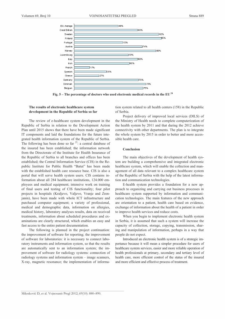
Volumen 69, Broj 10 VOJNOSANITETSKI PREGLED Strana 889
Milenkovi D, et al. Vojnosanit Pregl 2012; 69(10): 880–890.
The results of electronic healthcare systemdevelopment in the Republic of Serbia so far
The review of e-healthcare system development in theRepublic of Serbia in relation to the Development ActionPlan until 2015 shows that there have been made significantIT components and laid the foundations for the future inte-grated health information system of the Republic of Serbia.The following has been done so far 25: a central database ofthe insured has been established; the information networkfrom the Directorate of the Institute for Health Insurance ofthe Republic of Serbia to all branches and offices has beenestablished; the Central Information Service (CIS) in the Re-public Institute for Public Health “Batut” has been madewith the established health care resource base. CIS is also aportal that will serve health system users. CIS contains in-formation about all 284 healthcare institutions, 124.000 em-ployees and medical equipment; intensive work on trainingof final users and testing of CIS functionality; four pilotprojects in hospitals (Kraljevo, Valjevo, Vranje and Zren-janin), have been made with whole ICT infrastructure andpurchased computer equipment; a variety of professional,medical and demographic data, information on allergies,medical history, laboratory analyses results, data on receivedtreatments, information about scheduled procedures and ex-aminations are clearly structured, which enables an easy andfast access to the entire patient documentation.
The following is planned in the project continuation:the improvement of software for reporting; the improvementof software for laboratories: it is necessary to connect labo-ratory instruments and information system, so that the resultsare automatically sent to an information system; the im-provement of software for radiology systems: connection ofradiology systems and information system – image scanners,X-ray, magnetic resonance; the implementation of informa-
tion system related to all health centers (158) in the Republicof Serbia.
Project delivery of improved local services (DILS) ofthe Ministry of Health needs to complete computerization ofthe health system by 2011 and that during the 2012 achieveconnectivity with other departments. The plan is to integratethe whole system by 2015 in order to better and more acces-sible health care.
Conclusion
The main objectives of the development of health sys-tem are building a comprehensive and integrated electronichealthcare system, which will enable the collection and man-agement of all data relevant to a complex healthcare systemof the Republic of Serbia with the help of the latest informa-tion and communication technologies.
E-health system provides a foundation for a new ap-proach to organizing and carrying out business processes inhealthcare system supported by information and communi-cation technologies. The main features of the new approachare orientation to a patient, health care based on evidence,exchange of information about the health of a patient in orderto improve health services and reduce costs.
When you begin to implement electronic health systemin Serbia, it is assumed that such a system will increase thecapacity of collection, storage, copying, transmission, shar-ing and manipulation of information, perhaps in a way thatpeople do not expect.
Introduced an electronic health system is of a strategic im-portance because it will mean a simpler procedure for users ofhealthcare system services, easier and more reliable operation ofhealth professionals at primary, secondary and tertiary level ofhealth care, more efficient control of the status of the insuredand more efficient and effective process of treatment.
Fig. 5 – The percentage of doctors who used electronic medical records in the EU 24

Strana 890 VOJNOSANITETSKI PREGLED Volumen 69, Broj 10
Milenkovi D, et al. Vojnosanit Pregl 2012; 69(10): 880–890.
R E F E R E N C E S
1. International Information Support Centre. ICT and health [cited2006 September 25]. Available from:www.asksource.info/res_library/ict.htm
2. Marinkovi J, Kocev N. Medical informatics, In: Vidakovi A,editors. Industrial medicine I. Beograd: KCS; 1996. p.492 508. (Serbian)
3. Shortliffe EH, Cimino JJ.. Management of Information inHealthcare Organizations. Technicon Medical InformationSystem (TMIS) [cited 2011 december 6]. Available from:http://www.informatics-re-view.com/wiki/index.php/Technicon_Medical_Information_System_%28TMIS%29
4. Eysenbach G. What is e-health? J Med Internet Res 2001; 3(2):E20.
5. Commission Of The European Communities. e-Health - makinghealthcare better for European citizens: An action plan for aEuropean e-Health Area. Belgium, Brussels, 2004. [cited 2004April 30]. Available from:www.openclinical.org/publicreportsE-healthS
6. Fotheringham MJ, Owies D, Leslie E, Owen N. Interactive healthcommunication in preventive medicine: internet-based strate-gies in teaching and research. Am J Prev Med 2000; 19(2):113 20.
7. Laerum H, Karlsen TH, Faxvaag A. Effects of scanning andeliminating paper-based medical records on hospital physi-cians' clinical work practice. J Am Med Inform Assoc 2003;10(6): 588 95.
8. What Is an Electronic Patient Record? [cited 2011 April 23].Available from: http://www.wisegeek.com/what-is-an-electronic-patient-record.htm
9. Smart Card Alliance. Healthcare Applications. [cited 2010 Feb-ruary 8]. Available from: www.congseng.com/news-14-28-28.html
10. Istepanian R, Laxminarayan S, Pattichis CS. M-Health - EmergingMobile Health Systems Berlin: Springer; 2006.
11. Center for Information Technology. Partners HealthCare System.[cited 2007 February 23]. Available from:care.diabetesjournals.org/content/30/.../1137.f
12. Wang SJ, Middleton B, Prosser LA, Bardon CG, Spurr CD, CarchidiPJ, et al. A cost-benefit analysis of electronic medical recordsin primary care. Am J Med 2003; 114(5): 397 403.
13. Devedži V, Chaloub-Johansson C, Weinhara M, koordinatori.. De-velopment of Health Information System for Basic Health andPharmaceutical Services in Serbia. Copenhagen, Denmark:European Agency for Reconstruction; 2008. Available from:http://www.ehr-serbia.com/
14. Ministry of Information Technologies and Communication(MTIC). Ministry of Science and Technological Development(MSTD-RS) [updated 2009 November 9].Available from: http://www.seera-ei.eu/
15. Jovanovic-Milenkovic M, Radojicic Z, Milenkovic D, Vukmirovic D.Applying electronic documents in development of thehealthcare information system in the Republic of Serbia.Comp Sci Informat Sys 2009; 6(2) 111 26. (Serbian)
16. Ivanovi I. Activity center health information system. Belgrade:Institute of Public Health of Serbia; 2008. (Serbian)
17. Stojkanovi I. Improving the efficiency of health care by apply-ing new information technology in primary health care. Vranje:Healthcare Center Vranje; 2010. (Serbian)
18. Ministry of Defence of the Republic of Serbia. E-cards in the Ministryof Defence. Belgrade 2010. Available from:http://www.mod.gov.rs/novi_lat.php?action=fullnews&showcomments=1&id=2648(Serbian)
19. Karanovi N. The next steps. The first conference on informa-tion technology in the health system in Serbia. Belgrade:Pharmaceutical Association of the Republic of Serbia; 2009.(Serbian)
20. Commission of the european communities. e-Health - makinghealthcare better for European citizens: An action plan for aEuropean e-Health Area. Brussels, Belgium 2004. The Com-plete Document. [cited 2004 April 30]. Available:http://europa.eu/eur-lex/en/com/cnc/2004/com2004_0356en01.pdf
21. Šubarevi V. E-Health Belgrade City - expectations, initiatives,implementation, The first conference on information technol-ogy in the health system in Serbia. Belgrade: PharmaceuticalAssociation of the Republic of Serbia; 2008. (Serbian)
22. Vukmirovi D. Pavlovi K, Šuti V. The use of ICT in the Re-public of Serbia. Belgrade: Statistical Office of the Republic ofSerbia; 2010. (Serbian)
23. European Commission. Public health. eHealth. Availablehttp://ec.europa.eu/index_en.htm
24. Centers for Disease Control and Prevention. Percentage of PhysiciansUsing Electronic Medical Records and Using Electronic Medi-cal Record System by Practice size: United States 2005. At-lanta, USA: National Center for Health Statistics; 2005. 2008.
25. Teodosijevi N. The role of information technology in the healthsystem of Serbia, Round Table: Strengthening accountabilityand improving the efficiency of the health system of Serbia.Belgrade: Ministry of Health of the Republic of Serbia; 2009.(Serbian)
Received on November 25, 2010.Revised on March 29, 2011.Accepted on April 4, 2011.
OnLine-first, June, 2012.
Vojnosanit Pregl 2012; 69(10): 891–898. VOJNOSANITETSKI PREGLED Strana 891
Correspondence to: Tihomir V. Ili , Funkcijska neurološka dijagnostika, Vojnomedicinska akademija, Crnotravska 17, 11 000 Beograd,Srbija. Tel.: + 381 11 3609 064. E-mail: [email protected]
O P Š T I P R E G L E D I UDC: 57::612.829]::616.831-08DOI: 10.2298/VSP1210891I
Plasti na reorganizacija ljudskog motornog korteksaPlastic reorganisation of human motor cortex
Tihomir V. Ili *, Nela V. Ili †
*Medicinski fakultet Vojnomedicinske akademije, Univerzitet odbrane, Beograd,Srbija; †Klinika za fizikalnu medicinu i rehabilitaciju, Klini ki centar Srbije, Medicinski
fakultet Univerziteta u Beogradu, Beograd, Srbija
Klju ne re i:motorna kora; aktivnost, fizi ka; neurotransmiteri;mozak, ošte enje, hroni no; le enje.
Key words:motor cortex; motor activity; neurotransmitter agents;brain damage, chronic; therapeutics.
Uvod
Ube enja prema kojima su svojstvo nepromenjivosti iodsustvo mogu nosti preoblikovanja ili funkcionalne reorga-nizacije, relevantna obeležja centralnog nervnog sistema(CNS) odraslih sisara, predstavljala su jednu od centralnihdogmi neurobiologije XX veka. Za dugo vreme prevladavaoje konsenzus prema kojem razvojno stariji delovi mozga, ali isam neokorteks, nakon perioda ranog razvoja i maturacijenervnog sistema, predstavljaju stabilne i nepromenjive stru-kture. Jedini izuzetak, prema takvom konceptu, predstavljalisu procesi u enja, pra eni promenama sinapti ke transmisije,koji se dešavaju isklju ivo u regionima odgovornim za for-miranje memorije, poput hipokampusa i girusa dentatusa –lokacijama na kojim i u odraslom dobu dolazi do formiranjanovih neurona, drugim re ima, lokacijama sa visokim plasti-nim potencijalom. Me utim, kumulacija dokaza iz eksperi-
menata na životinjama, gde su primenjivane metode invaziv-ne mikrostimulacije i mapiranja moždane kore, ve od 80-tihgodina prošlog veka donose nova saznanja koja se odnose naplasti ne promene na razli itim nivoima, kako na nivo repre-zentacionih mapa, tako i na promene u aktivnosti manjihgrupa neurona ili ak i pojedina nih sinapsi. Korak po korak,usvajani su stavovi prema kojima plasti na reorganizacijamoždane kore zauzima svoje mesto u permanentnoj interak-ciji organizma sa spoljašnjom sredinom. Iz takvih shvatanjaproistekla je i savremena definicija kortikalnog plasticitetakojom se obuhvata niz adaptivnih promena funkcionalnih ilimorfoloških svojstava moždane kore u procesima interakcijesa spoljašnjom sredinom i/ili nakon patoloških procesa i oš-te enja mozga 1.
Motorna kora je zauzela centralnu ulogu u istraživanji-ma ove vrste, te postala na neki na in ogledni poligon name-njen razobli avanju starih dogmi i dokazivanju novih princi-
pa. Ne sasvim slu ajno, ovaj deo moždane kore, pored viso-kog potencijala plasti ne reorganizacije, pruža jedinstvenumogu nost kontinuiranog pra enja izlaznih signala, sa viso-kom vremenskom rezolucijom, putem registrovanja amplitu-da izazvanih motornih odgovora [(motorni evocirani poten-cijali (MEP)]. Me utim, prepreku prenosu saznanja ste enihu eksperimentima na životinjama, predstavljao je razvoj teh-nologije, kao i u brojnim oblastima medicinskih istraživanja.Razvojem metoda neinvazivne kortikalne stimulacije, kojedanas obuhvataju transkranijalnu magnetnu stimulaciju(TMS) i transkranijalnu stimulaciju istosmernom strujom(tSIS) (transcranial direct current stimulation – tDCS), po-krenut je niz bezbednih eksperimenata na ljudima, u kojimase pokušalo reprodukovati opažanje poreklom iz invazivniheksperimenata na životinjama, sa ciljem prevazilaženja bari-jere vrste, i integracije novih saznanja u oblast humane me-dicine – od fiziologije pa sve do mogu nosti modulacije na-rušenih funkcija.
Metode neizvazivne kortikalne stsimulacije
Od ove dve navedene metode, kako prema vremenuuvo enja, tako i prema višestrukom zna enju ove tehnike,TMS je predstavljala prekretnicu u okviru translacionih is-traživanja vezanih uz plasticitet mozga kod ljudi. Pored mo-gu nosti registrovanja izazvanih odgovora poreklom iz mo-tornog sistema, ime je obezbe eno kontinuirano pra enjepostignutih promena, primenom specifi nih obrazaca ponav-ljane stimulacije TMS-om podsti u se promene nadražljivo-sti motorne kore (korelat plasti ne reorganizacije) koje seodržavaju izvesno vreme i nakon prestanka stimulacije. Uprvoj nameni prostog registrovanja teku e moždane aktivno-sti, primena pojedina nog magnetnog pulsa (TMS), polaga-njem stimulativnog kalema na glavu ispitanika, u regionu re-

Strana 892 VOJNOSANITETSKI PREGLED Volumen 69, Broj 10
Ili TV, Ili NV. Vojnosanit Pregl 2012; 69(10): 891–898.
prezentacije primarne motorne kore, u skladu sa Faradey-ovim zakonom elektromagnetne indukcije, indukuje magnet-no polje, ije su linije toka sile usmerene vertikalno u odnosuna ravan kalema 2. Ovakvo promenjivo magnetno polje, na-dalje, indukuje sekundarno elektri no polje niskog intenzi-teta u samim površnim slojevima moždane kore (do dubine1–2 cm), te na taj na in pobu uje neuralne elemente motor-nog korteksa 3. Ovakva bezbolna i neinvazivna stimulacijaima za posledicu niz descendentnih pražnjenja (tzv. I-talasi)duž kortikospinalnih aksona sa visokom frekvencijom (oko600 Hz) koji vode aktivaciji alfa motoneurona izazivaju ipokrete i registrovanje kompleksnog sinusoidnog odgovorasa relativno kratkom latencijom (20–25 ms) u ciljnom miši ukontralateralne strane tela (elektromiografski signal). Slože-nija analiza nivoa ekscitabilnosti motorne kore kod ljudisprovodi se pomo u tehnike primene sparenih magnetnihpulseva (paired pulse stimulation) u kratkim interstimulus-nim intervalima (ISI) koji se uzastopno osloba aju kroz ististimulativni kalem 4. Pri kratkim ISI (1–4 ms) test puls bivainhibiran pripremnim pulsem (kondicioniraju im), za razlikuod dužih intervala (8–15 ms) kada test puls biva facilitiran.Brojni dokazi upu uju na to da test inhibicije pulsa priprem-nim stimulusom potpragovnog intenziteta jeste kortikalni fe-nomen, budu i da su serijom eksperimenata isklju eni dopri-nosi sa supkortikalnog ili spinalnog nivoa 5.
Me utim, za drugu namenu TMS-a, kao metode kojaintereferira sa moždanom aktivnoš u na na in da ak možeda menja obrasce funkcionisanja, primenjuju se isklju ivostrukturisani protokoli sa ponavljanjem stimulusa, koji su de-finisani intenzitetom, vremenskim konstantama koje odva-jaju uzastopne pulseve, kao i ukupnim brojem stimulusa. Fi-ziološki mehanizmi aktivacije neuralnih struktura, u ovomslu aju, nisu ni približno jednostavni kao pri primeni pojedi-na nih pulseva koji me usobno ne inteferiraju, budu i daponavljana stimulacija izaziva mešavinu ekscitatornih i inhi-bitornih efekata, a pored svega pojedini od aktiviranih neu-ralnih elemenata imaju projekcije ka daljim kortikalnim ilisupkortikalnim strukturama, ostvaraju i efekte i na udalje-nim mestima.
Razvoj novih arteficijelnih metoda manipulacije kor-tikalnim plasticitetom u procesu oporavka narušene motor-ne funkcije, zasnovan je na poznavanju relevantnih podsti-caja plasti noj reorganizaciji mozga, kako je to ve pretho-dno utvr eno u animalnim eksperimentima. Takva istraži-vanja pokazala su da pokreta i plasti nih promena mogubiti razli iti oblici eksperimentalnih intervencija daju iprimarni zna aj bihejvioralnim manipulacijama (slika 1):izmenama u prilivu aferentnih ulaznih signala u senzomo-torni korteks (poreklom bilo iz taktilnih ili proprioceptivnihprojekcija); iskustvu vezanom uz vršenje pokreta usmere-nih ka sticanju motorne veštine (motorni trening ili motor-no u enje), tzv. plasticitet podstaknut upotrebom; ali u istovreme ne zapostavljaju i niti sagledavanje preostalih mo-gu nosti: farmakološka modulacija plasticiteta primenomlekova sa specifi nim delovanjem na neurotransmiterske si-steme CNS-a; plasticitet podstaknut lezijama i patološkimprocesima u okviru CNS-a; plasticitet podstaknut stimula-cijom moždane kore.
Sl. 1 – Pokreta i kortikalnog plasticiteta
Manipulacije senzornim iskustvom
Somatosenzorne ulazne informacije neophodne su prili-kom obavljanja kompleksnih motornih zadataka, kao i tokomprocesa usvajanja novih motornih veština 6. Veoma dugo jepoznato da aferentni signali poreklom iz kutanih i miši nihreceptora mogu menjati nadražljivost neuronskih populacijasenzorimotorne kore mozga kod viših primata 7, ali i kodljudi 8. Zna aj aferentnih ulaznih signala definisali su ekspe-rimenti u kojima su pojedine strukture u okviru ovih ascen-dentnih projekcija bile ledirane. Tako je zapo eto sa pokuša-jima unilateralne deaferentacije ekstremiteta kod majmuma,presecanjem zadnjih korenova spinalnih nerava, što je razvi-jalo kod životinja sklonost da ne upotrebljavaju deaferento-vani ekstremitet 9. Komplementarni pravci istraživanja obu-hvatali su invazivna mapiranja somatosenzitivnog korteksanakon presecanja perifernih nerava 10, amputacija prstiju 11 ivešta ke sindaktilije (spajanje susednih prstiju životinja uzpomo hirurških šavova) 12. Ova istraživanja potpomogla sunastanak koncepta receptivnih polja (grupe perifernih recep-tora koji usmeravaju aferentni dotok informacija sa senzor-nih “površina” ka “pojedina nim” neuronima korteksa) izega su pretpostavljene mogu nosti topografske reorganiza-
cije somatosenzitivnog korteksa. Nedostaju a potvrda ovihmogu nosti kod ljudi uskoro se pojavila kroz magnetoence-falografsku (MEG) studiju kortikalne reorganizacije kod se-damnaestogodišnjaka, koji je u saobra ajnoj nesre i doživeonadlakatnu amputaciju 13. U ovom detaljnom prikazu boles-nika data je reorganizacija kortikalne reprezentacije šake ipodlaktice, tako što je bolesnik prilikom dodirivanja kože li-ca doživljavao percepciju dodira izgubljenog ekstremiteta.Sumnje nije više moglo biti – fenomen kortikalne reorgani-zacije kod ljudi postao je injenica.
Me utim, manipulacija dotokom senzornih informacijazapo eta je poja anjem aferentnih somatosenzitivnih ulaznihsignala. Ponavljana kutana stimulacija distalnih falangi (ja-godica) jednog ili više prstiju (pritisak na rotiraju i disk sa

Volumen 69, Broj 10 VOJNOSANITETSKI PREGLED Strana 893
Ili TV, Ili NV. Vojnosanit Pregl 2012; 69(10): 891–898.
neravnom površinom) majmuna tokom 60–90 dana, poka-zalo se, vodila je ka uve anju mapa kortikalne reprezentacijestimulisane šake na somatosenzitivnom korteksu 14. Opser-vacione studije, ponovnom primenom MEG, prikazale su dase sli ni fenomeni dešavaju i kod ljudi u slu ajevima poja a-ne profesionalne upotrebe šake. Tim putem, utvr eno je po-stojanje izrazite hemisferne asimetrije mapa somatosenzornereprezentacije šaka kod profesionalnih muzi ara na ži animinstrumentima, budu i da leva šaka odgovara visokozahtev-nom motornom zadatku hvatova na vratu instrumenta 15. Naosnovu ovih saznanja vezanih za plasti nu reorganizacijusomatosenzitivnog korteksa pristupilo se razli itim modeli-ma arteficijelne stimulacije aferentnih somatosenzitivnihprojekcija poreklom iz perifernih receptora, primenom elek-tri kih ili vibratornih stimulusa 16–19. Primenjivost ovakvihmodela poja ane aferentne modulacije u skorije vreme testirase i u klini kim pilot studijama 20, 21.
Suprotno ovom, deprivacija aferentnih somatosenzitiv-nih ulaznih signala nije sagledavana u okviru mogu nostiprimene u cilju oporavka motorne funkcije kod ljudi, s obzi-rom na poznate injenice, da se redukcijom dotoka ovih in-formacija, nakon primene lokalne anestezije remeti motornakontrola ak i kod zdravih ispitanika 22. Me utim, konceptsmanjenog dotoka aferentnih infromacija, u po etku sasvimzapostavljen kao mogu e terapijsko sredstvo, vremenom je,kroz patološke studije na ljudima, dobio novu dimenziju. Ši-renje kortikalne reprezentacije šake na štetu regiona lica, kodbolesnika sa facijalnom paralizom, bio je jedan od prvih na-laza u ovom smeru 23. Dakle, usvojeno je saznanje premakojem i redukcija dotoka aferentnih informacija u somato-senzitivnu koru može voditi ka porastu kortikalne ekscitabil-nosti susednog regiona – preduslovu plasti ne reorganizacije.Eksperimentalne studije na ljudima koristile su model priv-remene ishemijske deaferentacije podlaktice naduvavanjemmanžetne sfingomanometra iznad nivoa sistolnog pritiska,ime je izazivan porast MEP amplitude u miši ima proksi-
malno u odnosu na blok 24. U klini koj studiji koja se poslu-žila istim principom, motorni trening sprovo en neposrednonakon regionalnog anesteti kog bloka proksimalnog delabrahijalnog pleksusa, kod osoba sa parezom šake, koje sumoždani udar pretrpele pre više godina, rezultovao je zna-ajnim poboljšanjem motorne funkcije 25. Model dieferenta-
cije zasnovan je na kompeticiji susednih telesnih regija (npr.nadlaktica vs. šaka) za teritoriju reprezentacije senzorimotor-nog korteksa, pri emu je ovaj efekat posebno izražen u slu-aju uporednog motornog treninga pareti ne grupe miši a.
Plasticitet podstaknut upotrebom
U svakodnevnom životu ovek primenjuje niz raznovr-snih motornih veština koje je sticao tokom razvoja, krozkontinuirana ponavljanja i interakciju sa sredinom koja gaokružuje. Usvajanje novih veština postiže se kroz ponavljanaizvršavanja motornih zadataka (motorni trening), koji nadaljevode ka poboljšanju pokreta 26. Opisane vrste adaptacije uprocesu savladavanja pokreta, smatraju se danas jednim odnajsnažnijih stimulusa koji pokre u funkcionalnu reorgani-zaciju motornog korteksa.
Ovo se pre svega odnosi na u estala i ponavljana izvo-enja pojedinih pokreta, sa pažnjom usredsre enom na mo-
torni zadatak, u sklopu sticanja i/ili održavanja profesionalnemotorne veštine (vrhunski sportisti, muzi ki virtuozi).
Suprotno tome, u uslovima produžene redukcije senzo-rimotorne aktivnosti, kakva se vi a kod imobilizacija eks-tremiteta (prelomi, distorzije i sl) koje za posledicu imaju iz-razitu hipotrofiju, odnosno redukciju miši ne mase i snage,dolazi i do opadanja veštine motornog izvo enja. U nekolikodosadašnjih studija prikazana je reorganizacija primarnogmotornog korteksa kao posledica dužeg inaktiviteta, kod lju-di nakon imobolizacija ekstremiteta usled fraktura 27, 28. Stu-dija Liepert i sar. 28 prikazala je redukciju mapa kortikalnereprezentacije za miši e koji su bili imobilizovani. S drugestrane, u radu Zanette-ija i sar. 27 amplitude MEP ispitanikase umanjivala nakon prinudne redukcije upotrebe, ali u istovreme bez promena ekscitabilnosti na spinalnom nivou, imeje razrešena mogu nost plasti nih promena u segmentnoj di-stribuciji kao posledici inaktiviteta.
Mehanizam plasticiteta podstaknutog upotrebom u na-stanku promena kortikalne reprezentacije pojedinih pokretakod ljudi upoznat je kroz seriju fizioloških eksperimenata, ukojima je TMS primenjivan u kombinaciji sa farmakološkimagensima za koje je poznato da interferiraju sa sinapti kimplasticitetom. Blokada plasticiteta zavisnog od upotrebe pos-tignuta je primenom lorazepama, pozitivnog alosteri kogmodulatora na receptorima tipa GABAA i dekstrometorfana,leka koji blokira aktivnost NMDA-tipa glutamatnih recepto-ra, neophodnu za indukciju fenomena dugoro ne potencija-cije (DP) u motornoj kori. Suprotno ovome, primena D-amfetamina vodila je ja anju plasticiteta zavisnog od upotre-be na na in da su promene postajale dugotrajnije 29.
Farmakološka modulacija plasticiteta
Eksperimenti na životinjama u kojima je razmatranamodulacija plasticiteta farmakološkom manipulacijom razli-itim neurotransmiterskim sistemima ozna ila je noradrener-
gi ku i dopaminergi ku stimulaciju, kao potencijalne pro-motere kortikalnog plasticiteta, suprotno primeni agenasa saGABA-ergi kim delovanjem, koji ja aju i inhibiciju okludi-raju plasti ne promene 30. Naime, nor dren lin potencira fe-nomen DP u vizuelnom korteksu pacova, primenom specifi-nog obrasca repetitivne stimul cije koje oponaša teta ritam
hipokampusa (theta-burst), ime se pove v depol rizuju iodgovor n tet ni ku re kciju, i nadalje, u toku tr j nj togodgovor , povr tno pove v membr nsku provodljivostkontrolis nu NMDA tipom glut m tnih receptor 31. U mo-tornom korteksu p cov nor dren lin, n d lje, pove vekscit bilnost velikih pir mid lnih elij sloj V sl bljenjemsporih K+ struj , i j njem perzistentnog influks Na+ 32, štoje verov tno dovoljno z promovis nje fenomen DP u mo-tornom korteksu. Me utim, u klini koj pr ksi, neposrednprimen nor dren lin nije mogu a, usled niz neželjenihefek t , a primen mfet min t ko e je ogr ni en u pri-meni, s obzirom na k rdiov skul rne rizike ili eventu lnemogu nosti r zvoj dikcije 33. Stoga, pažnja je posebno us-merena ka mogu nostima pospešivanja dopaminergi ke tran-
Strana 894 VOJNOSANITETSKI PREGLED Volumen 69, Broj 10
Ili TV, Ili NV. Vojnosanit Pregl 2012; 69(10): 891–898.
smisije u procesima oporavka i kortikalne reorganizacije, našta upu uju i pojedine klini ke studije 34. Ovakve pretpostav-ke odnedavno su našle uporište i u fiziološkim studijama,primenom neinvazivne kortikalne stimulacije kod ljudi 35, 36.
Plasticitet podstaknut lezijama
Plasticitet podstaknut lezijama nervnog sistema obu-hvata itav niz poreme aja funkcija nervnog sistema, bilo uvidu ve opisanih izmena priliva aferentnih ulaznih signalasa nivoa perifernih receptora ili sa segmentnih nivoa (ošte-enja spinalne medule) ili neposrednih lezija supkortikalnih i
kortikalnih struktura (infarkt mozga, trauma, multipla sklero-za). Ovaj podsticaj verovatno odražava teleološko zna enjekortikalnog plasticiteta budu i da „sistem“ brzo odgovara re-organizacijom, u pokušaju ponovnog zadobijanja kontrolenad ošte enim ili izgubljenim funkcijama. Upravo ishemijskiinfarkt mozga predstavlja bolest – model sagledavanja plas-ticiteta podstaknutog lezijom u procesima oporavka, istra-žuju i istodobno sinergisti ko delovanje molekularnih, si-napti kih i regionalnih mehanizama plasticiteta 37. Nizom is-traživanja pokazano je na animalnim modelima i kod osobakoje su doživele moždani infarkt, da neošte eni regioni kor-teksa, u okolini lezije (penumbra), mogu prihvatiti (u ve ojili manjoj meri) senzomotorno procesiranje, koje je ranijepripadalo zonama nepovratno ošte enim ishemijom 38. Naeksperimentalnom modelu pokazane su dugotrajne promeneu smislu porasta ekscitabilnosti neurona, uz porast glutama-tergi ke aktivnosti posredovane putem NMDA i ne-NMDAtipova receptora do etiri nedelje nakon eksperimentalneokluzije a. cerebri mediae, kao i indukcije DP fenomena,koja je u perilezionalnom korteksu poja ana do sedmog dananakon akutnog razvoja moždane ishemije 39. Tako e, utvr e-no je i postojanje dugoro nog ošte enja gabaergi ke inhibi-torne transmisije, kako u neposrednom okruženju fokalne is-hemije, tako i u kontralateralnoj hemisferi 40. Sumarno i du-goro no pra enje promena kortikalne ekscitabilnosti poka-zalo je da se vrhunac promena meri trajanjem u nedeljama,ali da perzistira i mesecima nakon inicijalnog insulta. Naime,za period tzv. dugoro nog perioda oporavka nakon možda-nog infarkta ( 6 nedelja), na animalnom modelu pokazanaje poja ana sinaptogeneza, na apikalnim dendritima piramid-nih neurona sloja V u regionu periinfarciranog korteksa (narastojanju kra em od 0,5 mm u odnosu na granicu sa ishe-mijskim tkivom) 41. U istom smislu, ve od ranije prisutnasu zapažanja o zavisnosti procesa aksonske regeneracije (me-reno ekspresijom GAP 43) sa ishemijom tokom prve dve ne-delje 42. Me utim pored ja anja sinaptogeneze, anatomskestudije su pokazale da i lokalne i distalne intrakortikalneprojekcije bivaju izmenjene ishemijom, što se smatra svojs-tvom intrinzi ke plasti nosti samog korteksa 41.
U prou avanju funkcionalnih mehanizama nakon infar-kta mozga poseban doprinos pružile su studije u kojima jeprimenjivan TMS. Kod bolesnika sa visokim stepenommotornog hendikepa u akutnoj, ali i hroni noj fazi nakonmoždanog udara, opisana je redukcija inhibicije kako za oš-te enu 43, tako i za neošte enu hemisferu 44–46. Objašnjenjezapažanja vezanih za redukciju intrakortikalne inhibicije oš-
te enog motornog korteksa, u akutnoj fazi, moglo bi biti po-ve ana senzitivnost gabaergi kih neurona na ishemiju, ili štoje još verovatnije – kompenzatorni mehanizam „popuštanja“toni ke gabaergi ke inhibicije u nastojanju da se demaskirajuintrakortikalne horizontalne veze na putu reorganizacionihpromena kortikalnih mapa senzorimotornog korteksa. Nai-me, longitudinalno pra enje ovih bolesnika pokazuje da seredukcija inhibicije tako izražena u akutnoj fazi, ipak poste-peno normalizuje tokom vremena 46, 47.
Plasticitet podstaknut stimulacijom moždane kore
Pojavom neinvazivne tehnike TMS dobijena je metodakojom su se mogle oponašati invazivne stimulacije na ek-sponiranom korteksu eksperimentalnih životinja ili, još daljeod toga, na modelima pojedina ne neuronske sinapse. Usledtoga, prilikom elaboracije pojedinih modela neinvazivnekortikalne stimulacije upotrebljavaju se termini poreklom izbazi ne neurofiziologije. Otkri e prolongiranog poja anja si-napti ke transmisije nakon primene tetani ke stimulacije, fe-nomen poznat kao DP – long term potentiation (LTP), ozna-ilo je tokom poslednjih 40 godina jednu od najzna ajnijih
oblasti istraživanja u neuronaukama – plasticitet zavisan odaktivnosti (activity-dependent plasticity), sposobnost mozgada se preoblikuje u smislu memorije, u enja, poboljšanjamotornih i kognitivnih funkcija 48. Me utim, za razliku odjednostavnih modela stimulacije, izu avanjem vremenskespecifi nosti asocijativne sinapti ke modifikacije u regionuhipokampusa utvr eno je suštinsko zna enje redosleda pre- ipostsinapti kih pražnjenja 49. Ukoliko presinapti ko pražnje-nje prethodi postsinapti kom unutar vremenskog „prozora“od nekoliko desetina milisekundi, dolazi do indukcije DP, zarazliku od obratnog redosleda (post-pre) ime se izaziva sla-bljenje sinapti ke transmisije, fenomen poznat kao dugoro -na depresija (DD) – long term depression (LTD). Stoga,ovakav oblik DP/DD plasticiteta zavisnog od aktivnosti na-ziva se danas plasticitet vremenski zavisan od pražnjenja(spike timing-dependent plasticity) 50, a savremeni protokolineinvazivne stimulacije korteksa upravo nastoje da replici-raju navedene obrasce. Ipak, ovaj translacioni koncept i dodanas trpi kritike. Naime, sagledavaju i u celini strukturnu iorganizacionu kompleksnost cerebralnog korteksa, nije jed-nostavno uspostaviti neposrednu analogiju izme u animalnihmodela i primene na ljudima. Površna analogija govorila bida frekventno-zavisna kortikalna stimulacija primenom re-petitivne (rTMS)-a može na kortikalnim sinapsama ljudipodsta i promene koje su na mehanicisti kom nivou nalikonima koje vi amo pri indukciji DP/DD fenomena na ekspe-rimentalnim životinjama. Ove pretpostavke, naravno, podr-žane su zapažanjima prema kojima najdelotvorniji TMSprotokoli oponašaju prethodno uspešne obrasce stimulacije ueksperimentima na životinjama. Dalja analogija, koja se iupotrebljava kao argument, odnosi se na farmakološku mo-dulaciju indukovanih fenomena. Naime, ukoliko pojedinifarmakološki agens (sa poznatim delovanjem na neurotran-smisiju u CNS-u) prevenira indukciju DP ili DD kod glodara,pretpostavlja se sli no delovanje i na induktivne protokolekod ljudi. Me utim, pra enjem promena MEP amplitude kao

Volumen 69, Broj 10 VOJNOSANITETSKI PREGLED Strana 895
Ili TV, Ili NV. Vojnosanit Pregl 2012; 69(10): 891–898.
osnovnog pokazatelja plasti ne modulacije – izlazne infor-macije iz sistema, treba imati na umu da su originalno meha-nizmi DP i DD monosinapti ki doga aji, dok je MEP am-plituda kao posredni efekat, udaljena od mesta stimulacijeitave tri sinapse. Dakle, bez sumnje, optimalni na in pra enja
promena indukovanih stimulacijom bilo bi registrovanje „do-ga aja“ ve na prvoj sinapsi, što je za sada rutinski nedostup-no. Studije u kojima se obavljaju ove vrste tzv. direktnih regi-strovanja kod ljudi, uz pomo elektroda položenih u epiduralniprostor ispitanika, nose dimenziju invazivnih ispitivanja, iprimenjuju se samo u eksperimentima verifikacije pojedinihfizioloških principa (proof of principle studies). Ipak, primenametoda neinvazivne kortikalne stimulacije, uz puno razume-vanje ograni enja koje donosi, predstavlja najbolju aproksima-ciju sinapti kih doga aja kakvu danas poznajemo.
Uporedo sa razvojem niza protokola TMS, došlo je doponovnog „bu enja“ i tehnike nefokalne stimulacije korteksaistosmernom strujom niskog intenziteta (1–2 mA), tzv. (tSIS;tDCS). Suprotno pojedina nom magnetnom pulsu koji izazi-va MEP u ciljnom miši u, transkranijalna primena istosmer-ne struje niskog intenziteta (1–2 mA) uslovljava modulacijukortikalne ekscitabilnosti, fiziološkim mehanizmima razli i-tim u odnosu na TMS, a bez neposrednog izazivanja elektro-fiziološki merljivog odgovora na periferiji 51. Suštinska raz-lika ovih dvaju tehnika, pa tako i njima srodnih primena, od-nosi se na injenicu da magnetni puls primenjen nad kortek-som odgovara natpragovnom (suprathreshold) nadražaju safenomenološkim efektom izazivanja MEP u ciljnom miši u.Dakle, re je o neurostimulativnoj tehnici koja, uz odgova-raju i broj ponavljanja (obrasci repetitivne stimulacije), uz-rokuje i neuromodulatorni efekat. Za razliku od TMS, prikortikalnoj primeni elektri ne struje izazivamo isklju ivoneuromodulatorni efekat, budu i da je stimulacija potprago-vna (subthreshold) i ne uzrokuje MEP ve isklju ivo interfe-rira sa moždanom aktivnoš u.
Pionirske studije primenom magnetne stimulacije kor-teksa kod ljudi, sprovedene sredinom 90-tih godina, potvr-dile su princip uzlazne modulacije ekscitabilnosti motornekore rTMS pri frekvenciji od 20 Hz 52, kao i silaznu modu-laciju primenom obrasca niskofrekventne stimulacije od 1Hz 53. Me utim sprovo enje originalnih protokola kakve po-znajemo iz animalnih eksperimenata (npr stimulacija frek-vencijom 100 Hz tokom 1 sekunde), nisu, naravno, bile mo-gu e zbog tehni kih ograni enja (pregrejavanje kalema mag-netnog stimulatora), pre svega zbog toga što bi ovakva sti-mulacija kod osetljivih osoba mogla izazvati „nekontrolisa-ni“ porast ekscitabilnosti i eventualne epilepti ke napade.Ovo su razlozi zbog kojih studije kod ljudi ne predstavljajuautenti ne replike visokofrekventnih protokola stimulacije,na na in na koji se oni primenjuju na životinjama. Stoga, po-red obrazaca monotone kortikalne stimulacije, koji su defini-sani isklju ivo frekvencijom, razvijeni su i TMS protokoli ukojima se primenjuju strukturisani obrasci stimulacije, ilikortikalna stimulacija sparena sa aferentnom stimulacijom ustrogo definisanim ISI.
U cilju boljeg razumevanja fizioloških osnova interven-cionih protokola sa primenom TMS, mogu e su analogijeprema fiziološkim obrascima neuronalne aktivnosti od nivoapojedina nih neurona do itavih suppopulacija: toni ka praž-njenja (tonic spiking), karakterisana unimodalnom distribu-cijom intervala izme u pojedina nih pražnjenja; pražnjenja usalvama (bursting), dinami ko stanje u kojem neuroni opeto-vano okidaju u diskretnim grupama ili salvama pražnjenja,iza kojih sledi period mirovanja (quiescence) (u zavisnosti odbroja pražnjenja u salvama ovi nizovi se nazivaju dubleti,tripleti, kvadripleti itd); kao i prelaznim stanjima, izme udva navedena osnovna funkcionalna moda.
Slede i navedene analogije, protokoli rTMS koji seaktuelno primenjuju mogu se podeliti na (slika 2): I – frek-ventno-zavisne (analog plasticitetu zavisnom od aktivnosti –
Sl. 2 – Protokoli neinvazivne kortikalne stimulacije – mogu nosti bidirekcionalne modulacije plasticiteta. Panel A pokazujeobrasce visoko- (gornji deo) i niskofrekventne repetitivne transkranijalne magnetne stimulacije (rTMS) (donji deo); panel B
pokazuje theta-burst stimulaciju (TBS), u gornjem delu facilitatorni protokol intermitentne TBS (obratiti pažnju napostojanje pauza), u donjem delu inhibitorni protokol kontinuirane TBS; panel C prikazuje sparenu asocijativnu
stimulaciju (SAS) uz interstimulusne intervale (ISI) 25 ms (gornji deo) poja anje ekscitabilnosti motornog korteksa, zarazliku od inhibitornog protokola sa ISI 10 ms (donji deo); panel D – transkranijalna stimulacija istosmernom strujom
(tSIS), anodni oblik (gornji deo), katodna stimulacija (dole)

Strana 896 VOJNOSANITETSKI PREGLED Volumen 69, Broj 10
Ili TV, Ili NV. Vojnosanit Pregl 2012; 69(10): 891–898.
activity-dependent plasticity) – obrasci ritmi ke i toni kestimulacije moždane kore: a) sa unimodalnom distribucijomISI – niskofrekventna TMS (< 1 Hz), visokofrekventna TMS(> 5 Hz) i b) sa multimodalnom distribucijom ISI intervala(strukturisana stimulacija); pr. theta-burst TMS – obrazacstimulacije moždane kore pražnjenjem u salvama (multimo-dalna distribucija interstimulusnih intervala i intervala izme-u salvi) 54 – kontinuirani theta-burst TMS (kTBS); intermi-
tentni theta-burst TMS (iTBS); i II – intervalno zavisni pro-tokoli rTMS (plasticitet vremenski zavisan od pražnjenja –spike timing-dependent plasticity): a) kombinovana kortikal-na stimulacija rTMS i periferna elektri na stimulacija i pro-tokol sparene asocijativne stimulacije (SAS), kombinova-njem nisko frekventne rTMS i periferne aferentne stimulacijeu striktnom ISI 55 i b) repetitivni TMS u intervalima periodi-citeta I-talasa (spike-timing dependent plasticity) – primenaparova stimulusa 56, primena tripleta stimulusa 57 i primenakvadripleta stimulusa 58.
U pogledu dosadašnje primene predloženih protokolaizdvajaju se TBS i SAS protokoli, gde prvi odgovara inter-veniconom protokolu, kojim se oponašaju prirodni (inhe-rentni) ritmovi mreža neuronskih populacija, kao što je slu ajsa theta-ritmom (5 Hz) hipokampusa 59. Originalni TBSprotokol Huang-a i sar. 54, tzv. intermitentni obrazac teta-pražnjenja podrazumevao je 600 magnetnih pulseva, niskogintenziteta (80% vrednosti praga podražaja) u 20 epizoda odpo tri stimulusa (frekvencije od 50 Hz, što odgovara interva-lima izme u pojedina nog stimulusa od 20 msec), uz pauzeod po 8 sec, što je vodilo ka porastu MEP amplitude u perio-du od oko 20 min. Suprotno ovom, ukoliko se pauze izosta-ve, model nazvan kontinuiranim obrascem teta-pražnjenja,vodio je ka padu MEP amplitude.
Pored navedenog obrasca “ iste” kortikalne stimulacije,u upotrebi je i SAS protokol, koji oponaša obrazac tzv.Hebb-ovog asocijativnog plasticiteta, prema kojem snaga si-napsi može biti modulirana usaglašenom aktivnosti poreklomiz dva izvora, u striktnoj vremenskoj korelaciji – model plas-ticiteta vremenski zavisnog o pražnjenjima. Prva eksperi-mentalna replika ovog principa, primenom metoda neinvazi-vne kortikalne stimulacije kod ljudi upotrebila je perifernielektri ni stimulus (niske frekvencije, a trostruko višeg in-tenziteta u odnosu na perceptivni prag) prezentovan ispitani-ku iznad n. medianus u predelu ru nog zgloba, iza ega usle-di magnetni puls nad kontralateralnom hemisferom u odgo-varaju oj motornoj prezentaciji ciljnog miši a tenara, pri ISIod 25 milisec 55. Postintervenciono pra enje pokazalo je dasu promene odražene porastom amplitude izazvanih motor-nih odgovora (DP – sli an efekat) maksimalno izražene to-kom narednih sat vremena, da se na preintervencione vred-nosti vra aju kroz 24 asa, da su striktno topografski specifi-ne, te da zavise o aktivaciji NMDA receptora 60. Testira-
njem spinalne ekscitabilnosti (registrovanje F-odgovora) i
elektri nom stimulacijom moždanog stabla potvr eno jekortikalno poreklo opaženih fenomena. Naknadni eksperi-menti koji su pratili isti model utvrdili su depresiju amplitudeMEP, u trajanju od oko 90 min, ukoliko bi se ISI skratio na10 milisec (DD – sli an efekat). Efekat se može blokiratiprimenom NMDA antagonista (dekstrometorfan) ili antago-nista voltažno-zavisnih kalcijumskih kanala (nimodipin) 61.Ukupno uzevši, u nizu sprovedenih eksperimenata izvedenisu zaklju ci da procesima sli nim DP ili DD u regionu pri-marnog motornog korteksa kod ljudi (in vivo) upravljajustriktna pravila Hebb-ovog modela u enja. Ovaj protokol jedanas namenjen prvenstveno analizi fizioloških mehanizamaplasticiteta 62, 63 i patofizoloških abnormalnosti plasticitetakod neuroloških oboljenja 64.
Poznavanje navedenih pokreta a kortikalnog plastici-teta u patološkim stanjima i razumevanje mehanizama njiho-ve aktivacije u željenom pravcu, predstavlja osnov strategijeobnove neuroloških funkcija. Posebno zna enje ovog kon-cepta usmereno je ka potencijalu kompenzacije hroni nihmotornih deficita, nastalih više godina unazad, ime se na-pušta dugi period terapijskog nihilizma. Naposletku, u budu-im istraživanjima je neophodno posvetiti pažnju visokoj
interindividualnoj varijabilnosti promena kortikalne nadraž-ljivosti kod bolesnika, kao i injenici da klini ki ispoljen de-ficit esto predstavlja neto efekat uporednih kortikalnih i su-pkortikalnih ošte enja. Stoga, pred nama stoji put u pokušajušto egzaktnijeg definisanja obrazaca funkcionalnih abnor-malnosti, sa ciljem razvoja terapijskih programa prilago enihpojedincu. Razvoj kompleksnih neinvazivnih protokola kor-tikalne stimulacije i modulacije funkcije, u sinergiji sa odgo-varaju im farmakološkim agensima i programima motornogtreninga, otvaraju nove mogu nosti u tom pravcu.
Zaklju ak
Plasticitet motornog korteksa odnosi se na reorganiza-cione adaptivne promene pod uticajem relevantnih interak-cija sa spoljašnjom sredinom. Pored plasticiteta pokrenutoglezijom, funkcionalna svojstva motornog korteksa kod ljudimogu biti menjana putem bihejvioralnih i farmakološkihmanipulacija, kao i specifi nih obrazaca neinvazivne korti-kalne stimulacije. Transkranijalna magnetna stimulacija itSIS predstavljaju inovativne, bezbedne i neinvazivne tehni-ke modulacije nadražljivosti humanog motornog korteksa, upokušaju oponašanja stimulativnih protokola izvedenih iz si-napti ke fiziologije. Pored toga, tehnika TMS prikazala seprikladnom u prou avanju reprezentacionog plasticiteta nasistemskom nivou kod ljudi. Svrsishodna modulacija plasti -nih promena motornog korteksa primenom razli itih rehabi-litacionih strategija zasnovanih na detaljnom prou avanjupokreta a plasticiteta otvara novu nadu za one sa hroni nimmotornim deficitom.

Volumen 69, Broj 10 VOJNOSANITETSKI PREGLED Strana 897
Ili TV, Ili NV. Vojnosanit Pregl 2012; 69(10): 891–898.
L I T E R A T U R A
1. Ili TV. Adaptive brain plasticity. Mecthods of non-invasivemodulation. Belgrade: Print Land; 2010. (Serbian)
2. Hallett M. Transcranial magnetic stimulation: a primer. Neuron2007; 55(2): 187 99.
3. Cukic M, Kalauzi A, Ilic T, Miskovic M, Ljubisavljevic M. The in-fluence of coil-skull distance on transcranial magnetic stimula-tion motor-evoked responses. Exp Brain Res 2009; 192(1):53 60.
4. Kujirai T, Caramia MD, Rothwell JC, Day BL, Thompson PD, Fer-bert A, et al. Corticocortical inhibition in human motor cortex.J Physiol 1993; 471: 501 19.
5. Ili TV, Meintzschel F, Cleff U, Ruge D, Kessler KR, Ziemann U.Short-interval paired-pulse inhibition and facilitation of humanmotor cortex: the dimension of stimulus intensity. J Physiol2002; 545(Pt 1): 153 67.
6. Pearson K. Motor systems. Curr Opin Neurobiol 2000; 10(5):649 54.
7. Evarts EV. Motor cortex reflexes associated with learnedmovement. Science 1973; 179(4072): 501 3.
8. Marsden CD, Merton PA, Morton HB. Is the human stretch re-flex cortical rather than spinal? Lancet 1973; 1(7806): 759 61.
9. Taub E, Harger M, Grier HC, Hodos W. Some anatomical obser-vations following chronic dorsal rhizotomy in monkeys. Neu-roscience 1980; 5(2): 389 401.
10. Merzenich MM, Kaas JH, Wall JT, Sur M, Nelson RJ, Felleman DJ.Progression of change following median nerve section in thecortical representation of the hand in areas 3b and 1 in adultowl and squirrel monkeys. Neuroscience 1983; 10(3): 639 65.
11. Merzenich MM, Nelson RJ, Stryker MP, Cynader MS, SchoppmannA, Zook JM. Somatosensory cortical map changes followingdigit amputation in adult monkeys. J Comp Neurol 1984;224(4): 591 605.
12. Allard T, Clark SA, Jenkins WM, Merzenich MM. Reorganizationof somatosensory area 3b representations in adult owl mon-keys after digital syndactyly. J Neurophysiol 1991; 66(3):1048 58.
13. Ramachandran VS. Behavioral and magnetoencephalographiccorrelates of plasticity in the adult human brain. Proc NatlAcad Sci U S A 1993; 90(22): 10413 20.
14. Jenkins WM, Merzenich MM, Ochs MT, Allard T, Guíc-Robles E.Functional reorganization of primary somatosensory cortex inadult owl monkeys after behaviorally controlled tactile stimu-lation. J Neurophysiol 1990; 63(1): 82 104.
15. Elbert T, Pantev C, Wienbruch C, Rockstroh B, Taub E. Increasedcortical representation of the fingers of the left hand in stringplayers. Science 1995; 270(5234): 305 7.
16. Ridding MC, Brouwer B, Miles TS, Pitcher JB, Thompson PD.Changes in muscle responses to stimulation of the motor cor-tex induced by peripheral nerve stimulation in human subjects.Exp Brain Res 2000; 131(1): 135 43.
17. Rosenkranz K, Rothwell JC. Differential effect of muscle vibra-tion on intracortical inhibitory circuits in humans. J Physiol2003; 551(Pt 2): 649 60.
18. Kaelin-Lang A, Luft AR, Sawaki L, Burstein AH, Sohn YH, CohenLG. Modulation of human corticomotor excitability by so-matosensory input. J Physiol 2002; 540(Pt 2): 623 33.
19. McDonnell MN, Ridding MC. Afferent stimulation facilitatesperformance on a novel motor task. Exp Brain Res 2006;170(1): 109 15.
20. Sawaki L, Wu CW, Kaelin-Lang A, Cohen LG. Effects of so-matosensory stimulation on use-dependent plasticity inchronic stroke. Stroke 2006; 37(1): 246 7.
21. Klaiput A, Kitisomprayoonkul W. Increased pinch strength inacute and subacute stroke patients after simultaneous median
and ulnar sensory stimulation. Neurorehabil Neural Repair2009; 23(4): 351 6.
22. Aschersleben G, Gehrke J, Prinz W. Tapping with peripheralnerve block. a role for tactile feedback in the timing of move-ments. Exp Brain Res 2001; 136(3): 331 9.
23. Rijntjes M, Tegenthoff M, Liepert J, Leonhardt G, Kotterba S, MüllerS, et al. Cortical reorganization in patients with facial palsy.Ann Neurol 1997; 41(5): 621 30.
24. Ziemann U, Corwell B, Cohen LG. Modulation of plasticity inhuman motor cortex after forearm ischemic nerve block. JNeurosci 1998; 18(3): 1115 23.
25. Muellbacher W, Richards C, Ziemann U, Wittenberg G, Weltz D,Boroojerdi B, et al. Improving hand function in chronic stroke.Arch Neurol 2002; 59(8): 1278 82.
26. Bays PM, Wolpert DM. Computational principles of sensori-motor control that minimize uncertainty and variability. JPhysiol 2007; 578(Pt 2): 387 96.
27. Zanette G, Tinazzi M, Bonato C, di Summa A, Manganotti P, PoloA, et al. Reversible changes of motor cortical outputs follow-ing immobilization of the upper limb. Electroencephalogr ClinNeurophysiol 1997; 105(4): 269 79.
28. Liepert J, Tegenthoff M, Malin JP. Changes of cortical motor areasize during immobilization. Electroencephalogr Clin Neuro-physiol 1995; 97(6): 382 6.
29. Bütefisch CM. Plasticity in the human cerebral cortex: lessonsfrom the normal brain and from stroke. Neuroscientist 2004;10(2): 163 73.
30. Ziemann U, Meintzschel F, Korchounov A, Ili TV. Pharmacologi-cal modulation of plasticity in the human motor cortex. Neu-rorehabil Neural Repair 2006; 20(2): 243 51.
31. Bröcher S, Artola A, Singer W. Agonists of cholinergic andnoradrenergic receptors facilitate synergistically the inductionof long-term potentiation in slices of rat visual cortex. BrainRes 1992; 573(1): 27 36.
32. Foehring RC, Schwindt PC, Crill WE. Norepinephrine selectivelyreduces slow Ca2+- and Na+-mediated K+ currents in catneocortical neurons. J Neurophysiol 1989; 61(2): 245 56.
33. Grade C, Redford B, Chrostowski J, Toussaint L, Blackwell B. Meth-ylphenidate in early poststroke recovery: a double-blind, pla-cebo-controlled study. Arch Phys Med Rehabil 1998; 79(9):1047 50.
34. Acler M, Fiaschi A, Manganotti P. Long-term levodopa admini-stration in chronic stroke patients. A clinical and neurophysi-ologic single-blind placebo-controlled cross-over pilot study.Restor Neurol Neurosci 2009; 27(4): 277 83.
35. Kuo MF, Paulus W, Nitsche MA. Boosting focally-induced brainplasticity by dopamine. Cereb Cortex 2008; 18(3): 648 51.
36. Ili NV, Petroni I, Graji M, Ili TV. Efekti pojedina nih dozadiazepama i levodope na modulaciju plasticiteta motornogkorteksa kod zdravih osoba – TMS studija. Srp Arh Celok Lek2012; 140(1 2): 14 21
37. Gerloff C, Hallett M. Big news from small world networks afterstroke. Brain 2010; 133(Pt 4): 952 5.
38. Schaechter JD, Moore CI, Connell BD, Rosen BR, Dijkhuizen RM.Structural and functional plasticity in the somatosensory cortexof chronic stroke patients. Brain 2006; 129(Pt 10): 2722 33.
39. Centonze D, Rossi S, Tortiglione A, Picconi B, Prosperetti C, De Chi-ara V, et al. Synaptic plasticity during recovery from perma-nent occlusion of the middle cerebral artery. Neurobiol Dis2007; 27(1): 44 53.
40. Buchkremer-Ratzmann I, August M, Hagemann G, Witte OW.Electrophysiological transcortical diaschisis after cortical pho-tothrombosis in rat brain. Stroke 1996; 27(6): 1105 9; discus-sion 1109 11.
Strana 898 VOJNOSANITETSKI PREGLED Volumen 69, Broj 10
Ili TV, Ili NV. Vojnosanit Pregl 2012; 69(10): 891–898.
41. Brown CE, Aminoltejari K, Erb H, Winship IR, Murphy TH. Invivo voltage-sensitive dye imaging in adult mice reveals thatsomatosensory maps lost to stroke are replaced over weeks bynew structural and functional circuits with prolonged modes ofactivation within both the peri-infarct zone and distant sites. JNeurosci 2009; 29(6): 1719 34.
42. Carmichael ST, Chesselet MF. Synchronous neuronal activity is asignal for axonal sprouting after cortical lesions in the adult. JNeurosci 2002; 22(14): 6062 70.
43. Wittenberg GF, Bastings EP, Fowlkes AM, Morgan TM, Good DC,Pons TP. Dynamic course of intracortical TMS paired-pulse re-sponses during recovery of motor function after stroke. Neu-rorehabil Neural Repair 2007; 21(6): 568 73.
44. Liepert J, Hamzei F, Weiller C. Motor cortex disinhibition of theunaffected hemisphere after acute stroke. Muscle Nerve 2000;23(11): 1761 3.
45. Bütefisch CM, Netz J, Wessling M, Seitz RJ, Hömberg V. Remotechanges in cortical excitability after stroke. Brain 2003; 126(Pt2): 470 81.
46. Manganotti P, Acler M, Zanette GP, Smania N, Fiaschi A. Motorcortical disinhibition during early and late recovery afterstroke. Neurorehabil Neural Repair 2008; 22(4): 396 403.
47. Swayne OB, Rothwell JC, Ward NS, Greenwood RJ. Stages of mo-tor output reorganization after hemispheric stroke suggestedby longitudinal studies of cortical physiology. Cereb Cortex2008; 18(8): 1909 22.
48. Kujala T, Näätänen R. The adaptive brain: a neurophysiologicalperspective. Prog Neurobiol 2010; 91(1): 55 67.
49. Levy WB, Steward O. Temporal contiguity requirements forlong-term associative potentiation/depression in the hippo-campus. Neuroscience 1983; 8(4): 791 7.
50. Song S, Miller KD, Abbott LF. Competitive Hebbian learningthrough spike-timing-dependent synaptic plasticity. Nat Neu-rosci 2000; 3(9): 919 26.
51. Priori A, Berardelli A, Rona S, Accornero N, Manfredi M. Polariza-tion of the human motor cortex through the scalp. Neurore-port 1998; 9(10): 2257 -60.
52. Pascual-Leone A, Valls-Solé J, Wassermann EM, Hallett M. Re-sponses to rapid-rate transcranial magnetic stimulation of thehuman motor cortex. Brain 1994; 117( Pt 4): 847 58.
53. Chen R, Classen J, Gerloff C, Celnik P, Wassermann EM, Hallett M,et al. Depression of motor cortex excitability by low-frequencytranscranial magnetic stimulation. Neurology 1997; 48(5):1398 403.
54. Huang YZ, Edwards MJ, Rounis E, Bhatia KP, Rothwell JC. Thetaburst stimulation of the human motor cortex. Neuron 2005;45(2): 201 6.
55. Stefan K, Kunesch E, Cohen LG, Benecke R, Classen J. Induction ofplasticity in the human motor cortex by paired associativestimulation. Brain 2000; 123 Pt 3: 572 84.
56. Thickbroom GW, Byrnes ML, Edwards DJ, Mastaglia FL. Re-petitive paired-pulse TMS at I-wave periodicity markedlyincreases corticospinal excitability: a new technique formodulating synaptic plasticity. Clin Neurophysiol 2006;117(1): 61 6.
57. Sacco P, Turner D, Rothwell J, Thickbroom G. Corticomotor re-sponses to triple-pulse transcranial magnetic stimulation: Ef-fects of interstimulus interval and stimulus intensity. BrainStimul 2009; 2(1): 36 40.
58. Hamada M, Hanajima R, Terao Y, Arai N, Furubayashi T, Ino-mata-Terada S, et al. Quadro-pulse stimulation is more effectivethan paired-pulse stimulation for plasticity induction of thehuman motor cortex. Clin Neurophysiol 2007; 118(12):2672 82.
59. Larson J, Lynch G. Induction of synaptic potentiation in hippo-campus by patterned stimulation involves two events. Science1986; 232(4753): 985 8.
60. Stefan K, Kunesch E, Benecke R, Cohen LG, Classen J. Mechanismsof enhancement of human motor cortex excitability inducedby interventional paired associative stimulation. J Physiol 2002;543(Pt 2): 699 708.
61. Wolters A, Sandbrink F, Schlottmann A, Kunesch E, Stefan K, CohenLG, et al.A temporally asymmetric Hebbian rule governingplasticity in the human motor cortex. J Neurophysiol 2003;89(5): 2339 45.
62. Ziemann U, Ili TV, Pauli C, Meintzschel F, Ruge D. Learningmodifies subsequent induction of long-term potentiation-likeand long-term depression-like plasticity in human motor cor-tex. J Neurosci 2004; 24(7): 1666 72.
63. Ili NV, Saji J, Miskovi M, Krsti J, Milanovi S, Vesovi -Poti V,et al. The efficacy of two protocols for inducing motor cortexplasticity in healthy humans--TMS study. Gen Physiol Biophys2009; 28 Spec No: 228 34.
64. Morgante F, Espay AJ, Gunraj C, Lang AE, Chen R. Motor cor-tex plasticity in Parkinson's disease and levodopa-induced dys-kinesias. Brain 2006; 129(Pt 4): 1059 69.
Primljen 23. VIII 2010.Prihva en 30 VIII 2010.

Vojnosanit Pregl 2012; 69(10): 899–903. VOJNOSANITETSKI PREGLED Strana 899
Correspondence to: Jasna Mihailovi , Department of Nuclear Medicine, Oncology Institute of Vojvodina, Institutski put 4, 21 204,Sremska Kamenica, Serbia. Phone: +381 21 480 5459. E-mail: [email protected]
G E N E R A L R E V I E W UDC: 616.441-006.6-033.2-08::539.163DOI: 10.2298/VSP1210899M
Radionuclide treatment of metastatic disease in patients withdifferentiated thyroid carcinoma
Le enje metastatske bolesti radionuklidom kod bolesnika sa diferenciranimkarcinomom tiroideje
Jasna Mihailovi *‡, Jasna Trifunovi †‡
*Department of Nuclear Medicine, †Clinic of Internal Oncology, Oncology Institute ofVojvodina, Sremska Kamenica, Serbia; ‡Technical Faculty, University of Novi Sad,
Zrenjanin, Serbia
Key words:thyroid neoplasms; thyroidectomy; iodine; elements,radioactive; drug therapy; radiotherapy; radionuclideimaging; risk factors; prognosis.
Klju ne re i:tireoidna žlezda, neoplazme; tireoidektomija; jod;elementi, radioaktivni; le enje lekovima; radioterapija;scintigrafija; faktori rizika; prognoza.
Introduction
In general, the therapy of differentiated thyroid carci-noma (DTC), includes initial treatment and follow-up of pa-tients. Nowadays, there is no universal accepted consensus re-garding initial treatment of DTC patients. Several countrieshave their own guidelines and recommendations for treatmentof DTC 1–5. Beside all controversies, mostly accepted recom-mendation regarding initial treatment of DTC includes total or“near” total thyreoidectomy followed by the therapy with ra-dioactive iodine (131I). Radioiodine ablation of postoperativethyroid remnants is usually recommended in all DTC patientsregardless their stage (low or high risk of cancer-specificmortality and risk of relapse), due to easier monitoring of thy-roglobulin (Tg) 6. Thyroglobulin is a glycoprotein that is pro-duced by normal or neoplastic follicular thyroid cells. In theabsence of thyroglobulin antibodies (TgAb), undetectable Tgafter the thyroid stimulating hormone (TSH) stimulation is avalid parameter of remission and the absence of metastases.On the other hand, detectable or increasing Tg during follow-up indicates the appearance of metastatic disease. After ra-dioiodine ablation, a life-long suppressive therapy with L-thyroxine should be prescribed to all DTC patients. Rarely, apalliative therapy including external beam radiation therapyand chemotherapy is recommended 7.
After the initial treatment, all DTC patients should bemonitored life long, with the aim to detect persistent disease orrecurrence. Each check-up should include laboratory analyses(thyroid hormones, TSH, Tg and TgAb) and ultrasonographyof the neck. The result of diagnostic whole-body scintigraphy
(WBS) with 131I (131I-WBS) is influenced by thyroid carci-noma affinity to accumulate 131I in the presence of high con-centration of TSH achieved by one-month L-thyroxine with-drawal or with intramuscular application of human recombi-nant TSH 7. Whole-body scintigraphy is routinely performedas a routine check-up (a year after the radioiodine ablation),and thereafter in cases suspected for recurrence only 8. Mag-netic resonance imaging (MRI) and computed tomography(CT) are useful for detection of neck and mediastinal metasta-ses 9. Magnetic resonance is useful especially in non-iodine-avid and mediastinal metastases 10. Fluorodeoxyglucose (18F-FDG) positron emission tomography fused with computed to-mography (18F-FDG-PET/CT) is a modern diagnostic proce-dure that is important in detection of non-iodine-avid metasta-ses 11. Computed tomography is useful in visualization ofsmall lung metastases, but is rarely performed due to iodinecontrast interference with iodine therapy 12.
Local and regional metastases in differentiatedthyroid carcinoma patients
Local recurrence and/or regional neck metastases usu-ally appear during the first years of follow-up in approxi-mately of 5%–20% DTC patients. Regional neck metastasesrepresent about 60%–75% of all neck metastases. They areusually detected by neck ultrasound (in about 94% of cases),or by increased Tg levels or WBS (in 50% of cases) 13. How-ever, these recurrences are not palpable if they are soft, smallor located in a central neck compartment or behind the greatneck vessels. Metastatic neck lymph nodes appear at ultra-

Strana 900 VOJNOSANITETSKI PREGLED Volumen 69, Broj 10
Mihajlovi J, Trifunovi J. Vojnosanit Pregl 2012; 69(10): 899–903.
sound as round, hypoechoic, with microcalcification andcystic components, hypervascularized at Doppler ultrasoundwhich can detect metastatic lymph nodes in early stage ofdisease if they are a few milimeters sized only. Fine needleaspiration (FNA) of lymph node suspicious of recurrence isimportant in detection of regional metastases, since Tg de-termination in the aspirate increases accuracy of cytologicreport 9.
The Tg serum level is not detectable in about 20% ofpatients with isolated lymph node metastases on L-thyroxinetherapy. Thyroglobulin level remains undetectable after TSHstimulation in approximately 5% of these patients. Wholebody scintigraphy detects metastases in about 60%–80% ofpatients with clinical neck lymph node recurrences (Figure1). Mediastinal lymph node metastases are usually combinedwith distant lung metastases. Recurrences in soft tissues orinvading aero-digestive tract appear in less than 10% of allneck metastases 9.
The most important treatment of locoregional recur-rences is surgical removal of metastatic tissue. A mediastinaldissection with additional radioiodine therapy should be per-formed in case of bulky mediastinal metastases (even in thepresence of micrometastases of the lungs). The best effect of131I therapy is obtained when is a diameter of metastaticlymph nodes is less than 1 cm. Some authors suggest surgi-cal treatment as additional procedure after the 131I therapy asinitial treatment of recurrences 14. External beam radiationtherapy is recommended only in patients with non-iodine-avid metastases, those with incompleted surgery and patientswith invasion of aero-digestive tract and soft tissue 15. Acombination of external radiation and chemotherapy (doxo-rubicin in small doses of 10 mg/m2 weekly) is suggested inextensive and nonoperable recurrences 16.
Tubiana et al. 17 reported the ten-year survival rate of62% in DTC patients with local and regional recurrences.
Distant metastases in differentiated thyroidcarcinoma patients
Distant metastases appear approximately in 27% ofDTC patients 18; metastases presented at the time of diagno-sis (early metastases) occur in 9% of them, while metastaseswhich appear during the course of disease (late metastases)occur in 18% cases 19. According to our results, distant me-tastases appear in 21.2% of DTC patients; the frequency ofearly distant metastases is 8.5%, while late distant metastasesappear in 7.02%. Early metastases are more frequent in pap-illary carcinomas, while late metastases occur more often infollicular carcinomas. Appearance time of distant metastasesdoes not significantly affect disease-specific survival (DSS)of DTC patients 20.
Patients with high risk for distant metastases appear-ance are younger than 16 years, older than 45 years, thosewith histological subtypes of papillary carcinoma (tall cell,
columnar cell, diffuse sclerosing) and follicular carcinoma(widely invasive and poorly differentiated), patients withbulky tumors, those with tumors extended beyond thyroidcapsule and with nodal metastases, patients who underwentless extensive surgery than total or near total thyreoidec-tomy, or those with no post-surgical administration of ra-dioiodine ablation 9.
Lung metastases are often combined with lymph node re-currences in central neck and mediastinal compartment (Figure2). Papillary carcinomas usually extend lymphogeneously tothe lungs, while follicular carcinomas extend hematogeneouslyto the lungs and bones. Distant metastases are usually located inlungs (57%), bones (24%), spine, pelvis, long bones, ribs, ster-num, the base of the scull. One third of metastases in bones rep-resent solitary bone metastases. Lung metastases associatedwith bone metastases occur in 16% patients, while metastasesin liver, brain and skin appear in 3% of the patients 21.
Fig. 1 – Posttherapeutic 131I-WBS (whole-body scintigraphy) detects regional metastasis in the lymph node on the lateralright side of the neck (a – anterior view; b – posterior view)

Volumen 69, Broj 10 VOJNOSANITETSKI PREGLED Strana 901
Mihajlovi J, Trifunovi J. Vojnosanit Pregl 2012; 69(10): 899–903.
Surgery should be the primary treatment of bone metasta-ses. This treatment is not recommended if multiple lung metasta-ses are present. Radioactive iodine is accumulated only in 2/3patients with distant recurrences, but is able to destroy only asmall tumor foci. Radioactive iodine is performed in activityranged from 5.55 to 7.4 GBq in adults, every 6 months during thefirst 2 years, and thereafter annually until complete ablation of re-sidual uptake on posttherapeutic 131I-WBS. Most patients are
cured with cumulative activity of 18.5 GBq. Even the fact thatrisk of secondary carcinoma and leukemia increases with highercumulative activity of 131I, there is no limit for cumulative activ-ity which can be performed in DTC patients with metastatic dis-ease. If there is no uptake on posttherapeutic 131I-WBS, any fur-ther radioiodine therapy is useless (Figure 3) 9, 22. In these patientswith so-called non-iodine-avid metastases, the exact localizationof tumor deposits is possible with 18F FDG-PET/CT (Figure 4).
Fig. 2 – Posttherapeutic 131I-WBS (whole-body scintigraphy) shows regional recurrence in submandibular lymph node onthe right side, and distant metastases in the upper mediastinum and both lungs (a – anterior view; b – posterior view)
Fig. 3 – Posttherapeutic 131I-WBS (whole-body scintigraphy) of the patients with increased thyroglobulin serum level doesnot detect radioiodine uptake due to the presence of non-iodine-avid metastasis (a – anterior view; b – posterior view)
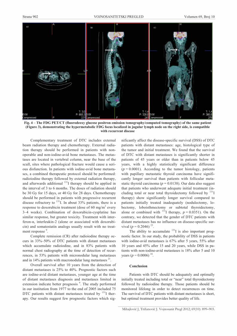
Strana 902 VOJNOSANITETSKI PREGLED Volumen 69, Broj 10
Mihajlovi J, Trifunovi J. Vojnosanit Pregl 2012; 69(10): 899–903.
Complementary treatment of DTC includes externalbeam radiation therapy and chemotherapy. External radia-tion therapy should be performed in patients with non-operable and non-iodine-avid bone metastases. The metas-tases are located in vertebral column, near the base of thescull, sites where pathological fracture would cause a seri-ous disfunction. In patients with iodine-avid bone metasta-ses, a combined therapeutic protocol should be performed:radioiodine therapy followed by external radiation therapy,and afterwards additional 131I therapy should be applied inthe interval of 3 to 6 months. The doses of radiation shouldbe 30 Gy for 15 days, or 40 Gy for 28 days. Chemotherapyshould be performed in patients with progressive recurrentdisease refractory to 131I. In about 33% patients, there is aresponse to doxorubicin treatment (dose of 60 mg/m2 every3–4 weeks). Combination of doxorubicin-cysplatine hassimilar response, but greater toxicity. Treatment with inter-feron- , interleukin-2 (alone or associated with doxorubi-cin) and somatostatin analogs usually result with no treat-ment response 9.
Complete remission (CR) after radioiodine therapy oc-curs in 33%–50% of DTC patients with distant metastaseswhich accumulate radioiodine, and in 83% patients withnormal chest radiography at the time of detection of recur-rences, in 53% patients with micronodular lung metastasesand in 14% patients with macronodular lung metastases 21.
Overall survival after 10 years from the detection ofdistant metastases is 25% to 40%. Prognostic factors suchare iodine-avid distant metastases, younger age at the timeof distant metastases diagnosis and metastases limited inextension indicate better prognosis 9. The study performedin our institution from 1977 to the end of 2005 included 75DTC patients with distant metastases treated by 131I ther-apy. Our results suggest few prognostic factors which sig-
nificantly affect the disease-specific survival (DSS) of DTCpatients with distant metastases: age, histological type ofthe tumor and initial treatment. We found that the survivalof DTC with distant metastases is significantly shorter inpatients of 45 years or older than in patients below 45years, with a highly statistically significant difference(p = 0.0001). According to the tumor histology, patientswith papillary metastatic thyroid carcinoma have signifi-cantly longer survival than patients with follicular meta-static thyroid carcinoma (p = 0.0138). Our data also suggestthat patients who underwent adequate initial treatment (in-cluding total or near total thyroidectomy followed by 131Itherapy) show significantly longer survival compared topatients initially treated inadequately (nodulectomy, lo-bectomy, loboisthmectomy or subtotal thyroidectomy,alone or combined with 131I therapy, p = 0.0351). On thecontrary, we detected that the gender of DTC patients withdistant metastases has no influence on disease-specific sur-vival (p = 0.2046) 23.
The ability to accumulate 131I is also important prog-nostic factor. In our study, the probability of DSS in patientswith iodine-avid metastases is 67% after 5 years, 55% after10 years and 45% after 15 and 20 years, while DSS in pa-tients with non-iodine-avid metastases is 18% after 5 and 10years (p = 0.0006) 24.
Conclusion
Patients with DTC should be adequately and optimallyinitially treated including total or “near” total thyroidectomyfollowed by radioiodine therapy. Those patients should bemonitored lifelong in order to detect recurrences on time.The survival of DTC patients with distant metastases is short,but optimal treatment provides better quality of life.
Fig. 4 – The FDG PET/CT (fluorodeoxy glucose positron emission tomography/computed tomography) of the same patient(Figure 3), demonstrating the hypermetabolic FDG focus localized in jugular lymph node on the right side, is compatible
with recurrent disease

Volumen 69, Broj 10 VOJNOSANITETSKI PREGLED Strana 903
Mihajlovi J, Trifunovi J. Vojnosanit Pregl 2012; 69(10): 899–903.
R E F E R E N C E S
1. The British Thyroid Association and the Royal College of Phy-sicians. Guidelines for the Management of Differentiated Thy-roid Cancer in Adults. Available from: www.british-thyroid-association.org/guidelines.htm [accessed 2006 February 10].
2. Cooper DS, Doherty GM, Haugen BR, Kloos RT, LeeSL, Mandel SJ,et al. Revised American Thyroid Association managementguidelines for patients with thyroid nodules and differentiatedthyroid cancer. Thyroid 2009; 19(11): 1167 214.
3. Pacini F, Schlumberger M, Dralle H, Elisei R, Smit JW, WiersingaW. European consensus for the management of patients withdifferentiated thyroid carcinoma of the follicular epithelium.Eur J Endocrinol 2006; 154(6): 787 803.
4. Luster M, Clarke SE, Dietlein M, Lassmann M, Lind P, Oyen WJ,et al. Guidelines for radioiodine therapy of differentiated thy-roid cancer. Eur J Nucl Med Mol Imaging 2008; 35(10):1941 59.
5. Dietlein M, Dressler J, Eschner W, Grünwald F, Lassmann M,Leisner B, et al. Procedure guidelines for radioiodine therapy ofdifferentiated thyroid cancer (version 3). Nuklearmedizin 2007;46(5): 213 9. (German)
6. Goldsmith SJ. Thyroid carcinoma. In: Khalikhali I, Maublant JC,Goldsmith SJ, editors. Nuclear oncology: diagnosis and therapy.Philadelphia: Lippincott Williams & Wilkins; 2001. p.197 219.
7. Shapiro B, Freitas J, Gross M. Follow-up of patients with well-differentiated thyroid cancer. In: Biersack H-J, Grunwald F,editors. Thyroid cancer. Berlin, Heidelberg: Springer-Verlag;2005. p. 199 219.
8. Stefanovi Lj, Guduri B, Šljapi N, Babi J, Miljkovi Lj, MaleševiM, et al. Protocols of diagnostics, treatment and control ofmalignant tumors at the Institute of oncology in SremskaKamenica. Archive of Oncology 1994; 2(2): 101 7.
9. Schlumberger M, Pacini F. Thyroid Tumors. Paris: Editions Nu-cleon; 2006.
10. Toubert ME, Cyna-Gorse F, Zagdanski AM, Noel-Wekstein S, Cat-tan P, Billotey C, et al. Cervicomediastinal magnetic resonanceimaging in persistent or recurrent papillary thyroid carcinoma:clinical use and limits. Thyroid 1999; 9(6): 591 7.
11. Freudenberg LS, Antoch G, Frilling A, Jentzen W, Rosenbaum SJ,Kühl H, et al. Combined metabolic and morphologic imagingin thyroid carcinoma patients with elevated serum thyroglobu-lin and negative cervical ultrasonography: role of 124I-PET/CT and FDG-PET. Eur J Nucl Med Mol Imaging 2008;35(5): 950 7.
12. Weber AL, Randolph G, Aksoy FG. The thyroid and parathyroidglands. CT and MR imaging and correlation with pathologyand clinical findings. Radiol Clin North Am 2000; 38(5):1105 29.
13. Frasoldati A, Pesenti M, Gallo M, Caroggio A, Salvo D, Valcavi R.Diagnosis of neck recurrences in patients with differentiatedthyroid carcinoma. Cancer 2003; 97(1): 90 6.
14. Travagli JP, Cailleux AF, Ricard M, Baudin E, Caillou B,Parmentier C,et al. Combination of radioiodine (131I) andprobe-guided surgery for persistent or recurrent thyroid carci-noma. J Clin Endocrinol Metab 1998; 83(8): 2675 80.
15. Tubiana M, Haddad E, Schlumberger M, Hill C, Rougier P, SarrazinD. External radiotherapy in thyroid cancers. Cancer 1985; 55(9Suppl): 2062 71.
16. Kim JH, Leeper RD. Treatment of locally advanced thyroid car-cinoma with combination doxorubicin and radiation therapy.Cancer 1987; 60(10): 2372 5.
17. Tubiana M, Schlumberger M, Rougier P, Laplanche A, Benhamou E,Gardet P, et al. Long-term results and prognostic factors in pa-tients with differentiated thyroid carcinoma. Cancer 1985;55(4): 794 804.
18. Shoup M, Stojadinovic A, Nissan A, Ghossein RA, Freedman S,Brennan MF, et al. Prognostic indicators of outcomes in pa-tients with distant metastases from differentiated thyroid car-cinoma. J Am Coll Surg 2003; 197(2): 191 7.
19. Haq M, Harmer C. Differentiated thyroid carcinoma with dis-tant metastases at presentation: prognostic factors and out-come. Clin Endocrinol (Oxf) 2005; 63(1): 87 93.
20. Mihailovic JM, Stefanovic LJ, Malesevic MD, Erak MD, TesanovicDD. Metastatic differentiated thyroid carcinoma: clinical man-agement and outcome of disease in patients with initial andlate distant metastases. Nucl Med Commun 2009; 30(7):558 64.
21. Schlumberger M, Challeton C, De Vathaire F, Travagli JP, Gardet P,Lumbroso JD, et al. Radioactive iodine treatment and externalradiotherapy for lung and bone metastases from thyroid carci-noma. J Nucl Med 1996; 37(4): 598 605.
22. Haq MS, Harmer C. Nonsurgical management of thyroid can-cer. In: Mazzaferri EL, Harmer C, Mallick UK, Kendall-Taylor P,editors. Practical Management of Thyroid Cancer. A Multidis-ciplinary Approach. London: Springer-Verlag; 2006. pp.171 91.
23. Mihailovic J, Stefanovic L, Malesevic M. Differentiated thyroid car-cinoma with distant metastases: probability of survival and itspredicting factors. Cancer Biother Radiopharm 2007; 22(2):250 5.
24. Mihailovic J, Stefanovic L, Malesevic M, Markoski B. The impor-tance of age over radioiodine avidity as a prognostic factor indifferentiated thyroid carcinoma with distant metastases. Thy-roid 2009; 19(3): 227 32.
Received on February 25, 2011.Revised on May 10, 2011.
Accepted on May 16, 2011.

Strana 904 VOJNOSANITETSKI PREGLED Vojnosanit Pregl 2012; 69(10): 904–907.
Correspondence to: Vukašin Badža, Faculty of Sport and Fhysical Education, Miše Dimitrijevi a 72/4, 21 000 Novi Sad, Serbia.Phones: +381 62 4563 88, +381 21 6542 980. E-mail: [email protected]
C A S E R E P O R T UDC: 616-001-073::[61:79DOI: 10.2298/VSP1210904B
Possibilities of thermovision application in sport and sportrehabilitation
Mogu nosti primene termovizije u sportu i sportskoj rehabilitaciji
Vukašin Badža*, Vojin Jovan evi †, Franja Fratri ‡, Goran Rogli §,Nenad Sudarov†
*Faculty of Sport and Physical Education, University of Novi Sad, Novi Sad, Serbia;†Provincial Secretariat for Sport, Novi Sad, Serbia; ‡University EDUCONS, Novi Sad,
Serbia; §Electromedical, Novi Sad, Serbia
Abstract
Introduction. Infrared thermography or thermovision is in-creasingly applicable in sport and sport rehabilitation. Ther-mic forms, thermic imprints, temperature and isotherm dis-tribution, temperature gradient change are the terms that aremore and more often met in sport medicine and medicine, ingeneral. Case report. We presented two examples of ther-movision application: in detection of muscle injury andchanges of the feet exposed to low temperature. In the firstexample the thermovision method was used for analysingheat distribution in an athlete with back muscles injury. Witha special original method of local cooling the place and degreeof injury was precisely localized and determined, respectively,regardless high environmental temperature. In the secondcase the thermovision method was for the first time appliedin a runner whose feet was exposed to low temperature. Sig-nificant hypothermia of the feet was detected by the methodand appropriate treatment was performed. Thanks to this theathlete had no harmful consequences. Conclusion. Ther-movision is fast and efficient in detecting different kind ofinjuries, so its increased use in the future can be expected.
Key words:sports medicine; rehabilitation; athletic injuries;thermal conductivity; treatment outcome.
Apstrakt
Uvod. Infracrvena termografija ili termovizija ima sve ve uprimenu u sportu i sportskoj rehabilitaciji. Termi ke forme,termi ki otisci, temperaturna i izotermalna distribucija,promene gradijenta temperature jesu pojmovi koji su sve e-š e prisutni u sportskoj medicini i medicini uopšte. Prikazbolesnika. Prikazali smo dva primera koriš enja termovizij-ske metode: za otkrivanje povrede miši a le a i promena nastopalima izloženim niskoj temperaturi. U prvom slu ajutermovizija je koriš ena u analizi distribucije toplote kodsportiste sa povredom le nih miši a. Zahvaljuju i specijal-noj, originalnoj metodi lokalnog hla enja, precizno je lokali-zovano mesto i odre en stepen povrede. U drugom slu ajutermovizija je po prvi put upotrebljena u otkrivanju prome-na na stopalima trka a izloženim niskoj temperaturi. Zahva-ljuju i ovoj metodi ustanovljena je zna ajna hipotermijastopala, preduzet je odgovaraju i tretman, tako da sportistanije imao štetnih posledica zbog hipotermije stopala. Zak-lju ak. Termovizija je brz i efikasan metod u otkrivanju ra-zli itih vrsta povreda, pa se može o ekivati njena sve ve aupotreba u budu nosti.
Klju ne re i:medicina, sportska; rehabilitacija; povrede, atletske;provodljivost, toplotna; le enje, ishod.
Introduction
Body temperature is determined by heat productiongenerated in metabolic processes and mechanisms that en-able the process of thermoregulation. The skin is particularlyimportant as a barrier between the outside and the inside ofthe body. Normal temperature of the body is between 36.2and 37.8 ºC. In normal conditions the inside body tempera-ture is several degrees higher than on the skin surface. Tem-perature starts dropping about 2.5 cm deep into the skin cre-
ating a temperature dissipation gradient. Peripheral tissues,like muscles, fat, skin, are able to function in a wider tem-perature range (20ºC to 40ºC), than inner organs that needlower temperature variation, ie. more stable temperature.Temperature changes on the skin have influence on bloodcirculation, on the receptors for heat in the skin and in thehipotalamus. Heat is lost through the following mechanisms:60% through radiation in infrared (IR) spectar, 25% throughan evaporation, 12% through air circulation, and 3% throughconducting.

Volumen 69, Broj 10 VOJNOSANITETSKI PREGLED Strana 905
Badža V, et al. Vojnosanit Pregl 2012; 69(10): 904–907.
In 1977, Clark et al. 1 reported temperature changes onthe skin during running. Infrared thermography was used forskin temperature visualization in two athletes during stillnessand during running at the air temperature of 20ºC and 11ºC,respectively. Temperature distribution was recorded on thefilm and analyzed. Paralelly, a method of measuring tem-perature with thermopar was used. It was concluded thatduring running the average skin temperature above musclesis significantly different from the average skin temperatureduring stillness. Both methods of measurement were identi-cal within 1ºC–5ºC. In 1979, Veghte et al. 2 did thermovi-sion measurements of temperature changes during trainingsessions. That study analyzed dynamic temperature changeson the skin connected to vascular changes following runningand other exercises. They noticed rising in temperature (withmaximum increase by 1.7ºC) with the increasing difficultyduring the exercise, as well as a significant increase in thetemperature of the skin on the challenged leg in comparisonwith the rested leg. Furthermore, Torri et al. 3 and Nakayamaet al. 4 noticed an increase in temperature when intensity ofthe exercise was increased by 20%, 50% and 80%, as well asa significant increase in the temperature of the leg skin thatwas challenged in comparison with the rested leg. They didstudies to determine increase and drop of temperature in dif-ferent sports, such as running, swimming, and cycling. Theinitial temperature drop was analyzed on the bicycle in tenhealthy males. The examination was done under the weightof 50W–150W in the ambience of a chamber with the tem-perature ranging from 10ºC to 40ºC and relative air humidityfrom 45% to 55%. Temperature was measured thermo-graphically and with a thermopar. A drop in tempeatureduring the exercise did not depend on the season of the year,although perspiration was greater at 40ºC than at 30ºC. Theyconcluded that the temperature drop was not influenced byperspiration but by vasoconstriction, probably caused bynon-thermic factors.
First articles on thermovision application to injuriesmonitoring and treatment were published about thirty yearsago 5, 6. Their authors concluded that injuries can be recog-nized as a local hypothermia, if an injury is not deep in thetissue.
Case report
We presented two examples of thermovision applica-tion: in detection of muscle injury and changes of the feetexposed to low temperature.
The applied protocol included control of room tem-perature and humidity before thermal recording, as well as20 min rest and termal body equilibrium (during termal re-cording, patient must be without movement).
Basic functions of thermal camera M3 were: All thepictures presented in LCD in black and white or in the pal-lette of 256 colours; picture performances; Spectral range: 8-14 m; Thermal sensitivity 120 mK, 30 C; Temperaturerange -20 C - +250 C, Resolution: 0.1 C
Modes of measurement were: auto hot-spot trace, spot,area, line profile, and isotherms.
The first case
Thermovision measurement was done on a 19-year-oldathlete, 181 cm high, weighing 75 kg. The athlete, a javelinthrower, was chosen after he had injured back during trainingin the gym, when he had been lifting weights.
The athlete was still after he had taken off his T-shirt sothat the body temperature became balanced. Air humiditywas 45%, and the air temperature was 28.5ºC. Musculuserector spinae was injured. In order to measure the degree ofback injury in the acute phase of the injury, pictures weretaken immediately and there was no waiting in order to lowerthe temperature in the room, because the precious timewould have been lost.
We applied an original, new method of local cooling ofthe back region by using spray for cooling injuries, and inthat way the first therapy was done. With even movements atthe distance of twenty centimeters cooling spray was applied.The technique of even spray application can be easily trainedusing autospray, where even colour application was prac-ticed. The same technique was applied for local cooling ofthe body. Soon after the application of cooling spray, regionsthat were warmer heated the lower temperature of the sprayand clearly pointed out the temperature contrast between theinjury and the surrounding tissue. It was necessary to deter-mine the extent of injury as soon as possible and locate itprecisely in order to start the therapy, as with this type ofinjury the most important period is several hours in the acutephase.
There were successive thermal pictures made in thetime intervals of 1 minute after applying the cooled spray(Figure 1). Application of cooled spray enabled us to use themethod of local cooling at the high temperature conditions of28.5ºC. In that way we were able to precisely localize anddetermine the extent of muscle injury, and at the same timeimmediately apply the first therapy.
Fig. 1 – Thermal presentation after the application ofcooling spray
Gradual heat domineering of the injured muscle thatpoints out the traumatized area (light blue region along thebackbone). A temperature region that is light blue, surrounding

Strana 906 VOJNOSANITETSKI PREGLED Volumen 69, Broj 10
Badža V, et al. Vojnosanit Pregl 2012; 69(10): 904–907.
the cooled area, is a normal phenomenon of heating of theborders of both the cooled and the non-cooled region. We areinterested in the light blue region that appears within thetreated region. It shows localization and the extent of injury.
Thermic picture was the same as the clinical picture ofthe athlete. Figure 2 shows a graph of thermal distribution
along the lines L1 and L2 (black and red lines). On the line L1there is a temperature difference of simmetrical points on themuscle of 0.5ºC, and on the line L2 of 1.2ºC. At the same timeit shows the direction of the injury that extends into the lowerpart of the back where the focus of the injury is. In recording 6(Figure 1) there is the thermal picture of temperature distribu-tion after 30 minutes. The injury is still clearly noticed.
The second case
In second case, termovision was applied after running.Thermovision measurement was done in a 45-year-old half-marathon runner, 182 cm high, weighing 78.5 kg. The trainersfor running made by a branded manufacturer were used. Thesocks were made from special materials chosen for running,so-called "dry-fit“. The procedure was as follows: expose feetat room temperature after runnning, rest 5 min in sitting posi-tion, take thermovision photoes. Measurement was done after10 km of running at the temperature of -2ºC. Fresh, wet snowhad fallen before. Immediately after the run, there was ameasurement with thermovision camera, done inside. Check-ing and making photos for comparison were done at one of thefollowing days in the ambience at similar temperatures, but indry weather, when feet were not wet.
In this case, a thermovision camera registered hypother-mia on the feet, especially on the toes, mostly on the thumb onthe right foot. This type of measurement was also new, andhad been never recorded before. Thermal picture when feetwere exposed to the temperature of -2ºC and humidity, isgiven in Figure 3. The coolest point was registered at the topof the toe-thumb (14.8ºC). Set squares on thermal picturesautomatically find the coolest points. In Figure 4, a tempera-ture gradient along the line L1 from Figure 3 is given. Figure 5represents a histogram with temperature distribution in themarked square. It can be seen that there was a significant hy-pothermia in that region. This hypothermia was quicklysanated and did not leave any consequences, except rednessand numbness feeling in the next 48 hours. Figure 6 shows atermal picture of the feet, that were dry, at the temperature of -2.5ºC. A significant difference can be seen in the foot tem-perature (the lowest registered temperature was 25.2ºC).
Fig. 3 – Thermal presentation of the feet
Fig. 4 – Temperature gradient along the line L1 shown inFigure 3
Fig. 5 – Distribution of the training temperature
Fig. 6 – Temperature distribution in the feet after training at
-2.5 ºC
Fig. 2 – A graph of thermal distribution

Volumen 69, Broj 10 VOJNOSANITETSKI PREGLED Strana 907
Badža V, et al. Vojnosanit Pregl 2012; 69(10): 904–907.
It is interesting that again the upper part of the right toe-thumb was the coolest point. It can be concluded again thatthere was a modest disturbance in peripheral circulation inthe right foot in this runner, the same as was niticed in hismedical history (the runner had modestly varicose veins onthe right leg, as a consequence of operation on the vermiformappendix). Besides, in the left leg there was also a significantlocal hypothermia of 16.8ºC.
Discussion
In the first case thermovision measurement of back in-jury (musculus erector spinae) determined the extent and lo-calization of the injury. A new original method was applied,a method of local cooling that enabled the measurement al-though the environmental temperature was high. In such asituation measurement must be applied quickly. That is alimitation of the method. Other limitations include conditionswhen an injury is deep inside the tissue, or when it is impos-sible to get appropriate enviromental condition.
In the first case presented, thermal picture, distributionof the temperature gradient and clinical picture of the injuredathlete were completely the same. The thermal picturesclearly show that the injury was asymmetrical (the injurywas localized on the right part of the backbone). Local cool-ing provided the first therapy and also allowed recording inthe high temperature conditions.
In the second case, the method detected local hypo-thermia of the feet exposed to the temperature of -2ºC andhumidity. Under the same ambiental conditions, but withdry feet, there was no hypothermia. The feet were againcooler, but the impact of lower temperature was insignifi-cant. Graphical representation of topology and thermaldistribution on the upper part of the right foot point outthat at the temperature below zero and if feet are wet,during the long runs (half-marathon, marathon) frostbitesof the first degree would occur. The wind influence wouldadditionally aggravate the situation because, according tothe chilly scale with the wind blowing at the speed of 10m/s, it would feel the temperature like that of -10ºC. At thesame speed of the wind and temperature of -8ºC, chillytemperature would be -18ºC 8. Additionally, thermaldisbalance was detected, as a direct consequence of thedifference in peripheral circulation of the left and rightrunner’s leg, which was the same as in the runner’s exist-ing history.
Conclusion
Thermovision is a fast and efficient method in detectingdifferent kind of injuries. Using a new cooling metod it ispossible to record termovision pictures even in high envi-romental temperatures. Increased use of this method can beexpected in the future.
R E F E R E N C E S
1. Clark RP, Mullan BJ, Pugh LG. Skin temperature during run-ning--a study using infra-red colour thermography. J Physiol1977; 267(1): 53 62.
2. Veghte JH, Adams WC, Bernauer EM. Temperature changesduring exercise measured by thermography. Aviat Space Envi-ron Med 1979; 50(7): 708 13.
3. Torii M, Yamasaki M, Sasaki T, Nakayama H. Fall in skin tem-perature of exercising man. Br J Sports Med 1992; 26(1):29 32.
4. Nakayama T, Ohnuki Y, Kanosue K. Fall in skin temperatureduring exercise observed by thermography. Jpn J Physiol 1981;31(5): 757 62.
5. Keyl W, Lenhart P. Thermography in sport injuries and lesionsof the locomotor system due to sport. Fortschr Med 1975;93(3): 124 6. (German)
6. oh M, Širok B. Use of thermovision method in sport training. FactaUniv Phys Educ Sport 2007; 5(1): 85 94.
7. Roberts C. Remote temperature sensing as a means of main-taing ski lift towers. Proc SPIE 1996; 2766: p. 121 30.
8. Evans JB. Meteorology and infrared measurements Proc SPIE1980; 254: p. 274 383.
Received on March 11, 2011.Revised on October 12, 2011.
Accepted on October 24, 2011.

Strana 908 VOJNOSANITETSKI PREGLED Vojnosanit Pregl 2012; 69(10): 908–912.
Correspondence to: Milan R. Radovanovi , Center for Urgent Medical Care, Clinical Center „Kragujevac, Magli ka 5A/18, 34 000 Kra-gujevac, Serbia. Phone: +381 34 338 029. E-mail: [email protected]
C A S E R E P O R T UDC: 616.89-008.441.3-06:[616.74+617.584/.588+616.61DOI: 10.2298/VSP1210908R
Heroin addict with gangrene of the extremities, rhabdomyolysis andsevere hyperkalemia
Heroinski zavisnik sa gangrenom ekstremiteta, rabdomiolizom i teškomhiperkalemijom
Milan R. Radovanovi *, Dragan R. Milovanovi †, Dragana Ignjatovi -Risti ‡,Mirjana S. Radovanovi §
*Center for Urgent Medical Care, †Psychiatric Clinic, ‡Department of ClinicalPharmacology, §Department for Anaesthesia and Resuscitation, Clinical Center
„Kragujevac“, Kragujevac, Serbia
Abstract
Introduction. Long-time consumption of narcotics leads toaltered mental status of the addict. It is also connected todamages of different organic systems and it often leads to ap-pearance of multiple organ failure. Excessive narcotics con-sumption or abuse in a long time period can lead to variousconsequences, such as atraumatic rhabdomyolysis, acute renalfailure and electrolytic disorders. Rhabdomyolysis is charac-terized by injury of skeletal muscle with subsequent release ofintracellular contents, such as myoglobin, potassium and cre-atine phosphokinase. In heroin addicts, rhabdomyolysis is aconsequence of the development of a compartment syndromedue to immobilization of patients in the state of unconscious-ness and prolonged compression of extremities, direct herointoxicity or extremities ischemia caused by intraluminal occlu-sion of blood vessels after intraarterial injection of heroin. Se-vere hyperkalemia and the development of acute renal failurerequire urgent therapeutic measures, which imply the applica-tion of either conventional treatment or a form of dialysis.Case report. We presented a male patient, aged 50, hospital-ized in the Emergency Center Kragujevac due to alteredmental status (Glasgow Coma Score 11), partial respiratory in-sufficiency (pO2 7.5 kPa, pCO2 4.3 kPa, SpO2 89 %), weak-ness of lower extremities and atypical electrocardiographicchanges. Laboratory analyses, carried out immediately afterthe patient’s admission to the Emergency Center, registeredthe following disturbances: high hyperkalemia level (K+ 9.9mmol/L), increased levels of urea (30.1 mmol/L), creatinine(400 mol/L), creatine phosphokinase – CK (120350 IU/L),CK-MB (2500 IU/L) and myoglobin (57000 g/L), withnormal levels of troponin I (< 0.01 g/L), as well as signs ofanemia (Hgb 92 g/L, Er 3.61 1012/L), infection (C-reactive
proteine 184 g/mL, Le 16.1 109/L) and acidosis (base ex-cess – 18.4 mmol/L, pH 7.26. Initial examination of the pa-tient revealed swelling and paleness of the right lower leg,signs of gangrene of the right foot and the 1st and the 4th toesof the left foot. The patient had normal values of arterialpressure (130/80 mmHg) and heart rate (64/min-1); roent-genographic lungs examination and computerized tomography(CT) brain examination did not reveal pathological changes inlung and brain parenchyma; toxicological analyses confirmedthe presence of heroin in patient’s organism. The patient wastreated by intensive conventional treatment (infusion of crys-talloid solutions, 8.4% solution of sodium bicarbonate, iv infu-sion of diuretics, calcium gluconate and short-acting insulin),and also by antibiotics and anticoagulants. Normalization ofkalemia and fast regression of electrocardiographic changeswere registered. The patient refused the suggested surgicaltreatment (fasciotomy, foot amputation). After stabilization ofkidney function and improvement of his mental state, the pa-tient agreed to undergo surgical procedure. Therefore, on theday 30 of hospitalization the above-knee amputation of theright leg was performed, and on the day 38 the transmetatarsalamputation of the left foot was carried out. After 46 days ofhospital treatment, the patient was released and sent to hometreatment. Conclusion. The routine laboratory diagnostics,which implies determining of the levels of potassium, urea,creatinine and CK in the serum of all hospitalized heroin ad-dicts can contribute to timely detection of hyperkalemia andacute kidney weakness and undertaking of appropriate thera-peutic measures.
Key words:heroin; overdose; rhabdomyolysis; hyperkalemia;gangrene; foot; diagnosis; differential.

Volumen 69, Broj 10 VOJNOSANITETSKI PREGLED Strana 909
Radovanovi RM, et al. Vojnosanit Pregl 2012; 69(10): 908–912.
Apstrakt
Uvod. Upotreba narkotika u dužem vremenskom periodudovodi do promena psihi kog stanja zavisnika. Ona je, ta-ko e, povezana sa ošte enjem razli itih organskih sistema iesto dovodi do pojave multiorganske insuficijencije. Ek-
scesivna upotreba narkotika ili njihova zloupotreba u du-žem vremenskom periodu može dovesti do razli itih pos-ledica, kao što su atraumatska rabdomioliza, akutna bubre-žna slabost i elektrolitski poreme aji. Rabdomioliza se ka-rakteriše ošte enjem skeletne muskulature i posledi nimosloba anjem intracelularnog sadržaja, kao što su mioglo-bin, kalijum i kreatin fosfokinaza. Kod heroinskih zavisnikarabdomioliza je posledica razvoja kompartment sindromazbog nepokretnosti bolesnika u besvesnom stanju i produ-žene kompresije ekstremiteta, direktne toksi nosti heroinaili ishemije ekstremiteta izazvane intraluminalnom okluzi-jom krvnih sudova nakon intraarterijske primene heroina.Teška hiperkalemija i razvoj akutne bubrežne slabosti zah-tevaju neodložnu primenu terapijskih mera, koje podrazu-mevaju primenu konvencionalnog ili nekog od oblika dija-liznog le enja. Prikaz slu aja. Prikazali smo muškarca, sta-rog 50 godina, hospitalizovanog u Urgentnom centru Kra-gujevac zbog izmenjenog stanja svesti (Glasgow Coma Score11), parcijalne respiratorne insuficijencije (pO2 7,5 kPa,pCO2 4,3 kPa, SpO2 89%), slabosti donjih ekstremiteta iatipi nih elektrokardiografskih promena. Laboratorijskimanalizama u injenim neposredno po prijemu bolesnika uUrgentni centar registrovani su slede i poreme aji: hiper-kalemija teškog stepena (K+ 9,9 mmol/L), povišene vred-nosti uree (30,1 mmol/L), kreatinina (400 mol/L), kreatinfosfokinaze – CK (120350 IU/L), CK-MB (2500 IU/L) imioglobina (57000 g/L), uz normalne vrednosti troponi-na I (< 0,01 g/L), kao i znaci anemije (Hgb 92 g/L, Er
3,61 1012/L), infekcije (C-reaktivni protein – CRP 184g/mL, Le 16,1 109/L), acidoze (bazni eksces – 18.4
mmol/L, pH 7,26). Inicijalnim pregledom bolesnika uo enisu otok i bledilo desne potkolenice, znaci gangrene desnogstopala i 1. i 4. prsta levog stopala. Bolesnik je imao nor-malne vrednosti arterijskog pritiska (130/80 mmHg) i sr a-ne frekvence (64/min-1), rendgengrafskim pregledom plu ai kompjuterizovanom tomografijom mozga nisu uo enepatološke promene u plu nom i moždanom parenhimu, atoksikološkim analizama potvr eno je prisustvo heroina uorganizmu bolesnika. Bolesnik je le en intenzivnom pri-menom konvencionalnog le enja (infuziona primena kris-taloidnih rastvora, 8,4% rastvora natrijum bikarbonata, ivprimena diuretika, kalcijum glukonata i insulina kratkogdejstva), kao i primenom antibiotika i antikoagulantnih le-kova. Registrovano je normalizovanje kaliemije i brza re-gresija elektrokardiografskih promena. Bolesnik je odbijaopredloženo hirurško le enje (fasciotomija, amputacija sto-pala). Nakon stabilizovanja bubrežne funkcije i poboljšanjapsihi kog statusa prihvatio je hirurški zahvat, tako da je 30.dana hospitalizacije u injena natkolena amputacija desnenoge, a 38. dana transmetatarzalna amputacija levog stopa-la. Nakon 46 dana hospitalnog le enja, le enje bolesnika jenastavljeno u ku nim uslovima. Zaklju ak. Rutinska labo-ratorijska dijagnostika koja podrazumeva odre ivanje vred-nosti kalijuma, uree, kreatinina i CK u serumu kod svih ho-spitalizovanih heroinskih zavisnika može doprineti pravov-remenom otkrivanju hiperkalemije i akutne bubrežne sla-bosti i preduzimanju odgovaraju ih terapijskih mera.
Klju ne re i:diacetilmorfin; predoziranost; rabdomioliza;hiperkaliemija; gangrena; stopalo; dijagnoza,diferencijalna.
Introduction
Upper normal levels of potassium (4.5–5.4 mmol/L), re-gardless the presence of other diseases, are associated with theincrease of cardiovascular mortality 1. The risk of appearanceof “malignant” cardiac arrhythmia and cardiac arrest rises withthe increase of potassium concentration in serum, and for ka-lemia level above 9 mmol/L the undertaken therapeutic meas-ures do not always give positive result. Narcotics abuse isamong less frequent causes of hyperkalemia occurrence. Opi-ates, psychotropic medications, alcohol and analgesics repre-sent substances which are often abused. In different ways, bothdirectly and indirectly, they lead to disturbance of organicsystems functioning and to appearance of diseases 2. In addi-tion to common complications which occur at long-term her-oin consumption and heroin overdosing, such as disorders ofconsciousness, respiratory depression and altered mental func-tions, these patients may develop numerous neurologic disor-ders and rhabdomyolysis 3–5. Severe atraumatic rhabdomyoly-sis results in a release of intracellular contents and subsequentincrease of potassium and myoglobin concentration in serum 6.Myoglobinuria leads to development of acute renal weaknesswhich must be treated in intensive care units by applying ei-ther conventional treatment or a form of dialysis 4, 5.
Case report
A male patient, aged 50, was hospitalized in the Emer-gency Center due to altered mental status (Glasgow ComaScore 11), respiratory insufficiency (pO2 7.5 kPa, pCO2 4.3kPa, SpO2 89%), weakness of lower extremities and electro-cardiogram changes (Figure 1). Ten hours prior to that, thepatient had been admitted to the Psychiatric Clinic with al-tered mental status (Glasgow Coma Score 10) and suspectedheroin overdosing. After the initial examination, there wereno indications for application of antidotes, while the signs ofsevere electrolytic disorders were the reason for continuingthe patient’s treatment in the Emergency Center.
The patient had been treated in the Psychiatric Clinicfor heroin addiction for approximately four previous yearsby methadone application (prior to the beginning of treat-ment, the patient had been a heroin user for 15 years). Thepatient demonstrated a lack of motivation for undergoingdetoxication treatment (methadone application), and duringprevious hospitalization in the Psychiatric Clinic (threemonths earlier), signs of renal weakness of the second de-gree (levels of urea of 11.6 mmol/L and creatinine of 145
mol/L) and initial right foot ischemia had been registeredin the patient.

Strana 910 VOJNOSANITETSKI PREGLED Volumen 69, Broj 10
Radovanovi RM, et al. Vojnosanit Pregl 2012; 69(10): 908–912.
Pathological findings were not registered by the initialphysical examination of heart and lungs during hospitalizationin the Emergency Center. The patient had normal values ofarterial pressure (130/80 mmHg) and heart rate (64/min-1).Swelling and paleness of the right lower leg, as well as thesigns of gangrene of the right foot and the 1st and the 4th toesof the left foot, were registered in the patient. Laboratoryanalyses revealed signs of anemia (Hgb 92 g/L, Er 3.61 1012/L), infection (creactive protein – CRP 184 g/mL, Le16.1 109/L) and acidosis (base excess – 18.4 mmol/L, pH7.26). Until the admission to the Emergency Center, the pa-tient had oliguria (diuresis lower than 20 mL/hr) and initiallaboratory analyses revealed severe hyperkalemia (K+ 9.9mmol/L), increased levels of urea (30.1 mmol/L), creatinine(400 mol/L), creatinine phosphokinase – CK (120350 IU/L),
CK-MB (2500 IU/L) and myoglobin (57000 g/L), with nor-mal levels of troponin I (< 0.01 g/L). The elevated levels ofaspartate transaminase (AST) – 720 IU/L, alanine aminotrans-ferase (ALT) – 235 IU/L and lactate dehydrogenase (LDH) –3860 IU/L were registered in the patient’s serum, with normalvalues of bilirubin. The initial concentration of fibrin degrada-tion fragment (d-Dimer) in the patient’s serum was 2970 g/L,and prolongation of prothrombin time (INR 1.8) was regis-tered as well. The presence of anti-HCV antibodies was regis-tered in the patient’s serum, and toxicological analyses (rapidimmunochromatographic Bio Gnost® assay) revealed the pres-ence of heroin, methadone and benzodiazepines in patient’sorganism. Radiographic lungs examination and computerizedtomography (CT) brain examination did not reveal pathologi-cal changes in the lung and brain parenchyma. Ultrasound ex-amination of right leg arteries revealed sclerotic changes of
walls and locally calcified lesions on femoral artery, withnormal flow all the way to its distal parts. From that part, dis-tally, in popliteal and tibial arteries the flow was not regis-tered; instead, a complete occlusion of those arteries by hyper-echogenic thrombi (old thrombosis) was detected. The patientwas treated by intensive conventional treatment (infusion ofcrystalloid solutions, 8.4% solution of sodium bicarbonate, ivinfusion of diuretics, calcium gluconate and short-acting insu-lin), and also by antibiotics and anticoagulants. There was noneed for applying mechanical ventilation; instead, oxygen wasapplied by nasal catheter. During the first 24 hours of hospi-talisation in the Emergency Center, mental status was normal-ised, as well as values of arterial blood gasses. Since the reg-istered diuresis was satisfactory, with gradual decrease of ka-lemia (Table 1) and rapid regression of electrocardiographic
changes (Figure 2), the application of a form of dialytic treat-ment was abandoned. From the 7th until the 23rd day of hos-pitalization the patient was treated in the Clinic for Urologyand Nephrology. Ultrasound examination did not reveal anychanges of position, size and shape of kidneys, but the pres-ence of bilateral calculosis was observed. On the 8th day ofhospitalization, creatinine clearance was 22 mL/min. Despite theexisting gangrene on lower extremities, on the tenth day of hos-pitalization the decrease of CK to the level below 500 IU/L wasregistered. The patient kept refusing the suggested surgicaltreatment, although he was treated in the Center for Cardiovas-cular Surgery since the 23rd day of hospitalization. After stabili-sation of the patient’s mental status, he agreed to undergo thesuggested surgical procedure. Therefore, on the 30th day ofhospitalization above-knee amputation of the right leg was per-formed, and on the 38th day the transmetatarsal amputation of
Fig 1. – Electrocardiogram at admission – decreased P-wave amplitude, extremely expanded QRS complexes – above 0.16 sand increased T wave amplitude
Table 1Serial presentation of laboratory analyses
Time K+
(mmol/L)CK
(IU/L)CK-MB(IU/L)
Urea(mmol/L)
Creatinine( mol/L)
Base excess(mmol/L)
Myoglobin( g/L)
At admission 9.9 120350 2500 30.1 400 -18.4 57000After 8 hoursAfter 24 hoursAfter 48 hours
8.26.65.7
1120008726051072
1700987473
33.934.527.5
489502460
-5.6-2.1-3.2
5820052300
–After 5 days 5.2 2760 62 26 382 +2.6 –After 10 days 4.6 486 24 23.5 288 +2.1 –K+ – potassium; CK – creatine phosphokinase; CK-MB – creatine phosphokinase-MB

Volumen 69, Broj 10 VOJNOSANITETSKI PREGLED Strana 911
Radovanovi RM, et al. Vojnosanit Pregl 2012; 69(10): 908–912.
the left foot was carried out. After 46 days of hospital treatment,the treatment was continued in home environment.
Discussion
Heroin addiction leads to nervous system damages, as aconsequence of psychic functions disturbance, and also to nu-merous, seemingly unusual somatic disorders. Acute rhabdo-myolysis is a syndrome which rarely occurs, but since it in-volves injuries of integrity of muscle cells plasma membraneand release of toxic intracellular contents into the circulatorysystem, it is characterised by potentially serious complica-tions 6, 7. Rhabdomyolysis may occur due to excessive physicalexertion, muscle injury, endocrinologic disorders, infectionsand exposure to various medications and toxins 3, 4.
Pathophysiology of rhabdomyolysis associated withheroin addicts is obscure. Most authors who study this disor-der indicate that pathogenesis of rhabdomyolysis is multi-factorial and that it occurs either due to acidosis, hypoxia,muscle compression, direct toxicity and immunologic reac-tions caused by heroin and various contaminants or due to in-fections or blood vessels damage caused by intravenous / in-traarterial injection of heroin 5, 6, 8–11.
Approximately one third of patients with rhabdomyolysisdevelop acute renal failure 12. Unlike pathophysiologic mecha-nisms cocaine overdosing, where vasospasm of renal arteriesand malignant hypertension are responsible for the develop-ment of renal insufficiency, heroin, through rhabdomyolysis,leads to massive myoglobinuria, renal tubules obstruction anddevelopment of acute renal failure 3, 13. These patients develophyperkalemia due to release of potassium from myocytes andthe development of acute renal failure. Besides conventionaltreatment, hyperkalemia often calls for application of someform of dialysis (continuous venovenous dialysis, hemofiltra-tion or hemodiafiltration) 12. The fact that only in the UnitedStates of America there are around 100,000 new registered her-oin users in a year emphasizes the importance of treating acuteand chronic complications caused by heroin consumption 2.
Hyperkalemia is a potentially fatal condition and is de-fined as a concentration of potassium higher than 5.5mmol/L. The risk of appearance of cardiac arrhythmia is di-rectly dependent on hyperkalemia level. In extreme hyper-kalemia, fatal arrhythmias such as ventricular fibrillation andasystole are often registered. However, with the application
of adequate therapy, the fatal outcome can be avoided evenin patients with serum potassium concentration above 10mmol/L 14. In patients with severe hyperkalemia electrocar-diograms can be different – from normal electrocardio-graphic records to records which may imitate the existenceof ventricular tachycardia or myocardial infarction 15, 16.Therefore, severe hyperkalemia must be treated in line withits etiology and accompanying disorders.
Our patient had the signs of severe hyperkalemia andacute renal failure. During previous hospitalizations in thePsychiatric Clinic, signs of foot ischemia and renal functiondecline had been registered in the patient. Since the patientwas a long-time heroin addict, he had most probably devel-oped heroin nephropathy over the years. American authorspoint out the fact that long-time consumption of cocaine andheroin leads to development of chronic renal failure throughthe appearance of segmental glomerulosclerosis and mem-branoproliferative glomerulonephritis 2.
In addition to that, long-time intravascular applicationof heroin and other drugs leads, initially, to damage of smalldiameter veins, and, over time, damage of large diameterveins and arteries; therefore, in addition to vasospasm, heroinaddicts develop many vascular complications on extremitiesblood vessels, ranging from trombophlebitis, aneurysms andarteriovenous fistulas to occlusions of blood vessels bythrombus or embolus 10, 17.
A preexisting renal failure in our patient, his rejectionof addiction treatment by application of methadone and re-curring use of heroin resulted in the appearance of severesomatic disorders, such as extremities ischemia, rhabdomy-olysis, acute renal failure and potentially fatal hyperkalemia.Aggressive application of infusion solutions, urine alkaliza-tion and acidosis correction led to stabilization of renal func-tion and correction of electrolytic disorders even without di-alytic treatment. Normal troponin levels, regardless of enor-mously high CK levels, as well as rapid regression of elec-trocardiographic changes made possible the exclusion of thepresence of acute coronary syndrome. Surgical treatment wassuggested (extremity amputation), which the patient acceptedonly after intensified psychiatric treatment.
In this paper, we have shown that timely application ofconventional treatment of severe hyperkalemia in heroin ad-dict with preexisting renal failure and gangrene of the ex-tremities may result in rapid regression of electrocardio-
Fig. 2 – Electrocardiogram registered 12 hours after the initial examination

Strana 912 VOJNOSANITETSKI PREGLED Volumen 69, Broj 10
Radovanovi RM, et al. Vojnosanit Pregl 2012; 69(10): 908–912.
graphic changes, normalization of kalemia and positive long-term prospects.
Conclusion
Consumption of heroin may lead to rhabdomyolysis andmyoglobinuria, subsequent renal function decline and severe
hyperkalemia which sometimes results in sudden appearanceof asystole and fatal outcome. The routine laboratory diag-nostics which implies determining of the levels of potassium,urea, creatinine and creatine phosphokinase in serum of allhospitalized heroin addicts can contribute to well-timed de-tection of these severe complications and undertaking of ap-propriate therapeutic measures.
R E F E R E N C E S
1. Fang J, Madhavan S, Cohen H, Alderman MH. Serum potassiumand cardiovascular mortality. J Gen Intern Med 2000; 15(12):885 90.
2. Jaffe JA, Kimmel PL. Chronic nephropathies of cocaine andheroin abuse: a critical review. Clin J Am Soc Nephrol 2006;1(4): 655 67.
3. Madhusoodanan S, Gupta S, Calleja G, Bogunovic O, Brenner R. ACase of Rhabdomyolysis After Intravenous Heroin Use. PrimCare Companion J Clin Psychiatry 2004; 6(5): 221 2.
4. Deighan CJ, Wong KM, McLaughlin KJ, Harden P. Rhabdomyoly-sis and acute renal failure resulting from alcohol and drugabuse. QJM 2000; 93(1): 29 33.
5. Mrsi V, Nesek Adam V, Grizelj Stojci E, Rasi Z, Smiljani A,Turci I. Acute rhabdomyolysis: a case report and literature re-view. Acta Med Croatica 2008; 62(3): 317 22. (Croatian)
6. O'Connor G, McMahon G. Complications of heroin abuse. Eur JEmerg Med 2008; 15(2): 104 6.
7. Chatzizisis YS, Misirli G, Hatzitolios AI, Giannoglou GD. Thesyndrome of rhabdomyolysis: complications and treatment.Eur J Intern Med 2008; 19(8): 568 74.
8. Kumar R, West DM, Jingree M, Laurence AS. Unusual conse-quences of heroin overdose: rhabdomyolysis, acute renal fail-ure, paraplegia and hypercalcaemia. Br J Anaesth 1999; 83(3):496 8.
9. Sahni V, Garg D, Garg S, Agarwal SK, Singh NP. Unusual com-plications of heroin abuse: transverse myelitis, rhabdomyolysis,compartment syndrome, and ARF. Clin Toxicol (Phila) 2008;46(2): 153 5.
10. Manekeller S, Tolba RH, Schroeder S, Lauschke H, Remig J, HirnerA. Analysis of vascular complications in intra-venous drug ad-dicts after puncture of femoral vessels. Zentralbl Chir 2004;129(1): 21 8. (German)
11. Gómez M, Castañeda M, Araujo AM, Pascual J, Martín MP, BatlloriM. Consequences of heroin consumption: compartmentalsyndrome and rhabdomyolysis. An Sist Sanit Navar 2006;29(1): 131 5. (Spanish)
12. Splendiani G, Mazzarella V, Cipriani S, Pollicita S, Rodio F, Cas-ciani CU. Dialytic treatment of rhabdomyolysis-induced acuterenal failure: our experience. Ren Fail 2001; 23(2): 183 91.
13. de Mendoza Asensi D, Rodríguez Jornet A, Carvajal Díaz A, AndreuNavarro FJ, Sala Rodó M, Cervantes García M. Acute renal insuf-ficiency associated to cocaine consumption. Rev Clin Esp2004; 204(4): 206 11. (Spanish)
14. Tran HA. Extreme hyperkalemia. South Med J 2005; 98(7):729 32.
15. Parham WA, Mehdirad AA, Biermann KM, Fredman CS. Hyper-kalemia revisited. Tex Heart Inst J 2006; 33(1): 40 7.
16. Feldman R. Rhabdomyolysis with life threatening hyperkalemiamasquerading as myocardial infarction in acute intentional poi-soning with oral morphine (MST). Pol Arch Med Wewn2001;105(3):235-9. (Polish)
17. Thalhammer C, Aschwanden M, Kliem M, Stürchler M, Jäger KA.Acute ischemia after intraarterial drug injection. Dtsch MedWochenschr 2004; 129(45): 2405 8. (German)
Received on April 8, 2011.Revised on Avgust 8, 2011.
Accepted on October 31, 2011.

Vojnosanit Pregl 2012; 69(10): 913–916. VOJNOSANITETSKI PREGLED Strana 913
Correspondence to: Vu i Rada, Clinical Centre Kragujevac, Internal medicine Clinic, Cardiology department Rada Vu i , Mostarska br25/a 34000 Kragujevac, Serbia. Phone: +381 34 382 183, +381 63 112 3610. E-mail: [email protected]
C A S E R E P O R T UDC: 577.15::[616.127-005.4-079.4:616.24-005.6/.7DOI: 10.2298/VSP1210913V
Elevation of troponin values in differential diagnosis of chest pain inview of pulmonary thromboembolism
Odre ivanje vrednosti troponina pri diferencijalnom dijagnostikovanju bola ugrudima sa stanovišta pulmonarne tromboembolije
Rada Vu i *, Slavko Kneževi †, Zorica Lazi ‡, Olivera Andreji ‡,Dragan Din i §, Violeta Iri - upi *, Vladimir Zdravkovi *
*Cardiology Department, †Gastroenterohepathology Department, ‡PulmonaryDepartment, Clinic of Internal Medicine, Clinical Centre Kragujevac, Kragujevac,
Serbia; §Faculty of Medicine of the Military Medical Academy, University of Defence,Belgrade, Serbia
Abstract
Introduction. Acute coronary syndrome, as unstableform of ischaemic heart disease, beside clinical presenta-tion and electrocardiographic abnormalities, is character-ized by increased value of troponin one of cardiospecificenzimes. Although troponin is a high specific and sensi-tive indicator of acute coronary syndrome, any heart mus-cle injury may induce its increasing, so there are someother diseases with the increased troponin value. Casereport. We presented a female patient with chest pain,admitted because of suspicioun of acute coronary sin-drome. Performed coronarography excluded ischemicheart disease. Considering symtomatology, electrocardio-graphic abnormalities, increased troponin and D-dimervalues, as well as echocardiography finding we consideredpulmonary embolism as a differential diagnosis, whichwas confirmed by pulmoangiography. Conclusion. Iso-lated increased troponin values are not enough for diag-nosis of acute coronary syndrome.
Key words:coronary disease; diagnosis, differential; pulmonaryembolism; chest pain; troponin I.
Apstrakt
Uvod. Akutni koronarni sindrom, kao nestabilni oblik is-hemijske bolesti srca, pored klini ke slike i elektrokardio-grafskih promena, karakteriše i porast vrednosti jednog odkardiospecifi nih enzima – troponina. Ipak, bez obzira na tošto je troponin visokospecifi an i senzitivan indikator akut-nog koronarnog sindroma, svako ošte enje sr anog miši amože dovesti do njegovog porasta, pa postoji odre en brojdrugih bolesti gde se posledi no javljaju povišene vrednostitroponina u perifernoj krvi. Prikaz bolesnika. Prikazana jebolesnica hospitalizovana zbog bolova u grudima i sumnjena akutni koronarni sindrom. Ura ena je koronarografijakojom je isklju eno postojanje ishemijske bolesti srca. S ob-zirom na simptomatologiju, promene u EKG-u, pozitivnevrednosti troponina i D-dimera, kao i ehokardiografski na-laz, diferencijalno dijagnosti ki razmatrana plu na embolija,koja je dokazana pulmoangiografijom. Zaklju ak. Povišenevrednosti sr anog troponina ne mogu izolovano obezbeditidijagnozu akutnog koronarnog sindroma.
Klju ne re i:koronarna bolest; dijagnoza, diferencijalna; plu a,embolija; bol u grudima; troponin I.
Introduction
Any cross-striped muscle fiber is composed of severalhundred to several thousand myofibrils, each of which con-tains 1,500 myosin and 3,000 actin filaments. Actin filamentis composed of three different protein components: F-actin,tropomyosin and troponin (Tn). Troponin achieves itsphysiological role in controlling the contraction of cardiacand skeletal muscle thanks to its structure, since it consists ofthree loosely related protein subunits: troponin I (TnI), tro-
ponin T (TnT) and troponin C (TnC) 1. Cardiac troponins(cTnT and cTnI) highly sensitive and specific indicators ofmyocardial damage, because it leads to the increase of tro-ponins in peripheral blood only 3 to 4 hours after the necro-sis of cardiac muscle cells, reaching a maximum within 12 to24 h, and after that it returns to the initial value in 7 to 10days 2 (cTnI < 0.01 ngmL-1 – laboratory Clinical Center Kra-gujevac).
In addition to troponin as markers of cardiac damagecreatinine phosphokinase (CK) and its isoenzyme CK-MB

Strana 914 VOJNOSANITETSKI PREGLED Volumen 69, Broj 10
Vu i R, et al. Vojnosanit Pregl 2012; 69(10): 913–916.
are also used, with their values increased in the peripheralblood only 8 to 12 h after myocardial necrosis. While CK-MB is highly specific for myocardial tissue necrosis, thespecificity of CK is very questionable since the value of thismarker rises in the necrosis of the brain, peripheral muscleand kidneys, too 3, 4. Furthermore, in the detection of myo-cardial necrosis myoglobin plays an important role thanks toits great sensitivity (90%). However, myoglobin is not suffi-ciently specific because it is released from damaged heart,but from skeletal muscle and the damaged tissue, too 3–5.Similar characteristics of myoglobin are shown by a lowmolecular weight protein, fatty-acid binding protein(FABP) 3.
It should be noted that all the above markers, with tro-ponin in the end, belong to the late acute coronary syndrome(ACS) detectors 2. Today, more and more attention is paid toearly detectors of ACS, such as leukocyte myeloperoxidase(MPO) 6 or ischemia modified albumin (IMA) 7, whose val-ues rise in myocardial ischemia in the absence of myocardialnecrosis, too. According to the latest research histamineplays more important role in early detection of cardiac is-chemia 8.
Case report
A 46-year-old female patient, was observed in theEmergency Department of Clinical Center Kragujevac, be-
cause of chest and abdominal pain, 2 hours before admission,with transitory loss of consciousness during hard physicalwork. The patient had been diagnosed psychosis since manyyears ago. At admission, the patient was afebril, tachypnoic
and pale. Auscultation of the chest revealed normal breathsound with late expiratory cracks on both side from the lowerparts. Initial tachycardia was registered (about 100beats/min.) Heart sounds were normal, blood pressure was140/90 mmHg. Other internal and neurological examinationsrevealed no signs of a disease.
Electrocardiogram (ECG) revealed sinus rhythm, fre-quency was about 100 beats per minute, ST depression hori-sontal type 0.5 mm in leads II, III, aVF, V5 and V6 andnegative T wave in aVL (Figure 1a). Basic laboratory analy-ses revealed reduced values of hemoglobin (105...108 g × L-1)and other results were in optimal range. Second ECG re-vealed significant evolution such as sinus bradycardia andST elevations 0.5 mm in leads II, III, aVF, negative T wavesin leads I, aVL, V2 i V3 with biphasic T in V5 and V6 (Fig-ure 1b). After this ECG evolutions, although there were ref-erent values of cardiospecific enzymes (cTnI < 0.01 ng·mL-1,CK-MB 10 ng·mL-1), a working diagnosis of ACS was setup. We expected elevations of cTnI and CK-MB consideringappearance of pain and first laboratory analyses from periph-eral blood had been taken not more than 2 h before. Doubleantiplateled and anticoagulant therapy was prescribed.
The surgeon was consulted since the patient had ab-dominal pain. Surgeon excluded acute surgery disesase andprescribed H+ pump blockers. Repeated laboratory analysesrevealed significant elevations of cTnI 0.18 ng·mL-1 and CK-MB was in optimal range, so the patient was admitted to the
Cardiology Department with the diagnosis of unstable an-gina. Performed echocardiography showed referent left ven-tricle (LV) systolic function, with no wall motions abnor-malities and LV hypertrophy. LV was normal and right ven-
a)
b)Fig. 1 – Electrocardiograms (ECG) of the presented patient: a) the first ECG; b) the second ECG

Volumen 69, Broj 10 VOJNOSANITETSKI PREGLED Strana 915
Vu i R, et al. Vojnosanit Pregl 2012; 69(10): 913–916.
tricle (RV) was bvot borderline size (30 mm) with overloadpressure at pulmonary artery and RV (42 mmHg) (Figure 2).
Fig. 2 – Transthoracic echocardiogram of the presentedpatient
Repeated cTnI CK-MB analysis showed further eleva-tions of cardiac troponin I (0.25 ng·mL-1). Considering clearclinical signs, laboratory tests and ECG, the diagnosis ACSwas confirmed, and urgent coronarography was indicated(Figure 3a and b). Performed coronarography showed angi-
ographically normal lesions-free coronary arteries, and ex-cluded ischemic heart disesae. Further diagnostic procedurewas directed to other disesaes with positive cTnI values. Per-formed laboratory analyses showed significant elevations ofD-dimer value (first value was 2,720 ng/mL, and repeatedone 2,220 ng/mL). A new working diagnosis was set up –pulmonary embolism (PE) and confirmed by computed to-mography (CT) pulmoangiography (multidetector computedtomography – MDCT) (Figure 4). Coumarin anticoagulantswere introduced into the treatment to maintain internationalnormalized ratio (INR) within the therapeutic range (2–3).
Deep venous doppler of the pelvic and lower extremitywas made and the findings were normal. The patient neitherused contraceptives nor gave information about the condi-tions that would make her lie in bed for long (recent surgery,
pregnancy, extensive injuries, etc.). An additional laboratorytests confirmed the presence of elevated values of lupus anti-coagulant (1.33) and the necessity of hematological andrheumatoid test was pointed to the patient. After two weeksthe patient was discharged in good condition with recom-mendation for the appropriate therapy.
Discussion
Elevated values of cardiac troponins, with appropriatesymptoms and ECG abnormalities, usually make a physicianto suspect of ACS. In contrast to conventional enzymes, whichhave their baseline levels, cardiac troponins are virtually im-measurable in healthy individuals, so that the least damage ofthe heart muscle can be easily detected. The combined analysisof four studies, estimating the predictive properties of individ-ual values of cardiac troponin I for acute myocardial infarc-tion, found a sensitivity of 39% and specificity of 91%. SerialcTnI increased the sensitivity of 90% to 100% 3.
However, despite the fact that troponin is highly spe-cific and sensitive indicator of ACS, any damage of heartmuscle (and not only of ischemic etiology) may lead to its
Fig. 4 – Pulmoangiographic finding of the presented patient
a) b)Fig. 3 – Selective coronarographic findings of the presented patient: a) left coronary artery (LCA);
b) right coronary artery (RCA)

Strana 916 VOJNOSANITETSKI PREGLED Volumen 69, Broj 10
Vu i R, et al. Vojnosanit Pregl 2012; 69(10): 913–916.
increase: renal failure, supraventricular tachycardia, acuteheart failure, pericarditis and myocarditis, PE, takotsubo car-diomyopathy, sepsis, stroke, heart contusion, heart surgery, adistinct physical exercise (eg marathon), and so on 2.
There are various mechanisms that lead to increased car-diac troponins in peripheral blood. Most often mentioned is is-chemia, which, if lasts enough, leads to irreversible damage ofheart muscle cells, ie necrosis. It can be caused by mechanicalor dynamically narrowing of coronary arteries (pathophysi-ological basis of ACS) 2, 3 or a sudden overload blood preasurein the pulmonary artery (RV damage mechanism in PE) 9. Onthe other side, repairing of ejection fraction of the LV aftersepsis or myocarditis, in which the values of troponin wereelevated, indicate the possibility of reversible damage of heartmuscle cells 2, 10. Studies show that troponins have high sensi-tivity in early detection of minor damage of cardiomyocytes inPE associated with RV dysfunction, but not in differentiationof cardiac from non-cardiac chest pain, because, in some pa-tients presenting with PE, elevated troponin I concentrationsabove the normal range are observed, as well 9.
In our case, starting from clinical signs, ECG abnormali-ties as well as laboratory findings, it was logical to suspectACS. We were directed to PE as differential possibility bycardiac ultrasound which showed a borderline dimension ofthe RV which is an independent predictor of mortality andnon-fatal clinical complications in PE 11 and enlarged values ofD-dimer after performed coronarography, although our patienthad intermediary Geneva (3 poens) and low Well (0 poen)score 12. We should say that appearing of opposite T waves in
precordial leads, was shown in our case, could be one of signsof PM 13. The reported sensitivities and specificities for the di-agnosis of PE of spiral CT vary (45%–100% and 78%–100%),and depend on the type of CT (single or multi-detector spiralCT). MDCT allows evaluation of pulmonary vessels down tosixth-order branches and significantly increases the rate of de-tection of PE in segmental and subsegmental levels 14.
Enlarged troponins values in patients with PE are presentbecause of acute RV pressure overload, impaired coronaryblood flow, and severe hypoxemia and they are independentpredictors for in-hospital mortality. Limitation of pericardialexpansion in the presence of dilated RV together with leftwardshift of the interventricular septum appear to contribute to thediminished LV preload and resultant decreased cardiac output.Hypoxemia, systemic arterial hypotension, and cardiogenicshock may further increase the propensity to ischemic damageand pre-existing cardiopulmonary abnormalities may contrib-ute to both the hemodynamic alteration and the risk of ische-mia and infarction induced by PE 15.
Conclusion
Elevated values of cardiac troponins cannot absolutelyindicate diagnosis of ACS, because laboratory is not a sin-gular arbiter. Together with clinical signs, ECG evolution,coronarography and echocardiographic searching it couldhelp in setting up the right diagnosis. If MDCT is available,then CT pulmonary angiogram can be used as the first-lineimaging investigation for the diagnosis of PE.
R E F E R E N C E S
1. Guyton AC, Hall JE. Textbook of Medical Physiology. 11 th ed.Philadelphia: Elsevier Saunders Inc; 2006.
2. Thygesen K, Mair J, Katus H, Plebani M, Venge P, Collinson P, et al.Recommendations for the use of cardiac troponin measure-ment in acute cardiac care. Eur Heart J 2010; 31(18):2197 204.
3. Zdravkovi - iri S. Markers of ischaemia and miocardial necro-sis. In: iri V, Lovi B. editors. Acute coronary syndrome Niš:Codex; 2008. p. 119 35. (Serbian)
4. Daubert MA, Jeremias A. The utility of troponin measurementto detect myocardial infarction: review of the current findings.Vasc Health Risk Manag 2010; 6: 691 9.
5. Karras DJ, Kane DL. Serum markers in the emergency depart-ment diagnosis of acute myocardial infarction. Emerg MedClin North Am 2001; 19(2): 321 37.
6. Morrow DA, Sabatine MS, Brennan ML, de Lemos JA, Murphy SA,Ruff CT, et al. Concurrent evaluation of novel cardiac bio-markers in acute coronary syndrome: myeloperoxidase andsoluble CD40 ligand and the risk of recurrent ischaemic eventsin TACTICS-TIMI 18. Eur Heart J 2008; 29(9): 1096 102.
7. Bali L, Cuisset T, Giorgi R, Monserrat C, Quilici J, Carrega L, et al.Prognostic value of ischaemia-modified albumin in patientswith non-ST-segment elevation acute coronary syndromes.Arch Cardiovasc Dis 2008; 101(10): 645 51.
8. Clejan S, Japa S, Clemetson C, Hasabnis SS, David O, Talano JV.Blood histamine is associated with coronary artery disease,cardiac events and severity of inflammation and atherosclero-sis. J Cell Mol Med 2002; 6(4): 583 92.
9. Meyer T, Binder L, Hruska N, Luthe H, Buchwald AB. Cardiactroponin I elevation in acute pulmonary embolism is associ-ated with right ventricular dysfunction. J Am Coll Cardiol2000; 36(5): 1632 6.
10. Piper HM, Schwartz P, Spahr R, Hütter JF, Spieckermann PG. Earlyenzyme release from myocardial cells is not due to irreversiblecell damage. J Mol Cell Cardiol 1984; 16(4): 385 8.
11. Margato R, Carvalho S, Ribeiro H, Mateus P, Fontes P, Moreira JI.Cardiac troponin I levels in acute pulmonary embolism. RevPort Cardiol 2009; 28(11): 1213 22. (English, Portuguese)
12. Torbicki A, Perrier A, Konstantinides S, Agnelli G, Galiè N, Pruszc-zyk P, et al. Guidelines on the diagnosis and management ofacute pulmonary embolism: the Task Force for the Diagnosisand Management of Acute Pulmonary Embolism of the Euro-pean Society of Cardiology (ESC). Eur Heart J 2008; 29(18):2276 315.
13. Cheng AS, Money-Kyrle A. Instructive ECG series in massivebilateral pulmonary embolism. Heart 2005; 91(7): 860 2.
14. Subramaniam RM, Blair D, Gilbert K, Sleigh J, Karalus N. Com-puted tomography pulmonary angiogram diagnosis of pulmo-nary embolism. Australas Radiol 2006; 50(3): 193 200.
15. Becattini C, Vedovati MC, Agnelli G. Prognostic value of tro-ponins in acute pulmonary embolism: a meta-analysis. Circula-tion 2007; 116(4): 427 33.
Received on April 18, 2011.Revised on August 15, 2011.
Accepted on August 25, 2011.
Vojnosanit Pregl 2012; 69(10): 917–927. VOJNOSANITETSKI PREGLED Strana 917
Correspondence to: Milorad Radusin, Boška Petrovi a 4, 21000 Novi Sad, Serbia. Phone: +381 21 2700429, +381 63 470 068.E-mail: [email protected], [email protected]
H I S T O R Y O F M E D I C I N E UDC: 616.921.5-036.21”1918/1919“DOI:
The Spanish Flu – Part II: the second and third waveŠpanska groznica – II deo: drugi i tre i talas
Milorad Radusin
Out-patient Clinic Tovariševo, Health Center „Dr Mladen Stojanovi “,Ba ka Palanka, Serbia
Key words:influenza, human; world war I; disease outbreakes;history, 20th century; serbia.
Klju ne re i:grip, humani; prvi svetski rat; epidemije; istorija, 20-tivek; srbija.
The second wave
The second wave of influenza pandemic of 1918 repre-sents a period in which the Spanish Flu showed its fulldeadly potential. It is usually said that this wave struck inautumn 1918, although the disease had spread even beforethis time.
A U.S. naval intelligence officer received a telegram onAugust 3, 1918 which he immediately stamped as a secretand classified document. While indicating that his sourcewas reliable, he reported to the competent authorities:
„I am confidentially advised ...that the disease now epi-demic throughout Switzerland is what is commonly knownas the black plague, although it is designated as Spanishsickness and grip“ 4.
The comparison of the Spanish Flu with plague was nota rarity already at the beginning of the second wave of thepandemic *. Doctors reached this conclusion, that it was aquestion of plague, based on the appearance of the lungs inautopsy. Until then, flu did not leave this kind of a picture,thus many believed that it was a question or either a new dis-ease or lung plague. An increase in the frequency of the ill-stricken with flu occurred in some places with mutual dis-tances of thousands of kilometers in August 1918, but thistime with a large number of severe cases. An epidemiologi-cal study, written in the States, relatively shortly after thepandemic, indicated that a progressive increase in the num-ber of flu cases was observed in the American military basesin the week ending August 4, 1918, whereas pneumoniacases started appearing in the week ending August 18,1918 4.
––––––––––* During the syphilis pandemic also, at the end of the XV and in
the XVI century, when the second stadium of the disease was violentand deadly, many believed that it was a question of – the plague!
The very day when the British command proclaimedthe end of the epidemic on August 10, there were so many illsoldiers in the French port Brest, that the overcrowded navalhospital was forced to stop admitting the newly ill-stricken.The increased death rate in Brest caused by flu, was ob-served already in July among the American troops, whichhad arrived from the military base in Arkansas 4.
Severe forms of the disease appeared on the Africancontinent also in mid August 1918. Military ships would gettheir coal supplies in Freetown, today’s capital of the Re-public of Sierra Leone, on the Atlantic coast. On August 27,1918 the crew of the British military ship „HMS Africa“ wasforced to load coal by itself, as the majority of African work-ers from the coal supply company were ill. Within a coupleof weeks, 51 crew members of that ship died of the SpanishFlu, which was 7% of the manpower. They got the diseasefrom those African workers who were still not affected withthe disease and with whom they were working, but who werealready in the incubation period 4.
A practically prophetic heading appeared in „The Ser-bian Newspaper” on August 16/29, 1918:
„INFLUENZA, FLU – It seems that the world is goingto be overwhelmed again by this infection this year“. Thefollowing was also included under this heading, signed byDr. Sima Petrovi : „We have already had this serial spread inone unit here on Corfu a few months ago, where in just a fewdays everyone fell ill, however they also all recovered easilyand happily in a couple of days. Further spread of the infec-tion among us was terminated by energetic isolation. How-ever, it seems now that it is present among the civilians ofthe town and that it will thus be easily brought in to us fromthere and from other sides. According to newspaper writings,it seems that this year’s spread has been more severe anddangerous in Spain, Switzerland and Germany, which havealready been overwhelmed by the disease. However, here in

Strana 918 VOJNOSANITETSKI PREGLED Volumen 69, Broj 10
Radusin M. Vojnosanit Pregl 2012; 69(10): 917–927.
the south, at least until now, the infection has had a lightform.“ 16.
It is possible to conclude from the text above that therehad not been any victims of the first pandemic wave amongthe Serbian soldiers on Corfu. However, considering othersources, this should not be taken for granted. The samewriting, on the other hand, testifies to the probable presenceof the disease on the island at the end of August.
The end of August and beginning of September markedthe beginning of mass deaths throughout the world caused bythe Spanish Flu. Millions were to die of this disease in thelast months of World War I.
The dying itself of the Spanish Flu was terrible, so ter-rible that the need of humankind to forget this pandemic, ispractically understandable. Before presenting the picture ofthe flourishing pandemic in this text, a description will begiven of the knowledge about the flu at the beginning and inthe course of the duration of the pandemic, and then of thesevere form of the disease, the form of it that claimed somany lives.
Dr. Aleksa Savi wrote at the time when the disease hadalready widely spread:
„The influenza is caused by contagious germs, found inthe nose, throat and lungs of the diseased. There are variousviews about the cause of the influenza and that issue remainsunresolved in the discussion. While the German schoolmaintained always, that the influenza was caused by a min-ute, motionless, short and very tiny bacillus, found by Pfeif-fer in 1890, French authors kept claiming that this bacilluswas not the cause of the disease. The truth is, according tothe reports from all countries and observations made on thisfront in the central bacteriological laboratory of the EasternArmy, that this bacillus was only found in a certain minornumber of cases. It is particularly interesting to note that thisbacillus was never found in the severe influenza cases, inwhich there are bronchopneumonic processes.“ 8.
Richard Friedrich Johannes Pfeiffer (1858–1945) be-lieved in 1892 that he had discovered the cause of influenza.His authority did not leave much room for doubt; a greatmajority of scientists of that time believed that Pfeiffer’s ba-cillus – Bacillus influenzae, today known as Hemophilus in-fluenzae, was quite certainly the cause of the flu. However,the text written by Dr. Aleksa Savi indicated doubt aboutPfeiffer’s bacillus being the cause of influenza, which wasthe stand of a minor number of doctors at that time.
Dr. Aleksa Savi concludes in his description of the ex-periments made by French scientists:
„Thus, according to this latest belief, the cause of theinfluenza would be an invisible microorganism, the so-called'Virus filtrant', as it is the cause of, for example, varicella(chickenpox) and variola (smallpox), scarlet fever and mea-sles.“ 8.
It turned out that Dr. Aleksa Savi was wrong in theprevious sentence in speaking about scarlet fever, but that hewas absolutely right about the influenza. More than decadeafter the Spanish Flu pandemic in 1931 Dr. Jovan St. Kuja iwrote that:: „the bacillus Pfeifferi s. b. Influenzae is still forthe time being the most important biological cause of influ-
enza“. He also pointed out the possibility of the disease be-ing caused by a virus 17. It was in this year that the influenzavirus was isolated from swine, and two years later, so wasthe human virus.
A clinical picture of the Spanish Flu was well depictedby Dr. Dimitrije Anti on a poster printed in Kragujevac inOctober 1918 (according to the Julian calender):
„There is almost no one who cannot be infected by theSpanish Flu, if just exposed to the danger of being infected.Maybe the spared ones are those who already had the samedisease in 1889 and 1890, which had also spread throughoutthe world at that time. Thus, the old and the young, men andwomen fell ill equally. What are the symptoms of the Span-ish Flu? It takes usually 2–3 days from the moment of entryof the contagious germ into the body till the manifestationsof the Spanish Flu. The first signs of the disease are: feelingcold, sometimes strong shivering, later fever, general weak-ness, headache, loin and joint pain, nausea sometimes withvomiting, appetite loss, cold, cough, bloodshot eyes andsometimes throat pain; the patient is usually constipated,having rarely diarrhea; his nose bleeds sometimes, thetongue is lined, dry, the patient frequently becomes deaf lateron. It is understood that all of the indicated signs may notappear in all patients and that some may not manifest. Thefever and the other signs of the disease last 3–5–7 days, afterwhich the fever starts decreasing and disappears completelyin 2–3 days, and with it the other signs of the disease. If thefever is not very high, many of the patients do not becomebedridden, but drag around and do their work. But many geta very high temperature, reaching as much as 39–40 or moredegrees and they feel so bad, that they have to go to bed, theinflammation which had started in the throat and trachea,lowers down to the larger, and then tinier bronchial branches,and very often also into the lung alveoles, causing thuspneumonia. Sometimes the influenza begins immediatelywith pneumonia, whereas it follows only after a few days ofillness in most cases. It is not rare that patients get rid of thehigh temperature first and feel quite well for 3–5 days and insome cases, even get out of bed; when all of a sudden, thefever gets back again with a high temperature, which are anintroduction to severe pneumonia. Pneumonia is very dan-gerous in case of influenza and ends after 3–5–10 days, veryfrequently in death.“ 18.
The Spanish Flu claimed many human lives throughprimary viral and secondary bacterial pneumonia. Peopledied more frequently of secondary bacterial infections, whichwere the result of bacterial invasion on the tissues alreadydamaged by virus. Death caused by primary viral pneumoniais still what makes the Spanish Flu special, what caused peo-ple to believe that it was a question of a new disease, even –plague. The diseased would die quickly, most frequently af-ter two to three days from the manifestation of the firstsymptoms, and the deadly outcome followed terrible suffer-ing of the diseased. The manner of dying was extremelystriking for the people in the vicinity of the diseased, evenmore so since this was the way in which mostly young andstrong people died, the ones who were believed to be themost resistant ones. There is a well-known description of the

Volumen 69, Broj 10 VOJNOSANITETSKI PREGLED Strana 919
Radusin M. Vojnosanit Pregl 2012; 69(10): 917–927.
Spanish Flu to be found in the letter written by Dr. RoyGrist, in the American military base Camp Devens on Sep-tember 29, 1918:
„These men start with what appears to be an ordinaryattack of LaGrippe or Influenza, and when brought to theHosp. they very rapidly develop the most vicious type ofPneumonia that has ever been seen. Two hours after admis-sion they have the Mahogany spots over the cheek bones,and a few hours later you can begin to see the Cyanosis ex-tending from their ears and spreading all over the face, untilit is hard to distinguish the coloured men from the white. It isonly a matter of a few hours then until death comes... It ishorrible.” 19 (Figure 1). The appearance of cyanosis on thecheeks and ears was an evil premonition. The body wouldnot get enough oxygen through lungs. It was really very dif-ficult to understand how it was possible for lungs to becomeuseless in such a short time. It is believed today that theyoung, strong people, the very ones who had had perfecthealth until then, developed an extremely intensive inflam-matory response to the antigenically new influenza virus.The occurrence of this strong inflammatory reaction in lungtissues, was the main obstacle to their normal functioning.Doctors who did autopsies, noticed that these young peoplehad actually suffocated in their own blood. It is believed to-day that they suffocated in the products of an intensive in-flammatory reaction, which is called „cytokine storm“.
Fig. 1 – The above graphic presentation shows the differencein the age specific death rate for influenza and pneumonia
per 100,000 persons in each age group, USA, in theinterpandemic years (the dashed line – the so-called “U
curve”) and in the pandemic year (solid line – the so-called“W curve”) 6
At the beginning of September 1918 the Spanish Fluwas present in many countries. The disease appeared in eachcountry in certain places, and took its toll. One gets the im-pression that it spread in a mysterious and unstoppable way.It appeared on broader and broader terrain, reaching everyplace, every family. By rule, the disease would still appearfirstly in military bases and barracks, to be brought to thecivil population, as a result of contacts with the members ofthe army. Embraces of spouses, who had not seen each otherfor months, kisses of fathers, who had just come back from
the front, were ways in which the deadly virus spread. Prac-tically by rule, the disease could be found firstly among sol-diers, before it spread into local environments.
Camp Devens, a military base from which Dr. RoyGrist wrote the already mentioned letter, has an importantrole in the United States. This base near Boston had around45,000 soldiers on September 6, 1918. Like other militarybases in the country, it was overcrowded with people, con-sidering that it was planned for some 10,000 soldiers less.Starting from August there is an increase in the number ofthose suffering from pneumonia in the base. The soldierskept falling ill, but this did not prevent normal operation atthe beginning. Intensive transport of people among bases andbetween America and Europe, was favourable for the spreadof the virus. What happened in Camp Devens was to followpractically everywhere. Around September 20, the SpanishFlu almost caused havoc in this military base. Around 20%of soldiers fell ill on September 22. Cases of pneumonia anddeadly outcomes became increasingly numerous. A total of342 soldiers were diagnosed with pneumonia, just on Sep-tember 24. The medical staff, who had also begun to fall illand die of the same disease, were so overburdened, that a de-cision was made on September 26, about not admitting anynew cases into the hospital! Dr. Roy Grist indicated in hisletter from this base, that a large number of doctors andmedical nurses had already succumbed to the Spanish Flu. Ashortage of coffins also occurred, and as for the long lines ofstacked corpses, Dr. Roy Grist pointed out that such picturewas more striking than any other, that could be seen inFrance, after the worst battle. Excruciating scenes were de-scribed by other doctors, who visited this camp at that time.An immense number of soldiers were lying in hospital. In itspremises hard coughing echoed, the foul smell of feces andurine penetrated the area, many patients had the ominous in-dicator – bluish face colour, many cough up blood... One ofthe most esteemed American doctors of the time, Dr. Wil-liam Henry Welch (1850–1934) upon visiting the influenzastricken Camp Devens said: „This must be some new kind ofinfection or plague“ 4.
The Spanish Flu appeared at the beginning of Septem-ber in its severest form throughout America. The disease ap-peared in the States in every town, taking its toll everywhere.The number of the dead of the Spanish Flu in this countryhas been estimated to 675,000 7.
The town Philadelphia was highlighted as an exampleof the conflict between public health and war propagandainterests. The Big Parade, the purpose of which was collec-tion of money from the citizens (the so-called Liberty LoanParade), was supposed to be organized in Philadelphia onSeptember 28, 1918, although the Spanish Flu was alreadypresent in the town since the beginning of September. Theflu was brought by sailors from Boston. The disease had al-ready claimed many lives, just before the parade. Doctorstried to prevent the parade from taking place, but the statewar logic differed and persistently incited the city authoritiesto organize the parade. The parade was thus held. It was af-terwards described as „magnificent“, but its effect on healthcondition of the population was catastrophic. Just three days

Strana 920 VOJNOSANITETSKI PREGLED Volumen 69, Broj 10
Radusin M. Vojnosanit Pregl 2012; 69(10): 917–927.
after it, every patient bed was occupied in thirty-one cityhospitals. Hundreds of residents of this town died daily, al-ready at the beginning of October. Terrible scenes could beseen. Corpses stayed in homes with the survivals, due to theshortage of coffins and paralysis of the mortuary services 4.
The Philadelphia case during the Spanish Flu pandemicis highlighted even today as an example of disastrously badpublic health measures. Efficient preventive measures en-tailed, primarily, quarantine isolation, with a series of rigor-ous regulations, which it was not possible to implement inthe largest part of the planet. Philadelphia was repeatingthroughout the world.
The United States represented the crucial factor whichgave the Allies the decisive advantage over the Central Pow-ers. The Central Powers had almost won the war during thefirst wave of the flu pandemic in 1918. However, the situa-tion changed in September 1918. The great German offen-sive did not succeed, and hundreds of thousands of Americansoldiers were arriving each month to Europe.
„The Serbian Newspaper” wrote about this on Septem-ber 8/21, 1918: „Reports from London say that 313,000 sol-diers have been transported from the States to Europe in thecourse of August.“ 20. There were already around two millionAmerican soldiers in Europe in September. The entire UnitedStates were fully dedicated to the victory of the Allies, andthe country functioned as an enormous war machinery. De-mocracy and human rights were suppressed onto the secondlevel. Rigorous laws were in force for prosecuting and pe-nalizing those who dared criticize the authorities. Every citi-zen had to contribute to the victory of the Allies.
The situation was similar in Europe, in which 2,640,000people died of the Spanish Flu, which was 1.1% of the Euro-pean population of the time 21. The number of deaths wasparticularly high in October and November 1918. The warwas still going on, thus censorship of information among theEuropean nations was of the same intensity as it was in theStates. There are even claims that it was not rare that Frenchdoctors diagnosed incorrectly, on purpose, the Spanish Flu,calling it cholera or dysentery, in the aim of covering up theactual situation 4. Still, the authorities closed down theschools in Paris, fearing that everything else could reducewar efforts. There were thousands of deaths.
The mayor of Cologne of that time, Konrad HermannJoseph Adenauer, (1876–1967), the future chancellor of theFederal Republic of Germany, said that the Spanish Flu wasexhausting so much thousands of sick people, that it madethem incapable of hatred 4. Life was strongly disrupted in theneutral Spain during the pandemic. The death rate caused bythe influenza amounted to 3.8% in the course of October1918. The death rate was significantly higher in some places.The death rate amounted to 10.1% in Zamora, a town in thewest of Spain in October. The mayor of Barcelona asked forhelp from military forces for transport and burial of the dead,as the available mortuary services had become insufficient.The first 4–5 pages of the Spanish papers consisted of obitu-aries, during the peak of the pandemic 1. The disease took itstoll in every European country. The European allies ignoredthe disease as much as they could in the last months of the
war. The Central Powers were being given the final blow andthere was no room for drastic prophylactic and hygienicmeasures, which would stop or at least slow down the spreadof the pandemic. The war logic was also against health inter-ests in the countries faced with a foreboding of defeat.
Absolute chaos resulted following the defeat of theCentral Powers, out which nothing good could have resultedin respect of public health either. While the ever presentmasks dominated on the American photos of the period,which were by the way useless, even that could not be seenin Europe: the flu was ignored to the greatest possible extent.In the photo of London celebration on November 11, 1918on the occasion of the signature of armistice, there was notany mask, but only happy people everywhere.
Mass gatherings of this kind helped to spread the virusin the same manner as had already happened in Philadelphia,end of September.
It is also not possible to see anything in the photos ofthe Serbian soldiers, after the penetration of the Thessalonikifront, which would indicate any protection against the Span-ish Flu.
The disease was particularly cruel in isolated humancommunities, which is explained by the absence of contact ofthe people from these environments with earlier forms of theinfluenza virus, thus the Spanish Flu virus was absolutelynew for them in antigenic respect. Some Eskimo settlementswere almost completely devastated. The death rate in manyEskimo communities was even above 70%. Out of 80 people72 died in Brevig Mission, a settlement in which search wasto be made, many decades after, for preserved lung tissues ofthe dead, for the purpose of getting the virus genetic mate-rial. Out of 300 people 176 died in another Eskimo settle-ment. The disease paralysed many Eskimo settlements.Many died because no one could take care of them in theirillness, give them food and light the fire. A hard fate befellalso many tribes in Africa, then the indigenous tribes inSouth America, as well as the population of the Pacific is-lands. The actual data will never be known.
A total of 8% of Europeans died of the Spanish Flu inGambia, and a British man noted down the situation hefound in the Gambian countryside:
„I found whole villages of 300 to 400 families com-pletely wiped out, the houses having fallen in on the unbur-ied dead, and the jungle having crept in within two months,obliterating whole settlements“.
A total of 4% of the population died of the influenza inCape Town. The virus killed almost 5% of the indigenouspopulation on the Pacific island Guam, 14% of the popula-tion died on the Fiji islands just between November 25 andDecember 10 in 1918. 22% in Samoa.
The case of American Samoa was, however, an excep-tion in the course of the Spanish Flu pandemic. Consideringextremely strict quarantine, no death cases were recorded inthis country! Evasion of the catastrophe by implementationof an extremely strict quarantine was also noted down inseveral minor American environments.
A total of 10% of the entire population died of theSpanish Flu in the Mexican state of Chiapas. The pandemic

Volumen 69, Broj 10 VOJNOSANITETSKI PREGLED Strana 921
Radusin M. Vojnosanit Pregl 2012; 69(10): 917–927.
took many lives in Russia, China, India... The real numberwill never be known. Recent sources point out several mil-lions of victims in India 4, while 390,000 people died of thisdisease in Japan 22.
Reliable statistical data can only be found in rare envi-ronments, whereas it is possible to conclude about the Span-ish Flu pandemic in the greatest part of the world just basedon incomplete data, memories, memoir writings, newspaperarticles and graves.
Medical science was already significantly developedat the time of the appearance of the Spanish Flu. It wasknown that microorganisms cause different diseases, and itwas also known that there are also even smaller infectiousparticles than the bacteria visible through a microscope.Vaccines and serums for prevention and treatment of infec-tious diseases were already being made. Antibiotics werestill not invented. The ways in which diseases were trans-mitted were known for a significant number of infectiousdiseases, and one of these diseases was also influenza.Thus, although some scientists believed already then, that avirus could be the cause of influenza, it was Pfeiffer’s ba-cillus, which was still considered as the scientificallyproven etiological factor.
While writing in Kragujevac at the peak of the SpanishFlu, Dr. Dimitrije Anti , listed the following preventivemeasures:
1. Do not visit patients nor those who have alreadydied; 2. Isolate the patient immediately preferably into aseparate room and prevent any kind of contact between thehealthy and the ill as well as with the infected objects fromhis room; 3. Make the patient spit only into the spittoon, andby no means on the floor or walls, as is the custom of ourpeople in the country. Put lime wash into spittoons andempty them either into lavatories or into dug out and filledup holes in the yard.; 4. Put immediately patients’ scarfs,towels, dirty clothes and bed linen into a container with lye,and only afterwards boil and wash them; 5. No one shouldstay for quite some time in the rooms in which patients hadstayed and then recovered or died, the walls of such roomsshould be whitewashed, the floors scoured with hot lye andwindows left open for several days; 6. Restaurants and innsshould not be visited as long as the disease is spreading andpeople falling ill, gatherings in general in closed premisesshould be avoided and schools should be closed down. Thosewho have the Spanish flu or have just recovered from it,should not mix with people for quite some time, unless theyabsolutely must; 7. The dead should be buried without theusual burial ceremony (it is of course understood, that allcontacts with the dead are absolutely out of the question).The usual memorial servings of food and drinks should alsobe left out; 8. The drunk and the exhausted get ill more easilyand recover with more difficulty.“ 18.
Dr. Aleksa Savi pointed out that particular attentionshould be given to mouth, nostril and throat care. He stressedthe necessity of rinsing the nose every morning and eveningwith a mild disinfectant device, brushing teeth several timesa day, and added that the „doctors and nurses should usesmall face masks, besides capes.“ 8.
The use of masks is ever-present in the American pho-tos from this period. They were really widely used in theStates. Yet, it is believed that their impact on the spread ofthe disease was very low or nonexistent. Masks were alsoused in other environments with developed public healthsystems, for example in Australia in 1919, the country whichthe pandemic circumvented in 1918 due to the efficient quar-antine isolation of passengers. Schools, somewhere alsocinemas and theatres, were closed throughout the world,mass gatherings were forbidden. Spitting, coughing, sneez-ing were particularly forbidden in the States, somewhere alsohand shaking or entry into public transportation means with-out masks (Figure 2). In an analysis which made a compari-son of the impact of the early introduction of public healthmeasures and mortality caused by the Spanish Flu in 23American towns, it was found that such measures did have asignificant impact on the reduction of the number of deaths.The mortality was significantly lower in Saint Louis thanksto the early and consistent implementation of preventivemeasures than in the previously mentioned Philadelphia 23.
Fig. 2 – A tram conductor in Seattle not allowing the manwithout a mask to get on the tram, because of the ongoing
Spanish Flu pandemic
According to the instructions given by Dr. Anti , thetreatment of the Spanish Flu should follow these steps:
„1. As soon as you feel any one of the above mentionedsigns of the Spanish Flu, go to bed straight away; 2. The pa-tient’s room should be clean with only the most essential

Strana 922 VOJNOSANITETSKI PREGLED Volumen 69, Broj 10
Radusin M. Vojnosanit Pregl 2012; 69(10): 917–927.
things in it, whereas all unnecessary furniture should be re-moved and taken to other rooms; windows in the roomshould be opened as frequently as possible, so that the pa-tient can get as much fresh air as possible; the room shouldbe moderately warm and not overheated; 3. It is advisable forthe patient to sweat all the time, he should therefore betucked in well and given every 2 hours warm linden flowertea or the chamomile tea or warm milk or sugared water forchildren; 4. The patient should frequently rinse his mouthwith lukewarm chamomile tea or lukewarm salt water; 5.The food should include: milk, yogurt, chicken soup withegg or beef soup, cooked fruit (apples, pears, plums). Thepatient is allowed to drink water and when he gets pneumo-nia it is good to give adult patients a glass or two of old wineor 3 small glasses of strong brandy per day; 6. When thecoughing is strong and when pneumonia begins, then com-presses should be placed around the chest and crosswiseover each shoulder using stale water from the room mixedwith vinegar. Compresses with brandy also have a good ef-fect. Dry cloths should be placed over the wet ones and thisshould be changed every 2 hours. Similar compresses onthe head and around the neck are also good for the patient.If the fever is too high, then it is possible to wipe the wholebody with a thick towel dipped in vinegar and water, re-peatedly every 2 hours; 7. during the illness, also in case ofpneumonia it is good to give the patient as a good refresh-ment, nutrient and strengthener the following drink: cook alittle cinnamon with a glass of water, filtrate it, stir into thefiltrated water 2 egg yolks, pour in 3 spoons of cognac orstrong brandy and add sugar until you get a drink with apleasant taste. A spoon of this should be given every hour;8. It is not advisable to bathe adults, children can takebaths, depending on the fever 2–3 times per day; 9. Uponrecovery from the Spanish Flu, one should take care forquite some time and avoid any kind of body exertion, be-cause due to a weakened heart, sudden death may resulteven after the recovery.“ 18.
The following have also been used in the treatment ofthe Spanish Flu: digitalis, aspirin, kinin, venesection... Peo-ple used alcohol compresses, garlic and everything elsewhich was believed to possibly have certain medicinal prop-erties. All this was in vain, there was no medicament againstthe Spanish Flu. The chances for a patient to recover in-creased if he went to bed, had good care, stayed in bed longenough even after the relief of the symptoms and signs of thedisease.
A lot of effort was made to find and get a vaccine andserum during the pandemic. People, volunteers, above allamong prisoners were also used in the experiments, con-ducted in search for the cause 24. This enormous effort wasdoomed to fail because of the wrong idea about the etiologicagent. Even if the cause had been experimentally proventhen, medicine was not able at its level of development of thetime, to produce an efficient vaccine against flu. Newspapersof the time reported frequently about proofs of the success-fulness of new vaccines, but the purpose of such news wasjust to maintain an optimistic spirit in that terrible period(Figure 3).
Fig. 3 – A postman in New York on Oct. 16, 1918
The second wave in Serbia
Serbia was an occupied country at the time of the sec-ond wave Spanish Flu appearance. The disease was presenton both sides of the Thessaloniki front on the day of the be-ginning of it’s penetration. In the memoir writing of a Ser-bian woman, the following is said about the presence of fluin the occupied Serbia:
„The Spanish Flu flourished that autumn, making allmy children ill, the younger ones caught a milder form,whereas the elder had a more severe and dangerous form ofit. Thus, my eldest daughter caught the Spanish Flu withcomplications, she was ill for a long time, and to her greatregret, was not able to welcome our army upon liberation.My mother got also ill and died, just two days before the endof the war at the age of sixty-two“ 25.
The Serbian army began the liberation of the fatherlandon September 15, 1918 (according to the Gregorian calen-der). The enemy was not able to withstand anymore the on-rush of the Serbian troops, thus Serbian soldiers were suc-cessfully liberating foot by foot their exhausted country.
In summing up the military medical corps experiencesof those days Dr. Aleksandar Nedok pointed out that the ap-pearance of flu burdened extremely the medical corps unitsof the advancing Serbian army. „The flu epidemic, whichwas spreading in these regions from Albania through thepopulation and moving troops, became a rage in a short time,causing significant difficulties in the work of the French andSerbian medical corps units, which was worsened by the lack

Volumen 69, Broj 10 VOJNOSANITETSKI PREGLED Strana 923
Radusin M. Vojnosanit Pregl 2012; 69(10): 917–927.
of background reinforcement...“ The organized field surgicalhospitals did not succeed in following the shifting front dueto inadequate transportability, and the presence of the Span-ish Flu paralysed additionally the Serbian military medicalcorps. Thus, for example, the field hospital of the Timok Di-vision with a large number of flu patients was left in Veles afew weeks, only to catch up later with the division in Kral-jevo 26. Serbian soldiers performed, thus, their liberation mis-sion during the very peak of the pandemic.
Many Serbian soldiers were to face their tragic fate inthe just liberated fatherland. A significant number of thosewho had survived hard battles, typhus, the „Albanian Gol-gotha“, hunger, malaria, became victims of the pandemic fluvirus. Dr. Aleksandar Radosavljevi noted down:
„Following the penetration of the Thessaloniki front, Iwent with my medical corps unit all the way to Sarajevo. Wesaw some sudden diseases appearing among our soldiers onour way from Thessaloniki via Skoplje, but we did not knowthat these diseases would become big epidemics. Only uponarrival in Raška, I sow that the local hospital was full of dis-eased citizens and soldiers, and that signs of „influenza“ hadappeared, as this disease was called at the time. I felt the se-verity and deadliness of this epidemic particularly when Icame to Kraljevo. I remember one of my good war friends,an active lieutenant, a serene and joyful man, born in Kral-jevo. Everybody called him 'Ti a the God' because of hisjoyful and serene nature. He had also passed through Albaniawith the Timok division of the first call-up from his nativeKraljevo and reached the Thessaloniki front alive andhealthy and following the penetration of this front, he arrivedwith us back to his native Kraljevo, glad to see his mother.He saw his mother, however having caught the Spanish Flu,he died two days later.“ 11.
A widespread view occurred among the people that theenemy had poisoned the wells and the food while retreating,and that this was the cause of the disease. However, Serbiandoctors knew that it was a question of a microorganismwhich had been present among the soldiers even before thepenetration of the Thessaloniki front, but the war excludedthe possibility of an active fight against the infection. Mili-tary victories had priority. Anyway, that was the rule in allarmies in those crucial, final months of the war. There wereprobably some protection measures among the Serbian sol-diers who were not involved in war actions. „The SerbianNewspaper“ published in the same issue in which it reportedthat Belgrade had been liberated, a text under the heading„Flu“, and from the witty content of this text, it is possible toconclude, that prophylactic measures had been implementedat least among the remaining Serbian soldiers on Corfu. It ispossible to read in this paper, which was still being publishedon Corfu, in the issue dated October 23/November 5 as fol-lows:
„At this time, when the greatest kindness is neededamong people, a furious unkindness is recommended. In-stead of gathering in theaters, concert halls, cinemas – thefavourite amusement places of the world of both genders andboth national costumes – you are forced to avoid each other,you must look for lonely places, preferably forests. The time
has come for people to become highland rebels. As soon assomeone sneezes in your company, you are taken over by adeadly fear, soaked in cold sweat“ 5. Anyhow, this kind ofwriting could not have been written on liberated Serbian soil,as the reality was far too cruel.
Dr. Dimitrije Anti wrote in Kragujevac about the dis-ease which „has taken over in a short time the entire Europe,thus also our fatherland, so that people are catching it inturns in all regions, with a large number of victims on a dailybasis, most of them being in their best years“ 18.
Only military rule governed Serbian towns and villagesimmediately after the liberation, whereas the other elementsof the state were waiting to be established. It was not possi-ble to properly keep statistic data, thus it was not possible toeasily get data on the dead.
The hospital staff in just liberated Vranje consistedmainly of women doctors and medical nurses from Australiaand New Zealand. These brave women represented the per-sonnel of the third field surgical hospital, which was the onlyone which managed to efficiently follow the liberationarmy 26. It is possible to see from their memoir writings whatthe Spanish Flu did in this just liberated town.
A great majority of hospitals on the territory of Serbiaof that time were found in absolutely devastated, the worstpossible condition *. Thus, the hospital in Vranje had partlybeen organized in the premises of the military barracks, sometime before the arrival of the Australians. The second ban-dage facility of the Drina division was already working therewith its staff and doctors. The sanitary hygienic conditionwas still terrible, to say the least.
According to the records kept by the above mentionedwomen doctors and medical nurses, 87 patients sufferingfrom the Spanish Flu were admitted and treated in the Vranjehospital in October, 114 in November, nd 62 in December.There is also evidence for the same period of those sufferingfrom „pneumonia, bronchitis and tuberculosis“, malaria, ty-phus... In total 334 suffering from the Spanish Flu, and 500suffering from „pneumonia, bronchitis and tuberculosis“were treated from October to April. However, these data donot speak of the total number of the diseased on the territoryof the municipality of Vranje, who quite certainly constituteda significantly greater number 28.
Many Serbian soldiers from this region came home totragic scenes, their dearest ones had died just a day or twobefore their return. „Laments and cries were heard almosteverywhere, instead of laughter and joy“ 28.
It is possible to conclude from the memoir writings ofŽivadin T. oki (1894–1988) that the number of the deadwas also very high in Belgrade: “A disease named the Span-ish Flu has started to strike ... There are not enough doctorsand even less medical material. Those who fall ill go to the(Main) Military Hospital on Vra ar, however few come outof it...” Živadin had himself had the flu: „I felt myself a hightemperature one morning. I did not go to the doctors, because
––––––––––* Mihailo Mika Petrovi , the father of Serbian war surgery, wrote
in his diary: „8.11.1918. According to Suboti , everything has beentaken away, everything demolished in the Belgrade and Niš hospital..“ 27

Strana 924 VOJNOSANITETSKI PREGLED Volumen 69, Broj 10
Radusin M. Vojnosanit Pregl 2012; 69(10): 917–927.
I knew where they would send me...“ He decided to report tothe commander and ask him to let him go home as he was ill,which the commander agreed to. His countryman Dika, alsoill, came along with him. Both of them were, quite obviously,spreading the disease. „Dika started to slow down, already inBelgrade and finally sat down. I saw him turning red in theface... He put his arms on his knees, leaned his head on hisarms and started crying. Bon Voyage, Živadin. When youcome back from your home, you will not find me among theliving“. Živadin got well, and looked for Dika’s body on theNew Cemetery in Belgrade: „... a grave-digger pointed out ashed where all those who had died in the past six days werestill located... They were lying naked... I could not believe thatso many had died... There were thousands thrown into the shedon a heap, just like pumpkins. When we arrived, the civilianshad started to take them out and throw them into a big hole“ 29.
Dr. Aleksandar Radosavljevi , who had collectedmemories about the Spanish Flu in Belgrade, writes:
„...the old Belgrade residents remember seeing burialprocessions passing every day, particularly through the street27. marta, at that time Ratarska street. Besides dead resi-dents, dead officers and soldiers were very often taken to theBelgrade cemetery, and military music with burial marchescaused great mourning amongst the Belgrade residents” 11.
Serbs fell ill and died of the Spanish Flu even far awayfrom their fatherland. It seems that „The Serbian Newspa-per“ published more freely obituaries for those people whodied far away from the Serbian government and army. TheAugust 16/29 issue announced about the death of a Serbianstudent in Switzerland, who died on August 9/22 „followinga short and severe flu at the age of 28, and is temporarilyburied in the chapel of Saint George’s cemetery in Geneva,from where he will be taken to Jagodina, upon the liberationof our beautiful fatherland and buried in the family tomb“ 30.There is an obituary in the issue dated October 9/22 dedi-cated to a Serbian woman „who was not destined to see theSerbian sun after great suffering, instead of which she diedof flu in three days at the age of 43 in Ajaccio on Corsica,between September 14 and 15 of the current year“ 31. Thesame issue reported that the Serbian prime minister, NikolaPaši , had fallen ill with flu in Paris.
The tasks of the Serbian army did not end with the lib-eration of the fatherland. Following the orders given byLouis Franchet d’Esperey, (1856–1942), the supreme com-mander of the Eastern Army, units of the Serbian army weresupposed to go pass onto the territory of Austria-Hungaryand occupy certain regions on behalf of the Entente. This or-der dated October 30, 1918 was in agreement with the wargoals of the Kingdom of Serbia from 1914 which entailedliberation of the Southern Slavs and establishment of acommon state 26, 32.
At the beginning of November, the Serbian army tookover part by part of the Austro-Hungarian territories of thetime. However, the Spanish Flu was already largely presentin these regions. Schools were closed down in Zagreb onOctober 10, 1918, considerably before the entry of Serbiantroops and a day later in Osijek and Sarajevo 14. Schoolswere closed down throughout Austria-Hungary and also in
those regions which are part of the present day Serbia.Schools were closed down in the village Tovariševo inBa ka on October 23 33. 300 death cases were registered inZemun in October 1918, whereas that number reached 408 inStara Pazova. A Croatian newspaper of that time wrote midOctober, that doctors in Split hardly manage to visit all pa-tients, that there are 8 to 10 funerals per day. The Croatianpaper „Obzor“ reported on October 13, 1918 that almost halfthe newspaper boys had fallen ill with flu, so that subscribersare kindly asked to come and get their copy of the newspaperat the newspaper office. The same paper reported on October19, that the state railway was forced, due to the illness of theemployees, to reduce the number of train lines. Zagreb horsecab drivers refused to transport Zagreb doctors, for fear ofthe disease, which was the cause for the intervention of themunicipal administration. It is possible to see in the pre-served documentation of the military administration of Bos-nia and Herzegovina, that around 40,000 people fell ill inOctober and that around 4,000 died, and that this disease be-came more frequent by the end of October 14. Of course, allthese data should be taken as the lower figure of the ill anddead cases, because the Spanish Flu could certainly not beregistered in less accessible regions.
Serbian soldiers thus performed their liberation missionin the period of greatest mortality caused by the pandemic.The disease was present among the soldiers before the be-ginning of the liberation actions, they came back to find thedisease in the liberated fatherland, and the Spanish Flu wasalso largely present on other territories of the future newstate. Serbian soldiers were thus dying of the Spanish Flualso on these new territories. On the other hand, the state in-terest imposed that no attention be paid to the pandemic, asthis would slow down the victorious advancement and wouldendanger the long desired goal. Austria-Hungary capitulatedon November 3, 1918.
A photo of the entry of Serbian soldiers into Novi Sadon November 9, 1918 shows a multitude of soldiers and ci-vilians, densely crowded, which illustrates excellent condi-tions for the spread of the disease. The disappearance of theold state and the establishment of a new one, created a publichealth vacuum, even chaos. People unavoidably came incontact with the army, gathered on various demonstrations,conditions for preventive measures disappeared. Even moreexpressed mortality occurs in Zagreb in the second half ofNovember and the first half of December, which is certainlyrelated to the termination of all kinds of public health activi-ties, more intensive contacts among people. Croatian sourcesindicate that a significant number of Serbian soldiers died ofthe Spanish Flu in this city. „Obzor“ dated November 9, re-ported that entire houses were left empty in Bosnia 14.
Dr. Aleksandar Radosavljevi remembers his arrival inSarajevo:
„When we arrived in Sarajevo with our medical corpsunit of the Timok Division of the first call-up, we startedimmediately working in the Sarajevo hospital. Only therewere we able to see that the Spanish Flu epidemic had spreadlargely. There were many ill soldiers and civilians in theSarajevo hospital. I even saw, among the ills, my acquain-

Volumen 69, Broj 10 VOJNOSANITETSKI PREGLED Strana 925
Radusin M. Vojnosanit Pregl 2012; 69(10): 917–927.
tance, Dr. Jefto Dedijer, a university assistant professor, whowas on his way back to his family from Switzerland. Dr.Jefto Dedijer, the father of Vladimir Dedijer, was hurryinghome, but fell ill in Sarajevo. I talked to him, thinking that Iwould see him again the next day, but unfortunately, he diedthat very day“ 11.
It is not easy to get the precise data about the number ofthe dead in the different environments. Documents were notkept with great precision, the demographic picture of the dif-ferent environments changed significantly in the war period.Still, it is possible to say with great certainty, based on theexisting data, that people died everywhere of the Spanish Fluin large numbers, that it was really a matter of a „terribleepidemic“.
A new cemetery was opened in the village Tovariševoin Ba ka in the course of the pandemic. A total of 55 peopleof Orthodox religion mainly very young, died in the villagefrom September 15, 1918 until November 28, 1918. The vil-lage had a population of a little over 2,500 Orthodox follow-ers in this period, however the number of soldiers from thevillage who succumbed to the flu, is not known. Dr. ŽivojinGavrilovi , who studied the consequences of the Spanish Flupandemic in 15 villages in south-eastern Ba ka, found that369 people died of it in them 34.
It is possible to find information that 775 people died ofthe Spanish Flu in the Zlatibor canton and that some houseswere completely devastated 35.
There were many Serbian victims of the Spanish Flu,one should also bear in mind that the Serbian population wasalso densely settled outside of Serbia at that time, on the veryterritories which were to be united with Serbia. The SpanishFlu took thousands of Serbian lives. The pandemic was ig-nored during its course, because of war goals, thus it has re-mained a mysterious, forgotten disease to date. To die of it,meant practically to get killed in the war. That is probablythe reason why it was neglected. However, the disease couldcertainly not have been ignored in that war time by familieswho lost their dearest ones. The Spanish Flu took young menand girls, children and adults at the peak of their vitality,leaving orphans everywhere, dooming children to a miser-able childhood, without one or both parents, bringing pain toelderly people, bringing them suffering for the rest of theirlife.
A text appeared in „The Serbian Newspaper“ dated No-vember 10/23, 1918, about lung auscultation, as a protest be-cause of the concealing of the tragic consequences of thepandemic:
„It is not sad when you listen to lungs, and nothing isheard in them. It is sad when even desperate consciousness isnot heard, which is screaming, imploring, begging us to getrid of magician and secret moves and significantly wrinkledeyebrows and meaningless expectoration of the many „hm!“and continuous annoying positioning of pince-nez, which isnot slipping anywhere and pretences to ourselves and to theworld. Medicine is not acting“ 36.
This bitterness related to doctors, of whom there werenot many anyway, is not strange. Little were they able to doanyway, despite everything.
Finally, it is possible to say, that Serbian doctors no-ticed after the pandemic, in many people, who had survivedthe Spanish Flu, neurological and psychiatric consequences.While speaking of the patients with the Spanish Flu sequelae,Dr. Aleksandar Radosavljevi , indicates:
„Almost all the beds were occupied in the NeurologicalDepartment at the Belgrade hospital, by the diseased with in-dicated severe symptoms. These patients left a very sad anddismal impression and fast death was often the consequenceof this disease. These patients were coming to the Belgradehospital until the end of 1927. We do not know how manydiseased there were, how many came and how many did notcome to the hospital” 11.
There were also other consequences. The Spanish Flu„transformed“ into tuberculosis in the never determined, butcertainly not small number of cases. Following the recoveryfrom the Spanish Flu, the impaired lungs by virus, wereprobably not efficient in fighting against the tuberculosis ba-cillus.
he third wave
The third wave of the Spanish Flu appeared in January1919. Although the war had already ended, there was notmuch data even about this last pandemic incursion. One getsthe impression that the disease did not appear everywhere inthe third wave, but that it was present only in some places,and that it took fewer lives in comparison with its secondwave. However, people were still dying of the Spanish dis-ease.
The third pandemic wave lasted in Spain from Januaryto June 1919. It was found that the disease appeared mostlyin the parts of the country which were struck by the firstwave of the disease, whereas the parts which had sufferedfrom the second wave, were mainly spared. The number ofthe dead of the flu in Spain reached about 147,000 in 1918,whereas the number of the dead in 1919 was a little over21,000 1.
he third wave was also present on the territory of thepresent Serbia. Unfortunately, the data are scarce. Dr. Živo-jin Gavrilovi indicated a total of 12 death cases in the vil-lages Ka and Gospo inci for 1919 34. Two people died ofthe Spanish Flu in the village Tovariševo at the beginning of1919. Influenza cases were registered in the hospital inVranje in the first four months of 1919 28.
The disease reached also Australia at the beginning of1919. However, it is not possible to talk about the third pan-demic wave in this case, as its second wave was not presentat all on this continent, due to the strict quarantine. The in-fluenza pandemic virus was significantly less deadly at thebeginning of 1919 than in the autumn of 1918, probably dueto certain genetic, i.e. antigenic changes of the virus. Still,the disease took also thousands of lives in Australia 4.
Over 2,600 people died of influenza and pneumonia inParis in February 1919. 4. This information is particularlysignificant, if we know that a peace conference was open inParis on January 18, 1919 for signing of the peace treatybetween the Allies and the defeated Central Powers. Maybe

Strana 926 VOJNOSANITETSKI PREGLED Volumen 69, Broj 10
Radusin M. Vojnosanit Pregl 2012; 69(10): 917–927.
it was here that influenza had, for the second time, an impacton the outcome and consequences of World War I. In thefirst case, it is said that it is possible that the Spanish Fluslowed down the strong German offensive at the crucialmoment, preventing thus the Central Powers to win the war.In the second case, influenza disabled, probably the mostimportant figure of the peace conference, the Americanpresident Thomas Woodrow Wilson, (1856–1924), at thevery important moment of the peace negotiations. Wilsonfell ill on April 3, and the cough paroxysms appeared so sud-denly that Wilson’s personal doctor thought that it was aquestion of an assassination attempt by poisoning. The firstsigns of recovery appeared on the fourth day, following avery severe course of the disease 4.
Before the beginning of the disease, Wilson had in-sisted on the peace treaty being acceptable for both sides.He was so persistent in this, that he even threatened toleave the peace conference, unless his principles were re-spected. Very soon after his disease, Wilson continued ne-gotiations, but the people close to him noticed a change inhis conduct. The American president had become forgetful,he concentrated with difficulty. His personality had some-how changed, and this was manifested in his refrainingfrom the political principles, he had advocated before hisdisease. Then Wilson accepted easily the requests made bythe French prime minister, Georges Benjamin Clemenceau,(1841–1929) which entailed extremely humiliating termsfor the defeated Germany. Before influenza, Wilson threat-ened to abort negotiations because of the same requestsmade by Georges Clemenceau. Finally, the humiliatingpeace terms were imposed on Germany, in which manyhistorians see the causes of the next world war. It is possi-ble that the neurological complications of the Spanish Flucaused the change in Wilson's conduct 4.
Many authors indicate 1920 instead of 1919, as the finalyear of the Spanish Flu pandemic. The disease appeared alsoafter the first half of 1919, but sporadically. The renownedCanadian doctor, Sir William Osler, (1849–1919) fell ill withthe Spanish Flu on September 29, 1919. He was working atthe Oxford University in England at that time. Influenza waspresent in that part of England to such an extent, that consid-erations were made about postponing the beginning of thelectures at Oxford. Following a short recovery, Dr. WilliamOsler got pneumonia, of which he died on December 29,1919, despite long, intensive treatment 4.
There is an indication of numerous death cases causedby influenza and its consequences in New York and Chicagoat the beginning of 1920 4.
There were also death cases in Serbia in 1920. Dr.Gavrilovi indicated a total of 8 in Ka , Budisava and Go-spo inci 34.
Many who had recovered from the Spanish Flu, felt theconsequences for long. Cases similar to the ones which weretreated in the Neurological Department of the Belgrade Hos-pital following the Spanish Flu pandemic, were reportedthroughout the world for years after the Spanish Flu. Dr.Aleksandar Radosavljevi described the appearance of thesepatients:
„... one of the main symptoms was a lethargic condi-tion, with a numb facial expression, so that the face of thediseased had the expression of a statue – or a mask“ 11. Thisdisease, which was believed to be the consequence of theSpanish Flu, was called „encephalitis lethargica“.
Conclusion
The Spanish Flu is a disease which claimed millions oflives in 1918 and 1919. It appeared at the end of the bloodyWorld War I, thus it is perceived in some way as an inevitablepart of the war reality. Namely, it is a known fact throughouthistory that war happenings are, by rule, followed by a signifi-cant number of victims of infectious diseases. Yet, the diseasetook in this case five times as many lives as the whole WorldWar I. That speaks enough about its significance.
The Spanish Flu was a disease which caused an enor-mous number of family tragedies, we could say that an oceanof tears has been shed because of this disease. Numerousyoung lives, millions of them, became victims of the pan-demic. Those who could have offered most, those fromwhom most was expected, the strongest ones, those were theones who ended their lives in terrible agony, cyanotic, fight-ing for air. Their dearest ones, who survived, were left withsuffering for life. The significance of the Spanish Flu is ex-actly in this and this is the very reason why we should notallow it to be forgotten. Millions of those who died of theSpanish Flu oblige us to remember this and to strive bymeans of our knowledge of this pandemic to prevent anysimilar future mass tragedy.
Acknowledgement
I wish to thank Prof. Dr. Brana Dimitrijevi , who haddedicated a lot of time and effort in analyzing this work,helping me with his suggestions to give it its final form.
I also wish to thank MSc Nataša Radusin-Bardi for hersuggestions and technical assistance in giving the work itsform.
R E F E R E N C E S
1. Trilla A, Trilla G, Daer C. The 1918 "Spanish flu" in Spain. ClinInfect Dis 2008; 47(5): 668 73.
2. Dimitrijevi B. A View into the History of Facial Prosthetics[updated 2011 June 24]. Available from:www.rastko.net/medicina (Serbian)
3. Diseases in German Army [Editorial]. Srpske Novine 1918May 15 (Julian calendar); p. 4. (Serbian)
4. Barry JM. The Story of the Deadliest Pandemic in History.New York: Penguin Books; 2005.
5. Influenza [Editorial]. Srpske Novine 1918 October 23 (Juliancalendar); p. 3. (Serbian)
6. Taubenberger JK, Morens DM. 1918 Influenza: the mother of allpandemics. Emerg Infect Dis 2006; 12(1): 15 22.

Volumen 69, Broj 10 VOJNOSANITETSKI PREGLED Strana 927
Radusin M. Vojnosanit Pregl 2012; 69(10): 917–927.
7. Kreiser CM. The Enemy Within. American History Magazine2006. pp. 22 29.
8. Savi . Influenza. Thessaloniki: Štamparska Radionica Mini-starstva Vojnog; 1918. (Serbian)
9. Olson DR, Simonsen L, Edelson PJ, Morse SS. Epidemiologicalevidence of an early wave of the 1918 influenza pandemic inNew York City. Proc Natl Acad Sci U S A 2005; 102(31):11059 63.
10. Oxford JS. The so-called Great Spanish Influenza Pandemic of1918 may have originated in France in 1916. Philos Trans RSoc Lond B Biol Sci 2001; 356(1416): 1857 9.
11. Dragi . The Health Conditions in Belgrade during WorldWar One Occupation and the Spanish Flu 1918-1919. Bel-grade: Srp Arh Celok Lek 1980; 9: 969 74. (Serbian)
12. Obituary [Editorial]. Srpske Novine 1918 April 21 (Julian cal-endar); p. 4. (Serbian)
13. Kolte IV, Skinhøj P, Keiding N, Lynge E. The Spanish flu inDenmark. Scand J Infect Dis2 008; 40(6 7): 538 46.
14. Hutinec B. The Echoes of the Spanish Flu Epidemic in theCroatian Public in 1918. Zagreb: Radovi zavoda za hrvatskupovijest Filozofskog fakulteta Sveu ilišta u Zagrebu 2006;38(1): 227 42. (Croatian)
15. iki D, Popovi B, ekanac R, ur i P, Zeljkovi J, Vidanovi M.Communicable diseases and their prevention and treatment ef-fected by the Serbian Army Medical Corps on the Salonikafront in 1917–1918 Vojnosanit Pregl 2008; 65(Suppl.): 59–69. (Serbian)
16. Petrovi S. Influenza, Flu. Srpske Novine 1918 August 16 (Jul-ian calendar); p. 2. (Serbian)
17. Kuja i . Influenza or flu. Belgrade: Štamparija Glavnog SavezaSrpskih zemljoradni kih Zadruga; 1931. (Serbian)
18. Anti D. The Spanish Flu (Influenza). Kragujevac: Unknownpublisher; 1918. (Serbian)
19. Radusin M. The Spanish Flu. Vox Medici 2006; 6: 36 8. (Serbian)20. American Reinforcement [Editorial]. Srpske Novine 1918
September 8 (Julian calendar); p. 3. (Serbian)21. Radovanovi Z. Flu. Belgrade: rhipelag; 2010. (Serbian)22. Kawana A, Naka G, Fujikura Y, Kato Y, Mizuno Y, Kondo T, et
al. Spanish influenza in Japanese armed forces, 1918-1920.Emerg Infect Dis 2007; 13(4): 590 3.
23. Bakalar N. How (and How Not) to Battle Flu: A Tale of 23Cities. New York: New York Times; 2007.
24. Kolata G. Flu: The Story of the Great Influenza Pandemicof 1918 and the Search for the Virus That Caused It (Witha new epilogue about avian flu!). New York: Touchstone;2008.
25. Vu eti -Mladenovi R. The Picture of the Belgrade Citizenry atthe Turn of the 19th into the 20th Century – new contribu-tions from the memories of Milice Babovi Baki . Belgrade:Currents of History; 2002; 1 2: 89 117. (Serbian)
26. Nedok . The Serbian Army Medical Corps in the breach ofthe Salonika Front and liberation of Serbia in the year 1918.Vojnosanit Pregl 2008; 65(Suppl): 27 34. (Serbian)
27. Dimitrijevi B. In the Garbage Can-notes of a Serbian ArmySurgeon 1916 1918. Belgrade: Apostrof; 2001. (Serbian)
28. Anti V, Vukovi Ž. Australian medical mission with the Ser-bian Army at the Salonika front. Vojnosanit Pregl 2008; 65(2):179 83. (Serbian)
29. Dimitrijevi B. The Diseases of a Serbian Soldier. Belgrade:Vukova zadužbina; 2008. (Serbian)
30. Private Announcement. [Editorial]. Srpske Novine 1918 Sep-tember 1 (Julian calendar); p. 4. (Serbian)
31. Disease of Mr. Paši [Editorial]. Srpske Novine 1918 October9 (Julian calendar); p. 6. (Serbian).
32. Nedok A. Military campaigns for the Northern borders of thefuture common state and the organisation of Army MedicalCorps in the period from 1918 to 1919. Vojnosanit Pregl 2008;65(Suppl): 93 7. (Serbian)
33. Radoj in MD. Tovariševo: From the Past of the Village and thePopulation. Novi Sad: Matica Srpska; 1991. (Serbian)
34. Gavrilovi Ž. The Spanish Fever Pandemic in the Šajkaška Re-gion in 1918-1919. Med Pregl 1995; 7 8: 277 80. (Serbian)
35. Popovi Lj. Užice – Then and Now [monograph on CD-ROM].Užice: Multisoft d.o.o. 1998. (Serbian)
36. Kosti A. Pulmonary Auscultation. Srpske Novine 1918, No-vember 10 (Julian calendar); p. 2. (Serbian)
Received on April 22, 2010.Revised on June 8, 2010.
Accepted on June 22, 2010.

Strana 928 VOJNOSANITETSKI PREGLED Vojnosanit Pregl 2012; 69(10): 928.
LETTER TO THE EDITOR / PISMO UREDNIKU
The Spanish Flu(A coment on the article: Radusin M. The Spanich Flu – Part I: thefirst wave. Vojnosanit Pregl 2012; 69(9): 812–7)
Španska groznica(Komentar o lanku: Radusin M. Španska groznica – I deo: prvi talas.
Vojnosanit Pregl 2012; 69(9): 812–7)
Dr. Milorad Radusin 1 in his article payed extremely highattention to diagnostic difficulties, especially during the firstwave of the Spanish flu epidemic and particularly with respectto the distinction between the Spanish flu and pneumonia, whichfrequently led, as a complication, to fatal outcomes, having afurther reflection also on hospital records of the diseased anddead.
Here are some more details which confirm this very con-nection.
In the report of the hospital Griton and Newnham Unit,which was situated in Thessaloniki within the Scottish Women’sHospitals (SWH) from 1915 to 1918 the following was mentio-ned, too 2: “122 patients were treated in the Ward from October1 till December 31, 1918; 74 of the latter were admitted with in-fluenza, mainly French. There were no fatal cases; 16 were ad-mitted with lung complications, 7 died. Of the surgical cases, 24got the influenza, five of which developed pneumonia. All gotwell. 8 cases occurred among Serbian orderlies (working underthe supervision of nurses). There were no complications, they allgot well. This points out to the significance of nursing in the ca-
se of influenza, having a fatal outcome only when it is compli-cated by pneumonia.”
In the 6-month report of SWH, the hospital “America”,situated in Vranje (1918/19) (end of October 1918 – May 1919)it was indicated that 334 cases were registered as suffering frominfluenza, wheras 500 diseased were at the same time recordedas suffering from pneumonia, bronchitis, TBC, although a cer-tain number of pneumonia cases were quite certainly complica-tions of the Spanish flu. In the 3-month report for May, June andJuly 1919, a distinction had been made between pneumonia andbronchitis (total of 84 cases) on one hand and tuberculosis (87)on the other, whereas influenza was still recorded separately (10cases). However, already in the 6-month report on mortality, theSpanish flu and pneumonia were classified together: Influenza& pneumonia = 80, with the same occurring in the following 3-month report: Influenza & pneumonia = 2 3.
Prof. Brana DimitrijeviMedical Academy of the Serbian Medical Society
E-mail: [email protected]
R E F E R E N C E S
1. Radusin M. The Spanish flu / Part I: the first wave. VojnosanitPregl 2012; 69(9): 812–7.
2. Shaw McLaren E. A History of the Scottish Women’s Hospi-tals. London-Toronto: Hodder and Stroughton; 1919. p. 386.
3. Shaw McLaren E. A History of the Scottish Women’s Hospi-tals. London-Toronto: Hodder and Stroughton; 1919. p. 402–5.

Vojnosanit Pregl 2012; 69(10): 929. VOJNOSANITETSKI PREGLED Strana 929
ERRATUM
Nataša Perkovi Vuk evi , Gordaba Babi , Zoran Šegrt, Goerdabna vukiovi Ercegovi , SnežanaJankovi , Ljubomir A imovi . Severe acute caffeine poisoning due to intradermal injections: meso-therapy hazard. Vojnosanit Pregl 2012; 69(8): 707–13.
In the article cited above, on the page 711, in the right column, line 7 and 12 from above, the wordhyperkalemia should be replaced by the word hypokalemia.
VOJNOSANITETSKI PREGLED
VOJNOMEDICINSKA AKADEMIJA
Crnotravska 17, 11040 Beograd, SrbijaTel/faks: +381 11 [email protected]@hotmail.com
Poziv za reklamiranje u 2012. godini
U prilici smo da vam ponudimo mogu nost oglašavanja i reklamiranja proizvoda i usluga u asopisu
„Vojnosanitetski pregled“ (VSP). To je sigurno najbolji vid i najzastupljeniji na in upoznavanja
eventualnih korisnika sa vašim uslugama i proizvodima.
asopis „Vojnosanitetski pregled“, zvani ni organ lekara i farmaceuta Vojske Srbije, nau no-
stru nog je karaktera i objavljuje radove iz svih oblasti medicine, stomatologije i farmacije. Radove
ravnopravno objavljuju stru njaci iz vojnih i civilnih ustanova i iz inostranstva. Štampa se na srpskom i
engleskom jeziku. asopis izlazi neprekidno od 1944. godine do sada. Jedini je asopis u zemlji koji izlazi
mese no (12 brojeva), na oko 100 strana A4 formata, a povremeno se objavljuju i tematski dodaci
(suplementi). Putem razmene ili pretplate VSP se šalje u 23 zemlje sveta. Radove objavljene u VSP-u
indeksiraju: Science Citation Index Expanded (SCIE), Journal Citation Reports/Science Edition, Index Medicus
(Medline), Excerpta Medica (EMBASE), EBSCO (preko ove baze VSP je on line dostupan od 2002. godine u
pdf formatu) i Biomedicina Serbica.
Cene reklama i oglasa u asopisu „Vojnosanitetski pregled“ u 2012. godini su:
1. Oglas u crno-beloj tehnici A4 formata za jedan broj 20 000,00 dinara2. Oglas u c/b tehnici A4 formata za celu godinu (11-12 brojeva) 200 000,00 dinara3. Oglas u boji A4 formata za jedan broj 35 000,00 dinara4. Oglas u boji A4 formata za celu godinu (11-12 brojeva) 330 000,00 dinara5. Oglas u boji na koricama K3 za jedan broj 50 000,00 dinara6. Oglas u boji na koricama K3 za celu godinu (11-12 brojeva) 455 000,00 dinara7. Oglas u boji na koricama K2 i K4 za jedan broj 55 000,00 dinara8. Oglas u boji na koricama K2 i K4 za celu godinu (11-12 brojeva) 530 000,00 dinara
Za sva obaveštenja, uputstva i ponude obratiti se redakciji asopisa „Vojnosanitetski pregled“.
Sredstva se upla uju na žiro ra un kod Uprave javnih pla anja u Beogradu broj: 840-941621-02 VMA (za
Vojnosanitetski pregled ili za VSP), PIB 102116082. Uplatnicu (dokaz o uplati) dostaviti li no ili poštom
(pismom, faksom, e-mail-om) na adresu: Vojnosanitetski pregled, Crnotravska 17, 11000 Beograd; tel/faks:
011 2669 689, e-mail: [email protected] ili [email protected]

Volumen 69, Broj 10 VOJNOSANITETSKI PREGLED Strana 931
UPUTSTVO AUTORIMA
Vojnosanitetski pregled (VSP) objavljuje radove koji ranije nisu nig-de publikovani, niti predati za publikovanje redosledom koji odre ujeure iva ki odbor. Uz rad, na posebnom listu, treba dostaviti: izjavu darad do sada nije objavljen ili prihva en za štampu u drugom asopisu;potpise svih koautora; ime, ta nu adresu, brojeve telefona (fiksne, mo-bilne, faks), e-mail adresu, kao i kopiju dokaza o pretplati na asopis zasve autore (i koautore). Za objavljene radove VSP zadržava autorskopravo. Primaju se radovi napisani samo na engleskom jeziku.
Od 1. januara 2012. godine Vojnosanitetski pregled prešao jena e-Ur: Elektronsko ure ivanje asopisa.
Svi korisnici sistema: autori, recezenti i urednici moraju biti regis-trovani jednozna nom e-mail adresom. Registraciju je mogu e izvršitina adresi:
http://scindeks-eur.ceon.rs/index.php/vspU VSP-u se objavljuju uvodnici, originalni lanci, prethodna ili
kratka saopštenja, revijski radovi tipa opšteg pregleda (uz uslov daautori navo enjem najmanje 5 autocitata potvrde da su eksperti u oblastio kojoj pišu), aktuelne teme ili metaanalize, kazuistika, lanci iz isto-rije medicine, li ni stavovi, naru eni komentari, pisma uredništvu, izve-štaji sa nau nih i stru nih skupova, prikazi knjiga, referati iz nau ne istru ne literature i drugi prilozi. Radovi tipa originalnih lanaka, pretho-dnih ili kratkih saopštenja, metaanalize i kazuistike objavljuju se uz ap-strakte na srpskom i engleskom jeziku.
Rukopis se piše pomo u IBM-PC kompatibilnog ra unara, sa prore-dom 1,5 sa levom marginom od 4 cm. Koristiti font veli ine 12, a na elnoizbegavati upotrebu bold i italic slova, koja su rezervisana za podnaslove.Originalni lanci, opšti pregledi i metaanalize ne smeju prelaziti 16 strani-ca (sa prilozima); aktuelne teme – osam, kazuistika – šest, prethodna saop-štenja – pet, a pisma uredniku, izveštaji sa skupova i prikazi knjiga – dvestranice.
U celom radu obavezno je koriš enje me unarodnog sistema mera(SI) i standardnih me unarodno prihva enih termina.
Za obradu teksta koristiti program Word for Windows verzije 97,2000, XP ili 2003, a samo izuzetno i neki drugi. Za izradu grafi kihpriloga koristiti standardne grafi ke programe za Windows, poželjnoiz programskog paketa Microsoft Office (Excel, Word Graph). Kodkompjuterske izrade grafika izbegavati upotrebu boja i sen enja poza-dine.
Prispeli radovi kao anonimni podležu ure iva koj obradi i recenzijinajmanje dva urednika/recenzenta. Primedbe i sugestije uredni-ka/recenzenata dostavljaju se autoru radi kona nog oblikovanja. Otisaklanka šalje se prvom autoru na korekturu, koji treba vratiti u roku od tri
dana. Rukopisi radova prihva enih za štampu ne vra aju se autoru.Priprema radaDelovi rada su: naslovna strana, apstrakt sa klju nim re ima,
tekst i literatura.1. Naslovna stranaa) Naslov treba da bude kratak, jasan i informativan i da odgovara
sadržaju rada. Podnaslove treba izbegavati.b) Ispisuju se puna imena i prezimena autora.c) Navode se puni nazivi ustanove i organizacijske jedinice u kojima
je rad obavljen i mesta u kojima se ustanove nalaze, sa jasnim obeleža-vanjem odakle je autor, koriste i standardne znake za fus-note.
2. Apstrakt i klju ne re iNa drugoj stranici nalazi se strukturisani apstrakt sa naslovom rada.
Kratkim re enicama na srpskom i engleskom jeziku iznosi se uvod icilj rada, osnovne procedure - metode (izbor ispitanika ili laboratorij-skih životinja; metode posmatranja i analize), glavni nalazi - rezultati(konkretni podaci i njihova statisti ka zna ajnost) i glavni zaklju ak.Naglasiti nove i zna ajne aspekte studije ili zapažanja. Strukturisaniapstrakt (250 re i) ima podnaslove: uvod/cilj, metode, rezultati i zak-lju ak. Za apstrakte na engleskom dozvoljeno je i do 450 re i. Struk-turisani apstrakt je obavezan za metaanalize (istog obima kao i za ori-ginalne lanke) i kazuistiku (do 150 re i, sa podnaslovima uvod, pri-kaz slu aja i zaklju ak). Ispod apstrakta, pod podnaslovom „Klju nere i“ predložiti 3–10 klju nih re i ili kratkih izraza koji oslikavaju sa-držinu lanka.
3. Tekst lankaTekst sadrži slede a poglavlja: uvod, metode, rezultate i diskusiju.
Zaklju ak može da bude posebno poglavlje ili se iznosi u poslednjempasusu diskusije. U uvodu ponovo napisati naslov rada, bez navo enjaautora. Navesti hipotezu (ukoliko je ima) i ciljeve rada. Ukratko iznetirazloge za studiju ili posmatranje. Navesti samo strogo relevantne po-
datke iz literature i ne iznositi opširna razmatranja o predmetu rada, kaoni podatke ili zaklju ke iz rada o kome se izveštava.
Metode. Jasno opisati izbor metoda posmatranja ili eksperimentnihmetoda (ispitanici ili eksperimentne životinje, uklju uju i kontrolne).Identifikovati metode, aparaturu (ime i adresa proizvo a a u zagradi) iproceduru, dovoljno detaljno da se drugim autorima omogu i reproduk-cija rezultata. Navesti podatke iz literature za uhodane metode, uklju u-ju i i statisti ke. Ta no identifikovati sve primenjene lekove i hemika-lije, uklju uju i generi ko ime, doze i na ine davanja. Za ispitivanja naljudima i životinjama navesti saglasnost eti kog komiteta.
Rezultate prikazati logi kim redosledom u tekstu, tabelama i ilustra-cijama. U tekstu naglasiti ili sumirati samo zna ajna zapažanja.
U diskusiji naglasiti nove i zna ajne aspekte studije i izvedene zak-lju ke. Posmatranja dovesti u vezu sa drugim relevantnim studijama, una elu iz poslednje tri godine, a samo izuzetno i starijim. Povezati zak-lju ke sa ciljevima rada, ali izbegavati nesumnjive tvrdnje i one zaklju -ke koje podaci iz rada ne podržavaju u potpunosti.
LiteraturaLiteratura se u radu citira kao superskript, a popisuje rednim broje-
vima pod kojima se citat pojavljuje u tekstu. Navode se svi autori, aliako broj prelazi šest, n a v o d i s e p r v i h š e s t i dodaje etal. Svi podaci o citiranoj literaturi moraju biti t a n i . Literatura se ucelini citira na engleskom jeziku, a iza naslova se navodi jezik lanka uzagradi. Ne prihvata se citiranje apstrakata, sekundarnih publikacija,usmenih saopštenja, neobjavljenih radova, službenih i poverljivih do-kumenata. Radovi koji su prihva eni za štampu, ali još nisu objavljeni,navode se uz dodatak „u štampi“. Rukopisi koji su predati, ali još nisuprihva eni za štampu, u tekstu se citiraju kao „neobjavljeni podaci“ (uzagradi). Podaci sa Interneta citiraju se uz navo enje datuma.
Primeri oblika referenci:urovi BM. Endothelial trauma in the surgery of cataract. Vojno-
sanit Pregl 2004; 61(5): 491–7. (Serbian)Balint B. From the haemotherapy to the haemomodulation. Beo-
grad: Zavod za udžbenike i nastavna sredstva; 2001. (Serbian)Mladenovi T, Kandolf L, Mijuškovi ŽP. Lasers in dermatology. In:
Karadagli , editor. Dermatology. Beograd: Vojnoizdava ki zavod &Verzal Press; 2000. p. 1437–49. (Serbian)
Christensen S, Oppacher F. An analysis of Koza's computational ef-fort statistic for genetic programming. In: Foster JA, Lutton E, Miller J,Ryan C, Tettamanzi AG, editors. Genetic programming. EuroGP 2002:Proceedings of the 5th European Conference on Genetic Programming;2002 Apr 3-5; Kinsdale, Ireland. Berlin: Springer; 2002. p. 182-91.
Abood S. Quality improvement initiative in nursing homes: theANA acts in an advisory role. Am J Nurs [serial on the Internet]. 2002Jun [cited 2002 Aug 12]; 102(6): [about 3 p.]. Available from:http://www.nursingworld.org/AJN/2002/june/Wawatch.htm
TabeleSve tabele štampaju se sa proredom 1,5 na posebnom listu hartije.
Obeležavaju se arapskim brojevima, redosledom pojavljivanja, u des-nom uglu (Tabela 1), a svakoj se daje kratak naslov. Objašnjenja se dajuu fus-noti, ne u zaglavlju. Za fus-notu koristiti slede e simbole ovim re-dosledom: *, †, ‡, §, ||, ¶, **, ††, ... . Svaka tabela mora da se pomene utekstu. Ako se koriste tu i podaci, obavezno ih navesti kao i svaki drugipodatak iz literature.
IlustracijeSlikama se zovu svi oblici grafi kih priloga i predaju se u tri primer-
ka i na disketi (CD). Fotografije treba da budu oštre, na glatkom i sjaj-nom papiru, do formata dopisnice. Slova, brojevi i simboli treba da sujasni i ujedna eni, a dovoljne veli ine da prilikom umanjivanja budu i-tljivi. Na svakoj slici treba na pole ini, tankom grafitnom olovkom, oz-na iti broj slike, ime prvog autora i gornji kraj slike. Slike treba obeležitibrojevima, onim redom kojim se navode u tekstu (Sl. 1; Sl. 2 itd.). Uko-liko je slika ve negde objavljena, obavezno citirati izvor.
Legende za ilustracije pisati na posebnom listu hartije, koriste i arap-ske brojeve. Ukoliko se koriste simboli, strelice, brojevi ili slova za ob-jašnjavanje pojedinog dela ilustracije, svaki pojedina no treba objasnitiu legendi. Za fotomikrografije navesti unutrašnju skalu i metod bojenja.
Skra enice i simboliKoristiti samo standardne skra enice, izuzev u naslovu i apstraktu.
Pun naziv sa skra enicom u zagradi treba dati kod prvog pominjanja utekstu.
Detaljno uputstvo može se dobiti u redakciji ili na sajtu:www.vma.mod.gov.rs/vsp/download/uputstvo_za_autore.pdf.

Strana 932 VOJNOSANITETSKI PREGLED Volumen 69, Broj 10
INSTRUCTIONS TO AUTHORS
Vojnosanitetski pregled (VSP) publishes only previously neitherpublished nor submitted papers in any other journals in the order de-termined by the Editorial Board. The following should be enclosedwith the manuscript: a statement that the paper has not been submittedor accepted for publication elsewhere; a consent signed by all theauthors that the paper could be submitted; the name, exact address,phone number, and e-mail address of the first author and co-authors.VSP reserves all copyrights.
From January 1, 2012 the Vojnosanitetski pregled was edited us-ing the service e-Ur: Electronic Journal Editing.
All users of the system: authors, editors and reviewrs have to beregistrated users with only one e-mail address. Registration shouldbe made on the web-address:
http://scindeks-eur.ceon.rs/index.php/vsp
VSP publishes: editorials, original articles, short communications,reviews/meta-analyses, case reports, from the medical history (generalor military), personal views, invited comments, letters to the editor, reportsfrom scientific meetings, book reviews, extensive abstracts of interestingarticles from foreign language journals, and other contributions. Originalarticles, short communications, meta-analyses and case reports are pub-lished with abstracts in both English and Serbian.
General review papers type will be accepted by the Editorial Boardonly if the authors prove themselves as the experts in the fields theywrite on by citing not less than 5 self-citations.
Papers should be written on IBM-compatible PC, using 12 pt font, anddouble spacing, with at least 4 cm left margin. Bold and italic lettersshould be avoided. Observational and experimental articles, reviews andmeta-analyses, should not exceed 16 pages (including tables and illus-trations); case reports – 6; short communications – 5; letters to the Edi-tor, reports on scientific meetings and book reviews – 2.
All measurements should be reported in the metric system interms of the International System of Units (SI). Standard, interna-tionally accepted terms should be used. Papers should be submittedon a diskette (CD) in triplicate (original and two copies).
MS Word for Windows (97, 2000, XP, 2003) is recommended forword processing; other programs are to be used only exceptionally. Il-lustrations should be made using standard Windows programs. Avoidthe use of colors in graphs.
Papers are reviewed anonymously by at least two editors and/or in-vited reviewers. Remarks and suggestions are sent to the author for finalcomposition. Galley proofs are sent to the first author for correctionsthat should be returned within 3 days. Manuscripts accepted for publica-tion are not being returned.
Preparation of manuscriptParts of the manuscript are: Title page; Abstract with key words;
Text; References.1. Title pagea) The title should be concise but informative. Subheadings should be
avoided;b) full name of each author;c) name and place of department(s) and institution(s) of affiliation,
clearly marked by standard footnote signs.2. Abstract and key wordsThe second page should carry a structured abstract with the title for
original articles, metanalyses and case reports. The abstract should statethe purposes of the study or investigation, basic procedures (selection ofstudy subjects or laboratory animals; observational and analytical meth-ods), main findings (giving specific data and their statistical signifi-cance, if possible), and the principal conclusions. It should emphasizenew and important aspects of the study or observations. S t r u c -t u r e d abstract should contain typical subtitles: background/aim,methods, results and conclusion. The abstract for metaanalyses and obr-ginal papers should have up to 450 words, and up to 150 words for casereports (with subtitles background, case report, conclusion). Below theabstract authors should provide, and identify as such, 3–10 key words orshort phrases that will assist indexers in cross-indexing the article andwill be published with the abstract.
3. TextThe text of original articles is divided into sections with the headings:
Introduction, Methods, Results, and Discussion. Long articles mayneed subheadings within some sections to clarify their content.
In the Introduction repeat the title of the article, excluding the namesof authors. State the purpose of the article and summarize the rationalefor the study or observation. Give only strictly pertinent references anddo not include data or conclusions from the work being reported.
Methods. Describe your selection of the observational or experimentalsubjects (patients or experimental animals, including controls) clearly. Iden-tify the methods, apparatus (manufacturer's name and address in parenthe-ses), and procedures in sufficient detail to allow other workers to reproducethe results. Give references to established methods, including statisticalmethods. Identify precisely all drugs and chemicals used, with genericname(s), dose(s), and route(s) of administration. State the approvement of theEthnics Committe for the tests in humans and enimals.
Results should be presented in logical sequence in the text, tables andillustrations. Emphasize or summarize only important observations.
Discussion is to emphasize the new and important aspects of the study andthe conclusions that result from them. Relate the observations to other rele-vant studies. Link the conclusions with the goals of the study, but avoid un-qualified statements and conclusions not completely supported by your data.
ReferencesReferences should be superscripted and numbered consecutively in the
order in which they are first mentioned in the text. The references mustbe verified by the author(s) against the original document. List allauthors, but if the number exceeds 6, give 6 followed by et al. Do notuse abstracts, secondary publications, oral communications, unpublishedpapers, official and classified documents. References to papers acceptedbut not yet published should be designated as ”in press“. Informationfrom manuscripts not yet accepted should be cited in the text as ”unpub-lished observations“. References are cited according to the InternationalCommittee of Medical Journal Editors. Uniform Requirements forManuscripts Submitted to Biomedical Journals. Ann Intern Med 1997;126: 36–47. Updated October 2001.
Examples of references:Jurhar-Pavlova M, Petlichkovski A, TrajkovD, Efinska-Mladenovska O,Arsov T, Strezova A, et al. Influence of the elevated ambient temperatureon immunoglobulin G and immunoglobulin G subclasses in sera ofWistar rats. Vojnosanit Pregl 2003; 60(6): 657–612.
DiMaio VJ. Forensic Pathology. 2nd ed. Boca Raton: CRC Press; 2001.Blinder MA. Anemia and Transfusion Therapy. In: Ahya NS, Flood K,Paranjothi S, editors. The Washington Manual of Medical Therapeutics, 30thedition. Boston: Lippincot, Williams and Wilkins; 2001. p. 413-28.Christensen S, Oppacher F. An analysis of Koza's computational effortstatistic for genetic programming. In: Foster JA, Lutton E, Miller J,Ryan C, Tettamanzi AG, editors. Genetic programming. EuroGP 2002:Proceedings of the 5th European Conference on Genetic Programming;2002 Apr 3-5; Kinsdale, Ireland. Berlin: Springer; 2002. p. 182-91.Abood S. Quality improvement initiative in nursing homes: the ANAacts in an advisory role. Am J Nurs [serial on the Internet]. 2002 Jun[cited 2002 Aug 12]; 102(6): [about 3 p.]. Available from:http://www.nursingworld.org/AJN/2002/june/Wawatch.htm
TablesType each table double-spaced on a separate sheet. Number tables
consecutively in the order of their first citation in the text in the upperright corner (Table 1) and supply a brief title for each. Place explanatorymatter in footnotes, using the following symbols, in this sequence: *, †,‡, §, ||, ¶, **, ††, ... . Each table has to be mentioned in the text. If youuse data from another source, acknowledge fully.
IllustrationsFigures are submitted in triplicate, and for the final version also on
diskette/CD. Photos should be sharp, glossy black and white photo-graphic prints, not larger than 203 × 254 mm. Letters, numbers, andsymbols should be clear and even throughout and of sufficient size thatwhen reduced for publication, each item will still be legible. Each figureshould have a label on its back indicating the number of the figure,author's name, and top of the figure. If a figure has been published, ac-knowledge the original source.
Legends for illustrations are typed on a separate page, with arabic nu-merals corresponding to the illustrations. Identify and explain each oneclearly in the legend symbols, arrows, numbers, or letters used to iden-tify parts of the illustrations. Explain the internal scale and identify themethod of staining in photomicrographs.
Abbreviations and symbolsUse only standard abbreviations. Avoid abbreviations in the title and
abstracts. The full term for which an abbreviation stands should precedeits first use in the text.
Detailed Instructions are available at the web site:www.vma.mod.gov.rs/vsp/download/instructions_to_authors.pdf.

VO
JNO
SAN
ITE
TSK
I P
RE
GL
ED
VO
JNO
ME
DIC
INSK
A A
KA
DE
MIJ
ACr
notra
vska
17,
110
40 B
eogr
ad, S
rbija
Tel/
Fax:
+38
1 11
266
9689
vmain
i1@
EU
net.r
s
asop
is „V
ojno
sani
tets
ki p
regl
ed“
izlaz
i god
išnje
u 12
bro
jeva
.G
odišn
ja pr
etpl
ata
za 2
012.
god
inu
iznos
i: 4
000
dina
ra z
a gr
aan
e Sr
bije,
8 00
0 di
nara
za
usta
nove
iz S
rbije
i 15
0 €
za st
rane
drž
avlja
ne i
usta
nove
. Sr
edstv
ase
upl
auj
u na
tek
ui r
aun
Voj
nom
edici
nske
aka
dem
ije B
eogr
ad k
od U
prav
e za
javna
pla
anja
u Be
ogra
du b
roj:
840-
9416
21-0
2V
MA
(z
a V
ojno
sani
tets
kipr
egle
d ili
za
VSP
), P
IB 1
0211
6082
. Upl
atni
cu (d
okaz
o u
plat
i) do
stavi
ti li
no il
ipo
štom
(pism
om, f
akso
m,
-om
). Z
a za
posle
ne u
MO
i V
ojsc
i Srb
ije m
ogu
aje
i pre
tplat
a u
12 m
ese
nih
rata
put
em tr
ajnog
nalo
ga, t
j. „o
dbija
njem
od
plat
e“.
Popu
njen
obr
azac
pos
lati n
a adr
esu
VSP
-a.
PR
IJA
VA
ZA
PR
ET
PL
AT
U N
A
ASO
PIS
„VO
JNO
SAN
ITE
TSK
I P
RE
GL
ED
“
Ime
i pre
zim
e ili
naz
iv u
stan
ove
Jedi
nstv
eni m
ati
ni b
roj g
raan
aPo
resk
i ide
ntifi
kaci
oni b
roj (
PIB)
za u
stan
ove
Mes
toU
lica
i bro
jTe
lefo
n /
tele
faks
Pret
plat
a na
as
opis
„Voj
nosa
nite
tski
pre
gled
“ (z
aokr
užiti
):1.
Lino
. Dok
az o
pre
tplat
i dos
tavl
jam u
z ov
u pr
ijavu
.2.
Za
prip
adni
ke M
O i
Voj
ske
Srbi
je:
Daje
m s
aglas
nost
da
sepr
iliko
m is
plat
e pl
ata
u Ra
unov
odst
veno
m c
entru
MO
iz m
ojih
prin
adle
žnos
ti ob
usta
vlja
izno
s mes
ene
rate
(pre
tplat
e).
3.V
irman
om p
o pr
ijem
u pr
ofak
ture
.
Dat
um _
____
____
____
___
Potp
is__
____
____
____
____
____
VO
JNO
SAN
ITE
TSK
I P
RE
GL
ED
VO
JNO
ME
DIC
INSK
A A
KA
DE
MIJ
ACr
notra
vska
17,
110
40 B
eogr
ad, S
rbija
Tel/
Fax:
+38
1 11
266
9689
vmain
i1@
EU
net.r
svm
avsp
@ho
tmail
.com
asop
is „V
ojno
sani
tets
ki p
regl
ed“
izlaz
i god
išnje
u 12
bro
jeva
.G
odišn
ja pr
etpl
ata
za 2
012.
god
inu
iznos
i: 4
000
dina
ra z
a gr
aan
e Sr
bije,
8 00
0 di
nara
za
usta
nove
iz S
rbije
i 15
0 €
za st
rane
drž
avlja
ne i
usta
nove
. Sr
edstv
ase
upl
auj
u na
tek
ui r
aun
Voj
nom
edici
nske
aka
dem
ije B
eogr
ad k
od U
prav
e za
javna
pla
anja
u Be
ogra
du b
roj:
840-
9416
21-0
2V
MA
(z
a V
ojno
sani
tets
kipr
egle
d ili
za
VSP
), P
IB 1
0211
6082
. Upl
atni
cu (d
okaz
o u
plat
i) do
stavi
ti li
no il
ipo
štom
(pism
om, f
akso
m,
-om
). Z
a za
posle
ne u
MO
i V
ojsc
i Srb
ije m
ogu
aje
i pre
tplat
a u
12 m
ese
nih
rata
put
em tr
ajnog
nalo
ga, t
j. „o
dbija
njem
od
plat
e“.
Popu
njen
obr
azac
pos
lati n
a adr
esu
VSP
-a.
PR
IJA
VA
ZA
PR
ET
PL
AT
U N
A
ASO
PIS
„VO
JNO
SAN
ITE
TSK
I P
RE
GL
ED
“
Ime
i pre
zim
e ili
naz
iv u
stan
ove
Jedi
nstv
eni m
ati
ni b
roj g
raan
aPo
resk
i ide
ntifi
kaci
oni b
roj (
PIB)
za u
stan
ove
Mes
toU
lica
i bro
jTe
lefo
n /
tele
faks
Pret
plat
a na
as
opis
„Voj
nosa
nite
tski
pre
gled
“ (z
aokr
užiti
):1.
Lino
. Dok
az o
pre
tplat
i dos
tavl
jam u
z ov
u pr
ijavu
.2.
Za
prip
adni
ke M
O i
Voj
ske
Srbi
je:
Daje
m s
aglas
nost
da
sepr
iliko
m i
splat
e pl
ata
u Ra
unov
odst
veno
m c
entru
MO
iz
moj
ih p
rinad
ležno
sti o
bust
avlja
izno
s mes
ene
rate
(pre
tplat
e).
3.V
irman
om p
o pr
ijem
u pr
ofak
ture
.
Dat
um _
____
____
____
___
Potp
is__
____
____
____
____
____