Regulation of the Atheroma-Enriched Protein, SPRR3, in Vascular Smooth Muscle Cells through Cyclic...
12
Vascular Biology, Atherosclerosis and Endothelium Biology Regulation of the Atheroma-Enriched Protein, SPRR3, in Vascular Smooth Muscle Cells through Cyclic Strain is Dependent on Integrin 11/Collagen Interaction Amy L. Pyle,* James B. Atkinson,* Ambra Pozzi, †‡ Jeff Reese, § Beate Eckes, ¶ Jeffrey M. Davidson,* Dan L. Crimmins,** and Pampee P. Young* † From the Departments of Pathology,* Internal Medicine, † Cancer Biology, ‡ and Pediatrics, § Vanderbilt University Medical Center, Nashville, Tennessee; the Department of Dermatology, ¶ University of Cologne, Cologne, Germany; The Department of Veterans Affairs Medical Center , Nashville, Tennessee; and the Department of Pathology and Immunology, ** Washington University School of Medicine, St. Louis, Missouri Atherosclerotic plaques express high levels of small proline-rich repeat protein (SPRR3) , a previously characterized component of the cornified cell enve- lope of stratified epithelia , where it is believed to play a role in cellular adaptation to biomechanical stress. We investigated the physiological signals and under- lying mechanism(s) that regulate atheroma-enriched SPRR3 expression in vascular smooth muscle cells (VSMCs). We showed that SPRR3 is expressed by VSMCs in both human and mouse atheromas. In cul- tured arterial VSMCs , mechanical cyclic strain , but neither shear stress nor lipid loading induced SPRR3 expression. Furthermore , this upregulation of SPRR3 expression was dependent on VSMC adherence to type I collagen. To link the mechanoregulation of SPRR3 to specific collagen/integrin interactions , we used blocking antibodies against either integrin 1 or 2 subunits and VSMCs from mice that lack specific collagen receptors. Our results showed a dependence on the 11 integrin for SPRR3 expression induced by cyclic strain. Furthermore , we showed that inte- grin 1 but not 2 subunits were expressed on VSMCs within mouse lesions but not in normal arteries. Therefore , we identified the enrichment of the mechan- ical strain-regulated protein SPRR3 in VSMCs of both human and mouse atherosclerotic lesions whose ex- pression is dependent on the collagen-binding integrin 11 on VSMCs. These data suggest that SPRR3 may play a role in VSMC adaptation to local biomechanical stress within the plaque microenvironment. (Am J Pathol 2008, 173:1577–1588; DOI: 10.2353/ajpath.2008.080042) Atherosclerosis is known to arise in regions of the vascu- lature subjected to altered hemodynamic stress. 1 Recent studies have shown that biomechanical stress caused by altered flow not only leads to atherosclerosis, but athero- sclerosis itself alters local biomechanics. 2,3 New, more sensitive technological innovations, such as intravascular ultrasound, have revealed that even early atherosclerotic lesions significantly affect vessel compliance. 2,4 Studies have considered changes in VSMCs gene expression in other vascular pathologies, 5,6 however little is known about how locally altered biomechanics affect VSMCs within the context of atheromas. In a previous study we showed that the protein SPRR3 is highly expressed in advanced atheromas of human arteries. 7 SPRR3 is a member of the family of small pro- line-rich repeat proteins, consisting of members that all possess glutamine- and lysine-rich head and tail do- mains and a proline-rich core. 8 The flexible core domain is believed to impart to cells an increased ability to stretch while the head and tail domains are anchored to other proteins. 8,9 Many members of the SPRR family of proteins serve as constituents of the cornified envelope, which is an insoluble protein complex formed under the plasma membrane in the uppermost layers of stratified squamous epithelium. 10 –12 The cornified envelope plays a major role in the mechanical and barrier properties of Supported by a National Institutes of Health Training Grant 5T32HL07751 (to A.L.P.), Pfizer Atorvastatin Research Award, and a grant from the American Heart Association (to P.P.Y.). Accepted for publication July 22, 2008. Supplemental material for this article can be found on http://ajp. amjpathol.org. Address reprint requests to P. Young, Vanderbilt University School of Medicine, Department of Pathology, 1161 21 st Ave. South. C2217A MCN, Nashville, TN 37232. E-mail: [email protected]. The American Journal of Pathology, Vol. 173, No. 5, November 2008 Copyright © American Society for Investigative Pathology DOI: 10.2353/ajpath.2008.080042 1577
-
Upload
vanderbilt -
Category
Documents
-
view
2 -
download
0
Transcript of Regulation of the Atheroma-Enriched Protein, SPRR3, in Vascular Smooth Muscle Cells through Cyclic...
Vascular Biology, Atherosclerosis and Endothelium Biology
Regulation of the Atheroma-Enriched Protein,SPRR3, in Vascular Smooth Muscle Cells throughCyclic Strain is Dependent on Integrin�1�1/Collagen Interaction
Amy L. Pyle,* James B. Atkinson,*�
Ambra Pozzi,†‡ Jeff Reese,§ Beate Eckes,¶
Jeffrey M. Davidson,*� Dan L. Crimmins,**and Pampee P. Young*†�
From the Departments of Pathology,* Internal Medicine,† Cancer
Biology,‡ and Pediatrics,§ Vanderbilt University Medical Center,
Nashville, Tennessee; the Department of Dermatology,¶ University
of Cologne, Cologne, Germany; The Department of Veterans
Affairs Medical Center�, Nashville, Tennessee; and the
Department of Pathology and Immunology,** Washington
University School of Medicine, St. Louis, Missouri
Atherosclerotic plaques express high levels of smallproline-rich repeat protein (SPRR3), a previouslycharacterized component of the cornified cell enve-lope of stratified epithelia, where it is believed to playa role in cellular adaptation to biomechanical stress.We investigated the physiological signals and under-lying mechanism(s) that regulate atheroma-enrichedSPRR3 expression in vascular smooth muscle cells(VSMCs). We showed that SPRR3 is expressed byVSMCs in both human and mouse atheromas. In cul-tured arterial VSMCs, mechanical cyclic strain, butneither shear stress nor lipid loading induced SPRR3expression. Furthermore, this upregulation of SPRR3expression was dependent on VSMC adherence totype I collagen. To link the mechanoregulation ofSPRR3 to specific collagen/integrin interactions, weused blocking antibodies against either integrin �1 or�2 subunits and VSMCs from mice that lack specificcollagen receptors. Our results showed a dependenceon the �1�1 integrin for SPRR3 expression inducedby cyclic strain. Furthermore, we showed that inte-grin �1 but not �2 subunits were expressed on VSMCswithin mouse lesions but not in normal arteries.Therefore, we identified the enrichment of the mechan-ical strain-regulated protein SPRR3 in VSMCs of bothhuman and mouse atherosclerotic lesions whose ex-pression is dependent on the collagen-binding integrin�1�1 on VSMCs. These data suggest that SPRR3 may play
a role in VSMC adaptation to local biomechanical stresswithin the plaque microenvironment. (Am J Pathol
2008, 173:1577–1588; DOI: 10.2353/ajpath.2008.080042)
Atherosclerosis is known to arise in regions of the vascu-lature subjected to altered hemodynamic stress.1 Recentstudies have shown that biomechanical stress caused byaltered flow not only leads to atherosclerosis, but athero-sclerosis itself alters local biomechanics.2,3 New, moresensitive technological innovations, such as intravascularultrasound, have revealed that even early atheroscleroticlesions significantly affect vessel compliance.2,4 Studieshave considered changes in VSMCs gene expression inother vascular pathologies,5,6 however little is knownabout how locally altered biomechanics affect VSMCswithin the context of atheromas.
In a previous study we showed that the protein SPRR3is highly expressed in advanced atheromas of humanarteries.7 SPRR3 is a member of the family of small pro-line-rich repeat proteins, consisting of members that allpossess glutamine- and lysine-rich head and tail do-mains and a proline-rich core.8 The flexible core domainis believed to impart to cells an increased ability tostretch while the head and tail domains are anchored toother proteins.8,9 Many members of the SPRR family ofproteins serve as constituents of the cornified envelope,which is an insoluble protein complex formed under theplasma membrane in the uppermost layers of stratifiedsquamous epithelium.10–12 The cornified envelope playsa major role in the mechanical and barrier properties of
Supported by a National Institutes of Health Training Grant 5T32HL07751(to A.L.P.), Pfizer Atorvastatin Research Award, and a grant from theAmerican Heart Association (to P.P.Y.).
Accepted for publication July 22, 2008.
Supplemental material for this article can be found on http://ajp.amjpathol.org.
Address reprint requests to P. Young, Vanderbilt University School ofMedicine, Department of Pathology, 1161 21st Ave. South. C2217A MCN,Nashville, TN 37232. E-mail: [email protected].
The American Journal of Pathology, Vol. 173, No. 5, November 2008
Copyright © American Society for Investigative Pathology
DOI: 10.2353/ajpath.2008.080042
1577

these tissues.13 A recent study identified other SPRR mem-bers as stress-inducible, cardioprotective proteins.14 BothSPRR1a and 2a/b were identified as downstream targets ofgp130 signaling that are strongly induced in cardiomyo-cytes in response to biomechanical stress.14 Ectopic over-expression of SPRR1a protected cardiomyocytes from isch-emic injury both in vivo and in vitro.14
Many proteins have been implicated in mechanosensingin VSMCs, especially integrins.15–18 Integrins are trans-membrane adhesion receptors that primarily bind extracel-lular matrix (ECM).19 They function in a non-covalentlybound heterodimer composed of an � and a � subunit.20
Studies have shown that chronic stretch and hypertensionincrease ECM production, as well as integrin expression onVSMCs.21–23 Current concepts suggest that mechanicalsignals are transmitted from integrin-ECM binding sites tothe cytoskeleton and hence to other transduction moleculesin the cytoplasm and nucleus.24
The recognized biomechanical disruptions around ath-erosclerotic plaques and the putative role of SPRR familymembers in epidermal and possibly cardiac biomechan-ics led us to test the hypothesis that SPRR3 gene expres-sion was regulated by mechanical stress in VSMCs. Thiswould represent a novel component of the molecularadaptation of VSMC to biomechanical alterations withinatheromas. We investigated the mechanism of transcrip-tional regulation of SPRR3 by cyclic strain (CS) and im-plicated signal transduction via integrin �1�1 and colla-gen in SPRR3 gene regulation in VSMCs.
Materials and Methods
Materials
Antibodies: anti-human SPRR3 (clone 4a; Alexis Biochemi-cals, San Diego, CA), anti- von Willebrand factor (Dako,Glostrup, Denmark), anti-mouse integrin �1 subunit (cloneHa 31/8; BD Pharmingen, San Diego, CA), anti-mouse inte-grin �2 subunit (clone Ha1/29; BD Pharmingen), anti-�-actin (clone AC-15; Sigma-Aldrich, St. Louis, MO), anti-smooth muscle myosin heavy chain (clone 1G12; Abcam,Cambridge, MA), and anti-smooth muscle �-actin (clone1A4, Sigma-Aldrich). We generated and affinity purified apolyclonal Armenian hamster anti-mouse SPRR3 againstthe peptide spanning amino acids V45-P56 of mSPRR3. Byimmunoblot, this antibody recognized a 30-kDa band frommouse esophagus lysate as well as COS-7 cells overex-pressing SPRR3 (Image Clone ID: 4288753). This band wascompeted away by preincubation with the immunizing pep-tide (Supplemental Figure S1A at http://ajp.amjpathol.org).
Animals
All animals and procedures were performed in accor-dance with the Vanderbilt Institutional Animal Care andUse Committee. C57Bl/6 (wild-type, WT), H-2Kb-tsA5825
(Tag; colony maintained by J.R. at VUMC), and �1-inte-grin26 and �2-integrin27 subunit null mice were sacrificedbetween 2 to 3 weeks of age by cervical dislocation andtheir thoracic aortas were harvested for VSMC isolation.
ApoE�/� and syngeneic Bl/6 WT control mice (gift of Dr. A.Hasty,28 Vanderbilt University) were maintained on regularchow diet and sacrificed at 6 months of age for histologicalevaluation of proximal aorta lesions.
Cell Culture
Protocols involving human tissues were reviewed by theVanderbilt Institutional Review Board. Human (h)VSMCswere isolated from excess aortic tissue from heart explants(in generous collaboration with Dr. Davis Drinkwater and Mr.Paul Chang). VSMCs were isolated from n � four donors.The vessels were cleaned of adventitia, cut longitudinally,and the lumen was scraped to remove the endothelium.Segments from the media were cut into 0.5 cm2 andcultured in SmGM2 media (Lonza, Basel, Switzerland)until outgrowth of cells. A similar procedure was used toobtain VSMCs from mice. Murine cells were maintainedin 15% fetal bovine serum/Dulbecco’s Modified EagleMedium/penicillin/streptomycin with or without 200 pMtransforming growth factor (TGF)�.29 Each independentisolation of VSMCs was assessed by immunofluores-cence with anti-smooth muscle �-actin (�-SMA, 1:1000),anti-smooth muscle myosin heavy chain (SM-MHC,1:250), and anti-von Willebrand factor (1:200). CS exper-iments were performed with cells that were 95% to 100%�-SMA and SM-MHC positive and von Willebrand factor-negative. VSMCs were maintained at 37°C in 5% CO2
and used between passages four to nine, except immor-tal Tag-VSMCs, which were culture expanded at 33°C inthe presence of 10 ng/ml interferon-� and subcultivatedfor 10 days (five passages) in regular media at 37°Cbefore use.30 VSMCs were treated with 75 �g/ml of low-density lipoprotein (LDL) or mildly oxidized LDL in me-dium containing 1% fetal bovine serum (kind gift of W. G.Jerome) for 48 or 72 hours.31
Biomechanical Stress Application
VSMCs were exposed to CS by plating cells at 1 � 104
cells/well on Flex I elastomer-bottom dishes or controlsolid Flex II plates (Flexcell Int, Hillsborough, NC) andallowed to adhere overnight. Plates were commerciallyprepared with type I collagen or pronectin F coating. Un-coated plates were treated with 100 �g/ml poly-L-lysine for3 hours at 37°C. In some experiments, VSMCs were prein-cubated 1 hour on ice with 2 �g antibody/1 � 104 cells.VSMCs were exposed to CS with 15% to 20% elongation for24 to 72 hours at 1Hz (60 cycles/min) via application of avacuum (15 to 20 kPa) by a computer-controlled mechan-ical strain unit (Flexercell 2000, Flexcell Int). All experimentsshown were performed a minimum of three times.
VSMCs were exposed to a constant level of shearstress using a cone-plate viscometer. Cone angle androtational velocity were selected to produce a steadyshear stress at either 5 or 10 dynes for 12 and 24 hours(1 dyne � 100mN). Following strain application cellswere washed with PBS and harvested in Trizol for RNAextraction, in 8M urea buffer for protein, or fixed forimmunofluorescence.
1578 Pyle et alAJP November 2008, Vol. 173, No. 5

Reverse Transcription and Semi-Quantitative
RNA from VSMCs was isolated with Trizol (Invitrogen,Carlsbad, CA) following the manufacturer’s instructions,and quality was confirmed by an A260/280 ratio �1.9, aswell as clear bands of 28S and 18S rRNA by gel electro-phoresis. Following isolation, RNA was treated withRNase-free DNase to eliminate contaminating DNA andprocessed with the RNeasy RNA cleanup kit (Qiagen,Valencia, CA). cDNA was generated using iScript cDNAsynthesis kit (Bio-Rad, Hercules, CA) from 1 �g RNA. ThecDNA was then used for real-time PCR using SYBR-green iQ PCR supermix and run in an iCycler Real-TimePCR thermal cycler (Bio-Rad). Primer sequences areshown in Table 1. The average cycle threshold fromtriplicate reactions was used to quantify the relativeamount mRNA present based on a standard curve. Thedifferences between samples were determined based onthe comparative Ct model.32 Data were compiled bycalculating ��Ct relative to the level of 18S RNA presentin each sample. A standard curve composed of serialdilutions of a positive control was run with each reaction.A single amplification product was confirmed by thepresence of a single band of the correct size by agarosegel electrophoresis, as well as by a single peak in a meltcurve analysis of the PCR products. Furthermore, sam-ples were only analyzed when the no-template controlreaction was negative. The results are presented as av-erage of fold change from non-stressed samples frommultiple, independent experiments; PCR analyses fromeach experiment performed in triplicate.
Immunofluorescence/Immunohistochemistry
Human tissue was obtained from autopsies (postmortemtime ranged from 2 to 12 hours) from males and femaleswhose cause of death was not vascular related. Regionswere from both proximal and distal large arteries (n � 38)and large veins (n � 8). Of these, n � 12 of the humanaortas contained primarily early atherosclerotic lesions(fatty streaks and intermediate lesions) and n � 4 aortasthat were histologically normal were analyzed by H&Eand SPRR3 immunohistochemistry. Histological analyseswere performed by a cardiovascular pathologist (Dr. J.Atkinson). Sections from formalin-fixed, paraffin-embed-
ded human arteries were stained as described.7 Briefly,the slides were deparaffinized and subjected to antigenretrieval in citrate buffer. The primary antibodies recognizedhuman SPRR3 (1:400) and �-SMA (1:1000). Frozen sec-tions of mouse proximal aortas fixed with cold acetonewere stained using antibodies against mouse SPRR3(3E9.1; 1:250), �1-integrin subunit (1:100), �2-integrinsubunit (1:200), and/or �-SMA (1:2000) followed by ap-propriate fluorescent-conjugated secondary antibodies.Human VSMCs grown on elastomer membranes werefixed for 20 minutes at room temperature with cold ace-tone, followed by permeabilization with 0.4% Triton inPBS. Subsequently, the membranes were cut out of thedishes. Each membrane was blocked for 1 hour with 10%goat serum and primary antibodies for SPRR3 (1:400),and SM-MHC (1:20) were applied in 3% goat serumovernight at 4°C and incubated with the appropriate sec-ondary antibodies the following day. Slides were viewedunder a Zeiss Axioplan microscope (Carl Zeiss MicroIm-aging, Thornwood, NY), and analyzed with MetaMorphImaging system (Molecular Devices, Sunnyvale, CA). Thestaining and colocalization was confirmed by fluorescentconfocal microscopy using a Zeiss upright LSM510 con-focal microscope and the images were analyzed usingLSM Image Browser (Zeiss).
Immunoblotting
VSMC lysates were harvested by scraping in urea buffer(8M Urea, 75 mmol/L Tris pH 8.0, 2% SDS, 5% �-mer-captoethanol, protease inhibitors) and centrifuged at10,000 � g for 10 minutes at 4°C and the debris pelletdiscarded. Immunoblotting was performed as describedelsewhere.33 Primary antibodies used were anti-mouseSPRR3 (3E9.1, 1:1000) and anti-�-actin (1:5000) withappropriate HRP-conjugated secondary antibodies.
Statistical Analysis
Statistical analyses were performed using GraphPadPrism5. All data are shown as � SEM. When comparingtwo samples, a Mann-Whitney U-test was used and forexperiments with multiple groups, a non-parametric Re-peated Measures analysis of variance with Bonferroni
Table 1. Primers Used for Semi-Quantitative RT-PCR
Primer sequence Tm Product size
18S forward 5�-CGCCGCTAGAGGTGAAATTCT-3� 60°C 100 bpreverse 5�-CGAACCTCCGACTTTCGTTCT-3�
hMMP-2 forward 5�-ATCGCTCAGATCCGTGGTG-3� 54°C 51 bpreverse 5�-CCAAATGAACCGGTCCTTGA-3�
hElastin forward 5�-TTCCCCGCAGTTACCTTTCC-3� 54°C 144 bpreverse 5�-AACCACCGCACCTGCAGA-3�
hSPRR3 forward 5�-ATGTCCTTCAACGGTCACTCC-3� 52°C 96 bpreverse 5�-CTCTTCGGTTGGTGGTCTAC-3�
mElastin forward 5�-CATCCGTCCATCTTGACTGCCTA-3� 60°C 147 bpreverse 5�-CAACCAGCCCACACAACCCT-3�
mSPRR3 forward 5�-CCCTTTGTCCCACCTCCT-3� 59°C 134 bpreverse 5�-TTGGTGTTTCCTGGTTGTG-3�
Mechanoregulation of SPRR3 in Atheromas 1579AJP November 2008, Vol. 173, No. 5

correction was used. P values �0.05 were consideredsignificant.
Results
SPRR3 Localizes to VSMCs in AtheroscleroticLesions
SPRR3 was detected even in early atherosclerosis withinsubendothelial intimal cells (Figure 1, A and C). We de-tected specific staining in five out of six fatty streaks andall six intermediate lesions examined, but not in normalregions within the same vessel (Figure 1, A–D). ExtensiveSPRR3 immunoreactive areas were identified in all ad-vanced atheromas (Figure 1, E–F). No specific stainingwas observed in normal veins (Figure 1J). Immunofluo-rescence was used to colocalize expression of SPRR3and �-SMA, a marker for VSMCs, within a human arterialatherosclerotic lesion.34 Numerous areas of colocaliza-tion indicated that a large number of VSMCs withinplaques expressed SPRR3 (Figure 1, G–I, arrowheads).There was also some evidence of extracellular depositionof SPRR3, which may be the result of protein secretion orcell death (Figure 1, G–I, arrows). SPRR3 staining wasalso evident in lesions within the smooth muscle cell-richfibrous caps of the proximal aortas of Apo E�/� mice(Figure 1, K and L). Human and murine esophagus werestained in parallel as positive control35 and secondaryantibodies alone were used as negative controls (Sup-plemental Figure S1B at http://ajp.amjpathol.org).
SPRR3 Gene Expression Is Regulated by CyclicStrain in VSMCs but not by Lipids or ShearStress
Independent preparations of primary hVSMCs were eachassessed by immunofluorescence for expression of SM�-actin and SM-MHC (Figure 2A) to confirm the VSMCphenotype in �90% of the cultured cells before use inexperiments. Furthermore, we demonstrated codistribu-tion in hVSMCs of SPRR3 with SM-MHC (Figure 2B),supporting the role of SPRR3 in biomechanics.36,37 Todetermine whether SPRR3 expression was influenced bybiomechanical stress when grown on native type I colla-gen, we measured SPRR3 mRNA levels by qRT-PCR inresponse to CS and shear stress. Collagen I was chosenas it is a predominant matrix protein in the arterial wall.38
Following CS application for 24–72 hours, SPRR3 tran-script levels were increased by 2.1 � 1.1- and 4.95 �0.7-fold after 48 and 72 hours, respectively, of continuousstrain as compared to unstrained cells (Figure 2C). As apositive control, we assessed concomitant upregulationof the previously studied stress-sensitive transcripts,elastin and MMP-2 (Figure 2, D–E).21,39 Elastin andMMP-2 transcripts were increased by 5.85 � 1.8- and2.11 � 0.4-fold, respectively, over unstrained controlVSMC after 48 hours of CS. By immunofluorescence,SPRR3 protein levels were shown to increase following 72hours CS (Figure 2F). As VSMCs are indirectly affected
by shear forces,40–43 we also investigated whetherSPRR3 was transcriptionally regulated by shear stress inculture. Human VSMCs were exposed to shear stress ateither an arterial level of 10 dynes or a venous level of 5dynes for 12 or 24 hours (Figure 2G shows data for only24 hours).41,42 There were no significant changes in tran-script levels of SPRR3 following application of shearstress. Since VSMCs within atheromas are exposed to alipid-rich environment, we investigated if SPRR3 expres-sion was influenced by lipids. Levels of SPRR3 weresignificantly reduced after exposure to oxidized LDL (Fig-ure 2 hours), while the addition of either oxidized orunoxidized LDL for 48 hours or 72 hours (only 48 hoursdata shown, Figure 2) did not increase SPRR3 transcriptlevels in VSMCs.
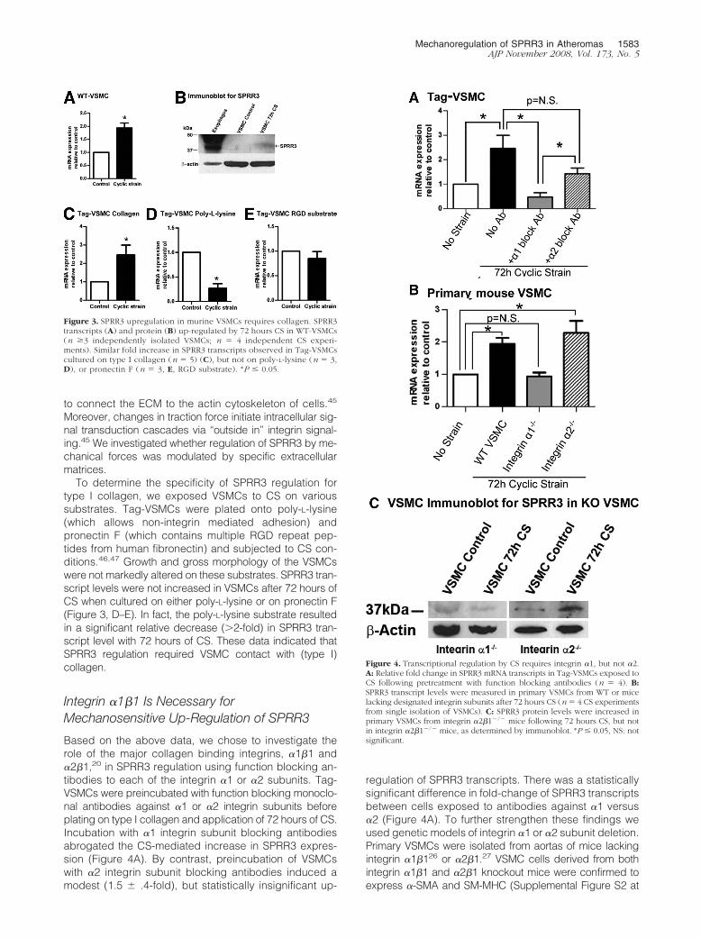
Due to the limited availability of primary VSMCs fromhuman aortic explants, it was of interest to determinewhether our results could be recapitulated using bothmouse primary wild-type VSMCs (WT-VSMC) as well asVSMC lines generated from the transgenic H-2Kb-tsA58mouse (Tag-VSMC). This mouse possesses a heat-labileT-antigen (Tag) expressed behind the mouse major histo-compatibility complex H-2Kb, which is widely expressedand is further inducible by interferon-�. Therefore, cells iso-lated from these animals can be conditionally immortalizedby growth at a permissive temperature of 33°C and in thepresence of interferon-�, but can be returned to normalprimary culture-like conditions when grown at 37°C.25 Asprimary cells must be used within nine passages, condi-tionally immortal Tag-VSMCs enabled us to circumventthe difficulty of obtaining sufficient numbers for extendedanalysis. We confirmed that both WT-VSMCs and Tag-VSMCs grown at 37°C expressed �-SMA and SM-MHC(Supplemental Figure S2 at http://ajp.amjpathol.org). BothWT and Tag-VSMCs were subjected to CS for 72 hoursand both demonstrated statistically significant upregula-tion of SPRR3 mRNA transcript levels between 2- to2.5-fold, respectively (Figure 3, A, C). Interestingly, bothtypes of murine VSMCs showed a less robust CS-asso-ciated regulation than human VSMCs under parallel con-ditions. To determine whether SPRR3 protein levels wereregulated by CS, cell homogenates from cyclicallystrained (72 hours) and non-strained WT-VSMC wereanalyzed by immunoblot. SPRR3 expression was de-tected in WT-VSMCs after 72 hours CS, but not in un-strained, control samples. Expression in mouse esopha-gus was expectedly very high (Figure 3B).35 Usinghuman and mouse primary VSMCs as well as mouseTag-VSMCs, we demonstrated consistent upregulation ofSPRR3 transcripts and protein by CS.
SPRR3 Mechanoregulation Is Dependent onBinding to Collagen
The mechanisms by which mechanical signals are rec-ognized by VSMCs and translated into molecular re-sponses are not completely understood.44 Intimal VSMCswithin atheromas are exposed to an environment rich inECM, particularly (type I) collagen.38 Collagen and otherECM components bind cell surface integrins, which serve
1580 Pyle et alAJP November 2008, Vol. 173, No. 5

Figure 1. SPRR3 is enriched in VSMCs in human and mouse atheromatous lesions. Representative sections from paraffin-embedded tissues of humanarteries obtained from different individuals containing fatty streaks show SPRR3-positive staining (brown) within the affected intima (A, C). Correspondingregions with normal histology from the same vessel (B, D, respectively) are negative. Arrowheads designate foam cells. SPRR3-stained human aortascontaining advanced atherosclerotic lesions (E–F) show extensive SPRR3 immunopositive staining within the fibrous cap (E, arrow) and dispersed throughthe lesion (F). An H&E of the lesion in (E) (inset) is shown with a box denoting the region magnified in (E). The tissue is fractured due to calcificationwithin the atheroma. No SPRR3-staining detected within the vessel wall of histologically normal human inferior vena cava, nor was it found in the medialVSMCs beneath any of the lesions (J, data not shown). The boxed area in (F) corresponds to the region of the vessel examined by indirectimmunofluorescence for costaining of SPRR3 (G, red) and �-SMA (H, green). I: Overlay in which colocalization is denoted with arrowheads. Note thepresence of extracellular SPRR3, which does not colocalize with 4,6-diamidino-2-phenylindole (I, arrows). Representative H&E-stained proximal aorticlesion from ApoE�/� (K) and its serial section (L) stained for SPRR3. Immunopositive area coincided with the VSMC-rich fibrous cap (indicated by arrows;L is the magnification of the boxed area in K). Vessel lumen (L), Lipid-Rich Core (LRC). Scale bar � 20 �m. Magnification � original �4 (inset, E), �10objective (A–F, J–K), �40 objective (G–I, L).
Mechanoregulation of SPRR3 in Atheromas 1581AJP November 2008, Vol. 173, No. 5

Figure 2. SPRR3 is regulated by CS in human VSMCs. A: Primary hVSMC cultures expressed both �-SMA and SM-MHC. B: SM-MHC (green) and SPRR3 (red)codistributed (yellow, arrowheads). mRNA levels of SPRR3 (C), elastin (D, 48 hours), MMP2 (E, 48 hours) after exposure to CS relative to unstrained controls(n � 4 independent CS experiments). Immunofluorescence of representative strained and unstrained cells from the same experiment demonstrated clearlyincreased cellular SPRR3 protein expression following 72 hours CS (F). Shear stress (24 hours) had no effect on SPRR3 transcript levels at either 5 or 10 dynes (G)(n �3 independent primary VSMC preps and CS experiments). Culturing hVSMCs with lipids (H, only 48 hours data shown) did not induce SPRR3 transcripts overcontrol (n � 4 independent experiments in triplicate). Scale bar � 20 �m. Magnification � original �20 (A), �20 (B). *P � 0.05.
1582 Pyle et alAJP November 2008, Vol. 173, No. 5
to connect the ECM to the actin cytoskeleton of cells.45
Moreover, changes in traction force initiate intracellular sig-nal transduction cascades via “outside in” integrin signal-ing.45 We investigated whether regulation of SPRR3 by me-chanical forces was modulated by specific extracellularmatrices.
To determine the specificity of SPRR3 regulation fortype I collagen, we exposed VSMCs to CS on varioussubstrates. Tag-VSMCs were plated onto poly-L-lysine(which allows non-integrin mediated adhesion) andpronectin F (which contains multiple RGD repeat pep-tides from human fibronectin) and subjected to CS con-ditions.46,47 Growth and gross morphology of the VSMCswere not markedly altered on these substrates. SPRR3 tran-script levels were not increased in VSMCs after 72 hours ofCS when cultured on either poly-L-lysine or on pronectin F(Figure 3, D–E). In fact, the poly-L-lysine substrate resultedin a significant relative decrease (�2-fold) in SPRR3 tran-script level with 72 hours of CS. These data indicated thatSPRR3 regulation required VSMC contact with (type I)collagen.
Integrin �1�1 Is Necessary forMechanosensitive Up-Regulation of SPRR3
Based on the above data, we chose to investigate therole of the major collagen binding integrins, �1�1 and�2�1,20 in SPRR3 regulation using function blocking an-tibodies to each of the integrin �1 or �2 subunits. Tag-VSMCs were preincubated with function blocking monoclo-nal antibodies against �1 or �2 integrin subunits beforeplating on type I collagen and application of 72 hours of CS.Incubation with �1 integrin subunit blocking antibodiesabrogated the CS-mediated increase in SPRR3 expres-sion (Figure 4A). By contrast, preincubation of VSMCswith �2 integrin subunit blocking antibodies induced amodest (1.5 � .4-fold), but statistically insignificant up-
regulation of SPRR3 transcripts. There was a statisticallysignificant difference in fold-change of SPRR3 transcriptsbetween cells exposed to antibodies against �1 versus�2 (Figure 4A). To further strengthen these findings weused genetic models of integrin �1 or �2 subunit deletion.Primary VSMCs were isolated from aortas of mice lackingintegrin �1�126 or �2�1.27 VSMC cells derived from bothintegrin �1�1 and �2�1 knockout mice were confirmed toexpress �-SMA and SM-MHC (Supplemental Figure S2 at
Figure 3. SPRR3 upregulation in murine VSMCs requires collagen. SPRR3transcripts (A) and protein (B) up-regulated by 72 hours CS in WT-VSMCs(n �3 independently isolated VSMCs; n � 4 independent CS experi-ments). Similar fold increase in SPRR3 transcripts observed in Tag-VSMCscultured on type I collagen (n � 5) (C), but not on poly-L-lysine (n � 3,D), or pronectin F (n � 3, E, RGD substrate). *P � 0.05.
Figure 4. Transcriptional regulation by CS requires integrin �1, but not �2.A: Relative fold change in SPRR3 mRNA transcripts in Tag-VSMCs exposed toCS following pretreatment with function blocking antibodies (n � 4). B:SPRR3 transcript levels were measured in primary VSMCs from WT or micelacking designated integrin subunits after 72 hours CS (n � 4 CS experimentsfrom single isolation of VSMCs). C: SPRR3 protein levels were increased inprimary VSMCs from integrin �2�1�/� mice following 72 hours CS, but notin integrin �2�1�/� mice, as determined by immunoblot. *P � 0.05, NS: notsignificant.
Mechanoregulation of SPRR3 in Atheromas 1583AJP November 2008, Vol. 173, No. 5

http://ajp.amjpathol.org). KO-VSMC primary cells wereplated on type I collagen, subjected to 72 hours CS, andassessed for changes in SPRR3 levels as compared tocontrol cells without strain. As expected, the VSMCs frommice lacking integrin �1�1 failed to up-regulate SPRR3whereas those from integrin �2�1�/� animals showed a2.3 � 0.6-fold (P 0.05) increase in SPRR3 transcriptand a corresponding increase in protein similar to WT-VSMCs (Figure 4, B and C). These data help explain whySPRR3 expression is limited to atherosclerotic regions ofthe vasculature; SPRR3 regulation will not occur in the
absence of integrin �1�1. It is of note that WT-VSMCsand Tag-VSMCs demonstrated enhanced SPRR3 regu-lation when the cells had been cultured for 48 hoursbefore stress in 200 pM TGF-� (Supplemental Figure S3Bat http://ajp.amjpathol.org). This observation was likelydue to the upregulation of the integrin �1 subunit whenVSMCs were cultured in 200 pM TGF-� (SupplementalFigure S3C at http://ajp.amjpathol.org); Furthermore, it isknown that VSMCs in prolonged culture down-regulateintegrin �1�1.48 Consistent with this, we did not observeSPRR3 regulation with cyclic strain using cells in pro-
Figure 5. Integrin �1, but not �2, is expressed in VSMCs in atheromas. Indirect immunofluorescence of representative proximal aorta lesion from ApoE�/� mice(H&E, A,E) showed coexpression of �-SMA (B, F, green) and integrin �1 (C, red). D: Merge, colocalization (yellow) marked by arrowheads. Integrin �2expression was not detected (G,H). Scale bar � 20 �m. Magnification � original �40. L indicates vessel lumen.
1584 Pyle et alAJP November 2008, Vol. 173, No. 5

longed culture (over nine passages) (Supplemental Fig-ure S3A at http://ajp.amjpathol.org). It should be noted,however, that there are numerous other cytokines andsignaling factors that are known to affect integrin expres-sion and these, as well as or in addition to TGF-�, may beinfluencing �1�1 integrin expression in atheromas.49
To confirm the relevance of integrin �1�1 in this sys-tem, we assessed whether �1�1 integrins were ex-pressed in VSMCs of atheromas. Proximal aortas fromApoE�/� mice containing atherosclerotic lesions werecostained with antibodies against �1 or �2 integrin sub-units and anti-�-SMA antibodies. Expression of integrin�1 subunit was colocalized with the VSMC marker (Figure5 A–D), whereas no discernible integrin �2 staining wasobserved in this mouse atherosclerosis model (Figure 5,E–H).
Discussion
This study was designed to investigate the physiologicalbasis for atheroma-enriched expression of the SPRR3protein demonstrated in both human and murine vascu-lature. Our results indicated that prolonged (�48 hours)CS of 20% resting length produced upregulation ofSPRR3 RNA and protein in VSMCs. By contrast, exposureof VSMCs to varying amounts of shear stress or lipidsfailed to increase SPRR3 transcript levels (Figure 2). Toour knowledge this is the first atheroma-specific proteinwhose expression is biomechanically regulated. It is ofsome interest that SPRR3 codistributed with SM-myosinheavy chain in hVSMCs (Figure 2B). SM-MHC has previ-ously been shown to be both biomechanically regulatedas well as biomechanically active and hence this findingsupports a role for SPRR3 in biomechanics.36,37 Becauseintegrins are hypothesized to serve as prime cellularmechanosensors, as they link the interior and exterior ofthe cell,22,50 we sought to elucidate the effect of ECM andintegrins in mechanoregulation of SPRR3 transcription.By exposing VSMCs to CS on various substrates, wedemonstrated that SPRR3 regulation by CS required typeI collagen, whereas cells grown on poly-L-lysine orpronectin F failed to regulate SPRR3 with CS. Since typeI collagen constitutes �70% of all collagen in atheroma-tous plaques, it represents the primary ECM circumscrib-ing VSMCs within lesions.38 Given that the CS effectrequired a collagenous substrate, we evaluated the roleof the major collagen binding receptors, �1�1 and �2�1,in SPRR3 gene regulation using both an immunoblockingapproach and a genetic deletion model. Taken together,the data suggest that the �1�1 collagen-binding integrinis required for mechanoregulation of SPRR3. Consistentwith published studies, we confirmed integrin �1 subunitexpression in VSMCs within murine lesions.51 Expressionof the integrin �2 subunit was not evident in murinelesions as is consistent with previous findings.48,52
Since VSMCs are exposed to mechanical forces suchas CS in healthy arteries as well as in atheromas, ourfindings do not fully explain why SPRR3 expression wasrestricted to plaques. One possibility for this phenome-non relates to the microenvironment of the plaque itself.
VSMCs as well as macrophage foam cells are known totake up lipid in atheromas, so we investigated if lipidloading could up-regulated SPRR3 transcript.53 LDL andox-LDL failed to enhance SPRR3 expression, althoughox-LDL loading resulted in decreased SPRR3 transcrip-tion. While it is unclear why this may have occurred, it isknown that lipid-loading VSMCs induces a variety of sig-naling pathways, as well as inducing an even more syn-thetic cellular phenotype.54–56 Therefore, cells with analtered phenotype, in the absence of biomechanicalstress, may down-regulate SPRR3. However, this raisesthe issue of VSMC phenotype (eg, contractile/differenti-ated versus synthetic) as a variable in this system.VSMCs in the context of the atherosclerotic plaque arewidely considered to reside in the synthetic state.56,57
Because this study investigates the regulation of SPRR3in the context of the atherosclerotic plaque, it is optimal touse cells in vitro under culture conditions that promote asynthetic phenotype. SM-MHC is a marker of a differen-tiated/contractile VSMC. As seen in Figure 2 and in Sup-plemental Figure 2, the VSMCs used in this study expresslow levels of this protein, suggesting that they are in arelatively synthetic state. Another potential explanationmay be that VSMCs within atheromas up-regulate �1�1expression. We have observed in vitro down-regulation ofthe integrin �1 subunit in some VSMCs after prolongedculture; these cells fail to demonstrate SPRR3 regulation(Supplemental Figure S3A at http://ajp.amjpathol.org).48 Itis known that lesions locally express high levels of TGF�,which is known to up-regulate collagen and surface inte-grin expression.58–60 We confirmed this finding by dem-onstrating that TGF� increased expression of �1�1 in cul-tured VSMCs. Accordingly, VSMCs up-regulated SPRR3 inresponse to CS more robustly when cultured in the pres-ence of 200 pM TGF� (Supplemental Figure S3B,C at http://ajp.amjpathol.org). We also demonstrated highly localizedexpression of �1�1 on VSMCs constituting murine prox-imal aorta lesions. Hence, one hypothesis is that theatheroma microenvironment (inflammation, altered localcompliance, etc), perhaps through increased TGF�, al-
Figure 6. Model of SPRR3 Regulation in VSMCs.
Mechanoregulation of SPRR3 in Atheromas 1585AJP November 2008, Vol. 173, No. 5

tered VSMC integrin expression and subsequent ECM/integrin signaling. However, other cytokines also presentin this microenvironment may also be responsible for theintegrin regulation.49 Moreover, to further examine if bio-mechanical stress alone could up-regulate SPRR3, we ex-amined aortas isolated from mice subjected to continuousinfusion of angiotensin II (Ang II) for 2 months, which in-duces systemic hypertension associated with increasedmedial thickness as compared to saline controls (samplesobtained from Dr. Nancy J. Brown, Vanderbilt University).61
We failed to detect either �1�1 or SPRR3 expression in arterialwall VSMCs in treated or control aortas (Supplemental FigureS4 at http://ajp.amjpathol.org). Together, the data suggestthat biomechanical stimulation alone in vivo or in vitro wasnot sufficient for upregulation of SPRR3 transcript and pro-tein expression in the absence of VSMC �1�1 integrin ex-pression. A model of this proposed mechanism is shownin Figure 6. Nevertheless, the atherosclerotic plaque is acomplex tissue with multiple variables that may affect thetranscription of SPRR3. While many excellent studies havehighlighted integrin/matrix interactions in regulating thephysiology and gene expression in VSMCs, to our knowl-edge our study is among the first to delineate a directrelationship between transcription of a mechanosensitivetarget gene and specific integrin/ECM signaling.
The function of SPRR3 in VSMCs within atheromasremains unclear. The architecture of SPRR3, which istransglutaminated via its N- and C-terminal domains toother structural proteins, including other SPRRs, is be-lieved to play a central role in the barrier and stressfunction of the cornified envelope.8 The central core ofSPRR3 is considered to have virtually no secondarystructure, a vital characteristic in its function as a flexiblecross-bridge between its binding partners.62,63 SPRR3 isa substrate for transglutaminase types I and III, which areexpressed in murine vessels.7,62,63 Interestingly, otherstudies have linked increased transglutaminase activityto VSMCs and sites of atherosclerosis,64,65 although noreports are available on the relationship between SPRR3(or any substrates) and transglutaminase gene family inVSMCs. It is tempting to speculate that SPRR3 may serveas a crosslinking substrate for transglutaminase to stabi-lize the cytoarchitecture of VSMCs, although the relation-ship between SPRR3 and transglutaminases in the vas-culature remains unexamined. Our data showing thatSPRR3 is exclusively enriched in VSMCs within athero-mas in response to mechanical stress implies that it mayplay a role in altered biomechanical compliance of thesmooth muscle cell within an atheromatous lesion. Effortsin our lab are currently underway to better understand therole of SPRR3 within VSMCs using gene deletion modelsand identifying its crosslinked partners.
It has been reported that early atherosclerotic lesions(undetectable by angiography but evident by intravascu-lar ultrasound and postmortem histopathology) result insignificantly reduced aortic compliance in a hyperlipid-emic rabbit model.2 These data have been confirmed byother studies by multiple groups.66–68 One mechanismby which even an early atheroma may alter local biome-chanics is from the direct effect of cholesterol on VSMCmembrane fluidity. Alternatively, alterations of the physi-
cal properties of the ECM can be transduced toVSMCs.69–71 While much data support that atherosclerosisitself alters vessel compliance and, thereby, biomechanicalproperties, the molecular changes within VSMCs in re-sponse to altered local biomechanics are poorly under-stood. This paper elucidates the mechanism by whichSPRR3 is regulated by mechanotransduction in VSMCswithin the context of the microenvironment of the atheroscle-rotic plaque.
Acknowledgments
Dr. Nancy Brown is graciously acknowledged for contrib-uting archival vascular tissue from Angiotensin II-infusedhypertensive mice. The authors also thank Drs. Samuel A.Santoro and W. Gray Jerome for their critical review ofthis manuscript and Sandy Olsen for her excellent tech-nical support in immunohistochemistry.
References
1. Davies PF, Polacek DC, Handen JS, Helmke BP, DePaola N: A spatialapproach to transcriptional profiling: mechanotransduction and thefocal origin of atherosclerosis. Trends Biotechnol 1999, 17:347–351
2. Hong MK, Vossoughi J, Mintz GS, Kauffman RD, Hoyt RF, Jr., CornhillJF, Herderick EE, Leon MB, Hoeg JM: Altered compliance and resid-ual strain precede angiographically detectable early atherosclerosisin low-density lipoprotein receptor deficiency. Arterioscler ThrombVasc Biol 1997, 17:2209–2217
3. Dart AM, Kingwell BA: Pulse pressure–a review of mechanisms andclinical relevance. J Am Coll Cardiol 2001, 37:975–984
4. Shaw JA, Kingwell BA, Walton AS, Cameron JD, Pillay P, Gatzka CD,Dart AM: Determinants of coronary artery compliance in subjects withand without angiographic coronary artery disease. J Am Coll Cardiol2002, 39:1637–1643
5. Nguyen KT, Frye SR, Eskin SG, Patterson C, Runge MS, McIntire LV:Cyclic strain increases protease-activated receptor-1 expression invascular smooth muscle cells. Hypertension 2001, 38:1038–1043
6. Julien MA, Wang P, Haller CA, Wen J, Chaikof EL: Mechanical strainregulates syndecan-4 expression and shedding in smooth musclecells through differential activation of MAP kinase signaling pathways.Am J Physiol Cell Physiol 2007, 292:C517–C525
7. Young PP, Modur V, Teleron AA, Ladenson JH: Enrichment of genesin the aortic intima that are associated with stratified epithelium:implications of underlying biomechanical and barrier properties of thearterial intima. Circulation 2005, 111:2382–2390
8. Candi E, Schmidt R, Melino G: The cornified envelope: a model of celldeath in the skin. Nat Rev Mol Cell Biol 2005, 6:328–340
9. Fischer DF, Sark MW, Lehtola MM, Gibbs S, van de Putte P, Backendorf C:Structure and evolution of the human SPRR3 gene: implications forfunction and regulation. Genomics 1999, 55:88–99
10. Kvedar JC, Manabe M, Phillips SB, Ross BS, Baden HP: Character-ization of sciellin, a precursor to the cornified envelope of humankeratinocytes. Differentiation 1992, 49:195–204
11. Kalinin AE, Kajava AV, Steinert PM: Epithelial barrier function: assem-bly and structural features of the cornified cell envelope. Bioessays2002, 24:789–800
12. Champliaud MF, Baden HP, Koch M, Jin W, Burgeson RE, Viel A:Gene characterization of sciellin (SCEL) and protein localization invertebrate epithelia displaying barrier properties. Genomics 2000,70:264–268
13. Cabral A, Voskamp P, Cleton-Jansen AM, South A, Nizetic D, BackendorfC: Structural organization and regulation of the small proline-richfamily of cornified envelope precursors suggest a role in adaptivebarrier function. J Biol Chem 2001, 276:19231–19237
14. Pradervand S, Yasukawa H, Muller OG, Kjekshus H, Nakamura T,St Amand TR, Yajima T, Matsumura K, Duplain H, Iwatate M, Wood-ard S, Pedrazzini T, Ross J, Firsov D, Rossier BC, Hoshijima M, Chien
1586 Pyle et alAJP November 2008, Vol. 173, No. 5

KR: Small proline-rich protein 1A is a gp130 pathway- and stress-inducible cardioprotective protein. EMBO J 2004, 23:4517–4525
15. Lehoux S, Tedgui A: Cellular mechanics and gene expression inblood vessels. J Biomech 2003, 36:631–643
16. Lehoux S, Tedgui A: Signal transduction of mechanical stresses in thevascular wall. Hypertension 1998, 32:338–345
17. Lammerding J, Kamm RD, Lee RT: Mechanotransduction in cardiacmyocytes. Ann NY Acad Sci 2004, 1015:53–70
18. Sadoshima J, Izumo S: The cellular and molecular response of car-diac myocytes to mechanical stress. Annu Rev Physiol 1997,59:551–571
19. Zhang Z-G, Bothe I, Hirche F, Zweers M, Gullberg D, Pfitzer G, Krieg T,Eckes B, Aumailley M: Interactions of primary fibroblasts and keratino-cytes with extracellular matrix proteins: contribution of �2�1 integrin.J Cell Sci, 2006, 119:1886–1895
20. Hynes RO: Integrins: bidirectional, allosteric signaling machines. Cell2002, 110:673–687
21. Sutcliffe MC, Davidson JM: Effect of static stretching on elastin pro-duction by porcine aortic smooth muscle cells. Matrix 1990,10:148–153
22. Davis MJ, Wu X, Nurkiewicz TR, Kawasaki J, Davis GE, Hill MA,Meininger GA: Integrins and mechanotransduction of the vascularmyogenic response. Am J Physiol Heart Circ Physiol 2001, 280:H1427–H1433
23. Wernig F, Mayr M, Xu Q: Mechanical stretch-induced apoptosis insmooth muscle cells is mediated by �1-integrin signaling pathways.Hypertension 2003, 41:903–911
24. Alenghat FJ, Ingber DE: Mechanotransduction: all signals point tocytoskeleton, matrix, and integrins. Sci STKE. 2002, 119:PE6
25. Jat P, Noble M, Ataliotis P, Tanaka Y, Yannoutsos N, Larsen L,Kioussis D: Direct derivation of conditionally immortal cell lines froman H-2Kb- tsA58 transgenic mouse. Proc Natl Acad Sci USA 1991,88:5096–5100
26. Gardner H, Kreidberg J, Koteliansky V, Jaenisch R: Deletion of inte-grin � 1 by homologous recombination permits normal murine devel-opment but gives rise to a specific deficit in cell adhesion. Dev Biol1996, 175:301–313
27. Holtkotter O, Nieswandt B, Smyth N, Muller W, Hafner M, Schulte V,Krieg T, Eckes B: Integrin � 2-deficient mice develop normally, arefertile, but display partially defective platelet interaction with collagen.J Biol Chem 2002, 277:10789–10794
28. Atkinson RD, Coenen KR, Plummer MR, Gruen ML, Hasty AH: Macro-phage-derived apolipoprotein E (apoE) ameliorates dyslipidemia andatherosclerosis in obese apoE deficient mice, Am J Physiol EndocrinolMetab 2007, 294:E284–E290
29. Stouffer GA, Owens GK: TGF-� promotes proliferation of culturedSMC via both PDGF-AA-dependent and PDGF-AA-independentmechanisms. J Clin Invest 1994, 93:2048–2055
30. Hasegawa K, Arakawa E, Oda S, Yanai N, Obinata M, Matsuda Y:Novel smooth muscle cell lines from transgenic mice harboring tem-perature-sensitive SV40 large T-antigen gene. Temperature-depen-dent expression of smooth muscle myosin heavy chain-1 and calpo-nin genes, J Mol Cell Cardiol 1997, 29:2177–2186
31. Cox BE, Griffin EE, Ullery JC, Jerome WG: Effects of cellular choles-terol loading on macrophage foam cell lysosome acidification. J LipidRes 2007, 48:1012–1021
32. Pfaffl MW: A new mathematical model for relative quantification inreal-time RT-PCR. Nucleic Acids Res 2001, 29:e45
33. Mayr M, Hu Y, Hainaut P, Xu Q: Mechanical stress-induced DNAdamage and rac-p38MAPK signal pathways mediate p53-dependentapoptosis in vascular smooth muscle cells. FASEB J 2002,16:1423–1425
34. Brisset AC, Hao H, Camenzind E, Bacchetta M, Geinoz A, SanchezJC, Chaponnier C, Gabbiani G, Bochaton-Piallat ML: Intimal smoothmuscle cells of porcine and human coronary artery express S100A4,a marker of the rhomboid phenotype in vitro. Circ Res 2007,100:1055–1062
35. Smolinski KN, Abraham JM, Souza RF, Yin J, Wang S, Xu Y, Zou TT,Kong D, Fleisher AS, Meltzer SJ: Activation of the esophagin pro-moter during esophageal epithelial cell differentiation. Genomics2002, 79:875–880
36. Somlyo AP, Somlyo AV: Signal transduction and regulation in smoothmuscle. Nature 1994, 372:231–236
37. Reusch P, Wagdy H, Reusch R, Wilson E, Ives HE: Mechanical strain
increases smooth muscle and decreases nonmuscle myosin expres-sion in rat vascular smooth muscle cells. Circ Res 1996, 79:1046–1053
38. Katsuda S, Kaji T: Atherosclerosis and extracellular matrix. J Athero-scler Thromb 2003, 10:267–274
39. Asanuma K, Magid R, Johnson C, Nerem RM, Galis ZS: Uniaxialstrain upregulates matrix-degrading enzymes produced by humanvascular smooth muscle cells. Am J Physiol Heart Circ Physiol 2003,284:H1778–H1784
40. Dobrin PB: Mechanical properties of arteries. Physiol Rev 1978,58:397–460
41. Yamawaki H, Pan S, Lee RT, Berk BC: Fluid shear stress inhibitsvascular inflammation by decreasing thioredoxin-interacting proteinin endothelial cells. J Clin Invest 2005, 115:733–738
42. Elhadj S, Akers RM, Forsten-Williams K: Chronic pulsatile shear stressalters insulin-like growth factor-I (IGF-I) binding protein release invitro. Ann Biomed Eng 2003, 31:163–170
43. Asada H, Paszkowiak J, Teso D, Alvi K, Thorisson A, Frattini JC, KudoFA, Sumpio BE, Dardik A: Sustained orbital shear stress stimulatessmooth muscle cell proliferation via the extracellular signal-regulatedprotein kinase 1/2 pathway. J Vasc Surg 2005, 42:772–780
44. Hu Y, Bock G, Wick G, Xu Q: Activation of PDGF receptor � invascular smooth muscle cells by mechanical stress. FASEB J 1998,12:1135–1142
45. Katsumi A, Orr AW, Tzima E, Schwartz MA: Integrins in mechano-transduction. J Biol Chem 2004, 279:12001–12004
46. Birukov KG, Shirinsky VP, Stepanova OV, Tkachuk VA, Hahn AW,Resink TJ, Smirnov VN: Stretch affects phenotype and proliferation ofvascular smooth muscle cells. Mol Cell Biochem 1995, 144:131–139
47. Tock J, Van Putten V, Stenmark KR, Nemenoff RA: Induction ofSM-�-actin expression by mechanical strain in adult vascular smoothmuscle cells is mediated through activation of JNK and p38 MAPkinase. Biochem Biophys Res Commun 2003, 301:1116–1121
48. Skinner MP, Raines EW, Ross R: Dynamic expression of � 1 � 1 and� 2 � 1 integrin receptors by human vascular smooth muscle cells. �
2 � 1 integrin is required for chemotaxis across type I collagen-coated membranes, Am J Pathol 1994, 145:1070–1081
49. Kim LT, Yamada KM: The regulation of expression of integrin recep-tors. Proc Soc Exp Biol Med 1997, 214:123–131
50. Shyy JY, Chien S: Role of integrins in endothelial mechanosensing ofshear stress. Circ Res 2002, 91:769–775
51. Schapira K, Lutgens E, de Fougerolles A, Sprague A, Roemen A,Gardner H, Koteliansky V, Daemen M, Heeneman S: Genetic deletionor antibody blockade of a1b1 integrin induces a stable plaque phe-notype in Apo E�/ � mice. Arterioscler Thromb Vasc Biol 2005,25:1917–1924
52. Grenache DG, Coleman T, Semenkovich CF, Santoro SA, Zutter MM:�2�1 integrin and development of atherosclerosis in a mouse model:assessment of risk. Arterioscler Thromb Vasc Biol 2003,23:2104–2109
53. Zhao GF, Seng JJ, Zhang H, She MP: Effects of oxidized low densitylipoprotein on the growth of human artery smooth muscle cells. ChinMed J (Engl) 2005, 118:1973–1978
54. Bochkov VN, Tkachuk VA, Hahn AW, Bernhardt J, Buhler FR, ResinkTJ: Concerted effects of lipoproteins and angiotensin II on signaltransduction processes in vascular smooth muscle cells. ArteriosclerThromb 1993, 13:1261–1269
55. Resink TJ, Rybin V, Bernhardt J, Orlov S, Buhler FR, Tkachuk VA:Cellular signalling by lipoproteins in cultured smooth muscle cellsfrom spontaneously hypertensive rats. J Vasc Res 1993, 30:169–180
56. Hayashi K, Takahashi M, Nishida W, Yoshida K, Ohkawa Y, Kitabatake A,Aoki J, Arai H, Sobue K: Phenotypic modulation of vascular smoothmuscle cells induced by unsaturated lysophosphatidic acids. CircRes 2001, 89:251–258
57. Shanahan CM, Weissberg PL: Smooth muscle cell heterogeneity:patterns of gene expression in vascular smooth muscle cells in vitroand in vivo. Arterioscler Thromb Vasc Biol 1998, 18:333–338
58. Zargham R, Pepin J, Thibault G: �8�1 Integrin is up-regulated in theneointima concomitant with late luminal loss after balloon injury. Car-diovasc Pathol 2007, 16:212–220
59. Kagami S, Kuhara T, Yasutomo K, Okada K, Loster K, Reutter W,Kuroda Y: Transforming growth factor-� (TGF-�) stimulates the ex-pression of �1 integrins and adhesion by rat mesangial cells. Exp CellRes 1996, 229:1–6
Mechanoregulation of SPRR3 in Atheromas 1587AJP November 2008, Vol. 173, No. 5

60. Kondo S, Kagami S, Urushihara M, Kitamura A, Shimizu M, Strutz F,Muller GA, Kuroda Y: Transforming growth factor-�1 stimulates col-lagen matrix remodeling through increased adhesive and contractivepotential by human renal fibroblasts. Biochim Biophys Acta 2004,1693:91–100
61. Weisberg AD, Albornoz F, Griffin JP, Crandall DL, Elokdah H, FogoAB, Vaughan DE, Brown NJ: Pharmacological inhibition and geneticdeficiency of plasminogen activator inhibitor-1 attenuates angiotensinII/salt-induced aortic remodeling. Arterioscler Thromb Vasc Biol2005, 25:365–371
62. Candi E, Paci M, Oddi S, Paradisi A, Guerrieri P, Melino G: Orderedstructure acquisition by the N- and C-terminal domains of the smallproline-rich 3 protein. J Cell Biochem 2000, 77:179–185
63. Candi E, Melino G, Sette M, Oddi S, Guerrieri P, Paci M: Acquisitionof ordered conformation by the N-terminal domain of the human smallproline rich 2 protein. Biochem Biophys Res Commun 1999,262:395–400
64. Haroon ZA, Wannenburg T, Gupta M, Greenberg CS, Wallin R, SaneDC: Localization of tissue transglutaminase in human carotid andcoronary artery atherosclerosis: implications for plaque stability andprogression. Lab Invest 2001, 81:83–93
65. Ou H, Haenderler J, Aebly MR, Kelly LA, Cholewa BC, Koike G,Kwitek-Black A, Jacob HJ, Berk BC, Miano JM: Retinoic acid-induced
tissue transglutaminase and apoptosis in vascular smooth musclecells. Circ Research 2000, 881–887
66. Hudetz AG, Mark G, Kovach AG, Kerenyi T, Fody L, Monos E:Biomechanical properties of normal and fibrosclerotic human cere-bral arteries. Atherosclerosis 1981, 39:353–365
67. Chau AH, Chan RC, Shishkov M, MacNeill B, Iftimia N, Tearney GJ,Kamm RD, Bouma BE, Kaazempur-Mofrad MR: Mechanical analysisof atherosclerotic plaques based on optical coherence tomography.Ann Biomed Eng 2004, 32:1494–1503
68. Hiro T, Leung CY, De Guzman S, Caiozzo VJ, Farvid AR, Karimi H,Helfant RH, Tobis JM: Are soft echoes really soft? Intravascularultrasound assessment of mechanical properties in human athero-sclerotic tissue. Am Heart J 1997, 133:1–7
69. Reddy GK: AGE-related cross-linking of collagen is associated withaortic wall matrix stiffness in the pathogenesis of drug-induced dia-betes in rats. Microvasc Res 2004, 68:132–142
70. Liu SQ, Tang D, Tieche C, Alkema PK: Pattern formation of vascularsmooth muscle cells subject to nonuniform fluid shear stress: medi-ation by gradient of cell density. Am J Physiol Heart Circ Physiol 2003,285:H1072–H1080
71. Liu SQ: Biomechanical basis of vascular tissue engineering. Crit RevBiomed Eng 1999, 27:75–148
1588 Pyle et alAJP November 2008, Vol. 173, No. 5