Regulation of HERG (KCNH2) potassium channel surface expression by diacylglycerol
13
RESEARCH ARTICLE Regulation of HERG (KCNH2) potassium channel surface expression by diacylglycerol Cia Ramstro ¨m • Hugh Chapman • Tero Viitanen • Emad Afrasiabi • Heli Fox • Johanna Kivela ¨ • Sanna Soini • Laura Korhonen • Dan Lindholm • Michael Pasternack • Kid To ¨rnquist Received: 10 June 2009 / Revised: 30 September 2009 / Accepted: 6 October 2009 / Published online: 27 October 2009 Ó Birkha ¨user Verlag, Basel/Switzerland 2009 Abstract The HERG (KCNH2) channel is a voltage- sensitive potassium channel mainly expressed in cardiac tissue, but has also been identified in other tissues like neuronal and smooth muscle tissue, and in various tumours and tumour cell lines. The function of HERG has been extensively studied, but it is still not clear what mecha- nisms regulate the surface expression of the channel. In the present report, using human embryonic kidney cells stably expressing HERG, we show that diacylglycerol potently inhibits the HERG current. This is mediated by a protein kinase C-evoked endocytosis of the channel protein, and is dependent on the dynein–dynamin complex. The HERG protein was found to be located only in early endosomes and not lysosomes. Thus, diacylglycerol is an important lipid participating in the regulation of HERG surface expression and function. Keywords Diacylglycerol Á Lipids Á Potassium channel Á Internalization Á Membranes Á Lysosomes Á Protein kinase C Introduction Potassium channels are expressed in virtually all cell types regulating a wide variety of physiological functions including heart rate, release of neurotransmitters and insulin secretion. Ion channels are modified by posttrans- lational changes such as channel phosphorylation [1]. The human ether a ´-go-go-related gene (HERG1 or KCNH2) was originally cloned from a human hippocampal cDNA library by homology to Drosophila EAG [2]. HERG encodes the a-subunit of a potassium channel with a structure resembling that of other voltage-gated potassium channels, i.e. six transmembrane domains, one of which acts as a voltage sensor, intracellular N (amino) and C (carboxy) termini, and a pore loop linking the S5 and S6 domains. The HERG channel was then found to underlie the rapid component of the cardiac delayed rectifier current (I Kr ) and hence plays a central role in mediating repolari- zation of the cardiac action potential [3]. Mutations in the HERG channel that cause a loss- or gain-of-function can result in type 2 long QT syndrome (LQTS) and one form of short QT syndrome, respectively, which are associated with cardiac arrhythmias and a risk of sudden death [4, 5]. Of more common occurrence are drug-induced LQTS and torsade de pointes tachyarrhythmia resulting primarily from HERG channel inhibition by class III antiarrhythmics, as well as by a wide range of non-cardiac drugs, including antihistamines, antimicrobial and psychiatric drugs [6]. HERG expression is, however, not limited to cardiac tissue. The channel has also been found in the hippocampus C. Ramstro ¨m Á E. Afrasiabi Á K. To ¨rnquist Department of Biology, A ˚ bo Akademi University, 20520 Turku, Finland C. Ramstro ¨m Á H. Chapman Á T. Viitanen Á H. Fox Á L. Korhonen Á D. Lindholm Á M. Pasternack Á K. To ¨rnquist Minerva Foundation Institute for Medical Research, Biomedicum Helsinki, 00290 Helsinki, Finland J. Kivela ¨ Á S. Soini Department of Pharmacology and Clinical Pharmacology, University of Turku, 20520 Turku, Finland K. To ¨rnquist (&) Department of Biology, A ˚ bo Akademi University, BioCity, Tykisto ¨katu 6, 20520 Turku, Finland e-mail: kid.tornqvist@abo.fi Cell. Mol. Life Sci. (2010) 67:157–169 DOI 10.1007/s00018-009-0176-2 Cellular and Molecular Life Sciences
Transcript of Regulation of HERG (KCNH2) potassium channel surface expression by diacylglycerol
RESEARCH ARTICLE
Regulation of HERG (KCNH2) potassium channel surfaceexpression by diacylglycerol
Cia Ramstrom • Hugh Chapman • Tero Viitanen • Emad Afrasiabi •
Heli Fox • Johanna Kivela • Sanna Soini • Laura Korhonen • Dan Lindholm •
Michael Pasternack • Kid Tornquist
Received: 10 June 2009 / Revised: 30 September 2009 / Accepted: 6 October 2009 / Published online: 27 October 2009
� Birkhauser Verlag, Basel/Switzerland 2009
Abstract The HERG (KCNH2) channel is a voltage-
sensitive potassium channel mainly expressed in cardiac
tissue, but has also been identified in other tissues like
neuronal and smooth muscle tissue, and in various tumours
and tumour cell lines. The function of HERG has been
extensively studied, but it is still not clear what mecha-
nisms regulate the surface expression of the channel. In the
present report, using human embryonic kidney cells stably
expressing HERG, we show that diacylglycerol potently
inhibits the HERG current. This is mediated by a protein
kinase C-evoked endocytosis of the channel protein, and is
dependent on the dynein–dynamin complex. The HERG
protein was found to be located only in early endosomes
and not lysosomes. Thus, diacylglycerol is an important
lipid participating in the regulation of HERG surface
expression and function.
Keywords Diacylglycerol � Lipids � Potassium channel �Internalization � Membranes � Lysosomes �Protein kinase C
Introduction
Potassium channels are expressed in virtually all cell types
regulating a wide variety of physiological functions
including heart rate, release of neurotransmitters and
insulin secretion. Ion channels are modified by posttrans-
lational changes such as channel phosphorylation [1]. The
human ether a-go-go-related gene (HERG1 or KCNH2)
was originally cloned from a human hippocampal cDNA
library by homology to Drosophila EAG [2]. HERG
encodes the a-subunit of a potassium channel with a
structure resembling that of other voltage-gated potassium
channels, i.e. six transmembrane domains, one of which
acts as a voltage sensor, intracellular N (amino) and C
(carboxy) termini, and a pore loop linking the S5 and S6
domains. The HERG channel was then found to underlie
the rapid component of the cardiac delayed rectifier current
(IKr) and hence plays a central role in mediating repolari-
zation of the cardiac action potential [3]. Mutations in the
HERG channel that cause a loss- or gain-of-function can
result in type 2 long QT syndrome (LQTS) and one form of
short QT syndrome, respectively, which are associated with
cardiac arrhythmias and a risk of sudden death [4, 5]. Of
more common occurrence are drug-induced LQTS and
torsade de pointes tachyarrhythmia resulting primarily
from HERG channel inhibition by class III antiarrhythmics,
as well as by a wide range of non-cardiac drugs, including
antihistamines, antimicrobial and psychiatric drugs [6].
HERG expression is, however, not limited to cardiac
tissue. The channel has also been found in the hippocampus
C. Ramstrom � E. Afrasiabi � K. Tornquist
Department of Biology, Abo Akademi University,
20520 Turku, Finland
C. Ramstrom � H. Chapman � T. Viitanen � H. Fox �L. Korhonen � D. Lindholm � M. Pasternack � K. Tornquist
Minerva Foundation Institute for Medical Research,
Biomedicum Helsinki, 00290 Helsinki, Finland
J. Kivela � S. Soini
Department of Pharmacology and Clinical Pharmacology,
University of Turku, 20520 Turku, Finland
K. Tornquist (&)
Department of Biology, Abo Akademi University, BioCity,
Tykistokatu 6, 20520 Turku, Finland
e-mail: [email protected]
Cell. Mol. Life Sci. (2010) 67:157–169
DOI 10.1007/s00018-009-0176-2 Cellular and Molecular Life Sciences

regulating the excitability of neurons [7], in jejunal smooth
muscle controlling electrical and contractile activities [8]
and in pancreatic b-cells regulating insulin secretion [9].
Furthermore, a growing number of studies show that
HERG is selectively up-regulated in several animal and
human tumours and tumour cell lines [10, 11].
Diacylglycerol (DAG) is one of the key lipid second
messengers generated from phosphatidyl inositol 4,5-
biphosphate (PIP2) by the action of receptor-activated
phospholipase C, PLC. In addition to its recognized role in
protein kinase C (PKC) activation, DAG also targets pro-
tein kinase D (PKD) [12], DAG kinases and chimaerins
[13] among others. Both DAG and its precursor PIP2 have
been shown to regulate a number of potassium channels.
DAG inhibits the voltage-gated Kv1.3 current of human T
lymphocytes [14] and in Xenopus oocytes inhibits the
minK current resembling the cardiac IKs current [15]. The
HERG current is regulated by PIP2 resulting in an increase
in current amplitude, acceleration of activation and slowing
of inactivation [16]. Another lipid second messenger, cer-
amide, whose diverse signalling includes PKC activation,
has also been shown to regulate the HERG channel. In rat
pituitary GH3 cells, an ERG current is inhibited by cera-
mide [17], while we have previously shown that ceramide
reduces the HERG current by decreasing the amount of
membrane-bound HERG protein [18]. The actions of these
lipids on the HERG channel appear devoid of PKC
involvement. However, investigations have shown that
PKC may indirectly [19, 20], or by direct phosphorylation
[21], decrease HERG current. As both ceramide and DAG
activates PKC, and both lipids seem to modulate HERG
channel function, it was of interest to further investigate the
mechanism of action of DAG on the channel. In this report,
we show that DAG evokes a time-dependent decrease in
HERG current. The underlying mechanism was found to be
a PKC-mediated decrease in plasma-membrane HERG
protein expression, due to a rapid endocytosis of the
channel.
Materials and methods
Cell culture
HEK293 cell line stably expressing HERG in the
pcDNA3.1 expression vector was selected for using G418
(A. G. Scientific, CA, USA). The cells were cultured in
DMEM supplemented with 10% foetal calf serum, peni-
cillin–streptomycin (BioWhittaker Cambrex Bio Science,
Verviers, Belgium) and G418 (0.2 mg/ml). SHSY5Y cells
were cultured in DMEM:Ham’s F12 medium (1:1) sup-
plemented with 10% foetal calf serum and penicillin–
streptomycin.
Patch-clamp recording
Whole-cell recordings of HEK293 cells expressing HERG
were performed using an EPC-9 amplifier and Pulse/Puls-
efit software (Heka, Lambrecht, Germany) as previously
described [18]. HERG-like tail currents present in
SHSY5Y neuroblastoma cells were recorded using a
modified version of a previously described protocol [22].
Briefly, cells were clamped to a holding voltage of –0 mV
and tail currents were evoked by a 200-ms step to
-120 mV applied every 5 s. At the beginning of the
experiment, cells were perfused with a 5.4-mM K? and
then with a 50.4-mM K? solution to enhance K? carried
inward currents. The first trace of each experiment recor-
ded in 5.4-mM K? solution was subtracted from
subsequent sweeps. For illustration purposes, the residual
current amplitudes were normalized against the largest
response obtained in the presence of 50.4 mM K? (given
the value of -1). For statistical analysis, the logarithms of
absolute current values (A pF-1) were used. Logarithm
values were normalized against respective controls (taken
as 100%) to prepare diagrams.
The electrodes had resistances of 2–6 MX when filled
with 150 mM KCl, 2 mM MgCl2, 5 mM BAPTA, 5 mM
Mg2ATP3 and 10 mM HEPES, pH 7.2. The standard
extracellular solution contained 150 mM NaCl, 5.4 mM
KCl, 1.8 mM CaCl2, 1 mM MgCl2 and 5 mM HEPES, pH
7.4. The high K? solution was otherwise identical, but
45 mM NaCl was replaced with KCl, ending to a [K?]0 of
50.4 mM. 1,2–dioctanoyl-sn-glycerol, DAG (Sigma, St.
Louis, MO, USA) and m-3M3FBS (Merck, Darmstadt,
Germany) were dissolved in DMSO and added to the
extracellular solution (the final vehicle concentration
was 0.1%). All experiments were carried out at room
temperature. The whole-cell recordings capacitance was
compensated for, as was series resistance by at least 70%.
Labelling of cell surface proteins
Cell surface proteins were biotinylated with a water-soluble
biotinylating reagent, sulfosuccinimidyl-2-(biotinamido)
ethyl-1,3-dithiopropionate (Sulfo-NHS-SS-biotin). Cells
were washed twice with PBS and cell surface proteins were
labeled with 1 mg/ml Sulfo-NHS-SS-biotin (Pierce Bio-
technology, Rockford, IL, USA) in PBS (30 min, ?4�C).
After two washes, non-reacted biotinylation reagent was
quenched with 100 mM glycine in PBS (20 min, ?4�C),
and after washes cells were lysed and HERG protein was
immunoprecipitated (see ‘‘Immunoprecipitation’’). The
precipitated HERG proteins were subjected to 6% SDS-
polyacrylamide gel electrophoresis, and biotin-labeled
HERG was detected by horseradish peroxidase-conjugated
strept-avidin (1:500; Pierce).
158 C. Ramstrom et al.

Western blot analysis
Membrane fractions were prepared as previously explained
[23]. Briefly, cells were incubated with 10 lM DAG for
60 min, and then scraped from the plates and broken by
sonication in a buffer (200 mM NaCl, 33 mM NaF, 10 mM
EDTA, 50 mM HEPES pH 7.5) supplemented with prote-
ase inhibitors (Roche Diagnostics, Mannheim, Germany).
The cells were then ultracentrifuged (100,000g for 1 h).
Protein concentrations were determined using the Pierce
protein assay (Pierce) and equal amounts of protein were
loaded on a 6% SDS-PAGE gel, followed by transfer to
nitrocellulose membranes (Amersham Biosciences, Buck-
ingham, England). Membranes were blocked with 5%
milk–TBS for 1 h at RT followed by incubation with pri-
mary antibodies: anti-HERG (1:1000; Alomone Labs,
Jerusalem, Israel) and secondary antibody (anti-rabbit,
1:2500; Pierce).
For translocation-studies of the PKC isoforms, the cells
were stimulated by 10 lM DAG for the indicated times,
and cytosolic and particulate fractions were prepared as
described by Kass et al. [24]. The samples were then stored
at –20�C. Cytosolic and particulate fractions (15 lg
of protein/sample) were subjected to SDS/PAGE (10%
polyacrylamide) for PKC analysis. The proteins were
transferred onto nitrocellulose membrane (Schleicher &
Schuell, Dassel, Germany). Western blot analysis was
performed using isoenzyme-specific PKC antibodies (Santa
Cruz Biotechnology, Santa Cruz, CA, USA). The second-
ary antibodies used were horseradish peroxidase-
conjugated anti-mouse and anti-rabbit antibodies (Sigma).
The proteins were detected by enhanced chemilumines-
cence. Densitometric analysis was performed using
MCID ? software for data acquisition and image analysis
(Imaging Research, St. Catherines, Ontario, Canada).
Results are expressed as % of PKC in the particulate
fraction compared to unstimulated control in 0 min.
Immunoprecipitation
Cells were treated with DAG (10 lM for 60 min). After
stimulation, cells were lysed in buffer (50 mM Tris,
150 mM NaCl, 1% NP40, 0.5% sodiumdeoxycholate, pH
7.5) supplemented with protease inhibitors (Roche).
Lysates were incubated with anti-HERG (8 ll antibody per
500 ll lysate; Alomone) overnight at ?4�C. Immuno-
complexes were precipitated using protein G-Agarose
(Roche) for 2 h at ?4�C and washed three times with
washing buffer (250 mM NaCl, 0.1% NP40, 50 mM Tris
pH 7.5). The beads were boiled in SDS-PAGE sample
buffer and samples separated using a 6% SDS-PAGE gel
followed by transfer to nitrocellulose membranes. Mem-
branes were probed with either anti-ubiquitin clone P4G7
(1:2500; Nordic Biosite, Taby, Sweden) or anti-ubiquitin
clone FK1 (1:1000; Affiniti Research Products, Exeter,
UK), and anti-HERG (1:1000; Alomone) antibodies.
Immunocytochemistry
For immuno-cytochemistry, cells were plated on poly-L-
lysine (Sigma) coated coverslips and fixed with methanol-
acetic acid (95:5) for 5 min at -70�C. After fixation wells
were washed with PBS, permeabilized with 0.1% Triton
X-100 for 10 min and blocked for 30 min with 5% normal
goat serum. Cells were incubated with anti-HERG (1:200;
Alomone) antibody over night in ?4�C and washed with
PBS. The unspecific sites were blocked with 5% goat
serum for 30 min followed by 1 h with goat anti-rabbit
FITC-conjugated secondary antibody (1:500, Alexis,
Lauflefingen, Switzerland) or Alexa Fluor 594 secondary
antibody (1:500; Invitrogen, Eugene, Oregon, USA). For
co-localization cells were incubated overnight at ?4�C
with antibodies against Lamp-1 (1:100; Santa Cruz
Biotechnology) or EEA1 (1:50; BD Transduction Labora-
tories, CA, USA) followed by anti-mouse Cy3-conjugated
secondary antibody (1:200; Jackson ImmunoResearch
Laboratories, West Grove, PA, USA). The cells were
examined using either a Nikon Eclipse TE300 microscope
equipped with an Ultra View confocal imaging system
(Perkin Elmer, Waltham, MA, USA) or a Leica TCS SP
confocal microscope (Leica, Heidelberg, Germany)
equipped with an Argon-Krypton laser (Omnichrome;
Melles Griot, Carlsbad, CA, USA). The figures were
acquired with Lecia TCS NT-software.
Metabolic labelling
Cells were starved for 1 h in serum-free DMEM without
methionine and cysteine, and containing 0.25% BSA. The
medium was changed to same DMEM containing
[35S]methionine/cysteine (100 lCi/ml; Amersham) and
cells were incubated for an additional 1 h. The labeling was
stopped by changing to DMEM with unlabeled methionine
and cysteine. Cells were then treated with DAG (10 lM for
60 min) and lysed at different time intervals (0, 3, 6 and
24 h). HERG protein was immunoprecipitated with anti-
HERG (see ‘‘Immunoprecipitation’’), subjected to 6% SDS-
polyacrylamide gel electrophoresis and 35S- labeled HERG
proteins were visualized with autoradiography.
Quantifications and statistics
Quantification of western blots was done using Image-
Quant software (BioRad Laboratories, CA, USA) and was
based on at least three independent experiments. All data
are expressed as mean ± SEM. Comparison of the
Diacylglycerol internalizes HERG 159

difference between two experimental groups was per-
formed using Student’s t test for unpaired data and
ANOVA was used for multiple comparisons in conjunction
with the Newman–Keuls test. p values of less than 0.05
were considered statistically significant.
Results
DAG modulates HERG currents in HEK293 cells
To investigate whether DAG has an effect on the HERG
channel, whole-cell patch-clamp recordings were performed
on HEK293 cells, stably expressing HERG, incubated with
either vehicle (DMSO; control) or 10 lM DAG in the
extracellular solution. After 40 min of incubation with
DAG, a decrease of the HERG current was evident both as
tail current (Fig. 1a) and as the instantaneous current fol-
lowing the relief of inactivation (Fig. 1b). The instantaneous
and tail HERG current densities were significantly
smaller with DAG treatment (Fig. 1c, d), the maximum tail
current density being 39.9 ± 6.5 pA/pF (n = 10) versus
61.4 ± 5.3 pA/pF (n = 8; p \ 0.05) in control. As with
ceramide [18], DAG did not affect the half-maximum acti-
vation voltage or slope factor of the HERG current but
significantly accelerated the deactivation time constants
(Table 1). A concentration-dependent effect of DAG was
also evident (Table 2).
Fig. 1 The effect of DAG on
HERG current density. aWhole-cell recordings from
HEK293 cells stably expressing
HERG exposed to either vehicle
(left) or 10 lM DAG (right).The voltage protocol consisted
of a 2-s depolarization, from the
holding potential of -80 mV, to
potentials between -70 and
?40 mV with tail currents
elicited on repolarization to
-60 mV for 4.5 s.b From the
holding potential, a 2-s
depolarization to ?40 mV was
followed by a brief
hyperpolarization step to
-100 mV to relieve
inactivation and then a step to
potentials between ?50 and
-20 mV. c–d Voltage-
dependence of HERG current
density. The current measured
as peak tail current (c, as
protocol in a) and instantaneous
current (d), i.e. the peak current
immediately following the end
of the hyperpolarization step as
in (b). For control (opensquare), n = 8 in both c and d,
while for 10 lM DAG (filledcircle), n = 10 and 5 in c and d,
respectively (*p \ 0.05,
**p \ 0.01)
160 C. Ramstrom et al.

DAG causes a reduction of surface HERG channels
in HEK293 cells
Our previous study showed that the ceramide-evoked
decrease in the HERG current was due to a decrease in
surface expression of the channel [18]. To investigate
whether the DAG-evoked decrease in HERG current was
also due to the same mechanism, the localization of HERG
protein was examined by immunocytochemistry and con-
focal microscopy. Exposing HERG expressing HEK293
cells, to DAG for different time periods caused a decrease
in surface expression already after 15 min. After 60 min,
virtually all surface expression had disappeared as shown
by immunocytochemistry (Fig. 2a). Clearly, exposure to
Table 1 Gating properties of the HERG channel in the presence and
absence of 10 lM DAG
CTRL DAG
Activation
V1/2 -18.2 ± 1.2 mV -20.9 ± 1.3 mV
k 6.1 ± 0.1 mV 5.8 ± 0.1 mV
n 8 10
Deactivation
At –60 mV
sf 294.9 ± 19.1 ms 163.0 ± 14.1 ms (p \ 0.001)
ss 1,337.3 ± 79.1 ms 630.4 ± 63.0 ms (p \ 0.0001)
At –120 mV
sf 12.0 ± 0.4 ms 8.1 ± 0.2 ms (p \ 0.0001)
ss 81.8 ± 5.2 ms 45.3 ± 4.1 ms (p \ 0.001)
n 7 5
Values for the voltage-dependence of activation were obtained from
Boltzmann fits to the peak tail currents at 260 mV (voltage protocol
as in Fig. 1a)
The deactivation time constants were obtained by a double expo-
nential fit to the tail current at 260 and 2120 mV after a 2-s
depolarization to ?40 mV
Table 2 Concentration–response effects of DAG on HERG current
Control 100
3 lM DAG 75.2 ± 7.1
10 lM DAG 64.7 ± 9.7*
Peak tail currents were measured as described in Fig. 1a
The cells were preincubated with the different concentrations of DAG
for 60 min. Tail currents at -60 mV were recorded following
depolarization to ?40 mV for 2 s from the holding potential of
-80 mV
The values are normalized to results obtained in control experiments,
which have been considered as 100%. The data given are the
mean ± SEM of 3–8 separate experiments
*P \ 0.05
Fig. 2 DAG reduces HERG surface expression. a Confocal images
of HEK293 cells stably expressing HERG and immunostained for
HERG (green) after treatment with DAG for 15, 30, and 60 min.
DAG induces a time-dependent internalization of HERG peaking at
60 min. Size bar 10 lm. b The effect of DAG on cell surface
expression of HERG can also be seen when analysing specific cell
surface expression of HERG protein by biotin labelling. Cells were
treated with DAG for 60 min whereupon cell surface proteins were
biotinylated with a biotinylating reagent, sulfo-NHS-SS-biotin.
HERG protein was immunoprecipitated and the biotinylated HERG
channels were detected by horseradish peroxidase conjugated strep-
tavidin. The blot shown is a representative of three separate
experiments. c Surface expression was analysed by western blot after
60 min incubation with DAG-treated (10 lM). Proteins were sepa-
rated and HERG was detected with monoclonal HERG antibody. The
higher molecular weight (155 kDa) corresponds to the fully glycos-
ylated mature HERG protein and the lower molecular weight band
(135 kDa) corresponds to the core-glycosylated immature HERG
protein
Diacylglycerol internalizes HERG 161
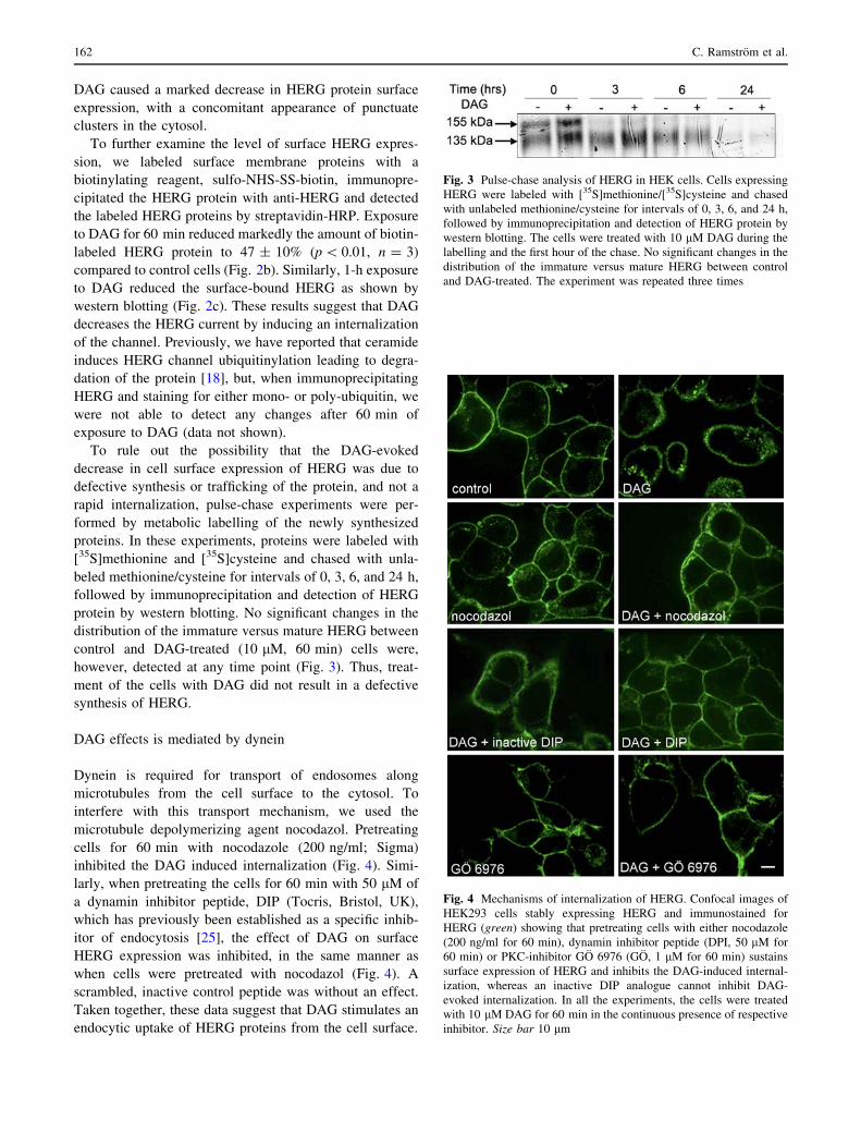
DAG caused a marked decrease in HERG protein surface
expression, with a concomitant appearance of punctuate
clusters in the cytosol.
To further examine the level of surface HERG expres-
sion, we labeled surface membrane proteins with a
biotinylating reagent, sulfo-NHS-SS-biotin, immunopre-
cipitated the HERG protein with anti-HERG and detected
the labeled HERG proteins by streptavidin-HRP. Exposure
to DAG for 60 min reduced markedly the amount of biotin-
labeled HERG protein to 47 ± 10% (p \ 0.01, n = 3)
compared to control cells (Fig. 2b). Similarly, 1-h exposure
to DAG reduced the surface-bound HERG as shown by
western blotting (Fig. 2c). These results suggest that DAG
decreases the HERG current by inducing an internalization
of the channel. Previously, we have reported that ceramide
induces HERG channel ubiquitinylation leading to degra-
dation of the protein [18], but, when immunoprecipitating
HERG and staining for either mono- or poly-ubiquitin, we
were not able to detect any changes after 60 min of
exposure to DAG (data not shown).
To rule out the possibility that the DAG-evoked
decrease in cell surface expression of HERG was due to
defective synthesis or trafficking of the protein, and not a
rapid internalization, pulse-chase experiments were per-
formed by metabolic labelling of the newly synthesized
proteins. In these experiments, proteins were labeled with
[35S]methionine and [35S]cysteine and chased with unla-
beled methionine/cysteine for intervals of 0, 3, 6, and 24 h,
followed by immunoprecipitation and detection of HERG
protein by western blotting. No significant changes in the
distribution of the immature versus mature HERG between
control and DAG-treated (10 lM, 60 min) cells were,
however, detected at any time point (Fig. 3). Thus, treat-
ment of the cells with DAG did not result in a defective
synthesis of HERG.
DAG effects is mediated by dynein
Dynein is required for transport of endosomes along
microtubules from the cell surface to the cytosol. To
interfere with this transport mechanism, we used the
microtubule depolymerizing agent nocodazol. Pretreating
cells for 60 min with nocodazole (200 ng/ml; Sigma)
inhibited the DAG induced internalization (Fig. 4). Simi-
larly, when pretreating the cells for 60 min with 50 lM of
a dynamin inhibitor peptide, DIP (Tocris, Bristol, UK),
which has previously been established as a specific inhib-
itor of endocytosis [25], the effect of DAG on surface
HERG expression was inhibited, in the same manner as
when cells were pretreated with nocodazol (Fig. 4). A
scrambled, inactive control peptide was without an effect.
Taken together, these data suggest that DAG stimulates an
endocytic uptake of HERG proteins from the cell surface.
Fig. 3 Pulse-chase analysis of HERG in HEK cells. Cells expressing
HERG were labeled with [35S]methionine/[35S]cysteine and chased
with unlabeled methionine/cysteine for intervals of 0, 3, 6, and 24 h,
followed by immunoprecipitation and detection of HERG protein by
western blotting. The cells were treated with 10 lM DAG during the
labelling and the first hour of the chase. No significant changes in the
distribution of the immature versus mature HERG between control
and DAG-treated. The experiment was repeated three times
Fig. 4 Mechanisms of internalization of HERG. Confocal images of
HEK293 cells stably expressing HERG and immunostained for
HERG (green) showing that pretreating cells with either nocodazole
(200 ng/ml for 60 min), dynamin inhibitor peptide (DPI, 50 lM for
60 min) or PKC-inhibitor GO 6976 (GO, 1 lM for 60 min) sustains
surface expression of HERG and inhibits the DAG-induced internal-
ization, whereas an inactive DIP analogue cannot inhibit DAG-
evoked internalization. In all the experiments, the cells were treated
with 10 lM DAG for 60 min in the continuous presence of respective
inhibitor. Size bar 10 lm
162 C. Ramstrom et al.

DAG- induced endocytosis of HERG is PKC dependent
Since DAG is an effective PKC activator, a plausible
pathway for the observed DAG-induced effect would be
through PKC. To test this possibility, we used the PKC
inhibitor GO6976 (Biosource International, CA, USA)
known to effectively inhibit the classical PKCa and bisoforms, to attenuate the effect of DAG on HERG. Pre-
treatment with GO6976 (1 lM for 60 min) completely
inhibited the reductive effect of DAG (Fig. 4). HEK293
cells express both PKCa, bI and bII (and the PKCd iso-
form), and to test if one or both isoforms are involved we
used HBDDE (Biomol Research Laboratories, Plymouth
Meeting, PA, USA), a specific PKCa inhibitor. A 1-h pre-
treatment with the inhibitor (50 lM) did not significantly
inhibit the DAG-induced endocytosis (data not shown),
implying that PKCa is not involved in mediating the DAG-
induced endocytosis of HERG protein. To confirm the
immunocytochemical data, whole-cell patch-clamp
recordings were performed on cells preincubated with
1 lM GO6976. As can be seen in Table 3, preincubating
the cells with GO6976 totally abolished the DAG-evoked
decrease in the HERG current. We also tested whether
10 lM of DAG was able to evoke a translocation of PKC
isoforms from the cytosol to the membrane fraction. We
could not observe a significant translocation of any isoform
from the cytosolic to the membrane fractions, but a sub-
stantial amount of PKCbI was detected in the membrane
fractions of the cells. Also, PKCa was detected in the
membrane fractions (Fig. 5).
To further investigate the regulation of HERG channels,
we stimulated the cells with 100 lM carbachol to activate
membrane receptors. As can be seen in Fig. 6, in these
cells, the HERG current was decreased. Furthermore, the
carbachol-evoked decrease in HERG current was attenu-
ated in cells treated with 1 lM GO6976. In addition,
incubating the cells with 100 lM carbachol caused a
marked decrease in HERG protein surface expression
(Fig. 7). This decrease was not observed in cells pretreated
with GO6976.
HERG is present in early endosomes
Next, we examined whether the punctuate clusters of
HERG appearing in the cytosol, upon exposure to DAG,
would be localized in endosomes or lysosomes. As seen in
Fig. 8, HERG and the early endosome marker EEA1 co-
localizes upon a 60-min exposure to DAG in small clusters
close to the cell surface. The co-localization with EEA1
was clearly time-dependent since it was not evident with
either a 30-min exposure to DAG, i.e. too short a period, or
with a 24-h exposure, too long (data not shown). Co-
localization with HERG and the lysosomal marker Lamp-1
was not seen at any of the time points used. These results
suggest that DAG targets the HERG channel for endo-
somes not leading to lysosomal degradation.
DAG causes a reduction of HERG channels
in SH-SY5Y cells
The above experiments were made in cells overexpressing
HERG channels. To verify our observations in cells
endogenously expressing HERG channels, we used SH-
SY5Y neuroblastoma cells. These cells express both
HERG channels and the truncated HERG1b channels [26].
Incubating the cells with 10 lM DAG for 60 min resulted
in a significant decrease in membrane expression of HERG,
but not of HERG1b, as measured by western blot. Pre-
treatment with the PKC inhibitor GO6976 completely
abrogated the effect of DAG on HERG (Fig. 9a, b).
In patch-clamp experiments, the relative tail currents
were markedly suppressed following applications of
10 lM DAG, -0.61 ± 0.09 versus control of -0.94 ±
0.01 (n = 8, p \ 0.05). In addition, perfusing the cells with
Table 3 Effects of GO6976 on DAG-evoked decrease in HERG
current
pA/pF
Control 57.8 ± 4.4
DAG 34.5 ± 5.7*
DAG ? GO 65.0 ± 4.9
Peak tail currents were measured as described in Fig. 1a
The cells were preincubated with 10 lM DAG for 60 min, or with
1 lM GO6976 for 60 min and then with 10 lM DAG in the presence
of GO6976 for another 60 min
Tail currents at -60 mV were recorded following depolarization to
?40 mV for 2 s from the holding potential of -80 mV. The data
given are the mean ± SEM of 3–12 separate experiments
*P \ 0.05
Fig. 5 Lack of translocation of PKC isoforms in response to DAG in
HEK293 cells stably expressing HERG. a The cells were treated with
10 lM DAG for the indicated times (min), and the cells were
fractioned into cytosolic c and membrane m fractions. The blots show
the effect of DAG on the distribution of PKCa. PKCbI, PKCbII, and
PKCd. The experiments were repeated three times
Diacylglycerol internalizes HERG 163

50 lM of the phospholipace C activator m-3M3FBS sig-
nificantly decreased the tail current (-0.44 ± 0.04 vs
control of -0.88 ± 0.03, n = 4, p \ 0.05). Pretreatment
of the cells with GO6976 did not inhibit the effect of DAG
(data not shown). However, in cells pretreated with
100 nM PMA to downregulate PKC, the effect of DAG on
the current was abolished (-0.84 ± 0.04 vs control of
-0.89 ± 0.02, n = 5, p = 0.37) (Fig. 9c). The normalized
values are shown in Fig. 9d. Thus, also in cells expressing
HERG endogenously, DAG reduces the membrane
expression of the channels protein through a PKC-depen-
dent mechanism.
Discussion
This study demonstrates that the current mediated by the
voltage-sensitive potassium channel HERG can be depres-
sed by DAG and, for the first time, that the mechanism for
the decrease is a PKC-mediated, dynamin-dependent
endocytosis of the channel. Our results introduce a new
mechanism of HERG channel regulation.
Many G protein-coupled receptors evoke the production
of DAG by two mechanisms: first, a rapid and transient
production of DAG as a result of PLC activation, followed
by a slower and more prolonged DAG production due to
activation of phospholipase D (PLD) [27, 28]. The differ-
ence in magnitude and duration of the DAG signal may
then result in activation of specific PKC-isoenzymes and
activation of separate signalling pathways, e.g., phosphor-
ylation of channel proteins or proteins regulating
endocytosis. The prolonged increase in DAG thus adds a
new level of regulation of channel function, e.g., by acti-
vating a PKC-evoked endocytosis of the channel protein.
This effect may be of significant importance in the regu-
lation of channel function.
The effect of PKC in regulating HERG channels is
controversial. Barros et al. [20] showed that in HERG-
expressing Xenopus oocytes, thyrotropin-releasing hor-
mone (TRH) through its receptor evoked a decrease in
current through a protein kinase C-dependent acceleration
of deactivation, and slower time course of activation. The
effect was mimicked by phorbol 12-myristate 13-acetate
(PMA), and inhibited by blocking PKC. In another study,
angiotensin II evoked a pronounced irreversible decrease in
Ikr in myocytes and in HEK cells expressing HERG [29].
The effect was mimicked by stimulating the cells with
PMA, and was attenuated if PKC was blocked. The above
results are thus in line with our observations, i.e., that both
the DAG-evoked and the carbachol-evoked decrease in
HERG current was attenuated when PKC was blocked.
However, in GH3B6 rat pituitary cells, TRH was shown to
decrease ERG currents by shifting the voltage dependence
of activation and by accelerating the time course of deac-
tivation in a PKC-independent manner [30]. In this study,
PMA evoked a PKC-independent shift in voltage depen-
dence of ERG activation and a decrease in the current, but
not a decrease in maximal tail current amplitude. Inter-
estingly, PKC was necessary for the recovery of a transient
TRH-evoked decrease of rat ERG current in GH3 rat
pituitary cells [31]. If PKC was blocked, the TRH-evoked
Fig. 6 Carbachol decreases HERG current density. a Current–
voltage plot of whole-cell HERG currents showing the decrease in
HERG conductance in response to Cch incubation. Dotted lines are
the 4-order polynomial functions fitted to data (circles) in control
conditions (black) or after incubation in 100 lM Cch (red). Conduc-
tance was measured by a linear fit (grey line) to polynomial function,
fit range 20 mV centred at EHERG. Aa Full-length 1.4-s current traces
in response to a voltage protocol used. The protocol constituted of 15
sweeps, each starting from a holding potential of –80 mV. After the
800-ms-long depolarization to ?20 mV, the membrane was clamped
to a series of potentials between ?10 and (-140 mV) for 200 ms and
then returned to the holding level. Inside the grey box are
representative recordings from control cell (black) and Cch exposed
cell (red) in an extended time scale. b Summary of HERG
conductance measurements done as described in a. Bars represent
the mean ± SEM of membrane conductance related to its capaci-
tance. Control (Ctrl) n = 66, 1 lM GO6976 incubation (GO) n = 31,
100 lM carbachol incubation (Cch) n = 25, pre-incubation in
GO ? Cch incubation (preGOCch) n = 38. *p \ 0.001 using the
two-sample independent t test. Pre-incubation with GO6976 signif-
icantly increase conductance compared with Cch-treated cells
164 C. Ramstrom et al.

decrease in current became irreversible. Furthermore, in
Xenopus laevis oocytes heterologously expressing HERG,
PMA decreased the current and shifted the voltage-
dependence of HERG activation towards more positive
potentials. This effect was suggested not to be mediated by
a direct PKC-evoked phosphorylation of the channel, as all
except one PKC-dependent phosphorylation sites were
mutated [19]. However, in a recent investigation, Cockerill
et al. [21] showed that stimulating HERG-expressing cells
with PMA, OAG or metacholine, PKC decreased HERG
current by a PKC-mediated phosphorylation of the pore-
forming unit of the channel. Thus, PKC may function
either as an enhancer or an inhibitor of the channel func-
tion. This discrepancy may, in part, be the result of the
different model systems used in the investigations. Dif-
ferent cell systems may express different isoforms of PKC,
further complicating the interpretation of the results. It
would thus be helpful to investigate which isoforms of
PKC that mediate the observed effects. Although a role for
either PKCa and b is implicated by the use of the fairly
PKCa/b selective inhibitor Go6976 (Cockerill et al. [21],
and our study), more precise results would be obtained by
downregulating PKC isoforms using specific siRNA
constructs.
Fig. 7 Carbachol decreases
HERG surface expression.
Confocal images of HEK293
cells stably expressing HERG
and immunostained for HERG
(green) showing that
stimulating the cells with
carbachol (100 lM for 60 min)
evokes internalization of the
channel protein. Pretreatment of
the cells with the PKC-inhibitor
GO6976 (GO, 1 lM for 60 min)
sustains surface expression of
HERG and inhibits the
carbachol-induced
internalization. Size bar 10 lm
Fig. 8 Internalized HERG is
present in early endosomes.
HERG (green) co-localizes,
upon 60 min of DAG treatment
(10 lM), with the early
endosome marker EEA-1 (red).
Co-localization of HERG and
EEA-1 is indicated by yellow.
Size bar 10 lm
Diacylglycerol internalizes HERG 165

A further twist was introduced when it was shown that
the PLC-evoked reduction of PIP2 was the signal that
reduced HERG activity [16]. According to this investiga-
tion, DAG-mediated activation of PKC was without any
effects. A role for PIP2 in our experiments also seems
apparent, as the charbachol-evoked decrease in HERG
conductance was only in part restored in cells pretreated
with the PKC antagonist GO6976. A PIP2-dependent
regulation has also been proposed for the neuronal KCNQ
potassium channel, although both calcium and PKC are
apparently participating in the fine-tuning of the regulation
(see [32] for a review).
In other potassium channels, an effect of PKC is also
evident. A rapidly activating delayed rectifier potassium
channel was enhanced by PKC through a reduction in
C-type inactivation [33], whereas a G-protein gated
inwardly rectifying K channel was inhibited for a pro-
longed period after transient stimulation with carbachol
[34]. In these experiments, inhibition of PKC attenuated the
effect of carbachol only in part, in a manner similar to our
results. Furthermore, transforming growth factor b1 regu-
lated Kir2.3 channels via a PKC-mediated down-regulation
of the conductance [35], whereas the Kir2.1b channel and a
G-protein-coupled inward rectifier channel were inhibited
by a direct PKC-mediated phosphorylation of the channel
proteins [36, 37]. In contrast, the surface expression of the
ROMK1 channel has been shown to be critically dependent
on PKC-evoked phosphorylation, as inhibition of PKC or
mutating PKC phosphorylation sites in the channel signi-
ficantly decreased the membrane expression of the channel
protein [38]. Thus, different channels are regulated by PKC
through different mechanisms.
The expression of proteins on the cell surface, e.g.,
tyrosine kinases [39], G-protein coupled receptors [40] and
some ion channels, can be regulated by endocytosis. The
surface expression of the epithelial sodium channel
(ENaC), was regulated by an ubiquitin-dependent endo-
cytosis [41]. A few reports indicating the importance of
endocytosis as a regulator mechanism of voltage-gated
potassium channels have also been published. Suppression
of Kir1.1 was regulated by clathrin-dependent endocytosis
[42], whereas a tyrosine kinase-induced inhibition of Kv1.2
was mediated by a dynamin-dependent endocytosis [39].
Furthermore, the surface expression of another potassium
Fig. 9 DAG decreases HERG membrane expression and current in
SH-SY5Y cells. a Western blot analysis of HERG in membranes from
SH-SY5Y cells treated with 10 lM DAG for 1 h. b Summary of
densitometric analysis of the distribution of HERG in the membrane
fraction. The values given are the mean ± SEM of N experiments. cHERG-like currents present in SHSY5Y cells are reduced in response
to treatment with DAG and PLC activation. Tail currents were
markedly suppressed following applications of 10 lM DAG (circle),
n = 8. The reductive effect of DAG was prevented by a 3-h pre-
incubation with 100 nM PMA (square), n = 5. Furthermore, 50 lM
of the PLC-inhibitor m-3M3FBS (triangle), n = 4, decreased the tail
current. A change from 5.4 to 50.4 mM K? extracellular solution
(control) enhanced tail currents (see inset). Amplitudes are presented
relative to the largest peak found during control period (considered as
-1). The wash-in periods of high [K?]0 solution and subsequent drug
application are indicated with horizontal lines. Values at 300 and
600 s time points (marked with arrows) were selected to represent
control and treated responses, respectively. d The currents shown in
(c) were normalized as described in ‘‘Materials and methods’’. Each
bar gives the mean ± SEM of 4–8 separate measurements. *p \ 0.05
b
166 C. Ramstrom et al.
channel, Kv1.5, was also regulated by a mechanism
dependent on endocytosis [43]. We have previously shown
that the ceramide-induced suppression of the voltage-gated
potassium channel HERG is regulated by ubiquitin-medi-
ated lysosomal degradation [18].
Our results suggested that PKC evoked dynamin-
dependent endocytosis of HERG channels. In both cardiac
myocytes and in neurons, PMA evoked a substantial
decrease in the ATP-sensitive potassium channel (KATP)
current through a PKC-evoked internalization via dynam-
in-dependent endocytosis [44]. In these experiments, no
phosphorylation of the channel was observed, but there was
an activation of the dynein motor complex. In addition,
PKC stimulated the endocytosis of GABAA receptors
through a dynamin-dependent pathway [45]. It is thus
possible that the prolonged or irreversible decrease in
HERG currents observed in previous studies in response to
activation of PKC is, at least in part, the result of a sub-
stantial internalization of the channel protein, in a manner
similar to what we report in the present study.
Treatment with DAG did not affect the surface expres-
sion of HERG1b. Presently, we do not know the reason for
this. However, in HERG1b, the amino terminus is trun-
cated compared with HERG. According to the results of
Cockerill et al. [21], this domain is important for PKC-
mediated phosphorylation and inactivation of HERG.
Further studies will reveal whether this domain is also
important for the internalization of HERG. Interestingly,
the PKC inhibitor GO6976 attenuated the internalization of
HERG evoked by DAG, but was ineffective in attenuating
the DAG-evoked decrease in HERG tail current in SH-
SY5Y cells. However, treatment with PMA, which down-
regulates all classical and novel isoforms of PKC, clearly
blocked the effect of DAG. Whether this result is due to
different isoforms of PKC in HEK cells and SH-SY5Y or
to the presence of HERG1b in the SH-SY5Y cells is
presently not known.
It must be pointed out that activation of PKC may also
enhance surface expression of channel proteins. Lan et al.
[46] and Lin et al. [47] showed that PKC both increased the
NMDA channel opening rate and delivered new NMDA
channels to the membrane by a mechanism dependent on
PKC. The latter mechanism was through SNARE-mediated
exocytosis. In addition, a 5-HT3 receptor-mediated current
in Xenopus oocytes was enhanced by a PKC-mediated
increase in the amount of receptor protein on the cell
membrane [48].
In the heart, autonomic signalling through a-adrenergic
and muscarinergic receptors are important determinants for
the regulation of contractility and heart rate [49, 50]. As
these receptors couple to the PLC-signalling pathway and
the production of DAG and activation of PKC, they are
probably of importance in regulating HERG. This is
supported by clinical observations in patients with LQTS.
These patients develop arrhythmias during physical or
emotional stress [51], which would suggest a link between
adrenergic stimulus and HERG potassium activity. The
b-adrenergic link to HERG is fairly well studied, showing
that elevated levels of cAMP regulate HERG channel
activity through protein kinase A. The a-adrenergic link is,
however, not that well studied. One plausible link could be
the DAG-induced suppression of HERG, since it is known
that adrenergic activity activates PLC [15] and generates
DAG through hydrolyses of PIP2. It is evident that the
relatively slow internalization of HERG observed in our
study cannot explain the sudden onset of arrhythmia.
However, as receptor-mediated production of DAG and the
activity of PKC is biphasic [27, 52], the latter prolonged
phase can affect HERG expression on the plasma mem-
brane. This could, at least in part, contribute to the onset of
arrhythmia during, e.g., emotional stress, especially in
conjunction with the reported inhibitory effect of receptor-
mediated decrease in PIP2 on HERG function [16].
In conclusion, DAG is a ubiquitous lipid second mes-
senger strongly associated with PKC activation. This lipid
has also been shown to be associated with regulation of ion
channels. In addition to the recent observation that HERG
can be down-regulated by a direct phosphorylation by PKC
[21], we show an additional mechanism for the regulation
of plasma membrane expression of HERG, i.e., a DAG-
evoked, PKC-mediated removal of the channel from the
cell surface. The plausible mechanism of the internaliza-
tion is a dynamin-dependent endocytosis of the channel.
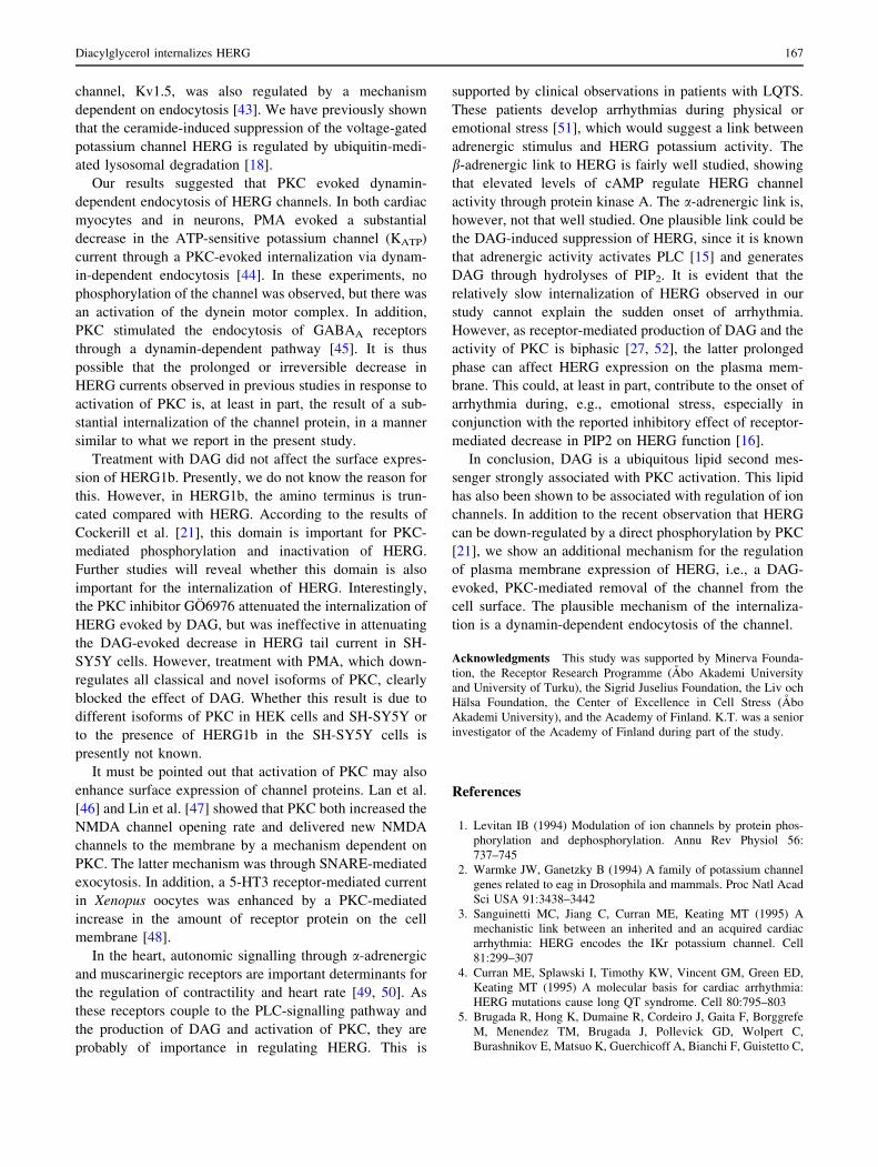
Acknowledgments This study was supported by Minerva Founda-
tion, the Receptor Research Programme (Abo Akademi University
and University of Turku), the Sigrid Juselius Foundation, the Liv och
Halsa Foundation, the Center of Excellence in Cell Stress (Abo
Akademi University), and the Academy of Finland. K.T. was a senior
investigator of the Academy of Finland during part of the study.
References
1. Levitan IB (1994) Modulation of ion channels by protein phos-
phorylation and dephosphorylation. Annu Rev Physiol 56:
737–745
2. Warmke JW, Ganetzky B (1994) A family of potassium channel
genes related to eag in Drosophila and mammals. Proc Natl Acad
Sci USA 91:3438–3442
3. Sanguinetti MC, Jiang C, Curran ME, Keating MT (1995) A
mechanistic link between an inherited and an acquired cardiac
arrhythmia: HERG encodes the IKr potassium channel. Cell
81:299–307
4. Curran ME, Splawski I, Timothy KW, Vincent GM, Green ED,
Keating MT (1995) A molecular basis for cardiac arrhythmia:
HERG mutations cause long QT syndrome. Cell 80:795–803
5. Brugada R, Hong K, Dumaine R, Cordeiro J, Gaita F, Borggrefe
M, Menendez TM, Brugada J, Pollevick GD, Wolpert C,
Burashnikov E, Matsuo K, Guerchicoff A, Bianchi F, Guistetto C,
Diacylglycerol internalizes HERG 167

Schimpf R, Brugada P (2004) Sudden death associated with
short-QT syndrome linked to mutations in HERG. Circulation
109:30–35
6. Yap YG, Camm AJ (2003) Drug induced QT prolongation and
torsades de pointes. Heart 89:1363–1372
7. Chiesa N, Rosati B, Arcangeli A, Olivotto M, Wanke E (1997) A
novel role for HERG K? channels: spike-frequency adaptation. J
Physiol 501:313–318
8. Farrelly AM, Ros S, Callaghan BP, Khoyi MA, Flemming N,
Horowitz B, Sanders KM, Keef KD (3003) Expression and
function of KCNH2 (HERG) in the human jejunum. Am J Physiol
Gastrointest Liver Physiol 284:G883–G895
9. Rosati B, Marchetti P, Crociani O, Lecchi M, Lupi RA, Arcangeli
A, Olivotto M, Wanke E (2000) Glucose- and arginine-induced
insulin secretion by human pancreatic beta-cells: the role of
HERG K(?) channels in firing and release. FASEB J. 14:
2601–2610
10. Lastraioli E, Guasti L, Crociani O, Polvani S, Hofmann G,
Witchel H, Bencini L, Calistri M, Messerini L, Scatizzi M,
Moretti PA, Wanke E, Olivotto M, Mugnai G, Arcangeli A
(2004) herg1 gene and HERG1 protein are overexpressed in
colorectal cancers and regulate cell invasion of tumor cells.
Cancer Res 64:606–611
11. Wang H, Zhang Y, Cao L, Han H, Wang J, Yang B, Nattel S,
Wang Z (2002) HERG K? channel, a regulator of tumor cell
apoptosis and proliferation. Cancer Res 62:4843–4848
12. Johannes FJ, Prestle J, Dieterich S, Oberhagemann P, Link G,
Pfizenmaier K (1995) Characterization of activators and inhibi-
tors of protein kinase C mu. Eur J Biochem 15:303–307
13. Canagarajah B, Leskow FC, Ho JY, Mischak H, Saidi LF,
Kazanietz MG, Hurley JH (2004) Structural mechanism for lipid
activation of the Rac-specific GAP, beta2-chimaerin. Cell
119:407–418
14. Attali B, Honore E, Lesage F, Lazdunski M, Bathain J (1992)
Regulation of a major cloned voltage-gated K? channel from
human T lymphocytes. FEBS Lett 303:229–232
15. Lo CF, Numann R (1998) Independent and exclusive modulation
of cardiac delayed rectifying K? current by protein kinase C and
protein kinase A. Circ Res 83:995–1002
16. Bian J, Cui J, McDonald TV (2001) HERG K(?) channel activity
is regulated by changes in phosphatidyl inositol 4, 5-bisphos-
phate. Circ Res 89:1168–1176
17. Wu S-N, Lo Y-K, Kuo B, Ching H-T (2001) Ceramide inhibits
the inwardly rectifying potassium current in GH3 lactotrophes.
Endocrinology 142:4785–4794
18. Chapman H, Ramstrom C, Korhonen L, Laine M, Wann KT,
Lindholm D, Pasternack M, Tornquist K (2005) Down-regulation
of the HERG (KCNH2) K? channel by ceramide: Evidence for
ubiquitin-mediated lysosomal degradation. J Cell Sci 118:
5325–5334
19. Thomas D, Zhang W, Wu K, Wimmer AB, Gut B, Wendt-Nor-
dahl G, Kathofer S, Kreye VA, Kathus HA, Schoels W, Kiehn J,
Karle CA (2003) Regulation of HERG potassium channel acti-
vation by protein kinase C independent of direct phosphorylation
of the channel protein. Cardiovasc Res 59:14–26
20. Barros F, Gomez-Varela D, Vitoria CG, Giraldez T, de la Pena P
(1998) Modulation of human erg K? channel gating by activation
of a G protein-coupled receptor and protein kinase C. J Physiol
511:333–346
21. Cockerill SL, Tobin AB, Torrecilla I, Willars GB, Standen NB,
Mitcheson JS (2007) Modulation of hERG potassium currents in
HEK-293 cells by protein kinase C. Evidence for direct phos-
phorylation f pore forming subunits. J Physiol 581:479–493
22. Arcangeli AFB, Becchetti A, Faravelli L, Coronello M, Mini E,
Olivotto M, Wanke E (1995) A novel inward-rectifying K?
current with a cell-cycle dependence governs the resting potential
of mammalian neuroblastoma cells. J Physiol 489:455–471
23. Zhou Z, Gong Q, Ye B, Fan Z, Makielski JC, Robertson GA,
January CT (1998) Properties of HERG channels stably expres-
sed in HEK 293 cells studied at physiological temperature.
Biophys J 74:230–241
24. Kass GEN, Duddy SK, Orrenius S (1989) Activation of hepato-
cyte protein kinase C by redox-cycling quinones. Biochem J
260:499–507
25. Nong Y, Huang YO, Ju W, Kalia LV, Ahmadian G, Wang YT,
Salter MW (2003) Glycine binding primes NMDA receptor
internalization. Nature 422:302–307
26. Crociani O, Guasti L, Balzi M, Becchetti A, Wanke E, Olivotto
M, Wymore RS, Arcangeli A (2003) Cell cycle-dependent
expression of HERG1 and HERG1B isoforms in tumor cells.
J Biol Chem 278:2947–2955
27. Liscovitch M (1992) Crosstalk among multiple signal-activated
phospholipases. Trends Biochem Sci 17:393–399
28. Asaoka Y, Nakamura S, Yoshida K, Nishizuka Y (1992) Protein
kinase C, calcium and phospholipid degradation. Trends Biochem
Sci 17:414–417
29. Wang YH, Shi CX, Dong F, Sheng JW, Xu YF (2008) Inhibition
of the rapid component of the delayed rectifier potassium current
in ventricular myocytes by angiotensin II via the AT1 receptor. Br
J Pharmacol 154:429–439
30. Schledermann W, Wulfsen I, Schwrz JR, Bauer CK (2001)
Modulation of rat erg1, erg2, erg3 and HERG K? currents by
thyrotropin-releasing hormone in anterior pituitary cells via the
native signal cascade. J Physiol 532:143–163
31. Gomez-Varela D, Giraldez T, de la Pena P, Dupuy SG, Garcia-
Manso D, Barros F (2003) Protein kinase C is necessary for
recovery from the thyrotropin-releasing hormone-induced r-ERG
current reduction in GH3 rat anterior pituitary cells. J Physiol
547:913–929
32. Delmas P, Brown DA (2005) Pathways modulating neural
KCNQ/M (Kv7) potassium currents. Nat Rev Neurosci 6:
850–862
33. Heath BM, Terrar DA (2000) Protein kinase C enhances the
rapidly activating delayed rectifier potassium current, IKr,
through a reduction in C-type inactivation in guinea-pig ven-
tricular myocytes. J Physiol 522:391–402
34. Leaney JL, Dekker LV, Tinker A (2001) Regulation of a G
protein-gated inwardly rectifying K? channel by a Ca(2?)-
independent protein kinase C. J Physiol 534:367–379
35. Perillan PR, Chen M, Potts EA, Simard JM (2002) Transforming
growth factor-beta 1 regulates Kir2.3 inward rectifier K? chan-
nels via phospholipase C and protein kinase C-delta in reactive
astrocytes from adult rat brain. J Biol Chem 277:1974–1980
36. Karle CA, Zitron E, Zhang W, Wendt-Nordahl G, Kathofer S,
Thomas D, Gut B, Scholz E, Vahl CF, Katus HA, Kiehn J (2002)
Human cardiac inwardly-rectifying K? channel Kir(2.1b) is
inhibited by direct protein kinase C-dependent regulation in
human isolated cardiomyocytes and in an expression system.
Circulation 106:1493–1499
37. Mao J, Wang X, Chen F, Wang R, Rojas A, Shi Y, Piao H, Jiang
C (2004) Molecular basis for the inhibition of G protein-coupled
inward rectifier K(?) channels by protein kinase C. Proc Natl
Acad Sci USA 101:1087–1092
38. Lin DH, Sterling H, Lerea KM, Giebisch G, Wang WH (2002)
Protein kinase C (PKC)-induced phosphorylation of ROMK1 is
essential for the surface expression of ROMK1 channels. J Biol
Chem 277:44278–44284
39. Nesti E, Everill B, Morielli AD (2004) Endocytosis as a mech-
anism for tyrosine kinase-dependent suppression of a voltage-
gated potassium channel. Mol Biol Cell 15:4073–4088
168 C. Ramstrom et al.

40. Delaney KA, Murph MM, Brown LM, Radhakrishna H (2002)
Transfer of M2 muscarinic acetylcholine receptors to clathrin-
derived early endosomes following clathrin-independent endo-
cytosis. J Biol Chem 277:33439–33446
41. Staub O, Abriel H, Plant P, Ishikawa T, Kanelis V, Saleki R,
Horisberger JD, Schild L, Rotin D (2000) Regulation of the
epithelial Na? channel by Nedd4 and ubiquitination. Kidney Int
57:809–811
42. Zeng WZ, Babich V, Ortega B, Quigley R, White SJ, Welling
PA, Huang CL (2002) Evidence for endocytosis of ROMK
potassium channel via clathrin-coated vesicles. Am J Physiol
Renal Physiol 283:F630–639
43. Choi WS, Khurana A, Mathur R, Viswanathan V, Steele DF,
Fedida D (2005) Kv1.5 surface expression is modulated by ret-
rograde trafficking of newly endocytosed channels by the dynein
motor. Circ Res 97:363–371
44. Hu K, Huang CS, Jan YN, Jan LY (2003) ATP-sensitive potas-
sium channel traffic regulation by adenosine and protein kinase
C. Neuron 38:417–432
45. Herring D, Huang R, Singh M, Dillon GH, Leidenheimer NJ
(2005) PKC modulation of GABAA receptor endocytosis and
function is inhibited by mutation of a dileucin motif within the
receptor beta 2 subunit. Neuropharmacology 48:181–194
46. Lan JY, Skeberdis VA, Jover T, Grooms SY, Lin Y, Araneda RC,
Zheng X, Bennet MVL, Zukin RS (2001) Protein kinase C
modulates NMDA receptor trafficking and gating. Nat Neurosci
4:382–390
47. Lin Y, Jover-Mangual T, Wong J, Bennet MVL, Zukin RS (2006)
PSD-95 and PKC converge in regulating NMDA receptor traf-
ficking and gating. Proc Natl Acad Sci USA 103:19902–19907
48. Sun H, Hu XQ, Moradel EM, Weight FF, Zhang L (2003)
Modulation of 5-HT3 receptor-mediated response and trafficking
by activation of protein kinase C. J Biol Chem 278:34150–34157
49. Thomas D, Kiehn J, Katus HA, Karle CA (2004) Adrenergic
regulation of the rapid component of the cardiac delayed rectifier
potassium curren, IKr, and the underlying hERG ion channel.
Basic Res Cardiol 99:279–287
50. Bian JS, McDonald TV (2007) Phosphatidylinositol 4, 5-bis-
phosphate interactions with the HERG K ? channel. Pflugers
Arch Eur J Physiol 455:105–113
51. Priori SG, Napolitano C, Paganini V, Cantu F, Schwatz PJ (1997)
Molecular biology of the long QT syndrome: impact on man-
agement. Pacing Clin Electrophysiol 20:2052–2057
52. Nishizuka Y (1992) Intracellular signaling by hydrolysis of
phospholipids and activation of protein kinase C. Science
258:607–614
Diacylglycerol internalizes HERG 169