REGIONAL BRAIN PERFUSION IN 10 NORMAL DOGS MEASURED USING TECHNETIUM-99m ETHYL CYSTEINATE DIMER...
247
FACULTY OF VETERINARY MEDICINE FUNCTIONAL BRAIN IMAGING IN THE DOG SINGLE PHOTON EMISSION TOMOGRAPHY AS A RESEARCH AND CLINICAL TOOL FOR THE INVESTIGATION OF CANINE BRAIN PHYSIOLOGY AND PATHOPHYSIOLOGY Kathelijne Peremans Proefschrift ter verkrijging van de graad van Doctor in de Diergeneeskundige Wetenschappen (PhD) aan de Faculteit Diergeneeskunde, Universiteit Gent 2002 Promotor: Prof. F.Verschooten Copromotor: Prof. R. Dierckx Vakgroep Medische Beeldvorming van de Huisdieren Salisburylaan 133 B-9820 Merelbeke
-
Upload
independent -
Category
Documents
-
view
0 -
download
0
Transcript of REGIONAL BRAIN PERFUSION IN 10 NORMAL DOGS MEASURED USING TECHNETIUM-99m ETHYL CYSTEINATE DIMER...

FACULTY OF VETERINARY MEDICINE
FUNCTIONAL BRAIN IMAGING IN THE DOG
SINGLE PHOTON EMISSION TOMOGRAPHY AS A RESEARCH ANDCLINICAL TOOL FOR THE INVESTIGATION OF CANINE BRAIN
PHYSIOLOGY AND PATHOPHYSIOLOGY
Kathelijne Peremans
Proefschriftter verkrijging van de graad van
Doctor in de Diergeneeskundige Wetenschappen (PhD)aan de Faculteit Diergeneeskunde,
Universiteit Gent2002
Promotor: Prof. F.VerschootenCopromotor: Prof. R. Dierckx
Vakgroep Medische Beeldvorming van de HuisdierenSalisburylaan 133B-9820 Merelbeke


c
TABLE OF CONTENTS
List of abbreviations
INTRODUCTION 1
1. General introduction 3
2. Principles of nuclear medicine 4
A. Historical overview 4
B. Current implementation of radiation in diagnostic imaging 5
C. Radioprotection: matter of concern 8
3. Nuclear Brain Imaging: “Mapping the functioning brain” 10
A. History of brain-behaviour research 10
B. Functional brain imaging with PET and SPET modalities 11
4. Nuclear imaging and behavioural disorders 12
A. Perfusion and serotonin-2A receptor alterations related with age 12
B. Impulsive behaviour 13
References 16
SCIENTIFIC AIMS 23
CHAPTER 1 REGIONAL BRAIN PERFUSION IN 10 NORMAL DOGSMEASURED USING TECHNETIUM-99m ETHYL CYSTEINATE DIMER SPECT 29
1. Summary 312. Introduction 333. Material and methods 354. Results 405. Discussion 416. References 44

TABLE OF CONTENTS
CHAPTER 2 USE OF AN AUTOMATIC REGISTRATION PROCEDURE FOR STANDARDISATION OF CANINEPERFUSION DATA OBTAINED WITH SINGLE PHOTONEMISSION TOMOGRAPHY 47
1. Introduction 492. Materials and methods 503. Results 554. Conclusion 565. References 57
CHAPTER 3 EVALUATION OF CEREBRAL NEUROTRANSMITTORPHYSIOLOGY AND PATHOPHYSIOLOGY WITHPET AND SPET IMAGING MODALITIES IN ANIMALMODELS: A REVIEW. 59
1. Summary 612. Introduction 623. Methodological considerations 634. Practical applications 685. Future prospects 746. References 76
CHAPTER 4 BIODISTRIBUTION AND DISPLACEMENT STUDIES OF THE SELECTIVE 5-HT2A RECEPTOR ANTAGONIST123I-5-I-R91150 IN THE NORMAL DOG 89
1. Summary 912. Introduction 923. Materials and methods 934. Results 965. Discussion 1016. Conclusive remarks 103
7. References 104
CHAPTER 5 REGIONAL BINDING INDEX OF THE SELECTIVE5-HT2A RADIOLABELLED ANTAGONIST 123 I-5-I-R91150 IN THE NORMAL CANINE BRAIN IMAGED WITH SINGLE PHOTON EMISSION TOMOGRAPHY 109
1. Summary 1112. Introduction 1123. Materials and methods 1144. Results 1205. Discussion 1216. References 123

TABLE OF CONTENTS
CHAPTER 6 THE AGING EFFECTS ON PERFUSION AND BINDING PARAMETERS OF A SELECTIVE SEROTONIN-2A LIGAND IN THE NORMAL CANINE BRAIN MEASURED WITH SINGLE PHOTON EMISSION TOMOGRAPHY 127
1. Summary 1292. Introduction 1303. Materials and methods 1324. Results 1385. Discussion 1476. Conclusion 1507. References 151
CHAPTER 7 A HYPOTHETICAL RESEARCH MODEL CONCERNING THE ROLE OF SEROTONINE AND THE FRONTAL CORTEX IN CANINE IMPULSIVITY 155
1. Summary 157
2. Introduction 1583. Methodology and results 1624. Discussion 1635. References 166
CHAPTER 8 EVALUATION OF THE BRAIN 5-HT2A RECEPTOR BINDING INDEX AND REGIONAL BRAIN PERFUSION IN THE IMPULSIVE, AGGRESSIVE DOGMEASURED WITH SPET 171
1. Summary 1752. Introduction 1763. Materials and methods 1784. Results 1865. Discussion 1936. Methodological considerations 1957. Future research directions 1978. References 198
GENERAL DISCUSSION 205
SUMMARY 215
SAMENVATTING 223
DANKWOORD 231
CURRICULUM VITAE 237


LIST OF ABBREVIATIONS
Alzheimer’s disease: AD
Beta-carboxymethoxy-iodophenyl-tropane: β-CIT
Binding index: BI
Blood brain barrier: BBB
Brain registration and automated SPECT semiquantification: BRASS
Brain uptake index: BUI
Cerebrospinal fluid: CSF
Computed tomography: CT
Dopamine transporter: DAT
Diethylenetriaminepentaacetic acid: DTPA
Equilibrum dissociation constant: Kd
N,N”-1,2-ethylene-diylbis-L-cysteine diethylester dihydrochloride: ECD
Full width at half maximum: FWHM
Gamma-aminobutyric acid receptor: GABA receptor
Glucoheptonate: GHA
d,l-hexamethylpropyleneamine-oxime: HMPAO
Iodobenzamide: IBZM
d,l-N-isopropyl-p-iodoamphetamine hydrochloride: IMP
Magnetic resonance imaging : MRI
Magnetic resonance spectroscopy: MRS
Maximal specific binding: Bmax
Mega becquerel: MBq
Multiple-time graphical analysis: MTGA
N-methyl-D-aspartate receptor: NMDA receptor
Non uniform attenuation coefficient: NUAC
Parkinson’s disease: PD
Partial volume effect: PVE
Positron emission tomography: PET
Regional cerebral blood flow: rCBF
Region of interest: ROI
Selective serotonin reuptake inhibitor: SSRI
Single photon emission tomography: SPET
Volume of interest: VOI
5-hydroxytryptamine (serotonin): 5-HT
5-hydroxytryptamine-2A: 5-HT2A99mTc: metastable Technetium123I: radioiodine


INTRODUCTION
“The notion of watching the brainthink, both in health and disease, hasbecome reality”
S. Lewis, N. Higgins from “Brain imaging inpsychiatry”, 1996

INTRODUCTION
INTRODUCTION
1. GENERAL INTRODUCTION
This thesis evaluates the use of single photon emission tomography (SPET) as functional
imaging modality in the study of canine brain and its relation to behavioural pathology. First, the use
of SPET in the study of in vivo brain perfusion was assessed.
Second, in vivo measurement of the serotonin-2A (5-HT2A) receptor binding in the dog brain was
evaluated with SPET. The clinical applications of these techniques in aged and in impulsive,
aggressive dogs were evaluated.
This introduction will serve to define the methodological context of the study.
First, since the use of nuclear medicine functional imaging modalities is not as wide-spread in
veterinary medicine as it is in human medicine, the first section of the introductory remarks present
some basic and general principles of nuclear medicine. A short historical overview will be given,
together with some notes on the current implementation of radiation in diagnostic imaging and
radiation protection in veterinary nuclear medicine.
A second section will narrow the focus of nuclear medicine to “brain” imaging and its
application in the study of pathological brain-behaviour relationships. First concerning the functioning
brain, there is a long tradition in the study of brain-behaviour interactions via post-mortem or surgical
and pharmacological interventional studies. Hence, a short overview on human and animal studies
from the “pre-imaging era” will be presented. This is followed by a short overview of the principles
of nuclear brain imaging, both perfusion or metabolism and receptor imaging, in physiological and
pathophysiological conditions.
A third section will narrow the focus even more to two conditions, old age and impulsive
behaviour, that will be evaluated with perfusion and serotonin-2A receptor studies, using SPET
imaging techniques and that will constitute the clinical application of this thesis. First, based on in
vitro and post-mortem neuropathological and biochemical studies, the rationale behind perfusion
imaging and serotonin-2A imaging in aged dogs is given. Second, impulsive behaviour is defined and
its relationship to brain abnormalities, both structurally and functionally, is given. The link of this
pathological condition and abnormalities in regional blood perfusion and serotonin-2A receptor is
explained.
3

INTRODUCTION
2. PRINCIPLES OF NUCLEAR MEDICINE
A. Historical overview
Only a few months after Röntgen discovered X-rays in 1895, Henri Becquerel demonstrated
the natural radiation properties of uranium salt crystals. Shortly thereafter, Pierre and Marie Curie
separated and identified radium, thorium and polonium, all three elements emitting radiation.
Radiation therapy was soon introduced for medical use by applying radium and later radon as local
implants to tuberculosis skin lesions. In the beginning of the twentieth century, radiation became a
popular health cure and was soon good for all ailments. Radioactive water was sold under several
forms as a drinkable tonicum, oil containing radium was used to cure arthritis and skin disorders and
radium containing electric blankets were claimed to combine the natural healing forces of electricity
and radiation. Although, at that time, virtually nothing was known about radiobiology, it was amazing
that the concept of radiation hormesis was already adopted. Indeed, high doses of radium were used
for therapy, utilizing its destructive properties, while small doses of radium were believed to have
stimulating properties, re-energizing weak and inactive cells.1 In the 1930’s an important step in the
introduction of artificial radioactivity was set by Enrico Fermi who discovered that chemical elements
became radioactive when irradiated with neutrons. This period was also important for the development
of equipment that measured radioactivity, thus resulting in the start of “radiobiology”, a science that
investigates the biological effects of radiation.
Half way the 20th century it was found that the uptake of strontium in pathological bone tissue
was increased before radiographic changes could be detected, paving the way for the diagnostic use of
radiation. Later, the improvement of specific detection systems, such as the Anger gamma camera in
1958, and the development of radionuclides, such as Technetium-99m, fastened the evolution of this
diagnostic imaging modality. The fact that the applicability of this technique kept growing can be
illustrated by the recognition of nuclear medicine as a separate speciality in human medicine, separate
from radiotherapy, internal medicine and radiology. In recent years progression has been made towards
the field of molecular imaging, consisting of establishing receptor imaging, the use of labelled
monoclonal antibodies and peptides and the introduction of labelled markers in gene therapy.
In 2002, this evolution in the field of nuclear medicine interestingly resulted, for example, in
the extension of the name of “European Journal of Nuclear Medicine” to “European Journal of Nuclear
Medicine and Molecular Imaging”.
4

INTRODUCTION
5
B. Current implementation of radiation in diagnostic imaging
Radiation used for diagnostic purposes consists of three types of radiation: X-rays, gamma-rays
(γ-rays) and beta+ (β+) rays. Radiography and related computed tomography, using X-rays, is
generally used for morphological imaging purposes while β+ and γ-rays are used for functional
diagnostic imaging, visualizing metabolism and measuring time related activity in selected organs. In
this study y emitting radio nuclides were used and will therefore be further discussed in this paragraph.
The major difference between X-rays and γ-rays lies in their origin and not their nature, since both are
electromagnetic rays. X-rays are generated outside the nucleus in the electron cloud, while gamma
rays develop in the nucleus. Another major difference is that X-rays are generated by the X-ray
machine, emitting radiation through the subject, causing a transmission or attenuation map of the
subject, registered on photographic film. Gamma-rays, generated from the injected radionuclide inside
the patient, are emitted from the subject itself and are detected by dedicated devices, forming an
emission map. This detecting device is the Anger gamma camera, equipped with a collimator. The
gamma camera is illustrated in Figure 1.
Fig 1: Gamma camera, mounted in gantry

INTRODUCTION
6
As was mentioned before, Single Photon Emission (Computed) Tomography (SPE(C)T) was
used in this study. This acquisition mode consists of a computer assisted rotation of one or multiple
detector-heads in a 360° circular or elliptical way round the patient, focussing the target organ and
registering emitted photons. The advantage of multiple heads is that the examination time is shortened.
Further, dedicated computer software will filter the obtained data and reconstruct them to a three
dimensional image. The advantage of this tomographic imaging is that the ratio of target/background
(= signal/noise) will be improved, since noise can be eliminated on the separate slices. Also,
localization of the lesion is facilitated since other structures, or other parts of the organ, will not
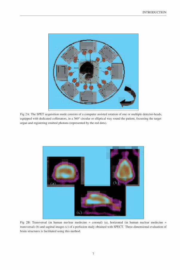
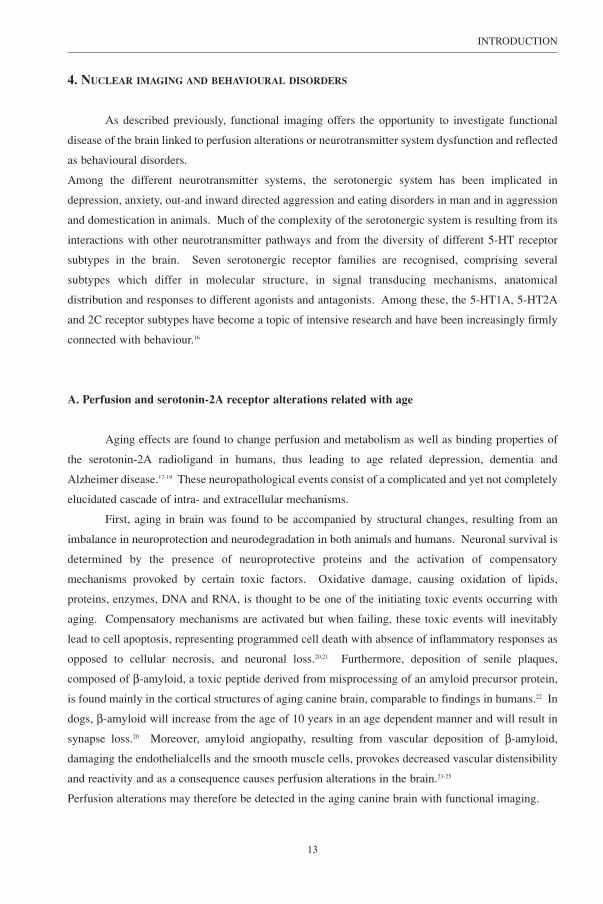
superimpose the region-of-interest. Principles of SPET imaging are illustrated in Figure 2A and 2B. It
is important to note the difference in nomenclature of the slice orientation used in this veterinary work,
compared to the nomenclature generally used in human nuclear medicine (Fig. 2B).
INTRODUCTION
7
Fig 2A: The SPET acquisition mode consists of a computer assisted rotation of one or multiple detector-heads,equipped with dedicated collimators, in a 360° circular or elliptical way round the patient, focussing the targetorgan and registering emitted photons (represented by the red dots).
Fig 2B: Transversal (in human nuvlear medecine = coronal) (a), horizontal (in human nuclear medecine =transversal) (b) and sagittal images (c) of a perfusion study obtained with SPECT. Three-dimensional evaluation ofbrain structures is facilitated using this method.

INTRODUCTION
8
Fig 3: Example of a total body bone scan of a dog, using the 99mTechnetium labelled methyl-diphosphonate tracerwith captation of radioactivity in the skeleton. Note the accumulation of radioactivity in the bladder (large arrow)and the site of the IV injection of the tracer (small arrow) and the presence of only one foreleg (the other wasamputated).
Selection of the radionuclide and its tracer are of major importance because they will determine
uptake in the target organ under investigation. As an example diphosphonates will be subject to
adsorption to the bone dependent on bone activity and therefore “isotope labelled diphosphonates” can
be used for investigating bone pathology involving increased bone metabolism. (Fig 3). Since the
application of brain imaging in veterinary nuclear medicine is new, the first part of this thesis is
dedicated to the investigation of radioligands, suitable for neuroimaging in dogs. In particular, the
99mTechnetium labelled N,N’’-1,2-ethylene-diylbis-L-cysteine diethyl ester dihydrochloride (ECD)
will be evaluated in a group of 10 normal dogs in Chapter 1 and the applicability of the specific 5-
HT2A receptor ligand 123I labelled R91150 for canine brain studies, will be determined in Chapter 3
and 4.

9
INTRODUCTION
C. Radioprotection: matter of concern
Radioprotection is an important issue to consider in both human as veterinary nuclear
medicine. As far as the animal is concerned, radiation hazards should not be overemphasized, since
life expectation precludes development of long term radiation effects. The most important measures
are aimed towards protection of the handler, owner and clinician. Hence, it is of importance that the
veterinary nuclear medicine physician informs staff and owners on these issues. First, since most
radiopharmaceuticals are eliminated by the liver and the kidneys, attention has to be paid to
contamination with faeces and urine. Hence, radioprotective measures are mainly directed to prevent
direct contact with contaminated fluids and therefore, gloves are worn. Second, distance from the
source, in this case the animal patient, is indicated, since the energy of radiation is inversely correlated
with distance to its source. If these radioprotective measures can not be guaranteed, the animal must
be kept in isolation in the medical imaging unit of the veterinary hospital until radiation hazards are
minimized. Concerning this matter, no general rules on a time-frame can be given since the half-life
(biological and physical) of the tracer and the injected dose have to be taken into account in
determining the amount of radiation and possible contamination in time.2 In this thesis the isotope
Technetium-99m (140keV) with a physical half-life of 6 hours was used for the perfusion studies. The
ligand used for the 5-HT2A receptor imaging was labelled with iodine-123 (160keV) which has a half-
life of 13 hours.

10
INTRODUCTION
Fig 4: Three views of a sculpture demonstrating the different modules of the working brain according to Gall.

11
INTRODUCTION
3. FUNCTIONAL BRAIN IMAGING
“MAPPING THE FUNCTIONING BRAIN”
A. History of brain-behaviour research
The brain is constructed of, and organized by a highly refined network of neurons and synapses
and is fueled by various neurotransmitters and neurohormones, in order to achieve three major goals:
basic survival, social interaction and the cognitive organization of living. The search to unravel brain
functions started early in man’s history. Medical case reports on brain pathology, written in 1700 BC,
were discovered in Luxor in 1862 by Edwin Smith. Hence, the knowledge that the “brain governed the
body”, was already introduced by the Egyptians. For a very long time, this statement formed the basis
of empirical neurosurgical strategies in behavioural pathologies that were mostly cruel and ineffective
at the same time.
In the realm of the emergence of criminology in the 19th century, and in an attempt to
understand the biological basis of deviant behaviour, “phrenology” became a separate science under
guidance of Franz Gall. His brain and skull maps were largely incorrect, but were the first attempt to
consider the brain as functionally inhomogeneous and consisting of functional “modules”. The
original phrenological concepts by Gall are depicted in Figure 4. By the end of the 19th century,
beginning of the 20th, more “sophisticated” investigational methods were used to map brain functions,
making an end to “phrenology”. First, case reports in humans on acquired brain lesions, accidentally
or disease-related, were the basis of a more correct definition of brain-behaviour relationship. In this
period, based on the post-mortem analysis of brains of patients who had cerebro-vascular accidents,
Broca and Wernicke were the first to correctly localize the language area in the brain, located in a
complete different area as was determined by the phrenologists. Further, with experiments such as
electrical stimulation in both man and animals, ablation studies and regional brain cooling in animals,
researchers tried to prove links between behavioural patterns and anatomical brain regions. This
research resulted in a rather premature introduction of this knowledge in the treatment of human
patients with behavioural disorders. Under impulse of Egas Moniz, frontal lobotomy was widely used
for patients with uncontrollable aggression. Paradoxically, Moniz was killed by one of his own
lobotomized patients. In that era, the neurosurgeon travelling between mental institutions and “treating
fourteen patients in one morning”, was a reality.
Only in the 1960s, with the emergence and the introduction of the first brain-dedicated
pharmaceuticals, a reasonable alternative was offered for lobotomy and electro-convulsion therapy.
Initially, serendipity lead to the awareness that certain neurotransmitters were involved in behavioural
and psychiatric disorders. Huge scientific efforts, often supported by pharmaceutical industry, lead to
the development of neuroleptics such as chlorpromazine, haloperidol and risperidone and anti-

12
INTRODUCTION
depressants such as imipramine and fluoxetine. Paradoxically, it was the application of these
psychotropics in the treatment of behavioural disorders, that finally offered a better insight in
neuroreceptor functioning and pathophysiology of behavioural and psychiatric disorders.
Recently, a tendency is set to turn away from the phenomenological approach, based on
nosological, i.e. syndromal clusters, towards a transnosological approach that is based on symptoms
that are apparent across nosological diagnostic categories. This approach proved to be more successful
in linking neuropathological and biochemical dysfunctions to behavioural symptoms and disorders.3 In
this context, “impulsivity” can be found as a common symptom in several psychiatric disorders, such
as personality disorders, eating disorders, impulsive behaviour disorders, suicide, and is consistently
brought in connection with a dysfunctional serotonergic system, throughout the diagnostic nosological
categories.4-8
B. Functional brain imaging with PET and SPET modalities
Since functional brain imaging allows the investigation of perfusion or metabolism and of brain
receptor status in vivo, a tendency to examine patients with behavioural disorders with positron
emission tomography (PET) and SPET modalities developed. Differences between these two
modalities will be discussed in Chapter 3. In this thesis, SPET is used for all investigation procedures.
First, to evaluate brain perfusion with SPET, two major radiopharmaceuticals are used: 99mTc-d,l-
hexamethyl-propylene-amine-oxime (HMPAO) and 99mTc-ECD. Due to their lipophilicity, both tracers
pass the blood brain barrier (BBB). They are incorporated into the brain cells and trapped by
conversion to hydrophilic compounds.9 With these compounds it is not only possible to evaluate and
delineate brain regions under resting conditions but also when activated during task performance in
normal and abnormal states, thus visualizing the “working” brain. Recently, the Ghent Molecular
Imaging Group developed a split-dose imaging paradigm to investigate patients before (rest condition)
and while (activation) undergoing cognitive challenge of the frontal cortex.10 Moreover, enhanced or
decreased perfusion can be demonstrated after pharmacological intervention.11,12 The important
advantage is that the functional state of the brain at the time of injection is “frozen” for some hours,
providing the possibility to perform the acquisition some time after injection, an advantage that is
exploited for visualization of epileptic foci during an epileptic fit.13,14 Second, research of the last 20
years has focussed on the development of specific radioligands to visualize and quantify different
neurotransmitter receptor systems in physiological and pathological conditions. Virtually all
neuroreceptor systems can be studied through a specific and receptor-dedicated radiolabelled tracer.15
Although the aetiology of disease is not addressed, functional imaging provides a mean to elucidate the
pathophysiology of behavioural disorders, to develop itself as a possible marker of a pathological
condition and to evaluate biochemical and physical receptor changes during treatment.
13
INTRODUCTION
4. NUCLEAR IMAGING AND BEHAVIOURAL DISORDERS
As described previously, functional imaging offers the opportunity to investigate functional
disease of the brain linked to perfusion alterations or neurotransmitter system dysfunction and reflected
as behavioural disorders.
Among the different neurotransmitter systems, the serotonergic system has been implicated in
depression, anxiety, out-and inward directed aggression and eating disorders in man and in aggression
and domestication in animals. Much of the complexity of the serotonergic system is resulting from its
interactions with other neurotransmitter pathways and from the diversity of different 5-HT receptor
subtypes in the brain. Seven serotonergic receptor families are recognised, comprising several
subtypes which differ in molecular structure, in signal transducing mechanisms, anatomical
distribution and responses to different agonists and antagonists. Among these, the 5-HT1A, 5-HT2A
and 2C receptor subtypes have become a topic of intensive research and have been increasingly firmly
connected with behaviour.16
A. Perfusion and serotonin-2A receptor alterations related with age
Aging effects are found to change perfusion and metabolism as well as binding properties of
the serotonin-2A radioligand in humans, thus leading to age related depression, dementia and
Alzheimer disease.17-19 These neuropathological events consist of a complicated and yet not completely
elucidated cascade of intra- and extracellular mechanisms.
First, aging in brain was found to be accompanied by structural changes, resulting from an
imbalance in neuroprotection and neurodegradation in both animals and humans. Neuronal survival is
determined by the presence of neuroprotective proteins and the activation of compensatory
mechanisms provoked by certain toxic factors. Oxidative damage, causing oxidation of lipids,
proteins, enzymes, DNA and RNA, is thought to be one of the initiating toxic events occurring with
aging. Compensatory mechanisms are activated but when failing, these toxic events will inevitably
lead to cell apoptosis, representing programmed cell death with absence of inflammatory responses as
opposed to cellular necrosis, and neuronal loss.20,21 Furthermore, deposition of senile plaques,
composed of β-amyloid, a toxic peptide derived from misprocessing of an amyloid precursor protein,
is found mainly in the cortical structures of aging canine brain, comparable to findings in humans.22 In
dogs, β-amyloid will increase from the age of 10 years in an age dependent manner and will result in
synapse loss.20 Moreover, amyloid angiopathy, resulting from vascular deposition of β-amyloid,
damaging the endothelialcells and the smooth muscle cells, provokes decreased vascular distensibility
and reactivity and as a consequence causes perfusion alterations in the brain.23-25
Perfusion alterations may therefore be detected in the aging canine brain with functional imaging.

14
INTRODUCTION
Second, aging is also accompanied by reduction of functioning of several neurotransmitter
systems, including the dopaminergic and the serotonergic system in animals and humans.18,26-36 In
humans decreased functioning of the serotonergic system is thought to be responsible for age-related
depression.17 Moreover, since the serotonergic system has an important role in the regulation of brain
microcirculation, dysfunction might lead to deficits in brain perfusion.37 Recently, the Ghent Molecular
Imaging Group demonstrated a reduction in brain serotonin-2A radioligand binding index in old
healthy volunteers and, to a larger extent, in patients with Alzheimer’s disease.38
Hence, concerning serotonine-2A receptor binding, one can expect that binding index
alterations may also be detected in the aging canine brain.
B. Impulsive behaviour
Impulsive behaviour, for animals defined as “incapacity to wait or to delay response”39 and for
humans as “acts related to inadequate self-control, impaired impulse control”39, result in reactions that
are “sudden” and “unpremeditated”40. This impulsive behaviour is often aggressive in nature and
therefore, has large impact on the victim and on the perpetrator.
It is important to define this “impulsive” behaviour in contrast to “normal” aggressive
behaviour in animals. Indeed, several types of aggression, such as maternal, intermale, territorial and
predatory aggression are described in animals, depending on different neuronal circuits and hormonal
mechanisms.41,42 These behavioural patterns are in-born and instinctive in nature and as such,
considered as normal coping activities to a set of environmental stimulus conditions. Therefore, they
are not pathological, although they can become beyond control, influenced by previous experiences or
situational determinants. Most of these reactions have been accepted by humans during domestication
of animals, as long as these reactions are appropriate in relation to the stimuli and as long as they can
be foreseen.43
Moreover, impulsive, aggressive behaviour can be of advantage under certain circumstances, such as
in a dangerous environment where quick reactions and high levels of arousal coupled to a high degree
of aggressivity are necessary as coping mechanisms. But, in safer habitats this behaviour will result in
unnecessary attacks leading to injury.44 Extensive research has been performed on impulsivity, pointing
at the frontal cortex and subcortical structures as the structural anatomical substrate and at lowered
activity of the serotonergic system as the biochemical substrate for impulsive behaviour.
First, studies on the anatomical and structural substrate of these impulsive disinhibited
disorders were based on acquired brain damage in humans and on ablation studies in animals. Phineas
Gage was one of the first clinical proves that the frontal cortex played an important, if not major role
in the expression of this abnormal, disinhibited behaviour.45 This previously conscientious railway
worker turned into an impulsive, aggressive drunk after an accident with a metal rod that perforated his

15
INTRODUCTION
left fronto-cortical lobe. This was later confirmed with frontal ablation studies in several animal
species, demonstrating that the frontal cortex exerted control over limbic drives of hunger and
aggression.46 Summarized, the frontal cortex will generate an adequate, premeditated response based
on the association of the limbic and sensoric information guided by possible previous experiences,
thereby permitting acceptable or “in-context” necessary reactions or inhibiting certain inappropriate
reactions.
In humans disturbed frontal perfusion and metabolism was demonstrated in murderers and aggressive
individuals with SPET47,48 and altered metabolism was found with PET49-51. It is important to notice that
in these cases disturbed frontal perfusion patterns were present without concurring structural
anatomical brain abnormalities, as was proven with CT or MRI and therefore represented true
functional perfusion and metabolism deficits.
No comparable functional imaging studies in animals are present at this moment. It can be
hypothesized that comparable functional deficits in the frontal brain structures of dogs with impulsive
behavioural disorder exist.
Second, studies on the functional biochemical deficits in the brain of impulsive subjects
pointed at the involvement of a deficient serotonergic system in impulsive behaviour.6,39,52-66 This is one
of the most replicated findings in biological psychiatry in human medicine. But also studies in other
species demonstrated a prominent role of serotonin in social adequate and inadequate behaviour.67-75
The cell bodies of the serotonergic neurons are located in the raphe nucleus, with widespread
projections to the limbic system, hypothalamus and the cortical regions, explaining their link with
mood and behavioural disorders. The influence of the serotonergic system on behaviour in animals and
man has been investigated with direct and indirect studies. A decreased metabolism, demonstrated by
decreased amounts of 5-hydroxy indolic acetic acid (5-HIAA, the principal metabolite of 5-HT) in
cerebrospinal fluid (CSF) was found both in humans as in animals with impulsive, aggressive and self-
injurious behaviour.6,60,61,69-72,77-80 Dietary tryptophan depletion increased aggressiveness and tryptophan
supplementation decreased aggressiveness in both animals and humans.57,63,68,81 Using pharmacological
probes, affecting the serotonin transporter mechanism and/or the receptor, aggressive behaviour both
in animals and humans could be influenced.67,82-88 More specific studies on the 5-HT2A receptor, using
measurements of 5-HT2A receptors density on platelets, demonstrated increases in binding index in
suicidal patients and in patients with aggressive personality disorders.54,66,89-92 Direct autoradiographic
studies showed increased density of 5-HT2A receptors in patients, committed suicide.73,93,94
Since the development of specific receptor radioligands for functional imaging, using SPET or
PET, these methods have received increasing attention in research on the serotonergic system in vivo,
under physiological and pathological conditions, with and without pharmacological interventions.
Concerning SPET imaging, a relatively new radioligand 123I-5-I-R91160 has been used to investigate
the serotonin-2A receptor status in normal volunteers.26,95 Recently, the Ghent Molecular Imaging
Group investigated human patients with recent suicide attempts8 and patients with eating disorders7

16
INTRODUCTION
and found a decreased cortical serotonin-2a binding index. No comparable functional imaging studies
on the serotonin-2A receptor in animals are present at this moment. One could expect comparable
functional deficits in the cortical brain structures of dogs with impulsive behavioural disorder.

17
INTRODUCTION
REFERENCES
1. Karli H. On the affective nature of human nature: a neurobiologist's reflections. In: Haug, M and Whalen, R.,ed. Animal models of human emotion and cognition. Washington DC: Am Psych Ass, 1999; 41-56.
2. Perkins AC. Nuclear Medicine, science and safety. London: John Libbey @ company Ltd, 1996.
3. Audenaert K. Functional neuroimaging in psychiatry: a psychopathological approach. Faculty of Medicine &Health Sciences, University Ghent2001 (PhD dissertation)
4. Nordstrom P, Asberg M. Suicide risk and serotonin. Int Clin Psychopharmacol 1992; 6[Suppl 6]: 12-21.
5. Virkkunen M, Kallio E, Rawlings R, Tokola R, Poland RE, Guidotti A, Nemeroff C, Bissette G, Kalogeras K,Karonen SL, . Personality profiles and state aggressiveness in Finnish alcoholic, violent offenders, fire setters,and healthy volunteers. Arch Gen Psychiatry 1994; 51: 28-33.
6. Virkkunen M, Goldman D, Nielsen D, Linnoila M. Low brain serotonin turnover rate (low CSF 5-HIAA) andimpulsive violence. J Psychiatry Neurosci 1995; 20: 271-275.
7. Audenaert K, Van Laere K, Dumont F, Vervaet M, Goethals I, Slegers G, Mertens J, van Heeringen C, DierckxR. Decreased 5-HT2A binding in patients with anorexia nervosa. J Nucl Med 2002; in press
8. Audenaert K, Van Laere K, Dumont F, Slegers G, Mertens J, van Heeringen C, Dierckx R. Decreased frontalserotonin 5-HT2a receptor binding index in deliberate self harm patients. Eur J Nucl Med 2001; 28: 175-182.
9. Leveille J, Demonceau G, Walovitch RC. Intrasubject comparison between Technetium-99m-ECD andTechnetium-99m- HMPAO in healthy human subjects. J Nucl Med 1992; 33: 480-484.
10. Audenaert K, Brans B, Van Laere K, Lahorte P, Versijpt J, van Heeringen C, Dierckx R. Verbal fluency as aprefrontal activation probe: a validation study using 99m-Tc-ECD brain SPECT. Eur J Nucl Med 2000; 27:1800-1808.
11. Susskind H, Weber DA, Ivanovic M, Wong CTC, DeHaan CE, Gavin PR. Quantitative 123I IMP and 99mHMPAOimaging in the dog following cocaine administration. Nucl Med biol 1996; 23: 343-352.
12. Cook EH, Jr., Metz J, Leventhal BL, Lebovitz M, Nathan M, Semerdjian SA, Brown T, Cooper MD.Fluoxetine effects on cerebral glucose metabolism. Neuroreport 1994; 5: 1745-1748.
13. Sadzot B, Debets R, Franck G. Biochemical and functional imaging for adult partial epilepsy: PET or SPECT.In: De Deyn, P. P., Dierckx, R., Alavi, A, and Pickut, B. A., ed. A textbook of SPECT in neurology andpsychiatry. London: John Libbey, 1997; 207-217.
14. Menzel C, Grünwald F, Hufnagel A, Pavics L, Reichman K, Ruhlman J, Elger C, Biersack H. Functionalneuroimaging with CGU-PET and rCBF-SPECT: targeting the epileptogenic focus. In: De Deyn, P. P.,Dierckx, R., Alavi, A, and Pickut, B. A., ed. A textbook of SPECT in neurology and psychiatry. London: JohnLibbey, 1997; 259-265.
15. Halldin C, Gulyas B, Langer O, Farde L. Brain radioligands-state of art and new trends. Q J Nucl Med 2001;45: 139-152.
16. Barnes NM, Sharp T. A review of central 5-HT receptors and their function. Neuropharmacology 1999; 38:1083-1152.
17. Nobler MS, Mann JJ, Sackeim HA. Serotonin, cerebral blood flow and cerebral metabolic rate in the geriatricmajor depression and normal aging. Brain Res Reviews 1999; 30: 250-263.
18. Meltzer C, Smith G, DeKosky S, Pollock B, Mathis C, Moore R, Kupfer D, and Reynolds C. Serotonin inaging, late life depression and Alzheimer's disease: the emerging role of functional imaging. 1998; 18: 407-430.
19. Moeller JR, Ishikawa T, Dhawan V, Spetsieris P, Mandel F, Alexander GE, Grady C, Pietrini P, Eidelberg D.The metabolic topography of normal aging. J Cereb Blood Flow Metab 1996; 16: 385-398.

18
INTRODUCTION
20. Head E, Thornton P, Tong L, Cotman C. Initiation and propagation of molecular cascades in human brainaging: insight from the canine model to promote successful aging. Prog Neuropsychopharmacol BiolPsychiatry 2000; 24: 777-786.
21. Kiatipattanasakul, W., Nakamura, S., Hossain, M., Nakayama, H., Uchino, T., Shumiya, S., Goto, N., and Doi,K. Apoptosis in the aged dog brain. Acta neuropathol 1996; 92: 242-248.
22. Hou Y, White RG, Bobik M, Marks JS, Russell MJ. Distribution of beta-amyloid in the canine brain.Neuroreport 1997; 8: 1009-1012.
23. Kawai M, Kalaria RN, Cras P, Siedlak SL, Velasco ME, Shelton ER, Chan HW, Greenberg BD, Perry G.Degeneration of vascular muscle cells in cerebral amyloid angiopathy of Alzheimer disease. Brain Res 1993;623: 142-146.
24. Prior R, D'Urso D, Frank R, Prikulis I, Pavlakovic G. Loss of vessel wall viability in cerebral amyloidangiopathy. Neuroreport 1996; 7: 562-564.
25. Thomas T, Thomas G, McLendon C, Sutton T, Mullan M. beta-Amyloid-mediated vasoactivity and vascularendothelial damage. Nature 1996; 380: 168-171.
26. Baeken C, D'haenen H, Flamen P, Terriere D, Chavatte K, Boumon R, Bossuyt A. 123I-5-I-R91150, a new singlephoton emission tomography ligand for 5-HT2A receptors: influence of age and gender in healthy subjects.Eur J Nucl Med 1998; 25: 1617-1622.
27. Gozlan H, Daval G, Verge D, Spampinato U, Fattaccini C, Gallissot M, El Mestikawy S, Hamon M. Agingassociated changes in serotonergic and dopaminergic pre-and postsynaptic neurochemical markers in the ratbrain. Neurobiol. Aging 1990; 11: 437-449.
28. Kakiuchi T, Nishiyama S, Sato K, Ohba H, Nakanishi S, Tsukada H. Age related reduction of {11C} MDL100,907 binding to central 5-HT2A receptors: PET study on the conscious monkey brain. Brain Res 2000; 883:135-142.
29. Robson L, Gower AJ, Kendall DA, Marsden CA. Age related behavioural, neurochemical and radioligandbinding changes in the central 5-HT system of Sprague-Dawley rats. Psychopharmacol 1993; 113: 274-281.
30. Wang G, Volkow ND, Logan J, Fowler JS, Schlyer DJ, Macgreggor RR, Hitzemann R, Gur R, Wolf AP.Evaluation of age-related changes in serotonin 5-HT2 and dopamine D2 receptor availibility in healthy humansubjects. Life Sci 1995; 56: 249-253.
31. Volkow ND, Ding Y, Fowler JS, Wang G, Logan J, Gatley SJ, Hitzemann R, Smith, G, Fields SD, Gur R.Dopamine transporters decrease with age. J Nucl Med 1996; 37: 554-559.
32. Volkow ND, Logan J, Fowler JS, Wang GJ, Gur RC, Wong C, Felder C, Gatley SJ, Ding YS, Hitzemann R,Pappas N. Association between age-related decline in brain dopamine activity and impairment in frontal andcingulate metabolism. Am J Psychiatry 2000; 157: 75-80.
33. Wong DF, Young D, Wilson PD, Meltzer CC, Gjedde A. Quantification of neuroreceptors in the living humanbrain:III. D2-like dopamine receptors: theory, validation, and changes during normal aging. J Cereb Blood Flow Metab 1997; 17: 316-330.
34. Rosier A, Dupont P, Peuskens J, Bormans G, Vandenberghe R, Maes F, Schiepers C, Verbruggen A,Mortelmans, L. Visualization of loss of 5-HT2A receptors with age in healthy using (18F) altanserin andpositron emission imaging. Psychiatry Res 1996; 25: 11-22.
35. Morris ED, Chefer SI, Lane MA, Muzic RF, Wong DF, Dannals RF, Matochik JA, Bonab AA, Villemagne V,Grant SJ, Ingram DK, Roth GS, London ED. Loss of D2 receptor binding with age in Rhesus monkeys:importance of correction for differences in striatal size. J Cereb Blood Flow Metab 1999; 19: 218-229.
36. Wong DF, Wagner HN, Dannals RF, Links JM, Frost JJ, Ravert HT, Wilson AA, Rosenbaum AE, Gjedde A,Douglas KH, Burns HD, Kuhar MJ. Effects of age on dopamine and serotonin receptors measured by positrontomography in the living human brain. Science 1984; 226: 1393-1396.

19
INTRODUCTION
37. Cohen Z, Bonvento G, Lacombe P, Hamel, E. Serotonin in the regulation of brain microcirculation. ProgNeurobiol 1996; 50: 335-362.
38. Versijpt J, Van Laere K, Dumont F, Decoo D, Vandecapelle M, Santens P, Goethals I, Audenaert K, Slegers G,Dierckx R, Korf J. Imaging of the 5-HT2A system: age-, gender-, and Alzheimer's disease-related findings.Neurobiol Aging 2002; in press.
39. Plutchik R, Van Praag H. The Nature of Impulsivity: Definitions, Ontology, Genetics, and Relations toaggression. In: Hollander, E. and Stein, D., ed. Impulsivity and aggression. New York: John Wiley and Sons,1995; 7-24.
40. Merriam-Webster Editorial Staff. Merriam-Webster's Collegiate Dictionary. Springfield: Merriam WebsterInc, 1999.
41. Olivier B, Mos J, Van Oorschot R, Hen R. Serotonin receptors and animal models of aggressive behavior.Pharmacopsychiat 1995; 28: 80-90.
42. Volavka J. Aggression among animals. In: Volavka, J., ed. Neurobiology of violence. Washington DC:American Psychiatric Press, Inc., 1995; 21-48.
43. Volavka J. The neurobiology of violence: an update. J Neuropsychiatry Clin Neurosc 1999; 11: 307-314.
44. Eichelman B. Animal models and evolutionary models of impulsive aggression. In: Hollander, E. and Stein,D., ed. Impulsivity and aggression. Chichester: John Wiley & Sons Ltd, 1995; 59-70.
45. Damasio H, Grabowski T, Frank R, Galaburda A, Damasio A. The return of Phineas Gage: clues about thebrain from the skull of the famous patients. Science 1994; 264: 1102-1105.
46. Fuster J. Animal neurophysiology. In: Fuster, J., ed. The prefrontal cortex: anatomy, physiology andneuropsychology of the frontal lobe. Philadelphia: Lippincott-Raven, 1997; 66-101.
47. Raine A, Buchsbaum M, Lacasse L. Brain abnormalities in murderers indicated by positron emissiontomography. Biol.Psychiatry 1997; 42: 495-508.
48. Amen D, Stubblefield M, Carmichael B, Thisted R. Brain SPECT findings and aggressiveness. Ann Clin Psych1996; 8: 129-137.
49. Volkow ND, Tancredi LR, Grant C, Gillespie H, Valentine A, Mullani N, Wang GJ, Hollister L. Brain glucosemetabolism in violent psychiatric patients: a preliminary study. Psychiatry Res 1995; 61: 243-253.
50. Miller BL, Darby A, Benson DF, Cummings JL, Miller MH. Aggressive, socially disruptive and antisocialbehaviour associated with fronto-temporal dementia. Br J Psychiatry 1997; 170: 150-154.
51. Hirono N, Mega MS, Dinov ID, Mishkin F, Cummings JL. Left frontotemporal hypoperfusion is associatedwith aggression in patients with dementia. Arch Neurol 2000; 57: 861-866.
52. Coccaro EF, Siever LJ, Klar HM, Maurer G, Cochrane K, Cooper TB, Mohs RC, Davis KL. Serotonergicstudies in patients with affective and personality disorders: correlates with suicidal and impulsive aggressivebehavior. Arch Gen Psychiatry 1989; 46: 587-599.
53. Kavoussi R, Armstead P, Coccaro E. The neurobiology of impulsive aggression. Psychiatr Clin North Am1997; 20: 395-403.
54. Coccaro EF, Kavoussi RJ, Sheline YI, Berman ME, Csernansky JG. Impulsive aggression in personalitydisorder correlates with platelet 5- HT2A receptor binding. Neuropsychopharmacology 1997; 16: 211-216.
55. Coccaro EF. Impulsive aggression and central serotonergic system function in humans: an example of adimensional brain-behavioral relationship. Int Clin Psychopharmacol 1992; 7: 3-12.
56. Placidi GP, Oquendo MA, Malone KM, Huang YY, Ellis SP, Mann JJ. Aggressivity, suicide attempts, anddepression: relationship to cerebrospinal fluid monoamine metabolite levels. Biol Psychiatry 2001; 50: 783-791.

20
INTRODUCTION
57. LeMarquand DG, Pihl RO, Young SN, Tremblay RE, Seguin JR, Palmour RM, Benkelfat C. Tryptophandepletion, executive functions, and disinhibition in aggressive, adolescent males. Neuropsychopharmacology1998; 19: 333-341.
58. Young SN, Pihl RO, Benkelfat C, Palmour R, Ellenbogen M, Lemarquand D. The effect of low brain serotoninon mood and aggression in humans. Influence of baseline mood and genetic factors. Adv.Exp Med Biol 1996;398: 45-50.
59. Manuck SB, Flory JD, McCaffery JM, Matthews KA, Mann JJ, Muldoon MF. Aggression, impulsivity, andcentral nervous system serotonergic responsivity in a nonpatient sample. Neuropsychopharmacology 1998;19: 287-299.
60. Linnoila M, Virkkunen M, Scheinin M, Nuutila A, Rimon R, Goodwin FK. Low cerebrospinal fluid 5-hydroxyindolacetic acid concentration differentiates impulsive form nonimpulsive violent behavior. Life Sci1983; 33: 2609-2614.
61. Cremniter D, Jamain S, Kollenbach K, Alvarez JC, Lecrubier Y, Gilton A, Jullien P, Lesieur P, Bonnet F,Spreux-Varoquaux O. CSF 5-HIAA levels are lower in impulsive as compared to nonimpulsive violent suicideattempters and control subjects. Biol Psychiatry 1999; 45: 1572-1579.
62. Bjork JM, Moeller FG, Dougherty DM, Swann AC, Machado MA, Hanis CL. Serotonin 2a receptor T102Cpolymorphism and impaired impulse control. Am J Med Genet. 2002; 114: 336-339.
63. Bjork JM, Dougherty DM, Moeller FG, Swann AC. Differential behavioral effects of plasma tryptophandepletion and loading in aggressive and nonaggressive men. Neuropsychopharmacology 2000; 22: 357-369.
64. Dolan M, Anderson IM, Deakin JF. Relationship between 5-HT function and impulsivity and aggression inmale offenders with personality disorders. Br J Psychiatry 2001; 178: 352-359.
65. Stoff DM, Pastiempo AP, Yeung JH, Cooper TB, Bridger WH, Rabinovich H. Neuroendocrine responses tochallenge with d,l-fenfluarmine and agression in disruptive behavior disorders of children and adolescents.Psychiatry Res 1991; 43: 263-276.
66. Rao ML, Hawellek B, Papassotiropoulos A, Deister A, Frahnert C. Upregulation of the platelet Serotonin2Areceptor and low blood serotonin in suicidal psychiatric patients. Neuropsychobiology 1998; 38: 84-89.
67. Dodman NH, Donnelly R, Shuster L, Mertens P, Rand W, Miczek K. Use of fluoxetine to treat dominanceaggression in dogs. J Am Vet.Med Assoc. 1996; 209: 1585-1587.
68. DeNapoli JS, Dodman NH, Shuster L, Rand WM, Gross KL. Effect of dietary protein content and tryptophansupplementation on dominance aggression, territorial aggression, and hyperactivity in dogs. J Am Vet.MedAssoc. 2000; 217: 504-508.
69. Mehlman PT, Higley JD, Faucher I, Lilly AA, Taub DM, Vickers J, Suomi SJ, Linnoila M. Low CSF 5-HIAAconcentrations and severe aggression and impaired impulse control in non human primates. Am J Psychiatry1994; 151: 1485-1491.
70. Westergaard GC, Suomi SJ, Higley DE, Mehlman PT. CSF 5-HIAA and aggression in female macaquemonkeys: species and interindividual differences. Psychopharmacology-Berl 1999; 146: 440-446.
71. Higley JD, Mehlman PT, Taub DM, higley SB, Suomi SJ, Linnoila M, Vickers JH. Cerebrospinal fluidmonoamine and adrenal correlates of aggression in free-ranging rhesus monkeys. Arch Gen Psychiatry 1992;49: 436-441.
72. Reisner IR, Mann JJ, Stanley M, Huang Y, Houpt KA. Comparison of cerebrospinal fluid monoaminemetabolite levels in dominant-aggressive and non- aggressive dogs. Brain Res 1996; 714: 57-64.
73. Popova NK, Kulikov AV, Nikulina EM, Kozlachkova EY, Maslova GB. Serotonin metabolism and serotonergicreceptors in Norway rats selected for low aggressiveness towards man. Aggress Behav 1991; 17: 207-213.

21
INTRODUCTION
74. Popova NK, Voitenko NN, Kulikov AV, Avgustinovich DF. Evidence for the involvement of central serotoninin mechanism of domestication of silver foxes. Pharmacol Biochem Behav 1991; 40: 751-756.
75. Popova NK, Voitenko NN, Trut LN. Changes in the content of serotonin and 5-hydroxyindoleacetic acid in thebrain in the selection of silver foxes according to behavior. Neurosci Behav Physiol 1976; 7: 72-74.
76. Stahl S. Essential psychopharmacology. Cambridge: Cambridge University Press, 1996.
77. Constantino JN, Morris JA, Murphy DL. CSF 5-HIAA and family history of antisocial personality disorder innewborns. Am J Psychiatry 1997; 154: 1771-1773.
78. Brown GL, Linnoila M. CSF serotonin metabolite (5-HIAA) studies in depression, impulsivity, and violence.J Clin Psychiatry 1990; 51: (suppl 4) 31-41.
79. Soderstrom H, Blennow K, Manhem A, Forsman A. CSF studies in violent offenders. I. 5-HIAA as a negativeand HVA as a positive predictor of psychopathy. J Neural Transm 2001; 108: 869-878.
80. Virkkunen M, Rawlings R, Tokola R, Poland RE, Guidotti A, Nemeroff C, Bissette G, Kalogeras K, KaronenSL, Linnoila M. CSF biochemistries, glucose metabolism, and diurnal activity rhythms in alcoholic, violentoffenders, fire setters, and healthy volunteers. Arch Gen Psychiatry 1994; 51: 20-27.
81. Chamberlain B, Ervin FR, Pihl RO, Young SN. The effect of raising or lowering tryptophan levels onaggression in vervet monkeys. Pharmacol.Biochem.Behav 1987; 28: 503-510.
82. Olivier B, Mos J. Rodent models of aggressive behavior and serotonergic drugs. Prog NeuropsychopharmacolBiol Psychiatry 1992; 16: 847-870.
83. Evenden JL. The pharmacology of impulsive behaviour in rats VII: the effects of serotonergic agonists andantagonists on responding under a discrimination task using unreliable visual stimuli. Psychopharmacology(Berl) 1999; 146: 422-431.
84. Fairbanks L, Melega W, Jorgensen M, Kaplan J, McGuire M. Social impulsivity inversely associated with CSF5-HIAA and fluoxetine exposure in vervet monkeys. Neuropsychopharmacology 2001; 24: 370-378.
85. Coccaro EF, Kavoussi RJ, Hauger RL. Serotonin function and antiaggressive response to fluoxetine: a pilotstudy. Biol Psychiatry 1997; 42: 546-552.
86. Medeiros JM, Silva CM, Sougey EB, Costa JA, Castro CM, Castro RM. Action of selective serotonin reuptakeinhibitor on aggressive behavior in adult rat submitted to the neonatal malnutrition. Arq Neuropsiquiatr. 2001;59: 499-503.
87. Raleigh MJ. Differential behavioral effects of tryptophan and 5-hydroxytryptophan in vervet monkeys:influence of catecholaminergic systems. Psychopharmacology (Berl) 1987; 93: 44-50.
88. Botchin MB, Kaplan JR, Manuck SB, Mann JJ. Low versus high prolactin responders to fenfluraminechallenge: marker of behavioral differences in adult male cynomolgus macaques. Neuropsychopharmacology1993; 9: 93-99.
89. Pandey GN, Pandey SC, Dwivedi Y, Sharma V, Janicak PG, Davis JM. Platelet serotonin-2A receptors: apotential biological marker for suicidal behavior. Am J Psychiatry 1995; 152: 850-855.
90. Biegon A, Essar N, Israeli M, et al. Serotonin 5HT2 receptor binding on blood platelets as a state dependentmarker in major affective disorder. Psychopharmacology (Berl) 1990; 102: 73-75.
91. Biegon A, Weizman A, Karp L, Ram A, Tiano S, Wolff M. Serotonin 5-HT2 receptor binding on bloodplatelets: a peripheral marker for depression? Life Sci 1987; 41: 2485-2492.
92. Alda M, Hrdina PD. Distribution of platelet 5-HT(2A) receptor densities in suicidal and non-suicidaldepressives and control subjects. Psychiatry Res 2000; 94: 273-277.
93. Arango V, Underwood MD, Gubbi AV, Mann JJ. Localized alterations in pre- and postsynaptic serotoninbinding sites in the ventrolateral prefontal cortex of suicide victims. Brain Res 1995; 688: 121-133.

22
INTRODUCTION
94. Arango V, Ernsberger P, Marzuk PM. Autoradiographic demonstration of increased serotonin 5HT2 and beta-adrenergic receptor binding sites in the brain of suicide victims. Arch Gen Psychiatry 1990; 47: 1038-1044.
95. Busatto GF, Pilowsky LS, Costa DC, Mertens J, Terriere D, Ell PJ, Mulligan R, Travis MJ, Leysen JE, Lui D,Gacinovic S, Waddington W, Lingford-Hughes A, Kerwin RW. Initial evaluation of 123I-5-I-R91150, a selective5-HT2A ligand for single photon emission tomography in healthy subjects. Eur J Nucl Med 1997; 24:119-124.

SCIENTIFIC AIMS


25
SCIENTIFIC AIMS
Research on the pathophysiology of abnormal canine behaviour in vivo is scarce due to the fact
that objective measurement of behaviour is difficult. The only available way to investigate the
disordered animal is by means of behavioural testing and elaborated questionnaires, which are no doubt
of significant importance but remain subjective (operator/owner dependent). It was the aim of this
study to evaluate the applicability of functional imaging of the brain in the dog, i.e. to evaluate this
technique as a tool to visualize and measure perfusion and the 5-HT2A receptor in normal dogs, aging
dogs and in dogs showing impulsive aggressive behaviour.
In this work we tried to address the following questions:
1. Can we measure canine brain perfusion with functional brain imaging, using the SPET
modality, and what is the normal distribution pattern in dogs without neurological or
behavioural disorders?
2. What are the technical issues to consider when performing receptor radioligand
imaging studies, using the SPET modality?
3. Is it possible to use the radioligand, 123I-5-I-R91150 to image and quantify the 5-HT2A
receptor in canine brain?
4. What is the normal distribution pattern of this receptor in the brain of dogs without
neurological or behavioural disorders?
5. What is the influence of age on brain perfusion and binding characteristics of the
specific serotonin-2A radioligand 123I-5-I-R91150 in normal aging canine brain?
6. Can we include impulsive aggressive behaviour in dogs as a clinical behavioural
disorder to investigation with this imaging modality?
7. Is it possible to demonstrate differences in brain perfusion and/or serotonin-2A
radioligand binding using SPET, between normal dogs and dogs showing impulsive,
aggressive behaviour?
Because information on the use of this modality for brain examinations in dogs is scarce, the
first part of this study consisted of the acquirement of normal databases of both perfusion and 5-HT2A
receptor ligand in normal adult individuals.
To measure brain perfusion, 99mTc-ECD was chosen as radiopharmaceutical. First, it is 99m-Technetium
labelled, a label generally preferred in nuclear medicine because of its excellent physical characteristics
for imaging (140 keV; T1/2 = 6 hours) and since it is relatively cheap and readily available from “on
site” 99Mo/99mTc generators. It also has superior imaging qualities compared to 99mTc-HMPAO.
Concerning the use of the radiopharmaceutical 99mTc-ECD in dogs, we did not perform elaborate
biodistribution studies, because it was already shown in a previous preclinical feasibility study
concerning the use of ECD in several species, that this tracer achieved low, but sufficient brain

26
SCIENTIFIC AIMS
penetration in the dog (1.2% ID) lasting 2.5 hours. However, we determined the time to achieve a
stable plateau for brain concentration after injection of the radiopharmaceutical in two dogs and found
it was as fast as 2 minutes (Fig.1) and a maximal brain concentration of 1,5% ID was registered.
Fig 1: 99mTc-ECD cerebral uptake curve in function of time in a normal dog. Dynamic frames of 6 seconds wereacquired for a period of 8 minutes. Brain uptake of the radiopharmaceutical stabilizes before 2 minutes post-injection.
The regional brain perfusion, using 99mTc-ECD and SPET, in 10 normal dogs, aged between 1
and 9 years old, including 5 males and 5 females, is determined in Chapter 1. In the next study,
described in brief in Chapter 2, a template is generated with accompanying region map, from the brain
perfusion data of 12 normal dogs (6 males and 6 females), aged younger than 8 years. This computer
operated procedure facilitates the fitting procedure of the individual patient data to the normal data base
and eliminates operator bias when determining regional activity with manually drawn VOI’s. In
Chapter 3 an overview is given on the imaging techniques and characteristics of radioligands, used for
neuroreceptor imaging. The differences between positon emission tomography (PET) and single
photon emission tomography (SPET) are briefly outlined in this chapter. The essential properties of
the radioligands and the major difficulties encountered in their development are discussed. This review
on the technical aspects is followed by a literature overview on the application of neuroreceptor
imaging in research on animal models.

27
SCIENTIFIC AIMS
Concerning the particular serotonin-2A receptor radioligand, 123I-5-I-R91150, a biodistribution study
had to be performed, because although the tracer has been used in humans, primates and rats, nothing
is known about its applicability in the dog. The specificity and reversibility of radioligand binding and
the optimal scanning time are evaluated in Chapter 4. In Chapter 5, the regional distribution pattern of
this serotonin-2A radioligand is evaluated in 10 normal dogs, 5 males and 5 females, age ranging from
1 to 9 years. Ample evidence exists from literature that age plays a significant role in both perfusion
and serotonin-2A receptor density. Therefore we investigate age influences in a larger group of
animals, in Chapter 6. The subjects are divided into two age categories. One group consists of dogs
younger than 96 months (N=12, 6 males and 6 females) and the other includes individuals aged
96 months and older (N=12, 5 males and 7 females).
Since persuasive evidence is provided in literature that impulsive, aggressive behaviour is
associated with altered brain functioning and involvement of the serotonergic system (Chapter 7), brain
perfusion and binding properties of the serotonin-2A radioligand are estimated in impulsive aggressive
dogs and compared with the data obtained from normal reference dogs (Chapter 8). The selection of
these dogs is based on a questionnaire, compiled from information found in literature and further
adapted towards recognition of impulsive individuals.


CHAPTER 1
Regional brain perfusion in 10 normal dogs measured usingTechnetium-99m-ethyl cysteinate dimer SPECT
K. Peremans1, P. De Bondt2, K. Audenaert2, K. Van Laere2, I.Gielen1,
M. Koole3, J. Versijpt2, H.Van Bree1, F.Verschooten1, R. Dierckx2
1Department of Medical Imaging, Faculty of Veterinary Medicine, Ghent University,
Salisburylaan 133, B-9820 Merelbeke, Belgium2Division of Nuclear Medicine Ghent University Hospital, De Pintelaan 185, B-9000 Gent,
Belgium3Institute for Nuclear Sciences, Ghent University, Proeftuinstraat 86, B-9000 Ghent,
Belgium
'
Adapted from: Veterinary Radiology & Ultrasound 2001; 42: 562-568


31
Chapter 1: Brain perfusion SPECT in normal dogs
Summary
Single photon emission computed tomography (SPECT) of the brain using perfusion tracers
allows estimation of regional brain perfusion. This allows in vivo examination of brain function in the
setting of neuropsychological and pathophysiological changes. However functional imaging data on
brain perfusion in dogs are limited.
Hence, the aim of this study was to determine the scintigraphic regional perfusion pattern of
the normal canine brain. Ten healthy shepherd type dogs were injected with 925 MBq Technetium-
99m ethyl cysteinate (ECD) 20 minutes prior to the examination. Acquisition was performed using a
triple head gamma camera equipped with fanbeam collimators. Uniform attenuation correction and
triple energy window correction were applied. Computed tomographic images were obtained from the
same dogs, reoriented along the orbito-meatal axis and SPECT perfusion data were coregistered to the
CT-volume data. Based on a morphological and suggested brain divisions, regions-of-interest (ROIs)
were defined for the bilateral frontocerebral, temporocerebral, parietocerebral, occipitocerebral,
cerebellar, and subcortical area. Regional count density was normalized on total counts. All dogs had
the highest uptake in the thalamic/striatal area compared to a rather homogeneous cerebral uptake. No
significant left/ right count differences were found, but a rostro-caudal gradient (+ 12-13%) was
present. In this group, age and gender did not influence the perfusion pattern.


33
Chapter 1: Brain perfusion SPECT in normal dogs
Introduction
Computed tomography (CT) and magnetic resonance imaging (MR) are the diagnostic imaging
techniques for evaluating structural brain pathology, while single photon emission computed
tomography (SPECT) and PET (positron emission tomography) enable three-dimensional visualisation
of functional parameters reflecting cerebral blood flow, brain metabolism and binding potential of
receptor ligands. SPECT perfusion imaging using 99mTechnetium labelled radiopharmaceuticals has
become a routine procedure in human nuclear medicine, enabling visualisation of regional blood flow
and under normal coupling also of brain metabolism. It is interesting to note that the first study that
demonstrated the coupling of cerebral perfusion and metabolism in the dog was already published in
1890.1
While 99mTc-HMPAO (hexamethylpropylene amine oxime) was the first 99mTc labelled tracer to
be commercialised for evaluation of rCBF, 99mTc-ECD (ethylcysteinate dimer) is a relatively new
lipophilic tracer with a faster blood clearance and chemically more stable than 99mTc-HMPAO. In the
human brain, the lipophilic 99mTc-ECD crosses the intact blood brain barrier (BBB) and is trapped
intracellular within 2 minutes in proportion to the cerebral blood flow, probably by de-esterification to
polar complexes.2,3 The optimal imaging time is between 30-120 min. post injection (p.i.) in humans,
but acquisition may be started as early as 10 min.4 No redistribution of the tracer occurs and the uptake
and distribution, which are proportional to rCBF, remain unchanged for two hours. Regional grey-to-
white matter contrast begins to decrease after 2 hours. Redistribution (difference in grey matter activity
pattern) starts from 4 hours onwards and is region dependent.3
Currently accepted clinical indications for SPECT functional brain imaging in humans are
diagnosis and follow-up of various neurological disorders, such as dementia, epilepsy and
cerebrovascular disorders. In psychiatry, brain SPECT especially using receptor-ligands, may be
considered a promising tool.5-7 In addition to studies in basal conditions, activation studies using
specific paradigms may be applied.8,9 These activation tasks generate detectable and reproducible
regional CBF changes during task performance.10
The radiopharmaceuticals used for conventional functional brain imaging of small animals
remain confined to the blood pool in the presence of an intact BBB, only diffusing into the parenchyma
when pathology disrupts this barrier. Classically 99mTc-DTPA (diethylenetriaminepentaacetic acid) and99mTc-GHA (glucoheptonate) were used. Planar brain imaging using these radioligands has been
applied in the diagnosis of infectious, vascular and tumoral lesions.11
Limited information is available on imaging using planar imaging or single photon emission
computed tomography (SPECT) in dogs with radiopharmaceuticals crossing the intact BBB. In one
study brain imaging is reported in 8 beagles with 99mTc-HMPAO and 123I-IMP (d,l-N-isopropyl-p-
iodoamphetamine hydrochloride). The authors describe the uptake, distribution and clearance of both
radiopharmaceuticals and quantify the effects of small doses of cocaine on the kinetics and localization

34
Chapter 1: Brain perfusion SPECT in normal dogs
of the tracers.12 In another study on the retention mechanism of 99mTc-ECD (ethyl cysteinate dimer) in
multiple species, including monkeys (6), ferrets (2), rabbit (1), miniswine (1) and dogs (2), a species-
specific brain retention of this lipophilic tracer is found. Retention was found to be good in human and
non-human primates, average in dogs and poor in rats, rabbits, ferrets and miniswine. These authors
find a brain uptake index (BUI) of 1.2% of the injected dose in dogs and a biological half-life (T1/2)
of 2.5h compared to 4.8-6.8% and 4h in humans.2
The purpose of the present study was to assess the feasibility of canine brain imaging with99mTc-ECD and high-resolution SPECT and to define its regional distribution pattern in the normal dog.
This information could be used as a reference atlas of brain perfusion in subsequent pathophysiologic
studies. The study constitutes the basis of a local project aiming at elucidating functional brain changes
in aggressive dogs using SPECT perfusion tracers and receptor ligands.

35
Chapter 1: Brain perfusion SPECT in normal dogs
Materials and methods
Ten healthy shepherd type dogs, 5 males and 5 females, aged between 1 and 9 years (mean age
= 5.5; SD = 2.9), with a body weight between 23 and 41 kg (mean weight= 31.9; SD= 6.4) were
studied. The dogs had no history of neurological disorders or behavioural abnormalities. These dogs
were used to being handled for intravenously injections and imaging procedures. The examination
procedures never resulted in excitation or aggression and were performed according to good animal
practice.99mTc-ECD (Neurolite®, Dupont Pharmaceuticals Ltd., Brussels, Belgium) was injected
intravenously (925 MBq (25mCi)) after sedation and prior to general anaesthesia. None of the dogs
experienced an adverse reaction to the radiopharmaceutical. Sedation was obtained with 10-30µg/kg
medetomidin hydrochloride IM (Domitor®, Pfizer) and general anaesthesia was induced with 2-3mg/kg
iso-propylphenol IV (Rapinovet®, Mallinckrodt) and maintained with halothane (Fluothane®, Zeneca)
to effect. All dogs were positioned in ventral recumbency. SPECT was performed with a triple head
gamma camera (Toshiba GCA-9300A, Dutoit Medical, Antwerp, Belgium), equipped with high
resolution fanbeam collimators (FWHM 7.8 mm). Acquisition was started 15 to 25 minutes after
injection of the tracer. Total acquisition time was 40 minutes. For each acquisition, 90 projection
images were obtained on a 128x128 matrix using a continuous scan mode by rotating each head 120°.
The images were reconstructed with filtered back projection after rebinning to parallel data and a
Butterworth-filter (cut-off 0.16 cycli/pixel, order 8). Pixel size was 1.72 mm. Sorensen attenuation
correction with a uniform attenuation coefficient of 0.12 /cm and triple-energy window scatter
correction were applied according to standard clinical settings, as described previously.13 In addition
to the perfusion study, computed tomographic brain imaging (scanner pace plus, GE Medical Systems,
Wisconsin, USA) was performed within 2 days. The dogs were positioned in dorsal recumbency.
Contiguous 5 mm transverse and dorsal scans were obtained using acquisition parameters of 120 kV
and 100 mA. The CT slices were formatted with a soft tissue window setting (WW = 150; WL = 50).
After the scan, images were reconstructed in the sagittal plane. Six consecutive dorsal plane images
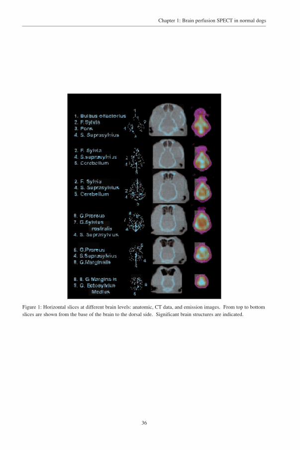
were used corresponding to anatomic levels of a stereotactic atlas of the dog’s brain14. (fig 1). The
SPECT perfusion data were manually fitted to the CT data set on a medical workstation using the
Multimodality software of Nuclear Diagnostics (Hägersten, Stockholm, Sweden). (fig 2).
36
Chapter 1: Brain perfusion SPECT in normal dogs
Figure 1: Horizontal slices at different brain levels: anatomic, CT data, and emission images. From top to bottomslices are shown from the base of the brain to the dorsal side. Significant brain structures are indicated.

37
Chapter 1: Brain perfusion SPECT in normal dogs
Figure 2: Example of 99mTc-ECD flow image, fitted on a corresponding CT image. This is a horizontal slice,located at the base of the brain.
Figure 3: Example of regions of interest (ROI) map drawn on brain slices. Irregular regions of interest aremanually drawn. This horizontal slice is taken at the lower middle part of the brain.

38
Chapter 1: Brain perfusion SPECT in normal dogs
For co-registration a mutual information cost function, minimalized by a down-hill simplex algorithm,
was used. The mutual information registration criterion allows fully automated, highly robust affine
registration of multimodal images without the need for pre-processing or user interaction.15
Irregular regions of interest (ROI) were drawn manually on the CT images for further
quantitative analysis. Consensus ROI’s were placed by two investigators (KP and PDB) on
frontocerebral, temporocerebral, parietocerebral, occipitocerebral, subcortical area and the cerebellum
based on the description by Redding (1978)25. (Fig 3). Further differentiation was not possible because
of the small size of the individual structures and the limited contrast of the CT imaging system and
limited resolution of the SPECT system.
From the SPECT data, average counts per pixel were calculated for all regions. A perfusion
index was obtained by normalising the average regional counts to total counts of all ROI’s. A rostro-
caudal gradient was defined as (R-C/R+C)*100, where R is the bilateral rostral (frontal) ROI count and
C the caudal (occipital) ROI count.
Non-parametric statistical analysis of the data was performed by means of the SPSS package
(Statistical Software Package for the Social Sciences, v9.0, SPSS Inc, USA). Correlations between age,
weight and regional cerebral blood flow were calculated with Spearman’s correlation test. Left to right
differences in frontocerebral, temporocerebral, parietocerebral and occipitocerebral cortex and rostro-
caudal differences between frontocerebral al and occipitocerebral perfusion were evaluated through the
related samples Wilcoxon Signed Rank Test. Differences in age, weight and regional cerebral blood
flow versus gender were evaluated with the independent sample Mann-Whitney U test. Differences in
standard deviation for the different regions were evaluated with the Mann-Whitney U test. Spearman’s
correlation coeficients were calculated for ROI size (pixels per ROI) and the measured activity (counts
per pixel). Level of significance was set at p =< 0.05
39
Chapter 1: Brain perfusion SPECT in normal dogs
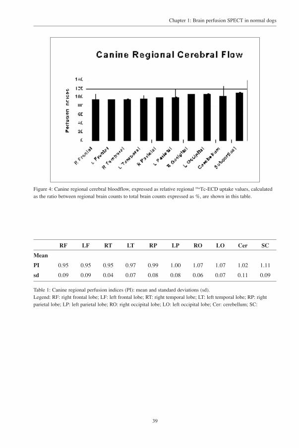
Figure 4: Canine regional cerebral bloodflow, expressed as relative regional 99mTc-ECD uptake values, calculatedas the ratio between regional brain counts to total brain counts expressed as %, are shown in this table.
RF LF RT LT RP LP RO LO Cer SC
Mean
PI 0.95 0.95 0.95 0.97 0.99 1.00 1.07 1.07 1.02 1.11
sd 0.09 0.09 0.04 0.07 0.08 0.08 0.06 0.07 0.11 0.09
Table 1: Canine regional perfusion indices (PI): mean and standard deviations (sd).Legend: RF: right frontal lobe; LF: left frontal lobe; RT: right temporal lobe; LT: left temporal lobe; RP: rightparietal lobe; LP: left parietal lobe; RO: right occipital lobe; LO: left occipital lobe; Cer: cerebellum; SC:

40
Chapter 1: Brain perfusion SPECT in normal dogs
Results
Characteristic examples of the brain perfusion pattern and the fitted ROI’s are shown in figure
1 and 3. The mean perfusion index for the separate regions is outlined in figure 4 and table 1. The
highest perfusion index was found in the thalamic region and the lowest in the frontocerebral cortex.
When comparing left versus right regional uptake in fronto-, temporo-, parieto- and occipito- cerebral
region, no statistically significant differences were found. A significant perfusion gradient was present
between right frontocerebral (rostral) and occipitocerebral (caudal) (+ 13%) perfusion (Wilcoxon’s
Z= 2.70; p<0.01) and left frontal and occipital (+ 12%) perfusion (Z= 2.29; p<0.05).
In this sample, there were no significant relationships between age or weight and any of the
regional cerebral uptake measurements. Also, when comparing male and female dogs, no significant
differences for gender concerning age, weight and regional cerebral uptake were found.

41
Chapter 1: Brain perfusion SPECT in normal dogs
Discussion
This study is, to our knowledge, the first report of regional cerebral blood flow in dogs using99mTc-ECD. In addition to the regional distribution pattern, the normal database is based on structural
correlation and provides semiquantitative data. In summary, it was found that regional brain uptake is
characterised by a rostro-caudal gradient, with highest uptake of the thalamic/striatal and
occipitocerebral regions and absence of demonstrable significant left-right perfusion differences. Age
and gender were not found to affect the results.
A significant rostro-caudal gradient was found in this series of adult dogs which is in
comparison with the results of SPECT examinations in adult humans.4,16,17 This pattern was observed
both with the eyes open and closed, and is probably related to specific metabolism of 99mTc-ECD in
the brain, although the intrinsic reason for such pattern is to date unexplained. In this series the most
active areas were the subcortical area and the occipitocerebral region. The standard deviation of the
perfusion index for the individual ROI’s varied between 0.04 and 0.09 for all regions except for the
cerebellum (0.11). (table 1). The higher, although not significant different, standard deviation for the
cerebellum may result from variable attenuation through the overlying tympanic bullae and occipital
bone for which the uniform attenuation does not account accurately. The higher uptake in the
subcortical area is in agreement with the results of rCBF studies with 123I-IMP in beagles12 where the
greatest regional uptake was found in the thalamus. No significant left-right differences were found.
In humans, lateralization is mainly found in the male, especially in the frontal area in relation to age.17
No gender difference was found in this group of dogs. No other data are available for animals
to compare our results with. In human studies, gender dimorphism in brain perfusion or metabolism
has been under debate.17,18 However, recent studies using advanced voxel-based statistical analysis
techniques have shown neocortical and cerebellar differences under resting conditions.17,20 With regard
to the influence of age, the statistical power of this feasibility study was limited to demonstrate
significant findings. In humans, a gradual decrease in rCBF with increasing age is found with a specific
frontotemporal pattern.17,18,21 Therefore, larger groups may need to be investigated before conclusions
can be drawn on aging and gender effects on canine brain perfusion.
METHODOLOGICAL STUDY LIMITATIONS:
General anaesthesia can influence the rCBF by reduction of blood pressure and heart rate after
halothane administration. Inhibition of CBF autoregulation in response to blood pressure changes, is
responsible for the vasodilatory effects of halothane and changed blood flow to various organs, with
specifically decreased vascular resistance in the brain, resulting in increased CBF.22 Since all dogs were
anaesthetised 10-15 min after the injection of the tracer, which at that stage is already trapped

42
Chapter 1: Brain perfusion SPECT in normal dogs
intracellular, it was not expected that this would influence rCBF. Medetomidine produces no
significant effect on brain metabolism but reduces cerebral blood flow in general. This suggests
uncoupling of cerebral metabolism and flow due to decreases in central catecholamine turnover.23 The
effects of the sedative given 40 minutes before anaesthesia could theoretically influence global
perfusion, but as in this study semiquantification was used with normalisation to total counts of the
individual brain, any global effect on brain perfusion would therefore be minimalized.
Since limited anatomic information is obtained with the flow images, these emission images
were co-registered on the individual CT images14. Stereotactic atlas was used to identify anatomical
landmarks to identify the different regions. Although in human functional brain mapping a stereotactic
atlas where brain images are resized into a defined stereotactic space is used24, such a system is not
available for dogs. The use of a corresponding three-dimensional coordinate system would allow a
more accurate group analysis of specific areas.
No consensus is found on the anatomic localisation of the different canine brain regions. We
used a classification25 which agrees largely with a published anatomic division26. The frontal lobe
(motor area, behaviour regulation and executive functions) is delineated caudally by the sulcus
cruciatus. Posterior to this sulcus is the parietal lobe (sensory cortex). The temporal lobe (memory
and auditory function) is situated ventral to the parietal and caudal to the frontal lobe. The occipital
lobe (visual reception and interpretation) lies caudal to the parietal lobe and caudodorsal to the
temporal lobe.
The anatomic delineation of the different cerebral regions used for SPECT coregistration and
analysis might be improved by the higher contrast and resolution power of magnetic resonance
imaging27. This technique may allow a more detailed study of the canine perfusion, corrected for
partial volume effects that originate from the limited SPECT resolution in comparison to the
dimensions of the objects under investigations.
PRACTICAL CONSIDERATIONS:
In this study a 99mTc labelled agent was used as it is generally preferred as a label for nuclear
medicine in humans because its physical characteristics for imaging are excellent (140 keV, physical
T1/2= 6.02 h) and because it is readily available from 99Mo/99mTc generators. 99mTc-ECD has a higher
brain-blood partition coefficient as compared to 99mTc-HMPAO because of its lower rate constant of
back-diffusion from brain to blood. It is also superior in its sensitivity to lesion detection and lesion
contrast because less protein binding in the blood is noted for 99mTc-ECD than for 99mTc-HMPAO.28,29
ECD was also chosen as radiopharmaceutical in this study because it is more stable on the shelf and
therefore more convenient to handle than HMPAO (in vitro stability is 6 hours after reconstitution

43
Chapter 1: Brain perfusion SPECT in normal dogs
compared to 30 min for HMPAO). On the other hand, HMPAO is more stable in the brain and has a
slightly higher extraction fraction, and stabilized forms have been commercialized.
In humans, a delay of at least 10 minutes between injection and acquisition is recommended.4
In our series the time interval was between 15 and 25 min because time, elapsing from inducing
anaesthesia and intubation of the animal, varied slightly. Injection of the tracer was performed in two
dogs in dynamic projections and adequate uptake was noted in the early stages of the acquisition.
The injected dose also varied slightly between dogs since all dogs were given 925 MBq
(25mCi) intravenously and weight variation (23-41 kg) was not taken into consideration. No influence
of weight on regional perfusion was noted. In humans the recommended dose varies between 10-25
MBq/ kg. In this study dose injected ranged between 16 and 32MBq/kg, which was higher since the
BUI is only 1.2% in the dog compared to 4.8% in humans. For personnel involved in acquiring the
SPECT study, a safe distance can be maintained because of general anaesthesia.
In human studies on rCBF either the cerebellum or the whole brain activity are the more
common references chosen to obtain region ratios.18 The cerebellum has advantages of higher
sensitivity for detection of abnormalities and relative stability in a number of diseases such as
Alzheimer's disease in humans. The lack of exact anatomic delineation and appropriate scatter
correction in the dog may increase regional variability and therefore a decrease in sensitivity for
detection of pathologic uptake values may result. The uniform attenuation coefficient of 0.12/cm
corresponds to an estimation of the theoretical broad-beam attenuation value in the absence of non-
uniform attenuation measurements. The influence of surrounding bone structures and air-filled cavities
may necessitate an optimization of this value since it has been shown a paradoxical skull effect may
lower the effective coefficient to be used in the correction algorithm.30 This phenomenon may be in
part responsible for the high activities found in the central brain areas (subcortical area) as compared
to cortical uptake.
Also partial volume effects may be responsible for this radially decreasing uptake, since small
region-of-interest size, influx from surrounding peripheral regions and the inclusion of white matter in
the cortical regions may artificially heighten the relative central values.

44
Chapter 1: Brain perfusion SPECT in normal dogs
REFERENCES
1. Roy CS, Sherrington CS. On the regulation of the blood supply of the brain. J Physiol 1890; 11: 85-108.
2. Walovitch RC, Cheesman EH, Maheu LJ, Hall KM. Studies of the retention mechanism of the brain perfusionimaging agent 99m-Tc-bicisate (99mTc-ECD). Journal Cereb Blood Flow Metab 1994; 14: S4-S11.
3. Ichise M, Golan H, Ballinger JR, Vines D, Blackman A, Moldofsky H. Regional differences in Technetium-99m- ECD clearance on brain SPECT in healthy subjects. J Nucl Med 1997; 38: 1253-1260.
4. Koyama M, Kawashima R, Ito H, Ono S, Sato K, Goto R, Kinomura S, Yoshioka S, Sato T, Fukuda H. SPECTimaging of normal subjects with Technetium-99m-HMPAO and Technetium-99m-ECD. J Nucl Med 1997; 38:587-592.
5. De Deyn P, Dierckx R, Alewi A, Pickert B. A Textbook of SPECT in Neurology & Psychiatry.London.Paris.Sydney: John Libbey & Company, 1997.
6. Report of the therapeutics and technology assessment subcommittee of the American Academy of Neurology.Assessment of brain SPECT. Neurology 1996; 46: 278-285.
7. Audenaert K, Van Laere K, Dumont F, Slegers G, van Heeringen C, Dierckx R. Decreased frontal serotonin 5-HT2a receptor binding potential in deliberate self harm. Eur J Nucl Med 2001; 28: 175-182.
8. Moretti, J. L., Caglar, M., and Weinmann, P. Cerebral perfusion imaging tracers for SPECT: which one tochoose? J Nucl Med 1995; 36: 359-363.
9. Biersack HJ, Klemm E, Reichmann K, Menzel C, Grünwald F. Activation Studies using SPECT. In: De Deyn,P. P., Dierckx, R. A., Alavi, A, and Pickut, B. A., ed. SPECT in Neurology and Psychiatry. London. Paris.Sydney: John Libbey, 1997; 67-72.
10. Audenaert K, Brans B, Van Laere K, Lahorte P, Versijpt J, van Heeringen C, Dierckx R. Verbal fluency as aprefrontal activation probe: a validation study using 99m-Tc-ECD brain SPECT. Eur J Nucl Med 2000; 27:1800-1808.
11. Daniel, G. B., Twardock, A. R., Tucker, R. L., and Shull, R. Brain scintigraphy. Prog Vet Neurol 1992; 3: 25-34.
12. Susskind H, Weber DA, Ivanovic M, Wong CTC, DeHaan CE, Gavin PR. Quantitative 123I IMP and 99mHMPAOimaging in the dog following cocaine administration. Nucl Med Biol 1996; 23: 343-352.
13. Van Laere K, Koole M, Kauppinen T, Monsieurs M, Bouwens L, Dierckx R. Non-uniform transmission inbrain SPET using 201-Tl, 99m-Tc and 153-Gd static line sources: antropomorphic dosimetry studies and brainquantification. J Nucl Med 2000; 41: 2051-2062.
14. Lion R, Liu C, Moffit R. A stereotactic atlas of the dog’s brain. Springfield, Illinois: C.C. Thomas, 1960.
15. Maes F, Collignon A, Vandermeulen D, Marchal G, Suetens P. Multimodality image registration bymaximization of mutual information. IEEE Trans Med Imaging 1997; 16: 187-198.
16. Tanaka F, Vines D, Tsuchida T, Freedman M, Ichise M. Normal patterns on 99mTc-ECD brain SPECT scansin adults. J Nucl Med 2000; 41: 1456-1464.
17. Van Laere K, Versijpt J, Audenaert K, Koole M, Goethals I, Achten E, Dierckx R. 99mTc-ECD brain perfusionSPET: variability, asymmetry and effects of age and gender in healthy adults. Eur J Nucl Med 2001; 28:873-887.
18. Catafou AM, Lomena FJ, Pavia J, Parellada E, Bernardo M, Setoain J, Tolosa E. Regional cerebral blood flowpattern in normal young and aged volunteers: a 99mTc-HMPAO SPET study. Eur J Nucl Med 1996; 23:1329-1337.
19. Gur R. Sex differences in regional glucose metabolism during the resting state. Science 1995; 267: 528-531.

45
Chapter 1: Brain perfusion SPECT in normal dogs
20. Jones K, Johnson KA, Becker JA, Spiers PA, Albert MS, Holman BL. Use of singular value decomposition tocharacterize age and gender differences in SPECT cerebral perfusion. J Nucl Med 1998; 39: 965-973.
21. Waldemar G, Hasselbalch SG, Andersen AR, Delecluse F, Petersen P, Johnsen A, Paulson OB. 99mTc-d,l-HMPAO and SPECT of the brain in normal aging. J Cereb Blood Flow Metab 1991; 11: 508-521.
22. Dormehl IC, Oliver DW, Hugo N. The primate model in neuropharmacology for cerebral blood flowdeterminations with HMPAO SPECT. In: De Deyn, P.P., Dierckx, R.A., Alavi, A, and Pickut, B.A., ed. A Textbook of SPECT in neurology and psychiatry. London: John Libbey & Company Ltd, 1997; 521-536.
23. Werner C. Effects of analgesia and sedation on cerebral blood flow, cerebral blood volume, cerebralmetabolism and intracranial pressure. Anaesthesist 1995; 44: S566-S572.
24. Talairach J, Tournoux P. Co-planar stereotactic atlas of the Human Brain. Stuttgart: Thieme MedicalPublishers, 1988.
25. Redding RW. Anatomy and Physiology. In: Hoerlein, B. F., ed. Canine neurology. Philadelphia. London.Toronto: W.B.Saunders Company, 1978; 7-51.
26. Schaller O. Systema nervosum. In: Schaller, O., ed. Illustrated Veterinary anatomical Nomenclature. Stuttgart:Ferdinand Enke Verlag, 1992; 414-509.
27. Lucignani G, Rizzo G, Messa C, Gilardi MC, Fazio F. Integration of brain imaging techniques. In: De Deyn,P.P., Dierckx, R.A., Alavi, A, and Pickut, B.A., ed. A textbook of SPECT in Neurology and Psychiatry.London.Paris.Sydney: John Libbey & Company Ltd, 1997; 547-560.
28. Schiepers C, Verbruggen A, Casaer P, De Roo M. Normal brain perfusion pattern of Technetium-99m-ethylcysteinate dimer in children. J Nucl Med 1997; 38: 1115-1120.
29. Matsuda H, Yagishita A. A non-invasive quantitative approach to 99mTc-ethyl cysteinate dimer. In: De Deyn,P.P., Dierckx, R.A., Alavi, A, and Pickut, B.A., ed. A textbook of SPECT in neurology and psychiatry. London-Paris-Rome-Sydney: John Libbey & Company Ltd, 1997; 501-505.
30. Van Laere K, Koole M, Versijpt J, Dierckx R. Influence of non-uniform attenuation correction on normal brainperfusion SPECT. Eur J Nucl Med 2001; 28: 90-98.

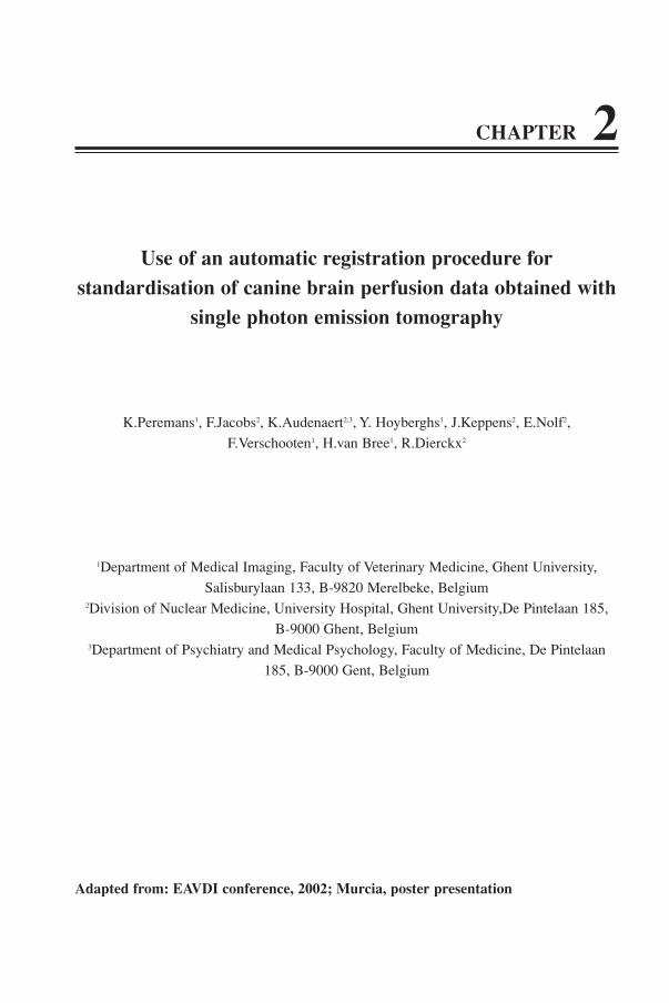
CHAPTER 2
Use of an automatic registration procedure forstandardisation of canine brain perfusion data obtained with
single photon emission tomography
K.Peremans1, F.Jacobs2, K.Audenaert2,3, Y. Hoyberghs1, J.Keppens2, E.Nolf2,
F.Verschooten1, H.van Bree1, R.Dierckx2
1Department of Medical Imaging, Faculty of Veterinary Medicine, Ghent University,
Salisburylaan 133, B-9820 Merelbeke, Belgium2Division of Nuclear Medicine, University Hospital, Ghent University,De Pintelaan 185,
B-9000 Ghent, Belgium3Department of Psychiatry and Medical Psychology, Faculty of Medicine, De Pintelaan
185, B-9000 Gent, Belgium
Adapted from: EAVDI conference, 2002; Murcia, poster presentation


49
Chapter 2: Automatic registration of perfusion data
Introduction
Interpretation of brain images obtained with SPET demands reliable and reproducible
quantification of the data. This requires an accurate registration method of the individual patient’s data
to a standardized image, on which the cortical and subcortical regions are delineated. Manual
registration is commonly implemented with special software (i.e. the multimodality registration
program from Hermes, Nuclear Diagnostics), enabling the alignment of the patient’s data manually to
a standardized image by reorientating data in three dimensions. Unfortunately this procedure is
operator dependent and as such can introduce errors in the final result. In addition, due to size and
shape differences between different canine breed and due to positional artefacts, manual registration
can introduce variability in the results of semi-quantification of the flow data. Also, manual
registration is time-consuming. More, the accuracy of the VOI’s placement largely depend on a
reproducible and objective registration of the patient’s data. Automated registration of the individual
patient’s perfusion data to a template combined with an automatically applied region map, consisting
of semi-quantitative “volume of interest” (VOI) analyses and a comparison of the obtained results to
normal values (mean and standard deviation) result in a more reliable analysis of brain perfusion
images, as compared to manual registration and visual interpretation.1
The aim of the study was to explore the value of automatic registration to a reference template
and to include a predefined VOI map on canine brain perfusion data, using the methodology of the
Brain Registration and Automated SPECT Semiquantification (BRASS) program from Nuclear
Diagnostics.

50
Chapter 2: Automatic registration of perfusion data
Materials and methods
A 99mTc-ECD image template was created including 12 normal dogs (6 males and 6 females),
aged between 12-84 months (mean: 49 months, SD: 29). The included individuals were free of
neurological disease or behavioural disorders. 99mTc-ECD (Neurolite®, Dupont Pharmaceuticals Ltd.,
Brussels, Belgium) was injected intravenously (925 MBq (25mCi)) after sedation and prior to general
anesthesia, in a quiet room with dimmed lights. Care was taken not to provoke anxiety. Sedation was
obtained with 10-30µg/kg medetomidin hydrochloride IM (Domitor®, Pfizer) and general anesthesia
was induced with 2-3mg/kg iso-propylphenol IV (Rapinovet®, Mallinckrodt) and maintained with
halothane (Fluothane®, Zeneca) to effect. All dogs were positioned in ventral recumbency with the
head placed in a special moulage. SPECT was performed with a triple head gamma camera (Toshiba
GCA-9300A, Dutoit Medical, Antwerp, Belgium), equipped with high resolution fanbeam collimators
(FWHM 7.8 mm). Acquisition was started 15 to 25 minutes after injection of the tracer depending on
the progress of induction of the anaesthesia. Total acquisition time was 20 minutes. For each
acquisition, 90 projection images were obtained on a 128x128 matrix using a continuous scan mode by
rotating each head 120°. Data were acquired for one main energy window and two scatter windows,
with a Toshiba GCA9300 triple headed gamma camera, equipped with low energy, high resolution fan-
beam collimators. All data were filtered, scatter corrected, re-binned to parallel data and reconstructed
with filtered back-projection (0.16 cycles/pixel; order 8), as described previously.2 Uniform Sorensen
attenuation (0.12/cm) was applied. Pixel size and slice thickness were 1.72 mm.
For the first part of the study, each reconstructed image was manually adjusted by two
experienced persons to one arbitrary chosen image, which was reoriented to a standardized position
along the X,Y and Z axis. (Fig 1A and B) and centred in the matrix. To register the individual image
to this “reference” image the multi modality software allowing for manual scaling, rotation and shifting
and thus adjusting 9 transformation parameters (X scale, Y scale, Z scale, X shift, Y shift, Z shift, XY
tilt, XZ tilt and YZ tilt) was used. This procedure was repeated twice to refine registration.
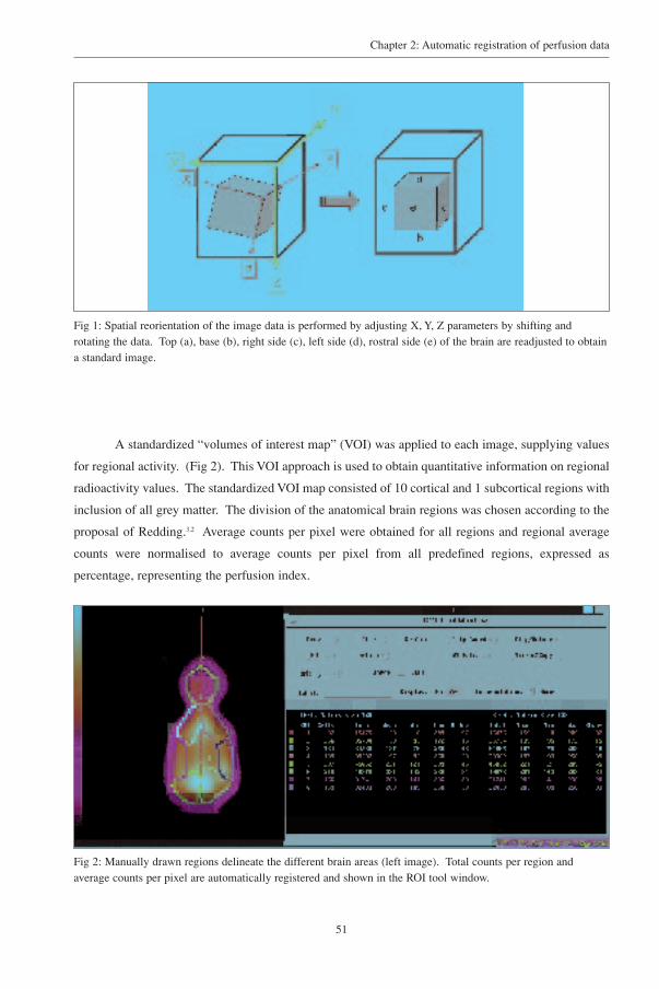
51
Chapter 2: Automatic registration of perfusion data
Fig 1: Spatial reorientation of the image data is performed by adjusting X, Y, Z parameters by shifting androtating the data. Top (a), base (b), right side (c), left side (d), rostral side (e) of the brain are readjusted to obtaina standard image.
A standardized “volumes of interest map” (VOI) was applied to each image, supplying values
for regional activity. (Fig 2). This VOI approach is used to obtain quantitative information on regional
radioactivity values. The standardized VOI map consisted of 10 cortical and 1 subcortical regions with
inclusion of all grey matter. The division of the anatomical brain regions was chosen according to the
proposal of Redding.3,2 Average counts per pixel were obtained for all regions and regional average
counts were normalised to average counts per pixel from all predefined regions, expressed as
percentage, representing the perfusion index.
Fig 2: Manually drawn regions delineate the different brain areas (left image). Total counts per region andaverage counts per pixel are automatically registered and shown in the ROI tool window.

52
Chapter 2: Automatic registration of perfusion data
For the second part of the study a mean image was created by sequentially adding all centred
images voxel by voxel to each other. (Fig 3). This composite template represents an average,
normalized distribution of counts in the brain for the three planes, creating a three dimensional mean
image. More, a coefficient of variance template is generated at the same time, representing the
standard deviation divided by the mean count distribution. (Fig 4). All individual images were then
re-registered on this mean image with an automatic registering algorithm, allowing shifting, scaling
and rotation in all three directions as described in the first part. The automatic registration procedure
is based on the principal axes transformation, resulting in an approximate fit, and on the downhill
simplex minimization algorithm, using the count difference method, to refine the fit.4 (Fig 5). A
regionmap, defined on the template image and using the same anatomically predefined regions (10
cortical and 1 subcortical region) as used in the previous part, was automatically applied on the
individual image data, generating the perfusion index for all regions. (Fig 6).
Standard deviations of normalized average radioactivity in the different brain regions were
obtained for both groups.
Fig 3: All individual image data for all slices are added and averaged (left image), creating a mean image(template) (right image).
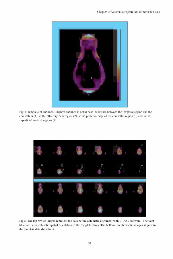
53
Chapter 2: Automatic registration of perfusion data
Fig 4: Template of variance. Highest variance is noted near the fissure between the temporal region and thecerebellum (1), in the olfactory bulb region (2), at the posterior edge of the cerebellar region (3) and in thesuperficial cortical regions (4).
Fig 5: The top row of images represent the data before automatic alignment with BRASS software. The faintblue line demarcates the spatial orientation of the template slices. The bottom row shows the images aligned tothe template data (blue line).

54
Chapter 2: Automatic registration of perfusion data
Fig 6: The left image demonstrates the manually drawn ROI’s. A region specific value (1-11), linked to aparticular colour pattern, is attributed to all predefined brain regions. The computer program will automaticallyapply this regionmap to newly acquired image data.

55
Chapter 2: Automatic registration of perfusion data
Results
No visual discrepancies were noted after the manual and the automatical fitting procedure,
although the standard deviation of perfusion index for the different cerebral regions was highest after
manual fit compared to the automatic registration. (Fig 7). Loss of resolution of the template image
due to amalgamating patient data is minimal and barely noted visually.
The average time spent by each operator to align the 12 image sets to the arbitrary choosen
image was 10 minutes per set, compared to a mean of 6 minutes for the automatic registration
procedure.
Fig 7: Standard deviations of regional perfusion data are given for the different brain regions. Highest values arefound for the manual fit.Legend: RF: right frontocortical region; LF: left frontocortical region; RT: right temporocortical region; LT: lefttemporocortical region; RO: right occipitocortical region; LO: left occipitocortical region; RP: rightparietocortical region; LP: left parietocortical region; SC: subcortical area; Cer: cerebellar area.

56
Chapter 2: Automatic registration of perfusion data
Conclusion
The template based automatic fit, using BRASS software from Nuclear Diagnostics, can be
used for registration of canine brain flow data and has the advantage that semi-quantification can be
performed more accurate and less subjectively. The higher standard deviations found with manual
fitting results from the fact that parameters for shifting, scaling and rotation are sequentially changed.
Since these parameters are dependent on each other, changing one influences the position of the others
and hampers optimal alignment.
On visual inspection virtually no difference was noted in positioning of the reoriented images
between both methods. Quality of the template appeared similar to the individual images although on
the template image background noise was reduced and the edges were slightly blurred and smoothed.
Overall, resolution was comparable with the original individual images.
The template of the coefficient of variance showed high values in the superficial cortical layers
and in the caudal border of the cerebellum, probably due to the variable negative skull effect which is
prominent in these regions. Difference in skull thickness could explain this phenomena. These
findings are similar to the regional variances found in a human study, using the same methodology.1
Registered regional activity in the olfactory bulb is prone to variety, presumably depending on the
activation state of this very important sensory region, which is beyond control.
This template based automated registration method provides a mean to compensate for size and
shape differences between different breeds, a major concern in canine brain imaging and enables to
adjust positional differences.

57
Chapter 2: Automatic registration of perfusion data
REFERENCES
1. Slomka P, Stephenson J, Reid R, Hurwitz G. Automated template-based quantification of brain SPECT. In: DeDeyn, P. P., Dierckx, R. A., Alavi, A, and Pickut, B. A., ed. Spect in neurology and psychiatry. London: JohnLibbey, 1997; 507-519.
2. Peremans, K, De Bondt, P, Audenaert, K, Van Laere, K, Gielen, I, Koole, M, Versijpt, J, Van Bree, H,Verschooten, F, and Dierckx, R. Regional brain perfusion in 10 healthy dogs measured using Technetium-99methyl cysteinate dimer SPECT: a normal database. Vet Radiol Ultrasound 2001; 42: 562-568.
3. Redding RW. Anatomy and Physiology. In: Hoerlein, B. F., ed. Canine Neurology. Philadelphia, London,Toronto: W.B.Saunders Company, 1978; 7-52.
4. Slomka P, Hurwitz G, Stephenson J, Cradduck T. Automated alignment and sizing of myocardial stress andrest scans to three-dimensional normal templates using an image registration algorithm. J Nucl Med 1995; 36:1115-1122.


CHAPTER 3
Evaluation of cerebral neurotransmitter physiology andpathophysiology with PET and SPET imaging modalities in
animal models: a review
K.Peremans1, K.Audenaert2,4, F. De Vos2, M.Vandecapelle3, H.van Bree1,
F.Verschooten1, G.Slegers3, R.Dierckx2
1From the Department of Medical Imaging, Faculty of Veterinary Medicine, Ghent
University, Salisburylaan 133, B-9820 Merelbeke, Belgium2Division of Nuclear Medicine, Ghent University Hospital, De Pintelaan 185,
B-9000 Gent, Belgium3Laboratory of Radiopharmacy, Faculty of Pharmaceutical Sciences, Ghent University,
Harelbekestraat 72, B-9000 Gent, Belgium4Department of Psychiatry and Medical Psychology, Faculty of Medicine,
De Pintelaan 185, B-9000 Gent, Belgium
Adapted from: submitted to Vlaams Diergeneeskundig Tijdschrift


61
Chapter 3: Review neuroreceptor radioligand imaging with PET and SPET
Summary
Since positron emission tomography (PET) and single photon emission tomography (SPET)
have become widely available as imaging modalities, studies have been performed on neuroreceptor
physiology and pathophysiology in humans and animals. Several neurological and psychiatric diseases
related to neurotransmitters and receptor functioning have been investigated.
In order to investigate the biological effects of new compounds, this imaging modality is
becoming more and more popular in the pharmaceutical industry, since pharmacokinetics and
pharmacodynamics of new drugs can now be evaluated in vivo, omitting extensive in vitro
examinations. The response to pharmaceuticals can be evaluated more objectively as compared to the
potentially subjective evaluation of the condition of the patient, based on clinical examination and
interpretation of neuropsychological tests.
This review focuses on the technique of this imaging modality and the nature of radioligands
used for neuroreceptor imaging. A brief overview of the research on neuroreceptor physiology,
neuropathology, drug evaluation and substance abuse, with the emphasis on animal models will be
given. Future prospects for implementation in veterinary medicine are discussed.

62
Chapter 3: Review neuroreceptor radioligand imaging with PET and SPET
Introduction
Already in the 19th century, Langley formulated the concept of “receptive substances”,
involving the interaction of drugs with tissue.1 Later, the Nobel prize winner Ehrlich proposed that
interaction with toxic substances was caused by the presence of certain cell surface groups (receptors)
showing binding properties with these toxins (ligands). This binding was due to high complementarity
between the stereochemical configuration of the ligand and the receptor with which it interacts.2 With
the formulation of the receptor concept, an explanation was found for the specific effects of tracer
amounts of substrate on a target organ.
Classification of receptors is based on their chemical specificity (e.g. the α- and β- adrenergic
receptors) or on their anatomical localisation (e.g. pre- and postsynaptic receptors, intra-and
extracellular receptors)3, while ligands are classified according to their ability to generate a response.
Full agonists will generate a maximal receptor response while partial agonists will never produce a
maximal activation of the receptor no matter how high a concentration is applied. Whether a ligand is
a full or partial agonist is a tissue- and drug- dependent phenomenon, although endogenous agonists
for a receptor type usually behave as full agonists. Antagonists block the response in a reversible,
irreversible, competitive, non-competitive or mixed way. Distribution and kinetics of the ligand (drug
or endogenous substrate) can be quantified with in vitro, ex vivo (injection of the ligand in vivo,
measuring tissue activity after death) and in vivo imaging techniques using radioactive labelled
molecules.4 In vitro techniques have been used to evaluate ligand-receptor responses and to quantify
receptors. In recent years, functional imaging techniques with positron emission tomography (PET)
and single photon emission tomography (SPET) are gradually gaining importance as ligand-receptor
interactions which can be evaluated in their natural environment in the living subject.4,5
This technology is now more commonly applied in drug development. Functional imaging is
also used as a research and diagnostic tool for studying neurologic and neuropsychiatric diseases in
humans, providing insight into complex receptor interactions. In this context, animal models were
introduced to investigate neuropathologic disorders, usually created by genetic, surgical or
pharmacological manipulation of the animal. In most cases rodents (ex vivo and in vitro) and non-
human primates are used, although it has been suggested that dogs would provide a valid alternative
model because they are widely available, rather cheap and easy to keep and handle, compared to non-
human primates. This has lead to the validation of different models of natural canine neuropathologic
entities for human disease.6,7 But, also from a veterinary point of view, this technique may facilitate
research on the mechanisms of neurologic and behavioral disorders in dogs and may monitor the
effects of therapy.
The aim of this article is to give a review of receptor imaging techniques and their
implementation, with emphasis on the in vivo imaging of neuroreceptors with PET and SPET
modalities. Special attention will be given to neuroreceptor research in which animal models are used
to illustrate the clinical potential of this modality for the investigation of causes and treatment of
behavioural diseases in animals.

63
Chapter 3: Review neuroreceptor radioligand imaging with PET and SPET
Methodological considerations
IMAGING METHODS:
1. Receptor imaging in vitro
Neurotransmitter receptors in frozen tissue sections can be visualized and quantified using
autoradiography. In order to achieve high spatial resolution, radionuclides with low β-emission
energies, such as 3H, 125I and 14C, are used. These radionuclides are bound to highly specific probes,
hereby providing a radiolabelled ligand (e.g. 3H-spiperone). These examinations can be performed in
vitro or ex vivo. Specific binding, which is binding of the radiolabeled ligand with the receptor, and
non-specific binding, being the activity persisting in the presence of an excess of a specific non
radioactive displacer, can be quantified, providing an estimation of receptor density (Bmax) and affinity
(1/Kd) (Kd= dissociation constant at equilibrum).5,8 Another method to evaluate the effects of receptor
binding is the measurement of the cellular responses elicited by the ligand-receptor interaction.
Changes in cyclic adenosine monophosphate (cAMP) concentration, glucose production or electrical
and mechanical responses enable biochemical characterisation of the receptor.4,9
Immunohistochemistry offers an alternative method to visualize receptors in vitro . However,
this technique does not fall under the scope of this article and will therefore not be discussed.
Since the natural environment of pre-and postsynaptic membranes is not accounted for in vitro,
assumptions on drug interaction or ligand-receptor behavior from in vitro experiments should be
regarded with caution when adopted to receptor estimation in vivo. Ex vivo techniques require the
death of the animal under investigation, since the β-particles, emitted during 3H,14C or 125I decay, are
strongly attenuated by tissue and therefore cannot be measured with external detecting devices.
2. Receptor imaging in vivo
In vivo techniques offer a more realistic representation of receptor behavior, because receptor
characteristics can be altered due to interaction with its intra-and extracellular regulatory mechanisms.10
The functional imaging modalities, PET and SPET enable external imaging of the receptor with ligands
labelled with γ and β+ emittors, thus permitting evaluation of biological processes in vivo in a safe and
non-invasive way. Due to the high affinity and specificity of the radioligand for the receptor and
because of the sensitivity of the imaging system, only tracer amounts, usually in the picomolar range,
of this radiolabeled ligand are applied, thus avoiding physiologic or toxic effects.4 In vivo sequential
studies are possible and time/activity curves in region of interests can be generated.

64
Chapter 3: Review neuroreceptor radioligand imaging with PET and SPET
Unfortunately, some of these interactions can give misleading receptor occupancy estimation.
As an example, cellular response to agonistic stimulation may desensitise or even internalize the G-
protein coupled receptor, a mechanism by which the receptor is translocated intracellular. This may
result in variability in visualization of the receptor.11 In addition, processes such as perfusion and, as a
consequence, delivery of the radioligand may influence receptor binding.12
PET IMAGING
In PET studies, positron emitting radionuclides are used. Following emission, these positrons
travel a few millimetres in matter before they interact with an electron and annihilation energy is
produced in the form of two opposite photons with an energy of 511 keV each. Both photons are
detected by external detectors. The most commonly used radionuclides are 15O (T1/2=2 min), 13N
(T1/2=10 min), 11C (T1/2=20 min), 18F (T1/2=110 min) and 76Br (T1/2=16 hours). The short half-lives of11C and 18F enable repetitive investigations within a short time interval in the same individual, using the
same or different radioligands, in identical or different test conditions.5 The advantage of 18F is that the
emitted positrons travel a very short distance before annihilation occurs, due to their lower kinetic
energy, hereby providing more exact localization of the emitted positrons.5
With PET, absolute quantification is possible and high spatial resolution (3-4 mm) can be
achieved. Disadvantage is that the radionuclide must be generated by a cyclotron which must be in the
immediate vicinity of the camera due to the short half life of most of these elements.5,9
SPECT IMAGING
SPECT is based on the emission of photons, registered by external detectors, so-called gamma-
cameras. Mostly Technetium (99mTc, T1/2=6h) or iodine (123I, T1/2=13h) are used. The longer half life
of these radionuclides precludes repetitive investigations on the same individual on the same day but
can be an advantage for studies on receptor occupancy requiring prolonged scanning times.8 Spatial
resolution depends on the collimator and camera configuration, but is in general lower than PET (7-10
mm vs 3-4 mm). Due to inherent physical problems such as attenuation, scatter and partial volume
effects, photon registration is not with certainty coming from the region of interest, therefore specific
binding is measured semi-quantitative.

65
Chapter 3: Review neuroreceptor radioligand imaging with PET and SPET
LIGAND PROPERTIES
In order to determine the receptor kinetics in vivo, the characteristics of the ligand are of crucial
importance.
1. Labelling of the ligand
The labelling of the ligand is not a simple procedure and it can alter its biological activity,
rendering it unsuitable for binding with the receptor. It can increase the lipophilicity of the ligand and
can enhance non-specific binding with plasma proteins and blood cells.10 Labelling with 11C usually
does not cause alteration of the physicochemical properties of the compound, but labelling with 18F or76Br may alter its properties. Labelling with either 99mTc or 123I usually has an important impact on the
physicochemical properties of the ligand.5,10 Another important factor in the labelling procedure is the
duration of radiosynthesis, since the half-life of the radionuclide has to be considered. Therefore, with
regard to PET tracers, this procedure should be kept as short as possible and the label should be
introduced as late in the synthetic procedure as possible.5 Specific radioactivity (MBq/mole) of the
synthetised radioligand has to be high, since low specific radioactivity results in need for higher ligand
concentrations, resulting in increasing risk for biological effects and in substantial occupation of
binding sites by unlabeled ligand.5
Exogenous antagonists are preferred because dilution with endogenous agonists is less likely
to happen, affinity for the receptor is usually higher and receptor responses interfering with the binding
estimation are less likely to occur.9,10
2. Selectivity and affinity:
Affinity is the chemical strength of the binding, selectivity refers to the strength of ligand
interaction with one receptor compared to interactions with other receptors (the ability to recognize
receptor molecules). Biological substrates frequently lack selectivity and high affinity. Since affinity
is formulated as the ratio of association and dissociation, high affinity would be a negative quality for
endogenous neurotransmitters since the receptors need the ability to react fast to stimuli. The opposite
is true for receptor probes used in research which must have high affinity and selectivity to make
visualization possible.13 Selectivity and affinity are closely related since high affinity for a receptor
results in high selectivity. Lack of selectivity may only be acceptable when the targeted receptors are
clearly separated anatomically from the other imaged receptors or receptor subtypes.5,8,9

66
Chapter 3: Review neuroreceptor radioligand imaging with PET and SPET
3. Biodistribution and metabolism:
During its way to the brain the radioligand might be trapped or bound to cells, plasma proteins
and other molecules. Lipophilicity, measured as log P, the value of the octanol-water partition
coefficient expressed in log units, is an essential property of the radiopharmaceutical in order to pass
the blood brain barrier but increases aspecific binding at the same time. Depending on the methods
used for measuring log P, values should be between 1 and 3.5 for neuroimaging tracers to be useful.14
Furthermore, tracer metabolism in vivo may result in both labelled and non-labelled metabolites. In
addition, labelled metabolites might bind to receptors or might be trapped in cells. Since PET and
SPET imaging detects only radioactivity it is not possible to distinguish between the signal produced
by the labelled parent molecule and the labelled metabolite. Therefore, ideally, metabolism should be
away from the investigated organ and metabolites should be non labelled. In case metabolites are
labelled, they should not cross the blood brain barrier or, if they do, should not be retained in the target
area. The degree of activity due to labelled metabolites can be estimated and corrective binding curves
generated, by measuring the metabolites in plasma during the study.5,8-10,12,15-35
4. Quantification of receptor binding:
PET and SPECT data can be analysed at three levels of sophistication. The simplest way is
visual inspection, determining areas of increased or decreased binding. This method is of limited value
since it is subjective and biased by the skills of the interpreter.
The next level of analyzing data is the semiquantitative or “reference region” approach on
condition that binding with the receptor is reversible and that a steady state between uptake and
washout of the radioligand after a single bolus injection, can be achieved. The ratio of binding in
regions with high density of receptors, and thus high specific binding, to that in low density areas,
representing the reference region, is calculated. This a priori knowledge of regional receptor density
and distribution, can be derived from autoradiographic or immunohistochemic studies.4,9 The main
advantage of the ratio method is that it provides a simple and quick semi-quantification for clinical use,
not requiring blood samples and not dependent on models and rate constants.36 However, the technique
is unreliable when a substantial amount of metabolites of the radioligand, with affinity for the receptor,
are formed that pass the blood brain barrier. Another possible confounder for this approach is that a
differential rate of wash out between receptor-rich areas and areas with non-specific binding may be
present or that there are heterogeneous non specific binding sites.8,36 In humans, but especially in
veterinary nuclear medicine, bloodflow changes, induced by exogenous administered compounds, such
as anaesthetics or sedatives, resulting in modulation of the capillary surface, can provoke changes in
the equilibral state.37

67
Chapter 3: Review neuroreceptor radioligand imaging with PET and SPET
Mathematical methods have been developed to quantify dynamic data in order to calculate Kd
(equilibrium dissociation constant) and Bmax (maximal specific binding which is proportional to
receptor density) more accurately.9 The compartmental model is based on different compartments in
which the tracer can pass or accumulate. Mostly, a three compartmental model that localizes the tracer
in different localizations, namely plasma (the input function), free or non specifically bound, and
specifically bound is used.9 Rate constants are introduced to describe the fractional transfer of ligand
along these compartments.8,9 Multiple-time graphical analysis (MTGA) is an alternative technique
used to quantify data over time as the radioactive tracer accumulates in tissue. This method is model
independent and as a consequence facilitates the processing of the data. Two different methods are
developed: the Patlak plot is used for a tracer that will be trapped for the length of the experiment in
one compartiment and that is considered essentially irreversibly bound38 and the Logan plot which is
applicable for reversible tracers39.
5. Testing new ligands
Preclinical testing of new ligands is usually performed both in vitro and in vivo studies.
Autoradiographic studies can provide information concerning affinity, selectivity and non-specific
binding but no data can be obtained concerning the biodistribution unless ex vivo or in vivo
experiments are conducted.
The advantage of SPET and PET is that the distribution of the radiotracer and its selectivity and
reversibility in relation to the receptor can be determined in the living subject in the so-called
biodistribution studies. Estimation of the specific binding can be evaluated with displacement
(“chaser” studies) or pre-treatment (“blocking” studies) with cold competitor reducing the specific
binding in tissue and leaving the non-specific bound fraction.5,8-10,17-20,22,24-29,31,34,35,40-73 Also, the optimal
scanning time, the time interval where a pseudo-equilibral state is achieved, can be determined, which
is essential when semi-quantification of receptor binding will be performed.
One of the great advantages of using animal models lies in the possibility of repeated intra-
subject scanning in order to compare tracers and to monitor therapeutic interventions.

68
Chapter 3: Review neuroreceptor radioligand imaging with PET and SPET
Practical applications
1. Confounding factors
Pathological changes in receptor occupancy may be due to altered synthesis or release of
neurotransmitters, or may be a consequence of altered blood flow (e.g. ischemia) or degenerative
processes.74 However, in order to evaluate pathology, one must also take physiological alterations in
receptor behavior into account.
• Age and gender:
Functional brain imaging studies in humans, in non-human primates and in rats demonstrated
that the binding of several neurotransmitter systems is influenced by age and gender and that
modulation of receptor binding parameters can occur due to hormonal influences such as during the
normal menstrual cycle and menopauze. 9,75,76,77-78,79-90,91,92
An age related decline of the cerebral 5-HT2A receptor has been demonstrated with 123I-5-I R91150 in
dogs93, using SPET, and with ((R)-(+)-[3-OCH3-11C]-MDL 100907 in primates, using PET.82 Regional
decreases of the 5-HT1A receptor were observed in primates using the PET tracer,
carbon-11 labelled N-(2-(4-(2-methoxy-phenyl)-1-piperazin-1-yl)ethyl)-N-(2-pyridyl)cyclohexane-
carboxamide (WAY-100635). Decreases of the serotonin transporter (SERT) were registered in aged
primates with PET using the difference in radioactivity between the active compound [11C]trans-
1,2,3,5,6,10-beta-hexahydro-6-[4-(methylthio)phenyl-pyrrolo-[2,1-a]isoquinoline ([11C]+McN5652)
and the inactive enantiomer [11C]-McN5652 during sequential scanning as a measure for specific
binding.94 Age related decrease of the dopamine-1 (D1) and D2 receptor binding, dopamine transporter
(DAT), phosphodiesterase-IV activity and muscarine cholinergic receptors were demonstrated with
respectively, carbon-11 labeled ((R)-(+)-8-chloro-2,3,4,5-tetrahydro-3-methyl-5-phenyl-1H-3-
benzazepin-7-ol) (SCH23390), 11C-raclopride, [11C](2beta-carbomethoxy-3beta-(4-fluorophenyl)-
tropane) (beta-CFT), [11C] rolipram and (+)N-[11C]-methyl piperidyl benzilate ((+)3-MPB and 4-MPB)
PET tracers in primates.75,86,95-97 In one study, binding alterations of the vesicular acetylcholine
transporter in aging rhesus monkeys, imaged with the PET tracer [18F] (+)-4-fluorobezyltrozamicol
([18F] FBT), showed individual differences, suggesting that the cholinergic system may undergo a
differential susceptibility to the aging process.98
• Influence of anaesthetic drugs and cerebral perfusion:
Sedation and anaesthesia are prerequisites for animal research on receptor imaging. The effects
of several anaesthetics are twofold. They can act on the receptor directly or indirectly through other
receptor systems, such as the anaesthetic ketamine, an antagonist of the N-methyl-D-aspartate
(NMDA) receptor, which interferes also indirectly with the dopaminergic and serotonergic system.99-103
They can also produce cerebral metabolic effects, blood pressure and perfusion changes.104-107 In one
69
Chapter 3: Review neuroreceptor radioligand imaging with PET and SPET
study different kinetics of the radiotracer [11C]-methamphetamine (MAP) were observed comparing
halothane and pentobarbital anaesthesia in rhesus monkeys.108 In another monkey study, isoflurane
anaesthesia enhanced the direct and indirect effects of the DAT inhibitors cocaine and GBR12909.109
On the other hand, low variability in radioligand binding with the D2 receptor was registered in a study
with primates, using the PET tracer [18F]-fluoroclebopride (FCP) between induction with ketamine or
with fluothane by mask induction.110 Nevertheless, it remains important to select the anaesthetic
combination that provokes lowest disturbances in the equilibrum reached between the receptor and its
radioligand and to stick to one anaesthetic protocol for this type of research.
• Influence of cellular responses and manipulation of other neurotransmitter systems
As mentioned previously, under the influence of agonists and in some cases also of antagonists,
receptor availability on the cellular membrane might change because of desensitisation and
internalisation mechanisms.11,111-113 It is hypothesized for the challenged (administration of an
exogenous chemical compound will provoke the release of endogenous neurotransmitter)
dopaminergic system, that internalisation might influence the binding parameters of the radioligands,
depending on their biochemical properties. The intracellular shift might result in lipophilic radioligands
entering the cell and low lipophilic not, resulting in a different binding profile. Also, the intracellular
environment (e.g. sodium concentration) might inhibit binding to the receptor for some and not for
other radioligands.11 Furthermore, endocytosis of receptors after internalization may result in “down-
regulation” of receptors by lysosomal degradation.111
Another important topic in research is the interaction of the different neurotransmitter systems
hereby using modulating pharmacological probes. These experiments contribute to a large extent to
the understanding of neuropsychiatric diseases and towards elucidating the efficacy of medication used
for these disorders. To enable visualization of endogenous fluctuations, the employed radioligands
should be sensitive to changes in neurotransmitter concentrations.114-117 The interaction of the
dopaminergic D2 system and the muscarinic cholinergic system was evaluated by sequential
pretreatment with anticholinergic drugs and dopaminergic antagonists, demonstrating respectively
reduced D2 receptor ([18F]N-methylspiroperidol, NMSP) and muscarine receptor radioligand ([N-11C-
methyl]benztropine) binding.118 Administration of scopolamine (a muscarine cholinergic antagonist)
resulted in increased dopamine synthesis, measured with [11C]-1-3,4-dihydrophenylalanine (DOPA), an
increased binding of the DAT radioligand 2beta-[11C]carbomethoxy-3beta-(4-fluorophenyl)tropane
(CFT) and decreased binding of the D2 ligand [11C]-raclopride. The extracellular level of dopamine,
measured with microdialysis, remained static despite the increased synthesis, due to enhanced DAT
availability but the affinity of the D2 receptor decreased.119 The dopaminergic regulation of the striatal
cholinergic activity was measured using the nicotinic cholinergic receptor PET tracer norchloro [18F]-
fluoroepibatidine (NFEP). It was demonstrated that pretreatment of the D2 receptor with agonists and
antagonists could respectively increase and decrease the binding of the cholinergic receptor
radioligand.114 The interaction of the serotonergic and dopaminergic system was examined with a

70
Chapter 3: Review neuroreceptor radioligand imaging with PET and SPET
serotonergic antagonist (altanserin) and the selective serotonin reuptake inhibitor (SSRI) citalopram.
Blocking the 5-HT2 receptor resulted in increased binding of the D2 receptor radioligand 11C-
raclopride in the extrastriatal region and in decreases in the striatal area, while, when enhancing the
serotonergic concentration in the synaptic cleft with SSRI’s, opposite effects were noted.120 In another
study, the effects of pharmacologic manipulation of gamma-aminobutyric acid (GABA) and
serotonergic receptors on the central cholinergic system were marked.121
2. Clinical applications
• Drug industry:
These imaging modalities have become a valuable tool in the development of new drugs.
Degree and duration of receptor occupancy and the effect of different drug dose regimes can be
evaluated. The advantage of functional imaging is that the doses of the labeled compounds are so low
that pharmacologic and toxicologic effects mean no risk to the individual. Therefore, information on
distribution and receptor binding can be obtained very early in the development of the drug.
Measurements of the labelled compound in plasma may provide information on metabolism.122
Two main strategies are followed for evaluation of pharmacologic receptor occupancy. Firstly, direct
studies in which the drug itself is labelled and secondly, indirect studies, in which the direct or indirect
effects of the drug on the receptor are investigated by the use of receptor specific radioligands.4,120-150
Receptor occupancy studies can be used also in the determination of the dose at which drugs
are therapeutically effective and have the slightest side effects.151,152 Using the radioligand 11C
raclopride, selective for D2 dopamine receptors, it was demonstrated that above 70% of receptor
binding corresponded to the “anti-psychotic” effect while above 80% occupancy of the receptors
resulted in catalepsy-extrapyramidal symptoms in animals.153,154 D2 receptor occupancy was evaluated
for different doses of 3 neuroleptic drugs (clozapine, risperidone and haloperidol) with the dopamine
D2 selective ligand 18F fallypride. Displacement of the ligand was demonstrated in rodent and primate
brain with decreasing potency observed for haloperidol > risperidone > clozapine and with D2 receptor
occupancy in the striatal as well as the extrastriatal region at therapeutic doses.150
• The influence of toxic substances on brain receptors
Insight in the pathophysiological changes, caused by neurotoxic substances at receptor level,
can be provided by functional imaging studies. This has been proved useful in the evaluation of toxic
effects of frequently abused drugs such as ketamine, amphetamines, cocaine and ecstasy.

71
Chapter 3: Review neuroreceptor radioligand imaging with PET and SPET
It was demonstrated with PET that ketamine had an impact on both the dopamine receptor,
imaged with 11C-raclopride, and the DAT, with 11C labelled cocaine analogs beta-CIT-FE (N-(2-
fluoroethyl)-2beta-carbomethoxy 3beta-(4-iodophenyl)tropane) and beta-CFT, in monkeys.99,103 In
primates acute administration of amphetamine lead to a decrease in striatal D2 receptors, imaged with18F-fallipride and of extrastriatal D2 receptors, measured with 11C-FLB457.117,155 A prolonged reduction
in binding of raioiodinated benzamide SPET tracers, (S)(-)-N-[(1-ethyl-2-pyrrolidinyl)methyl]
2-hydroxy-3-[I-123]iodo-6-methoxy-benzamide (iodobenzamide, IBZM) and (S)-5-iodo-7-((1-ethyl-
2-pyrrolidinyl)methyl) carboxamido-3-dihydrobenzofuran (iodobenzofuran, IBF) SPET tracers was
found after amphetamine injection, with an excellent correlation between reduction of D2 receptor
binding measured with SPET and peak dopamine release measured with microdialysis after various
doses.156 Chronic administration (10-14 days) showed long-term decreased striatal dopamine function
with the PET tracer 6-[18F]fluoro-L-DOPA and decreased binding of 11C-raclopride, suggesting down-
regulation of the D2 receptor.57,157,158 In another primate study the difference in wash-out rate of the
D2 radioligand [18F]FCP under influence of various psychostimulating drugs was compared and more
pronounced effects were detected after methamphetamine and amphetamine relative to cocaine and
methylphenidate, a result that is consistent with the ability of each drug to elevate synaptic dopamine
levels.159 A dog model demonstrated the effect of chronic metamphetamine intake on the cerebral
dopamine transporters showing long-term nigrostriatal dopaminergic deficits.160 A decrease in DAT
was demonstrated in primates with the PET tracer [11C]2 beta-carbomethoxy-3 beta-(4-fluorophenyl)-
tropane (WIN35428).161,162
In animal and human PET studies, using the serotonin transporter ligand ((+)-[11C]-McN5652),
repeated administration of 3,4-methylenedioxymethamphetamine (MDMA, ecstasy) was shown to
induce neurotoxic lesions to serotonergic neurons.163-165
Research has not been limited to illegal drugs but has also provided interesting information
concerning receptor behavior under influence of substances such as nicotine, alcohol and
benzodiazepines. The effect of chronic nicotine administration, in differing doses, on neuronal
acetylcholine (AcCh) receptors was evaluated in vivo with SPECT, with the 123I-A85380 ligand in
baboons, showing, at a dose equivalent to 20 cigarettes per day, an upregulation of this receptor.166 The
effect of nicotine on the dopaminergic system was shown to be insufficient to displace [11C]raclopride
in monkey brain.167
Animal experiments showed an in vivo association between alcohol intoxication, aggression
and serotonin transporter availability, measured with the SPET tracer [123I]methyl3 beta-(4-
iodophenyl)tropane-2-carboxylate (beta-CIT) in nonhuman primates.168
• Characterisation neuropsychiatric disorders:
Dysregulation of one or more neurotransmitter systems is implicated in the pathophysiology of
many neuropsychiatric disorders, such as Parkinson’s and Huntington’s disease, schizophrenia and
mood disorders.4,9
72
Chapter 3: Review neuroreceptor radioligand imaging with PET and SPET
Functional brain imaging with ligands can offer important information on the biochemistry of
neuropsychiatric diseases. Since animal models are used more frequently in research on human
neuropsychiatric disorders, functional imaging of the animal brain becomes increasingly important.
Also, recently, the advantage of using a natural occurring (in absence of genetic, surgical or
pharmacological manipulation) canine model for certain psychiatric disease entities such as obsessive
compulsive disease (OCD), cognitive dysfunction, anxiety and impulse control disorders, has been
emphasized, because of important similarities in pathophysiology.6
Ischemia
Brain ischemia, resulting from thrombotic or embolitic processes constitutes an increasing
problem in the elderly population. An animal model, using experimentally induced hypoxic ischemic
insults by unilateral arteria cerebri media occlusion in rats, showed lesions with change in cerebral
blood flow and an accompanying significant reduction in binding potential of 123I-iomazenil, a central
benzodiazepine receptor antagonist. The iomazenil distribution correlated well with neuronal
distribution which finding could have both diagnostic and therapeutic implications in human
neurology.169 A similar study in baboons, using the 11C-flumazenil tracer, reported a marked, early
and sustained decrease of binding reflecting synaptic damage.170 Changes in striatal D2 receptor
binding assessed with SPECT ligands, could be used to evaluate hypoxic-ischemic brain damage that
occurs in the absence of anatomical lesions.171
Degenerative dementia
The most common form of dementia in humans is Alzheimer’s disease (AD). This is a
neurodegenerative disease with a reduction in brain acetylcholine content in predelicted brain regions.
The iodine-123 labeled E-(R,R)-1-azabicyclo(2.2.2)oct-3-yl-alpha-hydroxy-alpha-(1-iodo-1-
propen-3-yl)-alha-phenylacetate (E-IQNP, M1 receptor) and Z-(R,R)-1-azabicyclo(2.2.2)oct-3-yl-
alpha-hydroxy-alpha-(1-iodo-1-propen-3-yl)-alha-phenylacetate (Z-IQNP, M2 receptor) tracers
designed for SPECT imaging of muscarinic acetylcholine receptors, first tested in monkeys and later
in humans, may be promising as a diagnostic tool in Alzheimer’s disease.172 Studies with the
cholinergic PET ligand 11C (+)3-MPB in monkeys, demonstrated an age related reduction of
cholinergic receptor density in frontal and temporal cortices, the regions affected initially in
Alzheimer’s disease patients.86 Moreover, current pharmacotherapeutic strategies with choline-esterase
inhibitors in the treatment of AD can be evaluated through serial scanning before and during treatment.
Parkinson’s disease and Parkinsonism
Parkinson’s disease (PD) is an extrapyramidal movement disorder which is associated with
degeneration of dopamine containing neurones in the nigrostriatal pathway. A parkinsonian animal
model for the disease is created by the injection of 1-methyl-4-1,2,3,6-tetrahydropyridine (MPTP), a

73
Chapter 3: Review neuroreceptor radioligand imaging with PET and SPET
toxic substance that destroys the substantia nigra containing dopaminergic neurons. It was applied in
monkeys174 and in minipigs175. PET imaging with the ligand 11C NS 2214 showed a reduced number of
catecholamine uptake sites in the MPTP treated minipig. An increase in postsynaptic dopamine D2
receptor binding, imaged with 11C-raclopride, suggest an upregulation of D2 receptors due to reduced
presynaptic dopamine availability9,89, in MPTP treated monkeys comparable to the findings in patients
with Parkinson’s disease.176 A decrease of DAT was found in MPTP treated primates, imaged with a
novel tracer for PET, 11C-PE2I177 and with the SPET tracer 99mTc-2beta-((N,N’-bis(2-
mercaptoethyl)ethylenediamino)methyl-3beta-(4-chlorophenyl) tropane) (TRODAT)178. Moreover, a
correlation was found in a primate study between DAT density, imaged with 123I beta-CIT for SPET,
and clinical staging of PD.179 A reduced binding of the 6-deoxy-6-beta-[18F]-fluoronaltrexone
(cyclofoxy) to the opioid receptors was found in MPTP lesioned monkeys, suggesting involvement of
this system in the physiopathology of PD.180
Schizophrenia
Alterations in neurotransmitter biochemistry have been examined extensively in schizophrenia,
a disease that has been found associated with an overactive neurotransmitter system.181,182 Research has
been focussed on the dopaminergic and serotonergic system, evaluating the pre- and postsynaptic
neurotransmitters (D2, 5-HT2A and DAT).183 In an animal model, using primates with temporal limbic
damage, the D2 receptor and DAT were imaged with the SPET tracers 123I-IBZM and the 123I-beta-CIT,
respectively.184,185 Further research on schizophrenia, concerning functional imaging and animal
models, has focussed on evaluation of therapy with neuroleptics, more specifically in elucidating the
extrapyramidal side effects (see above).
Aggressive and impulsive behavior
One of the most replicated findings in biological psychiatry is the reduction of the serotonin
metabolite 5-HIAA in the cerebrospinal fluid of violent suicide attempters186. Over years, it became
evident that dysfunctions of the serotonergic system play a role in impulsive behavior, such as
impulsive aggression, pyromania, kleptomania etc. A first study in dogs showed a significant increase
in cortical binding potential of the selective 123I-5-I-R91150 ligand in impulsive aggressive dogs.187

74
Chapter 3: Review neuroreceptor radioligand imaging with PET and SPET
Future prospects
Neurotransmitter receptor imaging may serve several purposes. First, it is used to study
neuropsychiatric diseases in order to elucidate the pathophysiology of disease in vivo and to clarify
clinical diagnostic and prognostic uncertainties. Instead of utilizing trial and error to see whether a
drug might help, this imaging technique may identify receptor abnormalities, with topographic
localization in the brain, giving guidance to instituting and monitoring effective therapy. Due to the
radiation burden, examination of young people, or repeated examinations at different ages of the same
individual cannot be carried out. Since radiation burden is not a constraint in animal studies, repeated
scanning of the same animal can be performed, starting from very young age to senescence. This could
offer important information on receptor development at very young age, before full maturation of the
central nervous system. From a veterinary point of view, the pathophysiology of behavioral problems,
such as certain forms of canine aggression and impulsivity, and the effect of medical treatment can be
investigated. Also, changes in neurotransmitter systems appear to be involved in age related cognitive
decline and in mood disorders, which currently are important research topics not only in humans, but
also in veterinary medicine.
Neurotransmitter imaging serves an important role in monitoring neurologic and
neuropsychiatric drug therapy. Pre- and post-therapy imaging can visualize effects of drugs and can
make a link with clinical progress or can explain unresponsiveness to therapy. It is an important and
indispensable tool for the pharmaceutical industry. It provides a means to in vivo evaluate the working
mechanism of the drug under investigation, to estimate dosage, to monitor side effects and to evaluate
the therapeutic effects of newly developed drugs. Since serial scanning is not a problem in animal
studies, drug-receptor interaction can also be monitored over time and related to clinical effects.
However, future research should also be directed towards resolving considerable
methodological problems. Most probably not all subtypes of the receptor families are yet known so
that interactions of the radioligand with a known receptor might not be so specific for that particular
receptor. A second limiting factor is the availability of ligands for different receptors, since the
stringent requirements of their design limits the variety of available probes. When implementation will
be considered in veterinary medicine, radiopharmaceuticals will have to be economical favourable.
Therefore, radiolabelled tracers for SPET will be most welcomed, as will pharmaceuticals labelled
with 99m-Tc. A third possible confounder is the receptor quantification method. Up to now, routinely
measurements of receptor binding are based on normalisation to a reference region. These methods
preclude differentiation between change in number and change in affinity of the receptor. This issue
will become important when both parameters change or when affinity changes and the number of
receptors remains the same. The latter situation implies a differential expression of certain subunits of

75
Chapter 3: Review neuroreceptor radioligand imaging with PET and SPET
the receptor rather than altered levels of endogenous transmitter, a situation with consequences for
therapeutic intervention. Another important issue is the response of the receptor to agonistic influences
in vivo. The effects can be unexpected, as is seen with the challenged dopaminergic system, using
different radioligands. Recent evidence suggested that besides simple binding competition between the
endogenous neurotransmitter and the radioligand, other mechanisms might underly the interactions.
In summary, neuroreceptor imaging provides important insights in pathophysiology of disease
resulting in more efficient treatment protocols. This technique may potentially be adopted by
veterinary medicine when appropriate and economical advantageous radiopharmaceuticals are
available. This imaging technique can be implemented in the research of behavioral disorders in the
dog; it can be used to evaluate the kinetics and dynamics of therapeutic drugs and to monitor outcome
of therapy in clinical cases. More, the use of larger animal models can be of relevance for
understanding human cognitive, emotional and behavioural characteristics and deviations.
76
Chapter 3: Review neuroreceptor radioligand imaging with PET and SPET
REFERENCES
1. Langley JN. On the physiology of salivary secretion II. On the mutual antagonism of atropin and pilocarpinhaving special reference to their relation in the submaxillary gland of the cat. J Physiol 1878; 1: 339-369.
2. Ehrlich P. On immunity with special reference to cell life: croonian lecture. Proc Roy Soc London 1900; 66:424-448.
3. Lingford-Hughes AR, Pilowsky LS. In vivo neuroreceptor imaging-methodology and applications inneuropsychiatry. In: De Deyn, P. P., Dierckx, R. A., Alavi, A., and Pickut, B. A., ed. SPECT in neurology andpsychiatry. London: John Libbey & Company Ltd, 1997; 89-106.
4. Dougherty D, Alpert N, Rauch S, Fischman A. In vivo neuroreceptor imaging techniques in psychiatric drugdevelopment. In: Dougherty, D. and Rauch, S., ed. Psychiatric neuroimaging research, contempory strategies.Washington DC: American Psychiatric Publishing,Inc, 2001; 193-205.
5. Halldin C, Gulyas B, Langer O, Farde L. Brain radioligands-state of art and new trends. Q J Nucl Med 2001;45: 139-152.
6. Overall, K. Natural animal models of human psychiatric conditions: assessment of mechanism and validity.Prog Neuro-Psychopharmacol & Biol Psychiat 2000; 24: 727-776.
7. Adams B, Chan A, Callahan H, Milgram N. The canine as a model of human cognitive aging: recentdevelopments. Prog Neuropsychopharmacol & Biol Psychiatry 2000; 24: 675-692.
8. Kerwin R, Pilowsky L. Traditional receptor theory and its application to neuroreceptor measurements infunctional imaging. Eur J Nucl Med 1995; 22: 699-710.
9. Pilowsky L. Imaging receptors in psychiatry. In: Lewis, S. and Higgins, N., ed. Brain imaging in psychiatry.London: Blackwell science, 1996; 108-115.
10. Stocklin G. Tracers for metabolic imaging of brain and heart. Eur J Nucl Med 1992; 19: 527-551.
11. Laruelle M, Huang Y. Vulnerability of positron emission tomography radiotracers to endogenous competition.Q J Nucl Med 2001; 45: 124-138.
12. Gjedde A. Kinetic analysis of radioligand binding in brain in vivo. In: Diksic, M. and Reba, R. C., ed.Radiopharmaceuticals and brain pathology studied with PET and SPECT. Florida: CRC Press, Inc, 1990; 337-355.
13. Aronstam RS. Receptor binding studies: general considerations. In: Eckelman, W. C. and Colombetti, L. G.,ed. Receptor-binding radiotracers. Florida: CRC Press, Inc, 1982; 5-15.
14. Eckelman WC. Nuclear imaging in drug discovery, development and approval. Boston: Birkhauer, 1993. 113-134.
15. Price JC, Lopresti BJ, Meltzer CC, Smith GS, Mason NS, Huang Y, Holt DP, Gunn RN, Mathis CA. Analysesof [(18)F]altanserin bolus injection PET data. II: consideration of radiolabeled metabolites in humans. Synapse2001; 41: 11-21.
16. Price JC, Lopresti B, Mason NS, Holt DP, Huang Y, Mathis CA. Analyses of [(18)F] altanserin bolus injectionPET data. I: consideration of radiolabeled metabolites in baboons. Synapse, 2001 2001; 41:1-10.
17. Shiue CY, Bai LQ, Teng RR, Arnett CD, Dewey SL, Wolf AP, McPherson DW, Fowler JS, Logan J, HollandMJ. A comparison of the brain uptake of N- (cyclopropyl[11C]methyl)norbuprenorphine([11C]buprenorphine) and N- (cyclopropyl[11C]methyl)nordiprenorphine ([11C]diprenorphine) in baboonusing PET. Int J Rad.Appl.Instrum.B 1991; 18: 281-288.

77
Chapter 3: Review neuroreceptor radioligand imaging with PET and SPET
18. Sandell J, Halldin C, Pike V, Chou YH, Varnas K, Hall H, Marchais S, Nowicki B, Wikstrom HV, Swahn CG,Farde L. New halogenated [11C]WAY analogues, [11C]6FPWAY and [11C]6BPWAY--radiosynthesis andassessment as radioligands for the study of brain 5-HT1A receptors in living monkey. Nucl Med biol 2001; 28:177-185.
19. Carson RE, Lang L, Watabe H, Der MG, Adams HR, Jagoda E, Herscovitch P, Eckelman WC. PET evaluationof [(18)F]FCWAY, an analog of the 5-HT(1A) receptor antagonist, WAY-100635. Nucl Med biol 2000; 27:493-497.
20. Shiue CY, Shiue GG, Mozley PD, Kung MP, Zhuang ZP, Kim HJ, Kung HF. P-[18F]-MPPF: a potentialradioligand for PET studies of 5-HT1A receptors in humans. Synapse 1997; 25: 147-154.
21. Welch MJ, Katzenellenbogen JA, Mathias CJ, Brodack JW, Carlson KE, Chi DY, Dence CS, Kilbourn MR,Perlmutter JS, Raichle ME, . N-(3-[18F]fluoropropyl)-spiperone: the preferred 18F labeled spiperone analogfor positron emission tomographic studies of the dopamine receptor. Int J Rad.Appl.Instrum.B 1988; 15:83-97.
22. Moerlein SM, Perlmutter JS, Welch MJ. Specific, reversible binding of [18F]benperidol to baboon D2receptors: PET evaluation of an improved 18F-labeled ligand. Nucl Med biol 1995; 22: 809-815.
23. Mach RH, Nader MA, Ehrenkaufer RL, Line SW, Smith CR, Luedtke RR, Kung MP, Kung HF, Lyons D,Morton TE. Comparison of two fluorine-18 labeled benzamide derivatives that bind reversibly to dopamine D2receptors: in vitro binding studies and positron emission tomography. Synapse 1996; 24: 322-333.
24. Foged C, Halldin C, Swahn CG, Ginovart N, Karlsson P, Lundkvist C, Farde L. [11C]NNC 22-0215, ametabolically stable dopamine D1 radioligand for PET. Nucl Med biol 1998; 25: 503-508.
25. Moerlein SM, Perlmutter JS. Binding of 5-(2'-[18F]fluoroethyl)flumazenil to central benzodiazepine receptorsmeasured in living baboon by positron emission tomography. Eur J Pharmacol 1992; 218: 109-115.
26. Foged C, Halldin C, Hiltunen J, Braestrup C, Thomsen C, Hansen HC, Suhara T, pauli S, Swahn CG, KarlssonP, Larsson S, Farde L. Development of 123I-labelled NNC 13-8241 as a radioligand for SPECT visualizationof benzodiazepine receptor binding. Nucl Med biol 1996; 23: 201-209.
27. Staley JK, van Dijck CH, Tan P, Al-Tikriti M, Ramsby Q, Klump H, Ng C, Garg P, Soufer R, Baldwin R, InnisR. Comparison of [(18)F]altanserin and [(18)F]deuteroaltanserin for PET imaging of serotonin(2A) receptorsin baboon brain: pharmacological studies. Nucl Med biol 2001; 28: 271-279.
28. Mulholland GK, Otto CA, Jewett DM, Kilbourn MR, Koeppe RA, Sherman PS, Petry NA, Carey JE, AtkinsonER, Archer S, . Synthesis, rodent biodistribution, dosimetry, metabolism, and monkey images of carbon-11-labeled (+)-2 alpha-tropanyl benzilate: a central muscarinic receptor imaging agent. J Nucl Med 1992; 33:423-430.
29. Bergstrom KA, Halldin C, Savonen A, Okubo Y, Hiltunen J, Nobuhara K, Swahn CG, Karlsson P, McPhersonD, Knapp FF, Jr., Larsson S, Schnell PO, Farde L. Iodine-123 labelled Z-(R,R)-IQNP: a potential radioligandfor visualization of M(1 )and M(2) muscarinic acetylcholine receptors in Alzheimer's disease. Eur J Nucl Med1999; 26: 1482-1485.
30. Jagust WJ, Eberling JL, Biegon A, Taylor SE, VanBrocklin HF, Jordan S, Hanrahan SM, Roberts JA, BrennanKM, Mathis CA. Iodine-123-5-iodo-6-nitroquipazine: SPECT radiotracer to image the serotonin transporter. J Nucl Med 1996; 37: 1207-1214.
31. Acton PD, Kung MP, Mu M, Plossl K, Hou C, Siciliano M, Oya S, Kung HF. Single-photon emissiontomography imaging of serotonin transporters in the non-human primate brain with the selective radioligand[(123)I]IDAM. Eur J Nucl Med 1999; 26: 854-861.
32. Acton PD, Choi SR, Hou C, Plossl K, Kung HF. Quantification of serotonin transporters in nonhuman primatesusing [(123)I]ADAM and SPECT. J Nucl Med 2001; 42: 1556-1562.
33. Fischman AJ, Bonab AA, Babich JW, Livni E, Alpert NM, Meltzer PC, Madras BK. [(11)C, (127)I] Altropane:a highly selective ligand for PET imaging of dopamine transporter sites. Synapse 2001; 39: 332-342.
78
Chapter 3: Review neuroreceptor radioligand imaging with PET and SPET
34. Goodman MM, Kilts CD, Keil R, Shi B, Martarello L, Xing D, Votaw J, Ely TD, Lambert P, Owens MJ, CampVM, Malveaux E, Hoffman JM. 18F-labeled FECNT: a selective radioligand for PET imaging of braindopamine transporters. Nucl Med biol 2000; 27: 1-12.
35. Lundkvist C, Halldin C, Ginovart N, Swahn CG, Farde L. [18F] beta-CIT-FP is superior to [11C] beta-CIT-FPfor quantitation of the dopamine transporter. Nucl Med biol 1997; 24: 621-627.
36. Verhoeff NL. Imaging neurotransmission and neuroreceptors-physiological and pharmacological basis. In:Costa, D., Morgan, G. F., and Lassen, N. A., ed. New trends in nuclear neurology and psychiatry. London:John Libbey & Company, 1993; 25-36.
37. Malizia A, Psych M. In vivo neuroreceptor characterization. In: Dougherty, D. and Rauch, S., ed. Psychiatricneuroimaging research, contempory strategies. Washington DC: American Psychiatric Publishing, Inc, 2001;233-247.
38. Patlack CS, Blasberg RG, Fenstermacher JD. Graphical evalaution of blood-to-brain transfer constants frommultiple-time uptake data. J Cereb Blood Flow Metab 1983; 3: 1-7.
39. Logan J, Fowler JS, Volkow ND, Wolf AP, Dewey S, Schlyer DJ, Macgreggor RR, Hitzemann R, Bendriem B,Gatley SG, Christman DR. Graphical analysis of reversible radioligand binding from time-activitymeasurements applied tp (N-11C-methyl)-(-)-cocaine PET studies in human subjects. J Cereb Blood FlowMetab 1990; 10: 740-747.
40. Eckelman WC. The testing of putative receptor binding radiotracers in vivo. In: Diksic, M. and Reba, R. C.,ed. Radiopharmaceuticals and brain pathology studied with PET and SPECT. Florida: CRC Press, Inc, 1990;41-68.
41. Besret L, Dauphin F, Huard C, Lasne MC, Vivet R, Mickala P, Barbelivien A, Baron JC. Specific in vivobinding in the rat brain of [18F]RP 62203: a selective 5-HT2A receptor radioligand for positron emissiontomography. Nucl Med biol 1996; 23: 169-171.
42. Lemaire C, Cantineau R, Guillaume M, Plenevaux A, Christiaens L. Fluorine-18-altanserin: a radioligand forthe study of serotonin receptors with PET: radiolabeling and in vivo biologic behavior in rats. J Nucl Med1991; 32: 2266-2272.
43. Samnick S, Ametamey S, Leenders KL, Vontobel P, Quack G, Parsons CG, Neu H, Schubiger PA.Electrophysiological study, biodistribution in mice, and preliminary PET evaluation in a rhesus monkey of 1-amino-3-[18F]fluoromethyl-5- methyl-adamantane (18F-MEM): a potential radioligand for mapping theNMDA-receptor complex. Nucl Med biol 1998; 25: 323-330.
44. Lever JR, Ilgin N, Musachio JL, Scheffel U, Finley PA, Flesher JE, Natarajan TK, Wagner HN, Jr., Frost JJ.Autoradiographic and SPECT imaging of cerebral opioid receptors with an iodine-123 labeled analogue ofdiprenorphine. Synapse 1998; 29: 172-182.
45. Gatley SJ, Lan R, Volkow ND, Pappas N, King P, Wong CT, Gifford AN, Pyatt B, Dewey SL, Makriyannis A.Imaging the brain marijuana receptor: development of a radioligand that binds to cannabinoid CB1 receptorsin vivo. J Neurochem 1998; 70: 417-423.
46. Farde L, Ginovart N, Ito H, Lundkvist C, Pike VW, McCarron JA, Halldin C. PET-characterization of[carbonyl-11C]WAY-100635 binding to 5-HT1A receptors in the primate brain. Psychopharmacology (Berl)1997; 133: 196-202.
47. Kessler RM, Votaw JR, de Paulis T, Bingham DR, Ansari MS, Mason NS, Holburn G, Schmidt DE, Votaw DB,Manning RG. Evaluation of 5-[18F]fluoropropylepidepride as a potential PET radioligand for imagingdopamine D2 receptors. Synapse 1993; 15: 169-176.
48. Mukherjee J, Yang ZY, Brown T, Roemer J, Cooper M. 18F-desmethoxyfallypride: a fluorine-18 labeledradiotracer with properties similar to carbon-11 raclopride for PET imaging studies of dopamine D2 receptors.Life Sci 1996; 59: 669-678.

79
Chapter 3: Review neuroreceptor radioligand imaging with PET and SPET
49. Suehiro M, Dannals RF, Scheffel U, Stathis M, Wilson AA, Ravert HT, Villemagne VL, Sanchez-Roa PM,Wagner HN, Jr. In vivo labeling of the dopamine D2 receptor with N-11C-methyl- benperidol. J Nucl Med1990; 31: 2015-2021.
50. Moerlein SM, Perlmutter JS, Markham J, Welch MJ. In vivo kinetics of [18F](N-methyl)benperidol: a novelPET tracer for assessment of dopaminergic D2-like receptor binding. J Cereb Blood Flow Metab 1997; 17:833-845.
51. Hwang DR, Kegeles LS, Laruelle M. (-)-N-[(11)C]propyl-norapomorphine: a positron-labeled dopamineagonist for PET imaging of D(2) receptors. Nucl Med biol 2000; 27: 533-539.
52. Endres CJ, Kolachana BS, Saunders RC, Su T, Weinberger D, Breier A, Eckelman WC, Carson RE. Kineticmodeling of [11C]raclopride: combined PET-microdialysis studies. J Cereb Blood Flow Metab 1997; 17: 932-942.
53. Villemagne VL, Dannals RF, Sanchez-Roa PM, Ravert HT, Vazquez S, Wilson AA, Natarajan TK, Wong DF,Yanai K, Wagner HN, Jr. Imaging histamine H1 receptors in the living human brain with carbon-11-pyrilamine. J Nucl Med 1991; 32: 308-311.
54. Karlsson P, Farde L, Halldin C, Swahn CG, Sedvall G, Foged C, Hansen KT, Skrumsager B. PET examinationof [11C]NNC 687 and [11C]NNC 756 as new radioligands for the D1-dopamine receptor.Psychopharmacology (Berl) 1993; 113: 149-156.
55. Yang ZY, Perry B, Mukherjee J. Fluorinated benzazepines: 1. Synthesis, radiosynthesis and biologicalevaluation of a series of substituted benzazepines as potential radiotracers for positron emission tomographicstudies of dopamine D-1 receptors. Nucl Med biol 1996; 23: 793-805.
56. Inoue O, Hosoi R, Kobayashi K, Itoh T, Gee A, Suzuki K. Different sensitivities to competitive inhibition ofbenzodiazepine receptor binding of 11C-iomazenil and 11C-flumazenil in rhesus monkey brain. Ann Nucl Med2001; 15: 137-139.
57. Lundkvist C, Halldin C, Ginovart N, Nyberg S, Swahn C, Carr AA, Brunner F, Farde L. 11C-MDL 100907, aradioligand for selective imaging of 5HT2A receptors with positron emission tomography. Life Sci 1996; 58:PL187-PL192.
58. Blin J, Pappata S, Kiyosawa M, Crouzel C, Baron JC. [18F]setoperone: a new high-affinity ligand for positronemission tomography study of the serotonin-2 receptors in baboon brain in vivo. Eur J Pharmacol 1988; 147:73-82.
59. Tsukada H, Takahashi K, Miura S, Nishiyama S, Kakiuchi T, Ohba H, Sato K, Hatazawa J, Okudera T.Evaluation of novel PET ligands (+)N-[11C]methyl-3-piperidyl benzilate ([11C](+)3-MPB) and itsstereoisomer [11C](-)3-MPB for muscarinic cholinergic receptors in the conscious monkey brain: a PET studyin comparison with. Synapse 2001; 39: 182-192.
60. Varastet M, Brouillet E, Chavoix C, Prenant C, Crouzel C, Stulzaft O, Bottlaender M, Cayla J, Maziere B,Maziere M. In vivo visualization of central muscarinic receptors using [11C]quinuclidinyl benzilate andpositron emission tomography in baboons. Eur J Pharmacol 1992; 213: 275-284.
61. Strijckmans V, Bottlaender M, Luo H, Ottaviani M, McPherson DW, Loc'h C, Fuseau C, Knapp FF, MaziereB. Positron emission tomographic investigations of central muscarinic cholinergic receptors with three isomersof [76Br]BrQNP. Eur J Nucl Med 1997; 24: 475-482.
62. Dewey SL, MacGregor RR, Brodie JD, Bendriem B, King PT, Volkow ND, Schlyer DJ, Fowler JS, Wolf AP,Gatley SJ, . Mapping muscarinic receptors in human and baboon brain using [N-11C- methyl]-benztropine.Synapse 1990; 5: 213-223.
63. Villemagne VL, Horti A, Scheffel U, Ravert HT, Finley P, Clough DJ, London ED, Wagner HN, Jr., DannalsRF. Imaging nicotinic acetylcholine receptors with fluorine-18-FPH, an epibatidine analog. J Nucl Med 1997;38: 1737-1741.

80
Chapter 3: Review neuroreceptor radioligand imaging with PET and SPET
64. Sihver W, Fasth KJ, Ogren M, Lundqvist H, Bergstrom M, Watanabe Y, Langstrom B, Nordberg A. In vivopositron emission tomography studies on the novel nicotinic receptor agonist [11C]MPA compared with[11C]ABT-418 and (S)(- )[11C]nicotine in rhesus monkeys. Nucl Med biol 1999; 26: 633-640.
65. Fujita M, Tamagnan G, Zoghbi SS, al Tikriti MS, Baldwin RM, Seibyl JP, Innis RB. Measurement ofalpha4beta2 nicotinic acetylcholine receptors with [123I]5-I-A-85380 SPECT. J Nucl Med 2000; 41: 1552-1560.
66. Livni E, Satterlee W, Robey RL, Alt CA, Van Meter EE, Babich JW, Wheeler WJ, O'Bannon DD, Thrall JH,Fischman AJ. Synthesis of [11C]dapoxetine.HCl, a serotonin re-uptake inhibitor: biodistribution in rat andpreliminary PET imaging in the monkey. Nucl Med biol 1994; 21: 669-675.
67. Jagust WJ, Eberling JL, Roberts JA, Brennan KM, Hanrahan SM, VanBrocklin H, Enas JD, Biegon A, MathisCA. In vivo imaging of the 5-hydroxytryptamine reuptake site in primate brain using single photon emissioncomputed tomography and [123I]5-iodo- 6-nitroquipazine. Eur J Pharmacol 1993; 242: 189-193.
68. Szabo Z, McCann UD, Wilson AA, Scheffel U, Owonikoko T, Mathews WB, Ravert HT, Hilton J, Dannals RF,Ricaurte GA. Comparison of (+)-(11)C-McN5652 and (11)C-DASB as serotonin transporter radioligandsunder various experimental conditions. J Nucl Med 2002; 43: 678-692.
69. Dresel SH, Kung MP, Huang X, Plossl K, Hou C, Shiue CY, Karp J, Kung HF. In vivo imaging of serotonintransporters with [99mTc]TRODAT-1 in nonhuman primates. Eur J Nucl Med 1999; 26: 342-347.
70. Wong DF, Yung B, Dannals RF, Shaya EK, Ravert HT, Chen CA, Chan B, Folio T, Scheffel U, Ricaurte GA.In vivo imaging of baboon and human dopamine transporters by positron emission tomography using[11C]WIN 35,428. Synapse 1993; 15: 130-142.
71. Bergstrom KA, Halldin C, Hall H, Lundkvist C, Ginovart N, Swahn CG, Farde L. In vitro and in vivocharacterisation of nor-beta-CIT: a potential radioligand for visualisation of the serotonin transporter in thebrain. Eur J Nucl Med 1997; 24: 596-601.
72. Malison RT, Vessotskie JM, Kung MP, McElgin W, Romaniello G, Kim HJ, Goodman MM, Kung HF. Striataldopamine transporter imaging in nonhuman primates with iodine- 123-IPT SPECT. J Nucl Med 1995; 36:2290-2297.
73. Tsukada H, Nishiyama S, Kakiuchi T, Ohba H, Sato K, Harada N. Is synaptic dopamine concentration theexclusive factor which alters the in vivo binding of [11C]raclopride?: PET studies combined withmicrodialysis in conscious monkeys. Brain Res 1999; 841: 160-169.
74. Drevets WC. Integration of structural and functional imaging. In: Dougherty, D. and Rauch, S., ed. Psychiatricneuroimaging research. Contemporary strategies. Washington: American Psychiatric publishing, Inc, 2001;249-290.
75. Morris ED, Chefer SI, Lane MA, Muzic RF, Wong DF, Dannals RF, Matochik JA, Bonab AA, Villemagne V,Grant SJ, Ingram DK, Roth GS, London ED. Loss of D2 receptor binding with age in Rhesus monkeys:importance of correction for differences in striatal size. J Cereb Blood Flow Metab 1999; 19: 218-229.
76. Wong DF, Wagner HN, Dannals RF, Links JM, Frost JJ, Ravert HT, Wilson AA, Rosenbaum AE, Gjedde A,Douglas KH, Burns HD, Kuhar MJ. Effects of age on dopamine and serotonin receptors measured by positrontomography in the living human brain. Science 1984; 226: 1393-1396.
77. Suzuki M, Hatano K, Sakiyama Y, Kawasumi Y, Kato T, Ito K. Age-related changes of dopamine D1-like andD2-like receptor binding in the F344/N rat striatum revealed by positron emission tomography and in vitroreceptor autoradiography. Synapse 2001; 41: 285-293.
78. Wong DF, Young D, Wilson PD, Meltzer CC, Gjedde A. Quantification of neuroreceptors in the living humanbrain:III. D2-like dopamine receptors: theory, validation, and changes during normal aging. J Cereb Blood Flow Metab 1997; 17: 316-330.
79. Lavalaye J, Booij J, Reneman L, Habraken JB, van Royen EA. Effect of age and gender on dopaminetransporter imaging with [123I]FP-CIT SPET in healthy volunteers. Eur J Nucl Med 2000; 27:867-869.

81
Chapter 3: Review neuroreceptor radioligand imaging with PET and SPET
80. Volkow ND, Ding Y, Fowler JS, Wang G, Logan J, Gatley SJ, Hitzemann R, Smith G, Fields SD, Gur R.Dopamine transporters decrease with age. J Nucl Med 1996; 37: 554-559.
81. Meltzer CC, Smith G, Price JC, Reynolds CF, Mathis CA, Greer P, Lopresti B, Mintun MA, Pollock BG, Ben-Eliezer D, Cantwell MN, Kaye W, DeKosky ST. Reduced binding of {18F} altanserin to serotonin type 2Areceptors in aging: persistence of effect after partial volume correction. Brain Res 1998; 813: 167-171.
82. Kakiuchi T, Nishiyama S, Sato K, Ohba H, Nakanishi S, Tsukada H. Age related reduction of {11C} MDL100,907 binding to central 5-HT2A receptors: PET study on the conscious monkey brain. Brain Res 2000; 883:135-142.
83. Baeken C, D'haenen H, Flamen P, Terriere D, Chavatte K, Boumon R, Bossuyt A. 123I-5-I-R91150, a new singlephoton emission tomography ligand for 5-HT2A receptors: influence of age and gender in healthy subjects.Eur J Nucl Med 1998; 25: 1617-1622.
84. Rosier A, Dupont P, Peuskens J, Bormans G, Vandenberghe R, Maes F, Schiepers C, Verbruggen A,Mortelmans L. Visualization of loss of 5-HT2A receptors with age in healthy using (18F) altanserin andpositron emission imaging. Psychiatry Res 1996; 25: 11-22.
85. Robson L, Gower AJ, Kendall DA, Marsden CA. Age related behavioural, neurochemical and radioligandbinding changes in the central 5-HT system of Sprague-Dawley rats. Psychopharmacol 1993; 113: 274-281.
86. Tsukada H, Kakiuchi T, Nishiyama S, Ohba H, Sato K, Harada N, Takahashi K. Age differences in muscariniccholinergic receptors assayed with (+)N-(11C)methyl-3-piperidyl benzilate in the brains of the consciousmonkeys. Synapse 2001; 41: 248-257.
87. Villares JC, Stavale JN. Age related changes in the N-methyl-D-aspartate receptor binding sites within thehuman basal ganglia. Exp Neurol 2001; 171: 391-404.
88. Seibyl JP, Woods SW, Zoghbi SS, Baldwin RM, Dey HM, Goddard AW, Zea-Ponce Y, Zubal G, Germine M,Smith EO, Heninger GR, Charney DS, Kung HF, Alavi A, Hoffer P, Innis R. Dynamic SPECT imaging ofdopamine D2 receptors in human subjects with iodine-123-IBZM. J Nucl Med 1992; 33: 1964-1971.
89. Wong DF, Brasic JR. In vivo imaging of neurotransmittor systems in neuropsychiatry. Clin Neurosci Res 2001;1: 35-45.
90. Kaasinen V, Nagren K, Hietala J, Farde L, Rinne JO. Sex differences in extrastriatal dopamine d(2)-likereceptors in the human brain. Am J Psychiatry 2002; 158: 308-311.
91. Biver F, Lotstra F, Monclus M, Wikler D, Damhaut P, Mendelwicz J, Goldman S. Sex difference in 5-HT2receptor in the living human brain. Neurosci Lett 1996; 204: 25-28.
92. Moses EL, Drevets WC, Smith G, Mathis CA, Kalro BN, Butters MA, Leondires MP, Greer PL, Lopresti B,Loucks TL, Berga SL. Effects of estradiol and progesterone administration on human serotonin 2A receptorbinding: a PET study. Biol Psychiatry 2000; 48: 854-860.
93. Peremans K, Audenaert K, Coopman F, Jacobs F, Blanckaert P, Verschooten F, Van Bree H, van Heeringen C,Mertens J, Slegers G, Dierckx R. Effects of aging on brain perfusion and serotonin-2A receptor binding in thenormal canine brain measured with single photon emission tomography. Prog Neuro-Psychopharmacol BiolPsychiat 2002; 26: 1393-1404.
94. Kakiuchi T, Tsukada H, Fukumoto D, Nishiyama S. Effects of aging on serotonin transporter availability andits response to fluvoxamine in the living brain: PET study with. Synapse 2001; 40: 170-179.
95. Harada N, Nishiyama S, Satoh K, Fukumoto D, Kakiuchi T, Tsukada H. Age-related changes in the striataldopaminergic system in the living brain: a multiparametric PET study in conscious monkeys. Synapse 2002;45: 38-45.
96. Harada N, Nishiyama S, Ohba H, Sato K, Kakiuchi T, Tsukada H. Age differences in phosphodiesterase type-IV and its functional response to dopamine D1 receptor modulation in the living brain: a PET study inconscious monkeys. Synapse 2002; 44: 139-145.

82
Chapter 3: Review neuroreceptor radioligand imaging with PET and SPET
97. Kakiuchi T, Ohba H, Nishiyama S, Sato K, Harada N, Nakanishi S, Tsukada H. Age-related changes inmuscarinic cholinergic receptors in the living brain: a PET study using N-[11C]methyl-4-piperidyl benzilatecombined with cerebral blood flow measurement in conscious monkeys. Brain Res 2001; 916: 22-31.
98. Voytko ML, Mach RH, Gage HD, Ehrenkaufer RL, Efange SM, Tobin JR. Cholinergic activity of aged rhesusmonkeys revealed by positron emission tomography. Synapse 2001; 39: 95-100.
99. Tsukada H, Harada N, Nishiyama S, Ohba H, Sato K, Fukumoto D, Kakiuchi T. Ketamine decreased striatal[(11)C]raclopride binding with no alterations in static dopamine concentrations in the striatal extracellularfluid in the monkey brain: multiparametric PET studies combined with microdialysis analysis. Synapse 2000;27: 95-103.
100. Lindefors N, Barati S, O'Connor W. Differential effects of single and repeated ketamine administration ondopamine, serotonin and GABA transmission in rat medial prefrontal cortex. Brain Res 1997; 759: 205-212.
101. Kim H, Park I, Park W. NMDA receptor antagonists enhance 5-HT2 receptor-mediated behavior, head-twitchresponse, in mice. Life Sci 1998; 63: 2305-2311.
102. Onoe H, Inoue O, Suzuki K, Tsukada H, Itoh T, Mataga N, Watanabe Y. Ketamine increases the striatal N-[11C]methylspiperone binding in vivo: positron emission tomography study using conscious rhesus monkey.Brain Res 1994; 663: 191-198.
103. Tsukada H, Nishiyama S, Kakiuchi T, Ohba H, Sato K, Harada N. Ketamine alters the availability of striataldopamine transporter as measured by [(11)C]beta-CFT and [(11)C]beta-CIT-FE in the monkey brain. Synapse2001; 42: 273-280.
104. Werner C. Effects of analgesia and sedation on cerebral blood flow, cerebral blood volume, cerebralmetabolism and intracranial pressure. Anaesthesist 1995; 44: S566-S572.
105. Ohata H, Iida H, Dohi S, Watanabe Y. Intravenous dexmedetomidine inhibits cerebrovascular dilation inducedby isoflurane and sevoflurane in dogs. Anesth Analg 1999; 89: 370-377.
106. Dormehl IC, Oliver DW, Hugo N. The primate model in neuropharmacology for cerebral blood flowdeterminations with HMPAO SPECT. In: De Deyn, P. P., Dierckx, R. A., Alavi, A, and Pickut, B. A., ed. ATextbook of SPECT in neurology and psychiatry. London: John Libbey & Company Ltd, 1997; 521-536.
107. Fulton B, Sorkin E. Propofol. An overview of its pharmacology and a review of its clinical efficacy in intensivecare sedation. Drugs 1995; 50: 636-657.
108. Mizugaki M, Nakagawa N, Nakamura H, Hishinuma T, Tomioka Y, Ishiwata S, Ido T, Iwata R, Funaki Y, ItohM, Higuchi M, Okamura N, Fujiwara T, Sato M, Shindo K, Yoshida S. Influence of anesthesia on braindistribution of [(11)C]methamphetamine in monkeys in positron emission tomography (PET) study. Brain Res2001; 911: 173-175.
109. Tsukada H, Nishiyama S, Kakiuchi T, Ohba H, Sato K, Harada N, Nakanishi S. Isoflurane anesthesia enhancesthe inhibitory effects of cocaine and GBR12909 on dopamine transporter: PET studies in combination withmicrodialysis in the monkey brain. Brain Res 1999; 849: 85-96.
110. Nader M, Grant K, Gage H, Ehrenkaufer R, Kaplan J, Mach R. PET imaging of dopamine D2 receptors with18F fluoroclebopride in monkeys: effects of isoflurane-and ketamine-induced anaesthesia.Neuropsychopharmacology 1999; 21: 589-596.
111. Gray JA, Roth BL. Paradoxical trafficking and regulation of 5-HT(2A) receptors by agonists and antagonists.Brain Res Bull. 2001; 56: 441-451.
112. Sanders-Bush E. Adaptive regulation of central serotonin receptors linked to phosphoinositide hydrolysis.Neuropsychopharmacology 1990; 3: 411-416.
113. Roth BL, Palvimaki EP, Berry S, Khan N, Sachs N, Uluer A, Choudhary MS. 5-Hydroxytryptamine2A (5-HT2A) receptor desensitization can occur without down-regulation. J Pharmacol Exp Ther 1995; 275: 1638-1646.
83
Chapter 3: Review neuroreceptor radioligand imaging with PET and SPET
114. Ding YS, Logan J, Bermel R, Garza V, Rice O, Fowler JS, Volkow ND. Dopamine receptor-mediatedregulation of striatal cholinergic activity: positron emission tomography studies withnorchloro[18F]fluoroepibatidine. J Neurochem 2000; 74: 1514-1521.
115. Chou YH, Karlsson P, Halldin C, Olsson H, Farde L. A PET study of D(1)-like dopamine receptor ligandbinding during altered endogenous dopamine levels in the primate brain. Psychopharmacology (Berl) 1999;146: 220-227.
116. Okauchi T, Suhara T, Maeda J, Kawabe K, Obayashi S, Suzuki K. Effect of endogenous dopamine onendogenous dopamine on extrastriated [(11)C]FLB 457 binding measured by PET. Synapse 2001; 41: 87-95.
117. Mukherjee J, Yang ZY, Lew R, Brown T, Kronmal S, Cooper MD, Seiden LS. Evaluation of d-amphetamineeffects on the binding of dopamine D-2 receptor radioligand, 18F-fallypride in nonhuman primates usingpositron emission tomography. Synapse 1997; 27: 1-13.
118. Dewey SL, Brodie JD, Fowler JS, MacGregor RR, Schlyer DJ, King PT, Alexoff DL, Volkow ND, Shiue CY,Wolf AP, . Positron emission tomography (PET) studies of dopaminergic/cholinergic interactions in thebaboon brain. Synapse 1990; 6: 321-327.
119. Tsukada H, Harada N, Nishiyama S, Ohba H, Kakiuchi T. Cholinergic neuronal modulation alters dopamineD2 receptor availability in vivo by regulating receptor affinity induced by facilitated synaptic dopamineturnover: positron emission tomography studies with microdialysis in the conscious monkey brain. J Neurosci2000; 20: 7067-7073.
120. Dewey SL, Smith GS, Logan J, Alexoff D, Ding YS, King P, Pappas N, Brodie JD, Ashby CR, Jr. Serotonergicmodulation of striatal dopamine measured with positron emission tomography (PET) and in vivomicrodialysis. J Neurosci 1995; 15: 821-829.
121. Dewey SL, Smith GS, Logan J, Brodie JD. Modulation of central cholinergic activity by GABA and serotonin:PET studies with 11C-benztropine in primates. Neuropsychopharmacology 1993; 8: 371-376.
122. Andrée B, Halldin C, Thorberg S, Sandell J, Farde L. Use of PET and the radioligand (carbonyl-11C)WAY-100635 in psychotropic drug development. Nucl Med biol 2000; 27: 515-521.
123. Galynker I, Schlyer DJ, Dewey SL, Fowler JS, Logan J, Gatley SJ, MacGregor RR, Ferrieri RA, Holland MJ,Brodie J, Simon E, Wolf AP. Opioid receptor imaging and displacement studies with [6-O-[11C]methyl]buprenorphine in baboon brain. Nucl Med biol 1996; 23: 325-331.
124. Kapur S, Cho R, Jones C, McKay G, Zipursky RB. Is amoxapine an atypical antipsychotic? Positron-emissiontomography investigation of its dopamine2 and serotonin2 occupancy. Biol Psychiatry 1999; 45: 1217-1220.
125. Trichard C, Paillere-Martinot ML, Attar-Levy D, Recassens C, Monnet F, Martinot JL. Binding ofantipsychotic drugs to cortical 5-HT2A receptors: a PET study of chlorpromazine, clozapine, and amisulpridein schizophrenic patients. Am J Psychiatry 1998; 155: 505-508.
126. Fischman AJ, Bonab AA, Babich JW, Alpert NM, Rauch SL, Elmaleh DR, Shoup TM, Williams SA, RubinRH. Positron emission tomographic analysis of central 5-hydroxytryptamine2 receptor occupancy in healthyvolunteers treated with the novel antipsychotic agent, ziprasidone. J Pharmacol Exp Ther 1996; 279: 939-947.
127. Larisch R, Estalji S, Hamacher K, Herzog HR, Klimke A, Muller-Gartner HW, Coenen HH. Measurement ofclomipramine induced synaptic changes of serotonin concentration using PET and [F-18]altanserin. J NuclMed 2000; 41: S533.
128. Villemagne VL, Wong DF, Yokoi F, Stephane M, Rice KC, Matecka D, Clough DJ, Dannals RF, Rothman RB.GBR12909 attenuates amphetamine-induced striatal dopamine release as measured by [(11)C]raclopridecontinuous infusion PET scans. Synapse 1999; 33: 268-273.
129. Villemagne VL, Rothman RB, Yokoi F, Rice KC, Matecka D, Dannals RF, Wong DF. Doses of GBR12909 thatsuppress cocaine self-administration in non- human primates substantially occupy dopamine transporters asmeasured by [11C] WIN35,428 PET scans. Synapse 1999; 32: 44-50.

84
Chapter 3: Review neuroreceptor radioligand imaging with PET and SPET
130. Chan GL, Doudet DJ, Dobko T, Hewitt KA, Schofield P, Pate BD, Ruth TJ. Routes of administration and effectof carbidopa pretreatment on 6- [18F]fluoro-L-dopa/PET scans in non-human primates. Life Sci 1995; 56:1759-1766.
131. Dewey SL, Smith GS, Logan J, Brodie JD. Modulation of central cholinergic activity by GABA and serotonin:PET studies with 11C-benztropine in primates. Neuropsychopharmacology 1993; 8: 371-376.
132. Oliver DW, Dormehl IC, Van der Schyf CJ, Neumeyer JL, Hugo N, Keeve R, Rossouw NT, Muller-GartnerHW, Castagnoli N, Jr. Effect of the haloperidol tetrahydropyridine metabolite 4-(4- chlorophenyl)-1-[4-(4-fluorophenyl)-4-oxobutyl]-1,2,3,6- tetrahydropyridine on dopamine receptor and transporter binding. Anonhuman primate 123I-iodobenzamide and 2 beta-carbomethoxy-3 beta-(4- iodophenyl)tropane singlephoton emission computed tomographic study. Arzneimittelforschung 1997; 47: 692-699.
133. Schmid L, Bottlaender M, Brouillet E, Fuseau C, Maziere M. Vigabatrin modulates benzodiazepine receptoractivity in vivo: a positron emission tomography study in baboon. J Pharmacol Exp Ther 1996; 276: 977-983.
134. Shiue CY, Shiue GG, Cornish KG, O'Rourke MF. PET study of the distribution of [11C]fluoxetine in a monkeybrain. Nucl Med biol 1995; 22: 613-616.
135. Vermeulen RJ, Drukarch B, Verhoeff NP, Goosen C, Sahadat MC, Wolters EC, van Royen EA, Stoof JC. Nodirect correlation between behaviorally active doses of the dopamine D2 agonist LY 171555 and displacementof [123I]IBZM as measured with SPECT in MPTP monkeys. Synapse 1994; 17: 115-124.
136. Bottlaender M, Brouillet E, Varastet M, Le Breton C, Schmid L, Fuseau C, Sitbon R, Crouzel C, Maziere M.In vivo high intrinsic efficacy of triazolam: a positron emission tomography study in nonhuman primates. J Neurochem 1994; 62: 1102-1111.
137. Sybirska E, Seibyl JP, Bremner JD, Baldwin RM, al Tikriti MS, Bradberry C, Malison RT, Zea-Ponce Y,Zoghbi S, During M, . [123I]iomazenil SPECT imaging demonstrates significant benzodiazepine receptorreserve in human and nonhuman primate brain. Neuropharmacology 1993; 32: 671-680.
138. Ding YS, Fowler JS, Dewey SL, Wolf AP, Logan J, Gatley SJ, Volkow ND, Shea C, Taylor DP. Synthesis andPET studies of fluorine-18-BMY 14802: a potential antipsychotic drug. J Nucl Med 1993; 34: 246-254.
139. de lS V, Chavoix C, Brouillet E, Hantraye P, Kunimoto M, Khalili-Varasteh M, Guibert B, Prenant C, MaziereM. In vivo benzodiazepine receptor occupancy by CL 218,872 visualized by positron emission tomography inthe brain of the living baboon: modulation by GABAergic transmission and relation with anticonvulsantactivity. Exp Brain Res 1991; 83: 397-402.
140. Brouillet E, Chavoix C, Hantraye P, Kunimoto M, Khalili-Varasteh M, Chevalier P, Frydman A, Gaillot J,Prenant C, Crouzel M, . Interaction of suriclone with central type benzodiazepine receptors in living baboons.Eur J Pharmacol 1990; 175: 49-55.
141. Brouillet E, Chavoix C, de lS, V, Hantraye P, Kunimoto M, Khalili-Varasteh M, Guibert B, Fournier D, DoddRH, Naquet R, . Anticonvulsant activity of the diaryltriazine, LY81067: studies using electroencephalographicrecording and positron emission tomography. Neuropharmacology 1989; 28: 351-358.
142. Hartvig P, Eckernas SA, Ekblom B, Lindstrom L, Lundqvist H, Axelsson S, Fasth KJ, Gullberg P, LangstromB. Receptor binding and selectivity of three 11C-labelled dopamine receptor antagonists in the brain of rhesusmonkeys studied with positron emission tomography. Acta Neurol Scand 1988; 77: 314-321.
143. Hartvig P, Eckernas SA, Lindstrom L, Ekblom B, Bondesson U, Lundqvist H, Halldin C, Nagren K,Langstrom B. Receptor binding of N-(methyl-11C) clozapine in the brain of rhesus monkey studied bypositron emission tomography (PET). Psychopharmacology (Berl) 1986; 89: 248-252.
144. Suhara T, Okauchi T, Sudo Y, Takano A, Kawabe K, Maeda J, Kapur S. Clozapine can induce high dopamineD(2) receptor occupancy in vivo. Psychopharmacology (Berl) 2002; 160: 107-112.
145. Unterwald EM, Tsukada H, Kakiuchi T, Kosugi T, Nishiyama S, Kreek MJ. Use of positron emissiontomography to measure the effects of nalmefene on D1 and D2 dopamine receptors in rat brain. Brain Res1997; 775: 183-188.

85
Chapter 3: Review neuroreceptor radioligand imaging with PET and SPET
146. Melega WP, Lacan G, Harvey DC, Huang SC, Phelps ME. Dizocilpine and reduced body temperature do notprevent methamphetamine- induced neurotoxicity in the vervet monkey: [11C]WIN 35. Neurosci Lett 1998;258: 17-20.
147. Ekesbo A, Torstenson R, Hartvig P, Carlsson A, Sonesson C, Waters N, Tedroff J, Langstrom B. Effects of thesubstituted (S)-3-phenylpiperidine (-)-OSU6162 on PET measurements of [11C]SCH23390 and[11C]raclopride binding in primate brains. Neuropharmacology 1999; 38: 331-338.
148. Smith DF, Gee AD, Hansen SB, Moldt P, Nielsen EO, Scheel-Kruger J, Gjedde A. Uptake and distribution ofa new SSRI, NS2381, studied by PET in living porcine brain. Eur Neuropsychopharmacol 1999; 9: 351-359.
149. Smith DF. Neuroimaging of serotonin uptake sites and antidepressant binding sites in the thalamus of humansand 'higher' animals. Eur Neuropsychopharmacol 1999; 9: 537-544.
150. Mukherjee J, Christian BT, Narayanan TK, Shi B, Mantil J. Evaluation of dopamine D-2 receptor occupancyby clozapine, risperidone, and haloperidol in vivo in the rodent and nonhuman primate brain using 18F-fallypride. Neuropsychopharmacology 2001; 25: 476-488.
151. Attar-Lévy D, Martinot J, Blin J, Dao-Castellana M, Crouzel C, Mazoyer B, Poirier M, Bourdel M, AymardN, Syrota A, Féline A. The cortical serotonin-2 receptors studied with positron-emission tomography and 18F-setorperone during depressive illness and antidepressant treatment with clomipramine. Biol Psychiatry 1999;45: 180-186.
152. Kapur S, Zipursky R, Remington G, Jones C, McKay G, Houle S. PET evidence that loxapine is an equipotentblocker of 5-HT2 and D2 receptors: implications for the therapeutics of schizophrenia. Am J Psychiatry 1997;154: 1525-1529.
153. Wadenberg ML, Kapur S, Soliman A, Jones C, Vaccarino F. Dopamine D2 receptor occupancy predictscatalepsy and the suppression of conditioned avoidance response behavior in rats. Psychopharmacology (Berl)2000; 150: 422-429.
154. Matsubara S, Matsubara R, Kusumi I, Koyama T, Yamashita I. Dopamine D1, D2 and serotonin2 receptoroccupation by typical and atypical antipsychotic drugs in vivo. J Pharmacol Exp Ther 1993; 265: 498-508.
155. Chou YH, Halldin C, Farde L. Effect of amphetamine on extrastriatal D2 dopamine receptor binding in theprimate brain: a PET study. Synapse 2000; 38: 138-143.
156. Laruelle M, Iyer RN, al Tikriti MS, Zea-Ponce Y, Malison R, Zoghbi SS, Baldwin RM, Kung HF, Charney DS,Hoffer PB, Innis RB, Bradberry CW. Microdialysis and SPECT measurements of amphetamine-induceddopamine release in nonhuman primates. Synapse 1997; 25: 1-14.
157. Ginovart N, Farde L, Halldin C, Swahn CG. Changes in striatal D2-receptor density following chronictreatment with amphetamine as assessed with PET in nonhuman primates. Synapse 1999; 31: 154-162.
158. Melega WP, Raleigh MJ, Stout DB, Huang SC, Phelps ME. Ethological and 6-[18F]fluoro-L-DOPA-PETprofiles of long-term vulnerability to chronic amphetamine. Behav Brain Res 1997; 84: 259-268.
159. Mach RH, Nader MA, Ehrenkaufer RL, Line SW, Smith CR, Gage HD, Morton TE. Use of positron emissiontomography to study the dynamics of psychostimulant-induced dopamine release. Pharmacol Biochem.Behav1997; 57: 477-486.
160. Mizugaki MT, Nakamura H, Hishinuma T, Tomioka Y, Ishiwata S, Suzuki H, Ido T, Iwata R, Funaki Y, ItohM. Positron emission tomography (PET) study of the alterations in brain distribution of[11C]methamphetamine in methamphetamine sensitized dog. Nucl Med biol 1995; 22: 803-807.
161. Villemagne V, Yuan J, Wong DF, Dannals RF, Hatzidimitriou G, Mathews WB, Ravert HT, Musachio J,McCann UD, Ricaurte GA. Brain dopamine neurotoxicity in baboons treated with doses of methamphetaminecomparable to those recreationally abused by humans: evidence from [11C]WIN-35,428 positron emissiontomography studies and direct in vitro determinations. J Neurosci 1998; 18: 419-427.
162. Harvey DC, Lacan G, Tanious SP, Melega WP. Recovery from methamphetamine induced long-termnigrostriatal dopaminergic deficits without substantia nigra cell loss. Brain Res 2000; 871: 259-270.
86
Chapter 3: Review neuroreceptor radioligand imaging with PET and SPET
163. Scheffel U, Szabo Z, Mathews WB, Finley PA, Dannals RF, Ravert HT, Szabo K, Yuan J, Ricaurte GA. In vivodetection of short-and long-term MDMA neurotoxicity- a positron emission tomography study in the livingbaboon brain. Synapse 1998; 29: 183-192.
164. Ricaurte GA, McCann UD, Szabo Z, Scheffel U. Toxicodynamics and long-term toxicity of the recreationaldrug, 3, 4- methylenedioxymethamphetamine (MDMA, 'Ecstasy'). Toxicol Lett 2000; 112-113: 143-146.
165. McCann UD, Szabo Z, Scheffel U, Dannals RF, Ricaurte GA. Positron emission tomographic evidence oftoxic effect of MDMA ("Ecstasy") on brain serotonin neurons in human beings. Lancet 1998; 352: 1433-1437.
166. Kassiou M, Eberl S, Meikle SR, Birrell A, Constable C, Fulham MJ, Wong DF, Musachio JL. In vivo imagingof nicotinic receptor upregulation following chronic (-)- nicotinic treatment in baboon using SPECT. Nucl Medbiol 2001; 28: 165-175.
167. Tsukada H, Miyasato K, Kakiuchi T, Nishiyama S, Harada N, Domino EF. Comparative effects ofmethamphetamine and nicotine on the striatal [(11)C]raclopride binding in unanesthetized monkeys. Synapse2002; 45: 207-212.
168. Heinz A, Higley JD, Gorey JG, Saunders RC, Jones DW, Hommer D, Zajicek K, Suomi SJ, Lesch KP,Weinberger DR, Linnoila M. In vivo association between alcohol intoxication, aggression, and serotonintransporter availability in nonhuman primates. Am J Psychiatry 1998 1998; 155: 1023-1028.
169. Toyama H, Matsumura K, Nakashima H, Takeda K, Takeuchi A, Koga S, Yoshida T, Ichise M. Characterizationof neuronal damage by iomazenil binding and cerebral blood flow in an ischemic rat model. Ann Nucl Med1998; 12: 267-273.
170. Sette G, Baron JC, Young AR, Miyazawa H, Tillet I, Barre L, Travere JM, Derlon JM, MacKenzie ET. In vivomapping of brain benzodiazepine receptor changes by positron emission tomography after focal ischemia inthe anesthetized baboon. Stroke 1993; 24: 2046-2057.
171. Zouakia A, Guilloteau D, Zimmer L, Besnard JC, Chalon S. Evolution of dopamine receptors in the rat afterneonatal hypoxia-ischemia: autoradiographic studies. Life Sci 1997; 60: 151-162.
172. Nobuhara K, Halldin C, Hall H, Karlsson P, Farde L, Hiltunen J, McPherson D., Savonen A, Bergstrom KA,pauli S, Swahn CG, Larsson SA, Schnell PO, Sedvall GZ-IQNP: a potential radioligand for SPECT imagingof muscarinic acetylcholine receptors in Alzheimer's disease. Psychopharmacology 2000; 149: 45-55.
173. Sorger D, Schliebs R, Kampfer I, Rossner S, Heinicke J, Dannenberg C, Georgi P. In vivo 125I-iodobenzovesamicol binding reflects cortical cholinergic deficiency induced by specific immunolesion of ratbasal forebrain cholinergic system. Nucl Med biol 2000; 27: 23-31.
174. Ossowska K, Lorenc-Koci E, Wolfarth S. Antiparkinsonian action of MK-801 on the reserpine-inducedrigidity: a mechanomyographic analysis. J Neural Transm Park Dis Dement Sect 1994; 7: 143-152.
175. Cumming P, Danielsen EH, Vafaee M, Falborg L, Steffensen E, Sorensen JC, Gillings N, Bender D, Marthi K,Andersen F, Munk O, Smith D, Moller A, Gjedde A. Normalization of markers for dopamine innervation instriatum of MPTP-lesioned miniature pigs with intrastriatal grafts. Acta Neurol Scand 2001; 103: 309-315.
176. Doudet DJ, Hoden JE, Jivan S, McGeer E, Wyatt RJ. In vivo PET studies of the dopamine D2 receptors inrhesus monkeys with long-term MPTP-induced parkinsonism. Synapse 2000; 38: 105-113.
177. Poyot T, Conde F, Gregoire MC, Frouin V, Coulon C, Fuseau C, Hinnen F, Dolle F, Hantraye P, BottlaenderM. Anatomic and biochemical correlates of the dopamine transporter ligand 11C-PE2I in normal andparkinsonian primates: comparison with 6- [18F]fluoro-L-dopa. J Cereb Blood Flow Metab 2001; 21: 782-792.
178. Fang P, Wu CY, Liu ZG, Wan WX, Wang TS, Chen SD, Chen ZP, Zhou X. The preclinical pharmacologic studyof dopamine transporter imaging agent [99mTc]TRODAT-1. Nucl Med biol 2000; 27: 69-75.
179. Eberling JL, Bankiewicz KS, Pivirotto P, Bringas J, Chen K, Nowotnik DP, Steiner JP, Budinger TF, JagustWJ. Dopamine transporter loss and clinical changes in MPTP-lesioned primates. Brain Res 1999; 832:184-187.

87
Chapter 3: Review neuroreceptor radioligand imaging with PET and SPET
180. Cohen RM, Carson RE, Aigner TG, Doudet DJ. Opiate receptor avidity is reduced in non-motor impairedMPTP-lesioned rhesus monkeys. Brain Res 1998; 806: 292-296.
181. Capuano B, Crosby IT, Lloyd EJ. Schizophrenia: genesis, receptorology and current therapeutics. Curr.MedChem. 2002; 9: 521-548.
182. Breier A, Su TP, Saunders R, Carson RE, Kolachana BS, de Bartolomeis A, Weinberger DR, Weisenfeld N,Malhotra AK, Eckelman WC, Pickar D. Schizophrenia is associated with elevated amphetamine-inducedsynaptic dopamine concentrations: evidence from a novel positron emission tomography method. Proc NatlAcad Sci U.S.A 1997; 94: 2569-2574.
183. Wong DF, Wagner HN, Jr., Tune LE, Dannals RF, Pearlson GD, Links JM, Tamminga CA, Broussolle EP,Ravert HT, Wilson AA, . Positron emission tomography reveals elevated D2 dopamine receptors in drug-naiveschizophrenics. Science 1986; 234: 1558-1563.
184. Heinz A, Saunders RC, Kolachana BS, Jones DW, Gorey JG, Bachevalier J, Weinberger DR. Striatal dopaminereceptors and transporters in monkeys with neonatal temporal limbic damage. Synapse 1999; 32: 71-79.
185. Tune LE, Wong DF, Pearlson GD, Strauss M, Young T, Shaya E, Dannals RF, Wilson AA, Ravert HT, Sapp J.Dopamine D2 receptor density estimates in schizophrenia: a positron emission study with 11C-N-methylspiperone. Psychiatry Res 1993; 49: 219-237.
186. Asberg M, Traskman L, Thoren P. 5-HIAA in the cerebrospinal fluid. A biochemical suicide predictor? ArchGen Psychiatry 1976; 33: 1193-1197.
187. Peremans K, Audenaert K, Coopman F, Slegers G, Van Bree H, Verschooten F, Dierckx R. Functional brainimaging of serotonin-2A receptors in impulsive dogs: a pilot study. Vlaams Dierg Tijdschrift 2002; 71:340-347


CHAPTER 4
Biodistribution and displacement of the selective 5-HT2Areceptor antagonist 123I-5-I-R91150 in the normal dog
K.Peremans1, K.Audenaert2, F.Jacobs2, F.Dumont3, F.De Vos2, C.Van de Wiele2,
M.Vandecapelle3, H.van Bree1, F.Verschooten1, G.Slegers3, J.Mertens4, R.Dierckx2
1Department of Medical Imaging, Faculty of Veterinary Medicine, Ghent University,
Salisburylaan 133, B-9820 Merelbeke, Belgium2Division of Nuclear Medicine, Ghent University Hospital, De Pintelaan 185,
B-9000 Gent, Belgium3Laboratory of Radiopharmacy, Faculty of Pharmaceutical Sciences, Ghent University,
Harelbekestraat 72, B-9000 Gent, Belgium
4VUB-cyclotron, Brussels, Belgium
Adapted from: Nuclear Medicine Communications, 2002; 23: 1019-1027.


91
Chapter 4: Biodistribution of 123I-5-I-R91150 in the dog
Summary
Increasing interest evolves in mapping receptors in vivo with functional imaging modalities such as
SPET (single photon emission tomography) and PET (positron emission tomography). Since SPET is
a more accessible functional imaging modality than PET and overall more economical, radioligands
suitable for this technique are more demanded.
Recently, 123I-5-I-R91150, a radioligand with high selectivity and affinity for 5-HT2A receptors
in the brain, has been introduced for SPET.
This study reports on the whole-body distribution and brain uptake of the selective 123I-5-I-
R91150 ligand in four normal dogs. The frontal to cerebellar ratio of uptake in time was determined
in three dogs. Time-activity curve of venous blood was determined in one dog. Maximal global brain
uptake was found at 10-60 minutes post-injection. Higher brain uptake was noted in the frontal cortical
areas compared to the cerebellum. The frontal-cerebellar ratio reached highest values at 90-180
minutes. Reversibility and pharmacological selectivity of ligand binding was demonstrated through
displacement and blocking studies with the 5-HT2A receptor antagonist ketanserin.
This study demonstrates that the specific 5-HT2A iodinated ligand can be used for imaging and
semi-quantification of the 5-HT2A receptors in the canine brain in vivo with SPET.

92
Chapter 4: Biodistribution of 123I-5-I-R91150 in the dog
Introduction
The serotonergic system has been brought under attention in human and veterinary medicine
literature because of its importance in mood disorders such as depression, in psychotic disorders such
as schizophrenia and disorders or conditions wherein impulsive behaviour is the prominent feature.1-3
Recent studies in man demonstrated a correlation between altered 5-HT2A receptor binding and
neuropsychiatric disorders.4 Dysfunctions of serotonin turnover have already been related to
aggressive behaviour in several animal species.5-14 In dogs a reduction of 5-hydroxy-indol-acetic acid
(5-HIAA), a metabolite of serotonin, was found in the cerebrospinal fluid in aggressive compared to
non-aggressive dogs.15 Also pharmacological modifications, based on the regulation of the serotonergic
system, are widely applied in the therapy of behavioural disorders in dogs.16
Initially the involvement of serotonin was demonstrated in man through indirect estimations of
serotonin turnover by measuring (5-HIAA), in cerebro-spinal fluid.17 Direct estimation of serotonergic
receptors was accomplished through post-mortem radioligand-based autoradiographic studies.18 In
recent years, nuclear imaging techniques with radiolabelled serotonin receptor antagonists have been
introduced.19 Several ligands were introduced for PET examinations such as [11C] labelled
ketanserin20, methylspiperone21, methylketanserine22 and bromine-LSD (MBL)23, [18F] labelled
fluoroethylaltanserin24, fluoroethylketanserin25 and setoperone26. The main problems encountered with
these ligands were that affinity for D2 receptors resulted in poor selectivity in some of them and that
in others the specific/ non specific ratios were low and therefore not ideal for imaging studies.27 Since
SPET is a more accessible and more economical imaging modality, increasing effort has been directed
towards the development of radioligands for this type of examination. Recently, 123I-5-I-R91150, a
highly selective iodinated antagonist of the 5-HT2A receptor with high affinity, was introduced for
SPET examinations. In vitro selectivity with regard to other 5-HT, α1, α2, D2 and D1 receptors was
at least a factor 50. Inhibition of binding of the radioligand with ketanserin and ritanserin and
displacement with ritanserin proved that the binding was reversible and that the compound was stable
in vitro.28
In vivo uptake of this radioligand has been measured in rodents, primates and humans.29,30
Reversibility of binding and selectivity was demonstrated in humans and primates with displacement
studies.27,31 Research and clinical studies in human populations with behavioural disorders have been
published using this radioligand.3,30,32-34
The aim of this study was to investigate 123I-5-I-R91150 binding in healthy dogs, to assess the
dynamic aspects of uptake and wash-out of brain activity. Reversibility of binding and the existence
of a pseudo-equilibral state was investigated, a necessary requirement for future semi-quantification,
as was the pharmacological selectivity of the ligand.

93
Chapter 4: Biodistribution of 123I-5-I-R91150 in the dog
Materials and methods
Four dogs (dog 1, German shepherd, male, 2 years; dog 2, Rottweiler, male, 2 years; dog 3,
German shepherd, male, 4 years; dog 4, Kaukasian shepherd, male, 4 years) were included in the study.
Their average weight was 37 kg (SD: 11). None of the dogs had a history of previous major disease
or neurological disorder. The study protocol was approved by the local ethical committee. The whole
body studies, SPET examinations and displacement studies were all performed on separate days for
logistical and practical reasons.
Tracer
123I-5-I-R91150 is synthesised by electrophilic substitution on the 5-position of the
methoxybenzamide group of R91150, followed by purification with high-performance liquid
chromatography. The product has a radiochemical purity of more than 99 % and is sterile and pyrogen
free. The specific activity of the injected tracer is 10 Ci/µmol. The tracer is a 5-HT2A antagonist with
high affinity (Kd = 0.11 nM). The selectivity of the ligand for 5-HT2A receptors with regard to other
neurotransmitter receptors such as other 5-HT receptors, including 5-HT2C and 5-HT1A, dopamine
receptors, adrenergic receptors and histamine receptors is at least a factor of 50.28,29 The tracer is
displaceable with ketanserin.31
Blood radioactivity curve
Venous blood samples (1ml) were taken from one dog (dog 1) at different time points (t= 15,
30, 45, 60, 75, 90 sec and 2, 3, 5, 7.5, 10, 12.5, 15, 20, 30, 60, 120 and 180 min) post-injection (p.i.).
These samples were used to calculate the total radioactivity in blood as a function of time.
Whole body scintigraphy
A whole body scintigraphy was performed in three dogs (dog 1, 2, 3) immediately after the
injection of 4.7-7.7 MBq/kg bodyweight. Fig.1. The acquisition was performed with a triple head
gamma camera (Irix, Marconi, Ohio, Cleveland). The dogs were premedicated with detomidine
(10-30 µg/kg IV) (Domitor®, Pfizer, Orion, Orion corporation, Espoo, Finland), anesthesia was induced
with propofol (2-3 mg/kg IV) (Rapinovet®, Schering Plough Animal Health, Brussels, Belgium) and
maintained with halothane (Fluothane® Zeneca, Destelbergen, Belgium) to effect. Anterior and

94
Chapter 4: Biodistribution of 123I-5-I-R91150 in the dog
posterior images were registered simultaneous by using two heads of the camera. On each dog, five
whole body acquisitions of 20 minutes were performed over a time period of 3 hours, obtaining data
at 30, 6’, 100, 130 and at 160 min. p.i. For quantification of radioactivity uptake after injection of the
tracer, regions of interest (ROI) were drawn manually over the total body and around different organs
(brain, ocular region, lung, liver, gallbladder, caudal abdomen, defined as the region between the caudal
delineation of the liver and the cranial border of the pelvis, and total body) on both anterior and
posterior whole body images on a medical workstation using the Multimodality software of Nuclear
Diagnostics (Hägersten, Stockholm, Sweden) on the earliest images. Shapes and sizes, i.e. number of
pixels, were kept constant over all subsequent images. For each ROI, i.e. each organ, the geometric
mean activity, corrected for physical decay, of total anterior and posterior counts was calculated. The
total body geometric mean activity calculated on the first image was taken as total injected activity,
considering that no urine was excreted prior to the first whole body scan. The activity in the total body
and different organs was expressed as the percentage of the injected activity (%IA) calculated by the
following equation: (geometric mean net counts in organ and total body)/(geometric mean counts in
first total body)x 100.
Figure 1: Total body image at 60 minutes: ventral (a) and dorsal (b) view.

95
Chapter 4: Biodistribution of 123I-5-I-R91150 in the dog
SPET brain imaging
SPET examinations of the brain were performed in three dogs (dog 1, 2, 4) using a triple head
gamma camera ((Toshiba GCA-9300A, Dutoit Medical, Antwerp, Belgium) up to 180 minutes p.i. of123I-5-I-R91150 at a dosage of 4.7-7.7 MBq/kg bodyweight. Each dog underwent 13 acquisitions
initially performed at a rate of one every 10 minutes (duration 6 minutes). The last 3 acquisitions were
performed at a rate of one every 35 minutes (duration 20 minutes). SPET data were reconstructed
using Ordered Subset Expectation Maximization (OSEM, 2 iterations, 4 subsets), a commercially
available software algorithm. Post-filtering was performed with a Butterworth filter (0,650
cycles/pixel, order 8). Images were analysed with ROI’s, kept constant and manually drawn around the
frontal cortex, the cerebellum and around the total brain of the SPET images. Regional radioactivity
uptake was expressed as activity per voxel normalized to the decay corrected injected activity (defined
at the different acquisition time intervals). The frontal region of interest was measured as the sum of
2 slices with highest frontal activity. The cerebellar region of interest was delineated on two
consecutive slices. The anatomic localisation of both frontal cortex and cerebellum was verified with
the help of computed tomography (CT) images, obtained from each dog, using commercially available
software based on a mutual information cost function, minimalized by a down-hill simplex algorithm.
The mutual information registration criterion allows fully automated, highly robust affine registration
of multimodal images without the need for pre-processing or user interaction. 35 The ratio of frontal to
cerebellar uptake was used as a relative measure of specific binding. The cerebellar uptake was used
as a reference for non-specific binding since this is a region void of 5-HT2A receptors. 36-39
Displacement study
One dog (dog 4) was injected with two different doses, on two separate days, of the 5-HT2A
antagonist ketanserin (1mg/kg and 5mg/kg bodyweight respectively) after two baseline SPET
acquisitions of 20 minutes duration, started at 140 minutes p.i. of 123I-5-I-R91150 (6.2 MBq/kg body
weight). Acquisitions of 10 minutes were obtained for 75 minutes after injection of ketanserin. The
acquisitions were performed with a triple head high resolution, fanbeam collimated gamma camera
(Toshiba GCA-9300A, Dutoit Medical, Antwerp, Belgium). An additional study was performed in this
same dog, three weeks after the last displacement study, with cold ketanserin, administered at a dose
rate of two times 30mg I.V. (1.5 mg/kg), one hour and one hour and a half prior to the administration
of the labelled tracer. One acquisition of 20 minutes was started 90 minutes after the tracer injection.

96
Chapter 4: Biodistribution of 123I-5-I-R91150 in the dog
RESULTS
Neither adverse effects were noted at or after injection of the radiopharmaceutical nor were any
side effects noted after injection of ketanserin.
Blood radioactivity curve
Blood clearance curve demonstrated a sharp decrease of radioactivity 3 minutes p.i. At 20
minutes a decrease of radioactivity to 2% of the injected dose was noted. The registered activity in the
blood remained at a stable plateau from 20-40 minutes onwards (1%). (Fig 2).
Figure 2: Radioactivity measured in time in blood as % injected dose
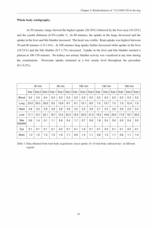
97
Chapter 4: Biodistribution of 123I-5-I-R91150 in the dog
Whole body scintigraphy:
At 30 minutes, lungs showed the highest uptake (20-29%) followed by the liver area (10-22%)
and the caudal abdomen (6-9%).(table 1) At 60 minutes, the uptake in the lungs decreased and the
uptake in the liver and bile bladder increased. The heart was visible. Brain uptake was highest between
30 and 60 minutes (1.0-1.6%). At 100 minutes lung uptake further decreased while uptake in the liver
(18-21%) and the bile bladder (0.7-1.7%) increased. Uptake in the liver and bile bladder reached a
plateau at 100-130 minutes. No kidney nor urinary bladder activity was visualized at any time during
the examination. Periocular uptake remained at a low steady level throughout the procedure
(0.1-0.2%).
30 min 60 min 100 min 130 min 160 min
Subj 1 Subj 2 Subj 3 Subj 1 Subj 2 Subj 3 Subj 1 Subj 2 Subj 3 Subj 1 Subj 2 Subj 3 Subj 1 Subj 2 Subj 3
Blood 0.3 0.3 0.4 0.3 0.3 0.3 0.3 0.3 0.3 0.3 0.3 0.3 0.3 0.3 0.2
Lung 25.0 20.5 29.0 9.5 13.9 8.7 8.1 15.1 8.0 7.5 13.7 7.5 7.0 12.4 7.0
Heart 4.8 3.5 3.9 3.9 2.8 2.9 3.2 2.5 2.8 2.7 2.5 2.6 2.6 2.0 2.4
Liver 17.1 10.7 22.1 18.7 12.5 22.3 18.2 20.0 21.0 18.2 19.8 20,5 17.9 19.7 20.5
Bile 0.6 1.4 0.1 1.1 0.9 0.4 1.7 0.7 0.8 1.8 0.4 0.6 2.0 0.4 0.5bladder
Eye 0.1 0.1 0.1 0.1 0.2 0.1 0.1 1.0 0.1 0.1 0.2 0.1 0.1 0.2 0.1
Brain 1.2 1.5 1.5 1.0 1.6 1.1 0.9 1.4 1.1 0.8 1.2 1.1 0.8 1.1 1.0
Table 1: Data obtained from total body acquisitions: tracer uptake (% of total body radioactivity) in differentregions

98
Chapter 4: Biodistribution of 123I-5-I-R91150 in the dog
SPET brain imaging
SPET images provided good anatomical definition of the frontal regions and showed low
uptake in the cerebellar area. (Fig 3). Peak uptake in the cerebellar area occurred slightly before peak
uptake in the frontal area, but never reached the same level of activity as the frontal area.
Figure 3: a. Horizontal slice of transmission data fitted to the CT slice taken from the same dog, facilitatingtopographic orientation on the emission data. b. On the emission images fronto-cortical (1) and temporo-cortical(2) uptake of radioactivity is visualized. Low uptake is noted in the cerebellar area (3).

99
Chapter 4: Biodistribution of 123I-5-I-R91150 in the dog
Specific binding of the ligand to the receptors could be estimated from the counts registered in
the fronto-cortical region (total binding) and the cerebellum (free and specific bound ligand). The
frontal to cerebellar ratio increased to a maximum of 1.85-2.39 at 90-100 minutes p.i. A constant ratio
was found between 90 and 180 minutes p.i. The peak ratio was consistently higher in dog 4. This dog
showed more anxiety and was less cooperative than the two other animals. (Fig 4).
Figure 4: Graphic illustration of fronto-cerebellar ratio in function of time, generated from the SPECT data.

100
Chapter 4: Biodistribution of 123I-5-I-R91150 in the dog
Displacement study
Intravenous administration of the receptor blocking agent ketanserin after administration of 123I-
5-I-R91150 resulted in rapid decrease of the radioactivity in the cortical areas. The ratio of fronto-
cortical/cerebellar activity (specific/non-specific binding) was 2.2-2.4 in the baseline study. After the
administration of cold (not labelled with 123I) ketanserin binding decreased to 1.5-1.7 with 1 mg/kg and
1.0-1.09 with 5mg/kg, indicating displacement of the radioligand from the receptors. (Fig 5).
Pretreatment with ketanserin led to uniform low binding of the 123I-5-I-R91150. (Fig 6).
Figure 5: Evaluation of total and specific binding in time (measured as activity/ pixel), after displacement with1mg/kg and 5 mg/kg ketanserin injected IV at 180 minutes after administration of labelled R 91150.
Figure 6: Binding of 123I-5-I-R91150: uptake in the different brain regions with (blocked) and without(unblocked) pretreatment with cold ketanserine.F= frontal cortex - T= temporal cortex - P= parietal cortex - O= occipital cortex - C= cerebellum

101
Chapter 4: Biodistribution of 123I-5-I-R91150 in the dog
Discussion
The tracer under investigation crossed the canine blood brain barrier and concentrated mainly
in the cortical regions, showing specific and reversible binding. Radioactivity in blood decreased fast
and remained stable thereafter, indicating a fast distribution to and no redistribution from various
organs. Redistribution would result in disruption of the steady state and as a consequence, would
preclude semi-quantification of receptor binding using the “ratio” method. On the total body scans it
was found that, in the early phase, highest activity was present in the lungs. Activity in the liver
predominated from 60 minutes onwards.
At no time the bladder shadow, nor the kidneys were visualized in our series. In humans,
highest activity in the liver and bowel is already evident on the early images and although the bladder
shadow is visualized at 180-210 minutes, the activity registered in this organ is low.30 This finding may
implicate that elimination of the tracer and its radioactive metabolites mainly occur by the intestinal
system rather than by the urinary system, probably as a result of plasma protein binding.
The maximal ligand uptake (1.0-1.6% of total body activity) in the canine brain is lower than
what is found in humans (2%)30. In general, low brain uptake is thought not to be caused by low
permeability through the blood brain barrier (BBB) but rather by low extractability due to high plasma
protein binding.31 More specifically, mass difference relative to bodyweight between the brain of the
examined dogs (100-120g) and the average human brain (1200g) has also to be considered. Despite
this low uptake, SPET imaging allows localization of the 5-HT2A receptors in the brain and gives
information regarding the dynamic binding of these receptors. Relative high uptake is noted in the
ocular area (0.1-0.2%). In one study performed in primates, this activity precluded accurate
measurement of the activity in the frontal cortex.31 This was not a problem in our study because of the
more rostral position of the eyes in the dog. Ketanserin did not affect the periocular activity in primate
studies nor did it in our displacement study, suggesting that this activity is caused by non specific
binding rather than by the presence of receptors in this area.31 This non specific binding could involve
melanin, since it is known that a variety of substances (amphetamine, (nor)ephedrine, prostaglandines,
adrenoreceptor agonists and antagonists, chloroquine, rifampicine, chlorpromazine, bezodiazepines..)
exhibit melanin binding properties.40-44
The maximal cortico-cerebellar ratio found in these dogs (ratio 1.85-2.39) between 90 and 180
minutes p.i. was similar but slightly higher than the ratios found in normal human (ratio 1.4 at 100
minutes) and non-human primates (ratio 1.5 at 60 minutes).30,31 In rats, the ratio of uptake in the frontal
cortex relative to the cerebellum (a region free from 5-HT2A receptors 45) was 10 at 60 minutes p.i. 29
The density of 5-HT2A receptors in the cortex is about three times higher than in the primate cortex,
thus explaining the higher cortico-cerebellar ratio in this species.31

102
Chapter 4: Biodistribution of 123I-5-I-R91150 in the dog
Cerebellar peak radioactivity was reached between 0-10 minutes in primate brain and at 5
minutes in humans.30,31 In our dogs, peak uptake of cerebellum and fronto-cortical area was reached on
the first image, at 6 minutes, as a result of vascular activity. After this peak, activity decreased over
time with a higher fronto-cortical activity compared to the cerebellar activity. The fronto-cortical to
cerebellar activity (specific/ non specific binding ratio) reached its maximal value at approximately 90
minutes p.i., remaining relatively stable until 180 minutes, indicating this to be the optimal scanning
time interval. These findings are similar to the findings in man and non-human primates. In healthy
humans peak activity in the frontal cortex was reached at approximately 100-120 minutes p.i. and
remained stable from approximately 100 minutes p.i. onwards30. In non-human primates a stable
plateau is reached at 150 minutes.31
Displacement with ketanserin resulted in a reduced radioactivity in the cortical regions of non-
human and human primates.27,31 In our study, a decrease of activity in all cortical areas was noted on
the first image at 10 minutes p.i., to the level observed in the cerebellum, remaining constant during
the following 60 minutes indicating displacement of the ligand from the receptors. Uniform low uptake
in the brain was noted after pre-treatment with ketanserin suggesting blockade of the receptors. These
findings indicate reversibility and specificity of binding, essential prerequisites for quantification of
receptors.
This study suggests that the radiolabelled 5-HT2A receptor antagonist R-91150 is a suitable
tracer to map and (semi)quantify receptor binding in the canine brain in order to investigate
abnormalities related to this receptor.

103
Chapter 4: Biodistribution of 123I-5-I-R91150 in the dog
Conclusive remarks
First, the use of dogs in research on new potential tracers is of practical interest. Since housing,
maintenance and handling of dogs is less tedious than primates and since dogs are also easy to obtain
and less expensive, it is worth the effort to investigate the use of dogs in this type of research.
Moreover, it has been proposed that certain canine behavioural disorders show resemblance to human
brain disease. Alterations in aging brain of dogs and humans show similar neuropathological changes,
including apoptosis, beta-amyloidal plaque formation and amyloidal angiopathy.46-48 Since the
serotonergic system has been brought in connection with mood disorders in aging, it would be of
scientific interest to evaluate the binding parameters of the 5-HT2A radioligand in canine aging brain.
Also, impulsive aggression in both man and dogs seems to be based on similar biological grounds, as
was shown with studies measuring cerebrospinal fluid concentrations of 5-hydroxyindoleacetic acid
(5-HIAA) in inward or outward directed aggression in man and aggressive behaviour in dogs, inversely
correlating with the unmediated character of the violent acts in man and with the unpredictability of
biting incidents in dogs.15,49-52 Also, recent research in veterinary science has focussed on the evaluation
of behaviour and cognitive status of dogs with tests and goal oriented questionnaires, making
assessment of canine behaviour more objective.53,54
Second, concerning the evaluation of the 5-HT2 receptor in particular, this receptor has been
brought in connection with depression, aggression, suicide and eating disorders in man.3,34,55,56 The
contribution of SPET in evaluating the status of this receptor is principal of practical value, since this
imaging modality is more economical and more accessible for most nuclear facilities. The availability
of the specific antagonist R91150, labelled with radioiodine, has already contributed to the in vivo
investigation of the binding properties of the 5-HT2A receptor in aging, suicidal behaviour, eating
disorders and therapeutic intervention with atypical neuroleptics in schizophrenic patients.3,32-34
It would therefore be interesting to evaluate the 5-HT2A receptor status with this radioligand
for SPET, in selected canine brain disease that show similarities in biological substrate to human
disorders. To evaluate the advantage of a canine model, future studies are necessary to examine the
binding parameters of this particular radioligand in pathologic states. This research modality could
have the additional advantage in that it may determine the impact of certain drugs on the 5-HT2A
receptor in canine brain.
ACKNOWLEDGMENTS
The authors would like to express their appreciation to Mr. H. De Bourdere and Mr.
M.Verdonck for providing technical support.

104
Chapter 4: Biodistribution of 123I-5-I-R91150 in the dog
REFERENCES
1. Busatto GF. Radioligands for brain 5-HT2 receptor imaging in vivo: why do we need them? Eur J Nucl Med1996; 23: 867-870.
2. Nobler MS, Mann JJ, Sackeim HA. Serotonin, cerebral blood flow and cerebral metabolic rate in the geriatricmajor depression and normal aging. Brain Res Reviews 1999; 30: 250-263.
3. Audenaert K, Van Laere K, Dumont F, Slegers G, Mertens J, van Heeringen C, Dierckx R. Decreased frontalserotonin 5-HT2a receptor binding index in deliberate self harm patients. Eur J Nucl Med 2001; 28: 175-182.
4. Meltzer CC, Smith G, Price JC, Reynolds CF, Mathis CA, Greer P, Lopresti B, Mintun MA, Pollock BG, Ben-Eliezer D, Cantwell MN, Kaye W, DeKosky ST. Reduced binding of {18F} altanserin to serotonin type 2Areceptors in aging: persistence of effect after partial volume correction. Brain Res 1998; 813: 167-171.
5. Olivier B, Mos J. Rodent models of aggressive behavior and serotonergic drugs. Prog NeuropsychopharmacolBiol Psychiatry 1992; 16: 847-870.
6. Olivier B, Mos J, Van Oorschot R, Hen R. Serotonin receptors and animal models of aggressive behavior.Pharmacopsychiat 1995; 28: 80-90.
7. Westergaard GC, Suomi SJ, Higley DE, Mehlman PT. CSF 5-HIAA and aggression in female macaquemonkeys: species and interindividual differences. Psychopharmacology-Berl 1999; 146: 440-446.
8. Doudet D, Hommer D, Higley JD, Andreason PJ, Moneman R, Suomi SJ, Linnoila M. Cerebral glucosemetabolism, CSF 5-Hiaa levels and aggressive behavior in rhesus monkeys. Am J Psychiatry 1995; 152: 1782-1787.
9. Higley JD, Mehlman PT, higley SB, Fernald B, Vickers J, Lindell SG, Taub DM, Suomi SJ, Linnoila M.Excessive mortality in young free-ranging male nonhuman primates with low cerebrospinal fluid 5-hydroxyindoleacetic acid concentrations. Arch Gen Psychiatry 1996; 53: 537-543.
10. Higley JD, Linnoila M. Low central nervous system serotonergic activity is traitlike and correlates withimpulsive behavior: a nonhuman primate model investigating genetic and environmental influences onneurotransmission. Ann N Y Acad Sci 1997; 836: 39-57.
11. Mehlman PT, Higley JD, Faucher I, Lilly AA, Taub DM, Vickers J, Suomi SJ, Linnoila M. Low CSF 5-HIAAconcentrations and severe aggression and impaired impulse control in non human primates. Am J Psychiatry1994; 151: 1485-1491.
12. Winberg S, Myrberg AA, Nilsson GE. Agonistic interactions affect brain serotonergic activity in anacanthopterygiian fish: the bicolor damselfish (Pomacentrus partitus). Brain Behav Evol 1996; 48: 213-220.
13. Popova NK, Voitenko NN, Kulikov AV, Avgustinovich DF. Evidence for the involvement of central serotoninin mechanism of domestication of silver foxes. Pharmacol Biochem Behav 1991; 40: 751-756.
14. Delville Y, Melloni RH, Ferris CF. Behavioral and neurobioligical consequences of social subjugation duringpuberty in golden hamsters. J Neurosci 1998; 18: 2667-2672.
15. Reisner IR, Mann JJ, Stanley M, Huang Y, Houpt KA. Comparison of cerebrospinal fluid monoaminemetabolite levels in dominant-aggressive and non- aggressive dogs. Brain Res 1996; 714: 57-64.
16. Reisner IR. Assessment, management, and prognosis of canine dominance-related aggression. Vet Clin NorthAm Small Anim Pract 1997; 27: 479-495.
17. Asberg M, Traskman L, Thoren P. 5-HIAA in the cerebrospinal fluid. A biochemical suicide predictor? ArchGen Psychiatry 1976; 33: 1193-1197.
18. Arango V, Underwood MD, Gubbi AV, Mann JJ. Localized alterations in pre- and postsynaptic serotoninbinding sites in the ventrolateral prefontal cortex of suicide victims. Brain Res 1995; 688: 121-133.

105
Chapter 4: Biodistribution of 123I-5-I-R91150 in the dog
19. Soares JC, van Dijck CH, Tan P, Soghbi SS, Garg P, Soufer R, Baldwin RM, Fujita M, Staley JK, Fu X, AmiciL, Seibyl J, Innis RB. Reproducibility of in vivo brain measures of 5-HT2A receptors with PET and {18F}deuteroaltanserin. Psychiatry Res: neuroimaging section 2001; 106: 81-93.
20. Baron JC, Samson Y, Comar D, Crouzel C, Deniker P, Agid Y. Etude in vivo des récepteurs sérotoninergiquescentraux chez l'homme par tomographie à émission de positions. Rev Neurol 1985; 141: 537-545.
21. Wong DF, Wagner HN, Dannals RF, Links JM, Frost JJ, Ravert HT, Wilson AA, Rosenbaum AE, Gjedde A,Douglas KH, Burns HD, Kuhar MJ. Effects of age on dopamine and serotonin receptors measured by positrontomography in the living human brain. Science 1984; 226: 1393-1396.
22. Frost JJ, Dannals RF, Mayberg HS, Links JM, Ravert HT, Kuhar MJ, Wagner HN. Regional localization ofserotonin-2 receptors in man using C-11-N-methylketanserin (NMKET) and PET. J Nucl Med 1987; 28:600-604.
23. Wong DF, Lever JR, Hartig PR, Dannals RF, Villemagne V, Hoffman BJ, Wilson AA, Ravert HT, Links JM,Scheffel U, Wagner HN. Lokalisation of serotonin 5-HT2 receptors in living human brain by positron emissiontotmography using N1-(11C-methyl)-2-Br-LSD. Synapse 1987; 1: 393-398.
24. Lemaire C, Damhaut P, Cantineau R, Plenevaux A, Christiaens L. NCA synthesis of an N-w-(18F)-fluoroethylanalog of altanserin, a serotonin S2 receptor ligand. J Label Compd Radiopharm 1991; 30: 374-375.
25. Moerlein SM, Perlmutter JS. Central serotonergic S2 binding in papio anubis measured in vivo with N-oo-(18F)fluoroethylketanserin and PET. Neurosci Lett 1991; 123: 23-26.
26. Attar-Lévy D, Martinot J, Blin J, Dao-Castellana M, Crouzel C, Mazoyer B, Poirier M, Bourdel M, AymardN, Syrota A, Féline A. The cortical serotonin-2 receptors studied with positron-emission tomography and 18F-setorperone during depressive illness and antidepressant treatment with clomipramine. Biol Psychiatry 1999;45: 180-186.
27. Sadzot B, Lemaire C, Maquet P, Salmon E, Plenevaux A, Degueldre C, Hermanne JP, Guillaume M, CantineauR, Comar D, Franck G. Serotonin 5HT2 receptor imaging in the human brain using positron emissiontotmography and a new radioligand, 18F-altanserin: results in young normal controls. J Cereb Blood FlowMetab 1995; 15: 787-797.
28. Terriere D, Janssen P, Gommeren W, Gysemans M, Mertens J, Leysen J. Evaluation of radioiodo-4-amino-N-(1-(4-fluorophenoxy)-propyl)-4-methyl-4-piperidinyl)-5-iodo-2-methoxybenzamide as a potential 5HT2receptor tracer for SPE(C)T. Nucl Med biol 1995; 22: 1005-1010.
29. Mertens J, Terriere D, Sipido V, Gommeren W, Janssen PMF, Leysen JE. Radiosynthesis of a newradioiodinated ligand for serotonin-5HT2-receptors, a promising tracer for gamma-emission tomography. J Label Compd Radiopharm 1995; 34: 795-801.
30. Busatto GF, Pilowsky LS, Costa DC, Mertens J, Terriere D, Ell PJ, Mulligan R, Travis MJ, Leysen JE, Lui D,Gacinovic S, Waddington W, Lingford-Hughes A, Kerwin RW. Initial evaluation of 123I-5-I-R91150, aselective 5-HT2A ligand for single photon emission tomography in healthy subjects. Eur J Nucl Med 1997;24: 119-124.
31. Abi-Dargham A, Zea-Ponce Y, Terriere D, Al-Tikriti M, Baldwin RM, Hoffer P, Charney D, Leysen JE,Laruelle M, Mertens J, Innis RB. Preclinical evaluation of (123I)R93274 as a SPECT radiotracer for imaging5-HT2A recpetors. Eur J Pharmacol 1997; 321: 285-293.
32. Baeken C, D'haenen H, Flamen P, Terriere D, Chavatte K, Boumon R, Bossuyt A. 123I-5-I-R91150, a newsingle photon emission tomography ligand for 5-HT2A receptors: influence of age and gender in healthysubjects. Eur J Nucl Med 1998; 25: 1617-1622.
33. Travis M, Busatto G, Pilowsky L, Mulligan R, Acton P, Gacinovic S, Mertens J, Terriere D, Costa D, Ell P,Kerwin R. 5-HT2a receptor blockade in patients with schizophrenia treated with risperidone and clozapine. ASPET study using the novel 5-HT2a ligand 123I-5-I-R-91150. Br J Psychiatry 1998; 173: 236-241.
34. Audenaert K, Van Laere K, Dumont F, Vervaet M, Goethals I, Slegers G, Mertens J, van Heeringen C, DierckxR. Decreased 5-HT2A binding in patients with anorexia nervosa. J Nucl Med 2002; in press.

106
Chapter 4: Biodistribution of 123I-5-I-R91150 in the dog
35. Maes F, Vandermeulen D, Suetens P. Automated multimodal image registration by maximization of mutualinformation: from theory, implementation and validation to a useful tool in routine clinical practice. IEEETrans Med Imaging 1997; 16: 187-198.
36. Pazos A, Probst A, Palacios J. Serotonin receptors in the human brain-IV. Autoradiographic mapping ofserotonin-2 receptors. Neuroscience 1987; 21: 123-139.
37. Pazos A, Cortes R, Palacios JM. Quantitative autoradiographic mapping of serotonin receptors in the ratbrain.II.Serotonin-2 receptors. Brain Res 1985; 346: 231-249.
38. Lopez-Gimenez JF, Vilaro MT, Palacios J, Mengod G. Mapping of 5-HT2A receptors and their mRNA inmonkey brain: (3H)MDL100.907 autoradiography and in situ hybridization studies. J Comp Neurol 2001; 429:571-589.
39. Hamada S, Senzaki K, Hamaguchi-Hamada K, Tabuchi K, Yamamoto H, Yamamoto T, Yoshikawa S, OkanoH, Okado N. Localization of 5-HT2A receptor in rat cerebral cortex and olfactory system revealed byimmunohistochemistry using two antibodies raised in rabbit and chicken. Molecular Brain Research 1998; 54:199-211.
40. Wepierre J, Cohen Y, Rapin J. Accumulation of ephedrine, norephedrine, amphetamine and tyramine labeledwith carbon 14 in pigmented and non-pigmented eyes of rats. Arch Int Pharmacodyn Ther 1975; 217: 342-350.
41. Debing I, Ijzermen A, Vauquelin G. Melanosmoe binding and oxidation-reduction properties of synthetic L-DOPA-melanin as in vitro tests for drug toxicity. Mol Pharmacol 1988; 33: 470-476.
42. Aula P, Kaila T, Huupponen R, Salminen L, Lisalo E. Prostaglandin F2 alpha binding to bovine ocular andsynthetic melanin in vitro. Pharmacol Toxicol 1989; 65: 100-103.
43. Steiner K, Buhring KU, Merck E. The melanin binding of bisopropol and its toxicological reference. Lens EyeToxic Res 1990; 7: 319-333.
44. Koike T, Kitazuma H, Mukai H. Tissue distribution of NS-49, a phenethylamine alpha 1A adrenoreceptoragonist. Arzneimittelforschung 2001; 51: 402-407.
45. Leysen JE, Janssen P, Heylen L, Gommeren W, Van Gompel P, Lesage AS, Megens AAHP, Schotte A.Receptor interactions of new antipsychotics: relation to pharmacodynamic and clinical effects. Int J PsychiatryClin Pract 1998; 2: S3-S17.
46. Anderson A, Ruehl W, Fleischman L, Stenstrom K, Entriken T, Cummings B. DNA damage and apoptosis inthe aged canine brain: relationship to Abeta deposition in the absence of neuritic pathology. 2000; 24: 787-799.
47. Torp R, Head E, Cotman C. Ultrastructural analyses of beta-amyloid in the aged dog brain: neuronal b-amyloidis localized to the plasma membrane. Prog Neuropsychopharmacol Biol Psychiatry 2000; 24:801-810.
48. Head E, Thornton P, Tong L, Cotman C. Initiation and propagation of molecular cascades in human brainaging: insight from the canine model to promote successful aging. Prog Neuropsychopharmacol BiolPsychiatry 2000; 24: 777-786.
49. Brown GL, Linnoila M. CSF serotonin metabolite (5-HIAA) studies in depression, impulsivity, and violence.J Clin Psychiatry 1990; 51: supl 31-41.
50. Virkkunen M, Rawlings R, Tokola R, Poland RE, Guidotti A, Nemeroff C, Bissette G, Kalogeras K, KaronenSL, Linnoila M. CSF biochemistries, glucose metabolism, and diurnal activity rhythms in alcoholic, violentoffenders, fire setters, and healthy volunteers. Arch Gen Psychiatry 1994; 51: 20-27.
51. Coccaro E, Kavoussi R, Trestman R, Gabriel M, Cooper T, Siever LJ. serotonin function in human subjects:intercorrelations among central 5-HT indices and aggressiveness. Psychiatry Res 1997; 73: 1-14.
52. Constantino JN, Morris JA, Murphy DL. CSF 5-HIAA and family history of antisocial personality disorder innewborns. Am J Psychiatry 1997; 154: 1771-1773.

107
Chapter 4: Biodistribution of 123I-5-I-R91150 in the dog
53. Kiatipattanasakul W, Nakamura S, Hossain M, Nakayama H, Uchino T, Shumiya S, Goto N, Doi K. Apoptosisin the aged dog brain. Acta neuropathol 1996; 92: 242-248.
54. Adams B, Chan A, Callahan H, Milgram N. The canine as a model of human cognitive aging: recentdevelopments. Prog Neuropsychopharmacol & Biol Psychiatry 2000; 24: 675-692.
55. Biegon A, Essar N, Israeli M, et al. Serotonin 5HT2 receptor binding on blood platelets as a state dependentmarker in major affective disorder. Psychopharmacology (Berl) 1990; 102: 73-75.
56. Stockmeier CA. Neurobiology of serotonin in depression and suicide. In: Mann, J. J. and Stoff, D. M., ed. Thepsychobiology of suicide. New York: The New York Academy of Sciences, 1997;


CHAPTER 5
Regional binding index of the selective 5-HT2A radiolabelledantagonist 123 I-5-I-R91150 in the normal canine brain
imaged with single photon emission tomography
K.Peremans1, K.Audenaert2, F.Coopman3, F.Jacobs2, F.Dumont4, G.Slegers4,
F.Verschooten1, H.van Bree1, J.Mertens5, R.Dierckx2
1From the department of Medical Imaging, Faculty of Veterinary Medicine, Ghent
University Salisburylaan 133, B-9820 Merelbeke, Belgium2Division of Nuclear Medicine Ghent University Hospital, De Pintelaan 185,
B-9000 Gent, Belgium3Department of Animal Nutrition, Genetics, Breeding and Ethology, Faculty of Veterinary
Medicine, Ghent University, Heidestraat 19, B-9820 Merelbeke4Laboratory of Radiopharmacy, Faculty of Pharmaceutical Sciences, Ghent University,
Harelbekestraat 72, B-9000 Gent, Belgium5VUB-cyclotron, Brussels, Belgium
Adapted from: accepted for publication in the Veterinary Radiology and Ultrasound


111
Chapter 5: Regional distribution of 123I-5-I-R91150 in the normal canine brain
Summary
The pattern of the specific 5-HT2A (5-hydroxytryptamine 2A receptor) antagonist 123I-5-I
R91150 was measured in 10 healthy dogs without neurologic and behavioural abnormalities. Eight
cortical regions (left and right fronto-, temporo-, parieto-, and occipito-cortical area), and one global
subcortical region (including the thalamic system) were compared with a reference region lacking
receptors, i.e. the cerebellum. The 123I labelled radioligand was injected intravenously 100-120 minutes
prior to acquisition. Both transmission and emission data were obtained with a triple head gamma-
camera equipped with high resolution fanbeam collimators. The emission data were corrected for
scatter and attenuation. To delineate different cerebral regions more accurately, the regions of interest
(ROI) defined in a former study on brain perfusion measured with 99mTc-ethyl cysteinate dimer (ECD)
in the same dogs were used. The co-registration of the 99mTc-ECD and the 123I-5-I-R91150, obtained
from each dog, was realized with the help of corresponding transmission maps. By normalizing each
regional cerebral activity to the activity observed in the cerebellum, the regional radioactivity (binding
index) could be relatively quantified. Highest brain uptake was noted in the fronto-cortical brain areas
(right: 1.85, left: 1.89), followed by the temporo-cortical region (right: 1.58, left: 1.56). Least uptake
was noted in the more caudal and middle brain regions (occipito-(right: 1.46, left: 1.41), parieto-
cortical (right: 1.30, left: 1.26) and striatal region (1.19)). Neither gender nor age influence was noted
in this series.
The 123I labelled serotonin-2A receptor ligand appears to have similar cortical binding in the
normal canine brain as shown in humans and other animal species. A frontocortical to occipitocortical
(rostro-caudal) binding index gradient was identified within the dog that has not been seen in imaging
studies from humans and other animal species. The significance of these results will need further
investigation. This normative data can be used to compare regional brain uptake of the 123I-
radioligand to dogs with behavioral disorders related to the serotonergic system, in future studies.

112
Chapter 5: Regional distribution of 123I-5-I-R91150 in the normal canine brain
Introduction
Serotonin (5-HT, 5-hydroxytryptamine) is a central and a peripheral neurotransmitter. It is
synthesized from the amino acid L-tryptophan by sequential hydroxylation and decarboxylation. It is
stored in pre-synaptic vesicles, released from central and peripheral nerve-terminals during neuronal
firing and metabolized by the mono-amino-oxidase (MAO) enzyme to 5-hydroxy indole acetic acid
(5-HIAA).1 Impairments of the serotonergic neurotransmitter have been associated with a variety of
human mental disorders, including psychotic disorders, mood and anxiety disorders, eating disorders
and dementia.2
A lower 5-HIAA concentration in cerebrospinal fluid (CSF) is thought to be an indication of
reduced serotonergic turnover. This reduced 5-HIAA in CSF is found in subjects that attempt suicide
or harm themselves, in violent offenders including murderers, in impulsive patients such as arsonists
and aggressive alcoholics, and in children with conduct disorders, e.g. children injuring animals
frequently and deliberately.3-6 Low 5-HIAA in CSF is also found in mammals and non-human
primates. In non-human primates a lower 5-HIAA in CSF is detected in violent individuals.7-9 In
aggressive, impulsive dogs, defined as sudden, unpredictable, poorly inhibited aggressive behavior, a
decrease in 5-HIAA is detected in the cerebrospinal fluid, a result maintained after controlling for
breed and irrespective of age and gender.10 It is concluded from these studies that the serotonergic
system plays an important role in impaired impulse control and modulates aggressive behaviour
throughout a large variety of animal species.8,10
Within the serotonergic system several 5-HT receptor types and subtypes have been
recognized1,11,12, each with a particular function. Because of its involvement in neuro-psychiatric
conditions such as mood and anxiety disorders, in aggression and impulsivity, attention has been
focused on the 5-HT2A receptor. Distribution of the 5-HT2A receptors was initially investigated post-
mortem with autoradiography13-16 or with immuno-histochemistry17 in both animals and humans. In
humans, primates and rats, these receptors are found predominantly in the rostral cerebral cortical
areas, to a somewhat lesser extent in the caudal regions and absent in the cerebellum.17-19 Based on post-
mortem brain receptor studies in violent suicide victims, increased 5-HT2A receptor densities were
found in cortical areas, predominantly in the frontal cortex.20,21 However, fast and unpredictable post-
mortem alterations in receptor density and affinity are an important methodologic weakness of post-
mortem brain research.
Nuclear medicine techniques enable in vivo examination of neuro-psychiatric disorders and
results of treatment18. Radioligands with selective in vivo binding affinity to the 5-HT2A receptor have
been developed for positron emission tomography (PET) 22-24 and single photon emission computed
tomography (SPECT).19 SPECT makes use of radionuclides with a longer half life and therefore does
not require a cyclotron in the vicinity of the camera and it is therefore a more accessible technique. 123I-
5-I-R91150 (R93274), a highly selective radioiodinated antagonist of the 5-HT2A receptors was

113
Chapter 5: Regional distribution of 123I-5-I-R91150 in the normal canine brain
synthesized for SPECT imaging.25,26 The feasibility of this tracer was tested in rats, non-human
primates and humans.19,27 The specificity of cortical binding of this radioligand to 5-HT2A receptors
was investigated in primates and dogs through displacement studies with ketanserin, a 5-HT2A
antagonist.27,32 In rats, the ratio of radioactivity in the frontal cortex relative to the cerebellum, a region
free from 5-HT2A receptors, is 10:1 at 60 minutes. In non-human primates, the cortex-cerebellar ratio
is 1.5 at 60 minutes.27 In sequential SPECT brain scans in humans the cortico-cerebellar ratio (1.5- 1.8)
reaches a plateau between 90 and 110 minutes, remaining stable from then onwards, thus reflecting a
pseudoequilibrium.19 Since these pilot studies, several studies have been carried out in normal humans
and in pathologic states.28-31
Since nothing was found concerning the characteristics of this radioligand in dogs, we
performed preliminary studies and demonstrated that the binding characteristics of the selective123I-5-I-R91150 radioligand for the 5-HT2A receptor fulfilled the requirements for semi-quantification
in the canine brain, using the ratio of activity in a region to a reference region. Binding was reversible
and a steady state (pseudo-equilibrum) was reached from 90 minutes onwards.32
The aim of this study was to investigate the distribution pattern of the regional binding of123I-5-I-R91150 radioligand for 5-HT2A receptor imaging in the canine brain and consequently
determine the distribution pattern of 5-HT2A receptors within the brain in a group of 10 normal dogs
using SPECT attenuated and scatter corrected images. Knowledge of the normal distribution pattern
will enable studies of brain pathology associated with canine behavioral abnormalities caused by
altered binding properties of the 5-HT2A receptor.

114
Chapter 5: Regional distribution of 123I-5-I-R91150 in the normal canine brain
Materials and methods
Subjects
Ten normal healthy shepherd type dogs were studied. There were 5 intact males and 5 intact
females. Age varied between 1 and 7 years (mean: 4.1; standard deviation (SD): 2.3) and weight
between 13 and 41 kg (mean: 30.0 kg; SD: 9.0). This study was approved by the local ethical
committee.
TRACER
123I-5-I-R91150 was synthesized by electrophilic substitution on the 5-position of the
methoxybenzamide group of R91150, followed by purification with high-performance liquid
chromatography. The product had a radiochemical purity of more than 99 % and was sterile and
pyrogen free. A specific activity of 370 GBq/µmol was obtained.
The tracer is a 5-HT2A antagonist with high affinity (Kd = 0.11 nM) and selectivity for 5-
HT2A receptors. The selectivity of the ligand for 5-HT2A receptors with regard to other
neurotransmitter receptors such as other 5-HT receptors, including 5-HT2C and 5-HT1A, dopamine
receptors, adrenoreceptors and histamine receptors is at least a factor of 50. The tracer is displaceable
with ketanserin, a competitor for receptor binding.25,26 27
In this study, the decay corrected injected activity ranged from 4.07-7.77 MBq/kg (mean:
5.55MBq/kg; SD: 0.04) IV. The interval between injection and acquisition ranged from 100 to 120
minutes (mean: 113 minutes; SD: 8.6).32
SPECT acquisition
The SPECT acquisitions were performed under general anesthesia. Premedication to
anesthesia was obtained with 10-30µg/kg IV medetomidin hydrochloride (Domitor®, Pfizer, Orion,
Orion corporation, Espoo, Finland). General anesthesia was induced with 2-3 mg/kg iso-propylphenol
(Rapinovet®, Schering Plough Animal Health, Brussels, Belgium) intravenously and maintained with
halothane (Fluothane®, Zeneca, Destelbergen, Belgium) to effect. The dogs were positioned in ventral
recumbency. The SPECT acquisition was performed with a triple head gamma camera (Toshiba GCA-
9300A, Dutoit Medical, Antwerp, Belgium), equipped with high resolution fanbeam collimators (full
width at half maximum (FWHM) of 7.8 mm). A single photon transmission computed tomography
scan (TCT-scan) of 4 minutes was obtained prior to the emission scan, using three 153-Gadolinium

115
Chapter 5: Regional distribution of 123I-5-I-R91150 in the normal canine brain
(Gd) rod sources. This TCT images, having sufficient contrast to recognize bone and sinuses, were
obtained for later co-registration of serotonin-2A receptor emission data with 99mTc-ECD perfusion
emission data, obtained in a former study.33
Following the TCT scan, emission images were acquired over 20 minutes, thus obtaining the
whole brain volume within the single scanning session. For each acquisition, 90 projection images
were obtained on a 128x128 matrix using a continuous scan mode by rotating each head twice over
120°. The emission data sets were reconstructed with filtered back projection after rebinning to
parallel data and applying a Butterworth-filter (cut-off 0.16 cycles/pixel, order 8). Pixel size was 1.72
mm. Uniform Sorensen attenuation correction with linear attenuation coefficient of 0.12/cm and triple-
energy window scatter correction were applied.
After reconstruction, data were transferred to another computer equipped with Hermes
software from Nuclear Diagnostics (Hägersten, Stockholm, Sweden) for processing. Each receptor
radioligand image was co-registered with a formerly obtained perfusion image from the same dog.
Because of the poor anatomic definition of neuroreceptor imaging studies, the simultaneously acquired
transmission data were used to optimise co-registration of both data sets. (Fig 1).
Figure 1: The emission data of the 123I-5-I-R91150 study (right set of images) are automatically fitted to theirtransmission data (TCT data) (left set of images), provided that the dog’s head did not move between bothacquisitions. The bone structures of the skull are recognizable on the TCT images. Note the high uptake in theocular region (circular marking).

116
Chapter 5: Regional distribution of 123I-5-I-R91150 in the normal canine brain
Co-registration of the TCT data was performed with the multimodality software of Nuclear
Diagnostics. This computer program, displaying images in a dual window setting, allows for manual
co-registration by providing tools for rotation and translation images in all three dimensions. The
parameter values used to register the TCT map of the receptor radioligand image to the TCT map of
the perfusion image were then applied to the emission image of the receptor radioligand, thus co-
registering this image with the perfusion image. This allowed us to use the regions of interest defined
on the perfusion images for the serotonin-2A receptor images. (Fig 2a, b, c, d). The predefined and
standardized regions of interest consisted of eight cortical brain regions (left and right fronto-,
temporo-, parieto- and occipitocortical), one global subcortical region and the cerebellum. The
regional classification was performed according to prior definitions (1978) and was also described in
more detail in a former study on brain perfusion in the dog.33,34
Figure 2: a. Overview of horizontal TCT slices from the skull. Manual co-registration of the TCT data of thereceptor radioligand studies (right images) to the TCT images of the perfusion study (left images) is performed byrotation, shifting and scaling of the TCT data of the receptor radioligand studies, using the Multi-Modalitysoftware from Hermes (Nuclear Diagnostics).

117
Chapter 5: Regional distribution of 123I-5-I-R91150 in the normal canine brain
Figure 2: b. Detail of a TCT image from a perfusion study (left image) and from the receptor radioligand study(right image). Using the sliding window facility, the TCT image of the receptor radioligand study will besuperimposed on the TCT image of the perfusion study, allowing visual inspection of the fitting procedure. Theuse of a different color scale simplifies this procedure.
Figure 2:c. The parameters (rotation, shift and scale) used for co-registration of the TCT data of both studies areextended to the emission data of the receptor radioligand images. By this procedure the emission data of bothstudies are adjusted to each other.

118
Chapter 5: Regional distribution of 123I-5-I-R91150 in the normal canine brain
Figure 2:d. Horizontal slices taken at the level of the lower midbrain. The predefined, standardized regions ofinterest on the perfusion image are automatically extended to the emission image of the 123I-5-I-R91150 study.This procedure is performed for all slices. 2: left fronto-cortical region, 3: right fronto-cortical region, 4: lefttemporo-cortical region, 5: right temporo-cortical region, 6: subcortical region, 7: cerebellum

119
Chapter 5: Regional distribution of 123I-5-I-R91150 in the normal canine brain
The binding index (BI) was operationally estimated as (counts/pixel in regional cortex) /
(counts/pixel in reference region). This BI is proportional to the concentration of available receptors
under near equilibral conditions. To apply this semi-quantification method using a BI as a measure for
specific binding (binding to the receptor under investigation), a reference region is needed. This region
must be void of the concerned receptor and must have similar concentration of free and non-specific
bound radioligand as the region under examination. The cerebellum satisfies these conditions and is
therefore selected as a reference region for studies involving the 5-HT2A receptor.
Statistics
Correlations between age, weight, injected dose/kg bodyweight and regional activity were
calculated using the Spearman’s correlation. Differences in age, weight, dose and regional activity
versus gender were evaluated with the independent samples Mann-Whitney U test. Left to right BI in
fronto-, parieto-, temporo- and occipito-cortical regions and rostro-caudal gradient, were evaluated
through the related samples Wilcoxon Signed Rank Test. At first, the null hypotheses on laterality was
that there were no differences between left and right sided binding indices. Secondly, the null
hypothesis on rostro-caudal serotonin-2A binding was that no difference between frontal and occipital
binding index was present. Level of significance was set at p < 0.05.

120
Chapter 5: Regional distribution of 123I-5-I-R91150 in the normal canine brain
Results
Highest uptake was noted in the fronto-cortical cerebral region (right fronto-cortical region
(RF): 1.85, SD: 0.07; left fronto-cortical region (LF): 1.89, SD: 0.09) followed by the temporo-cortical
region (right temporo-cortical region (RT): 1.58, SD: 0.15; left temporo-cortical region (LT): 1.56, SD:
0.11). The parieto-cortical (right parieto-cortical region (RP): 1.30, SD: 0.39; left parieto-cortical
region (LP): 1.26, SD: 0.35), occipito-cortical (right occipito-cortical region (RO): 1.46, SD: 0.21; left
occipito-cortical region (LO): 1.41, SD: 0.21) and subcortical regions (1.19, SD: 0.40) had a lower BI.
The lowest activity was, as expected, noted in the cerebellar region set at 1.0. (Fig 3). There was no
correlation between injected dose and regional activity. There were no significant differences between
male and female subjects concerning age, weight, injected dose/kg bodyweight and regional activity,
in any of the regions.
When comparing bilateral regional activity of the fronto-, temporo- and occipito-cortical
region, no significant difference was found. There was a significant correlation between right and left
parietal binding index (_=0.67; p=0.02). There was a significant fronto-occipito-cortical gradient (left
and right; Z=-2.67; p=0.008).
Figure 3: Graphical representation of the mean registered activity normalized to the cerebellum in the differentbrain regions. Legend: RF: right fronto-cortical region, LF: left fronto-cortical region, RT: right temporo-cortical region, LT:left temporo-cortical region, RP: right parieto-cortical region, LP: left parieto-cortical region, RO: right occipitalregion, LO: left occipital region, SC: subcortical region, C: cerebellum

121
Chapter 5: Regional distribution of 123I-5-I-R91150 in the normal canine brain
Discussion
Results are in agreement with previously published human and animal studies on the in vivo
distribution pattern of 123I-5-R91150.19,27 Despite the low ligand uptake (1-1.6%) the high resolution,
fanbeam collimated SPECT provides useful information about receptor binding in different brain
structures.
In rats, the density of 5-HT2A receptors in the cortex is about three times higher than in the
primate cortex. This contributes to the higher cortico-cerebellar ratio in this species. In our series,
highest relative uptake was found in the cerebral cortex (fronto-cortical region having highest values),
followed by the subcortical region and least activity, as expected, was found in the cerebellum. In
humans, a similar pattern of regional variation was found with highest binding in the cerebral cortex,
followed by the striatal area, and lowest in the cerebellum, compatible with data from autoradiographic
studies.15,19 The rostro-caudal cortical gradient recognized in this group is not replicated in the few
existing studies using this ligand. The regional binding index of the cortical regions is also more
homogenous in human studies. However, a small variability in highest and lowest regional binding
index is apparent between existing human studies. 19,31 More, in some studies not all cortical regions are
evaluated29,31. This was also the situation in one primate study in which fronto-cortical activity was not
measured due to technical reasons, and parieto-cortical binding index was not mentioned.27 A possible
explanation for this higher BI in the fronto-cortical brain regions in our series could be the species
dependent distribution of these receptors.14 Larger studies are needed, to verify the presence of this
gradient. In primates, a high uptake was noted in the ocular and periocular region, contaminating the
region of interest of the frontal cortical area.27 A similar high uptake in the ocular area was noted in
our series. Since ocular uptake in the study with primates27 and in our preliminary study on dogs32, was
not influenced by ketanserin, this activity is most probably caused by non-specific binding to undefined
substances (proteins, lipids, other receptors...).
In humans, an age related decline of 5-HT2A receptor density was found both with
autoradiographic studies and with in vivo studies, using positron emission tomography (PET) or
SPET.28,35-37 In this study only a weak, non significant correlation was found with age and right fronto-
cortical binding index, but the number of subjects is too small to draw definite conclusions regarding
this matter. Gender difference was not found in a human study including 26 normal volunteers28 nor
was this present in this study.
Methodologic considerations and limitations:
General anesthesia is necessary in order to perform these studies. It is therefore important to
consider the effects of the anaesthetics used. Drugs having an effect on the serotonergic system,
directly or indirectly, have to be excluded. However, global cerebral blood flow (CBF) alterations as
result of the anaesthetic drugs, are inevitable.

122
Chapter 5: Regional distribution of 123I-5-I-R91150 in the normal canine brain
Medetomidine is an α2 agonist and produces no direct effect on the 5-HT2A receptors, or on
brain metabolism, but reduces cerebral blood flow in general, suggesting uncoupling of cerebral
metabolism and flow due to decreases in central catecholamine turnover.38 Induction of general
anesthesia obtained with iso-propylphenol also decreases blood flow. Halothane, isoflurane and
sevoflurane cause cerebral vasodilation and decreased vascular resistance in the brain.39 In one study
it was demonstrated that the use of dexmedetomidine prior to isoflurane and sevoflurane significantly
attenuated dilation of cerebral arterioles induced by these volatile anaesthetics.40 It is unlikely that the
regional variation of radioactivity is due to global flow alterations in the brain. Because all dogs
received the same anaesthetic protocol, individual blood flow pattern changes would be minimalized.
Another possible confounding factor is the partial volume effect, which increases as the size of
the object and resolution (FWHM) of the system decrease. It may be assumed that the activity due to
specific binding, registered in the cortical regions, will be more affected by the partial volume effect
than the activity registered in the area chosen as a reference for non-specific binding.
Conclusions:
This study presents data from canine brain uptake patterns of the 5-HT2A receptor radioligand,123I-5-I-R91150 using SPECT imaging. The findings in this study agree with previous studies in normal
human and non-human primates. Recognition of the normal distribution pattern of the 5-HT2A
receptor will enable evaluation of pathologic states of this receptor. The diagnostic value of this
technique will be tested in future investigations on canine neuropsychological disease involving the
serotonergic system. Also, this technique has the potential to become an attractive tool to image the
influence of pharmacologic interventions on receptor binding status and to correlate alterations in
binding index with therapeutic outcome.

123
Chapter 5: Regional distribution of 123I-5-I-R91150 in the normal canine brain
REFERENCES
1. Stahl S. Essential psychopharmacology. Cambridge: Cambridge University Press, 1996.
2. Ogren SO, Misane I. Animal models for studying serotonin (5-HT) receptor subtypes: relationship to 5-HTsystem pathologies. Nucl Med biol 1998; 25: 747-749.
3. Virkkunen M, Nuutila A, Goodwin FK, Linnoila M. Cerebrospinal fluid metabolite levels in male arsonists.Arch Gen Psychiatry 1987; 44: 241-247.
4. Virkkunen M, Goldman D, Nielsen D, Linnoila M. Low brain serotonin turnover rate (low CSF 5-HIAA) andimpulsive violence. J Psychiatry Neurosci 1995; 20: 271-275.
5. Asberg M, Traskman L, Thoren P. 5-HIAA in the cerebrospinal fluid. A biochemical suicide predictor? ArchGen Psychiatry 1976; 33: 1193-1197.
6. Cremniter D, Jamain S, Kollenbach K, Alvarez JC, Lecrubier Y, Gilton A, Jullien P, Lesieur P, Bonnet F,Spreux-Varoquaux O. CSF 5-HIAA levels are lower in impulsive as compared to nonimpulsive violent suicideattempters and control subjects. Biol Psychiatry 1999; 45: 1572-1579.
7. Higley JD, Mehlman PT, higley SB, Fernald B, Vickers J, Lindell SG, Taub DM, Suomi SJ, Linnoila M.Excessive mortality in young free-ranging male nonhuman primates with low cerebrospinal fluid 5-hydroxyindoleacetic acid concentrations. Arch Gen Psychiatry 1996; 53: 537-543.
8. Higley JD, Linnoila M. Low central nervous system serotonergic activity is traitlike and correlates withimpulsive behavior: a nonhuman primate model investigating genetic and environmental influences onneurotransmission. Ann N Y Acad Sci 1997; 836: 39-57.
9. Doudet D, Hommer D, Higley JD, Andreason PJ, Moneman R, Suomi SJ, Linnoila M. Cerebral glucosemetabolism, CSF 5-Hiaa levels and aggressive behavior in rhesus monkeys. Am J Psychiatry 1995; 152:1782-1787.
10. Reisner IR, Mann JJ, Stanley M, Huang Y, Houpt KA. Comparison of cerebrospinal fluid monoaminemetabolite levels in dominant-aggressive and non- aggressive dogs. Brain Res 1996; 714: 57-64.
11. Olivier B, Mos J, Van Oorschot R, Hen R. Serotonin receptors and animal models of aggressive behavior.Pharmacopsychiat 1995; 28: 80-90.
12. Tecott LH, Julius DJ. A new wave of serotonin receptors. Curr Opin Neurobiol 1993; 3: 310-315.
13. Mengod G, Vilaro MT, Raurich A, Lopez-Gimenez JF, Cortes R, Palacios JM. 5-HT receptors in mammalianbrain: receptor autoradiography and in situ hybridization studies of new ligands and newly identified receptors.Histochem.J 1996; 28: 747-758.
14. Lopez-Gimenez JF, Vilaro MT, Palacios J, Mengod G. Mapping of 5-HT2A receptors and their mRNA inmonkey brain: (3H)MDL100.907 autoradiography and in situ hybridization studies. J Comp Neurol 2001; 429:571-589.
15. Pazos A, Probst A, Palacios J. Serotonin receptors in the human brain-IV. Autoradiographic mapping ofserotonin-2 receptors. Neuroscience 1987; 21: 123-139.
16. Pazos A, Cortes R, Palacios JM. Quantitative autoradiographic mapping of serotonin receptors in the ratbrain.II.Serotonin-2 receptors. Brain Res 1985; 346: 231-249.
17. Hamada S, Senzaki K, Hamaguchi-Hamada K, Tabuchi K, Yamamoto H, Yamamoto T, Yoshikawa S, OkanoH, Okado N. Localization of 5-HT2A receptor in rat cerebral cortex and olfactory system revealed byimmunohistochemistry using two antibodies raised in rabbit and chicken. Molecular Brain Research 1998; 54:199-211.
18. Lingford-Hughes AR, Pilowsky LS. In vivo neuroreceptor imaging-methodology and applications inneuropsychiatry. In: De Deyn, P. P., Dierckx, R. A., Alavi, A., and Pickut, B. A., ed. SPECT in neurology andpsychiatry. London: John Libbey & Company Ltd, 1997; 89-106.

124
Chapter 5: Regional distribution of 123I-5-I-R91150 in the normal canine brain
19. Busatto GF, Pilowsky LS, Costa DC, Mertens J, Terriere D, Ell PJ, Mulligan R, Travis MJ, Leysen JE, Lui D,Gacinovic S, Waddington W, Lingford-Hughes A, Kerwin RW. Initial evaluation of 123I-5-I-R91150, a selective5-HT2A ligand for single photon emission tomography in healthy subjects. Eur J Nucl Med 1997; 24: 119-124.
20. Hrdina PD, Demeter TB, Vu P, Sotonyi P, Palkovits M. 5HT uptake sites and 5HT2 receptors in brain ofantidepressant-free suicide victims/depressives: increases in 5-HT2 sites in cortex and amygdala. Brain Res1993; 614: 37-44.
21. Mann JJ, Stanley M, McBride PA, McEwen BS. Increased serotonin 2 and beta adrenergic receptor binding inthe frontal cortex of suicide victims. Arch Gen Psychiatry 1986; 43: 954-959.
22. Attar-Lévy D, Martinot J, Blin J, Dao-Castellana M, Crouzel C, Mazoyer B, Poirier M, Bourdel M, AymardN, Syrota A, Féline A. The cortical serotonin-2 receptors studied with positron-emission tomography and 18F-setorperone during depressive illness and antidepressant treatment with clomipramine. Biol Psychiatry 1999;45: 180-186.
23. Lundkvist C, Halldin C, Ginovart N, Nyberg S, Swahn C, Carr AA, Brunner F, Farde L. 11C-MDL 100907, aradioligand for selective imaging of 5HT2A receptors with positron emission tomography. Life Sci 1996; 58:PL187-PL192.
24. Meyer JH, Kapur S, Houle S, DaSilva J, Owczarek B, Brown GM, Wilson AA, Kennedy S. Prefrontal cortex(-HT2 receptors in depression: an [18F]setoperone PET imaging study. Am J Psychiatry 1999; 1029-1034.
25. Mertens J, Terriere D, Sipido V, Gommeren W, Janssen PMF, Leysen JE. Radiosynthesis of a newradioiodinated ligand for serotonin-5HT2-receptors, a promising tracer for gamma-emission tomography. J Label Compd Radiopharm 1995; 34: 795-801.
26. Terriere D, Janssen P, Gommeren W, Gysemans M, Mertens J, Leysen J. Evaluation of radioiodo-4-amino-N-(1-(4-fluorophenoxy)-propyl)-4-methyl-4-piperidinyl)-5-iodo-2-methoxybenzamide as a potential 5HT2receptor tracer for SPE(C)T. Nucl Med biol 1995; 22: 1005-1010.
27. Abi-Dargham A, Zea-Ponce Y, Terriere D, Al-Tikriti M, Baldwin RM, Hoffer P, Charney D, Leysen JE,Laruelle M, Mertens J, Innis RB. Preclinical evaluation of (123I)R93274 as a SPECT radiotracer for imaging5-HT2A recpetors. Eur J Pharmacol 1997; 321: 285-293.
28. Baeken C, D'haenen H, Flamen P, Terriere D, Chavatte K, Boumon R, Bossuyt A. 123I-5-I-R91150, a newsingle photon emission tomography ligand for 5-HT2A receptors: influence of age and gender in healthysubjects. Eur J Nucl Med 1998; 25: 1617-1622.
29. Audenaert K, Van Laere K, Dumont F, Slegers G, Mertens J, van Heeringen C, Dierckx R. Decreased frontalserotonin 5-HT2a receptor binding index in deliberate self harm patients. Eur J Nucl Med 2001; 28: 175-182.
30. Audenaert K, Van Laere K, Dumont F, Vervaet M, Goethals I, Slegers G, Mertens J, van Heeringen C, DierckxR. Decreased 5-HT2A binding in patients with anorexia nervosa. J Nucl Med 2002; in press.
31. Travis M, Busatto G, Pilowsky L, Mulligan R, Acton P, Gacinovic S, Mertens J, Terriere D, Costa D, Ell P,Kerwin R. 5-HT2a receptor blockade in patients with schizophrenia treated with risperidone and clozapine. ASPET study using the novel 5-HT2a ligand 123I-5-I-R-91150. Br J Psychiatry 1998; 173: 236-241.
32. Peremans K, Audenaert K, Jacobs F, Dumont F, De Vos F, Van de Wiele C, Vandecapelle M, Van Bree H,Verschooten F, Slegers G, Mertens J, Dierckx R. Biodistribution and displacement studies of the selective 5-HT2A receptor antagonist 123I-5-I-R91150 in the normal dog. Nuc Med Comm 2002; 23: 1019-1027.
33. Peremans, K., De Bondt, P., Audenaert, K., Van Laere, K., Gielen, I., Koole, M., Versijpt, J., Van Bree, H.,Verschooten, F., and Dierckx, R. Regional brain perfusion in 10 healthy dogs measured using Technetium-99methyl cysteinate dimer SPECT: a normal database. Vet Radiol Ultrasound 2001; 42: 562-568.
34. Redding RW. Anatomy and Physiology. In: Hoerlein, B. F., ed. Canine Neurology. Philadelphia, London,Toronto: W.B.Saunders Company, 1978; 7-52.

125
Chapter 5: Regional distribution of 123I-5-I-R91150 in the normal canine brain
35. Rosier A, Dupont P, Peuskens J, Bormans G, Vandenberghe R, Maes F, Schiepers C, Verbruggen A,Mortelmans L. Visualization of loss of 5-HT2A receptors with age in healthy using (18F) altanserin andpositron emission imaging. Psychiatry Res 1996; 25: 11-22.
36. Marcusson J, Morgan D, Winblad B, Finch C. Serotonin-2- binding sites in human frontal cortex andhippocampus. Selective loss of S-2A sites with age. Brain Res 1984; 311: 51-56.
37. Meltzer CC, Smith G, Price JC, Reynolds CF, Mathis CA, Greer P, Lopresti B, Mintun MA, Pollock BG, Ben-Eliezer D, Cantwell MN, Kaye W, DeKosky ST. Reduced binding of {18F} altanserin to serotonin type 2Areceptors in aging: persistence of effect after partial volume correction. Brain Res 1998; 813: 167-171.
38. Werner C. Effects of analgesia and sedation on cerebral blood flow, cerebral blood volume, cerebralmetabolism and intracranial pressure. Anaesthesist 1995; 44: S566-S572.
39. Dormehl IC, Oliver DW, Hugo N. The primate model in neuropharmacology for cerebral blood flowdeterminations with HMPAO SPECT. In: De Deyn, P. P., Dierckx, R. A., Alavi, A, and Pickut, B. A., ed. A Textbook of SPECT in neurology and psychiatry. London: John Libbey & Company Ltd, 1997; 521-536.
40. Ohata H, Iida H, Dohi S, Watanabe Y. Intravenous dexmedetomidine inhibits cerebrovascular dilation inducedby isoflurane and sevoflurane in dogs. Anesth Analg 1999; 89: 370-377.

CHAPTER 6
Effects of aging on brain perfusion and serotonin-2Areceptor binding in the normal canine brain measured with
single photon emission tomography
Kathelijne Peremans1, Kurt Audenaert2, Peter Blanckaert3, Filip Jacobs4,
Frank Coopman1, Francis Verschooten1, Henri van Bree1, Kees van Heeringen2,
John Mertens5, Guido Slegers3, Rudi Dierckx4
1Department of Medical Imaging, Faculty of Veterinary Medicine, Ghent University,
Salisburylaan 133, B-9820 Merelbeke, Belgium2Department of Psychiatry and Medical Psychological, Faculty of Medicine,
De Pintelaan 185, B-9000 Ghent, Belgium3Laboratory of Radiopharmacy, Faculty of Pharmaceutical Sciences, Ghent University,
Harelbekestraat 72, B-9000 Ghent, Belgium4Division of Nuclear Medicine, Ghent University Hospital, De Pintelaan 185,
B-9000 Ghent, Belgium5VUB-Cyclotron, Brussels, Belgium
Adapted from: Progress in Neuropsychopharmacology and Biological Psychiatry, 2002;26: 1393-1404.


129
Chapter 6: Aging effects on regional perfusion and serotonin-2A binding
Summary
Normal aging is associated with a decrease in number and size of neurons, loss of synapses and
neuronal branching and with reduced functioning neurotransmitter systems, such as the serotonergic
system. These structural and functional alterations have important impact on the behavioural, cognitive
and affective status of the individual. With the introduction of functional brain imaging in veterinary
medicine, the canine brain can be examined in vivo, evaluating changes in perfusion, metabolism and
neurotransmitter systems. Since cognitive decline is recognised in the aging dog, it was our aim to
investigate whether age related changes concerning cerebral perfusion and binding index of the
selective 5-HT2A receptor ligand 123I-5-I-R91150 could be found in the canine brain. A group of
twelve normal, aging dogs, aged 96 months or older, was compared to a normal reference group
(N=12), younger than 96 months. SPET images were obtained, using the radiopharmaceutical 99mTc-N,N”-1,2-ethylene-diylbis-L-cysteine diethylester dihydrochloride (99mTc-ECD) for evaluation of
the regional perfusion and the selective radioligand 123I-5-I-R91150 for visualization of the 5-HT2A
receptor. Regional decrease of cerebral blood-flow was noted in the fronto- and temporocortical area
and in the subcortical region. Age was negatively correlated with perfusion in the left and right fronto-
cortical region. The binding index of the neuroreceptor radioligand was decreased in the fronto-
cortical region, with a significant negative correlation with age in the right fronto-cortical area. No
correlation was found between alteration of perfusion and binding index of the receptor ligand,
suggesting that age related perfusion differences do not influence the binding of this radioligand.
These results suggest that age related effects should be considered in functional canine brain imaging.

130
Chapter 6: Aging effects on regional perfusion and serotonin-2A binding
Introduction
Increasing interest has evolved on the pathophysiology of aging processes in the canine brain,
not only to serve the dogs’ own benefit but also to create a model for research on the human aging brain.
Natural similarities with humans in symptomatology and neuropathologic findings1 compared to
rodents, and the ease to maintain and handle dogs compared to primates makes this model more
favourable. Dogs are also more easy to obtain and are less expensive than primates.2,3 The most
obvious histological alterations in the aging canine brain are cell loss and accumulation of β-amyloid
in brain tissue and cerebral vessels4,5, comparable to the alterations found in the aging human brain1,2,6.
Brain cell apoptosis has been observed in dogs, showing a relationship with the amount of amyloid
deposition7 and correlating well with a dementia index, according to Uchino8. Similar apoptotic
alterations of brain cells have been found to a larger extent in Alzheimer’s disease patients, in
comparison to their age matched controls.9
Behavioural and cognitive observations in the aging dog, including the evaluation of the effect
of environmental stimuli, pharmacological probes and nutritional interventions, pointed at a
sociobiological substrate of canine age-related behaviour.1,10,11 Cortical atrophy and an increase in
cerebrospinal fluid containing ventricular spaces was demonstrated with post-mortem investigations
and with morphologic imaging techniques, such as magnetic resonance imaging (MRI) and computed
tomography (CT).12,13 Unfortunately, the structural changes in age-associated brain pathology occur
later than functional disturbances and show a broad overlap with normal age related changes as is found
in human brain.14,15 Furthermore, structural changes do not correlate with observed neurological
disease, as was demonstrated in a study on canine brain ventricular size in normal and pathologic
conditions.16
The introduction of functional brain imaging in veterinary medicine recently led to the
evaluation of the normal canine regional cerebral blood perfusion (rCBF) pattern with 99mTc-N,N”-1,2-
ethylene-diylbis-L-cysteine diethylester dihydrochloride (ECD)17, enabling also the evaluation of brain
metabolism, coupled with cerebral perfusion (already demonstrated in 1890 in dogs)18, and to the
evaluation of neurotransmitter receptor binding with specific radioligands (e.g. 123I-5I-R91150)19,20,
using SPET. Concerning brain perfusion or metabolism in aged subjects, controversy exists about the
presence of a global decrease of human21-24 and canine13 brain perfusion and metabolism, but evidence
for regional decrease is consistent in human literature. Most often, in human studies, reduction in
frontal and temporal perfusion or metabolism is demonstrated in aged, but not cognitive disturbed,
subjects.22-28 Moreover, regional decreased metabolism has been demonstrated in aging non-
anaesthetised dogs.29 In general, research concerning in vivo SPET and PET receptor imaging with
selective ligands could demonstrate age related reduced binding to serotonergic and dopaminergic
receptors in the human brain.30-34 An age dependent decrease in dopaminergic, serotonergic and
adrenergic receptor density was also found in rats and primates.35-40 Increasing evidence emerges that
131
Chapter 6: Aging effects on regional perfusion and serotonin-2A binding
the serotonergic system, involved in the regulation of mood and behaviour, plays a major role in the
behavioural changes seen in age related disorders.21,41 In vivo SPET imaging of the serotonin-2A
receptor, was already carried out in a reference population of healthy volunteers and in some clinical
populations in human neuropsychiatric studies. These studies demonstrated an age-dependent
reduction in serotonin-2A binding index. Up to now, animal studies were only limited to small,
preclinical, rodent and primate studies. Recently, we demonstrated the feasibility of the technique in
healthy canine subjects.19,20 In analogy with human studies in old aged subjects, one could expect a
reduction in serotonin-2A binding index in old aged but cognitively intact dogs.
A possible relation between changes in brain perfusion and brain serotonin-2a binding index
could exist through a mechanism by which the presence of serotonin decreases perfusion by altering
vascular tone of the cerebral vasculature.42 Changes in the permeability of the blood brain barrier
(BBB) have been observed when brainstem serotonin raphe neurons are manipulated43, an interesting
finding in view of the age related BBB alterations found recently on MRI in dogs13. After all, the
serotonergic system plays a role in the innervation of blood vessels and in the modulation of brain
perfusion (and metabolism).43,44 Also, decreased perfusion on itself, may alter the availability of
neurotransmitter precursors and in the same line of thinking, altered metabolism may decrease
neurotransmitter production.
The aim of this study was to investigate the effects of aging on regional cerebral blood flow
and on the binding parameters of the 5-hydroxytryptamine-2A (5-HT2A) ligand. For clinical purposes,
knowledge of normal variations and alterations in the distribution pattern of flow tracers in aging brain
is necessary when investigating pathological states. In future, knowledge on the perfusion and
serotonin-2A status can help in the diagnosis of age-related cognitive decline versus dementia in dogs
and can serve as a model for pathophysiological and pharmaceutical intervention studies on age-related
cognitive decline and dementia in human subjects.

132
Chapter 6: Aging effects on regional perfusion and serotonin-2A binding
Methods
SUBJECTS
Twelve “aged” dogs, five males and seven females, all aged 96 months and older, and a
reference group (6 males, 6 females), aged less than 96 months, were included. Table 1. All these dogs
were privately owned and housed since young age. None of the dogs in both groups had a history of
neurological disorders or behavioural abnormalities. No obvious cognitive decline was present in the
“old age” group. None of the dogs were on medication or received a specific diet. The Uchino
dementia index 8, based on a questionnaire, attributing a value to the cognitive status of the animal, was
used to rule out dementia in the aging animals. This questionnaire evaluated cognitive functions such
as eating behaviour, social cognition and recognition, housetraining, sensoric and motoric activity and
changes in sleep-wake cycle. In addition, computed tomographic brain imaging was performed to rule
out brain pathology (scanner Pace Plus, GE Medical Systems, Wisconsin, USA)
The examination procedures never resulted in excitation, anxiety or aggression and were
performed according to good animal practice. The two examinations, brain perfusion SPECT and brain
5-HT2A receptor binding SPECT, were performed on separate occasions to account for the half-life of
the radionuclides and as a safety precaution, related to the higher sensitivity of older individuals for
general anaesthesia.45

133
Chapter 6: Aging effects on regional perfusion and serotonin-2A binding
Gender Age Race Weight (kg)
(months)
Old aged group
1 F 120 German shepherd 30
2 F 144 Bobtail 26
3 F 108 German shepherd 36
4 F 96 Rotweiler 48
5 F 120 German shepherd 36
6 M 144 Tibetan Terrier 14
7 F 96 Rotweiler 40
8 M 144 Bouvier 38
9 M 108 German shepherd 32
10 M 108 German shepherd 45
11 F 132 Border collie 20
12 M 108 Belgian shepherd 36
Reference group
1 F 36 German shepherd 26
2 F 84 Belgian shepherd 34
3 M 84 Cross-bred shepherd 33
4 F 12 Border collie 16
5 M 84 Cross-bred shepherd 40
6 M 48 German shepherd 38
7 M 12 German shepherd 30
8 F 72 Belgian shepherd 28
9 M 48 German shepherd 30
10 F 24 German shepherd 25
11 M 12 Cross-bred shepherd 23
12 F 72 German shepherd 29
Table 1: Demographical data of the included dogs

134
Chapter 6: Aging effects on regional perfusion and serotonin-2A binding
Radiopharmaceuticals
SEROTONIN-2A RECEPTOR BINDING TRACER
The receptor binding studies were performed with 123I-5-I-R91150 as radiotracer. 123I-5-I-
R91150 is synthesized by electrophilic substitution on the 5-position of the methoxybenzamide group
of R91150, followed by purification with high-performance liquid chromatography. The product has a
radiochemical purity of more than 99 % and is sterile and pyrogen free. A specific activity of 370
MBq/µmol is obtained. The tracer is a 5-HT2A antagonist with high affinity (Kd = 0.11 nM) and
selectivity for 5-HT2A receptors. The selectivity of the ligand for 5-HT2A receptors with regard to
other neurotransmitter receptors such as other 5-HT receptors, including 5-HT2C and 5-HT1A,
dopamine receptors (D1 and D2), α1 and α2 adrenergic receptors and histamine receptors is at least a
factor of 50. The tracer is displaceable with the 5-HT2 antagonist ketanserin.46,47
The IV injected activity ranged from 4.1-7.8 MBq/kg (0.11-0.21 mCi/kg) and the
radiopharmaceutical was injected 100 minutes prior to image acquisition. The optimal scanning time,
the time when pseudo-equilibral conditions are reached between free and bound radiotracer, was
determined in a preliminary study from 90 minutes onwards.
BRAIN PERFUSION TRACER
Perfusion studies were performed with ECD labelled with 99mTechnetium. The perfusion tracer99mTc-ECD (Neurolite®, Dupont Pharmaceuticals Ltd, Brussels, Belgium) is a lipophilic compound,
permitting blood brain barrier passage. It is chemically stable in vitro for several hours after
reconstitution. Its fast cerebral retention, rapid blood clearance of metabolites and fast clearance from
the extracerebral tissues results in a favourable signal to noise ratio.48 Care was taken that all
procedures provoked minimal excitement of the animal. The catheter was placed intravenously 10
minutes before injection of the tracer to avoid tension accompanying this procedure. The
radiopharmaceutical was injected 15-20 minutes prior to the induction of general anaesthesia, in the
examination room, free of noise and with dimmed light. The injected activity ranged from 16-32
MBq/kg (0.43-0.86 mCi/kg).

135
Chapter 6: Aging effects on regional perfusion and serotonin-2A binding
Image acquisition
ANAESTHETIC PROTOCOL
All animals required anaesthesia in both imaging protocols. The anaesthetic protocol was
evaluated in prior studies on brain perfusion and receptor binding.17,19,20 Sedation was obtained with 10-
30µg/kg medetomidin hydrochloride IM (Domitor®, Pfizer) and general anaesthesia was induced with
2-3mg/kg iso-propylphenol IV (Rapinovet®, Mallinckrodt) and maintained with halothane (Fluothane®,
Zeneca) to effect.
ACQUISITION AND PROCESSING PROTOCOL
All dogs were positioned in ventral recumbence. The head was positioned on a sort of
moulage, in order to minimize individual positioning variability. SPECT was performed with a triple
head gamma camera (Toshiba GCA-9300A, Dutoit Medical, Antwerp, Belgium), equipped with high
resolution fan beam collimators (full width at half maximum (FWHM): 7.8 mm).
For both studies, total acquisition time was 24 minutes, including a transmission (2 x 2 min)
prior to the emission (2 x 10 min) acquisition. For each acquisition, 90 projection images were
obtained on a 128x128 matrix using a continuous scan mode by rotating each head 120°. The images
were reconstructed with filtered back projection after rebinning to parallel data, and applying a
Butterworth-filter (cut-off 0.16 cycli/pixel, order 8). Pixel size was 1.72 mm. Uniform Sorensen
attenuation correction with a linear attenuation coefficient of 0.12 /cm and triple-energy window scatter
correction were applied. The individual image perfusion data were automatically registered to a
template, generated from 12 normal dogs, 6 males and 6 females (aged between 12-84 m, mean 49 m;
SD: 29), using BRASS software (Brain Registration and Automated SPECT Semiquantification,
Nuclear diagnostics). On this template, 11 manually drawn volumes-of-interest (VOI) were defined
with inclusion of all grey matter. (Fig 1). The division of the anatomical brain regions was chosen
according to the proposal of Redding.17,49

136
Chapter 6: Aging effects on regional perfusion and serotonin-2A binding
Figure 1: Horizontal slice at the level of the lower mid-brain, showing the manually drawn volumes of interest ona perfusion image. 2. right frontal cortex; 3. left frontal cortex; 4. left temporal cortex; 5. right temporal cortex; 6.subcortical region; 7. cerebellum

137
Chapter 6: Aging effects on regional perfusion and serotonin-2A binding
The use of this template in the brain perfusion studies eliminates subjectivity due to operator
dependent region definition and the automatic registration facilitates the fitting procedure. This
template could not directly be used for the serotonin-2A ligand studies since automatic registration is
not applicable for the majority of these studies because of the higher intersubject variability and lack
of anatomical reference. For the serotonin-2A ligand studies, the emission data were matched with
the emission 99mTc-ECD perfusion data with the aid of their corresponding transmission data, showing
sufficient contrast to recognize bone and sinuses. Fitting of these data was performed with the
Multimodality software of Nuclear Diagnostics (Hägersten, Stockholm, Sweden). This computer
program, displaying images in a dual window setting, allows for manual co-registration by providing
tools for scaling, rotation and translation images in all three dimensions. The values of the parameters
used to register the transmission computed tomography (TCT) map of the radioligand data to the TCT
map of the perfusion data were then applied to the radioligand emission data. Since regions of interest
were predefined on the perfusion data, we were able to extent the division of anatomical regions to the
serotonin-2A receptor radioligand data.
DATA INTERPRETATION
The uptake in the cerebellum (a region free from 5-HT2A receptors) was used as a reference
for non-specific binding in addition to free ligand. Radioactivity measured in the cortical areas was
assumed to represent the total activity (i.e. specific + non specific activity + free ligand).50 The binding
index (BI) was operationally estimated as (counts/pixel in regional cortex) / (counts/pixel in
cerebellum). This binding index is directly related to the in vivo receptor density and affinity under
near equilibral conditions.51 Concerning the perfusion data a regional perfusion index was obtained by
normalising the regional counts per pixel to the registered cerebellar activity per pixel * 100.
STATISTICAL ANALYSIS
The equality of age, weight, injected dose per kg body weight of 123I-5I-R91150 and 99mTc-ECD
between the two groups was evaluated according to the Student’s t-test. The equality of gender-
distributions was evaluated through Pearson’s Chi Square. Paired Student’s t-statistics were applied to
evaluate left-right differences within diagnostic groups. Independent Student’s t-statistics were applied
to evaluate differences in binding index and perfusion index between diagnostic groups. Pearson
correlation analysis was used to examine any relationships between binding index, perfusion index and
injected tracer dose per kg body weight. In order to evaluate radioligand binding and perfusion
differences across groups, a general linear model procedure was applied and significance levels were
adjusted for multiple comparisons through Bonferroni-correction.
138
Chapter 6: Aging effects on regional perfusion and serotonin-2A binding
Results
Besides ventricular enlargement, no structural abnormalities were present on CT images
obtained from all aged dogs.
DESCRIPTION OF POPULATION
Aged dogs had a mean age of 119 months (SD 18; range 96-144) and reference dogs (young
to middle age) had a mean age of 49 months (SD 29; range 12-84), yielding a statistical significant
difference (t= -7.0; P< 0.001). Gender was not significantly different (χ2=0.17; P=0.68) between aged
dogs (5 males; 7 females) and the young to middle age group (6 males; 6 females). There was no
significant difference in weight (t= -1.20; P=0.24) between aged dogs (mean 33.4 kg; SD 9.8) and
reference dogs (mean 29.3 kg; SD 6.6). The dementia index, according to Uchino8 was below 21 in all
aged dogs, indicating absence of cognitive dysfunction.
REGIONAL BRAIN PERFUSION
Injected dose of the radiopharmaceutical 99mTc-ECD per kg body weight was not significantly
different (t=0.37; P=0.72) between aged dogs (mean 0.72 mCi/kg; SD 0.12) and reference dogs (mean
0.74 mCi/kg; SD 0.09). Regional brain perfusion in none of the studied regions in both populations
was correlated with injected dose.
Table 2 depicts regional cerebral perfusion. Regional brain perfusion was significantly lower
in aged dogs compared to the reference dogs in the right (t= 2.56; P= 0.02) and left frontocortical
region (t= 3.60; P< 0.001), right (t= 4.07; P= 0.001) and left temporocortical region (t=3.18; P=0.01).
(Fig 2). There was also a significant lower perfusion (t= 3.67; P= 0.001) in the subcortical region in
aged dogs compared to reference dogs. There were no other significant differences between the
reference and aged subjects.

139
Chapter 6: Aging effects on regional perfusion and serotonin-2A binding
Regional cerebral perfusion
Reference subjects Aged subjects Statistics
Region (N=12) (N=12)
Mean SD Mean SD t P
Right Frontal 91.4 4.0 87.6 1.9 2.56 0.02
Left Frontal 92.5 4.9 87.5 2.1 3.60 < 0.01
Right Temporal 92.9 4.1 86.5 3.6 4.07 0.001
Left Temporal 92.2 4.4 85.8 5.3 3.18 0.01
Right Parietal 97.4 3.7 95.6 2.0 1.51 0.15
Left Parietal 96.9 4.5 94.0 2.6 1.93 0.07
Right Occipital 97.7 4.0 95.0 3.2 1.79 0.09
Left Occipital 96.8 4.0 94.2 2.7 1.90 0.07
Subcortical 95.3 3.5 87.0 7.0 3.67 0.001
Subcortical-cortical ratio 1.01 0.03 0.96 0.07 2.08 < 0.05
Right fronto-occipital ratio 0.94 0.03 0.92 0.03 1.06 0.30
Left fronto-occipital ratio 0.96 0.05 0.93 0.04 1.51 0.14
Table 2: Mean and standard deviation of the regional cerebral brain perfusion in cortical and subcortical regionsas assessed with 99mTc-ECD as perfusion tracer, in aged dogs and reference dogs.

140
Chapter 6: Aging effects on regional perfusion and serotonin-2A binding
Figure 2: Horizontal slices at the level of the lower mid-brain demonstrate decreased perfusion in the fronto- andtemporocortical brain regions in a reference dog (A) compared to an old dog (B)
Comparisons of left-right, cortical-subcortical and anterior-posterior perfusion ratios are
presented in Table 3. In the aged dogs population there was a significant difference between right and
left perfusion in the parietal cortex (r= 0.82; P< 0.01). Right and left frontal (r= 0.58; P< 0.05) and
temporal (r= 0.87; P< 0.001) perfusion were correlated. Cortical and subcortical perfusion were
significantly correlated (r= 0.60; P< 0.05) in the reference but not in the aged group.

141
Chapter 6: Aging effects on regional perfusion and serotonin-2A binding
Regional cerebral perfusion
Reference subjects (N= 12) Aged subjects (N= 12)
RegionalDifference
Paired T CorrelatDifference
Paired T CorrelatPaired comparisons
Mean SDstatistics Statistics
Mean SDstatistics Statistics
R-L Frontal cortex -2.00 2.52 -1.29 0.80** 0.08 1.83 0.16 0.58*
R-L Temporal cortex 0.75 4.07 0.64 0.91*** 0.67 2.84 0.81 0.87***
R-L Parietal cortex 0.50 3.55 0.49 0.64* 1.58 2.39 2.29* 0.49
R-L Occipital cortex 0.83 2.95 0.98 0.70** 0.83 3.51 0.82 0.32
Subcortical-Cortical 0.52 3.03 0.60 0.60* -3.80 6.60 2.00 0.35
Right Fronto-Occipital gradient -6.25 2.6 -8.33*** 0.42 -7.42 3.32 -7.75*** 0.25
Left Fronto-Occipital gradient -4.33 4.52 -3.32** 0.84* -6.67 3.7 -6.24*** -0.16
*P< 0.05; ** P< 0.01; *** p< 0.001Table 3: Mean and standard deviation of the left-right and subcortical-cortical regional brain perfusion. asassessed with 99mTc-ECD as perfusion tracer, in aged dogs and reference dogs. Correlation coefficients arecalculated.

142
Chapter 6: Aging effects on regional perfusion and serotonin-2A binding
There was a significant correlation between the mean global cortical perfusion and age in the
whole population (r=-0.36; p=0.04), which was absent in both populations when considered as two
separate groups. (Fig 3).
Concerning the regional cerebral blood flow, a significant correlation between age and left
frontal perfusion (r=-0.71; P< 0.001) and right frontal perfusion (r= -0.52; P< 0.01) was found.
Figure 3: Scatterplot of mean cortical brain perfusion and age in the total population
SEROTONIN-2A RECEPTOR BINDING INDEX
Injected serotonin-2A tracer (123I-5I-R91150) dose per kg body weight was not significantly
different (t= -0.03; P=0.98) between aged dogs (mean 0.19 mCi/kg; SD 0.08) and reference dogs (mean
0.19 mCi/kg; SD 0.06). In both the aged dogs and reference dogs group, the serotonin-2A binding
index in all studied regions was not correlated to injected dose per kg body weight.
Table 4 depicts regional serotonin-2A binding indices. Regional serotonin-2A binding index
was significantly decreased in aged dogs in the right (t= 3.67; P= 0.001) and left frontal cortex (t= 5.53;
P< 0.001) compared to the reference group. (Fig 4). There were no other significant regional
differences between the reference and aged subjects.

143
Chapter 6: Aging effects on regional perfusion and serotonin-2A binding
Serotonin-2a Receptor Binding Index
Reference subjects Aged subjects Statistics
(N=12) (N=12)
Mean SD Mean SD t P
Right Frontal 186.6 8.2 170.9 12.3 3.67 0.001
Left Frontal 188.1 8.2 165.3 11.8 5.53 <0.001
Right Temporal 163.3 16.3 160.0 21.3 0.42 0.68
Left Temporal 160.4 17.9 156.3 22.5 0.50 0.62
Right Parietal 132.8 32.2 134.3 35.0 -0.11 0.95
Left Parietal 131.5 28.3 139.9 35.1 -0.65 0.55
Right Occipital 147.9 20.3 138.2 33.6 1.74 0.10
Left Occipital 138.9 20.9 125.1 25.9 1.15 0.26
Thalamus 120.9 36.8 97.8 27.0 1.76 0.09
Subcortical-cortical ratio 1.0 0.03 0.96 0.07 2.08 0.05
Right fronto-occipital ratio 1.28 0.17 1.41 0.34 -1.16 0.26
Left fronto-occipital ratio 1.38 0.21 1.42 0.43 -0.3 0.77
Table 4: Mean and standard deviation of the left-right and subcortical-cortical regional brain
perfusion. as assessed with 99mTc-ECD as perfusion tracer, in aged dogs and reference dogs.
Correlation coefficients are calculated.

144
Chapter 6: Aging effects on regional perfusion and serotonin-2A binding
Figure 4: Horizontal slices at the level of the lower mid-brain. Higher radioactivity is registered in the leftfrontocortical region of the reference dog (A) compared to the old dog (B). Note the lack of radioactivity in thecerebellar area.
Comparison of serotonin-2A binding index ratios concerning left-right, cortical-subcortical
and anterior-posterior brain regions are presented in Table 5. A significant difference between the right
and left sided binding index in the frontal cortex (t= 2.7; P= 0.02) was present in the aged group. The
serotonin-2A binding index in right and left cerebral cortex was correlated only in the parietal cortex
(r= 0.80; P= 0.002) in the younger population. In the aged group, correlation with age was found in
the frontal (r= 0.82; P= 0.001), the temporal (r= 0.50; P= 0.01), in the parietal (r= 0.72; P= 0.009) and
in the occipital (r= 0.75; P= 0.005) cortex. A significant lower binding index was found in the
subcortical region both in the reference population (t= -3.68; P= 0.004) and in the old dogs population,
(r=-5.36; P< 0.001) compared to cortical binding.

145
Chapter 6: Aging effects on regional perfusion and serotonin-2A binding
Serotonin-2a Receptor Binding Indexn
Reference subjects (N= 12) Aged subjects (N= 12)
RegionalDifference
Paired T CorrelatDifference
Paired T CorrelatPaired comparisons
Mean SDstatistics Statistics
Mean SDstatistics Statistics
R-L Frontal cortex - 1.5 7.7 - 0.67 0.57 5.7 7.3 2.67 * 0.82 **
R-L Temporal cortex 2.8 15.8 0.62 0.58 3.8 21.9 0.59 0.50 **
R-L Parietal cortex 1.3 19.6 0.24 0.80** -5.6 26.4 -0.73 0.71 **
R-L Occipital cortex 9.0 20.6 1.51 0.50 3.1 24.6 0.43 0.75 **
Subcortical-Cortical -35.3 33.2 -3.68** 0.50 -49.8 32.1 -5.4*** 0.03
Right Fronto-Occipital gradient 38.7 19.6 6.85 *** 0.29 42.8 29.4 5.04 *** 0.51
Left Fronto-Occipital gradient 49.2 22.2 7.67 *** 0.026 40.2 29.0 4.80 ** 0.70 *
*P< 0.05; ** P< 0.01; ***P< 0.001Table 5: Mean and standard deviation of the left-right and subcortical-cortical regional serotonin-2a bindingindex, as assessed with the 123I-5I-R91150 tracer, in aged dogs and reference dogs. Correlation coefficients arecalculated.
A significant correlation was present between age and right frontal serotonin-2A binding index
(r= -0.60; P= 0.04). This is plotted as Fig 5.
Figure 5: Scatterplot of right frontal serotonin 2a binding index and age in reference dogs population.

146
Chapter 6: Aging effects on regional perfusion and serotonin-2A binding
REGIONAL BRAIN PERFUSION AND SEROTONIN-2A BINDING INDEX
There were no significant correlations between serotonin binding index and regional brain
perfusion in the reference population and in the population of aged dogs.

147
Chapter 6: Aging effects on regional perfusion and serotonin-2A binding
Discussion
CEREBRAL PERFUSION STUDIES
In this study, decreased global cerebral perfusion was present, combined with a significant
regional decrease in fronto-, temporocortical and subcortical region perfusion in the aged group
compared with the reference group. Regression analysis revealed a significant decrease of perfusion
in the frontal cortex with age. Our finding of a global reduction of cortical perfusion is in line with
most, but not all, findings in human medicine literature indicating a decrease in global age-related brain
perfusion and metabolism.22,23,27,52 However, in dogs, a dynamic contrast-enhanced MRI study could not
reveal global vascular volume changes related to age.13 With regard to regional changes, a decrease in
both left and right sided frontal and temporal regions and the subcortical region was found in this study.
This finding is comparable to most human studies showing a decrease in metabolism and perfusion
with age, mainly in the fronto-, temporo-, parietocortical and hippocampal, limbic area and basal
ganglia22,23,25-27,53 and is also in line with the findings in a study on regional metabolism in dogs,
demonstrating continuous decline of the metabolic rate in some brain areas including the frontal and
temporal cortical regions after the age of six years29. In human studies, these age related changes in
the rCBF are already evident in early to mid-adulthood in the absence of cognitive decline or other
functional degenerative processes14,54 and in dogs a significant decrease in metabolic rate is found
between 1 and 6 years in the majority of examined brain regions29. We could not demonstrate these
early and mid-adulthood changes in rCBF. To our belief, the relatively small number of reference
subjects in the adulthood group lacks power to investigate the age of onset of perfusion decline in
normal dogs.
The regional reduced uptake of the radiopharmaceutical in the aging brain could result from a
decreased blood supply as a result of a tendency to cerebral vasoconstriction caused by decreased
distensibility of the vessel wall due to altered vascular innervation and responsiveness21 and/or amyloid
angiopathy55. Moreover, β-amyloid accumulation is most consistently found in the frontal cortex at an
early stage in the canine aging process56 which is in line with the perfusion alterations in the frontal
cortical area in this study. On the other hand, due to tissue loss or reduction in neuronal synapses, local
requirements for energy and oxygen might be reduced, resulting in decreased blood flow. A third
reason of decreased blood supply could lie in the fact that enzymatic retention of the
radiopharmaceutical itself might be altered with age.22,25
A significant anterior-posterior perfusion gradient, to the detriment of the frontal regions, was
found in the aged group. This is in accordance with human PET and SPET studies14,23,25,28 Indeed, the
posterior brain (cerebellum, parieto-and occipitocortical region) seems to be less vulnerable to aging
effects. This finding could fit the clinical observation that the cognitive, affective and behavioral
changes, seen with aging, are more related to altered functioning of the frontal cortex region and that
148
Chapter 6: Aging effects on regional perfusion and serotonin-2A binding
the posterior regions, involved in receptive sensory functions (occipital cortex) are less involved in the
aging process.25 Only in the parietocortical region, a right/left hemispheric difference was found in the
aged subjects. Although there is no clear explanation for this asymmetry, this finding is in line with
some human studies, indicating that the left hemisphere appears to be more vulnerable to aging
effects.25,28 However, other studies only found minimal or no hemispheric differences and assumed
symmetry between homologous regions.23,26,53
No gender influence was noted in this series. Since no functional imaging studies have been
performed on dogs, no comparison with former findings is possible. Nevertheless, a post-mortem
study in aged dogs on ventricular size revealed no gender differences.12 In humans, a controversial and
probably marginal gender difference with PET and SPET studies is noted for the frontal, temporal,
parietal and cerebellar areas.14,24,25,57
In human studies, increased standard deviations are found in the aged group in most cerebral
regions, presumably due to intersubjective variable cerebral atrophy.22,26 However, in this study,
intersubject variability in rCBF values was lower in the aged group compared with the reference group,
most likely due to the smaller age variability in the aged group.
SEROTONIN-2A RECEPTOR BINDING STUDIES
A decreased binding index was found only in the fronto-cortical area in the aged group
compared to the reference group. In humans, a more widespread age related reduction is found with18F-altanserine (a selective 5-HT2A ligand for PET) and with the same 123I-5-I-R91150 ligand for
SPECT, involving the whole neocortex.34,58 In one study with 18F-N-methylspiperone (a PET ligand
for dopamine D2 and serotonin 5-HT2A receptors), the decline of 5-HT2 receptor availability with age
was greater in the frontocortical area than in the occipitocortical region.32 In monkeys, reduced binding
was observed in the cortical areas but no decrease was found in the subcortical regions.35
Tracer uptake did not seem to be influenced by age related regional perfusion changes since no
correlation with perfusion was present.
No influence of gender was found in this series, which is in accordance with findings in human
studies.34,58
METHODOLOGICAL CONSIDERATIONS
Firstly, in contrast to human studies, general anaesthesia is required to perform these
examinations. With regard to the perfusion studies, the radiopharmaceutical was injected 15 minutes
prior to induction of general anaesthesia, at which stage the tracer is already trapped intracellular.

149
Chapter 6: Aging effects on regional perfusion and serotonin-2A binding
Concerning the ligand studies, global cerebral blood flow alterations as result of the general
anaesthesia, are inevitable and may influence the pseudo-equilibral state of the tracer. However, it is
unlikely that the regional variation of activity is due to effects of anaesthetics. Also, all dogs received
exactly the same anaesthetic protocol for both studies.
Secondly, in order to obtain semi-quantitative perfusion data, the different regions have to be
compared with a reference area, unaffected by the physiological or pathological alterations being
investigated. In this study, cerebral regions were normalized to the cerebellum, since this region seems
to be least affected by aging changes in humans and in dogs.26,28,59-61 When using total counts as a
normalization entity, perfusion impairment in some regions, influencing the total brain counts, might
result in false relative rCBF changes.26 Another methodological consideration implies crossed
cerebellar diaschisis. In this study reductions in cortical perfusion in the old dog population were
symmetrical, hence, if existent, one would expect bilateral “crossed” reductions in cerebellar perfusion.
Due to the use of the semi-quantification method this phenomena cannot be ruled out. However, it is
unlikely that this would affect the semi quantitative results of altered cortical perfusion. Based on the
aforementioned positive criteria of using the cerebellum as reference region, possible crossed
cerebellar diaschisis effects were neglected.
Thirdly, cortical atrophy may increase the partial volume effects (PVE), confounding the semi-
quantitative measurements of both perfusion and ligand data. The answer to this problem is co-
registration of functional and structural data, such as obtained with MRI, providing voxel based
morphometry and correction for partial volume effects. In human literature, these corrections for PVE
have been mainly performed on PET studies on brain metabolism in research on Alzheimer disease,
confirming the regions of hypometabolism after correction for atrophy.62,63 In conclusion, although part
of the metabolic alterations seen with PET are the result of structural changes, these changes reflect
decrease in neuronal size and shrinkage of synapses and therefore may relate to age induced functional
and, coupled, perfusion alterations.23 Concerning the influence of cortical atrophy on the binding
parameters of the 5-HT2A ligand, it was shown in one human study, with the radioligand 18F-altanserin,
that the wide spread cortical reduced activity remained after partial volume correction, obtained by
MRI derived regional correction factors.33

150
Chapter 6: Aging effects on regional perfusion and serotonin-2A binding
Conclusion
A database is generated for the normal aging canine brain, including perfusion data and binding
parameters for the selective 5-HT2A ligand. A significant decreased perfusion is noted in the fronto-
and temporocortical regions and the subcortical area. Binding parameters for the 123I-5-I-R91150
selective ligand decrease in the frontal regions, but only the right frontal area is correlated with age.
Although partial volume effects cannot be ruled out based on this investigation, studies on human brain
find similar age related changes, remaining when corrected for atrophy. When examining the diseased
brain it is essential to have normal age matched data. Another important conclusion of this report is
that age correction should be considered when interpreting perfusion and serotonin-2A radioligand
data in functional imaging studies of the dog’s brain.
Future research, focusing on the regional perfusion changes and alterations in binding
parameters of the serotonin-2A receptor, in the very old aged dogs and dogs with a declined cognitive
status, is mandatory. A comparative investigation between different sized dogs would be interesting
since it is shown in dogs that size is inversely correlated with lifespan.64 Further studies should also
include a larger number of dogs in the group younger than 96 months, to evaluate the linearity of the
decrease of the serotonin-2A radioligand binding since in humans a more rapid decline is observed in
early adult life.58,65 These studies should also explore the time course of aging effects on regional
perfusion indices. Another possible field of future investigation is the impact of pharmacologic
interventions on brain perfusion and receptor kinetics in old aged and clinically demented dogs. It
would be of considerable interest to extent this methodology to the investigation of aging effects in the
canine brain to other neurotransmitter systems. The radioligands currently implemented in research
and clinical evaluation of normal and pathological aging are 123I-2 β-carbomethoxy-3 β-(4-iodophenyl)
tropane (123I β-CIT), for visualization of the dopamine and serotonin transporters, and 123I-epidepride or
123I iodobenzamide (IBZM), for the D2 receptor. Unfortunately, at this moment not all
neurotransmitter systems can be evaluated with SPET, due to the technical difficulties encountered in
the production of selective radioligands.
Acknowledgment
The authors would like to express their appreciation to Mr. M. Verdonck for his technical
support.

151
Chapter 6: Aging effects on regional perfusion and serotonin-2A binding
REFERENCES
1. Head E, Thornton P, Tong L, Cotman C. Initiation and propagation of molecular cascades in human brainaging: insight from the canine model to promote successful aging. Prog Neuropsychopharmacol BiolPsychiatry 2000; 24: 777-786.
2. Torp R, Head E, Cotman C. Ultrastructural analyses of beta-amyloid in the aged dog brain: neuronal b-amyloidis localized to the plasma membrane. Prog Neuropsychopharmacol Biol Psychiatry 2000; 24: 801-810.
3. Adams B, Chan A, Callahan H, Milgram N. The canine as a model of human cognitive aging: recentdevelopments. Prog Neuropsychopharmacol Biol Psychiatry 2000; 24: 675-692.
4. Morys J, Narkiewicz O, Maciejewska B, Wegiel J, Wisniewski HM. Amyloid deposits and loss of neurones inthe claustrum of the aged dog. Neuroreport 1994; 5: 1825-1828.
5. Cummings B, Head E, Ruehl W, Milgram N, Cotman C. Beta-amyloid accumulation correlates with cognitivedysfunction in the aged canine. Neurobiol Learn Memory 1996; 66: 11-23.
6. Andersson A, Eriksson A, Marcusson J. Unaltered number of brain serotonin uptake sites in suicide victims. J Psychopharmacol 1992; 6: 509-513.
7. Anderson A, Ruehl W, Fleischman L, Stenstrom K, Entriken T, Cummings B. DNA damage and apoptosis inthe aged canine brain: relationship to Abeta deposition in the absence of neuritic pathology. ProgNeuropsychopharmacol & Biol Psychiatry 2000; 24: 787-799.
8. Kiatipattanasakul W, Nakamura S, Hossain M, Nakayama H, Uchino T, Shumiya S, Goto N, Doi K. Apoptosisin the aged dog brain. Acta neuropathol 1996; 92: 242-248.
9. Lassmann H, Bancher C, Breitschopf H, Wegiel J, Bobinski M, Jellinger K, Wisniewski HM. Cell death inAlzheimer's disease evaluated by DNA fragmentation in situ. Acta Neuropathol (Berl) 1995; 89: 35-41.
10. Siwak C, Callahan H, Milgram N. Adrafinil: effects on behavior and cognition in aged canines. ProgNeuropsychopharmacol Biol Psychiatry 2000; 24: 709-726.
11. Milgram N, Zicker S, Head E, Cotman C. Effect of an antioxidant-diet and cognitive enrichement on age -dependent cognitive dysfunction in dogs. 2002; Hill's european symposium on canine brain ageing: 32-37.
12. Gonzales-Soriano J, Marin Garcia P, Contreras-Rodriguez J, Martinez-Sainz P, Rodriguez-Veiga E. Age-related changes in the ventricular system of the dog brain. Ann Anat 2001; 183: 283-291.
13. Su M, Head E, Brooks W, Wang Z, Muggenburg B, Adam G, Sutherland R, Cotman C, Nalcioglu O. MRimaging of anatomic and vascular characteristics in a canine model of human aging. Neurobiol.Aging 1998;19: 479-485.
14. Loessner A, Alavi A, Lewandrowski K-U, Mozley D, Souder E, Gur R. Regional cerebral function determinedby FDG-PET in healthy volunteers: normal patterns and changes with age. J Nucl Med 1995; 36: 1141-1149.
15. Mielke R, Kessler J, Szelies B, Herholz K, Wienhard K, Heiss WD. Normal and pathological aging--findingsof positron-emission-tomography. J Neural Transm 1998; 105: 821-837.
16. Esteve-Ratsch B, Kneissl S, Gabler C. Comparative evaluation of the ventricles in the yorkshire terrier and thegerman shepherd dog using low-field MRI. Vet Radiol & US 2001; 42: 410-413.
17. Peremans K, De Bondt P, Audenaert K, Van Laere K, Gielen I, Koole M, Versijpt J, Van Bree H, VerschootenF, Dierckx R. Regional brain perfusion in 10 healthy dogs measured using technetium-99m ethyl cysteinatedimer SPECT: a normal database. Vet Radiol Ultrasound 2001; 42: 562-568.
18. Roy CS, Sherrington CS. On the regulation of the blood supply of the brain. J Physiol 1890; 11: 85-108.
19. Peremans K, Audenaert K, Jacobs F, Dumont F, De Vos F, Van de Wiele C, Vandecapelle M, Van Bree H,Verschooten F, Slegers G, Mertens J, Dierckx R. Biodistribution and displacement studies of the selective 5-HT2A receptor antagonist 123I-5-I-R91150 in the normal dog. Nuc Med Comm 2002; 23: 1019-1027.

152
Chapter 6: Aging effects on regional perfusion and serotonin-2A binding
20. Peremans K, Audenaert K, Coopman F, Jacobs F, Dumont F, Slegers G, Verschooten F, Van Bree H, MertensJ, Dierckx R. Regional binding index of the radiolabelled selective 5-HT2A antagonist 123I-5-I-R91150 in thenormal canine brain imaged with single photon emission computed tomography. Vet Radiol Ultrasound 2002;in press.
21. Nobler MS, Mann JJ, Sackeim HA. Serotonin, cerebral blood flow and cerebral metabolic rate in the geriatricmajor depression and normal aging. Brain Res Reviews 1999; 30: 250-263.
22. Markus HS, Ring H, Kouris K, Costa DC. Alterations in regional cerebral blood flow, with increased temporalinterhemispheric asymmetries, in the normal elderly: an HMPAO SPECT study. Nucl Med Commun. 1993; 14:628-633.
23. Petit-Taboue MC, Landeau B, Desson JF, Desgranges B, Baron JC. Effects of healthy aging on the regionalcerebral metabolic rate of glucose assessed with statistical parametric mapping. Neuroimage 1998; 7: 176-184.
24. Waldemar G, Hasselbalch SG, Andersen AR, Delecluse F, Petersen P, Johnsen A, Paulson OB. 99mTc-d,l-HMPAO and SPECT of the brain in normal aging. J Cereb Blood Flow Metab 1991; 11: 508-521.
25. Van Laere K, Versijpt J, Audenaert K, Koole M, Goethals I, Achten E, Dierckx R. 99mTc-ECD brain perfusionSPET: variability, asymmetry and effects of age and gender in healthy adults. Eur J Nucl Med 2001; 28: 873-887.
26. Catafau AM, Lomena FJ, Pavia J, Parellada E, Bernardo M, Setoain J, Tolosa E. Regional cerebral blood flowpattern in normal young and aged volunteers: a 99mTc-HMPAO SPET study. Eur J Nucl Med 1996; 23: 1329-1337.
27. Goto R, Kawashima R, Ito H, Koyama M, Sato K, Ono S, Yoshioka S, Fukuda H. A comparison of Tc-99mHMPAO brain SPECT images of young and aged normal individuals. Ann Nucl Med 1998; 12: 333-339.
28. Matsuda H, Tsuji S, Shuke N, Sumiya H, Tonami N, Hisada K. Noninvasive measurements of regional cerebralblood flow using technetium-99m hexamethylpropylene amine oxime. Eur J Nucl Med 1993; 20: 391-401.
29. London ED, Ohata M, Takei H, French AW, Rapoport SI. Regional cerebral metabolic rate for glucose inbeagle dogs of different ages. Neurobiol Aging 1983; 4: 121-126.
30. Wong DF, Wagner HN, Dannals RF, Links JM, Frost JJ, Ravert HT, Wilson AA, Rosenbaum AE, Gjedde A,Douglas KH, Burns HD, Kuhar MJ. Effects of age on dopamine and serotonin receptors measured by positrontomography in the living human brain. Science 1984; 226: 1393-1396.
31. Wong DF, Young D, Wilson PD, Meltzer CC, Gjedde A. Quantification of neuroreceptors in the living humanbrain:III. D2-like dopamine receptors: theory, validation, and changes during normal aging. J Cereb BloodFlow Metab 1997; 17: 316-330.
32. Wang G, Volkow ND, Logan J, Fowler JS, Schlyer DJ, Macgreggor RR, Hitzemann R, Gur R, Wolf AP.Evaluation of age-related changes in serotonin 5-HT2 and dopamine D2 receptor availibility in healthy humansubjects. Life Sci 1995; 56: 249-253.
33. Meltzer CC, Smith G, Price JC, Reynolds CF, Mathis CA, Greer P, Lopresti B, Mintun MA, Pollock BG, Ben-Eliezer D, Cantwell MN, Kaye W, DeKosky ST. Reduced binding of {18F} altanserin to serotonin type 2Areceptors in aging: persistence of effect after partial volume correction. Brain Res 1998; 813: 167-171.
34. Rosier A, Dupont P, Peuskens J, Bormans G, Vandenberghe R, Maes F, Schiepers C, Verbruggen A,Mortelmans L. Visualization of loss of 5-HT2A receptors with age in healthy using (18F) altanserin andpositron emission imaging. Psychiatry Res 1996; 25: 11-22.
35. Kakiuchi T, Nishiyama S, Sato K, Ohba H, Nakanishi S, Tsukada H. Age related reduction of {11C} MDL100,907 binding to central 5-HT2A receptors: PET study on the conscious monkey brain. Brain Res 2000; 883:135-142.
36. Bigham M, Lidow M. Adrenergic and serotonergic receptors in aged monkey neocortex. Neurobiol.Aging1995; 16: 91-104.

153
Chapter 6: Aging effects on regional perfusion and serotonin-2A binding
37. Morris ED, Chefer SI, Lane MA, Muzic RF, Wong DF, Dannals RF, Matochik JA, Bonab AA, Villemagne V,Grant SJ, Ingram DK, Roth GS, London ED. Loss of D2 receptor binding with age in Rhesus monkeys:importance of correction for differences in striatal size. J Cereb Blood Flow Metab 1999; 19: 218-229.
38. Robson L, Gower AJ, Kendall DA, Marsden CA. Age related behavioural, neurochemical and radioligandbinding changes in the central 5-HT system of Sprague-Dawley rats. Psychopharmacol 1993; 113: 274-281.
39. Gozlan H, Daval G, Verge D, Spampinato U, Fattaccini C, Gallissot M, El Mestikawy S, Hamon M. Agingassociated changes in serotonergic and dopaminergic pre-and postsynaptic neurochemical markers in the ratbrain. Neurobiol Aging 1990; 11: 437-449.
40. Nabeshima T, Yamada K, Hayashi T, Hasegawa T, Ishihara S, Kameyama T, Morimasa T, Kaneyuki T,Shohmori T. Changes in muscarinic cholinergic, PCP, GABAA, D1 and 5-HT21 receptor binding but not inbenzodiazepine receptor binding in the brains of aged rats. Life Sci 1994; 55: 1585-1593.
41. Meltzer C, Smith G, DeKosky S, Pollock B, Mathis C, Moore R, Kupfer D, Reynolds C. Serotonin in aging,late life depression and Alzheimer's disease: the emerging role of functional imaging.Neuropsychopharmacology 1998; 18: 407-430.
42. Volkow ND, Logan J, Fowler JS, Wang GJ, Gur RC, Wong C, Felder C, Gatley SJ, Ding YS, Hitzemann R,Pappas N. Association between age-related decline in brain dopamine activity and impairment in frontal andcingulate metabolism. Am J Psychiatry 2000; 157: 75-80.
43. Cohen Z, Bonvento G, Lacombe P, Hamel E. Serotonin in the regulation of brain microcirculation. ProgNeurobiol 1996; 50: 335-362.
44. Cudennec A, Duverger D, Serrano A, Scatton B, MacKenzie E. Influence of ascending serotonergic pathwayson glucose use in the conscious rat brain. II. Effects of electrical stimulation of the rostral raphé nuclei. BrainRes 1988; 444: 227-246.
45. Magnusson K, Scanga C, Wagner A, Dunlop C. Changes in anaesthetic sensitivity and glutamate receptors inthe aging canine brain. J Gerontol A Biol Sci Med Sci 2000; 55: B448-454.
46. Mertens J, Terriere D, Sipido V, Gommeren W, Janssen PMF, Leysen JE. Radiosynthesis of a newradioiodinated ligand for serotonin-5HT2-receptors, a promising tracer for gamma-emission tomography. J Label Compd Radiopharm 1995; 34: 795-801.
47. Terriere D, Janssen P, Gommeren W, Gysemans M, Mertens J, Leysen J. Evaluation of radioiodo-4-amino-N-(1-(4-fluorophenoxy)-propyl)-4-methyl-4-piperidinyl)-5-iodo-2-methoxybenzamide as a potential 5HT2receptor tracer for SPE(C)T. Nucl Med biol 1995; 22: 1005-1010.
48. Ichise M, Golan H, Ballinger JR, Vines D, Blackman A, Moldofsky H. Regional differences in technetium-99m- ECD clearance on brain SPECT in healthy subjects. J Nucl Med 1997; 38: 1253-1260.
49. Redding RW. Anatomy and Physiology. In: Hoerlein, B. F., ed. Canine Neurology. Philadelphia, London,Toronto: W.B.Saunders Company, 1978; 7-52.
50. Travis M, Busatto G, Pilowsky L, Mulligan R, Acton P, Gacinovic S, Mertens J, Terriere D, Costa D, Ell P,Kerwin R. 5-HT2a receptor blockade in patients with schizophrenia treated with risperidone and clozapine. ASPET study using the novel 5-HT2a ligand 123I-5-I-R-91150. Br J Psychiatry 1998; 173: 236-241.
51. Kerwin R, Pilowsky L. Traditional receptor theory and its application to neuroreceptor measurements infunctional imaging. Eur J Nucl Med 1995; 22: 699-710.
52. Buijs PC, Krabbe-Hartkamp MJ, Bakker CJ, de Lange EE, Ramos LM, Breteler MM, Mali WP. Effect of ageon cerebral blood flow: measurement with ungated two- dimensional phase-contrast MR angiography in 250adults. Radiology 1998; 209: 667-674.
53. Tanaka F, Vines D, Tsuchida T, Freedman M, Ichise M. Normal patterns on 99mTc-ECD brain SPECT scansin adults. J Nucl Med 2000; 41: 1456-1464.

154
Chapter 6: Aging effects on regional perfusion and serotonin-2A binding
54. Mozley P, Sadek A, Alavi A, Gur R, Muenz L, Bunow B, Kim H, Stecker M, Jolles P, Newberg A. Effects ofaging on the cerebral distribution of technetium-99m hexamethylpropylene amine oxime in healthy humans.Eur J Nucl Med 1997; 24: 754-761.
55. Prior R, D'Urso D, Frank R, Prikulis I, Pavlakovic G. Loss of vessel wall viability in cerebral amyloidangiopathy. Neuroreport 1996; 7: 562-564.
56. Head E, McCleary R, Hahn FF, Milgram NW, Cotman CW. Region-specific age at onset of beta-amyloid indogs. Neurobiol Aging 2000; 21: 89-96.
57. Gur RC, Mozley LH, Mozley PD, Resnick SM, Karp JS, Alavi A, Arnold SE, Gur RE. Sex differences inregional cerebral glucose metabolism during a resting state. Science 1995; 267: 528-531.
58. Baeken C, D'haenen H, Flamen P, Terriere D, Chavatte K, Boumon R, Bossuyt A. 123I-5-I-R91150, a newsingle photon emission tomography ligand for 5-HT2A receptors: influence of age and gender in healthysubjects. Eur J Nucl Med 1998; 25: 1617-1622.
59. Leenders K, Perani D, Lammertsma A, Heather J, Buckingham P, Healey M, Gibbs J, Wise R, Hatazawa J,Herold S, Beany R, Brooks D, Spinks T, Rhodes C, Frackowiak R, Jones T. Cerebral bloodflow , blood volumeand oxygen utilization- normal values and effect of age. Brain 1990; 113: 27-47.
60. Hou Y, White RG, Bobik M, Marks JS, Russell MJ. Distribution of beta-amyloid in the canine brain.Neuroreport 1997; 8: 1009-1012.
61. Krausz Y, Bonne O, Gorfine M, Karger H, Lerer B, Chisin R. Age-related changes in brain perfusion of normalsubjects detected by 99mTc-HMPAO SPECT. Neuroradiology 1998; 40: 428-434.
62. Alavi A, Newberg A, Souder E, Berlin J. Quantitative analysis of PET and MRI data in normal aging andAlzheimer's disease: atrophy weighted total brain metabolism and absolute whole brain metabolism as reliablediscriminators. J Nucl Med 1993; 34: 1681-1687.
63. Meltzer C, Zubieta J, Brandt J, Tune L, Mayberg H, Frost J. Regional hypometabolism in Alzheimer's diseaseas measured by positron emission tomography after correction for effects of partial volume averaging.Neurology 1996; 47: 454-461.
64. Li Y, Deeb B, Pendergrass W, Wolf N. Cellular proliferative capacity and life span in small and large dogs. JGerontol A Biol Sci Med Sci 1996; 51: B403-B408.
65. Sheline YI, Mintun MA, Moerlein SM, Snyder AZ. Greater loss of 5-HT(2A) receptors in midlife than in latelife. Am J Psychiatry 2002; 159: 430-435.

CHAPTER 7
Functional brain imaging of serotonin-2A receptors inimpulsive dogs: a pilot study
K. Peremans1,3, K. Audenaert2,3, F. Coopman1, Y. Hoybergs4, G. Slegers5,
H. Van Bree1, F. Verschooten1 and R. Dierckx3
1Department of Medical Imaging, Faculty of Veterinary Medicine, University of Ghent,
Salisburylaan 133, B-9820 Merelbeke, Belgium2Department of Psychiatry and Medical Psychology, University Hospital,
De Pintelaan 185, B-9000 Gent, Belgium3Division of Nuclear Medicine, University Hospital, De Pintelaan 185,
B-9000 Gent, Belgium4Department of Clinical Biology and Small Animal Medicine, Faculty of Veterinary
Medicine, University of Ghent, Salisburylaan 133, B-9820 Merelbeke, Belgium5Laboratory of Radiopharmacy, Faculty of Pharmaceutical Sciences, Ghent University,
Harelbekestraat 72, B-9000 Ghent, Belgium
Adapted from: Vlaams Diergeneeskundig tijdschrift, 2002; 71: 340-347


157
Chapter 7: Review anatomical and biochemical substrate impulsivity and aggression
Summary
Impulsive, disinhibited behaviour in dogs, exposed as aggression, has major impact on public
health. Measures might be taken without real understanding of the underlying pathology. It would
therefore be both of pathophysiological as well as clinical relevance if a research paradigm could be
developed to investigate this aberrant behaviour in dogs. This article first aims at reviewing literature
concerning functional-anatomical and biochemical research on animal impulsivity. Based on this
research, a hypothesis involving the prefrontal serotonin-2A receptor in the frontal cortex is generated
and the feasibility of estimating the 5-HT2A receptor with Single Photon Emission Computed
Tomography (SPECT) and the highly selective receptor radioligand 123I-5-I-R91150 is presented in a
pilot study. Confirmation of this hypothesis may offer an in vivo research tool to investigate
behavioural disorders and a modality to monitor the effects of pharmacologic interventions and
behaviour therapy.

158
Chapter 7: Review anatomical and biochemical substrate impulsivity and aggression
Introduction
DEFINITION AND CLASSIFICATION OF AGGRESSIVE AND IMPULSIVE BEHAVIOUR
In literature, there is a striking lack of agreement on definitions and concepts related to
aggression and impulsivity.1 A range of experts with a divergent expertise, from ethologists, zoologists,
veterinarians and law enforcement officials are involved in the study of aggressive and violent
behaviour, with each his own viewpoint and own operant definition and classification.2
Aggression in animal research can be defined as “a manifestation of competition, the active demand by
two or more individuals for a common limited source, such as food, reproduction (access to mating),
or territory”. In this context, predatory aggression, intermale aggression, maternal aggression and
territorial aggression are described. These types of behaviours are in-born and instinctive in nature and
hence, are not deviant, although they can be beyond control.2 Domestication of animals included the
acceptance of these types of animal aggression by the humans. The aggressive behaviour of the animal
is considered as normal and is tolerated as long as it is appropriate in relation to the environmental
conditions and stimuli.
Disinhibition and impulsivity in human research are defined as “acts related to inadequate self-control
or impaired impulse-control”.3 In animal research, impulsivity is related to the “incapacity to wait or
to delay response”.4 The “inappropriateness” of impulsive behaviour can be illustrated by the literal
definition of the word impulse as “ a sudden spontaneous inclination or incitement to some usually
unpremediated action”.5 Since this impulsive and disinhibited behaviour is inappropriate and difficult
to foresee, compared to more predictable and “appropriate” aggressive behaviour, it is more threatening
and must therefore serve as a focus for treatment. Moreover and beyond the academic discussion of
the most correct term, this inappropriate “impulsive behaviour”, often aggressive in nature, seems to
have a different biological basis as compared to the appropriate “aggressive behaviour”.3 Since human
biological psychiatric research offers an acceptable research hypothesis on impulsive behaviour, often
but not always violent in nature, the research strategy described in this article is directed to the study
of “impulsive animal subjects” and not to “highly aggressive” ones that show no impulsivity.
ANATOMICAL BRAIN-BEHAVIOUR RELATIONSHIPS IN AGGRESSION AND IMPULSIVITY
As a general conclusion of anatomical studies, both subcortical and cortical brain regions were
found to be involved in aggression and impulsivity. Initially based on ablation and stimulation studies
in animals, a model of aggressive drive, driven by the subcortical limbic system, and supervisory
inhibition, directed by the (pre)frontal cortex was proposed from the 30s to the 60s and further
developed and refined up to now.

159
Chapter 7: Review anatomical and biochemical substrate impulsivity and aggression
In this model, on one hand, the limbic system can be considered as a primitive system, directed
towards survival, where unrefined feelings are generated and pleasant versus unpleasant emotions are
discriminated. It constitutes of the hypothalamus, amygdala, the hippocampus, septal nuclei and
anterior cingulate gyrus. The hypothalamus is the central core from which all emotions derive their
motive force but the emotional states that are elicited in the hypothalamus are very primitive (pleasant
versus unpleasant), undirected (reflexive) and unrefined (on/off manner)6 and seem to provide the
immediate discharge of tensions in an almost reflexive manner, without concern or understanding of
its consequences. For instance, stimulation of the lateral hypothalamus induced extremes in
emotionality, including intense attacks of rage accompanied by biting and attack upon any moving
object.7 If the hypothalamus nucleus is destroyed, aggressive and attack behaviour is abolished.6 In
contrast to the primitive hypothalamus, the more recently developed amygdala is pre-eminent in the
control and mediation of all higher-order emotional and motivational activities, including aggression.
Firstly, it is able to modulate and even control rudimentary emotional forces governed by the
hypothalamus.6 Secondly, the amygdala, via environmental surveillance, can discover a potentially
threatening stimulus, and then act to excite and drive the hypothalamus to take appropriate action.6
The role of the amygdala is described as necessary for the animal’s selection of aggressive (or
submissive) behaviour appropriate for a given social situation2 and lesions disrupt the ability of the
animal to conform its aggressive (or submissive) behaviour to established social norms.
On the other hand, already since the third decade of the twentieth century, it was described that
the prefrontal cortex is involved in the behavioural inhibition of drives. This finding was based on the
observation of an abnormal voracity and aggressiveness in animals with large frontal lesions.8 These
ablation studies had far more than a passing historical significance since the early reports in which
“placidity” in primates after frontal lesions was described, led to the common neurosurgical practice of
Egas Moniz. He instituted prefrontal lobotomy as a form of treatment for certain emotional disorders,
especially aggressive conduct disorders, in human psychiatry. The behavioural consequence of
ablation studies largely depended on the anatomical region of the prefrontal cortex that was removed.
The prefrontal cortex in mammals can be divided in a medial frontal cortex, comprising the orbital and
inferior convexity, and a lateral frontal cortex, comprising the dorsal convexity. Both lesions of the
inferomedial frontal cortex and the dorsolateral cortex are associated to a possible increase in
aggressive conflicts, albeit based on totally different underlying cognitive mechanisms.
BIOCHEMICAL SUBSTRATES IN AGGRESSION AND IMPULSIVITY
The influence of the neurotransmitters serotonin, norepinephrine and dopamine, and of
hormones, particularly sex hormones, is demonstrated in aggressive behaviour in animals.9 In
biological psychiatry, most research on impulsivity was carried out with probes investigating the
160
Chapter 7: Review anatomical and biochemical substrate impulsivity and aggression
serotonin system. Studying the impact of the serotonin system on behaviour can be done by indirect
and direct studies. Indirect studies involve the measurement of serotonin and its principal metabolite
5-HIAA in plasma and cerebrospinal fluid (CSF), the manipulation of the serotonergic system by
interfering in the synthesis, by quantification of the serotonin receptors on blood platelets and by
influencing the serotonergic system with pharmacological interventions.10-17 Direct studies involve
brain measurements of serotonin and its metabolites via in vivo microdialysis studies, assessment of
receptor density and function with postmortem autoradiographic and immunohistochemical studies
and with functional brain imaging studies in vivo.18-23 Most studies point at a deficient serotonergic
system. However, important research limitations lie in the quick postmortem alterations of brain
receptors, in the unproven correlation between peripheral and central nervous measurements and in the
imprecise targeting of one type of neurotransmitter or receptor. But besides methodological problems,
an important shortcoming of the majority of these techniques is the absence of any topographical
localization of the biochemical deficit.
FUNCTIONAL NEUROIMAGING TECHNIQUES
Both single photon emission computed tomography (SPECT) and positron emission
tomography (PET) can be used for in vivo imaging of the brain using specific radioligands. These
techniques offer access to the “living” brain, creating the possibility to investigate pathophysiology of
behavioural disorders such as aggression and impulsivity, to investigate the effect of environmental
stimuli and to evaluate the effect of pharmacological interventions. Another important advantage is
that the interaction of different neurotransmitter systems can be investigated with so-called challenging
experiments, where altered binding properties of a specific radioligand to receptors of a particular
neurotransmitter system may be explored by manipulation of another neurotransmitter system.23 In the
context of the investigation of impulsivity and aggression, functional neuroimaging studies with
SPECT or PET, using specific radiolabelled tracers to assess brain perfusion or metabolism,
demonstrated prefrontal hypoperfusion or hypometabolism in the prefrontal and temporal cortex in
violent humans.24 (Fig 1).
Recently, the feasibility of the serotonin-2A receptor radioligand 123I-5I-R91150 in the
estimation of the 5-HT2A canine brain receptor index was demonstrated.25,25,26 (Fig 2). In the further
described pilot study, this specific radioligand was used to investigate the 5-HT2A receptor binding
index in impulsive, aggressive dogs.

161
Chapter 7: Review anatomical and biochemical substrate impulsivity and aggression
Figure 1: Perfusion defects are seen in the left fronto-cortical brain region (large arrows) compared to thecontralateral side (small arrows) in an aggressive human patient. These SPET images (transversal slices) areobtained with the radiopharmaceutical ethyl cysteinate dimer (ECD) labeled with 99m-Technetium.
Figure 2: The left image demonstrates the regional brain perfusion in a normal dog. The right image reflects thedistribution of the serotonin-2a receptor, visualized with the specific radioligand 123I-5-I-R91150. Bothhorizontal slices are taken at the same level in the brain, for comparison. Arrow 1 marks the frontocortical area.On the right image this region shows high radioactivity as compared with the region marked with arrow 2. Thisactivity represents binding of the radioligand with the receptor. In the cerebellar area, a region void of 5-HT2Areceptors, low radioactivity is seen on both images (arrow 2).

162
Chapter 7: Review anatomical and biochemical substrate impulsivity and aggression
Methodology and results
In a pilot study, including 4 impulsive, aggressive dogs compared to age matched non-
impulsive individuals, a significant difference was found in 6 of the 10 brain regions examined (the
frontal, temporal, parietal and occipital cortical region, the subcortical area and the cerebellum (used
as a reference region)). The demographical and behavioural data of these dogs are depicted in Table 1.
These biting incidents were unpredictable, without the classical preceding warning ritual signals and
more, the severity of the attacks were out of proportion to the provoking stimuli. All four dogs were
referred after thorough examination by clinicians, experienced in the examination and therapy of
behavioural disorders in dogs. The imaging examination was performed with the specific radioligand123I-5-I-R91150. The selectivity of this ligand for 5-HT2A receptors with regard to other
neurotransmitter receptors such as other 5-HT receptors, including 5-HT2C and 5-HT1A, dopamine
receptors, adrenergic receptors and histamine receptors is at least a factor of 50.27,28 The examination
was performed using a triple head gamma camera, dedicated for brain investigations. The Mann
Whitney U test was used to compare differences in regional binding index between the impulsive and
the normal dogs. Level of significance was set at p < 0.05.
The binding index was significantly increased in the frontal, temporal and occipital region. The
binding in the mean cortical regions was also significantly higher. There was no difference present
between the two groups in the subcortical regions.
Breed Age Sex Victims Bites without Severity of Predictability
warning bites of bites
- Owner
GR 12m M - Family, friends >5 bite Superficial No prodromal
- Strangers incidents wounding warning signs
- Owner
R 48m M - Family, friends >5 bite Perforating No prodromal
- Strangers incidents bites warning signs
- Owner
LR 18m M - Family >5 bite Superficial No prodromal
- Strangers incidents wounding warning signs
- Owner
JR 20m M - Family, friends >5 bite Severe No prodromal
- Strangers incidents wounding warning signs
Table 1: Demographical and behavioural dataLegend: GR= Golden Retriever; JR= Jack Russell ; LR= Labrador Retriever ; R= Rotweiler

163
Chapter 7: Review anatomical and biochemical substrate impulsivity and aggression
Discussion
This pilot study demonstrates a higher serotonin-2A binding index in the frontal, temporal and
occipital cortex. The involvement of serotonin dysfunction in impulsivity, found is this pilot study, is
in keeping with the literature. As was mentioned before, up to now, in vivo studies demonstrating
involvement of the serotonergic system in impulsivity, consisted mainly of indirect studies. First, in
research on the feasibility of the 5-HIAA-CSF probe, a significant correlation between levels of
5-HIAA in the cortex and in the cerebrospinal fluid was demonstrated29 and it was shown that the level
of 5-HIAA in CSF is reflecting presynaptic serotonergic activity in brain30. Numerous studies applied
this paradigm to the study of impulsivity and aggression in humans, mammals and rodents. In research
in humans, the reduction of 5-HIAA levels in CSF in violent suicide attempters, in patients with
increased lifetime aggressive incidents and in subjects committing impulsive offences is one of the
most established and replicated findings in biological psychiatry.31,32 Measurements of 5-HIAA
concentration in the cerebrospinal fluid of non-human primates were inversely correlated with
escalated aggression and wounds requiring medical intervention33 or with excessive mortality due to
aggressive interactions and risk taking behaviour (leaving the flock at a young, immature age,
performing leaps in trees at dangerous heights and over risky long distances)34,35,36. In dogs, a study
showed that CSF 5-HIAA was lower in the aggressive group as compared to normal subjects. A very
interesting finding in this report was that the levels of 5-HIAA were significantly lower in a subgroup
of dogs, exhibiting impulsive aggression compared to dogs that warned before biting.37 This was an
indication that an impaired serotonergic turnover was especially related to impulsivity, and to a lower
extent to aggression.
Secondly, and also indirect in nature, experimental challenges that interfered negatively with
the synthesis of serotonin were carried out. In human studies, dietary tryptophan depletion lead to
behavioural inhibition in aggressive patients38 or lead to impulsivity in individuals with a genetic
vulnerability to alcohol abuse39. Muricidal behaviour in rats was increased by a dietary reduction of
tryptophan (a serotonin precursor), by drugs eliciting relatively selective axonal degeneration of
serotonin-containing neurons, or by blocking tryptophan hydroxylase – the rate-limiting enzymes for
serotonin synthesis –with p-chlorophenylalanine (PCPA).2,40,41
Tryptophan free food mixtures increased and tryptophan rich mixtures decreased aggression in
monkeys.42 This might implicate that tryptophan-supplemented low protein diets could be used to
reduce aggression in dogs.43
A third important support for the role of serotonin in impulsive and aggressive behaviour lies
in the use of serotonergic enhancing drugs such as the selective serotonin re-uptake inhibitors and the
serotonergic tricyclic anti-depressants. In humans, the anti-aggressive effect on impulsive aggressive
personality-disordered individuals is demonstrated.44 In monkeys, fluoxetine-treated subjects had
significantly lower scores on a social impulsivity index then did drug-free subjects.36 In dogs,
164
Chapter 7: Review anatomical and biochemical substrate impulsivity and aggression
fluoxetine was demonstrated to be useful in the management of dominance aggression in dogs.45 In
rats, aggressive behaviour could be reduced with the treatment of citalopram.46 In animal studies
agonists of the serotonin-1A and serotonin-2A receptor reduced impulsivity in rats.47
Concerning our finding on the serotonin-2A receptor, in human medicine, a peripheral model
on platelets is available to evaluate the brain serotonin-2A receptor status.48 These platelet studies on
impulsive subjects, such as impulsive suicide attempters, demonstrated an increased platelet 5-HT2A
binding.44 It would be of great practical interest to study the possible correlation of brain receptor
occupancy with platelet receptor binding in dogs. This would, in theory, allow screening on blood
samples, providing a tool that may give access to a large series of normal and impulsive individuals.
But, this pathway has to be elucidated further, since a study exists that demonstrate that alterations of
5-HT2A receptors on platelets may not indicate similar changes in central 5-HT2A receptors.49
In this pilot study, mostly cortical regions and not subcortical regions seem to be inflicted in
impulsive aggressive behaviour in dogs. This finding could be partially artefactual due to the limited
number of subjects and due to the methodological problem of adequately scanning subcortical regions,
resulting from technical limitations of the SPECT technique. Nevertheless, there is a reasonable
hypothesis that can explain the disturbed frontal cortex binding index and its link to frontal cortex
related behavioural disturbances in impulsive dogs.
Based on theories with respect to the role of frontal cortex in behaviour50, two major parts of
the mammal brain, the orbito-frontal and the dorsolateral prefrontal cortex may play a role in the
regulation of behaviour. First, regarding the orbitofrontal and medial cortex, there is evidence that at
least a part of the canine’s medial prefrontal cortex – the pregenual area – is, like the orbital cortex in
the monkey, essential for the inhibition of inappropriate behavioural responses.8 For example, lesions
in monkeys and humans and medial frontal lesions in dogs and cats were related to an increased
number of aggressive conflicts because these subjects were found to be more easily distractable, and
hence overreacting to stimuli. The subjects suffer from the basic inability to inhibit behavioural
reactions when they need to be inhibited.51 Secondly, lesions of the dorsolateral cortex can also add to
desinhibitive behaviour. For example, large lobectomies that spare the posterior orbital area tend to
increase the aggressiveness of the monkey.52 The effect is peculiar because it occurs in conjunction
with a general diminution of the communicative expressions that normally accompany aggression.
There is a decrement of threats and of all those symbolic gestures and moves with which the normal
monkey precedes aggression or asserts dominance and position in the hierarchy. The dorsolateral
monkey attacks others, often without apparent motive or warning, not guided by the customary
interchange of signals.8 The precise neuropsychological deficits in dorsolateral animals, like
dorsolateral humans, are executive in nature.53 Executive functioning can be defined as the capacity to
organize cognitive-specific resources to allow development of contextually sensitive plans and flexible
responses.54 Hence, the dorsolateral cortex can be interpreted as the siege of execution capacity of most
other cognitive functions and motor behaviour, based on sensory input. Dysexecutive subjects fail to

165
Chapter 7: Review anatomical and biochemical substrate impulsivity and aggression
interact with the environment because of unplanned and unorganized responses, often unpredicted and
hence impulsive in nature. Research in monkeys and large animals showed that dorsolateral animals
had a marked delayed-response deficit8, incapacity to use previously patterns of avoidance55 and failed
integration and the recognition of communicative signals56. Dorsolateral deficits not only provoke
behaviour deficits but also reduce learning abilities because of reduced working memory capacities.54
This has an impact on behavioural therapy strategies in human psychiatry but presumably also in
veterinary medicine.
In general, further exploration of this technique makes sense, not only regarding research on
the pathophysiology of this aberrant behaviour, but also from a clinical point of view. At least, this
investigation can add to the public debate on canine aggressive behaviour. Indeed, canine aggression
towards man, often in an impulsive way, has major impact on the victims, varying from superficial
wounds to permanent disability and even lethal physical damage, especially in children.57 Besides
physical injury, it has also important consequences on the psychological function of the attacked
person, with post-traumatic stress disorder and avoiding behaviour as possible consequences.58 Since
dog bites are recognized as a public health problem59, authorities are “forced” to take measures, varying
from elimination of dogs that provoked incidents to restrained breeding. In the absence of objective
tests, providing measurable data on evaluating and predicting canine impulsive aggression, evidence-
based preventive action, such as breeding restriction, and therapeutic interventions are not available,
and hence, elimination of subjects-at-risk is the most logical solution in practice in the majority of
cases.
Acknowledgements
The authors express their sincere gratitude to Dr. R. De Meester and Dr. T. De Keuster who
placed the patients at our disposal and who provided carefully checked case histories and clinical
findings.

166
Chapter 7: Review anatomical and biochemical substrate impulsivity and aggression
REFERENCES
1. Kavoussi R, Armstead P, Coccaro E. The neurobiology of impulsive aggression. Psychiatr Clin North Am1997; 20: 395-403.
2. Volavka J. Neurobiology of violence. Washington DC: American Psychiatric Press, 1994.
3. Plutchik R, Van Praag H. The Nature of Impulsivity: Definitions, Ontology, Genetics, and Relations toaggression. In: Hollander, E. and Stein, D., ed. Impulsivity and aggression. New Yorl: John Wiley and Sons,1995; 7-24.
4. Soubrie E, Bizot J. Monoaminergic control of waiting capacity (impulsivity) in animals. In: Van Praag, H.,Plutchik, R., and Apter, A., ed. Violence and Suicidality: Perspectives in Clinical and Biological Research.New York: Brunner/Mazel, 1990; 257-272.
5. Merriam-Webster Editorial Staff. Merriam-Webster's Collegiate Dictionary. Springfield: Merriam WebsterInc, 1999.
6. Joseph R. The Limbic System. In: Neuropsychiatry, Neuropsychology, and Clinical neuroscience. Baltimore:Williams and Wilkins, 1996; 161-205.
7. Flynn J, Edwards S, Bandler R. Changes in sensory and motor systems during centrally elicited attack. BehavSci 1971; 100: 156-162.
8. Fuster J. The prefrontal cortex: anatomy, physiology and neuropsychology of the frontal lobe. Philadelphia:Lippincott-Raven, 1997.
9. Volavka J. Aggression among animals. In: Volavka, J., ed. Neurobiology of violence. Washington DC:American Psychiatric Press, Inc., 1995; 21-48.
10. Dodman NH, Donnelly R, Shuster L, Mertens P, Rand W, Miczek K. Use of fluoxetine to treat dominanceaggression in dogs. J Am Vet.Med Assoc. 1996; 209: 1585-1587.
11. LeMarquand DG, Pihl RO, Young SN, Tremblay RE, Seguin JR, Palmour RM, Benkelfat C. Tryptophandepletion, executive functions, and disinhibition in aggressive, adolescent males. Neuropsychopharmacology1998; 19: 333-341.
12. Manuck SB, Flory JD, McCaffery JM, Matthews KA, Mann JJ, Muldoon MF. Aggression, impulsivity, andcentral nervous system serotonergic responsivity in a nonpatient sample. Neuropsychopharmacology 1998;19: 287-299.
13. Rao ML, Hawellek B, Papassotiropoulos A, Deister A, Frahnert C. Upregulation of the platelet Serotonin2Areceptor and low blood serotonin in suicidal psychiatric patients. Neuropsychobiology 1998; 38: 84-89.
14. Bergqvist PB, Bouchard C, Blier P. Effect of long-term administration of antidepressant treatments onserotonin release in brain regions involved in obsessive-compulsive disorder. Biol Psychiatry 1999; 45: 164-174.
15. Cremniter D, Jamain S, Kollenbach K, Alvarez JC, Lecrubier Y, Gilton A, Jullien P, Lesieur P, Bonnet F,Spreux-Varoquaux O. CSF 5-HIAA levels are lower in impulsive as compared to nonimpulsive violent suicideattempters and control subjects. Biol Psychiatry 1999; 45: 1572-1579.
16. Moore P, Landolt HP, Seifritz E, Clark C, Bhatti T, Kelsoe J, Rapaport M, Gillin JC. Clinical and physiologicalconsequences of rapid tryptophan depletion. Neuropsychopharmacology 2000; 23: 601-622.
17. Alda M, Hrdina PD. Distribution of platelet 5-HT(2A) receptor densities in suicidal and non-suicidaldepressives and control subjects. Psychiatry Res 2000; 94: 273-277.
18. Arango V, Ernsberger P, Marzuk PM. Autoradiographic demonstration of increased serotonin 5HT2 and beta-adrenergic receptor binding sites in the brain of suicide victims. Arch Gen Psychiatry 1990; 47: 1038-1044.

167
Chapter 7: Review anatomical and biochemical substrate impulsivity and aggression
19. Meltzer C, Smith G, DeKosky S, Pollock B, Mathis C, Moore R, Kupfer D, Reynolds C. Serotonin in aging,late life depression and Alzheimer's disease: the emerging role of functional imaging. 1998; 18: 407-430.
20. Ciccocioppo R, Angeletti S, Colombo G, Gessa G, Massi M. Autoradiographic analysis of 5-HT2A bindingsites in the brain of Sardinian alcohol-preferring and nonpreferring rats. Eur J Pharmacol 1999; 373: 13-19.
21. Cho R, Kapur S, Du L, Hrdina P. Relationship between central and peripheral serotonin 5-HT2A receptors: apositron emission tomography study in healthy individuals. Neurosci Lett 1999; 261: 139-142.
22. Fujita M, Charney DS, Innis RB. Imaging serotonergic neurotransmission in depression: hippocampalpathophysiology may mirror global brain alterations. Biol Psychiatry 2000; 48: 801-812.
23. Lombardo I, Abi-Dargham A, Kegeles L, Laruelle M. "Functional" neuroreceptor imaging. In: Dougherty, D.and Rauch, S., ed. Psychiatric neuroimaging research, contempory strategies. Washington DC: Americanpsychiatric Publishing, Inc, 2001; 207-232.
24. Raine A, Buchsbaum M, LaCasse L. Brain abnormalities in murderers indicated by positron emissiontomography. Biol.Psychiatry 1997; 42: 495-508.
25. Peremans K, Audenaert K, Jacobs F, Dumont F, De Vos F, Van de Wiele C, Vandecapelle M, Van Bree H,Verschooten F, Slegers G, Mertens J, Dierckx R. Biodistribution and displacement studies of the selective 5-HT2A receptor antagonist 123I-5-I-R91150 in the normal dog. Nuc Med Comm 2002; 23: 1019-1027.
26. Peremans K, Audenaert K, Coopman F, Jacobs F, Dumont F, Slegers G, Verschooten F, Van Bree H, MertensJ, Dierckx R. Regional binding index of the radiolabelled selective 5-HT2A antagonist 123I-5-I-R91150 in thenormal canine brain imaged with single photon emission computed tomography. Vet Radiol Ultrasound 2002;in press.
27. Terriere D, Janssen P, Gommeren W, Gysemans M, Mertens J, Leysen J. Evaluation of radioiodo-4-amino-N-(1-(4-fluorophenoxy)-propyl)-4-methyl-4-piperidinyl)-5-iodo-2-methoxybenzamide as a potential 5HT2receptor tracer for SPE(C)T. Nucl Med biol 1995; 22: 1005-1010.
28. Mertens J, Terriere D, Sipido V, Gommeren W, Janssen PMF, Leysen JE. Radiosynthesis of a newradioiodinated ligand for serotonin-5HT2-receptors, a promising tracer for gamma-emission tomography. J Label Compd Radiopharm 1995; 34: 795-801.
29. Stanley M, Traskman-Bendz L, Dorovini-Zis K. Correlations between aminergic metabolites simultaneouslyobtained from human CSF and brain. Life Sci 1985; 37: 1279-1286.
30. Coccaro EF, Siever LJ, Klar HM, Maurer G, Cochrane K, Cooper TB, Mohs RC, Davis KL. Serotonergicstudies in patients with affective and personality disorders: correlates with suicidal and impulsive aggressivebehavior. Arch Gen Psychiatry 1989; 46: 587-599.
31. Asberg M, Traskman L, Thoren P. 5-HIAA in the cerebrospinal fluid: a biochemical suicide predictor? ArchGen Psychiatry 1976; 33: 1193-1197.
32. Virkkunen M, Goldman D, Nielsen D, Linnoila M. Low brain serotonin turnover rate (low CSF 5-HIAA) andimpulsive violence. J Psychiatry Neurosci 1995; 20: 271-275.
33. Westergaard GC, Suomi SJ, Higley JD, Mehlman PT. CSF 5-HIAA and aggression in female macaquemonkeys: species and interindividual differences. Psychopharmacology (Berl) 1999; 146: 440-446.
34. Mehlman PT, Higley JD, Faucher I, Lilly AA, Taub DM, Vickers J, Suomi SJ, Linnoila M. Low CSF 5-HIAAconcentrations and severe aggression and impaired impulse control in non human primates. Am J Psychiatry1994; 151: 1485-1491.
35. Higley JD, Mehlman PT, higley SB, Fernald B, Vickers J, Lindell SG, Taub DM, Suomi SJ, Linnoila M.Excessive mortality in young free-ranging male nonhuman primates with low cerebrospinal fluid 5-hydroxyindoleacetic acid concentrations. Arch Gen Psychiatry 1996; 53: 537-543.

168
Chapter 7: Review anatomical and biochemical substrate impulsivity and aggression
36. Fairbanks L, Melega W, Jorgensen M, Kaplan J, McGuire M. Social impulsivity inversely associated with CSF5-HIAA and fluoxetine exposure in vervet monkeys. Neuropsychopharmacology 2001; 24: 370-378.
37. Reisner IR, Mann JJ, Stanley M, Huang YY, Houpt KA. Comparison of cerebrospinal fluid monoaminemetabolite levels in dominant-aggressive and non-aggressive dogs. Brain Res 1996; 714: 57-64.
38. Bjork JM, Dougherty DM, Moeller FG, Swann AC. Differential behavioral effects of plasma tryptophandepletion and loading in aggressive and nonaggressive men. Neuropsychopharmacology 2000; 22: 357-369.
39. LeMarquand DG, Benkelfat C, Pihl RO, Palmour RM, Young SN. Behavioral disinhibition induced bytryptophan depletion in nonalcoholic young men with multigenerational family histories of paternalalcoholism. American Journal of Psychiatry 1999; 156: 1771-1779.
40. Gibbons JL, Barr GA, Bridger WH, Leibowitz SF. Effects of para-chlorophenylalanine and 5-hydroxytryptophan on mouse killing behavior in killer rats. Pharmacol.Biochem.Behav 1978; 9: 91-98.
41. Gibbons JL, Barr GA, Bridger WH, Leibowitz SF. Manipulations of dietary tryptophan: effects on mousekilling and brain serotonin in the rat. Brain Res 1979; 169: 139-153.
42. Chamberlain B, Ervin FR, Pihl RO, Young SN. The effect of raising or lowering tryptophan levels onaggression in vervet monkeys. Pharmacol.Biochem.Behav 1987; 28: 503-510.
43. DeNapoli JS, Dodman NH, Shuster L, Rand WM, Gross KL. Effect of dietary protein content and tryptophansupplementation on dominance aggression, territorial aggression, and hyperactivity in dogs. J Am Vet.MedAssoc. 2000; 217: 504-508.
44. Coccaro EF, Kavoussi RJ, Sheline YI, Berman ME, Csernansky JG. Impulsive aggression in personalitydisorder correlates with platelet 5- HT2A receptor binding. Neuropsychopharmacology 1997; 16: 211-216.
45. Dodman NH, Donnelly R, Shuster L, Mertens P, Rand W, Miczek K. Use of fluoxetine to treat dominanceaggression in dogs. J Am Vet.Med Assoc. 1996; 209: 1585-1587.
46. Medeiros JM, Silva CM, Sougey EB, Costa JA, Castro CM, Castro RM. Action of selective serotonin reuptakeinhibitor on aggressive behavior in adult rat submitted to the neonatal malnutrition. Arq Neuropsiquiatr. 2001;59: 499-503.
47. Evenden JL. The pharmacology of impulsive behaviour in rats VII: the effects of serotonergic agonists andantagonists on responding under a discrimination task using unreliable visual stimuli. Psychopharmacology(Berl) 1999; 146: 422-431.
48. Arora RC, Meltzer HY. Increased serotonin2 (5-HT2) receptor binding as measured by 3H-lysergic aciddiethylamide (3H-LSD) in the blood platelets of depressed patients. Life Sci 1989; 44: 725-734.
49. Cho R, Kapur S, Du L, Hrdina PD. Relationship between central and peripheral serotonin 5-HT2A receptors:a positron emission tomography study in healthy individuals. Neurosci.Lett. 1999; 261: 139-142.
50. Cummings JL. Frontal-subcortical circuits and human behavior. Arch Neurol 1993; 50: 873-880.
51. Grueninger W, Pribram K. Effects of spatial and nonspatial distractors on performancy latency of monkeyswith frontal lesions. J Comp Physiol Psychol 1969; 68: 203-209.
52. Miller M. Dorsolateral frontal lobe lesions and behavior in the macaque: Dissociation of threat and aggression.Physiol Behav 1976; 17: 209-213.
53. Fuster JM. Executive frontal functions. Exp.Brain Res 2000; 133: 66-70.
54. Baddeley A, Della Sala S. Working memory and executive control. In: Roberts, A., Robbins, T., andWeizkrantz, L., ed. The Prefrontal Cortex. Executive and Cognitive Functions. Oxford: Oxford UniversityPress, 1998; 9-21.
55. Brody E, Rosvold H. Influence of prefrontal lobotomy on social interaction in a monkey group. PsychosomMed 1952; 14: 406-415.

169
Chapter 7: Review anatomical and biochemical substrate impulsivity and aggression
56. Myers R. Role of prefrontal and anterior temporal cortex in social behavior and affect in monkeys. ActaNeurobiol Exp 1972; 32: 567-579.
57. Tuggle DW, Taylor DV, Stevens RJ. Dog bites in children. J Pediatr.Surg. 1993; 28: 912-914.
58. Rossman BR, Bingham RD, Emde RN. Symptomatology and adaptive functioning for children exposed tonormative stressors, dog attack, and parental violence. J Am Acad.Child Adolesc.Psychiatry 1997; 36: 1089-1097.
59. Voelker R. Dog bites recognized as public health problem. JAMA 1997; 277: 278

171
CHAPTER 8
Evaluation of the brain 5-HT2A receptor binding index andregional brain perfusion in the impulsive aggressive dog
measured with SPET
Kathelijne Peremans1, Kurt Audenaert2, Frank Coopman3, Peter Blanckaert4,
Filip Jacobs5, Andreas Otte5, Francis Verschooten1, Henri van Bree1, Kees van Heeringen2,
John Mertens6, Guido Slegers4, Rudi Dierckx5
1Department of Medical Imaging, Faculty of Veterinary Medicine, Ghent University,
Salisburylaan 133, B-9820 Merelbeke, Belgium2Department of Psychiatry and Medical Psychology, Faculty of Medicine,
De Pintelaan 185, B-9000 Ghent, Belgium3Department of Animal Nutrition, Genetics, Breeding and Ethology,
Faculty of Veterinary Medicine, Ghent University, Gent, Belgium4Laboratory of Radiopharmacy, Faculty of Pharmaceutical Sciences, Ghent University,
Harelbekestraat 72, B-9000 Gent, Belgium5Division of Nuclear Medicine, Ghent University Hospital, De Pintelaan 185,
B-9000 Gent, Belgium6VUB-Cyclotron, Brussels, Belgium
Adapted from: submitted to Neuropsychopharmacology


173
Chapter 8: SPET imaging in impulsive aggression
“Violence has persistently resisted policies intended to
control it. It is possible that these policies have been
failing because they address the consequences rather than
the origins of violence”
J.Volavka, Neurobiology of violence, 1995


175
Chapter 8: SPET imaging in impulsive aggression
Summary
Impulsive aggression in dogs has major impact on human Public Health. A better insight in the
pathophysiology of this phenomenon can lead to a more adequate diagnosis and treatment. Indirect in
vivo research on peripheral body fluids and post mortem studies in animals and humans indicate a
deficient serotonergic system in general and disturbances in the serotonin-2A receptor in particular.
To include dogs in this study, owners were asked to describe the overall behaviour of the dogs
and their conduct during aggressive assaults. Finally, 19 dogs were retained for this study, showing,
according to different behavioural specialists, impulsive aggressive behaviour. Functional imaging
studies were performed on all of these dogs. Single photon emission tomography (SPET) was used to
measure regional brain perfusion using 99mTc-ethyl cysteinate dimer (ECD). The 5-HT2A receptor
binding properties were investigated using the selective radioligand 123I-5-I-R91150.
Whereas no alterations were found in the regional cerebral perfusion, a significant increased uptake of
the serotonin-2A radioligand was noted in all cortical areas.

176
Chapter 8: SPET imaging in impulsive aggression
Introduction
Impulsive, aggressive behaviour is experienced as distressing for the victim and for the
perpetrator. Moreover, unpredictable aggressive behaviour towards people living in close relation with
dogs has a major impact, varying from superficial wounds to permanent disability and even lethality.1
Apart from physical injury, these insults have also important psychological consequences on the
victim.2
Research on impulsivity and its functional neuroanatomical substrate in the living subject has
a long tradition. Already in the nineteenth century case studies reported on the involvement of the
(pre)frontal cortex in impulsive behaviour. Famous case reports, such as Phineas Gage, indicated that
anatomical lesions to the prefrontal cortex could alter social adequate behaviour into impulsive
unadapted behaviour.3 Research animal studies demonstrated that ablation of the frontal cortex or
prefrontal cooling experiments could provoke disinhibited behaviour and resulted in unplanned,
unmediated reactions.4,5
From a neurobiochemical viewpoint, the involvement of a deficient serotonergic system in
impulsive behaviour is one of the most established findings in biological psychiatry in human
medicine.6-16 Studies in other species, such as primates, dogs and rats also demonstrate a prominent role
of serotonin in social adequate and inadequate behaviour.17-25 The influence of the serotonergic system
on behaviour in animals and man has been investigated with direct and indirect studies. Indirect studies
consist of measuring biological markers on peripheral body fluids. In research on impulsivity and
aggression most often the measurement of 5-hydroxy indol acetic acid (5-HIAA), the major serotonin
metabolite, has been carried out in cerebrospinal fluid. Studies in rats and silver foxes indicated that
socialization and domestication over generations is paralleled by the acquirement of higher 5-HIAA
concentrations, and hence, higher brain serotonin turnover.26-28 On the other hand, in primates, involved
in escalating aggressive incidents leading to severe wounds, and in dogs, attacking in an unpredictable
way, an inverse correlation was found between impulsive aggressive behaviour and CSF 5-HIAA.19,22,29-
31 Likewise, human studies demonstrated a reduction of 5-HIAA in violent suicide attempters8,14,32,33 and
individuals showing impulsive outward-directed aggressive behaviour34,35.
The involvement of serotonin in behavioural disorders can also indirectly be deducted from the
fact that dietary reduction of tryptophan (the precursor of serotonin) leads to increased impulsivity and
disinhibited behaviour in aggressive patients36,37 and muricidal behaviour in rats38. Moreover, tryptophan
rich mixtures lead to decreased aggression in monkeys.39 The serotonergic turnover can also be
enhanced by pharmacological interventions, using serotonergic agonistic drugs, resulting in reduced
aggression and impulsivity in humans, monkeys, dogs and rats.12,17,25,40,41
Besides evidence of the aforementioned involvement of the serotonin system in impulsivity,
some indirect studies focus more specifically on the postsynaptic receptor systems. One model
measures the serotonin-2A receptor density on blood platelets to indirectly estimate serotonin-2A

177
Chapter 8: SPET imaging in impulsive aggression
density. This has been extensively studied in humans, revealing a significant increased receptor
serotonin-2A density in suicidal patients and impulsive aggressive individuals.42-47
Possible confounders of these indirect measurements of the function of the serotonergic system
include that they may not truly reflect events in the brain48 and that they lack spatial information and
give no specifications on post-synaptic binding and receptor specificity.
The development of suitable radiopharmaceuticals for functional imaging modalities enable
investigations on brain perfusion and –metabolism and can, therefore, help in the evaluation of the
functional status of the frontal cortex and the subcortical structures in vivo. Moreover, this technique
allows visualization and evaluation of several neurotransmitter systems and their receptors in vivo with
highly selective radioligands.
With PET imaging modalities, perfusion and metabolism can both be evaluated quantitatively.
SPET radiopharmaceuticals image perfusion and regarding the 99mTc-labelled tracer N,N’’-1,2-
ethylene-diylbis-L-cysteine diethyl ester dihydrochloride (ECD), also to some extent metabolism. The
advantage of SPET is that it is a more accessible technique and economically more advantageous
compared to PET. In humans, disturbed perfusion was demonstrated in murderers and aggressive
individuals with SPET49,50 and altered metabolism was found with PET51. Previous SPET studies in
dogs, using the radiopharmaceutical 99mTc-ECD, demonstrated the regional distribution pattern of brain
perfusion in a healthy reference and in aging dogs.52,53
Studies on the serotonin-2A receptor have been performed in humans and animals both with
PET and SPET. A selective serotonin-2A receptor radioligand, 123I-5-I-R91150, was synthetised for
SPET imaging.54-57 This radioligand has been used to investigate the serotonin-2A receptor status in
normal volunteers57, human patients with recent suicide attempts58 and in patients with eating
disorders59. In dogs, the feasibility and regional distribution pattern of this tracer was first
demonstrated60,61,61, followed by the investigation of the binding properties of this receptor radioligand
in a healthy reference group and in a group of aged dogs53.
In this study, the results of regional cerebral perfusion and the 5-HT2A receptor binding index,
visualized using SPET, in the impulsive, aggressive dog are presented.

178
Chapter 8: SPET imaging in impulsive aggression
Materials and methods
DEMOGRAPHICAL DATA
Nineteen dogs (15 males, 4 females, mean age 31.5 months), showing impulsive aggression,
defined as aggressive assaults without warning and, therefore, unpredictable in nature (table 1), were
compared to a reference group (6 males, 6 females, mean age 49 months) of dogs which never showed
this behaviour. None of the included dogs had a history of treatment with psychotropics or
(tryptophane-rich) diet. None of the dogs in both groups had a history of neurological disorders. In
the reference group one male dog was neutered, in the aggressive group four male dogs were neutered.
Breeds varied substantially in the aggressive group (table 1). In the reference group all dogs included
were shepherd type dogs. All dogs from the aggressive group were referred by behavioural consultants.
BEHAVIOURAL CONSULT
In the consult, general behaviour of the dog, showing unprovoked aggression towards man or
other dogs, was discussed. In addition to the detailed questioning on the dog’s general behaviour, the
circumstances and the physical manifestation of the aggressive insults, and especially their
predictability, were evaluated. The questionnaire was a compilation of tests as proposed in the
literature62-64 and was adapted towards recognising especially impulsive behaviour, including repeated
questions in order to control the objectivity of the answers of the owners (Ödberg, personal
communication)

179
Chapter 8: SPET imaging in impulsive aggression
ID N° Breed Age Gender Victims Bites without Severity warning of bites
1 BB 24m M - Family, friends R 0 � 4- Strangers- Family, friends
2 R 48m MC - Strangers R 0 � 4- Dogs
4 R 12m M - Owner 3 0 � 1- Dogs
5 DD 20m M - Family, friends R 0 � 3- Strangers
6 BS 54m M - Family R 1- Strangers
7 JR 20m MC - Family, friends R 0 � 4- Strangers
8 GR 24m M - Owner, family, friends R 3- Strangers
9 R 18m M - Family R 0 � 1
10 KS 18m FC - Family, friends R 1- Strangers
11 D 24m M - Family , friends R 0 � 3- Strangers
12 BM 42m F - Family, friends 4 0 � 1- Strangers
13 BS 60m F - Owner, family R 0 � 1- Strangers
14 BB 12m F - Owner, friends R 0 � 1- Strangers- Friends
15 BS 76m MC - Strangers R 0 � 4- Dogs- Family
16 PB 18m M - Strangers R 0 � 2- Dogs
17 LR 18m MC - Owner, family R 0 � 1- Strangers- Family
18 EB 72m M - Strangers R 0 � 2- Dogs
14 GR 12m M - Owner, family, friends R 2 � 3- Strangers
3 DD 24m M - Family, friends R 0 � 1
Table 1: Breed age, gender and impulsive behaviour characteristics in the aggressive dogsLegend: BB= Berger de Beauce; BS= Belgian shepherd; PB= Pitbull; LR= Labrador Retriever; EB = EnglishBuldog; R= Rotweiller; DD= Deutsche Dogge; KS= Kaukasian Shepherd; JR= Jack Russel; GR= GoldenRetriever; D= Dobberman; BM= Bull mastiff; M= male; F= female; MC= male castrated; FC= female castratedSeverity of bites: 0 = no wounds; 1= blue mark; 2 = superficial wound; 3 = laceration; 4 = medical treatment;R = > 5 bite accidents;
180
Chapter 8: SPET imaging in impulsive aggression
Imaging procedures
The examination procedures were performed according to good animal practice and were
approved by the local ethical committee. Computed tomographic brain imaging was performed on
separate days due to safety precautions concerning general anaesthesia, to rule out anatomical brain
pathology (scanner Pace Plus, GE Medical Systems, Wisconsin, USA).
The two functional imaging examinations, brain perfusion SPECT and brain 5-HT2A receptor
binding SPECT, were performed on separate occasions to account for the half-life of the radionuclides.
The head was positioned on a sort of moulage, in order to minimize intra-individual positioning
variability.
RADIOPHARMACEUTICALS
Brain perfusion tracer
Perfusion studies were performed with the 99mTc-labelled tracer N,N’’-1,2-ethylene-diylbis-L-
cysteine diethyl ester dihydrochloride (ECD; bicisate; Neurolite™). This tracer, which preponderantly
indicates cerebral perfusion, is a lipophilic compound, permitting blood brain barrier passage. ECD
was used in this study because it is Technetium labelled and is chemically stable in vitro for several
hours after reconstitution, making it more convenient to handle than 99mTc-hexamethylpropylene amine
oxime (HMPAO). Moreover, the fast cerebral retention, rapid blood clearance of metabolites and fast
clearance from the extracerebral tissues, result in a favourable signal to noise ratio, providing superior
imaging qualities and higher lesion detection sensitivity compared to HMPAO.65-68
The IV injected activity ranged from a mean of 0.70 mCi/kg (SD: 0.17) in the aggressive group
to a mean of 0.74 mCi/kg (SD: 0.09) in the reference group. Care was taken that all proceedings
provoked minimal excitement of the animal. The catheter was placed intravenously 10 minutes before
injection of the tracer to avoid tension accompanying this procedure The radiopharmaceutical was
injected 20-25 minutes prior to induction of the general anaesthesia, in the examination room, free of
noise and with dimmed light.
Serotonin-2A receptor binding tracer
The receptor binding studies were performed with 123I-5-I-R91150. The tracer is synthesized
by electrophilic substitution on the 5-position of the methoxybenzamide group of R91150, followed by
purification with high-performance liquid chromatography. The product has a radiochemical purity of
more than 99 % and is sterile and pyrogen free. A specific activity of 10 Ci/µmol is obtained.
The tracer is a 5-HT2A antagonist with high affinity (Kd = 0.11 nM) and selectivity for
5-HT2A receptors. The selectivity of the ligand for 5-HT2A receptors with regard to other

181
Chapter 8: SPET imaging in impulsive aggression
neurotransmitter receptors such as other 5-HT receptors, including 5-HT2C and 5-HT1A, dopamine
receptors (D1 and D2), adrenergic receptors (α1 and α2) and histamine receptors is at least a factor of
50. The tracer is displaceable with the 5-HT2 antagonist ketanserin54-56,60. The IV injected activity
ranged from a mean of 0.19 mCi/kg (SD: 0.07) in the reference group to 0.21 mCi/kg (SD: 0.09) in the
group of aggressive dogs. The radiopharmaceutical was injected 90-100 minutes prior to image
acquisition. The optimal scanning time, the time when pseudo-equilibral conditions are reached
between free and bound radiotracer, necessary for semi-quantification of the regional binding index,
was determined from 90 minutes onwards in a preliminary study.60
ANAESTHETIC PROTOCOL
All animals inevitable required anaesthesia in both imaging protocols. The anaesthetic
protocol was evaluated in prior studies on brain perfusion and receptor binding.52,60 Sedation was
obtained with 10-30mg/kg medetomidin hydrochloride IM (Domitor®, Pfizer) 30 minutes prior to the
examination and general anaesthesia was induced with 2-3mg/kg iso-propylphenol IV (Rapinovet®,
Mallinckrodt) and maintained with halothane (Fluothane‚®, Zeneca) to effect.
ACQUISITION AND PROCESSING PROTOCOL
All dogs were positioned in ventral recumbence, with the head placed on a moulage to ensure
similar intra-individual positioning. SPECT was performed with a triple headed gamma camera
(Toshiba GCA-9300A, Dutoit Medical, Antwerp, Belgium), equipped with high resolution fan beam
collimators (FWHM 7.8 mm) and 153gadolinium rods for transmission scanning purposes.
For both studies, total acquisition time was 24 minutes, including a transmission (TCT) (2 x 2
min) prior to the emission (2 x 10 min) acquisition. For each acquisition, 90 projection images were
obtained on a 128 x 128 matrix using a continuous scan mode by rotating each head 120°. The images
were reconstructed with filtered back projection after rebinning to parallel data, and application of a
Butterworth filter (cut-off 0.16 cycles/ pixel, order 8). Pixel size was 1.72 mm. Uniform Sorensen
attenuation correction with a linear attenuation coefficient of 0.12 /cm and triple-energy window scatter
correction were applied according to standard clinical settings, as described previously.69 The
individual image perfusion datasets were automatically registered to a template, generated from 12
normal dogs, 6 males and 6 females (aged between 12-84 m, mean 49 m; SD: 29), using BRASS
software (Brain Registration and Automated SPECT Semiquantification, Nuclear diagnostics). The
use of this template in the brain perfusion studies eliminates subjectivity due to operator dependent
region definition and the automatic registration facilitates the fitting procedure, necessary to

182
Chapter 8: SPET imaging in impulsive aggression
compensate for intraindividual differences in anatomical brain size and shape, and in head positioning.
On this template, 11 manually drawn volumes-of-interest (VOI) were defined with inclusion of all grey
matter. (Fig 1).
The template is generated from the addition of all images from the reference group. On this
mean image the volumes-of-interest are manually drawn, creating a regionmap. This template with its
regionmap are then used for automatic registration of patient data.
The division of the anatomical brain regions was chosen according to the proposal of
Redding.52,70 Regional radioactivity was normalized to the activity registered in the global cerebellum
(both hemispheres). A similar template for the receptor radioligand studies could not be used since
automatic registration is not applicable for the majority of these studies due to higher intersubject
variability and lack of anatomical reference. For the serotonin-2A ligand studies, the emission data
were matched with the emission 99mTc-ECD perfusion data with the aid of their corresponding
transmission data, which show sufficient contrast to recognize bone and sinuses. Fitting of these
transmission data was performed with the Multimodality software of Nuclear Diagnostics (Hägersten,
Stockholm, Sweden). This software, displaying images in a dual window setting, allows for manual
co-registration by providing tools for scaling, rotation and translation images in all three dimensions.
The values of the parameters used to register the transmission computed tomography (TCT) map of the
radioligand data to the TCT map of the perfusion data were then applied to the radioligand emission
data. Since regions of interest were predefined on the perfusion data, we were able to extend the
division of anatomical regions to the serotonin-2A receptor radioligand data.
The uptake in the global cerebellum (a region poor of 5-HT2A receptors)71-73 was used as a
reference for non-specific binding in addition to free ligand. Radioactivity measured in the cortical
areas was assumed to represent the total activity (i.e. specific + non specific activity + free ligand). The
binding index (BI) was operationally estimated as (counts/pixel in regional cortex) / (counts/pixel in
cerebellum). This binding index is proportional to the concentration of available receptors under
pseudo- equilibral conditions.74

183
Chapter 8: SPET imaging in impulsive aggression
Figure 1: The template is generated from the addition of all images from the reference group. On this meanimage the volumes-of-interest are manually drawn, creating a regionmap. This template with its regionmap arethen used for automatic registration of patient data.
184
Chapter 8: SPET imaging in impulsive aggression
Statistics
The equality of age, weight, injected dose per kg body weight of 123I-5I-R91150 and 99mTc-ECD
between the two groups was evaluated according to the independent samples Student’s t-test. Since age
was different, although not statistically significant (P= 0.08), age was taken into account as
confounding variable, since interaction between age and perfusion and binding index of the serotonin
radioligand are reported in literature.53,75-81
The equality of gender and gonadal status were evaluated with the Fischer Exact's test. Both
in the reference group and in the impulsive aggressive group, there were no differences between the
male and female subjects concerning the perfusion index and the 123I-5-I-R91150 binding index. A
selective interaction between gonad status and perfusion and serotonin receptor radioligand binding
index in impulsive dogs was inspected with scatter plots, comparing frontal perfusion and receptor
radioligand binding index in neutered versus intact dogs. Since no differences between the intact and
neutered subjects concerning regional serotonin-2A binding index and concerning regional cerebral
blood perfusion were found, in the impulsive aggressive group, gender and gonad status were not taken
into account as a covariate in further analyses. (Fig 2, 3).
There was no significant difference in weight between impulsive dogs and reference dogs.
Since injected dose of tracers was adjusted to weight, weight as such was not taken into account as a
covariate.
Paired Student’s t-statistics were applied to evaluate left-right differences within diagnostic
groups. In order to evaluate binding and perfusion differences across groups, a general linear model
procedure was applied. Pearson correlation analyses were used to examine any relationships between
binding index, perfusion index and injected tracer dose per kg body weight.

185
Chapter 8: SPET imaging in impulsive aggression
Demographical and experimental data
Impulsive Reference
subjects subjects Statistics
(N=19) (N=12)
Demographical data Mean (SD) Mean (SD) t / χ2 P
Age 31.5 (24.1) 49.0 (29.6) t = 1.81 0.08
Weight 35.5 (15.6) 29.3 (6.6) t = -1.29 0.21
Gender 15 M / 4F 6M / 6F χ2= 2.82 0.13 (FE)
Gonad status 15 I / 4 N 11 I / 1 N χ2= 1.52 0.36 (FE)
Experimental data Mean (SD) Mean (SD) t P
Inj dose/kg 123I-R91150 0.22 (0.09) 0.19 (0.07) t = -0.98 0.34
Inj dose/kg 99mTc-ECD 0.71 (0.17) 0.74 (0.09) t = 0.64 0.53
FE = Fisher Exact’s Test
Table 2: Demographic and experimental data from impulsive and reference subjects. Gender: Male (M) versusFemale (F) subjects. Gonad status: Intact (I) versus Neutered (N) subjects. In the experimental data, the injecteddose per kg body weight of the respective tracers is given.

186
Chapter 8: SPET imaging in impulsive aggression
Results
DEMOGRAPHICAL DATA
Although statistically not significant (χ2=2.82; P=0.13), gender was different between the
group of impulsive dogs (15 males; 4 females) and the group of reference dogs (6 males; 6 females).
A selective interaction between gender and perfusion and between gender and binding index of the
serotonin-2A ligands in impulsive dogs was inspected with scatter plots, comparing frontal index in
males and females, and demonstrated that both perfusion and radioligand binding index of the female
subjects fell in the range of these of the male individuals. (Fig 2, 3). Gonad status, intact versus
neutered, was not significantly different between impulsive dogs and reference dogs. Neutered
subjects fell in the range of the intact subjects in the impulsive group. (Fig 2).
Figure 2: Scatter plot of the frontal perfusion index of all subjects (reference and impulsive dogs). Referencesubjects and impulsive subjects are separated visually by the black bold line. Legend: M= male; F= female; MI= male with intact gonads; MN= male neutered; FI= female with intact gonads;FN= female neutered

187
Chapter 8: SPET imaging in impulsive aggression
Figure 3: Scatter plot of the frontal serotonin-2A binding index of all subjects (reference and impulsive dogs).Reference and impulsive subjects are separated visually by the black bold line. Legend: M= male; F= female; MI= male with intact gonads; MN= male neutered; FI= female with intact gonads;FN= female neutered.

188
Chapter 8: SPET imaging in impulsive aggression
BEHAVIOURAL CONSULTS
The number of assaults and the severity of bites are summarized in Table 1. The main
complaint in all cases was that the bite incidents were unpredictable and that the provoking stimulus
was not in proportion to the intensity of the attack and associated with positive signals (for example,
petting….) as well as with negative (for example, punishment….) interactions. Usually multiple bites
were registered during the incident, not always leading to medical intervention. It is marked that owner
and family members are most often the assaulted persons. Concerning eating habits, most animals in
the aggressive group, were labeled greedy. Most owners reported decreased learning abilities of their
dogs in the sense that it seemed difficult to learn them to do or not to do things. Submissive behaviour,
nervousness, hyperkinetic behaviour was frequently observed and tail chasing, self mutilation and
excessive barking was occasionally reported.
REGIONAL BRAIN PERFUSION
Injected dose per kg body weight of 99mTc-ECD was not significantly different between the two
groups (Table 2) nor correlated with the regional brain perfusion. Both in the group of the impulsive
dogs and the reference dogs, there were no significant left-right differences in regional brain perfusion
index in none of the regions (Table 3).
Left-Right comparison of cortical brain perfusion index
Impulsive subjects (N=19) Reference subjects (N=12)
Right Leftt P
Right Leftt P
Mean (SD) Mean (SD) Mean (SD) Mean (SD)
Frontal 93.9 (5.6) 94.4 (5.4) -0.54 0.60 91.4 (1.4) 92.5 (1.3) -1.29 0.22
Temporal 91.5 (9.9) 92.6 (9.3) -1.26 0.22 92.9 (1.2) 92.2 (1.3) 1.40 0.19
Parietal 100.2 (7.0) 98.9 (7.4) 1.43 0.17 97.4 (1.1) 96.9 (1.3) 0.49 0.64
Occipital 99.8 (7.4) 99.0 (6.2) 0.74 0.47 97.7 (1.2) 96.8 (1.2) 0.92 0.38
Table 3: Left-Right comparison of cortical brain perfusion index from impulsive and reference subjects. Meansand Standard Deviation (SD) data are given. All data are age corrected to the mean age of the group (37.9months) applying linear correction.
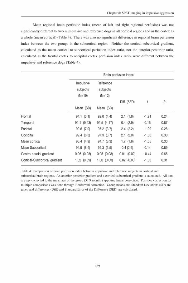
189
Chapter 8: SPET imaging in impulsive aggression
Mean regional brain perfusion index (mean of left and right regional perfusion) was not
significantly different between impulsive and reference dogs in all cortical regions and in the cortex as
a whole (mean cortical) (Table 4). There was also no significant difference in regional brain perfusion
index between the two groups in the subcortical region. Neither the cortical-subcortical gradient,
calculated as the mean cortical to subcortical perfusion index ratio, nor the anterior-posterior ratio,
calculated as the frontal cortex to occipital cortex perfusion index ratio, were different between the
impulsive and reference dogs (Table 4).
Brain perfusion index
Impulsive Reference
subjects subjects
(N=19) (N=12)
Diff. (SED) t P
Mean (SD) Mean (SD)
Frontal 94.1 (5.1) 92.0 (4.4) 2.1 (1.8) -1.21 0.24
Temporal 92.1 (9.43) 92.5 (4.17) 0.4 (2.9) 0.16 0.87
Parietal 99.6 (7.0) 97.2 (3.7) 2.4 (2.2) -1.09 0.28
Occipital 99.4 (6.3) 97.3 (3.7) 2.1 (2.0) -1.06 0.30
Mean cortical 96.4 (4.9) 94.7 (3.3) 1.7 (1.6) -1.05 0.30
Mean Subcortical 94.9 (8.4) 95.3 (3.5) 0.4 (2.6) 0.14 0.89
Costro-caudal gradient 0.96 (0.08) 0.95 (0.03) 0.01 (0.02) -0.44 0.66
Cortical-Subcortical gradient 1.02 (0.09) 1.00 (0.03) 0.02 (0.03) -1.03 0.31
Table 4: Comparison of brain perfusion index between impulsive and reference subjects in cortical andsubcortical brain regions. An anterior-posterior gradient and a cortical-subcortical gradient is calculated. All dataare age corrected to the mean age of the group (37.9 months) applying linear correction. Post-hoc correction formultiple comparisons was done through Bonferroni correction. Group means and Standard Deviations (SD) aregiven and differences (Diff) and Standard Error of the Difference (SED) are calculated.

190
Chapter 8: SPET imaging in impulsive aggression
5-HT2A RECEPTOR BINDING INDEX
The injected dose per kg body weight of 123I-5I-R91150 was not significantly different
between the two groups (Table 2) and did not correlate with the regional binding index of the ligands.
No significant left/right binding index differences were found in the cortical areas neither in the
aggressive impulsive group, nor in the reference group (Table 5).
Left-Right comparison of cortical serotonin-2A binding index
Impulsive subjects (N=19) Reference subjects (N=12)
Right Leftt P
Right Leftt P
Mean (SD) Mean (SD) Mean (SD) Mean (SD)
Frontal 241.0 (19.8) 236.3 (15.5) 1.56 0.14 186.6 (8.2) 188.0 (8.2) -0.67 0.52
Temporal 218.7 (23.8) 219.0 (35.0) -0.04 0.97 163.3 (16.3) 160.4 (17.9) 0.62 0.55
Parietal 157.2 (32.4) 160.6 (36) -0.53 0.60 132.8 (32.2) 131.5 (28.3) 0.24 0.82
Occipital 177.2 (24.1) 176.7 (35.3) 0.10 0.92 147.9 (20.3) 138.9 (20.98) 1.51 0.16
Table 5: Left-Right comparison of cortical serotonin-2A binding index from impulsive and reference subjects.Means and Standard Error data are given. All data are age corrected to the mean age of the group (37.9 months)applying linear correction. Post-hoc correction for multiple comparisons was done through Bonferroni correction.
Mean regional serotonin-2A binding index (mean of left and right regional binding index) was
significantly different between impulsive and reference dogs in all cortical regions and in the global
cortex (mean cortical) (Table 6) (Fig 4). There was no significant difference in serotonin-2A receptor
binding index in the subcortical regions. Neither the cortical-subcortical gradient, calculated as the
mean cortical to subcortical binding index ratio, nor the anterior-posterior ratio, calculated as the
frontal cortex to occipital cortex binding index ratio, were different between the impulsive and
reference dogs (Table 6).

191
Chapter 8: SPET imaging in impulsive aggression
Fig 4: Serotonin-2A radioligand horizontal images from (A) a normal reference dog and (B) an aggressive dog.Increased activity is most visible in the left cortical areas of image B.
Serotonin-2A binding index
Impulsive Reference
subjects subjects
(N=19) (N=12)
Diff. (SED) t P
Mean (SD) Mean (SD)
Frontal 238.7 (16.5) 187.3 (7.2) 51.3 (5.1) 10.1 <0.001
Temporal 218.9 (25.9) 161.8 (15.2) 57.1 (8.2) 6.9 <0.001
Parietal 158.9 (31.22) 132.2 (28.7) 26.7 (11.2) -2.4 0.02
Occipital 177.0 (28.1) 143.4 (17.8) 33.6 (9.1) 3.7 0.001
Mean cortical 198.4 (17.4) 156.2 (11.2) 42.2 (5.7) 7.4 <0.001
Mean Subcortical 126.2 (40.1) 120.9 (36.8) 5.3 (14.3) 0.37 0.71
Costro-caudal grad 1.39 (0.30) 1.32 (0.17) 0.07 (0.10) 0.69 0.49
Cortical-Subcortical grad. 1.88 (1.16) 1.69 (1.58) 0.19 (0.49) 0.38 0.71
Table 6: Comparison of serotonin-2A binding index between impulsive subjects and reference subjects in corticaland subcortical brain regions. An anterior-posterior gradient and a cortical-subcortical gradient is calculated. Alldata are age corrected to the mean age of the group (37.9 months) applying linear correction. Post-hoc correctionfor multiple comparisons was done through Bonferroni correction. Group means and Standard Deviations (SD)are given and differences (Diff) and Standard Error of the Difference (SED) are calculated.

192
Chapter 8: SPET imaging in impulsive aggression
REGIONAL BRAIN PERFUSION AND SEROTONIN-2A BINDING INDEX
There were no significant correlations between serotonin binding index and regional brain
perfusion in the reference population and in the impulsive dogs population (reference group: all regions
> temporal cortex: r=-0.31; P= 0.32 and the impulsive group: all regions > occipital cortex: r= 0.13;
P= 0.57).

193
Chapter 8: SPET imaging in impulsive aggression
Discussion
The major findings of this report are a statistically higher mean serotonin-2A binding index in
all cortical regions, but not in subcortical regions, in the impulsive subjects as compared to the
reference subjects but no mean regional perfusion differences between the two groups. The absence of
gender differences for serotonin-2A binding and regional brain perfusion is in line with the study of
5-HIAA in aggressive and reference dogs22, human impulsivity studies on platelets82, studies on
primates29. In terms of the gonad status, a larger proportion of dogs in the impulsive aggressive group
was neutered. As neutering is a common treatment in problems with aggression in animals, as was in
this group, it can certainly affect the study results. A correlation between plasma testosterone and
aggressive acts in male offenders was found in humans83, and an interaction between the sex-hormone
system and the serotonergic system was suggested in a study on aggressive behaviour in rats84.
However, in this study, the frontal binding index measurements of the neutered subjects fell in the range
of the intact subjects. The reported decreased learning abilities in the impulsive aggressive group might
be related to frontocortical dysfunction. This is shown with frontal ablation or cooling experiments
resulting in rats and primates, resulting in impairment of learning and task performances.4,85 The
absence of perfusion deficits in this study is in disagreement with human studies, where a decreased
frontal and subcortical perfusion and metabolism is found in murderers and aggressive indivi-
duals.49-51,86,87
The statistically higher serotonin-2A binding index in all cortical regions found in this study in
the impulsive subjects, confirm the involvement of the serotonergic system in impulsive aggressive
behaviour. In so far, as methodological differences do not preclude the following comparisons, our
results are in line with the higher levels of 5-HT2A receptor binding in post-mortem brains of suicide
victims42,88,89, but not with a report on reduced prefrontal 5-HT2A binding index, measured with the
same serotonin-2A receptor ligand as used in this study, in vivo in patients that very recently attempted
suicide58.
Confounders, such as the impact of possible post-mortem brain alterations in vitro, the impact
of the acute stress-relief of stress episode in the immediacy of a very recent suicide attempt, the
distinction between outward-directed (assault) and inward-directed (suicide attempt) aggression in vivo
and the absence of impulsivity ratings in the in vitro and in vivo aforementioned studies, make a
reasonable comparison difficult.
It is tempting to make a hypothetical link between the results of the indirect studies and the
results of our study that showed a significant higher cortical serotonin-2A binding index. Following
the classical theory on receptor alterations, theoretically, a mechanism of up-regulation of post synaptic
5-HT2A receptors as a compensatory change for the reduced presynaptic serotonergic activity could be
put forward. However, typical up- and down-regulation of receptors from over- and underexposure to
5-HT does not appear to apply to these type of receptors. Lesions to the serotonergic cell bodies,
194
Chapter 8: SPET imaging in impulsive aggression
leading to a dramatic reduction in presynaptic serotonin levels leave the post-synaptic serotonin-2A
receptors unchanged.90 In addition, serotonin-2 receptors not only will down regulate in the presence
of agonists but also, paradoxically, in the presence of antagonists.91,92 The exact mechanism of this
reduction is not clear but may be related to the receptor itself by downregulation or internalization or
to changes in gene transcription and/or translation.91 On the other hand, serotonin-selective uptake
inhibitors (SSRI’s), increasing the serotonin level in the synaptic cleft, induce variable effects on the
receptor.91 Hence, it is unlikely that a classical receptor up-regulation, accompanying a decrease of
serotonin pre-synaptically, will explain the findings in this study.
Another putative mechanism for up-regulation of 5-HT2A receptors in the brain may be related
to modulation through other neurotransmitter systems or through neuro-endocrinological pathways,
such as the hypothalamic-pituitary-adrenal axis (HPA axis). In chronic stress situations, the α2
adrenergic receptors show altered functioning93,94, possibly contributing to disturbances in related
receptor systems such as the serotonergic system. It has been demonstrated in rats that administration
of dexamethasone or exposition to stress (forced swimming) causes an up-regulation of 5-HT2A
receptors without any modification of 5-HT or 5-HIAA concentrations in the brain.95,96

195
Chapter 8: SPET imaging in impulsive aggression
Methodological considerations
We must take into consideration that the serotonin-2A binding index might be artefactually
increased.
First, an increase in cerebral blood perfusion could deliver more tracer to the brain. But this is
unlikely in this study since no significant differences in regional brain perfusion were found between
impulsive and reference subjects. The trend (p=0.08) towards a higher perfusion in the cortical regions
in the impulsive subjects is unlikely to account for the large significant differences in serotonin-2A
binding index.
Second, external manipulations such as medication or diets could act as confounding factors.
This is unlikely since all dog owners and all treating veterinarians or behavioural therapists were
interviewed and all pharmaceutical or dietary interventions were ruled out. In conclusion, all dogs
were psychopharmacological medication-naïve and were not on a diet.
Third, a confounding factor may include breed variability, since the reference group consisted
predominantly of shepherd-type dogs while the aggressive impulsive group was composed of a larger
diversity of breeds. Westergaard et al. (1999) demonstrated that small between-species variations were
accompanied by large differences in CSF 5-HIAA in primates but concluded that this finding also
accounted for interspecies temperament difference (high intensity, escalating aggressive acts and
wounding accompanying lower CSF 5-HIAA.29 Moreover, none of the species-specific subgroups of
the dogs in this study were out of the range of the other species subgroup members and also, shepherd
type dogs were also included in the impulsive group. However, we cannot rule out that the
heterogeneity of the impulsive aggressive population is an influencing factor for the observed
difference. Finally, this study could not demonstrate whether this finding was state-dependent or trait-
related. Indeed, owners came to search for advise often immediately after a serious assault by the dog.
Nevertheless, due to methodological reasons, all dogs, both the reference and the impulsive subjects,
had to wait at least two weeks for their first scan. All dogs were kept and treated in comparable
conditions. By consequence, the findings tend to be rather trait-related than state-dependent.
There are also methodological considerations due to the animal research paradigm, more
specifically, in contrast to human studies, the need of anaesthesia in animals and the smaller cortical
area and brain volume compared to human brain.
First, in contrast to human studies, general anaesthesia is required to perform these
examinations. The anaesthetics used in this study have a predominantly global effect on the brain
(cortical, subcortical and cerebellar region) without hemispheric preponderances, as opposed to the
more regional effects of some anaesthetics such as ketamine hydrochloride. Given the relative
quantitation with reference to the cerebellum, a potential effect of anaesthetics would be ruled out.
Also, with regard to the perfusion studies, the radiopharmaceutical was injected 20-25 minutes prior to
induction of general anaesthesia, at which stage the tracer is already trapped intracellularly.

196
Chapter 8: SPET imaging in impulsive aggression
Concerning the serotonin-2A radioligand studies, global cerebral blood flow alterations as result of the
general anaesthesia, are inevitable and may influence the pseudo- equilibral state of the tracer. But, it
is unlikely that the regional variations, found in this study, are due to the effects of anaesthetics, since
all dogs from both groups received exactly the same anaesthetic protocol for both studies, precluding
individual effects of different products.
Second, in order to obtain semi-quantitative perfusion data, the different regions have to be
compared with a reference area, unaffected by the physiological or pathological alterations being
investigated. In the perfusion study, cerebral regions were normalized to the cerebellum, since this
region seems to be least affected by aging changes.97-100 When using total counts as a normalization
entity, false relative rCBF changes might result from impairment in some regions, influencing the total
brain counts.97 In the radioligand study, the cerebellum was also used as a reference region since this
structure contains few 5-HT2 receptors. This procedure has the disadvantage that it is dependent on a
very secure estimation of cerebellar activity and that a small artifact herein can have large impact on
other regional brain activity estimations. On the other hand, it has the advantage that small differences
in activity in brain regions have a greater chance to be discovered since they are not included in the
reference region. Moreover, to our knowledge, there are no reports that the cerebellum is involved in
the pathophysiology of impulsive aggressive behaviour.

197
Chapter 8: SPET imaging in impulsive aggression
Future research directions
Future studies should at first replicate these findings in larger populations. It would be of
interest if peripheral measurements of serotonin metabolites and platelet serotonin-2A receptor binding
index, preferentially with the same radioligand, could be assessed simultaneously. This would offer
the opportunity to have access to a larger number of animals. In addition, further research should also
include neuro-endocrinological measurements, such as HPA-axis hormones and sex-hormones.
Furthermore, future research should aim at follow-up studies in both populations. Since
repeated examinations from a radio protective point of view are more acceptable for dogs, compared
to humans, due to their limited life-span, and since repeated, state-of-the-art performed, anaesthesia is
well-tolerated, this offers the opportunity to examine individuals on several occasions, evaluating the
“state” or “trait” nature of pathological alterations and monitor perfusion and serotonin-2A binding
index after therapeutic interventions. In this time interval, different intervention-strategies, ranging
from pharmaceutical to behavioural therapy, could be evaluated and compared.
Conclusion: The imaging techniques used in this study can help to improve insight information on the
pathophysiology of impulsive behaviour and the diagnosis of subjects or groups at risk. This may lead
to more adequate preventive breeding programs in stead of the “blind” elimination of breeds. In
addition, animal research on impulsivity can potentially serve as a representative model for the study
of impulsive behaviour in humans.
Acknowledgements
We would like to express our gratitude to the “Martin Gaus” dog school, specialised in
education and re-education of dogs, with special thanks to Rudy Verbraecken and Monique De Roeck
for referral of dogs. Rudy De Meester and Tiny De Keuster, veterinary behavioural therapists are also
acknowledged for referral of cases. We would also like to thank Marnix Verdonck for his technical
assistance.

198
Chapter 8: SPET imaging in impulsive aggression
REFERENCES
REFERENCES
1. Tuggle DW, Taylor DV, Stevens RJ. Dog bites in children. J Pediatr.Surg. 1993; 28: 912-914.
2. Rossman BR, Bingham RD, Emde RN. Symptomatology and adaptive functioning for children exposed tonormative stressors, dog attack, and parental violence. J Am Acad.Child Adolesc.Psychiatry 1997; 36: 1089-1097.
3. Damasio H, Grabowski T, Frank R, Galaburda A, Damasio A. The return of Phineas Gage: clues about thebrain from the skull of the famous patients. Science 1994; 264: 1102-1105.
4. Bauer R, Fuster J. Delayed-matching and delayed response deficit from cooling dorsolateral prefrontal cortexin monkeys. J Comp Physiol Psychol 1976; 90: 243-249.
5. Fuster J. Animal neurophysiology. In: Fuster, J., ed. The prefrontal cortex: anatomy, physiology andneuropsychology of the frontal lobe. Philadelphia: Lippincott-Raven, 1997; 66-101.
6. Cochran E, Robins E, Grote S. Regional serotonin levels in brain: a comparison of depressive suicides andalcoholic suicides with controls. Biol Psychiatry 1976; 11: 283-294.
7. Coccaro E, Kavoussi R, Trestman R, Gabriel M, Cooper T, Siever LJ. serotonin function in human subjects:intercorrelations among central 5-HT indices and aggressiveness. Psychiatry Res 1997; 73: 1-14.
8. Coccaro EF, Siever LJ, Klar HM, Maurer G, Cochrane K, Cooper TB, Mohs RC, Davis KL. Serotonergicstudies in patients with affective and personality disorders: correlates with suicidal and impulsive aggressivebehavior. Arch Gen Psychiatry 1989; 46: 587-599.
9. Brown GL, Linnoila M. CSF serotonin metabolite (5-HIAA) studies in depression, impulsivity, and violence.J Clin Psychiatry 1990; 51:
10. Coccaro EF. Impulsive aggression and central serotonergic system function in humans: an example of adimensional brain-behavioral relationship. Int Clin Psychopharmacol 1992; 7: 3-12.
11. Kavoussi R, Armstead P, Coccaro E. The neurobiology of impulsive aggression. Psychiatr Clin North Am1997; 20: 395-403.
12. Coccaro E, Kavoussi R. Fluoxetine and impulsive aggressive behavior in personality-disordered subjects. Arch Gen Psychiatry 1997; 54: 1081-1088.
13. LeMarquand DG, Pihl RO, Young SN, Tremblay RE, Seguin JR, Palmour RM, Benkelfat C. Tryptophandepletion, executive functions, and disinhibition in aggressive, adolescent males. Neuropsychopharmacology1998; 19: 333-341.
14. Cremniter D, Jamain S, Kollenbach K, Alvarez JC, Lecrubier Y, Gilton A, Jullien P, Lesieur P, Bonnet F,Spreux-Varoquaux O. CSF 5-HIAA levels are lower in impulsive as compared to nonimpulsive violent suicideattempters and control subjects. Biol Psychiatry 1999; 45: 1572-1579.
15. Placidi GP, Oquendo MA, Malone KM, Huang YY, Ellis SP, Mann JJ. Aggressivity, suicide attempts, anddepression: relationship to cerebrospinal fluid monoamine metabolite levels. Biol Psychiatry 2001; 50: 783-791.
16. Dolan M, Anderson IM, Deakin JF. Relationship between 5-HT function and impulsivity and aggression inmale offenders with personality disorders. Br J Psychiatry 2001; 178: 352-359.
17. Olivier B, Mos J. Rodent models of aggressive behavior and serotonergic drugs. Prog NeuropsychopharmacolBiol Psychiatry 1992; 16: 847-870.
199
Chapter 8: SPET imaging in impulsive aggression
18. Botchin MB, Kaplan JR, Manuck SB, Mann JJ. Low versus high prolactin responders to fenfluraminechallenge: marker of behavioral differences in adult male cynomolgus macaques. Neuropsychopharmacology1993; 9: 93-99.
19. Mehlman PT, Higley JD, Faucher I, Lilly AA, Taub DM, Vickers J, Suomi SJ, Linnoila M. Low CSF 5-HIAAconcentrations and severe aggression and impaired impulse control in non human primates. Am J Psychiatry1994; 151: 1485-1491.
20. Kaplan JR, Shively CA, Fontenot MB, Morgan TM, Howell SM, Manuck SB, Muldoon MF, Mann JJ.Demonstration of an association among dietary cholesterol, central serotonergic activity, and social behaviorin monkeys. Psychosom Med 1994; 56: 479-484.
21. Summers CH, Greenberg N. Activation of central biogenic amines following aggressive interaction in malelizards, Anolis carolinensis. Brain Behav Evol. 1995; 45: 339-349.
22. Reisner IR, Mann JJ, Stanley M, Huang Y, Houpt KA. Comparison of cerebrospinal fluid monoaminemetabolite levels in dominant-aggressive and non- aggressive dogs. Brain Res 1996; 714: 57-64.
23. Higley JD, Linnoila M. Low central nervous system serotonergic activity is traitlike and correlates withimpulsive behavior: a nonhuman primate model investigating genetic and environmental influences onneurotransmission. Ann N Y Acad Sci 1997; 836: 39-57.
24. Delville Y, Melloni RH, Ferris CF. Behavioral and neurobioligical consequences of social subjugation duringpuberty in golden hamsters. J Neurosci 1998; 18: 2667-2672.
25. Fairbanks L, Melega W, Jorgensen M, Kaplan J, McGuire M. Social impulsivity inversely associated with CSF5-HIAA and fluoxetine exposure in vervet monkeys. Neuropsychopharmacology 2001; 24: 370-378.
26. Popova NK, Voitenko NN, Trut LN. Changes in the content of serotonin and 5-hydroxyindoleacetic acid in thebrain in the selection of silver foxes according to behavior. Neurosci Behav Physiol 1976; 7: 72-74.
27. Popova NK, Voitenko NN, Kulikov AV, Avgustinovich DF. Evidence for the involvement of central serotoninin mechanism of domestication of silver foxes. Pharmacol Biochem Behav 1991; 40: 751-756.
28. Popova NK, Kulikov AV, Nikulina EM, Kozlachkova EY, Maslova GB. Serotonin metabolism and serotonergicreceptors in Norway rats selected for low aggressiveness towards man. Aggress Behav 1991; 17: 207-213.
29. Westergaard GC, Suomi SJ, Higley JD, Mehlman PT. CSF 5-HIAA and aggression in female macaquemonkeys: species and interindividual differences. Psychopharmacology (Berl) 1999; 146: 440-446.
30. Higley JD, Mehlman PT, Taub DM, higley SB, Suomi SJ, Linnoila M, Vickers JH. Cerebrospinal fluidmonoamine and adrenal correlates of aggression in free-ranging rhesus monkeys. Arch Gen Psychiatry 1992;49: 436-441.
31. Higley JD, Mehlman PT, higley SB, Fernald B, Vickers J, Lindell SG, Taub DM, Suomi SJ, Linnoila M.Excessive mortality in young free-ranging male nonhuman primates with low cerebrospinal fluid 5-hydroxyindoleacetic acid concentrations. Arch Gen Psychiatry 1996; 53: 537-543.
32. Asberg M, Traskman L, Thoren P. 5-HIAA in the cerebrospinal fluid: a biochemical suicide predictor? ArchGen Psychiatry 1976; 33: 1193-1197.
33. Virkkunen M, Nuutila A, Goodwin FK, Linnoila M. Cerebrospinal fluid metabolite levels in male arsonists.Arch Gen Psychiatry 1987; 44: 241-247.
34. Linnoila M, Virkkunen M, Scheinin M, Nuutila A, Rimon R, Goodwin FK. Low cerebrospinal fluid 5-hydroxyindolacetic acid concentration differentiates impulsive form nonimpulsive violent behavior. Life Sci1983; 33: 2609-2614.
35. Soderstrom H, Blennow K, Manhem A, Forsman A. CSF studies in violent offenders. I. 5-HIAA as a negativeand HVA as a positive predictor of psychopathy. J Neural Transm 2001; 108: 869-878.
36. Bjork JM, Dougherty DM, Moeller FG, Swann AC. Differential behavioral effects of plasma tryptophandepletion and loading in aggressive and nonaggressive men. Neuropsychopharmacology 2000; 22: 357-369.
200
Chapter 8: SPET imaging in impulsive aggression
37. LeMarquand DG, Benkelfat C, Pihl RO, Palmour RM, Young SN. Behavioral disinhibition induced bytryptophan depletion in nonalcoholic young men with multigenerational family histories of paternalalcoholism. Am J Psychiatry 1999; 156: 1771-1779.
38. Gibbons JL, Barr GA, Bridger WH, Leibowitz SF. Manipulations of dietary tryptophan: effects on mousekilling and brain serotonin in the rat. Brain Res 1979; 169: 139-153.
39. Chamberlain B, Ervin FR, Pihl RO, Young SN. The effect of raising or lowering tryptophan levels onaggression in vervet monkeys. Pharmacol.Biochem.Behav 1987; 28: 503-510.
40. Dodman NH, Donnelly R, Shuster L, Mertens P, Rand W, Miczek K. Use of fluoxetine to treat dominanceaggression in dogs. J Am Vet.Med Assoc. 1996; 209: 1585-1587.
41. Medeiros JM, Silva CM, Sougey EB, Costa JA, Castro CM, Castro RM. Action of selective serotonin reuptakeinhibitor on aggressive behavior in adult rat submitted to the neonatal malnutrition. Arq Neuropsiquiatr. 2001;59: 499-503.
42. Arora RC, Meltzer HY. Serotonergic measures in the brains of suicide victims: 5-HT2 binding sites in thefrontal cortex of suicide victims and control subjects. Am J Psychiatry 1989; 146: 730-736.
43. Biegon A, Essar N, Israeli M, et al. Serotonin 5HT2 receptor binding on blood platelets as a state dependentmarker in major affective disorder. Psychopharmacology (Berl) 1990; 102: 73-75.
44. Pandey GN, Pandey SC, Janicak PG, Marks RC, Davis JM. Platelet serotonin-2 receptor binding sites indepression and suicide. Biol Psychiatry 1990; 28: 215-222.
45. McBride PA, Brown RP, De Meo M, Keilp J, Mieczkowski T, Mann JJ. The relationship of platelet 5-HT2receptor indices to major depressive disorder, personality traits and suicidal behaviour. Biol Psychiatry 1994;35: 295-308.
46. Hrdina PD, Bakish D, Chudzik J, Ravindran A, Lapierre YD. Serotonergic markers in platelets of patients withmajor depression: upregulation of 5-HT2 receptors. J Psychiatry Neurosci 1995; 1995: 11-19.
47. Coccaro E, Kavoussi R, Sheline YI, Berman ME, Csernansky JUG. Impulsive aggression in personalitydisorder correlates with platelet 5-HT2A receptor binding. Neuropsychopharmacology 1997; 16: 211-216.
48. Cho R, Kapur S, Du L, Hrdina PD. Relationship between central and peripheral serotonin 5-HT2A receptors:a positron emission tomography study in healthy individuals. Neurosci.Lett. 1999; 261: 139-142.
49. Raine A, Buchsbaum M, Lacasse L. Brain abnormalities in murderers indicated by positron emissiontomography. Biol.Psychiatry 1997; 42: 495-508.
50. Amen, D., Stubblefield, M., Carmichael, B., and Thisted, R. Brain SPECT findings and aggressiveness. AnnClin Psych 1996; 8: 129-137.
51. Volkow ND, Tancredi LR, Grant C, Gillespie H, Valentine A, Mullani N, Wang GJ, Hollister L. Brain glucosemetabolism in violent psychiatric patients: a preliminary study. Psychiatry Res 1995; 61: 243-253.
52. Peremans K, De Bondt P, Audenaert K, Van Laere K, Gielen I, Koole M, Versijpt J, Van Bree H, VerschootenF, Dierckx R. Regional brain perfusion in 10 healthy dogs measured using technetium-99m ethyl cysteinatedimer SPECT: a normal database. Vet Radiol Ultrasound 2001; 42: 562-568.
53. Peremans K, Audenaert K, Coopman F, Jacobs F, Blanckaert P, Verschooten F, Van Bree H, van Heeringen C,Mertens J, Slegers G, Dierckx R. Effects of aging on brain perfusion and serotonin-2A receptor binding in thenormal canine brain measured with single photon emission tomography. Prog Neuro-Psychopharmacol & BiolPsychiat 2002; 26: 1393-1404.
54. Terriere D, Janssen P, Gommeren W, Gysemans M, Mertens J, Leysen J. Evaluation of radioiodo-4-amino-N-(1-(4-fluorophenoxy)-propyl)-4-methyl-4-piperidinyl)-5-iodo-2-methoxybenzamide as a potential 5HT2receptor tracer for SPE(C)T. Nucl Med biol 1995; 22: 1005-1010.

201
Chapter 8: SPET imaging in impulsive aggression
55. Mertens J, Terriere D, Sipido V, Gommeren W, Janssen PMF, Leysen JE. Radiosynthesis of a newradioiodinated ligand for serotonin-5HT2-receptors, a promising tracer for gamma-emission tomography. J Label Compd Radiopharm 1995; 34: 795-801.
56. Abi-Dargham A, Zea-Ponce Y, Terriere D, Al-Tikriti M, Baldwin RM, Hoffer P, Charney D, Leysen JE,Laruelle M, Mertens J, Innis RB. Preclinical evaluation of (123I)R93274 as a SPECT radiotracer for imaging5-HT2A recpetors. Eur J Pharmacol 1997; 321: 285-293.
57. Busatto GF, Pilowsky LS, Costa DC, Mertens J, Terriere D, Ell PJ, Mulligan R, Travis MJ, Leysen JE, Lui D,Gacinovic S, Waddington W, Lingford-Hughes A, Kerwin RW. Initial evaluation of 123I-5-I-R91150, aselective 5-HT2A ligand for single photon emission tomography in healthy subjects. Eur J Nucl Med 1997;24: 119-124.
58. Audenaert K, Van Laere K, Dumont F, Slegers G, Mertens J, van Heeringen C, Dierckx R. Decreased frontalserotonin 5-HT2a receptor binding index in deliberate self harm patients. Eur J Nucl Med 2001; 28: 175-182.
59. Audenaert K, Van Laere K, Dumont F, Vervaet M, Goethals I, Slegers G, Mertens J, van Heeringen C, DierckxR. Decreased 5-HT2A binding in patients with anorexia nervosa. J Nucl Med 2002; in press.
60. Peremans K, Audenaert K, Jacobs F, Dumont F, De Vos F, Van de Wiele C, Vandecapelle M, Van Bree H,Verschooten F, Slegers G, Mertens J, Dierckx R. Biodistribution and displacement studies of the selective 5-HT2A receptor antagonist 123I-5-I-R91150 in the normal dog. Nuc Med Comm 2002; 23: 1019-1027.
61. Peremans K, Audenaert K, Coopman F, Jacobs F, Dumont F, Slegers G, Verschooten F, Van Bree H, MertensJ, Dierckx R. Regional binding index of the radiolabelled selective 5-HT2A antagonist 123I-5-I-R91150 in thenormal canine brain imaged with single photon emission computed tomography. Vet Radiol Ultrasound 2002;in press.
62. Marder A, Voith V. Canine aggression evaluation. In: Voith, V. and Borchelt, P., ed. Readings in companionanimal behaviour. New Jersey: Trenton, 1996; 227-229.
63. Goodloe L. Issues in description and measurements of temperament in companion dogs. In: Voith, V. andBorchelt, P., ed. Readings in companion animal behaviour. New Jersey: Trenton, 1996; 32-39.
64. Overall K. Canine aggression. In: Overall, K., ed. Clinical behavioral medecine for small animals. St Louis:Mosby, 1997; 293-322.
65. Leveille J, Demonceau G, Walovitch RC. Intrasubject comparison between technetium-99m-ECD andtechnetium-99m- HMPAO in healthy human subjects. J Nucl Med 1992; 33: 480-484.
66. Moretti JL, Defer G, Tamgac F, Weinmann P, Belin C, Cesaro P. Comparison of brain SPECT using 99mTc-bicisate (L,L-ECD) and [123I]IMP in cortical and subcortical strokes. J Cereb Blood Flow Metab 1994; 14Suppl 1: S84-S90.
67. Ichise M, Golan H, Ballinger JR, Vines D, Blackman A, Moldofsky H. Regional differences in technetium-99m- ECD clearance on brain SPECT in healthy subjects. J Nucl Med 1997; 38: 1253-1260.
68. Matsuda H, Yagishita A. A non -invasive quantitative approach to 99mTc-ethyl cysteinate dimer. In: De Deyn,P. P., Dierckx, R. A., Alavi, A, and Pickut, B. A., ed. A textbook of SPECT in neurology and psychiatry.London-Paris-Rome-Sydney: John Libbey & Company Ltd, 1997; 501-505.
69. Van Laere K, Koole M, Kauppinen T, Monsieurs M, Bouwens L, Dierckx R. Non-uniform transmission inbrain SPET using 201-Tl, 99m-Tc and 153-Gd static line sources: antropomorphic dosimetry studies and brainquantification. J Nucl Med 2000; 41: 2051-2062.
70. Redding RW. Anatomy and Physiology. In: Hoerlein, B. F., ed. Canine Neurology. Philadelphia, London,Toronto: W.B.Saunders Company, 1978; 7-52.
71. Pazos A, Cortes R, Palacios JM. Quantitative autoradiographic mapping of serotonin receptors in the ratbrain.II.Serotonin-2 receptors. Brain Res 1985; 346: 231-249.

202
Chapter 8: SPET imaging in impulsive aggression
72. Pazos A, Probst A, Palacios J. Serotonin receptors in the human brain-IV. Autoradiographic mapping ofserotonin-2 receptors. Neuroscience 1987; 21: 123-139.
73. Lopez-Gimenez JF, Vilaro MT, Palacios J, Mengod G. Mapping of 5-HT2A receptors and their mRNA inmonkey brain: (3H)MDL100.907 autoradiography and in situ hybridization studies. J Comp Neurol 2001; 429:571-589.
74. Kerwin R, Pilowsky L. Traditional receptor theory and its application to neuroreceptor measurements infunctional imaging. Eur J Nucl Med 1995; 22: 699-710.
75. Moeller JR, Ishikawa T, Dhawan V, Spetsieris P, Mandel F, Alexander GE, Grady C, Pietrini P, Eidelberg D.The metabolic topography of normal aging. J Cereb Blood Flow Metab 1996; 16: 385-398.
76. Mielke R, Kessler J, Szelies B, Herholz K, Wienhard K, Heiss WD. Normal and pathological aging--findingsof positron-emission-tomography. J Neural Transm 1998; 105: 821-837.
77. Baeken C, D'haenen H, Flamen P, Terriere D, Chavatte K, Boumon R, Bossuyt A. 123I-5-I-R91150, a newsingle photon emission tomography ligand for 5-HT2A receptors: influence of age and gender in healthysubjects. Eur J Nucl Med 1998; 25: 1617-1622.
78. Meltzer CC, Smith G, Price JC, Reynolds CF, Mathis CA, Greer P, Lopresti B, Mintun MA, Pollock BG, Ben-Eliezer D, Cantwell MN, Kaye W, DeKosky ST. Reduced binding of {18F} altanserin to serotonin type 2Areceptors in aging: persistence of effect after partial volume correction. Brain Res 1998; 813: 167-171.
79. Nobler MS, Mann JJ, Sackeim HA. Serotonin, cerebral blood flow and cerebral metabolic rate in the geriatricmajor depression and normal aging. Brain Res Reviews 1999; 30: 250-263.
80. Kakiuchi T, Nishiyama S, Sato K, Ohba H, Nakanishi S, Tsukada H. Age related reduction of {11C} MDL100,907 binding to central 5-HT2A receptors: PET study on the conscious monkey brain. Brain Res 2000; 883:135-142.
81. Van Laere K, Versijpt J, Audenaert K, Koole M, Goethals I, Achten E, Dierckx R. 99mTc-ECD brain perfusionSPET: variability, asymmetry and effects of age and gender in healthy adults. Eur J Nucl Med 2001; 28: 873-887.
82. Pandey GN, Pandey SC, Dwivedi Y, Sharma V, Janicak PG, Davis JM. Platelet serotonin-2A receptors: apotential biological marker for suicidal behavior. Am J Psychiatry 1995; 152: 850-855.
83. Virkkunen M, Rawlings R, Tokola R, Poland RE, Guidotti A, Nemeroff C, Bissette G, Kalogeras K, KaronenSL, Linnoila M. CSF biochemistries, glucose metabolism, and diurnal activity rhythms in alcoholic, violentoffenders, fire setters, and healthy volunteers. Arch Gen Psychiatry 1994; 51: 20-27.
84. Bonson KR, Johnson RG, Fiorella D, Rabin RA, Winter JC. Serotonergic control of androgen-induceddominance. Pharmacol Biochem.Behav 1994; 49: 313-322.
85. Volavka J. Neurobiology of violence. Washington DC: American Psychiatric Press, 1995.
86. Miller BL, Darby A, Benson DF, Cummings JL, Miller MH. Aggressive, socially disruptive and antisocialbehaviour associated with fronto-temporal dementia. Br J Psychiatry 1997; 170: 150-154.
87. Hirono N, Mega MS, Dinov ID, Mishkin F, Cummings JL. Left frontotemporal hypoperfusion is associatedwith aggression in patients with dementia. Arch Neurol 2000; 57: 861-866.
88. Stanley M, et al. Alterations in pre- and postsynaptic serotonergic neurons in suicide victims.Psychopharmacol Bull 1983; 19: 684-687.
89. Arango V, Ernsberger P, Marzuk PM. Autoradiographic demonstration of increased serotonin 5HT2 and beta-adrenergic receptor binding sites in the brain of suicide victims. Arch Gen Psychiatry 1990; 47: 1038-1044.
90. Sanders-Bush E. Adaptive regulation of central serotonin receptors linked to phosphoinositide hydrolysis.Neuropsychopharmacology 1990; 3: 411-416.

203
Chapter 8: SPET imaging in impulsive aggression
91. Gray JA, Roth BL. Paradoxical trafficking and regulation of 5-HT(2A) receptors by agonists and antagonists.Brain Res Bull. 2001; 56: 441-451.
92. Aloyo VJ, Dave KD, Rahman T, Harvey JA. Selective and divergent regulation of cortical 5-HT(2A) receptorsin rabbit. J Pharmacol Exp Ther 2001; 299: 1066-1072.
93. Du L, Faludi G, Palkovits M, Bakish D, Hrdina P. Serotonergic genes ans suicidality. Crisis 2001; 22: 54-60.
94. Popova NK, Kulikov AV, Avgustinovich DF, Shigantsov SN. Brain serotonergic system and anxiety in C57BLand CBA mice strains. Zhurnal Vysshei Nervnoi Deyatelnosti Imenti I P Pavlova 1996; 46: 348-354.
95. Popova NK, Kulikov AV, Avgustinovich DF, Voitenko NN, Trut LN. Effects of domestication on the basicenzymes of serotonin metabolism and serotonin receptors in the silver foxes. Genetika 1997; 33: 370-374.
96. Perry BD, Giller EL, Jr., Southwick SM. Altered platelet alpha 2-adrenergic binding sites in posttraumaticstress disorder. Am J Psychiatry 1987; 144: 1511-1512.
97. Stone E. Stress and neurotransmitter receptors. In: Sen, A. and Lee, T., ed. Receptors and ligands in psychiatry.New York: Cambridge Univ Press, 1988; 400-423.
98. Takao K, Nagatani T, Kitamura Y, Kawasaki K, Hayakawa H, Yamawaki S. Chronic forced swim stress of ratsincreases frontal cortical 5-HT2 receptors and the wet-dog shakes they mediate, but not frontal cortical beta-adrenoceptors. Eur J Pharmacol 1995; 294: 721-726.
99. Katagiri H, Kagaya A, Nakae S, Morinobu S, Yamawaki S. Modulation of serotonin2A receptor function inrats after repeated treatment with dexamethasone and L-type calcium channel antagonist nimodipine. ProgNeuropsychopharmacol Biol Psychiatry 2001; 25: 1269-1281.
100. Catafau AM, Lomena FJ, Pavia J, Parellada E, Bernardo M, Setoain J, Tolosa E. Regional cerebral blood flowpattern in normal young and aged volunteers: a 99mTc-HMPAO SPET study. Eur J Nucl Med 1996; 23: 1329-1337.
101. Matsuda H, Tsuji S, Shuke N, Sumiya H, Tonami N, Hisada K. Noninvasive measurements of regional cerebralblood flow using technetium-99m hexamethylpropylene amine oxime. Eur J Nucl Med 1993; 20: 391-401.
102. Leenders K, Perani D, Lammertsma A, Heather J, Buckingham P, Healey M, Gibbs J, Wise R, Hatazawa J,Herold S, Beany R, Brooks D, Spinks T, Rhodes C, Frackowiak R, Jones T. Cerebral bloodflow , blood volumeand oxygen utilization- normal values and effect of age. Brain 1990; 113: 27-47.
103. Krausz Y, Bonne O, Gorfine M, Karger H, Lerer B, Chisin R. Age-related changes in brain perfusion of normalsubjects detected by 99mTc-HMPAO SPECT. Neuroradiology 1998; 40: 428-434.


205
GENERAL DISCUSSION


207
General Discussion
General discussion
Gradually, more and more research is focussing on clinical behavioural problems encountered
in small animals, and this from a veterinary clinical point of view as well as concerning the possible
use of larger animal models in research on human brain disorders and related deviant behaviour.1
In the discussion we will address two aspects of this work. In the first part we will raise the
methodological issues and in the second we will discuss the clinical applications.
METHODOLOGICAL ISSUES
Functional versus structural imaging
Structural neuroimaging studies with CT or MRI offer insight in brain-behaviour relationships
that involved anatomical lesions of the brain. But very often, behavioural disturbances are not
accompanied with structural lesions. Therefore, structural imaging is only helpful to rule out organic
pathology. Functional imaging modalities, including functional magnetic resonance imaging (fMRI),
magnetic resonance spectroscopy (MRS), PET and SPET offer the opportunity to examine the
functioning brain in vivo. Moreover, with the introduction of functional imaging, using SPET and PET,
in the research of brain-behaviour relationship, the possibility is created to quantify perfusion,
metabolism and the neuroreceptor status in vivo.
SPET as a research tool
We used the SPET technique in this studies, despite the better resolution and more accurate
quantification, provided by PET. Nevertheless SPET remains a more economical and applicable
modality for general use, since it does not require a cyclotron in the immediate vicinity of the camera.
It also remains a valid modality to evaluate regional binding kinetics and to provide semiquantitative
indices of regional perfusion and receptor binding. Some technical aspects have been discussed in the
preceding chapters.
Partial volume effects and attenuation
We did not subdivide the fronto-cortical and subcortical region in smaller structural and
functional regions. In these studies, partial volume effects would increase inter-and intra-individual
variability to an undesirable extent. Also, the lack of detail on the CT images, precluded adequate
subdivision of the fronto-cortical and subcortical regions, as is done in human studies. Nevertheless,
even with this division, partial volume effects cannot be excluded and can result in an underestimation
of regional radioactivity values. A second technical factor is the attenuation applied to the acquired
data. The broad beam (uncollimated source) attenuation coefficient used in this study was 0.12/cm

208
General Discussion
which is slightly lower than the narrow beam (collimated source) attenuation coefficient used for
correction for attenuation of photons with an energy of 140 keV in water (0.15/cm).2 The usefulness
of a non uniform attenuation coefficient (NUAC) is beyond any doubt in studies of heterogeneous
structures such as the thoraco-abdominal region but the clinical relevance for brain SPET is under
debate.2 Brain is a rather homogenous structure, although the thickness of the skull at certain parts can
influence attenuation, mainly in the superficial cortical structures and the cerebellum. When applying
uniform attenuation, the superficial cortical activity will be underestimated, compared to the deeper
structures (subcortical regions), resulting from increased attenuation of the overlying skull (paradoxical
skull effect).2,3 Since we used group comparisons, the effects are expected to be minimal.
Feasibility of perfusion and 5-HT2A receptor imaging
We demonstrated in these studies that SPET is a valid modality to image and measure both
perfusion and the serotonin-2A receptor occupancy in physiological and pathophysiological states of
the canine brain. However, to our belief, the technique of functional neuroimaging cannot stand alone.
It has to be carried out after careful behavioural examination of the animal and has to be accompanied
by anatomical imaging techniques to exclude organic lesions. Ideally, in its research phase, it needs
post-mortem validation with autoradiography or immunohistochemical techniques and should be
accompanied by investigation of other biochemical markers, to rule out metabolic, hormonal and other
neurotransmitter influences and to explore possible peripheral markers, reflecting central alterations.
Applicability in daily veterinary practice
We must also stress the questionable applicability for common daily use in veterinary practice.
An important methodological problem concerns the requirement of a brain dedicated gamma camera
to optimise resolution and shorten acquisition procedures. Furthermore, receptor imaging requires
specific receptor ligands and up to now, radioligands for SPET are scarce. As is the case for most
receptor ligands, the tracer used in this study is labelled with 123I (in the order of 500 euros per dog)
which is too expensive to apply on routinely basis. It is therefore logical to conclude that this technique
offers definite advantages as a research model for behavioural problems in dogs and as a model for
certain human brain disease and behavioural abnormalities, but that practical application in veterinary
medicine is still limited. Tracer labelling with 99mTechnetium might offer a more economical solution.

209
General Discussion
CLINICAL APPLICATION
The study of the feasibility of this SPET technique, both in the assessment of perfusion as in
the evaluation of serotonin-2a binding index in the normal canine brain, was the first goal of our
research. Its clinical application was evaluated in the study of the brain of the aging dog and the
impulsive dog.
Aging dogs
First, concerning the perfusion alterations found in aged dogs, the increased anterior-posterior
gradient observed is in agreement with findings in human aging brain. A possible explanation for this
observation could be that regions, serving cognitive and affective behaviour, are more affected than the
more sensory areas4,5, which in turn could account for the cognitive and mood disorders encountered in
aging individuals.
Second, the most important methodological confounder concerns the cortical atrophy,
accompanying aging alterations. Nevertheless, regional hypometabolism in patients with Alzheimer
disease, measured with PET, were confirmed after atrophy corrections based on MRI derived volume
correction factors in some human studies.6,7
Third, the global decrease found in this study is in line with some but not all human studies on
brain perfusion.8-11 On the other hand, the regional decreases found in this study are comparable with
human studies.5,8-10,12,13 In humans, age related changes in rCBF are already noted in early to mid
adulthood without accompanying cognitive decline or other degenerative processes. Concerning the
altered 5-HT2A receptor radioligand binding mainly affecting the frontocortical cortex in our study,
this is in partial agreement with human and primate studies In most studies a more general cortical
decrease is found in normal aging brain14,15, although some studies report a region dependent decrease
and an anterior-posterior gradient in normal aging and in Alzheimer patients.16,17 The absence of
decreased binding in the other cortical regions in our study could also be artefactually induced by the
larger standard deviation found in the temporo-, parieto and occipitocortical regions, accounting for a
less sensitive detection of marginal decreases in binding index which could therefore mask more
general decreases. In addition, it is also noteworthy to mention that a few human studies report a non-
linear decline of serotonin-2A radioligand binding with the largest decline before the age of 40.14,17,18 It
could therefore be possible that the observed tendency to decreased binding in other regions, except for
the parieto-cortical area, does not reach levels of significance in the age groups we examined. These
observations emphasize the need to include a larger population reference dogs in order to validate the
time of onset and the evolution of altered binding of the 5-HT2A receptor radioligand in smaller age
groups.

210
General Discussion
Fourth, from previous research on rats and primates it is known that the activity of the
serotonergic system also alters during brain maturation. It would therefore be of considerable interest
to monitor the development of the serotonin-2A receptor in animals under different rearing conditions.
In view of this, the SPET receptor imaging procedure could offer a feasible research paradigm to follow
receptor status during brain development.
Impulsive, aggressive dogs
No perfusion alterations were found in this study. In human studies concerning aggressive and
antisocial behaviour, focal regional perfusion and metabolism alterations are consistently reported in
the prefrontal cortical regions but alterations in other regions differ between studies.19-24 Since the brain
structures of dogs are small, dividing the fronto-cortical and the subcortical structures into their
structural and functional subdivisions (dorso-lateral and orbital fronto-cortical region, thalamus,
amygdala..) would increase the partial volume effects to an impermissible degree precluding accurate
quantification. Therefore we could only evaluate global alterations in regional perfusion, which could
allow intra-regional compensation of focal increases and decreases. As an example, in the study of
Raine et al (1997) decreased metabolism was present in some parts of the subcortical areas while in
other parts increases were found. Another explanation could lie in the possible smaller contribution of
the frontal cortex in the regulation of canine behaviour.
Concerning the serotonin-2A radioligand binding, a general increase was found in the
impulsive, aggressive group, indicating that the serotonergic system is involved in this behavioural
disorder in dogs. This finding is in line with post-mortem evaluation of serotonin-2A receptor binding
with autoradiography in suicide victims.25 We must take care of oversimplification by addressing only
one receptor or even one neurotransmitter system. It has to be kept in mind that possible effects of
downstream events following receptor activation and interaction of different neurotransmitter systems
may contribute to the pathophysiology of the investigated disorders. As an example, we did not
investigate the involvement of the adrenergic system. Soubrie and Bizot (1990) found in their
experiments with rats that pharmacological enhancement of both serotonin and noradrenergic
transmission decreased impulsivity (defined as capacity to wait or tolerance to delay of reward).26
Furthermore, it is known that the α2 adrenergic receptor is involved in the modulation of the
serotonergic system.27 It has been demonstrated in rats that chronic stress not only results in altered
frontocortical densities of the 5-HT2 receptor but also of the adrenergic receptors.28-32 Also in humans,
altered functioning of the adrenergic system has been found in combination with an altered
serotonergic system in depression.33
The investigation of the effect of the hormonal status on the serotonergic system would make
an interesting research subject, since it is known from literature that estrogens have a modulating
role.27,34-36 Not only from a veterinary pathophysiological, but also from a human medicine
neuropsychiatric viewpoint, this research could shed a light on the influence of hormones on the
211
General Discussion
behavioural, affective and cognitive status in humans and could lead to a better understanding of
behavioural disorders such as the Pre Menstrual tension Syndrome (PMS), which is now empirically
treated with serotonergic drugs.34
The possibility to perform longitudinal studies in man is restricted due to radioprotective
measures. Therefore studies are limited to cross sectional investigations, or at most one follow-up
study, and by consequence, hamper differentiation between state or trait alterations of neurotransmitter
systems.37 It would be of considerable interest to evaluate the impulsive aggressive dogs after 6 months
and longer (without any form of treatment) to evaluate the consistency of our findings. Moreover,
future studies will also focus on pharmacologic manipulation of the serotonergic system in impulsive,
aggressive dogs, to monitor the serotonergic system with both clinical evaluation and functional
imaging. This imaging modality provides also the possibility to monitor the effects of diet
interventions, e.g. tryptophan augmentation, suggested to influence aggressive behaviour.34,38,39

212
General Discussion
REFERENCES
1. Overall K. Natural animal models of human psychiatric conditions: assessment of mechanism and validity. ProgNeuro-Psychopharmacol & Biol Psychiat 2000; 24: 727-776.
2. Van Laere K, Koole M, Kauppinen T, Monsieurs M, Bouwens L, Dierckx R. Non-uniform transmission in brainSPET using 201-Tl, 99m-Tc and 153-Gd static line sources: antropomorphic dosimetry studies and brainquantification. J Nucl Med 2000; 41: 2051-2062.
3. Licho R, Glick SJ, Xia W, Pan TS, Penney BC, King MA. Attenuation compensation in 99mTc SPECT brainimaging: a comparison of the use of attenuation maps derived from transmission versus emission data in normalscans. J Nucl Med 1999; 40: 456-463.
4. Mielke R, Kessler J, Szelies B, Herholz K, Wienhard K, Heiss WD. Normal and pathological aging--findingsof positron-emission-tomography. J Neural Transm 1998; 105: 821-837.
5. Van Laere K, Versijpt J, Audenaert K, Koole M, Goethals I, Achten E, Dierckx R. 99mTc-ECD brain perfusionSPET: variability, asymmetry and effects of age and gender in healthy adults. Eur J Nucl Med 2001; 28:873-887.
6. Meltzer C, Zubieta J, Brandt J, Tune L, Mayberg H, Frost J. Regional hypometabolism in Alzheimer's diseaseas measured by positron emission tomography after correction for effects of partial volume averaging. 1996;47: 454-461.
7. Alavi A, Newberg A, Souder E, Berlin J. Quantitative analysis of PET and MRI data in normal aging andAlzheimer's disease: atrophy weighted total brain metabolism and absolute whole brain metabolism as reliablediscriminators. 1993; 34: 1681-1687.
8. Markus HS, Ring H, Kouris K, Costa DC. Alterations in regional cerebral blood flow, with increased temporalinterhemispheric asymmetries, in the normal elderly: an HMPAO SPECT study. Nucl Med Commun. 1993; 14:628-633.
9. Petit-Taboue MC, Landeau B, Desson JF, Desgranges B, Baron JC. Effects of healthy aging on the regionalcerebral metabolic rate of glucose assessed with statistical parametric mapping. Neuroimage 1998; 7: 176-184.
10. Goto R, Kawashima R, Ito H, Koyama M, Sato K, Ono S, Yoshioka S, Fukuda H. A comparison of Tc-99mHMPAO brain SPECT images of young and aged normal individuals. Ann Nucl Med 1998; 12: 333-339.
11. Buijs PC, Krabbe-Hartkamp MJ, Bakker CJ, de Lange EE, Ramos LM, Breteler MM, Mali WP. Effect of ageon cerebral blood flow: measurement with ungated two- dimensional phase-contrast MR angiography in 250adults. Radiology 1998; 209: 667-674.
12. Catafau AM, Lomena FJ, Pavia J, Parellada E, Bernardo M, Setoain J, Tolosa E. Regional cerebral blood flowpattern in normal young and aged volunteers: a 99mTc-HMPAO SPET study. Eur J Nucl Med 1996; 23: 1329-1337.
13. Tanaka F, Vines D, Tsuchida T, Freedman M, Ichise M. Normal patterns on 99mTc-ECD brain SPECT scans inadults. J Nucl Med 2000; 41: 1456-1464.
14. Rosier A, Dupont P, Peuskens J, Bormans G, Vandenberghe R, Maes F, Schiepers C, Verbruggen A, MortelmansL. Visualization of loss of 5-HT2A receptors with age in healthy using (18F) altanserin and positron emissionimaging. Psychiatry Res 1996; 25: 11-22.
15. Baeken C, D'haenen H, Flamen P, Terriere D, Chavatte K, Boumon R, Bossuyt A. 123I-5-I-R91150, a new singlephoton emission tomography ligand for 5-HT2A receptors: influence of age and gender in healthy subjects. EurJ Nucl Med 1998; 25: 1617-1622.
16. Wang G, Volkow ND, Logan J, Fowler JS, Schlyer DJ, Macgreggor RR, Hitzemann R, Gur R, Wolf AP.Evaluation of age-related changes in serotonin 5-HT2 and dopamine D2 receptor availibility in healthy humansubjects. Life Sci 1995; 56: 249-253.
17. Blin J, Baron JC, Dubois B, Crouzel C, Fiorelli M, Attar-Levy D, Pillon B, Fournier D, Vidailhet M, Agid Y.Loss of brain 5-HT2 receptors in Alzheimer's disease. In vivo assessment with positron emission tomographyand [18F]setoperone. Brain 1993; 116 ( Pt 3): 497-510.

213
General Discussion
18. Sheline YI, Mintun MA, Moerlein SM, Snyder AZ. Greater loss of 5-HT(2A) receptors in midlife than in latelife. Am J Psychiatry 2002; 159: 430-435.
19. Brower MC, Price BH. Neuropsychiatry of frontal lobe dysfunction in violent and criminal behaviour: a criticalreview. J Neurol Neurosurg Psychiatry 2001; 71: 720-726.
20. Amen D, Stubblefield M, Carmichael B, Thisted R. Brain SPECT findings and aggressiveness. Ann Clin Psych1996; 8: 129-137.
21. Volkow ND, Tancredi L. Neural substrates of violent behaviour. A preliminary study with positron emissiontomography. Br.J Psychiatry 1987; 151: 668-673.
22. Raine A, Buchsbaum M, Lacasse L. Brain abnormalities in murderers indicated by positron emissiontomography. Biol.Psychiatry 1997; 42: 495-508.
23. Hirono N, Mega MS, Dinov ID, Mishkin F, Cummings JL. Left frontotemporal hypoperfusion is associated withaggression in patients with dementia. Arch Neurol 2000; 57: 861-866.
24. Miller BL, Darby A, Benson DF, Cummings JL, Miller MH. Aggressive, socially disruptive and antisocialbehaviour associated with fronto-temporal dementia. Br J Psychiatry 1997; 170: 150-154.
25. Stanley M, Mann JJ. Increased serotonin-2 binding sites in frontal cortex of suicide victims. Lancet 1983; i: 214-216.
26. Soubrie E, Bizot J. Monoaminergic control of waiting capacity (impulsivity) in animals. In: Van Praag, H.,Plutchik, R., and Apter, A., ed. Violence and Suicidality: Perspectives in Clinical and Biological Research. NewYork: Brunner/Mazel, 1990; 257-272.
27. Stahl S. Essential psychopharmacology. Cambridge: Cambridge University Press, 1996.
28. Goodall EM, Cowen PJ, Franklin M, Silverstone T. Ritanserin attenuates anorectic, endocrine and thermicresponses to d-fenfluramine in human volunteers. Psychopharmacology (Berl) 1993; 112: 461-466.
29. Takao K, Nagatani T, Kitamura Y, Kawasaki K, Hayakawa H, Yamawaki S. Chronic forced swim stress of ratsincreases frontal cortical 5-HT2 receptors and the wet-dog shakes they mediate, but not frontal cortical beta-adrenoceptors. Eur J Pharmacol 1995; 294: 721-726.
30. Flugge G, Ahrens O, Fuchs E. Monoamine receptors in the prefrontal cortex of Tupaia belangeri during chronicpsychosocial stress. Cell Tissue Res 1997; 288: 1-10.
31. Flugge G. Effects of cortisol on brain alpha2-adrenoceptors: potential role in stress. Neurosci Biobehav.Rev1999; 23: 949-956.
32. Flugge G, Ahrens O, Fuchs E. Beta-adrenoceptors in the tree shrew brain. II. Time-dependent effects of chronicpsychosocial stress on [125I]iodocyanopindolol bindings sites. Cell Mol Neurobiol 1997; 17: 417-432.
33. Shiloh R, Nutt D, Weizman A. Atlas of psychiatric pharmacotherapy. London: Martin Dunitz Ltd, 1999.
34. Volavka J. Neurobiology of violence. Washington DC: American Psychiatric Press, 1995.
35. Fink G, Sumner BE, Rosie R, Grace O, Quinn JP. Estrogen control of central neurotransmission: effect onmood, mental state, and memory. Cell Mol Neurobiol 1996; 16: 325-344.
36. Kaye W, Gwirtsman H, Brewerton T, George D, Wurtman R. Bingeing behaviour and plasma amino acids: Apossible involvement of brain serotonin in bulimia nervosa. Psychiatry Res 1988; 23: 31-43.
37. Audenaert K. Functional neuroimaging in psychiatry: a psychopathological approach. Faculty of Medicine &Health Sciences, University Ghent 2001 (PhD dissertation)
38. Kaplan JR, Shively CA, Fontenot MB, Morgan TM, Howell SM, Manuck SB, Muldoon MF, Mann JJ.Demonstration of an association among dietary cholesterol, central serotonergic activity, and social behavior inmonkeys. Psychosom Med 1994; 56: 479-484.
39. DeNapoli JS, Dodman NH, Shuster L, Rand WM, Gross KL. Effect of dietary protein content and tryptophansupplementation on dominance aggression, territorial aggression, and hyperactivity in dogs. J Am Vet.MedAssoc. 2000; 217: 504-508.


SUMMARY


217
Summary
Summary
In the Introduction of this thesis, the evolution and technique of imaging involving
radionuclides is described. A brief review is given on the history of the evolution of radioactivity for
medical purposes. With the introduction of more efficient imaging modalities such as PET and SPET,
brain imaging with radionuclides have gained an important place in investigation of normal physiology
and of the pathophysiology of neuropsychiatric disorders in human medicine. The technical aspects of
functional imaging are further explained in the introduction. In veterinary medicine no comparable
imaging studies have been performed.
In the introduction, we postulated some questions regarding the applicability of functional
brain imaging in dogs. These questions formed the core of this research project. The results will be
summarized per chapter.
1. Can we measure brain perfusion with functional brain imaging, using the SPET
modality, in the canine brain and what is the normal distribution pattern in dogs
without neurological or behavioural disorders?
In the first chapter regional brain perfusion was investigated in 10 normal dogs. In this study 99mTc-ECD was used to measure perfusion with SPET. Since perfusion is coupled under normal
conditions with metabolism and since its trapping mechanism relies on its metabolism from a lipophilic
to a hydrophilic compound, it provides also an indirect measure of neuronal metabolism. Neither age
nor gender influences were noted but the number of included animals (N=10, age between 1 and 9
years) was too small to draw definite conclusions. An important anterior-posterior gradient was
demonstrated. No left to right differences were registered. Methodological confounders were
discussed concerning partial volume effect and the effect of the anaesthesia. Since the examined
structures are small, partial volume effects may play a significant role. The application of anaesthetics,
which is an inevitable procedure, is a second confounder, although the effect is minimalized since the
tracer is injected before induction of anaesthesia and is trapped within minutes in the brain, reaching
stable values for at least 60 minutes. In the second chapter the application of a standardised, automated
method was investigated to semi-quantify regional perfusion in a more objective way with less
interindividual variance, offering the opportunity to compare data more accurately. The validity of this
procedure was demonstrated in this chapter.
In conclusion, the feasibility of perfusion SPET imaging and the normal regional perfusion
indices in normal canine brain are demonstrated in this chapter. An automatic registration
procedure, using dedicated software can be used to semiquantify regional activities in order to
make inter- and intra-individual comparison more accurate.
218
Summary
2. What are the technical issues to consider when performing receptor radioligand
studies?
To outline the technicalities of receptor radioligand studies in vivo, we first reviewed the literature in
the third chapter, with emphasis on animal studies. The advantage of neuroreceptor imaging is that
physiology and physiopathology of neurotransmitter systems can be examined in vivo,
pharmacodynamics-and kinetics of newly created drugs can be evaluated and the effect of treatment
can be monitored. Moreover, the behaviour of the receptor and its transmitters can be followed in their
natural surroundings including intra- and extracellular regulation and influences from other
neurotransmitter systems. On the other hand, these modulating endogenous influences make
interpretation of the results not always straight forward. Also, major difficulties are encountered in the
production of radioligands. First, the creation of new ligands for SPET is hampered by the fact that
the radionuclide may not alter the binding properties of the used receptor ligand. Technetium is the
preferred radionuclide since it is easy to obtain from a generator “on site”, but it is also large and can
therefore interfere with the stereotactic properties of the ligand, thereby changing its binding
characteristics. The other commonly used radionuclide for SPET is 123I, which has the disadvantage
that it is expensive, enhances lipophilicity and can also interfere with the binding properties of the
ligand. Second, the lipophilicity, a characteristic necessary for blood brain barrier (BBB) passage,
increases non specific binding to non target molecules. Third, although the tracer should have high
affinity for the receptor, an equilibrial state has to be installed for the quantification procedure, which
is precluded when the tracer is trapped too avid when affinity for the receptor is too high. Fourth, the
specificity of the radioligand is not always 100% for one receptor and more, since not all subtypes of
the different receptor systems are known, specific binding to a receptor subtype might not be so
specific. Fifth, (semi)quantification in SPET examinations relies on the ratio method, precluding
differentiation between receptor density and receptor affinity. Several neurotransmitter systems have
already been imaged in different animal species, all studies in function of research on human disease.
In conclusion, this chapter points at the methodological issues that have to be considered in SPET
functional receptor imaging with radioligands. All animal studies performed up to now are
performed for human medicine use.
219
Summary
3. Is it possible to use the radioligand, 123I-5-I-R91150 to image and quantify the 5-HT2A
receptor in canine brain?
In the fourth chapter the applicability of the specific radioliogand 123I-5-I-R91150 was evaluated in the
canine brain. First, in order to investigate the selectivity of the tracer, displacement and blocking
studies were performed with the cold (unlabelled) 5-HT2A antagonist ketanserine, displacing the
radioligand or making the receptor unavailable for the tracer. Binding was displaceable, proving
selectivity and reversibility of binding. Second, in order to semiquantify regional data using the ratio
method, a steady state has to be achieved. This implies a stable state between free, non-specific and
receptor bound ligand. In order to achieve this situation, no redistribution should occur from other
organs, which was demonstrated in this study with time activity curves for most organs. Third, the
optimal scanning time, the time that a steady state is achieved for at least the time of the acquisition,
was determined from 90 minutes post-injection onwards, lasting throughout the whole biodistribution
study (180 minutes).
In conclusion, this chapter describes the biodistribution characteristics of 123I-5I-R91150 and
proves the feasibility of this tracer for serotonin-2A receptor imaging in the canine brain.
4. If so, what is the normal distribution pattern of this receptor in the brain of dogs
without neurological or behavioural disorders?
In the fifth chapter the regional distribution of the serotonin-2A receptor radioliogand was measured in
the normal canine brain. Ten dogs were included, aged between 1 and 9 years and gender was equally
distributed. Highest regional radioactivity was found in the fronto-cortical region and lowest was
registered in the cerebellum. A significant rostro-caudal gradient was noted in this series, which is not
reported in human studies, although not all of these studies report regional activity of all cerebral areas.
A possible explanation for the higher frontal radioactivity in this study could be due to species
dependent regional distribution of this receptor. Larger studies and post-mortem information are
needed to verify this gradient in dogs. Neither age nor gender influences were noted in this study, but,
as was mentioned in the perfusion studies, the number of included subjects is too small to draw definite
conclusions. Methodological confounders in this study were similar to those described for the
perfusion studies, including the inevitable use of general anaesthesia. To minimize effects on the
binding parameters of the radioligands, care was taken that anaesthetics used had no direct effects on
the 5-HT2A receptor and had furthermore global, rather than regional effects on the brain circulation.

220
Summary
In conclusion, the serotonin-2A receptor tracer 123I-5I-R91150, is suitable for assessing the
regional brain 5-HT2A receptor binding index in dogs. Creation of this normal database forms
the base for studies on abnormal canine behaviour.
5. What is the influence of age on brain perfusion and binding characteristics of the
specific serotonin-2A radioligand 123I-5-I-R91150 in normal aging brain?
In the sixth chapter perfusion and serotonin-2A binding index were evaluated in the normal aging
canine brain. Twelve dogs (age >8 years) were compared with a group of young to middle age dogs
(N=12, age <8 years). None of the aged dogs suffered from dementia as was determined with a
questionnaire. A significant global decrease of perfusion and a regional decrease was found in the
fronto- and temporocortical area, with no left-right differences. A linear decline was found between
age and cortical perfusion. The 5-HT2A receptor binding index was decreased in the frontal region,
again without left to right differences. No correlation was shown between perfusion and binding index
of the receptor radioligand, indicating that the decreased binding index of 123I-5-I R91150 in this study
was not caused by perfusion alterations. Besides its importance as a database of normal aging dogs for
comparison with pathological aging individuals, this study illustrates the necessity of age correction
when examining perfusion and serotonin-2A radioligand binding index.
In conclusion, this chapter describes the perfusion and serotonin-2A binding index alterations in
the brain of normal aged dogs, imaged and quantified with SPET.
6. Can we include impulsive aggressive behaviour in dogs as a clinical behavioural
disorder to investigate with this imaging modality?
In the seventh chapter a research paradigm was expounded concerning the relation of perfusion
and the serotonergic system in impulsive aggression, based on a literature study. In this review
emphasis is laid on the pathophysiological base of abnormal coping in animals, rendering them
unsuitable to socialize and in extremes, to survive. Evidence from literature is given that a
dysfunctioning frontal cortex plays a major role in this kind of behavioural disorders since this
structure has a major regulating influence on the basic drives of the limbic system. From a
neurobiochemical point of view, it is shown that the serotonergic system has an important role in signal
transducing in the limbic system as well in the cortical regions and as such, is primordial in the well
functioning of these structures. In a small pilot study changes of binding of the serotonin-2A
radioligand, 123I-5-I-R91150, were present in dogs showing impulsive, aggressivity.

221
Summary
In conclusion, this literature review and the pilot study confirms the validity of the inclusion of
impulsive, aggressive dogs in imaging studies on brain perfusion and serotonin-2A receptor
evaluation.
7. Is it possible to demonstrate differences in brain perfusion and/or serotonin-2A
radioligand binding using SPET between normal dogs and dogs showing impulsive,
aggressive behaviour?
In the eight chapter the status of the 5-HT2A receptor and the cerebral perfusion was investigated in a
group of impulsive, aggressive dogs (N=19), compared with a group of normal behaving dogs (N=12).
Selection of the impulsive, aggressive animals was based on a questionnaire. None of the dogs suffered
from neurological disease. No significant differences were found in brain perfusion but a significant
increase in binding index of the serotonin-2A receptor radioligand was found in all cortical regions.
In conclusion, this chapter demonstrates the alterated binding properties of the serotonin-2A
receptor radioligand in impulsive, aggressive behaviour in dogs, imaged and quantified with
SPET.


SAMENVATTING


225
Samenvatting
Samenvatting
In de inleiding van dit werk wordt de evolutie toegelicht van ioniserende straling voor medische
toepassingen naast een korte uiteenzetting over de techniek van de beeldvorming met radionucliden.
Met de introductie van beeldvormende modaliteiten zoals PET en SPET, krijgt deze techniek een
belangrijke plaats in het onderzoek naar de normale physiologie en de pathophysiologie van
neuropsychiatrische afwijkingen bij de mens. In diergeneeskunde zijn er tot op heden geen
vergelijkbare studies uitgevoerd.
We brachten 7 vragen naar voor in de inleiding, betreffende de bruikbaarheid van functionele
beeldvorming bij het onderzoek van de hersenfunctie bij de hond, dewelke onderzocht werden in deze
thesis. De antwoorden hierop worden per hoofdstuk gegeven.
1. Is het mogelijk hersenperfusie te meten met behulp van SPET beeldvorming, bij
honden? Wat is de normale regionale perfusie bij honden zonder neurologische of
gedragsafwijkingen?
In het eerste hoofdstuk onderzochten we de regionale hersenperfusie bij 10 normale honden. We
maakten gebruik van de zoekstof 99mTc-ECD. Deze methode geeft tevens een indirecte evaluatie van
het hersenmetabolisme, gezien onder normale omstandigheden perfusie en metabolisme gekoppeld
zijn en gezien de opname van de zoekstof afhankelijk is van metabole conversie van een lipofiel naar
een hydrofiel product. Er werden geen leeftijds- noch geslachtsverschillen opgemerkt in deze studie
maar het aantal honden was te klein om tot een definitieve conclusie te komen (N=10, leeftijd tussen 1
en 9 jaar). Verder vonden we een belangrijke rosto-caudale gradiënt en was er geen links-rechts
verschil. Methodologische problemen werden aangekaart in dit hoofdstuk. Een eerste probleem is het
partieel volume effect, dat niet onderschat mag worden gezien de kleine structuren die in beeld
gebracht worden. Het niet te vermijden gebruik van algemene anaesthesie is een tweede belangrijke
factor, maar het effect is waarschijnlijk niet groot, daar de zoekstof zeer snel opgenomen wordt in de
cel na IV inspuiting, die gebeurt vòòr inductie van de anesthesie. In het tweede hoofdstuk wordt een
geautomatiseerde registratie en semiquantificatie methode getest voor de perfusie data. Het voordeel
van deze procedure is dat de perfusie data automatisch op mekaar worden gepast, waardoor de
interindividuele variatie afneemt en de vergelijking van inter- en intra-individuele data accurater maakt.
Het gebruik van functionele beeldvorming voor hersendoorbloeding onderzoek kan toegepast
worden bij de hond. Met behulp van de automatische registratie en semiquantificatie procedure
daalt de interindividuele variantie gevoelig.

226
Samenvatting
2. Waarmee moeten we rekening houden bij het uitvoeren van receptor radioligand
studies?
In het derde hoofdstuk wordt een overzicht gegeven van de literatuur met betrekking tot de technische
factoren van receptoronderzoek en quantificatie. Tevens worden ook de studies met betrekking tot
dieren bekeken. Het voordeel van neuroreceptor-beeldvorming met PET en SPET is dat de fysiologie
en pathofysiologie van neurotransmitter systemen in vivo kunnen geëvalueerd worden. Tevens kan
men hiermee ook de farmacodynamische en -kinetische eigenschappen van nieuwe medicamenten
uittesten. Een bijkomend voordeel, in vergelijking met in vitro of post-mortem studies, is dat het
gedrag van de receptor en de neurotransmitters gevolgd kan worden in hun natuurlijke omgeving met
intra- en extracellulaire regelende mechanismen en beïnvloeding van andere neurotransmitter
systemen. Deze modulerende endogene invloeden maken echter interpretatie van de resultaten niet
altijd éénvoudig. In dit overzicht worden ook de problemen bekeken die rijzen bij het aanmaken van
nieuwe radioliganden. Een eerste vereiste voor een bruikbare ligand is dat het isotoop de
bindingskarakteristieken van het ligand niet mag veranderen. 99mTechnetium is het meest gebruikte
isotoop in nucleaire beeldvorming gezien het gemakkelijk en relatief goedkoop te genereren is uit een
generator die in de buurt van de camera staat. Het nadeel is dat dit element groot is en al snel
interfereert met de stereotactische eigenschappen van de receptor ligand, waarbij deze de mogelijkheid
tot binding met de receptor verliest. Het andere veel gebruikte isotoop is 123I. Dit heeft dan weer het
nadeel dat het erg duur is, het de lipofiele eigenschap van het complex versterkt en het eveneens
bindingseigenschappen van de ligand kan veranderen. De lipofiele eigenschap van de radioligand is
noodzakelijk om door de bloed-hersen-barrière te geraken, maar deze eigenschap geeft eveneens
aanleiding tot een verhoogde, niet specifieke binding met andere moleculen. Een geschikte receptor
radioligand moet over de nodige affiniteit en specificiteit beschikken. Ondanks het feit dat de ligand
een hoge affiniteit moet hebben voor de receptor in kwestie, mag deze affiniteit niet te sterk zijn om de
semiquantificatie procedure, gebaseerd op een evenwichtstoestand tussen specifiek, niet specifiek
gebonden en vrij ligand, niet te belemmeren. Een bijkomend probleem van receptor beeldvorming is
dat de meeste gebruikte radioliganden niet 100% specifiek zijn voor de te onderzoeken receptor en dat
daarenboven waarschijnlijk nog niet alle subtypes van bepaalde neurotransmitter-systemen gekend
zijn, waardoor de veronderstelde specificiteit van binding mogelijk niet zo specifiek is. Als laatste punt
dient vermeld te worden dat de semikwantificatie methode geen onderscheid mogelijk maakt tussen
verandering in aantal of verandering in affiniteit van de onderzochte receptor.
Verschillende receptoren zijn reeds in beeld gebracht bij dieren voornamelijk in functie van onderzoek
naar de pathofysiologie van humane afwijkingen. Onderzoeken in functie van specifieke
diergeneeskundige problemen zijn tot op heden nog niet gebeurd.

227
Samenvatting
Dit hoofdstuk bespreekt de methodologische factoren die in acht genomen moeten worden
wanneer men receptor onderzoek uitvoert in vivo, met functionele beeldvorming gebruik
makend van PET en SPET.
3. Is het mogelijk om de specifieke serotonine-2A receptor radioligand, 123I-5-I-R91150,
te gebruiken voor beeldvorming en kwantificatie van deze receptor in
hondenhersenen?
In het vierde hoofdstuk werd de toepasbaarheid van de 123I-5-I-R91150 als specifieke radioligand voor
de 5-HT2A receptor bekeken. In eerste instantie werd de selectiviteit en de affiniteit van de ligand
bekeken met behulp van verdringing en blokkeren van de receptor met de niet radioactief gemerkte
concurrerende ligand ketanserine. De radioligand kon verdrongen worden waarbij reversibiliteit en
specificiteit werd aangetoond. Bovendien kon de koude ligand, toegediend vóór de radioligand,
binding verhinderen. Een tweede belangrijk punt in deze studie was het onderzoek naar het optreden
van een evenwichtstoestand tussen niet specifieke en specifieke gebonden en vrije radioligand, een
toestand die essentieel is voor semikwantificatie van de regionale activiteit. Deze situatie kan maar
optreden als er geen redistributie gebeurt vanuit andere organen. Dit werd aangetoond in deze studie
aan de hand van tijd/activiteitscurven voor verschillende organen. Het optimaal scanmoment, het
moment dat er een evenwichtstoestand ontstaat gedurende minstens de tijd nodig voor de acquisitie,
werd eveneens bepaald. Dit evenwicht werd bereikt vanaf 90 minuten na inspuiting van het radioligand
en bleef gedurende de hele biodistributiestudie bestaan (180 minuten).
Als besluit kan gesteld worden dat de specifieke serotonin-2A receptor radioligand, 123I-5-I-
R91150, gebruikt kan worden voor het in beeld brengen en kwantificeren van de serotonine-2A
receptor in hondenhersenen.
4. Wat is de regionale distributie van de specifieke serotonine-2A radioligand, 123I-5-I-
R91150, in normale honden zonder neurologische of gedragsafwijkingen?
In het vijfde hoofdstuk werd de regionale distributie van de serotonine-2A radioligand onderzocht bij
10 normale honden tussen 1 en 9 jaren. De hoogste radioactiviteit werd geregistreerd in de
frontocorticale regio’s en de laagste, zoals verwacht, in het cerebellum. Er werden geen geslachts-
noch leeftijdsverschillen aangetoond maar zoals al vermeld bij de perfusie studie is het aantal honden
te beperkt om definitieve besluiten te trekken. De rostrocaudale gradiënt die vastgesteld werd in deze
studie is niet terug te vinden in humane studies, waar de corticale activiteit globaal het hoogste is in
vergelijking met de subcorticale en cerebellaire activiteit. Nochtans dient opgemerkt dat in een aantal

228
Samenvatting
studies de activiteit van niet alle corticale structuren wordt gerapporteerd. Een mogelijke verklaring
voor de hogere frontale geregistreerde radioactiviteit kan de diersoort afhankelijke regionale distributie
van 5-HT2A receptoren zijn. In ieder geval dient deze gradient geverifieerd te worden aan de hand van
uitgebreidere klinische studies en post-mortale studies. Er werden geen geslachts- noch
leeftijdsverschillen aangetoond. Maar, zoals al vermeld voor de perfusie studies, is de groep te klein
voor definitieve conclusies. Methodologische problemen waren gelijkaardig zoals beschreven voor de
perfusie studies. Om effecten van de anesthesie zo laag mogelijk te houden werden producten gebruikt
die geen rechtstreekse invloed hebben op de 5-HT2A receptoren en die eerder een globaal dan een
regionaal effect hebben.
Als besluit kan gesteld worden dat deze methode bruikbaar is voor het bepalen van de regionale
distributie van de 5-HT2A receptor in hondenhersenen. De registratie van
regionale waarden bij normale honden kan als basis dienen voor studies op dieren met afwijkend
gedrag.
5. Wat is de invloed van leeftijd op hersenperfusie en binding van de serotonine-2A
radioligand in normale verouderende honden?
In het zesde hoofdstuk werd doorbloeding en binding van de 5-HT2A radioligand bekeken in 12 oude
honden (>8 jaren) in vergelijking met een referentie groep (N=12; leeftijd < 8 jaren). Bij geen enkele
oude hond werd er dementie vastgesteld (gebaseerd op een vragenlijst). Een significante globale daling
van de perfusie en een regionale daling werd vastgesteld in fronto- en temporocorticale regio’s, zonder
links-rechts verschillen. De binding van de serotonine-2A radioligand was verminderd in de frontale
regio, zonder links-rechts verschillen. Er werd geen correlatie gevonden tussen doorbloeding en 5-
HT2A radioligand binding, wat erop wijst dat perfusieveranderingen niet aan de basis liggen van deze
veranderde receptorbinding. Naast het belang als databank van normale verouderende honden, duidt
deze studie ook op de noodzakelijkheid van leeftijdscorrectie bij het uitvoeren van perfusie en
serotonine-2A receptor radioliganden studies.
Deze studie beschrijft leeftijdsafhankelijke veranderingen in regionale hersenperfusie en in
bindingskarakteristieken van de serotonine-2A radioligand in beeld gebracht met SPET.

229
Samenvatting
6. Zijn de neurobiologische kenmerken van impulsieve agressie van die aard dat we dit
afwijkend gedrag kunnen invoegen in deze studie, betreffende perfusie en serotonine-
2A receptor beeldvorming?
In het zevende hoofdstuk werd een onderzoeksparadigma ontwikkeld betreffende de relatie tussen
hersenperfusie en het serotonerge systeem bij impulsieve agressie bij dieren, gebaseerd op een
literatuurstudie. In dit overzichtsartikel wordt vooral aandacht besteed aan de pathofysiologie van
abnormaal agressief en impulsief gedrag hetwelk het dier onmachtig maakt normaal te functioneren in
groep, waarbij het ook zelfdestructief gedrag kan vertonen en het in het algemeen verlaagde
overlevingskansen heeft. Abnormaal agressief gedrag is niet gemakkelijk te omschrijven bij dieren
omwille van het feit dat een zekere vorm van agressie essentieel is als natuurlijk middel om te
overleven, voedsel te vergaren en zich voort te planten. In de vrije natuur is het voor het in groep
levende individu uitermate belangrijk voldoende sociale vaardigheden te bezitten in de omgang met
groepsleden, die bepalend zijn voor de rang in de hiërarchie en overlevingskansen binnen deze groep.
Als normale vormen van agressie worden maternale, territoriale, prooi gerelateerde en dominante
agressie aanvaard. Een slecht functionerende frontale cortex speelt een belangrijke rol in abnormaal
gedrag omdat deze structuur een regulerende rol heeft over het limbische systeem. Vanuit biochemisch
oogpunt vindt men dat het serotonerge systeem een belangrijke rol speelt in het vlot doorgeven van
signalen zowel in de cortex als in het limbische systeem. In een beperkte pilootstudie werd tevens
aangetoond, dat veranderingen in binding van de serotonine-2A radioliogand aanwezig waren in de 4
geïnclineerde agressieve individuen.
Op basis van de literatuur studie en de pilootstudie kan besloten worden dat het zin heeft om
perfusie en serotonine-2A binding te onderzoeken in agressieve, impulsieve honden.
7. Zijn er aantoonbare verschillen tussen normale honden en impulsieve, agressieve
honden wat betreft hersendoorbloeding en bindingskarakteristieken van de
serotonine-2A receptor radioligand, gebruik makend van SPET?
In het achtste hoofdstuk worden perfusie parameters en serotonine-2A receptor radioligand bindingen
vergeleken tussen een referentie groep honden (N=12) en een groep impulsieve, agressieve honden. De
agressief, impulsieve honden werden geselecteerd op basis van een vragenlijst. Geen enkele hond leed
aan enige vorm van neurologische afwijking. Perfusie was niet significant verschillend tussen beide
groepen, maar er werd wel een significant hogere corticale binding van de serotonine-2A receptor
radioligand aangetroffen bij de agressieve groep in vergelijking met de referentie groep.
In dit hoofdstuk wordt aangetoond dat er verandering is in bindingskarakteristieken van de
serotonine-2A receptor radioligand in agressieve, impulsieve dieren, in beeld gebracht met SPET.


DANKWOORD

233
Dankwoord
Dankwoord
In mijn dankwoord richt ik mij in eerste instantie tot mijn promotor Prof. Francis Verschooten, waar ik
tevens dit werk aan opdraag. Sinds jaren heeft hij voor de nodige ruggesteun gezorgd bij mijn
opleiding zowel als radioloog als nadien als “nuclearist”. Zijn nimmer aflatende zorg mij te overtuigen
dat “meten” “weten” is en zijn interesse voor het brein en vooral het afwijkende brein, naast het
paardenbeen, heeft ertoe bijgedragen dat dit werk is ontstaan. Los van het feit dat hij van in het begin
mijn mentor was op gebied van beroepsactiviteiten, is hij ook mijn beste, meest betrouwbare
kameraad! Francis, dank je voor wat je al die jaren hebt betekend en nog zal betekenen. Ik hoop dat
je, ondanks het einde nadert van jouw zéér productieve carrière, nog met raad en daad meehelpt aan
het vervolg van dit verhaal…..
Speciale dank aan Prof. Rudi Dierckx, co-promotor, die mij introduceerde op de “P7”, dienst nucleaire
geneeskunde, UZ, Gent. Hij trachtte mij al snel te overtuigen, met nimmer aflatende moed, een
doctoraat te schrijven. Met deze kan ik je zeggen “mission accomplished”. Dank je Rudi, voor “je
voorzet”, kameraadschap en logistieke ondersteuning tijdens de hele periode en de steun die ik nog
altijd krijg bij het ontwikkelen van de nucleaire diergeneeskunde! Ik hoop dat we nog veel kunnen
publiceren in “nationale en internationale geneeskundige en diergeneeskundige tijdschriften”.
Prof. Guido Slegers dank ik, voor zijn vriendschap, kritische zin, ten gepaste tijde scherpe
opmerkingen en raadgevingen. Ik ben je erg dankbaar, Guido, want zonder jouw milde donatie van de
5-HT2A radioligand was dit alles niet mogelijk geweest. Ik hoop dat we nog jaren deze aangename
samenwerking kunnen verder zetten in het onderzoek van nieuwe radiopharmaca en in de toekomstige
uitbouw van de nucleaire diergeneeskunde.
Dank ook aan Dr. John Mertens, de vader van de ligand die in dit werk gebruikt werd. Je hebt mij via
ontelbare e-mailtjes, dag en nacht, verlost van vragen en er tegelijkertijd wel een hoop bijgegeven…..Ik
hoop dat er in dit werk geen enkele 2a voorkomt, indien wel, is het ditmaal de schuld van de computer.
Dr. Frank Coopman, de mede bezieler van het agressie project. Frank, ik heb je leren kennen als een
erg begeesterd en betrouwbaar iemand die er altijd ten gepaste tijde was ondanks het feit dat je het zelf
druk had met je doctoraat! Ik hoop dat onze toekomstige projecten in dezelfde aangename sfeer
verlopen.
Yves Hoybergs en Iris Van Soens, mijn twee anesthesisten, wil ik speciaal bedanken voor hun hulp en
betrouwbaarheid! Het is niet evident dat jullie bereid waren om ’s avonds nog uren te vullen met
honden te verdoven en dit met niet aflatend enthousiasme en “werkvreugde”.
Marnix Verdonck, dé logistieke steun. Marnix heeft jaren één, maar meer frequent tweemaal per week
honden vervoerd van en naar de P7. En dit terwijl je ondertussen je kinderen moest ophalen van
school. Een dankwoordje ook voor je echtgenote en je jongens voor hun geduld.

234
Dankwoord
Speciale dank aan de mensen van de dienst Pathologie, in het bijzonder Prof. Rick Ducatelle en Prof.
Rik Thoonen, voor hun interesse in het onderzoek en hun bereidwillige hulp bij de preparatie van
hersenmateriaal voor autoradiografisch onderzoek. Mijn dank ook aan Dr. Patrick Van der Heyden
(VUB) en Ilse Verheijen voor hun hulp bij mijn eerste stappen in de autoradiografie.
Verder zijn er nog een groot aantal mensen die betrokken waren bij dit werk en die ik hierbij oprecht
wil danken voor hun bijgedragen steentje:
Medische Beeldvorming: dank aan Prof. Piet van Bree onze nieuwe vagroepvoorzitter, en aan Ingrid
Gielen, die met haar mooie CT beeldjes er toch wat anatomische herkenning inbracht! Dank ook aan
Ivan Van Lancker en Frank De Smet voor hun hulp bij het aanvullen van de reeks “oudjes” en hun steun
in moeilijke dagen. Verder dank aan alle collega’s op de medische beeldvorming en het technisch
personeel die mij daarom niet altijd rechtstreeks maar dan toch onrechtstreeks ondersteunde met
welwillende hulp en vriendschap.
Prof. Kees Dik, dank je voor je hulp bij het doorworstelen van mening verhaal, je steun bij
voordrachten en je kameraadschap. Ik heb ook enorm veel gehad aan je menige kritische vragen die
me toch soms uit het lood brachten!!
Nucleaire Geneeskunde: Dr. Filip Jacobs, Dr. Pieter De Bondt, Dr. Filip De Vos, Dr. Christophe Van de
Wiele, Dr. Michel Koole, Dr. Jan Versijpt, Johan Keppens, Erik Nolf en het personeel, Norbert
Hamerlynck, Martine D’halluin, Marleen Vanderkerken, Jean Ketsman, Walter Verweire en Jean-Pierre
Van Haelst die mij in de pionierstijd geholpen hebben met raad en daad…De mensen van het
secretariaat, bestaande uit twee lieve dames waarvoor geen inspanning teveel was, Sonja Van
Haesebrouck en Denise Welvaert, dank aan u beiden.
A warm thank you for Prof. Andreas Otte, who came on the scene just yesterday and almost succeeded
in making me write a second thesis! I much appreciate your interest and your very helpful comments.
I hope we can now enjoy publishing many other veterinary stories.
Radiopharmacie: Filip Dumont, Marleen Vandecapelle, Peter Blanckaert, trouwe makers van de nodige
specifieke activiteit…
Dienst Psychiatrie: Prof. Cees.van Heeringen en Dr. Dirk Van den Abbeele die hun steun betuigden met
raad en daad! Prof. Myriam Vervaet wil ik ook hartelijk danken voor haar luisterend oor en haar geduld
bij het in de banen leiden van mijn voordracht….

235
Dankwoord
Verder wil ik Dr. Rudi De Meester, Dr. Tiny De Keuster en de mensen van de Martin Gaus school, met
name Rudy Verbraeken en Monique De Roeck, bedanken voor het doorsturen van patiënten materiaal
en hun interesse in dit project.
Ik zou hierbij ook Prof.Frank Gasthuys willen bedanken, die het mogelijk maakte dat we over een
volwaardig gebouw kunnen beschikken waar we niet alleen de gamma camera’s kunnen herbergen
maar tevens ook de dieren kunnen huisvesten zodat de nucleaire diergeneeskunde een volwaardige unit
kan worden.
En, dank aan de mensen die per toeval, maar zeker niet met opzet vergeten zijn (een onvermijdelijk
gevolg van een overwerkte hippocampus), in deze lange lijst.
En als laatsten, nochtans niet de minsten.
Mijn ouders, die me de mogelijkheid hebben gegeven te studeren, naar Engeland te gaan en zo mijn
opleiding uit te bouwen. Het is jullie tot op heden waarschijnlijk nog niet duidelijk wat het verband is
tussen paardenbenen en hondenhersenen. De verklaring is simpel en ligt hoofdzakelijk in het feit dat
er op het moment dat ik dit onderzoek aanvatte er praktische bezwaren waren tegen het onderzoek bij
paarden, de camera was te klein, het acquisitiebed te licht en er was ook geen ingang voor paarden
voorzien. Nochtans was het een verzuchting van Norbert Hamerlynck (P7) telkens hij me zag: “er staat
een paard in de gang.”
Erik Geysels, always on the right time, at the right place, in the righ moment! Kameraad van jaren, die
altijd op het geschikte moment de geschikte hulp en oplossing bood.
Dr.Kurt Audenaert was de eerste die het geweldig vond dat er een dierenarts opdook op de nucleaire
afdeling in het UZ. Hij zag zijn droom werkelijkheid worden: het scannen van hersenen van bavianen.
De ene droom werd al gauw door ondergetekende onderdrukt, maar een andere kwam in de plaats.
Kurt, je bent een meester in het aan het werk zetten van neuronen … en vooral ze aan het werk te
houden, wat in dit geval soms het meest uitdagende was! Hopelijk kunnen we nog jaren de zucht naar
het ontrafelen van het serotonerge systeem voortzetten, in minder serotonine uitputtende
omstandigheden en blijft er misschien nog tijd over voor het onderdompelen in (meer wereldse) andere
zaken... Dank je voor je nimmer aflatende steun en liefde ondanks mijn soms suboptimaal
functionerende frontale cortex, limbische en serotonerge systeem!! Dank ook aan Kurt’s ouders voor
het kaarsje dat ze brandden en de liefdevolle steun en het vertrouwen die ze gaven gedurende deze
laatste maanden.


CURRICULUM VITAE


239
Curriculum vitae
Curriculum vitae
Kathelijne Peremans studeerde in 1983 af aan de faculteit diergeneeskunde, RUG, met onderscheiding.
Gedurende twee jaren was ze werkzaam aan de universiteit van Liverpool, Engeland, waar ze een
opleiding genoot in radiologie en orthopedische chirurgie van het paard onder toezicht van Dr. Wynn-
Jones. Tijdens die periode volgde ze opleidingen in fractuurbehandeling en arthroscopie bij het paard,
in Ohio en in Colorado State University, USA. In 1985 behaalde ze het “Certificate of Veterinary
Radiology”. Van 1985 tot 1991 werkte ze als assistente op de dienst chirurgie van de huisdieren,
faculteit diergeneeskunde, RUG, waar ze zich verder bekwaamde in radiologie, echografie en
arthroscopie van het paard onder leiding van prof Verschooten. Als bijkomende opdrachten verrichtte
ze anaesthesie van het paard en chirurgie van het rund. Vanaf 1991 werkte ze als tweedelijns
consultante in dierenkliniek Noorderwijk-Voortkapel en in opdracht van verschillende dierenartsen,
voornamelijk voor radiologie, orthopedische diagnostiek en behandeling van paarden. In 1994
verkreeg ze de titel “Diplomate of the European College of Veterinary Diagnostic Imaging”. Vanaf
1996 tot 2001 verplaatste haar werkterrein zich naar een eigen opgebouwde kliniek: “Aan de
Watergang”, waar ze voornamelijk tweedelijns mankheidsonderzoeken, keuringen en chirurgische
ingrepen bij het paard deed. In 1997 ging ze in opleiding voor nucleaire geneeskunde aan het UZ,
Gent, onder leiding van prof. Dierckx. In 1999 werd de aanzet gegeven tot hersenbeeldvorming bij de
hond met behulp van radionucliden en SPET, dewelke leidde tot deze thesis. Vanaf oktober 2001 is ze
fulltime werkzaam op de dienst medische beeldvorming van de huisdieren onder leiding van prof.
Verschooten en prof. van Bree. Ze is auteur en co-auteur van verschillende nationale en internationale
artikels en was spreker op internationale congressen.