Nuclear magnetic resonance parameters in water dimer
12
REGULAR ARTICLE Nuclear magnetic resonance parameters in water dimer Teemu S. Pennanen • Perttu Lantto • Mikko Hakala • Juha Vaara Received: 6 April 2010 / Accepted: 30 June 2010 / Published online: 15 August 2010 Ó Springer-Verlag 2010 Abstract The changes in the computed nuclear magnetic resonance (NMR) parameters of the water dimer with respect to their monomer values were monitored as the geometry of the dimer was systematically varied. Nuclear magnetic shielding constants, shielding tensor anisotropies, nuclear quadrupole coupling constants and spin–spin cou- pling constants for the hydrogen bond donor and acceptor molecules were calculated at hybrid density-functional theory level. The dimer geometry was specified through the intermolecular oxygen–oxygen distance R OO and the hydrogen bond angle a. A grid of 120 geometries was selected by systematically varying these two parameters. The other geometrical parameters of the dimer were allowed to relax, keeping the two parameters fixed. As the dimer geometry was varied, all NMR parameters were observed to be smoothly behaving. Characteristic changes as a function of the intermolecular geometry were observed. These include, besides the well-known deshielding of the donor hydrogen shielding constant, also influences on the donor deuterium quadrupole coupling constant, as well as the shielding anisotropy of the donor and acceptor oxygens. We discuss the contributions to the total dimerisation effect from, on the one hand, the dominant direct interaction effect at a fixed geometry and, on the other hand, from the quan- titatively relevant indirect, geometric effect. A fundamental ambiguity of this partitioning is demonstrated. By forging the general, smooth trends in all the studied NMR para- meters into a specific geometric definition, we find our data to be in agreement with the widely used distance criterion for hydrogen bonding in water, R OO B 3.5 A ˚ . Keywords Hydrogen bonding Nuclear shielding Quadrupole coupling Spin–spin coupling Property surfaces Density-functional theory Direct and indirect interaction effects 1 Introduction The local microscopic structure of liquid water has recently attracted a lot of attention. This is mainly due to recent X-ray absorption measurements and their interpretation [1] that challenge the traditional on-average-tetrahedral view on the immediate environment of an individual water molecule in the liquid [2, 3]. Wernet et al. [1] concluded that liquid water is mainly comprised of rings and chains of water molecules where, on average, each molecule has only two strong hydrogen bonds with their neighbours. Since this controversial statement, much effort has been invested in finding out whether these measurements could be interpreted in terms of traditional tetrahedral structural Dedicated to professor Pekka Pyykko ¨ on the occasion of his 70th birthday and published as part of the Pyykko ¨ Festschrift Issue. Electronic supplementary material The online version of this article (doi:10.1007/s00214-010-0782-y) contains supplementary material, which is available to authorized users. T. S. Pennanen (&) P. Lantto J. Vaara NMR Research Group, Department of Physics, University of Oulu, P.O. Box 3000, 90014 Oulu, Finland e-mail: teemu.pennanen@oulu.fi M. Hakala Department of Physics, University of Helsinki, P.O. Box 64, 00014 Helsinki, Finland T. S. Pennanen J. Vaara Laboratory of Physical Chemistry, Department of Chemistry, University of Helsinki, A.I. Virtasen aukio 1, P.O. Box 55, 00014 Helsinki, Finland 123 Theor Chem Acc (2011) 129:313–324 DOI 10.1007/s00214-010-0782-y
Transcript of Nuclear magnetic resonance parameters in water dimer

REGULAR ARTICLE
Nuclear magnetic resonance parameters in water dimer
Teemu S. Pennanen • Perttu Lantto •
Mikko Hakala • Juha Vaara
Received: 6 April 2010 / Accepted: 30 June 2010 / Published online: 15 August 2010
� Springer-Verlag 2010
Abstract The changes in the computed nuclear magnetic
resonance (NMR) parameters of the water dimer with
respect to their monomer values were monitored as the
geometry of the dimer was systematically varied. Nuclear
magnetic shielding constants, shielding tensor anisotropies,
nuclear quadrupole coupling constants and spin–spin cou-
pling constants for the hydrogen bond donor and acceptor
molecules were calculated at hybrid density-functional
theory level. The dimer geometry was specified through
the intermolecular oxygen–oxygen distance ROO and the
hydrogen bond angle a. A grid of 120 geometries was
selected by systematically varying these two parameters.
The other geometrical parameters of the dimer were allowed
to relax, keeping the two parameters fixed. As the dimer
geometry was varied, all NMR parameters were observed to
be smoothly behaving. Characteristic changes as a function
of the intermolecular geometry were observed. These
include, besides the well-known deshielding of the donor
hydrogen shielding constant, also influences on the donor
deuterium quadrupole coupling constant, as well as the
shielding anisotropy of the donor and acceptor oxygens. We
discuss the contributions to the total dimerisation effect
from, on the one hand, the dominant direct interaction effect
at a fixed geometry and, on the other hand, from the quan-
titatively relevant indirect, geometric effect. A fundamental
ambiguity of this partitioning is demonstrated. By forging
the general, smooth trends in all the studied NMR para-
meters into a specific geometric definition, we find our data
to be in agreement with the widely used distance criterion for
hydrogen bonding in water, ROO B 3.5 A.
Keywords Hydrogen bonding � Nuclear shielding �Quadrupole coupling � Spin–spin coupling �Property surfaces � Density-functional theory �Direct and indirect interaction effects
1 Introduction
The local microscopic structure of liquid water has recently
attracted a lot of attention. This is mainly due to recent
X-ray absorption measurements and their interpretation [1]
that challenge the traditional on-average-tetrahedral view
on the immediate environment of an individual water
molecule in the liquid [2, 3]. Wernet et al. [1] concluded
that liquid water is mainly comprised of rings and chains of
water molecules where, on average, each molecule has
only two strong hydrogen bonds with their neighbours.
Since this controversial statement, much effort has been
invested in finding out whether these measurements could
be interpreted in terms of traditional tetrahedral structural
Dedicated to professor Pekka Pyykko on the occasion of his 70th
birthday and published as part of the Pyykko Festschrift Issue.
Electronic supplementary material The online version of thisarticle (doi:10.1007/s00214-010-0782-y) contains supplementarymaterial, which is available to authorized users.
T. S. Pennanen (&) � P. Lantto � J. Vaara
NMR Research Group, Department of Physics,
University of Oulu, P.O. Box 3000, 90014 Oulu, Finland
e-mail: [email protected]
M. Hakala
Department of Physics, University of Helsinki,
P.O. Box 64, 00014 Helsinki, Finland
T. S. Pennanen � J. Vaara
Laboratory of Physical Chemistry, Department of Chemistry,
University of Helsinki, A.I. Virtasen aukio 1, P.O. Box 55,
00014 Helsinki, Finland
123
Theor Chem Acc (2011) 129:313–324
DOI 10.1007/s00214-010-0782-y

models [4, 5], or whether the traditional evidence for the
tetrahedral view could be re-interpreted to support the non-
tetrahedral rings-and-chains model [6].
The water dimer is a basic hydrogen-bonded unit and is of
crucial importance in understanding the hydrogen bond
network in liquid water [7]. Many studies have been devoted
to probe the structure, energetics, and various other properties
of the dimer, both theoretically and experimentally [8–12].
Nuclear magnetic resonance (NMR) spectroscopy [13] is an
important technique in molecular structure determination,
both in the liquid and solid state. The NMR parameters of
water are known to be sensitive to both intramolecular
rovibrational effects [14–16] and hydrogen bonding with
water environment [17, 18] but no direct experimental
NMR data on the water dimer exists. The dimeric parameters
can be indirectly investigated from gas-to-liquid shifts
[17], when interpreted through the pairwise additive
approximation [19].
There are many theoretical studies on the NMR
parameters in water dimer. Some older papers consider
geometry dependence of the nuclear shielding tensor r
[20, 21] while more recent studies scrutiny the computa-
tional requirements of various NMR parameters at the
equilibrium geometry [22–24]. In a recent study, the per-
formance of three density-functional theory (DFT) func-
tionals in predicting the hydrogen bonding effects on NMR
shielding constants in (H2O)2 were tested against high-
level ab initio results as a function of geometry [25].
In the present study, we investigate several NMR
parameters, the shielding constants r, shielding anisotro-
pies Dr, nuclear quadrupole coupling constants (NQCCs)
v, and spin–spin coupling constants J for the donor and
acceptor molecules in water dimer, as functions of inter-
molecular geometry. Changes with respect to the monomer
parameters are monitored systematically as functions of
two geometrical parameters defining the dimer arrange-
ment, the intermolecular oxygen–oxygen distance ROO and
the hydrogen bond angle a. This provides a two-dimen-
sional surface of the properties with respect to these
coordinates. We concentrate on the data for nuclei (nuclear
pairs for J coupling), for which the most noticeable changes
occur. These are usually both the oxygen centres and the
hydrogen involved in the hydrogen bond. The numerical
data for all cases are supplied in the Electronic Supple-
mentary Information.
Since the NMR parameters are sensitive to the imme-
diate environment of the nuclei, the dimer geometry vari-
ations and the associated changes in the NMR parameters
suggest a means to formulate an NMR-based hydrogen
bond definition. However, the overall smoothness of the
property hypersurfaces does not justify an unambiguous
criterion, and our outcome resembles closely the widely
used geometric criteria [26–28] for hydrogen bonding.
Analysing the impact of dimerisation from another point
of view, the total change in an NMR parameter can be
divided into direct and geometric parts [29]. The geometric
contribution refers to the indirect effect due to the intra-
molecular geometry change, whereas the direct contribu-
tion results from the presence of the neighbour without
allowing for the geometric relaxation of the monomers.
While the dominating effect is found to result from the
direct interaction, the analysis involves a fundamental
ambiguity related to the partition into geometric relaxation
and direct effects.
2 Theory and computational details
2.1 NMR parameters
Nuclear magnetic shielding is a measure of the modification
of the external magnetic field at the site of the nucleus
caused by the electron distribution responding to the mag-
netic field [30]. Thus, shielding carries information about the
molecular electronic structure. For isotropic media such as
liquid water, rapid tumbling of the molecules averages the
shielding tensor so that only the shielding constant, one-third
of the trace of the time-averaged shielding tensor, contri-
butes to the spectrum. NMR experiments only determine
chemical shifts with respect to a similar nucleus in a refer-
ence compound, not the absolute shielding constants.
For isotropic liquids, the peak splitting in the spectrum
is caused by indirect nuclear spin–spin coupling, which is
an electron-mediated interaction between the magnetic
dipoles of the nuclei [31]. Similarly as for shielding,
motional averaging reduces the tensor to its isotropic
average, the spin–spin coupling constant.
Also of interest in the present work are the anisotropic
parameters [13], the anisotropy of the 1H and 17O shielding
tensors, as well as the 2D and 17O NQCCs, which affect
spectra in anisotropic surroundings such as liquid crystals
or the solid state and operate through relaxation processes
also in isotropic media [32]. Dr manifests itself, e.g., in the
chemical shift anisotropy relaxation [33].
Nuclear quadrupole coupling constant describes the
interaction between the nuclear electric quadrupole
moment (possessed by nuclei with spin C1) and the elec-
tric field gradient tensor at the site of the nucleus [34].
NQCC causes rapid relaxation and can be determined from
T1 relaxation time measurements [35].
2.2 Quantum-chemical calculations
Dimer geometries were obtained by a constrained geometry
optimisation, where the distance between the oxygens (ROO)
and the hydrogen bond angle, a ¼ \ðH1� O1 � � �O2),
314 Theor Chem Acc (2011) 129:313–324
123
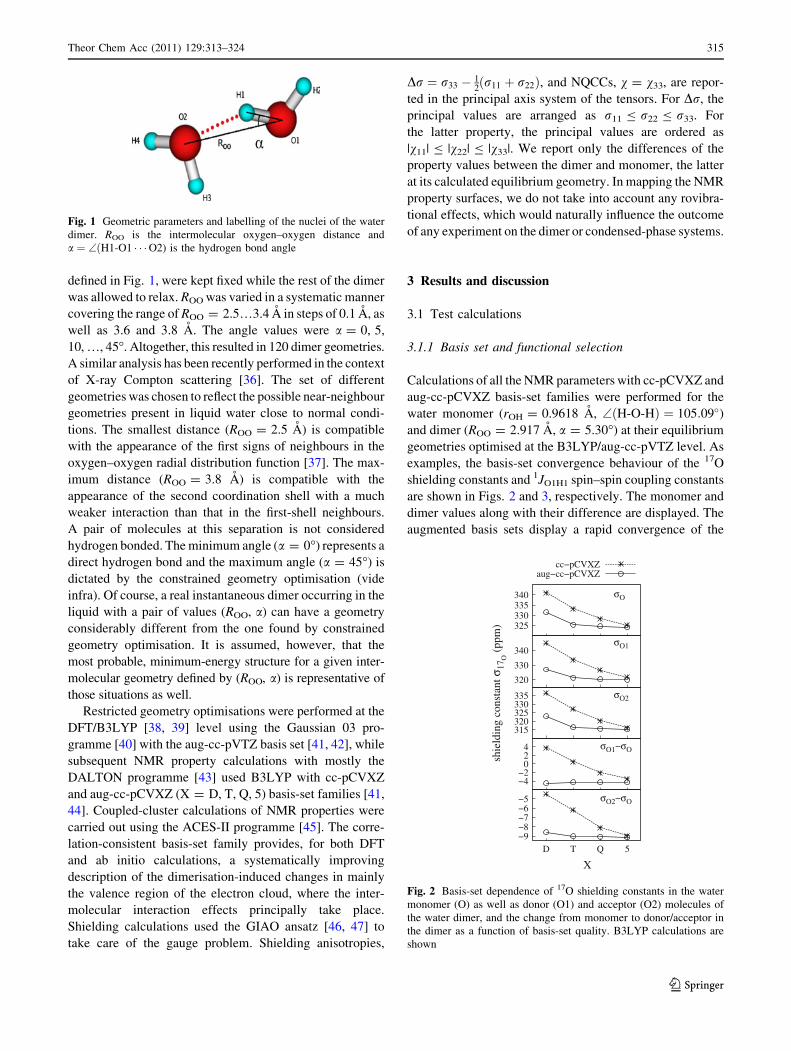
defined in Fig. 1, were kept fixed while the rest of the dimer
was allowed to relax. ROO was varied in a systematic manner
covering the range of ROO = 2.5…3.4 A in steps of 0.1 A, as
well as 3.6 and 3.8 A. The angle values were a = 0, 5,
10, …, 45�. Altogether, this resulted in 120 dimer geometries.
A similar analysis has been recently performed in the context
of X-ray Compton scattering [36]. The set of different
geometries was chosen to reflect the possible near-neighbour
geometries present in liquid water close to normal condi-
tions. The smallest distance (ROO = 2.5 A) is compatible
with the appearance of the first signs of neighbours in the
oxygen–oxygen radial distribution function [37]. The max-
imum distance (ROO = 3.8 A) is compatible with the
appearance of the second coordination shell with a much
weaker interaction than that in the first-shell neighbours.
A pair of molecules at this separation is not considered
hydrogen bonded. The minimum angle (a = 0�) represents a
direct hydrogen bond and the maximum angle (a = 45�) is
dictated by the constrained geometry optimisation (vide
infra). Of course, a real instantaneous dimer occurring in the
liquid with a pair of values (ROO, a) can have a geometry
considerably different from the one found by constrained
geometry optimisation. It is assumed, however, that the
most probable, minimum-energy structure for a given inter-
molecular geometry defined by (ROO, a) is representative of
those situations as well.
Restricted geometry optimisations were performed at the
DFT/B3LYP [38, 39] level using the Gaussian 03 pro-
gramme [40] with the aug-cc-pVTZ basis set [41, 42], while
subsequent NMR property calculations with mostly the
DALTON programme [43] used B3LYP with cc-pCVXZ
and aug-cc-pCVXZ (X = D, T, Q, 5) basis-set families [41,
44]. Coupled-cluster calculations of NMR properties were
carried out using the ACES-II programme [45]. The corre-
lation-consistent basis-set family provides, for both DFT
and ab initio calculations, a systematically improving
description of the dimerisation-induced changes in mainly
the valence region of the electron cloud, where the inter-
molecular interaction effects principally take place.
Shielding calculations used the GIAO ansatz [46, 47] to
take care of the gauge problem. Shielding anisotropies,
Dr ¼ r33 � 12
r11 þ r22ð Þ, and NQCCs, v = v33, are repor-
ted in the principal axis system of the tensors. For Dr, the
principal values are arranged as r11 B r22 B r33. For
the latter property, the principal values are ordered as
|v11| B |v22| B |v33|. We report only the differences of the
property values between the dimer and monomer, the latter
at its calculated equilibrium geometry. In mapping the NMR
property surfaces, we do not take into account any rovibra-
tional effects, which would naturally influence the outcome
of any experiment on the dimer or condensed-phase systems.
3 Results and discussion
3.1 Test calculations
3.1.1 Basis set and functional selection
Calculations of all the NMR parameters with cc-pCVXZ and
aug-cc-pCVXZ basis-set families were performed for the
water monomer (rOH = 0.9618 A, \ðH-O-HÞ ¼ 105:09�)and dimer (ROO = 2.917 A, a = 5.30�) at their equilibrium
geometries optimised at the B3LYP/aug-cc-pVTZ level. As
examples, the basis-set convergence behaviour of the 17O
shielding constants and 1JO1H1 spin–spin coupling constants
are shown in Figs. 2 and 3, respectively. The monomer and
dimer values along with their difference are displayed. The
augmented basis sets display a rapid convergence of the
Fig. 1 Geometric parameters and labelling of the nuclei of the water
dimer. ROO is the intermolecular oxygen–oxygen distance and
a ¼ \ðH1-O1 � � �O2) is the hydrogen bond angle
340335330325
σO
cc−pCVXZaug−cc−pCVXZ
340
330
320
shie
ldin
g co
nsta
nt σ
17O (
ppm
)
σO1
335330325320315
σO2
420
−2−4
σO1−σO
−5−6−7−8−9
5QTD
X
σO2−σO
Fig. 2 Basis-set dependence of 17O shielding constants in the water
monomer (O) as well as donor (O1) and acceptor (O2) molecules of
the water dimer, and the change from monomer to donor/acceptor in
the dimer as a function of basis-set quality. B3LYP calculations are
shown
Theor Chem Acc (2011) 129:313–324 315
123

interaction-induced effect on NMR parameters. It turns
out that the aug-cc-pCVTZ basis performs well in all
cases other than the 1JO1H1 spin–spin coupling constant,
which would require the quadruple zeta level, as can be seen
in the bottom part of Fig. 3. Regarding the overall perfor-
mance and computational cost, aug-cc-pCVTZ appears to
be a good choice for production calculations, keeping in
mind the nice performance with shieldings and NQCCs but
only a restricted validity when the spin–spin coupling is
concerned.
Other studies have found the performance of the DFT/
B3LYP level of theory suitable for qualitative NMR cal-
culations of water using aug-cc-pVDZ for shieldings [48]
and a triple-zeta basis set supplemented with tight func-
tions for spin–spin couplings [49]. Kongsted et al. [25]
found that the DFT error for the acceptor oxygen shielding
constant as a function of dimer geometry differs between
the different DFT functionals, as referenced to high-level
CCSD(T) results. Table 1 compares the present DFT,
Hartree-Fock and coupled-cluster calculations to the liter-
ature data for both the monomer and the dimer, both at
their equilibrium geometry.
The results reveal that the performance of B3LYP is no
inferior to that of the recommended [25] KT3 functional
[57] when all NMR parameters are compared against the
CC reference or, in the monomer case, to experiment.
Small differences should have no effect on overall con-
clusions based on the present B3LYP results. B3LYP
shows a reasonable overall performance with respect to the
present CC data, as well as recent high-level calculations,
thus validating the choice of this functional for the pro-
duction calculations. The significance of the DFT errors is
diminished by the fact that the present work concentrates
on the trends occurring in the property surfaces of the
dimer, which are obtained as differences between the dimer
and monomer values. These trends are only slightly
affected by the systematic errors of the method.
One might question the applicability of DFT for rela-
tively weak interactions taking place between molecules.
Whereas accurate intermolecular interaction energies
typically require using correlated ab initio methods, the
corresponding effects on NMR parameters, determined by
one-electron operators sampling a region of space weighed
heavily to the proximity of nuclei, are seen to be rather well
reproduced at the DFT level. In the case of water dimer, the
electrostatic and monomer overlap effects play key roles in
the NMR property surfaces, and both can be expected to be
captured reasonably by DFT. In fact, even in the case of a
xenon dimer, where the energetics obtains a major dis-
persion contribution, already the Hartree-Fock theory is
able to convey the qualitative features of inter-atomic
NMR property surfaces [58, 59].
3.1.2 Basis-set superposition error tests
The importance of basis-set superposition error (BSSE)
effects [60] was tested using the counterpoise (CP) cor-
rection scheme of Boys and Bernardi [61], modified to
include changes in the monomer geometry [62]. The ratio
of the CP correction term to the uncorrected interaction-
induced change is displayed in Table 2 for each NMR
parameter of the water dimer at ROO = 2.9 A and a = 0�geometry. The conclusion is that BSSE needs not to be
corrected for in the cases of nuclear shielding, shielding
anisotropy, or NQCC with the presently used combination
of method and basis set. These properties may indicate
large relative BSSE in some specific cases but this
behaviour can be traced back to the small magnitude of the
interaction-induced change, causing any correction to
appear large in the relative sense. On the contrary, spin–
spin couplings show significant BSSE effects up to more
than 40% for OH couplings of the donor molecule and up
to 10% for the acceptor. In production calculations, we
decide not to correct for BSSE, keeping in mind the pos-
sible consequences for spin–spin couplings.
We note that Kongsted et al. [25] also obtained quite
small BSSE effects on shielding in their study of (H2O)2
using the aug-cc-pVTZ basis set. In the same context,
already Chesnut and Rusiloski [21] mention the little need
of performing CP corrections for the water dimer when
basis sets containing diffuse functions are employed.
3.2 Production calculations
3.2.1 Geometry and potential energy
The optimised dimer geometry at the B3LYP/aug-cc-pVTZ
level has ROO = 2.917 A and a = 5.30�. The former value
−50−60−70
1JOH
cc−pCVXZaug−cc−pCVXZ
−50−60−70
spin
−sp
in c
oupl
ing
cons
tant
1 J (H
z)1JO1H1
−2−3−4−5−6
5QTDX
1JO1H1−1JOH
Fig. 3 Basis-set dependence of 1JO1H1 spin–spin coupling constants
for monomer (OH), dimer (O1H1), and their difference. The
hydrogen atom H1 participates in the hydrogen bond. B3LYP
calculations are shown
316 Theor Chem Acc (2011) 129:313–324
123

Table 1 Calculated NMR properties of water monomer and dimer at different levels of theory
B3LYP KT3 HFb CCc Literature
Monomera
rO 325.5 325.0 326.6 336.3 325.3d
rH 31.08 31.40 30.52 30.79 30.05e
DrO 54.7 50.7 56.5 49.1 46.9f
DrH 19.16 18.61 20.35 19.92 19.08f
vO 10.38 11.06 10.63 9.78 10.11g
vD 0.322 0.314 0.321 0.329 0.308h
1JOH -68.7 -71.1 – -73.7 -78.7i, -80.6j
2JHH -5.58 -8.86 – -6.81 -7.34j
Dimerk
rO1 -4.2 -3.4 -0.8 -3.6 -3.4l, -1.3m
rO2 -9.0 -8.5 -6.5 -7.1 -7.3l, -5.6m
rH1 -3.13 -3.04 -3.13 -2.99 -2.94l, -2.45m
rH2 0.40 0.40 0.48 0.37 0.36n
rH3 -0.65 -0.67 -0.58 -0.61 -0.48n
DrO1 11.5 11.7 7.7 10.1 8.5o
DrO2 -12.5 -10.8 -11.4 -10.8
DrH1 11.13 10.90 10.82 10.90 12.03o
DrH2 -0.08 0.01 0.04 -0.04 0.38o, 0.0p
DrH3 -0.27 -0.25 -0.13 -0.19 -0.6p
vO1 -0.78 -0.78 -0.75 -0.70
vO2 -0.25 -0.30 -0.19 -0.22
vD1 -0.041 -0.040 -0.043 -0.041
vD2 0.004 0.004 0.005 0.004
vD3 -0.005 -0.005 -0.005 -0.0051JO1H1 -4.9 -4.6 – -4.0 -4.5q, -7.3r
1JO1H2 -1.6 -1.8 – -1.1 -0.7q, -2.7r
2JH1H2 0.22 0.49 – 0.30 -0.14q, -0.35r
1JO2H3 -3.4 -3.6 – -2.6 -2.4q, -4.5r
2JH3H4 -0.03 0.04 – 1.75 -0.1q, -0.4r
DFT results using the B3LYP and KT3 functionals, as well Hartree-Fock (HF) and coupled-cluster (CC) data are shown for the shielding constants (r, in ppm), shieldinganisotropies (Dr, ppm), quadrupole couplings (v, MHz) and spin–spin couplings (J, Hz). For the dimer, changes with respect to the monomer values are givena Unless otherwise indicated, present calculations using the aug-cc-pCVTZ basis set and the optimised (at the B3LYP/aug-cc-pVTZ level) monomer (rOH = 0.9618 A,\H� O� H ¼ 105:09�) and dimer (ROO = 2.9173 A, a = 5.30�) geometriesb Spin–spin couplings not calculated at the HF levelc r;Dr and v with CCSD(T), J with CCSDd Analysis of experimental spin-rotation data in Ref. [50]. Best estimate of the equilibrium geometry value 337.7 ppm is obtained by subtracting the computed rovibrationalcontribution [50]
e Analysis of experimental gas-phase data in Ref. [51]. An estimate of the equilibrium geometry value 30.6 ppm is obtained by subtracting the computed rovibrationalcontribution -0.55 ppm at 300 K [14] from the experimental valuef MCSCF calculation including rovibrational contributions at 300 K of -2.4 and -1.08 ppm for 17O (in 1H2
17O) and 1H (in 1H216O), respectively [14]. The equilibrium geometry
values are 49.3 and 20.16 ppm for 17O and 1H, respectivelyg Microwave experiment [52]. An estimate of the equilibrium geometry value 9.93 MHz is obtained by subtracting the computed rovibrational contribution 0.18 MHz at 298 K[16] from the experimental valueh Molecular beam experiment [53]. An estimate of the equilibrium geometry value 0.312 MHz is obtained by subtracting the computed rovibrational contribution -0.004 MHzat 298 K [16] from the experimental valuei NMR measurement in cyclohexane-d12 solution at 293 K [54]. An estimate of the equilibrium geometry value -83.0 Hz is obtained by subtracting the computed rovibrationalcontribution 4.34 Hz at 300 K [15] from the experimental valuej NMR measurement in CD3NO2 solution at 323 K (1JOH) and 297 K (2JHH) [55]. Estimates of the equilibrium geometry values -85.0/-7.9 Hz are obtained by subtracting thecomputed rovibrational contributions 4.39/0.55 Hz at 340/300 K [15] from the experimental values of 1JOH/2JHH
k For the dimer, the changes in the parameters with respect to the monomer values, a(dim) - a(mon), are indicated. Only one result (H3/D3) for the acceptor hydrogens isdisplayed because the hydrogens have symmetric positions at the equilibrium geometryl Ref. [25]. CCSD(T)/aug-cc-pVTZ level of theory, at the dimer geometry optimised at the same levelm Ref. [23]. CCSD/aug-cc-pVTZ level of theory, at the experimental dimer geometry [8]n Ref. [21]. HF/6-311??G(2d,2p) level of theory, at the experimental dimer geometryo Ref. [20]. HF/4-31G level of theoryp Ref. [56]. HF calculationq Ref. [22]. RASSCF(RAS1)/HIIIa level of theory, at the experimental dimer geometry [8]r Ref. [22]. As in footnote q but with the dimer immersed in a dielectric continuum model simulating liquid water
Theor Chem Acc (2011) 129:313–324 317
123

resides within the rather large error margins of the experi-
mental result by Dyke [9], ROO = 2.98 ± 0.33 A. Klopper
et al. [11] obtained in their benchmark CCSD(T) calcula-
tions ROO = 2.912 ± 0.005 A and a = 5.5�. Consequently,
the present B3LYP/aug-cc-pVTZ geometry agrees extre-
mely well with the best existing estimate.
During each point of the restricted geometry optimisa-
tion, the dimer parameters ROO and a are not allowed to
change while the rest of the geometry relaxes. The overall
appearance of the dimer after optimisation is quite different
for small a angles when compared with large angles.
Figure 4 illustrates the gradual changes. With moderate a(0�–15�), the orientation of the monomers with respect to
each other in the dimer closely resembles that of the global
equilibrium geometry [11]. The hydrogen bond angle is
not restricted to lie in the symmetry plane of the dimer,
however. As a increases further (20�–30�), monomer ori-
entations gradually evolve, finally (35�–45�) ending up into
a situation, where the distinction of the donor and the
acceptor molecules has disappeared. At this point, the
hydrogen bond angle a equals 45�. Because the molecules
are in symmetric positions with respect to one another, also
the other angle a0, the equivalent of a with the roles of the
donor and acceptor interchanged, is also 45�. If the angle ais increased over 45�, the donor and acceptor labels swap,
and a0 takes the role of a as the hydrogen bond angle. In
vibration-rotation-tunnelling spectroscopy, this type of
process is called ‘‘donor-acceptor-interchange’’ and it is
one of the tunnelling processes in the water dimer [63, 64].
The sequence of geometries in a restricted geometry opti-
misation of the water dimer as the hydrogen bond angle
gradually increases resembles the rearrangement pathway
for the donor-acceptor-interchange tunnelling, the hydro-
gen bond angle being the reaction coordinate. Thus, the
symmetric geometry where both a and a0 equal 45� can be
seen as a transition state.
Figure 5 shows the variation of the covalent bond
lengths and intramolecular angles of the donor and
acceptor as a function of ROO and a. The covalent O1H1
bond directly involved in the hydrogen bond elongates
upon dimerisation, with the elongation amounting to less
than 0.01 A and being most noticeable near the equilibrium
Table 2 Basis set superposition errors estimated with the modified
counterpoise method [62] at the B3LYP/aug-cc-pCVTZ level for the
near-equilibrium dimer geometry ROO = 2.9 A and a = 0�
Aa A(dim) - A(mon) BSSEabsolute BSSE% [A(dim) -
A(mon)]CP
rO1 -4.73 -0.04 -0.92 -4.77
rO2 -9.10 0.06 0.62 -9.04
rH1 -3.22 -0.01 -0.22 -3.23
DrO1 9.53 -0.08 -0.83 9.47
DrO2 -13.12 -0.02 -0.19 -13.12
DrH1 11.51 0.01 0.07 11.52
vO1 -0.82 0.00 -0.17 -0.82
vO2 -0.25 -0.01 -2.34 -0.25
vD1 -0.041 0.000 0.38 -0.0411JO1H1 -5.05 2.24 44.33 -2.811JO2H3 -3.75 0.38 10.03 -3.38
Results with and without the BSSE, as well as the absolute and rel-
ative BSSE are displayeda Shielding constants and anisotropies in ppm, quadrupole couplings
in MHz, and spin–spin couplings in Hz
Fig. 4 Changes in the overall
dimer geometry as the hydrogen
bond angle a changes from 0� to
45�
0
0.002
0.004
0.006
0.008
2.63.0
3.43.8
10203040
0.002
0.004
0.006
0.008
ΔRH1O1 (Å)
ROO
α
ΔRH1O1 (Å)
0
0.001
0.002
0.003
2.63.0
3.43.8
102030400.000
0.001
0.002
0.003
0.004
ΔRH3O2 (Å)
ROO
α
ΔRH3O2 (Å)
−2
0
2
4
2.6 3.0 3.4 3.810203040
−1.0
0.0
1.0
2.0
ΔβH1−O1−H2(°)
ROO
α
ΔβH1−O1−H2(°)
−0.003
−0.002
−0.001
0
2.63.0
3.43.8
10203040
−0.002
−0.001
0.000
0.001
ΔRH2O1 (Å)
ROOα
ΔRH2O1 (Å)
−0.001
0
0.001
0.002
2.63.0
3.43.8
10203040
−0.001
0.000
0.001
ΔRH4O2 (Å)
ROOα
ΔRH4O2 (Å)
−2
0
2
2.63.0
3.43.8 10 20 30 40
0.0
1.0
2.0
ΔβH3−O2−H4(°)
ROO α
ΔβH3−O2−H4(°)
Fig. 5 Variation of the donor and acceptor bond lengths and angles
in water dimer as a function of the intermolecular oxygen–oxygen
distance ROO and hydrogen bond angle a. B3LYP/aug-cc-pVTZ level
of theory
318 Theor Chem Acc (2011) 129:313–324
123

geometry. The other covalent bonds are considerably less
affected. As the monomers approach each other, the bond
angles of both the acceptor and the donor remain close to the
free monomer value all the way down to the equilibrium
distance of the dimer. An even closer encounter makes both
angles open up by a couple of degrees.
The potential energy surface of the dimer is illustrated in
Fig. 6. The energy surface has a repulsive core region at
small ROO values and approaches zero for large ROO. As a
function of a the changes are modest. Around the equi-
librium value of ROO at about 2.9 A, the energy landscape
has a valley as a traverses its range, indicating the rear-
rangement pathway for the donor-acceptor-interchange
tunnelling. The potential energy at the equilibrium geom-
etry is about -19 kJ/mol and the energy of the transition
state is about 4.4 kJ/mol higher, with no corrections
applied for basis-set superposition error.
3.2.2 Property surfaces
Table 1 points out the major changes caused by hydrogen
bonding on the NMR parameters of the water dimer at the
equilibrium geometry. They occur in the nuclei that are
directly involved in the O2_H1–O1 bond. Among their
parameters, the most dramatic effects occur for the
anisotropy of the acceptor and donor oxygens, DrO2 and
DrO1, which undergo a 20% (10 ppm) decrease and
increase, respectively, as well as DrH1 that increases by
50% from its monomer value to ca. 30 ppm. Large changes
in the shielding constant and anisotropy of H1 with
ROO \ 3.5 A were already found by Ditchfield [20], who
also pointed out that the anisotropy may be more indicative
of hydrogen bonding than changes in the isotropic shield-
ing constants. In a similar vein, the NQCC of D1 decreases
by roughly 40 kHz upon dimer formation. The other
parameters change relatively little, with a characteristic
decrease taking place in the shielding constants (apart from
the small increase in the case of the non-bonded donor
hydrogen, H2), as well as the one- and two-bond spin–spin
couplings.
Figures 7, 8, 9 and 10 show the dimer property surfaces
for some of the shielding constants, shielding anisotropies,
NQCCs, and intramolecular spin–spin coupling constants,
respectively, computed as differences with respect to the
corresponding monomer values. Small but representative
set of test calculations confirm that the general trends and
overall behaviour is similar between B3LYP vs. CCSD(T)
for shieldings and B3LYP vs. CCSD for J couplings. Thus,
general conclusions based on the B3LYP property surfaces
should be reliable.
The depicted surfaces have their maxima (in the abso-
lute sense) usually near (ROO = 2.5 A, a = 0�) because at
small intermolecular distance the repulsive intermolecular
interaction strongly affects the properties. The effect of the
neighbouring molecule diminishes smoothly as the inter-
molecular separation increases. While all the parameters
are clearly dependent on ROO, the dependence on the
hydrogen bond angle a varies. This can in some cases be
traced back to the dependence on the distance between the
nucleus or nuclei in question and the closest atom in the
neighbouring molecule. For the donor oxygen nucleus
(O1), the distance to the closest atom in the acceptor, the
oxygen O2, is not changing as a changes in the constrained
geometry optimisation. Consequently, the effect of a on
rO1 and DrO1 is modest. For O2 and H1, the nuclei that are
directly involved in the hydrogen bonding, there is a more
−20−16−12−8−4 0
2.7 3.1 3.510203040
−20.0−16.0−12.0 −8.0 −4.0 0.0
E (kJ/mol)
−12.9
ROOα
E (kJ/mol)
Fig. 6 B3LYP/aug-cc-pCVTZ potential energy surface of the water
dimer as a function of the intermolecular oxygen–oxygen distance
ROO and hydrogen bond angle a. The marked point indicates the
equilibrium geometry and the black projected curve is an equipoten-
tial contour corresponding to the value at the point ROO = 3.5 A and
a = 0�.
−12−8−4 0 4
2.7 3.1 3.510203040−12.0
−8.0
−4.0
0.0σO1 (ppm)
−0.40
ROOα
σO1 (ppm)
−30−20−10 0 10
2.7 3.1 3.510203040
−21.0
−14.0
−7.0
0.0σO2 (ppm)
−1.60
ROOα
σO2 (ppm)
−8−6−4−2 0
2.7 3.1 3.510203040
−8.0 −6.0 −4.0 −2.0 0.0
σH1 (ppm)
−1.15
ROOα
σH1 (ppm)
Fig. 7 As Fig. 6 but for the nuclear shielding constant of the water
dimer for the donor oxygen O1, acceptor oxygen O2, and hydrogen-
bonded hydrogen H1
Theor Chem Acc (2011) 129:313–324 319
123

pronounced dependence on a in the shielding constant and
anisotropy. The distance between the hydrogen-bonded
hydrogen H1 and the acceptor oxygen O2, nearest
neighbours for each other, depends on a, and so do most of
the parameters of these nuclei. DrO2 even changes to have
a positive sign in the depicted portion of the surface, at
large a values.
While the shielding anisotropies have a noticeable
a-dependence for all the observed nuclei, in general the
investigated spin–spin couplings are mostly dependent only
on the ROO distance, with little curvature of the surfaces in
the direction of changing a. The NQCCs of O1, O2, and D1
are all non-negligibly dependent on the hydrogen bond
angle, although the range of overall alteration is small for
the acceptor oxygen. We also checked the dimerisation
effects on the direction of the principal axis 3, the reference
direction for the shielding anisotropy r33, and observed
that its direction tends to turn towards the neighbour. These
directional changes are modest for hydrogen-bonded
hydrogen H1 and donor oxygen O1, and very small for
acceptor oxygen O2.
3.2.3 Direct interaction effect and indirect geometric
effect
The effect of dimerisation on the NMR properties is ana-
lysed further by dividing it into two contributions. On the
one hand, the direct effect focuses on the intermolecular
influence on the properties at a fixed geometry. On the
other hand, the indirect effect is caused by the dimerisa-
tion-induced geometry change of the monomers [29].
0 10 20 30 40
2.73.13.510203040
0.0 10.0 20.0 30.0
ΔσΟ1 (ppm)
3.40
ROOα
ΔσΟ1 (ppm)
−30−20−10 0 10 20
2.7 3.1 3.510203040−30.0−20.0−10.0
0.0 10.0 20.0
ΔσΟ2 (ppm)
−3.00
ROOα
ΔσΟ2 (ppm)
0 10 20 30
2.73.13.510203040
0.0
10.0
20.0ΔσΗ1 (ppm)
4.80
ROOα
ΔσΗ1 (ppm)
Fig. 8 As Fig. 6 but for the nuclear shielding anisotropy of the water
dimer for the donor oxygen O1, acceptor oxygen O2, and hydrogen-
bonded hydrogen H1
−2−1.5−1−0.5 0
2.7 3.1 3.510203040
−2.0 −1.5 −1.0 −0.5 0.0
χO1 (MHz)
−0.27
ROOα
χO1 (MHz)
−0.4−0.3−0.2−0.1 0
2.73.13.5
1020
3040
−0.4
−0.2
0.0
χO2 (MHz)−0.17
ROO
α
χO2 (MHz)
−60−45−30−15 0
2.7 3.1 3.510203040
−60.0−45.0−30.0−15.0
0.0χD1 (kHz)
−20.0
ROOα
χD1 (kHz)
Fig. 9 As Fig. 6 but for the nuclear quadrupole coupling of the water
dimer for the donor oxygen O1, acceptor oxygen O2, and hydrogen-
bonded deuterium D1
−12
−8
−4
0
2.6 3.0 3.4 3.810203040
−10.0 −8.0 −6.0 −4.0 −2.0 0.0
JH1O1 (Hz)
−1.70
ROOα
JH1O1 (Hz)
−4
−2
0
2.6 3.0 3.4 3.810203040
−4.0
−2.0
0.0
2.0JH2O1 (Hz)
−0.35
ROOα
JH2O1 (Hz)
−8−6−4−2 0
2.6 3.0 3.4 3.810203040
−8.0 −6.0 −4.0 −2.0 0.0
JO2Hnear (Hz)
−1.60
ROOα
JO2Hnear (Hz)
Fig. 10 As Fig. 6 but for the nuclear spin–spin coupling constant
surfaces of the water dimer for 1JH1O1, 1JH2O1, and 1JO2Hnear, where
Hnear denotes the hydrogen nucleus of the acceptor which is closest to
the donor molecule
320 Theor Chem Acc (2011) 129:313–324
123

However, this often-considered separation is somewhat
artificial and contains an ambiguity as demonstrated by the
data in Table 3 and the following consideration.
The break-down of the total dimerisation effect into
direct and geometric contributions can be realised by using
one of two intermediate states that manifest either the
former or the latter part, but not both at the same time. If
we start with a monomer in its equilibrium geometry, and
as a first step transform it into a monomer in a geometry
that it assumes in a dimer, we take the upper route with the
intermediate state 1 in Table 3. In the second step, the
neighbour molecule is introduced and we end up at an
interacting dimer at the dimer geometry. The geometric
and direct effects on properties can be computed as the
changes in the two steps. The other possible choice is to
first introduce the neighbour molecule without allowing the
geometry of the monomers to change (state 2 in the Table)
and only subsequently letting the geometry relax. In prin-
ciple, the two choices result in distinct analyses into the
direct and indirect contributions. It is emphasised that
neither of the two paths joining the monomer and dimer
situations are physically realised as such; they only serve as
a means of analysing the total interaction effect. We use
both approaches, and the results are gathered in Table 3.
In eight out of twelve cases, the direct effect is larger
than the indirect geometric effect by about a factor of 10 or
more. Properties related to the acceptor oxygen O2 change
primarily through the direct effect because the geometry
changes of the acceptor molecule are modest. Also the
dimerisation effects on the shielding anisotropies DrO1 and
DrH1 as well as the quadrupole coupling vO1 arise chiefly
due to the direct interaction. In the remaining cases, the
absolute magnitude of the geometric effect constitutes
more than 10% of the total effect and may reach as much as
40%, which is the case for both the donor oxygen shielding
rO1 and the quadrupole coupling of deuterium D1 directly
involved in the hydrogen bond. These are the two centres
that form the covalent bond that undergoes the largest
changes upon dimerisation. The dimerisation-induced
changes in the spin–spin couplings 1JO1H1 and 1JO1H2 of
the donor molecule also bear considerable geometric con-
tributions. While the direct and geometric effects occur
mostly in the same direction, they may be opposite as well,
notably in 1JO1H1.
To demonstrate the fundamental ambiguity in the pres-
ent type of analysis, we also tested the influence on the
results of the order in which the geometric and direct
effects are applied. Selecting either route one or two in
Table 3 is an important factor only for the shielding con-
stant of the donor oxygen, where the two approaches give a
notably different partition into geometric and direct con-
tributions. In all the other cases, the order is unimportant.
Again, as the O1–H1 covalent bond length is the one that
clearly changes in dimerisation, the operations of geometry
change and the introduction of the neighbour are not
independent in this case, in contrast to the other parameters
where they appear additive to a good approximation.
The discussion above suggests that the pairwise additive
approximation (PAA) that has been found valid, e.g., for
the shielding constant rXe in monoatomic xenon gas [65]
may not be a sensible one for rO in liquid water. In this
approximation, it is assumed that each contribution from
the neighbouring atoms can be separately evaluated and
Table 3 Comparison of the direct intermolecular interaction effect
and the indirect geometric effect in the NMR parameters of the water
dimer
The diagram shows the different partitions into direct and geometric
effects obtained using two auxiliary intermediate states (see text for
details)
Theor Chem Acc (2011) 129:313–324 321
123

accumulated, and that the investigated molecule does not
have to be allowed to relax geometrically after adding each
neighbour, since the indirect effect can be added afterwards
if necessary. The latter condition fails if the geometry
change and the addition of the neighbour are not inde-
pendent operations. For comparison, while the NQCC of
the deuterium directly involved in the hydrogen bond
obtains a large geometric contribution, the order of the two
mentioned operations is not important. Indeed, PAA has
been demonstrated to be a good approximation for quad-
rupole couplings in liquid water [19].
In cases where the pairwise additive approximation
holds, the present intermolecular property surfaces may
provide a powerful avenue for evaluating the NMR
parameters of bulk and surface water, without resorting to
expensive, explicit quantum-chemical supermolecule cal-
culations of simulation snapshots. The surface data can be
fitted to a suitable functional form, an NMR force field
[66], which can be used to calculate the parameters based
on the instantaneous position information alone.
3.2.4 Property surfaces and hydrogen bonding
The NMR parameters are sensitive to the details of the
electronic structure near the resonant nucleus. Interaction
with neighbouring molecules changes the electronic
structure and thus affects the NMR parameters. The other
way around, the changes in the NMR parameters could in
principle be used to identify the neighbours of the mole-
cule, e.g., to judge whether the molecule is involved in a
hydrogen bond. Based on the computed property surfaces
for the water dimer, we may try to identify a useful defi-
nition of a hydrogen bond that exploits the changes in the
NMR parameters caused by dimerisation. Such definitions
are typically expressed geometrically.
In the present data, the dimer parameters approach those
of an isolated water molecule at a large separation of the
monomers. Utilising this tendency, a suitable cut-off dis-
tance criterion can be selected that determines when the
dimer parameter deviates enough from the corresponding
monomer parameter, to be considered as having a strong
interaction with its neighbour. A general feature of all the
present NMR parameter surfaces is that they are smooth,
revealing no abrupt changes, as well as well converged to
the monomer values as a function of distance within our
range of intermolecular geometries. Even the somewhat
more strict, commonly used cut-off distance of about 3.5 A
can on the basis of our data be used to define an approxi-
mate range where the interaction appreciably affects the
NMR parameters. In contrast, the interaction energy seems
not be converged at this distance.
For the angle a, there cannot be an NMR-based, gen-
erally applicable cut-off value that would be independent
of ROO. This is due to the fact that some nuclei, especially
the hydrogen-bonded centre H1 turns away from the
acceptor molecule with increasing a, eventually becoming
effectively free. At the same time, the oxygens stay at a
separation ROO, with the consequence that their NMR
parameters continue to be affected regardless of the angle.
Hence, the oxygens do not become free as a increases. This
means that there cannot be any generally valid range of afor hydrogen bonding that would be defined through
changes in NMR parameters when compared to the
monomer. However, if a increases without limit, the
hydrogen atom H1 no longer stays in between the two
molecules and eventually ceases to participate in hydrogen
bonding. Consequently, we must from this purely geo-
metric point-of-view restrict a, and the natural upper limit
for a equals 45� because at this point the restricted
geometry optimisation pushes the dimer into a geometry
where two hydrogen bonds coexist, as depicted in Fig. 4.
Summarising, a hydrogen bond definition that is com-
patible with the NMR data can be expressed as ROO B
3.5 A and a(H1–O1_O2) B 45�. For comparison, the
geometric ‘‘strong’’ hydrogen bond criterion of Mezei and
Beveridge [26] (ROO B 3.3 A and a B 45�) and the defi-
nition of Luzar and Chandler [27] (ROO B 3.5 A and
a B 30�) are in a good agreement with the present sug-
gestion. A very recent discussion about the different defi-
nitions can be found in the article by Kumar, Schmidt and
Skinner [28]. Comparison with techniques other than NMR
shows, e.g., that the range of the first coordination shell in
liquid water as determined experimentally from the oxy-
gen–oxygen radial distribution function, is about 3.5 A
[37], which indicates the range of the hydrogen bonding
interaction. The use of NMR parameters to identify
hydrogen bonding complements and reinforces the validity
of the widely used hydrogen bond definitions. Our findings
are in unison with the traditional notion of the hydrogen
bond, and the overall smoothness of the NMR property
surfaces prevents us from giving any novel definition.
4 Conclusions
We have monitored the changes in the NMR parameters of
the water dimer, calculated at the DFT/B3LYP/aug-cc-
pCVTZ level of theory, as functions of two intermolecular
coordinates, the oxygen–oxygen distance ROO and the
hydrogen bond angle a ¼ \ðH1� O1 � � �O2). The studied
parameters are the nuclear shielding constants, shielding
anisotropies, and quadrupole coupling constants, as well as
the intramolecular spin–spin coupling constants.
The behaviour of the intramolecular geometry, interaction
energy, and NMR parameters was illustrated as property
surfaces where the changes induced by dimerisation are
322 Theor Chem Acc (2011) 129:313–324
123

seen as functions of ROO and a, with all the other intra- and
intermolecular geometric variables optimised. The dimeri-
sation mainly affects the parameters of the donor and
acceptor oxygens as well as the hydrogen centre H1. Par-
ticularly, the shielding anisotropies of these nuclei, as well
as the quadrupole coupling constant of the deuterium
directly involved in the hydrogen bond, undergo large
changes upon dimerisation. In general, smooth property
surfaces are obtained for all the parameters, indicating no
abrupt changes on the (ROO, a) surface. The dependence on
the ROO distance is mostly a monotonic decay to the
monomer value of the parameter at large ROO, with the only
exception constituted by the quadrupole coupling of the
acceptor oxygen, which features a saddle surface. The
dependence on a is generally weaker and more varied than
that on ROO.
Analysis of the total dimerisation influence on the main
NMR parameters was performed in terms of the contribu-
tions from an indirect effect via the geometry change and
direct intermolecular effect at fixed geometry. The direct
interaction effect is found to always dominate the total
change, but the indirect influence should generally be
included as well. It is shown, however, that this analysis is
not unique, in the sense that the operations of relaxing to
the dimer geometry and introducing the neighbouring
molecule are generally not additive. This manifests itself
by a different partitioning of the total interaction effect into
the indirect and direct contributions, depending on the
order in which the two steps are taken. In practice, of the
currently investigated NMR properties of the water dimer,
only the nuclear shielding constant of the donor oxygen is
significantly affected by this. For parameters such as this,
approximating the total intermolecular interaction effect in
terms of pairwise additive intermolecular contributions
may not be valid.
Pairwise additivity justifies the use of a preparameter-
ised NMR force field, obtainable from fitting a suitable
model function to our property surfaces. Such a paramet-
risation would enable efficient calculation of the NMR
parameters of differently coordinated water molecules in
the bulk and at surfaces, based on coordinate data alone.
Analysis of the NMR property surfaces can be attempted
with the purpose of obtaining an NMR-based hydrogen
bond definition expressed in terms of ROO and a. Due to the
smoothness of the property surfaces, the analysis is not
capable of producing fundamentally new insight into the
applicable geometric definitions. Our result, ROO B 3.5 A
and a B 45�, coincides with the widely used definitions.
Acknowledgments TP is grateful to the financial support from the
Graduate School of Computational Chemistry and Molecular Spec-
troscopy, Oulu University Scholarship Foundation, Research Foun-
dation of Orion Corporation, The Finnish Foundation for Economic
and Technology Sciences—KAUTE, Magnus Ehrnrooth Foundation,
and Finnish Cultural Foundation. The authors belong to the Finnish
Center of Excellence in Computational Molecular Science (CMS,
2006-11). PL is Academy Research Fellow of the Academy of
Finland. Computational resources were partially provided by CSC—
Scientific Computing Ltd, Espoo, Finland.
References
1. Wernet Ph, Nordlund D, Bergmann U, Cavalleri M, Odelius M,
Ogasawara H, Naslund LA, Hirsch TK, Ojamae L, Glatzel P,
Pettersson LGM, Nilsson A (2004) Science 304:995–1012
2. Stillinger FH (1980) Science 209:451
3. Eisenberg D, Kauzmann W (1969) The structure and properties
of water. Clarendon, Oxford
4. Prendergast D, Galli G (2006) Phys Rev Lett 96:215502
5. Wang RLC, Kreuzer HJ, Grunze M (2006) Phys Chem Chem
Phys 8:4744
6. Soper AK (2005) J Phys Condens Matter 17:S3273
7. Bukowski R, Szalewicz K, Groenenboom GC, van der Avoird A
(2007) Science 315:1249
8. Odutola JA, Dyke TR (1980) J Chem Phys 72:5062
9. Dyke TR (1984) Top Curr Chem 120:85
10. Halkier A, Koch H, Jørgensen P, Christiansen O, Nielsen IMB,
Helgaker T (1997) Theor Chem Acc 97:150
11. Klopper W, van Duijneveldt-van de Rijdt JGCM, van Duijneveldt
FB (2000) Phys Chem Chem Phys 2:2227
12. Hakala M, Nygard K, Manninen S, Huotari S, Buslaps T, Nilsson
A, Pettersson LGM, Hamalainen K (2006) J Chem Phys
125:084504
13. Levitt MH (2001) Spin dynamics: basics of nuclear magnetic
resonance. Wiley, Chichester
14. Vaara J, Lounila J, Ruud K, Helgaker T (1998) J Chem Phys
109:8388
15. Wigglesworth RD, Raynes WT, Sauer SPA, Oddershede J (1998)
Mol Phys 94:851
16. Olsen L, Christiansen O, Hemmingsen L, Sauer SPA, Mikkelsen
KV (2002) J Chem Phys 116:1424
17. Pennanen TS, Vaara J, Lantto P, Sillanpaa AJ, Laasonen K,
Jokisaari J (2004) J Am Chem Soc 126:11093, and references
therein
18. Pennanen TS, Lantto P, Sillanpaa AJ, Vaara J (2007) J Phys
Chem A 111:182
19. Muller MG, Kirchner B, Vogt PS, Huber H, Searles DJ (2001)
Chem Phys Lett 346:160
20. Ditchfield R (1976) J Chem Phys 65:3123
21. Chesnut DB, Rusiloski BE (1993) J Phys Chem 97:2839
22. Pecul M, Sadlej J (1999) Chem Phys Lett 308:486
23. Pecul M, Lewandowski J, Sadlej J (2001) Chem Phys Lett
333:139
24. Pecul M, Sadlej J, Helgaker T (2003) Chem Phys Lett 372:476
25. Kongsted J, Aidas K, Mikkelsen KV, Sauer SPA (2008) J Chem
Theory Comput 4:267
26. Mezei M, Beveridge DL (1981) J Chem Phys 74:622
27. Luzar A, Chandler D (1996) Phys Rev Lett 76:928
28. Kumar R, Schmidt JR, Skinner JL (2007) J Chem Phys
126:204107
29. Astrand P-O, Mikkelsen KV, Ruud K, Helgaker T (1996) J Chem
Phys 100:19771
30. Ramsey NF (1950) Phys Rev 78:699
31. Ramsey NF (1953) Phys Rev 91:303
32. Kowalewski J, Maler L (2006) Nuclear spin relaxation in liquids:
theory, experiments, and applications. Taylor & Francis, Boca
Raton, FL
Theor Chem Acc (2011) 129:313–324 323
123

33. Abragam A (1961) The principles of nuclear magnetism. Oxford
University Press, Oxford
34. Slichter CP (1990) Principles of magnetic resonance, 2nd edn.
Springer, Berlin
35. Struis RPWJ, De Bleijser J, Leyte JC (1987) J Phys Chem
91:1639
36. Nygard K, Hakala M, Manninen S, Andrejczuk A, Itou M,
Sakurai Y, Pettersson LGM, Hamalainen K (2006) Phys Rev E
74:031503
37. Sorenson JM, Hura G, Glaeser RM, Head-Gordon T (2000)
J Chem Phys 113:9149
38. Becke AD (1993) J Chem Phys 98:5648
39. Stephens PJ, Devlin FJ, Chabalowski CF, Frisch MJ (1994)
J Phys Chem 98:11623
40. Frisch MJ, Trucks GW, Schlegel HB, Scuseria GE, Robb MA,
Cheeseman JR, Montgomery JA Jr, Vreven T, Kudin KN, Burant
JC, Millam JM, Iyengar SS, Tomasi J, Barone V, Mennucci B,
Cossi M, Scalmani G, Rega N, Petersson GA, Nakatsuji H, Hada
M, Ehara M, Toyota K, Fukuda R, Hasegawa J, Ishida M,
Nakajima T, Honda Y, Kitao O, Nakai H, Klene M, Li X, Knox
JE, Hratchian HP, Cross JB, Bakken V, Adamo C, Jaramillo J,
Gomperts R, Stratmann RE, Yazyev O, Austin AJ, Cammi R,
Pomelli C, Ochterski JW, Ayala PY, Morokuma K, Voth GA,
Salvador P, Dannenberg JJ, Zakrzewski VG, Dapprich S, Daniels
AD, Strain MC, Farkas O, Malick DK, Rabuck AD, Raghava-
chari K, Foresman JB, Ortiz JV, Cui Q, Baboul AG, Clifford S,
Cioslowski J, Stefanov BB, Liu G, Liashenko A, Piskorz P,
Komaromi I, Martin RL, Fox DJ, Keith T, Al-Laham MA, Peng
CY, Nanayakkara A, Challacombe M, Gill PMW, Johnson B,
Chen W, Wong MW, Gonzalez C, Pople JA (2004) Gaussian 03,
revision C.02. Gaussian Inc., Wallingford, CT
41. Dunning ThH Jr (1989) J Chem Phys 90:1007
42. Kendall RA, Dunning ThH Jr, Harrison RJ (1992) J Chem Phys
96:6796
43. DALTON, a molecular electronic structure program, release 2.0
(2005) See http://www.kjemi.uio.no/software/dalton/dalton.html
44. Woon DE, Dunning ThH Jr (1995) J Chem Phys 103:4572
45. Stanton JF, Gauss J, Watts JD et al, ACES II Mainz-Austin-
Budapest version; see also Stanton JF, Gauss J, Watts JD,
Lauderdale WJ, Bartlett RJ (1992) Int J Quantum Chem, Quantum
Chem Symp 26:879. Current version, see http://www.aces2.de
46. Wolinski K, Hinton JF, Pulay P (1990) J Am Chem Soc 112:8251
47. Helgaker T, Jørgensen P (1991) J Chem Phys 95:2595
48. Cybulski H, Sadlej J (2006) Chem Phys 323:218
49. Peralta JE, Scuseria GE, Cheeseman JR, Frisch MJ (2003) Chem
Phys Lett 375:452
50. Puzzarini C, Cazzoli G, Harding ME, Vazquez J, Gauss J (2009)
J Chem Phys 131:234304
51. Raynes WT (1978) Spec Per Reps Nucl Magn Reson 7:1
52. Bellet J, Lafferty WJ, Steenbeckeliers G (1973) J Mol Spectrosc
47:388
53. Garvey RM, DeLucia FC (1988) Can J Phys 55:1115
54. Wasylishen RE, Friedrich JO (1987) Can J Chem 65:2238
55. Sergeyev NM, Sergeyeva ND, Strelenko YA, Raynes WT (1997)
Chem Phys Lett 277:142
56. Holler R, Lischka H (1981) Chem Phys Lett 84:94
57. Keal TW, Tozer DJ (2004) J Chem Phys 121:5654
58. Hanni M, Lantto P, Runeberg N, Jokisaari J, Vaara J (2004)
J Chem Phys 121:5908
59. Hanni M, Lantto P, Ilias M, Jensen HJAa, Vaara J (2007) J Chem
Phys 127:164313
60. van Duijneveldt FB, van Duijneveldt-van de Rijdt JGCM, van
Lenthe JH (1994) Chem Rev 94:1873
61. Boys SF, Bernardi F (1970) Mol Phys 19:553
62. Xantheas SS (1996) J Chem Phys 104:8821
63. Dyke TR (1977) J Chem Phys 66:492
64. Watanabe Y, Taketsugufoot T, Wales DJ (2004) J Chem Phys
120:5993
65. Hanni M, Lantto P, Vaara J (2009) Phys Chem Chem Phys
11:2485
66. Vaara J (2007) Phys Chem Chem Phys 9:5399
324 Theor Chem Acc (2011) 129:313–324
123