Mechanical design parameters for detection of nuclear signals by magnetic resonance force microscopy
10
LA-UR -cIn- 2901 , .. a ,.. . “, ,, , . ... . ..1. ., : 1., 1,1, ! ! , !, .. . .,.. .!1, #:, l,. .,l . .. . ..m ,,..1 .,:,,., ,., ..... ... . ‘I l., ;, , .1,.. . , ,, 1. , .“. ’41- 1IJ., It, —— ———- . . ---- . . -— ----- . .. . . . . . . ... . . . . .. . . .. . . . . . ....... . .. . ---- .-. .— ,1 ..1, 114 Gr~’~Jory tJ. Mtmro, ,Jaw:k A. Ihn I on, Jlrucc l,,lm,lrt i nc, Mi]ri Iyn ll,Iwlcy, .Jdwdnlc (!. %)lcm, Sidn(:y S.inqar, ,J(}t]ll ,J. ~J~nrmcr, Sf?ppc3 Pcnl ti lil ilfld l,durcl (). Si 1lcrud l) I,S(”l.AIMlili ,. ,., . I II (( I ,, ..
-
Upload
independent -
Category
Documents
-
view
1 -
download
0
Transcript of Mechanical design parameters for detection of nuclear signals by magnetic resonance force microscopy
LA-UR -cIn- 2901
, .. a ,.. . “, ,, , . . . . ...1. ., : 1.,1,1,! ! , !, . . . .,.. .!1, #:, l,. .,l . . . . ..m ,,..1 .,:,,., ,., . . . . . . . . . ‘ I l., ;, , .1,.. . , ,, 1. , .“. ’41- 1IJ., It,—— ———- . . ---- . . -— ----- . .. . . . . . . ... . . . . . . . . . . . . . . . ... . . . . . .. . ---- .-. .—
,1 ..1, 114 Gr~’~Jory tJ. Mtmro, ,Jaw:k A. Ihn I on, Jlrucc l,,lm,lrt i nc,Mi]ri Iyn ll,Iwlcy, .Jdwdnlc (!. %)lcm, Sidn(:y S.inqar,,J(}t]ll ,J. ~J~nrmcr, Sf?ppc3 Pcnl t i lil ilfld l,durcl (). Si 1lcrud
l) I,S(”l.AIMlili
,. ,., .
III ((
I
,,..
About This Report
This official electronic version was created by scanning the best available paper or microfiche copy of the original report at a 300 dpi resolution. Original color illustrations appear as black and white images. For additional information or comments, contact: Library Without Walls Project Los Alamos National Laboratory Research Library Los Alamos, NM 87544 Phone: (505)667-4448 E-mail: [email protected]

1 lt.1~ Intllrn;ltioniil [TA(T1-?hlSymposium Proclwlings, Buston, MA, August, 18-21, 1993
MECHANICAL DESIGN PARAMETERS FOR DETECTIONOF NUCLEAR SIGNALS BY MAGNETIC RESONANCE
FORCE MICROSCOPY
Gregory J. Moore, Jack A. HanIon, Bruce Lamartine,Marilyn Hawley, Johndale C. Solem, Sidney Singer,John J. Jarmer, Seppo Penttila and Laurel O. Sillerud
Biomedical NMR FacilityDivision of Life Sciences
Los Alamos National Laboratory,Los Alamos, New Mexico 87545
U.S.A.
Ryszard J, Pryputniewicz
Center for Holographic and Laser TechnologyDepartment of Mechanical Engineering
Worcester Polytechnic InstituteWorcester, Massachusetts 01609
U.S.A.
ABSTRACT
Recent theoretical work has shown that mechanical detection ofmagnetic resonance from a single nuclear spin is in principle possible. Thistheory has recently been experimentally validated by the mechanica! detectionof olcctron spin resonance signals using microscalo cantilevers, Currently weam extending this Iochnology in an attempt to detect nuclear signals which arolhme orders of magnitude Iowcr in intnnsity than electron signals. In ordor to“Ichieve the needed thousard-folcl improvcmont in scmsitivity we havo(Irldc)rt;lkcm tho dovolopmcnt of optimi~cd mechanical cantilevers and highly[ml;irizod :;;Imples.
~“initr?c!h?mnnt modollnq Is Llsf?d ;1s a toot to SIITI(IIWO carltilevor boarndynilrTIK;S m(l to optimize tho mochanicnl propmlios including (3, resonantIrnllucrwy, ;Impllt(l(k) of vitmlliml ;Ind spring (XvlStilrlt. Sim(llntions :Irc)comp;irnrj 10 ox~wllnl(?ll t:; II:;irlg Ilot(?rmlyrl(! 1101(.)(]rilrTl intorforomo try.N;lllc)fiit)ric~ltiorl of o~)lir-ni~(lrj cilrllil(~v(?ts \~iil ior) rnillim] v;ill ho diroctod by tllnoIll(x)ln(! r)f Itl(lso :;ill)(ll; itiorl:; ;III(I (!xlwrimorlt:;,
I

11th International IJA(’KM Symposium ~’rocwxiin~s, Boston, MA, August 18-21, 1993
1. INTRODUCTION
Magnetic resonance force microscopy (MRFM) is the use of mechanicallydetected magnetic resonance (MR) signals to image small objects with highresolution and to simultaneously identify the chemical species. Until recently,MR signals have only been detected by electrical means which have a
sensitivity limit of approximately 1015 nucleons. Recent work by Sidles et alhas shown that the theoretical limit for mechanical detection is a singlenucleonl ‘4. Rugar et al have experimentally validated this theory by themechanical detection of electron spin resonance (ESR) signals usingmicroscale cantilevers516.
Currently, we are extending this technology in an attempt to detectnuclear signals which are three orders of magnitude lower in intensity thanelectron signals. A subsequent goal is to determine the ultimate experimentalsensitivity of the technology with the goal of detecting a single nuclear spin.The main focus of our investigations to date has been to improve the sensitivity,using both simulation and experiment to design and develop optimizedmechanical cantilevers and highly polarized samples.
2. BACKGROUND
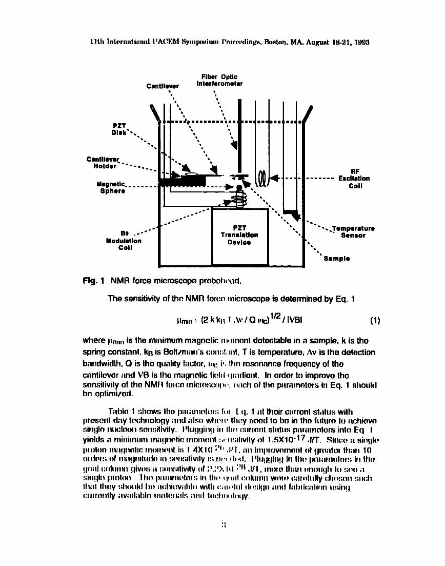
A schematic diagram of the NMR force microscope probehead is shownin Fig. 1, The probehead is an ev; ~cuated chamber and consists of amechanical cantilever, similar to those ilsed in atomic force microscopy, towhich the samp!e is mounted. The paramotcrrs of typical commercial cantileversinclude a Q of 2000 and a resonant froqucmcy of 8 K1-fz, A s~nall magneticsphere provides the tnagnetic field gradient which ic responsible for the highresolution of the rnicroscopo, An rf coi! allows ff)l excitation of the sample whiloa 60 modulation coil pfovictcs the capability to slowly sweep Iho main mngnoticfield. When t m sarnplo is al a critic;~l distance with respect to the fieldgenerated by Ihc magnetic splmrc, the cimtitovor will oscillato ilt its rcmonnntfrcquoncy. The (:ar]tilcvor vihriltiotl:; (:;~Il ho rlotnctnd by thn fitmr optic
intorforomntm wl~ich h;ls ;I sensitivity on tllo ordw of 0,1 Angstroms.f)ositicming of the compcmrmts is iwcomplishod with various PZT trilnslntiondovi(ms, l“tlis ptdmlmld is dosignd to 1)0 insottml ir~lo i] Hc)3-14n4 dilutionlofti!j(?ri~toi which c;~r] m;iil~!;litl ii tonlporilt(it [>” of 1omK, Ttm clilutmn rofriqor;ltoriipr)iitilt[ls is c(mtilinod wittli~~ il 7,5T sll~](?rcor)[i(l(:tillf] vortic;ll l)(m) rl)ilf]l~(]t. Alllii:l OWilVO W;lvo fllliflo is ;Ilt; wtlm{ to 111P Ilrdmt]n;ld to ;Illow for (Iyllilrl]i(;
l~[l(;l(}ilt poliltl/iltiol:~~” (I IN I’) of III(! :;ill~)~)l(~. At 2.5’1, 70 G1-fz mi(:low;lvn:; ;II(}
II:;(!(I It) itti~[llilt[~ ttlr! :;; III I1)Io for 1110 IINI’ lw~](:u(l[lrn
1lth Intrrnnt ionnl [ lA(Vthl Sympwium I’rmvdinhm, ?hton, MA, Aupwt 1U21, 10!!3
PZTDisk’-.
‘u“m.
Ca:gtl:c--------!
Maglwllc. ------,Sphoro
90 ..---”Moduhtlon
Coil
Flhr Opllc
Cantllowr Intarfaromator
Fig. 1
lml
lb
LPZT
Trmnddon
Dedco
NMR force microscope probol~{~i~d.
bb
b
■ ■
✎✍
,1b
RF------- Exclttilon
Coil
Thesensifivity ofthc NMRfor(:r? lllicro.xope isdeteminedby Eq. 1
pm,, :- (2 k k[l r .\\g/ Q (IE) ‘E/ IVBI (1)
where ~lmlllis the rmnimum magnetic HIIm(mt dotoctablo m a sample, k is tho
spring constant. k~ is Boltznmnis C(NN;1,1111,T is temperature, AV is fhe detection
bandwidth. Q is Iho qunlity fnctor, o~: i’, III(? msnnnnce frequency of tho
cilntilcvor iitld W3 is Iho milgnntic Iinl(I ~Iiildblll. In ordor to improvo thas(wwitivily of ttm NMI 1 Iotco mi(:to:;(x]lII I, (!:mli of thn pnmmotors in Eq. 1 shouldtm optimizod.
T;IIIIo 1 !;htlWSIh(i l)ill;illl(~[[!l:i Ii II [ (~. I ilt Ihoir curronl st,~tus withprmmnl (lily Iochnology ill]d i~lso WII[’I I I It][!y IMNYd to tIO in tho fulurn to ilct]k)vn
nily]lo nu(:looll scm:;itivily. l]luq!iit~!l ill Illf? curmnl stntus pilrilln~hJrS into Eq Iyinlds il minimum nm!~rwtic Ilmm(!lll :.1I I:;llivily ot 1.5X 1()- 1”~.l~. Sifwn il sity~l(?~}l(]t(m lll;l(~ll(lllC 1110111 (’111 i:; I .4X 1() “f’ ,111, illl iminov(mnnl d f~r(liltol tllilll 10
(N(bfs 01 fllilf~Illlll(k! ill :;(vwitiwly l:; Ill II (1141, I]hlqqinq in ttl(?~)illillll(llfll!; ifl [110.fjoill (Xdlllllll qiv(m il :;i~lmllivily (~1;’.;’)f 10 “t! .1/1 , ‘I;IIJIII Itlilll (Nlollfjll It) !;{?(1 il
:iinf]l(~ ~)mlon “1II(! ~Nllillllilt(?l!; ili 111(’~IIlill [:{)1[111111W[}lf) [:illflllllly (:11!1!;(111!; IIC:II
Itlill lll(!~ !:I1OIII(I 1)() il{:tli(!Vill)lc! will] 1:1111‘1111[Il!:;iqll ;lll(~ fill) liCillif)ll lu;illq~
cinr(mtly ;lV:lll; ltll(! Illill(!llill!; :111(!I(ll:t Illf d(~(ly,
:;

11th lntlwu~timd IIA(V?hl Symponium I%wcwdinhw,IWtun, MA, Augimt lH-21, 1993
Table 1 Technical Challenges to Achieve Single Proton %nsitivity.
Current SonaltlvltyParameter Status Goal Increase
Cantilever 200 pm 5 ~mLength
3X1O’
Cantilever 8 KHz 7.2 MHzFrequency
Cantilever Q 2000 2X10⁶ 3.1X1O’
CantileverSpring 0.1 N/m 0.01 N/m 3.1
Oonstant (k]
MagneticField 80 T/m 8X10⁷ T/m
Gradientl,3xld
MagneticSpher(! 1 mm 300 ADinmotor
Tomporaturo 300 K lK 1.7XI0’
3. EXPERIMENTAL APPROACH
A:; di:ww!(l iil)~ Ivn, W(I ;m! oxlmditlg curwml Ml IFM Icclmoloqy in ;III
;Ilfmnl)l 1(1d!lt?d Illl(;li?ill %lf[flill!; illl(l ill~;[) to (_!Xl)lolo” 1110lJllllllil!() iii:t]imf; il)l(l

To date, commercially available cantilevers designed for use in scanningtunneling (STM) and atomic force microscopy (AFM) have been used in !vlRFMinstruments. However, the properties of these cantilevers are not optimized forMRFM experiments. Our approach has been to use finite element modeling(FEM) as a tool to simulate cantilever beam dynamics and to optimize themechanical properties including Q, resonant frequency, amplitude of vibrationand spring constant. Simulations are then compared to experiments usingheterodyne hologram interferometry (HHI).
Previous, MRFM experi~ nents have been performed at room temperatureand at low magnetic field strer.cjths resulting in polarizations on the order of10-6. The nuclear polarization in a nuclear magnetic resonance (NMR)experiment is described by Eq. 2,
(nT-nJ)/n yhBo/2kBT (2)
where (nr - n.J.) / n is the nuclear polarization, y is the gyromagletic ratio, // is
Plank’s constant, B. is the magnetic field strength , kB is Boltzman’s constantand T is the temperature. Our focus I;as been to improve the achievab!~.nuclear polariz;ltions using a three-fold approach: 1) increased magnetic fieldstrength (2.5 T), 2) decreased temperature (1K), and 3) use of samples polarized
by dynamic nuclear polariza!ion8.
4. RESULTS
A.n example of FEM and HHI of ii c;mtilcvcr beam is shown in Fig. 2. Thisc:lntilover bem is in second torsion mode at approximately 5.2KHz, Note thooxc~?llcnt agreement be!wecm the t)ei~m dyni~mics prodictod by simulation andthoso fwlnd experimentally, N:mofi~briciition of op!iimizod cantilovcrs willultillli~t~ly tm clircctod hy Ihc mltcomn of thwxr simulations and oxpcriments.
!1

1 lth lnttltmi~tmnal LTA(’IIMSyn}p(wi(ll]) I’r-(mwlings, Boston, NIA, August 18-21, 1993
A B c
Fig. 2 Cilniilrwcr bcwrn in SC?[:(lI1[Il[w;iol~ mode. (A ill~d f3) Finilc clnmcntmodeling of cantlhwcr beam t!y Iii II II II::l; :,1(1(?ZII)[I !I.)1) VI(JWS tll(! Sli(lwll,
r(}spcwtivnly. (C) H(?torodym I1OIIJT II ,1111 It II f?rfmollwt ty 01 CillltilOVcl Im;lm
ms(~rliltirlfj ;Il Tl,?Kt+~.

1lth Intl’rn. ilionn] ITA(’EM Symposi~lm l’r[m.ldin~s, Ihston, MA, August 18-21, 1993
1p
magnetic
I gradisntsing e crystal quartz st)here
Fig.
cantilever beam hol~s made by ion milling ‘
3 Proposed cantilever beam design for increased sensitivity.
H-1 Signal (Toluene)60
&56.5$% —
G9.-s
“E ‘]()-d
%L1
/\
,QQ)
\..o- - -—~—---- --- ”-------- ------------ ----- . .——-—.
t-
._.._,__.._T.7T-..7.- ~.-
1
,.I
.
1 lth International UACEM Sympwium Proccrding~, Bo4xm, MA, August 18-21, 1993
5. DISCUSSION
The large improvement in nuclear polarization achieved by the DNPprocedure shcu!d dramatically increase the signal-to-noise of the NMR forcemicroscopy experiment. Using this added signal intensity, a straightfonvardcalculation of the sensitivity of this experiment based on previous works showsthat we should be able to design an experimental device capable of detectingon the order cf 109 nuclear spins. Further improvements in sensitivity shouldbe achieved by the design and fabrication of more sensitive cantilevers withlarge Q and higher resonant frequencies based on the outcome of simulationand experimtmt.
Overcoming the technical challenges facing the development of a singlemolecule NMR force microscope will require considerable effort. However thepotential advantages of such a tool over present day technology such asscanning tunneling and atomic force microscopy, justify this effort, namely:
● The imaging is noncontact and nondestructive.● The imaging field is three dimensional and reaches below the
scanned surface.● NMR is the only method which gives direct chemical information
about the sample.The successful development of an NMR force microscope would make directmolecular imaging a reality and would provide a valuable tool for diagnosticand structural studies in materials science, microelectronics, molecular biologyand pharmaceutical development.
6. ACKNOWLEDGMENTS
The authors would like to af;knowlcc!gu helpful tilscussions with J.A.Sidlus of thcrUniversity of Washington nnd t). 13L!Cnr of IBM during the owlystages of this research. This work wns porformml under Ihe auspices of theU.S. Departmcmt of Energy.

1 lth Internatimml LJACEM Symposium }’roc(v~dings, Boston, MA, August 18-21, 1993
1.
2.
3.
4.
5.
6.
7.
8.
7. REFERENCES
J.A. Sidles, Noninductive Detec?ion of Single-Proton Magnetic Resonance,App/ied Physics LeHers 58(24), 2854, 1991.
J.A Sidles, Folded Stern-Gerlach Experiment as a Means of DetectingMagnetic Resonance in Individual Nuclei, Phys. Rev. Lslt. 68, 1124, 1992.
J.A. Sidles, J.L. Garbini, and G.P. Drobny, The Theory of Oscillator-Coupled Magnetic Resonance with Potential Applications to MolecularImaging, Rev. Sci. hstr. 63, 3881-3899, 1992.
J.A. Sidles and D. Rugar, Signal-to-Noise Ratios in Electrical andMechanical Detection of Magnetic Resonance, submitted to Phys. Rev.Leitm
D. Rugar, C.S. Yannoni, and J.A. Sidles, Force Detection of MagneticResonance, Nature 360,563, 1992.
0. Zuger and D. Rugar, Magnetic Resonance Force Microscopy, submittedto Science.
T.17. Albrecht, P. Grutter, D. Rugar, and D.P.E. Smith, A Low TemperatureFiber Optic Interferometer, UIlrarr?icroscopy 42-44, 1638, 1992.
C.D. Jefferies, Dynamic Nuchmr Orientation, Interscience, New York, 1953.