Raynaud's Phenomenon with Focus on Systemic Sclerosis
20
Citation: Maciejewska, M.; Sikora, M.; Maciejewski, C.; Alda-Malicka, R.; Czuwara, J.; Rudnicka, L. Raynaud’s Phenomenon with Focus on Systemic Sclerosis. J. Clin. Med. 2022, 11, 2490. https://doi.org/10.3390/jcm11092490 Academic Editor: Masutaka Furue Received: 15 March 2022 Accepted: 27 April 2022 Published: 28 April 2022 Publisher’s Note: MDPI stays neutral with regard to jurisdictional claims in published maps and institutional affil- iations. Copyright: © 2022 by the authors. Licensee MDPI, Basel, Switzerland. This article is an open access article distributed under the terms and conditions of the Creative Commons Attribution (CC BY) license (https:// creativecommons.org/licenses/by/ 4.0/). Journal of Clinical Medicine Review Raynaud’s Phenomenon with Focus on Systemic Sclerosis Magdalena Maciejewska 1 , Mariusz Sikora 2, * , Cezary Maciejewski 3 , Rosanna Alda-Malicka 1 , Joanna Czuwara 1 and Lidia Rudnicka 1 1 Department of Dermatology, Medical University of Warsaw, Koszykowa 82A, 02-008 Warsaw, Poland; [email protected] (M.M.); [email protected] (R.A.-M.); [email protected] (J.C.); [email protected] (L.R.) 2 National Institute of Geriatrics, Rheumatology and Rehabilitation, Sparta ´ nska 1, 02-637 Warsaw, Poland 3 1st Department of Cardiology, Medical University of Warsaw, 02-091 Warsaw, Poland; [email protected] * Correspondence: [email protected] Abstract: Raynaud’s phenomenon is a painful vascular condition in which abnormal vasoconstriction of the digital arteries causes blanching of the skin. The treatment approach can vary depending on the underlying cause of disease. Raynaud’s phenomenon can present as a primary symptom, in which there is no evidence of underlying disease, or secondary to a range of medical conditions or therapies. Systemic sclerosis is one of the most frequent causes of secondary Raynaud’s phenomenon; its appearance may occur long before other signs and symptoms. Timely, accurate identification of secondary Raynaud’s phenomenon may accelerate a final diagnosis and positively alter prognosis. Capillaroscopy is fundamental in the diagnosis and differentiation of primary and secondary Ray- naud’s phenomenon. It is helpful in the very early stages of systemic sclerosis, along with its role in disease monitoring. An extensive range of pharmacotherapies with various routes of administra- tion are available for Raynaud’s phenomenon but a standardized therapeutic plan is still lacking. This review provides insight into recent advances in the understanding of Raynaud’s phenomenon pathophysiology, diagnostic methods, and treatment approaches. Keywords: Raynaud’s phenomenon; microcirculation; systemic sclerosis; vasculopathy; capillaroscopy; iloprost; alprostadyl; sulodexide 1. Introduction Raynaud’s phenomenon (RP) is defined as intermittent, excessive vasoconstriction of the microvasculature, triggered by cold exposure or emotional stress [1]. The classic clinical picture involves changes in skin colour from white (ischemia), to blue (cyanosis), and red (reperfusion). These changes are associated with a significant burden of pain and hand-related disability [2]. Raynaud’s phenomenon most often occurs in the fingers/toes. Less commonly, the nose, tongue, nipples, and pinnae of the ears may be involved [3]. Raynaud’s phenomenon can be subdivided into primary (idiopathic) and secondary forms [Table 1]. Primary Raynaud’s phenomenon (PRP) has an estimated prevalence of 5% in the general population, and most often occurs in young women [3]. Both forms of Raynaud’s phenomenon are more common in cold climates [4]. Patients with PRP have a younger age of onset (usually between the age of 15 and 30) than those with secondary Raynaud’s phenomenon (SRP), and the thumb is usually not involved [5]. The latest diagnostic criteria for PRP are: history of episodic, acral, bi- or triphasic colour change; normal nailfold capillaries; antinuclear antibodies (ANAs) titer < 1:40 (i.e., negative); no association with underlying systemic disease; and no history of collagen vascular disease [6]. A large population-based cohort study revealed that low body weight and previous involuntary weight loss are significantly associated with an increased risk of RP in both men and women [7]. J. Clin. Med. 2022, 11, 2490. https://doi.org/10.3390/jcm11092490 https://www.mdpi.com/journal/jcm
-
Upload
khangminh22 -
Category
Documents
-
view
1 -
download
0
Transcript of Raynaud's Phenomenon with Focus on Systemic Sclerosis

Citation: Maciejewska, M.; Sikora,
M.; Maciejewski, C.; Alda-Malicka, R.;
Czuwara, J.; Rudnicka, L. Raynaud’s
Phenomenon with Focus on Systemic
Sclerosis. J. Clin. Med. 2022, 11, 2490.
https://doi.org/10.3390/jcm11092490
Academic Editor: Masutaka Furue
Received: 15 March 2022
Accepted: 27 April 2022
Published: 28 April 2022
Publisher’s Note: MDPI stays neutral
with regard to jurisdictional claims in
published maps and institutional affil-
iations.
Copyright: © 2022 by the authors.
Licensee MDPI, Basel, Switzerland.
This article is an open access article
distributed under the terms and
conditions of the Creative Commons
Attribution (CC BY) license (https://
creativecommons.org/licenses/by/
4.0/).
Journal of
Clinical Medicine
Review
Raynaud’s Phenomenon with Focus on Systemic SclerosisMagdalena Maciejewska 1 , Mariusz Sikora 2,* , Cezary Maciejewski 3 , Rosanna Alda-Malicka 1,Joanna Czuwara 1 and Lidia Rudnicka 1
1 Department of Dermatology, Medical University of Warsaw, Koszykowa 82A, 02-008 Warsaw, Poland;[email protected] (M.M.); [email protected] (R.A.-M.); [email protected] (J.C.);[email protected] (L.R.)
2 National Institute of Geriatrics, Rheumatology and Rehabilitation, Spartanska 1, 02-637 Warsaw, Poland3 1st Department of Cardiology, Medical University of Warsaw, 02-091 Warsaw, Poland;
[email protected]* Correspondence: [email protected]
Abstract: Raynaud’s phenomenon is a painful vascular condition in which abnormal vasoconstrictionof the digital arteries causes blanching of the skin. The treatment approach can vary depending onthe underlying cause of disease. Raynaud’s phenomenon can present as a primary symptom, inwhich there is no evidence of underlying disease, or secondary to a range of medical conditions ortherapies. Systemic sclerosis is one of the most frequent causes of secondary Raynaud’s phenomenon;its appearance may occur long before other signs and symptoms. Timely, accurate identification ofsecondary Raynaud’s phenomenon may accelerate a final diagnosis and positively alter prognosis.Capillaroscopy is fundamental in the diagnosis and differentiation of primary and secondary Ray-naud’s phenomenon. It is helpful in the very early stages of systemic sclerosis, along with its rolein disease monitoring. An extensive range of pharmacotherapies with various routes of administra-tion are available for Raynaud’s phenomenon but a standardized therapeutic plan is still lacking.This review provides insight into recent advances in the understanding of Raynaud’s phenomenonpathophysiology, diagnostic methods, and treatment approaches.
Keywords: Raynaud’s phenomenon; microcirculation; systemic sclerosis; vasculopathy; capillaroscopy;iloprost; alprostadyl; sulodexide
1. Introduction
Raynaud’s phenomenon (RP) is defined as intermittent, excessive vasoconstrictionof the microvasculature, triggered by cold exposure or emotional stress [1]. The classicclinical picture involves changes in skin colour from white (ischemia), to blue (cyanosis),and red (reperfusion). These changes are associated with a significant burden of pain andhand-related disability [2]. Raynaud’s phenomenon most often occurs in the fingers/toes.Less commonly, the nose, tongue, nipples, and pinnae of the ears may be involved [3].
Raynaud’s phenomenon can be subdivided into primary (idiopathic) and secondaryforms [Table 1]. Primary Raynaud’s phenomenon (PRP) has an estimated prevalence of5% in the general population, and most often occurs in young women [3]. Both forms ofRaynaud’s phenomenon are more common in cold climates [4].
Patients with PRP have a younger age of onset (usually between the age of 15 and 30)than those with secondary Raynaud’s phenomenon (SRP), and the thumb is usually notinvolved [5]. The latest diagnostic criteria for PRP are: history of episodic, acral, bi-or triphasic colour change; normal nailfold capillaries; antinuclear antibodies (ANAs)titer < 1:40 (i.e., negative); no association with underlying systemic disease; and no historyof collagen vascular disease [6].
A large population-based cohort study revealed that low body weight and previousinvoluntary weight loss are significantly associated with an increased risk of RP in bothmen and women [7].
J. Clin. Med. 2022, 11, 2490. https://doi.org/10.3390/jcm11092490 https://www.mdpi.com/journal/jcm
J. Clin. Med. 2022, 11, 2490 2 of 20
Symptoms of SRP depend on the frequency, severity, and duration of blood vesselspasm. Episodes usually last minutes but may last several hours, potentially causing digitalulceration, irreversible ischemia, necrosis, and secondary infection. Consequently, timelyand accurate recognition of SRP is crucial as it alters patient management and prognosis [4].
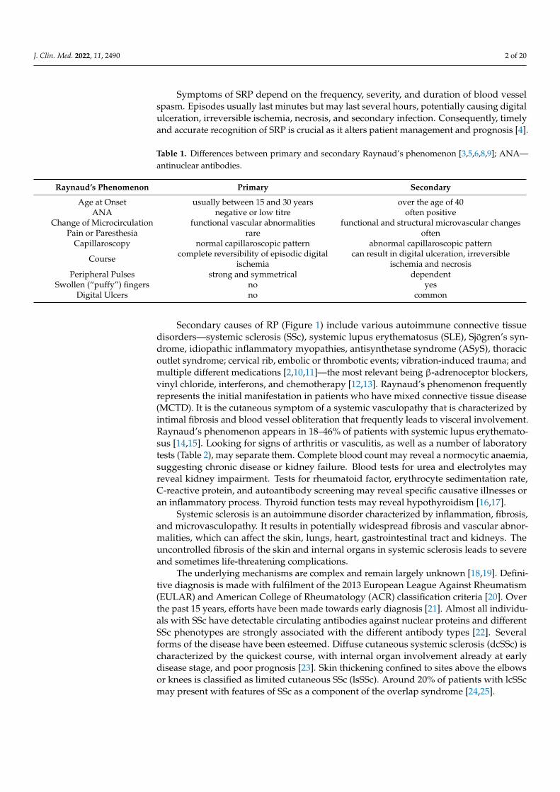
Table 1. Differences between primary and secondary Raynaud’s phenomenon [3,5,6,8,9]; ANA—antinuclear antibodies.
Raynaud’s Phenomenon Primary Secondary
Age at Onset usually between 15 and 30 years over the age of 40ANA negative or low titre often positive
Change of Microcirculation functional vascular abnormalities functional and structural microvascular changesPain or Paresthesia rare often
Capillaroscopy normal capillaroscopic pattern abnormal capillaroscopic pattern
Course complete reversibility of episodic digitalischemia
can result in digital ulceration, irreversibleischemia and necrosis
Peripheral Pulses strong and symmetrical dependentSwollen (“puffy”) fingers no yes
Digital Ulcers no common
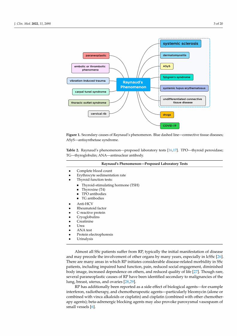
Secondary causes of RP (Figure 1) include various autoimmune connective tissuedisorders—systemic sclerosis (SSc), systemic lupus erythematosus (SLE), Sjögren’s syn-drome, idiopathic inflammatory myopathies, antisynthetase syndrome (ASyS), thoracicoutlet syndrome; cervical rib, embolic or thrombotic events; vibration-induced trauma; andmultiple different medications [2,10,11]—the most relevant being β-adrenoceptor blockers,vinyl chloride, interferons, and chemotherapy [12,13]. Raynaud’s phenomenon frequentlyrepresents the initial manifestation in patients who have mixed connective tissue disease(MCTD). It is the cutaneous symptom of a systemic vasculopathy that is characterized byintimal fibrosis and blood vessel obliteration that frequently leads to visceral involvement.Raynaud’s phenomenon appears in 18–46% of patients with systemic lupus erythemato-sus [14,15]. Looking for signs of arthritis or vasculitis, as well as a number of laboratorytests (Table 2), may separate them. Complete blood count may reveal a normocytic anaemia,suggesting chronic disease or kidney failure. Blood tests for urea and electrolytes mayreveal kidney impairment. Tests for rheumatoid factor, erythrocyte sedimentation rate,C-reactive protein, and autoantibody screening may reveal specific causative illnesses oran inflammatory process. Thyroid function tests may reveal hypothyroidism [16,17].
Systemic sclerosis is an autoimmune disorder characterized by inflammation, fibrosis,and microvasculopathy. It results in potentially widespread fibrosis and vascular abnor-malities, which can affect the skin, lungs, heart, gastrointestinal tract and kidneys. Theuncontrolled fibrosis of the skin and internal organs in systemic sclerosis leads to severeand sometimes life-threatening complications.
The underlying mechanisms are complex and remain largely unknown [18,19]. Defini-tive diagnosis is made with fulfilment of the 2013 European League Against Rheumatism(EULAR) and American College of Rheumatology (ACR) classification criteria [20]. Overthe past 15 years, efforts have been made towards early diagnosis [21]. Almost all individu-als with SSc have detectable circulating antibodies against nuclear proteins and differentSSc phenotypes are strongly associated with the different antibody types [22]. Severalforms of the disease have been esteemed. Diffuse cutaneous systemic sclerosis (dcSSc) ischaracterized by the quickest course, with internal organ involvement already at earlydisease stage, and poor prognosis [23]. Skin thickening confined to sites above the elbowsor knees is classified as limited cutaneous SSc (lsSSc). Around 20% of patients with lcSScmay present with features of SSc as a component of the overlap syndrome [24,25].
J. Clin. Med. 2022, 11, 2490 3 of 20
J. Clin. Med. 2022, 11, 2490 4 of 21
Figure 1. Secondary causes of Raynaud’s phenomenon. Blue dashed line—connective tissue dis-eases; ASyS—antisynthetase syndrome.
2. Pathophysiology The pathophysiological mechanisms behind RP are not entirely understood; gener-
ally, it is characterized by excessive vasoconstriction of the digital arteries, precapillary arterioles, and cutaneous arteriovenous anastomoses [33].
In PRP, vasospasm of the digital and cutaneous vessels is believed to occur as a con-sequence of an increased alpha2c-adrenergic response, and does not result in vascular pa-thology [34]. Ascherman et al. put forward an autoimmune etiology, proposing cy-tokeratin 10 (K10) as a potential autoantigen. Their study on mice showed that anti-K10 antibodies can mediate ischemia, similar to that seen in primary Raynaud’s Phenomenon [35].
In SRP, affected endothelial cells exhibit amplified exocytosis of endothelin-1 and ul-tra-large von Willebrand factor (ULVWF), which contribute, respectively, to increased vasospasm and capillary thrombosis. In addition, it is believed that increased transform-ing growth factor β (TGFβ), endothelin-1, cytokines, and angiotensin II drive the process of myofibroblast proliferation, vascular fibrosis and dropout in SSc patients [34]. Nitric oxide (NO) has a complex role in the disease process [36]. A decrease in endothelial for-mation of NO results in diminished vascular relaxation and extended vasoconstriction. Conversely, overproduction of NO leads to increased generation of reactive oxygen spe-cies and plays a pathogenic role in fibrosis.
Gualtierotti et al. found that markers of endothelial damage are regularly elevated in patients with PRP at their first assessment, even when there are no capillaroscopic abnor-malities or autoantibodies detectable. They are particularly increased in patients with very early SSc. The plasma concentration of tissue-type plasminogen activator (t-PA) and von Willebrand factor (vWF)—two markers of endothelial damage, as well as interleukin-6 (IL-6)—a pro-inflammatory cytokine, were evaluated. After a 36-month follow-up, those with higher basal concentrations of markers of endothelial damage had developed con-nective tissue disease. Von Willebrand factor analysis showed clear differences between primary and secondary RP patients. These findings suggest that markers of endothelial damage are elevated in RP patients who go on to develop SSc or other connective tissue diseases, even in the absence of capillaroscopic abnormalities [37].
Figure 1. Secondary causes of Raynaud’s phenomenon. Blue dashed line—connective tissue diseases;ASyS—antisynthetase syndrome.
Table 2. Raynaud’s phenomenon—proposed laboratory tests [16,17]. TPO—thyroid peroxidase;TG—thyroglobulin; ANA—antinuclear antibody.
Raynaud’s Phenomenon—Proposed Laboratory Tests
• Complete blood count• Erythrocyte sedimentation rate• Thyroid function tests:
� Thyroid-stimulating hormone (TSH)� Thyroxine (T4)� TPO antibodies� TG antibodies
• Anti-HCV• Rheumatoid factor• C-reactive protein• Cryoglobulins• Creatinine• Urea• ANA test• Protein electrophoresis• Urinalysis
Almost all SSc patients suffer from RP, typically the initial manifestation of diseaseand may precede the involvement of other organs by many years, especially in lcSSc [26].There are many areas in which RP initiates considerable disease-related morbidity in SScpatients, including impaired hand function, pain, reduced social engagement, diminishedbody image, increased dependence on others, and reduced quality of life [27]. Though rare,several paraneoplastic causes of RP have been identified secondary to malignancies of thelung, breast, uterus, and ovaries [28,29].
RP has additionally been reported as a side effect of biological agents—for exampleinterferon, radiotherapy, and chemotherapeutic agents—particularly bleomycin (alone orcombined with vinca alkaloids or cisplatin) and cisplatin (combined with other chemother-apy agents); beta-adrenergic blocking agents may also provoke paroxysmal vasospasm ofsmall vessels [6].

J. Clin. Med. 2022, 11, 2490 4 of 20
Kim et al. reported a case of RP in a 70-year-old woman, with no history of connectivetissue disease, secondary to pembrolizumab therapy for gallbladder cancer [30]. A furthercase report on the development of IL-17A antagonist (secukinumab)-related RP in a 35-year-old female patient with ankylosing spondylitis has also been described [31]. Additionally,Bouaziz et al. described a patient with proven COVID-19 infection presenting with RP andchilblain appearance of the hands [32].
2. Pathophysiology
The pathophysiological mechanisms behind RP are not entirely understood; generally,it is characterized by excessive vasoconstriction of the digital arteries, precapillary arterioles,and cutaneous arteriovenous anastomoses [33].
In PRP, vasospasm of the digital and cutaneous vessels is believed to occur as a conse-quence of an increased alpha2c-adrenergic response, and does not result in vascular pathol-ogy [34]. Ascherman et al. put forward an autoimmune etiology, proposing cytokeratin 10(K10) as a potential autoantigen. Their study on mice showed that anti-K10 antibodies canmediate ischemia, similar to that seen in primary Raynaud’s Phenomenon [35].
In SRP, affected endothelial cells exhibit amplified exocytosis of endothelin-1 andultra-large von Willebrand factor (ULVWF), which contribute, respectively, to increasedvasospasm and capillary thrombosis. In addition, it is believed that increased transforminggrowth factor β (TGFβ), endothelin-1, cytokines, and angiotensin II drive the process ofmyofibroblast proliferation, vascular fibrosis and dropout in SSc patients [34]. Nitric oxide(NO) has a complex role in the disease process [36]. A decrease in endothelial formation ofNO results in diminished vascular relaxation and extended vasoconstriction. Conversely,overproduction of NO leads to increased generation of reactive oxygen species and plays apathogenic role in fibrosis.
Gualtierotti et al. found that markers of endothelial damage are regularly elevatedin patients with PRP at their first assessment, even when there are no capillaroscopicabnormalities or autoantibodies detectable. They are particularly increased in patients withvery early SSc. The plasma concentration of tissue-type plasminogen activator (t-PA) andvon Willebrand factor (vWF)—two markers of endothelial damage, as well as interleukin-6(IL-6)—a pro-inflammatory cytokine, were evaluated. After a 36-month follow-up, thosewith higher basal concentrations of markers of endothelial damage had developed con-nective tissue disease. Von Willebrand factor analysis showed clear differences betweenprimary and secondary RP patients. These findings suggest that markers of endothelialdamage are elevated in RP patients who go on to develop SSc or other connective tissuediseases, even in the absence of capillaroscopic abnormalities [37].
A recent study by Taher et al. demonstrated use of a non-invasive NO-dependentmethod to identify peripheral microvascular endothelial dysfunction in patients withSRP. The association between SRP and microvascular peripheral endothelial dysfunctionwas also significant after adjusting for confounding variables, including conventionalrisk factors for cardiovascular disease, and vasoactive medications. This also remainedsignificant in women after stratifying only by sex. It was emphasized that detection ofmicrovascular peripheral endothelial dysfunction at an early stage could help to identifyindividuals with SRP who are at risk of developing connective tissue disease, as well ascardiovascular disease. Early detection could additionally indicate who may benefit fromfrequent screening, prompt initiation of preventative treatments, and modification of riskfactors [38].
3. Genetics
A genetic predisposition for RP has been demonstrated in two studies demonstratinggreater concordance amongst monozygotic than dizygotic twins. Heritability for RP isreported to be 55–64% [39,40].
Polymorphisms in various genes encoding ion channels or vasoactive agents havebeen hypothesized to result in the RP phenotype. It is suggested that genetic variation

J. Clin. Med. 2022, 11, 2490 5 of 20
in temperature-responsive or vasospastic genes may underlie RP manifestation. Severalstudies have investigated candidate genes that could potentially regulate vascular reactiv-ity [41,42]. Munir et al. aimed to evaluate the association between RP and single nucleotidepolymorphisms (SNPs). Temperature-sensing receptor channels called thermo-sensitivetransient receptor potential (TRP) ion channels include TRPA1 and TRPM8. These arecold-sensing and have been proposed to mediate cold-induced vascular responses in skinin vivo. This is linked, at least in part, to the expression of these channels on perivascularsensory nerves [41]. Calcitonin-related polypeptides, alpha and beta (CALCA, CALCB),encode the peptide hormones calcitonin, calcitonin gene-related peptide, and katacalcin bytissue-specific alternative RNA splicing of gene transcripts and cleavage of inactive pre-cursor proteins. Calcitonin is involved in the regulation of calcium levels and phosphorusmetabolism. Calcitonin gene-related peptide functions as a vasodilator [43]. NO derivedfrom neuronal nitric oxide synthase (nNOS) facilitates the restorative vasodilator responseafter cold exposure; thus, the gene encoding nNOS (NOS1) has also been investigated [41].Munir et al. found that one polymorphic variant within the NOS1 gene was significantlyassociated with RP in the general population [42].
4. Diagnosis
A detailed medical history, laboratory tests, and nailfold capillaroscopy form thebasis of RP diagnosis [17]. Follow-up nailfold capillaroscopy should be performed every12 months in patients with significant nailfold videocapillaroscopy disturbances present atbaseline [44]. Laboratory investigations should comprise a full blood count, inflammatorymarkers, thyroid function, and ANA testing by indirect immunofluorescence (accompaniedby ELISA or solid-phase immunoassays to determine antigen specificities where possible).A negative ANA with cytoplasmic stain could indicate anti-synthetase antibodies, such asanti-Jo-1, or rarer SSc-specific autoantibodies such as anti-eukaryotic initiation factor 2Bautoantibodies (anti-EIF2B) [45].
5. Capillaroscopy
Nailfold capillaroscopy is a simple, non-invasive technique that allows both qualita-tive and quantitative evaluation of the microcirculation, thus enabling early detection ofabnormalities. At the nailfold, capillaries are positioned parallel to the surface of the skin,allowing full morphological assessment [46,47]. Among the most important indications forcapillaroscopy are the differential diagnosis of primary and secondary RP.
Capillaroscopy is included in the 2013 American College of Rheumatology (ACR)/European League Against Rheumatism (EULAR) recommendations [20]. It is considered akey investigation in both the very early phases of the disease, and in monitoring diseaseprogression (Figure 2). Cutolo et al. proposed three progressive capillaroscopic patterns inSSc—‘early’, ‘active’, and ‘late’. The ‘early’ pattern is defined as the presence of a few giantcapillaries, single microhemorrhages, and preservation of capillary architecture withoutcapillary loss. ‘Active’ presents as numerous giant capillaries and microhemorrhages, milddisturbance of the capillary architecture and moderate capillary loss. The ‘late’ pattern ischaracterized by severe capillary loss with extensive avascular areas, disorganization of thecapillary architecture and ramified/bushy capillaries [48].
A clinical expert-based, fast track decision algorithm was developed to facilitate differ-entiation of a “non-scleroderma pattern” from a “scleroderma pattern” on capillaroscopicimages. The algorithm demonstrated excellent reliability when used by capillaroscopistswith varied expertise levels compared to principal experts, and corroborated with externalvalidation [49,50].

J. Clin. Med. 2022, 11, 2490 6 of 20
J. Clin. Med. 2022, 11, 2490 7 of 21
Qualitative analysis is subjective, and quantitative analysis is time-consuming when done manually. A study performed by Cutolo et al. accomplished validation of fully au-tomated AUTOCAPI software for measuring the absolute capillary number over 1 lin-ear/mm in NVC images. The software was subsequently optimized to assess capillary number in the shortest possible time and with the lowest possible error, in both healthy subjects, and those with SSc [61].
1 mm 1 mm (a) (b)
1 mm 1 mm (c) (d)
Figure 2. Nailfold videocapillaroscopic (×200) patterns of microangiopathy: (a)—normal; capillaro-scopic characteristics: density—normal, 8 capillaries in 1 linear mm; dimension: within normal lim-its; morphology: normal shapes of capillaries; haemorrhages: absent (b)—early; capillaroscopic characteristics: density—7 capillaries in 1 linear mm; dimension: presence of giants (homogeneous enlargement of all three limbs of the capillary with the diameter ≥ 50 µm); morphology: hairpin shaped capillaries; haemorrhages: absent (c)—active; capillaroscopic characteristics: density—low-ered, 4 capillaries in 1 linear mm; dimension: presence of giants (homogeneous enlargement of all three limbs of the capillary with the diameter ≥ 50 µm); morphology: presence of abnormally shaped capillary; haemorrhages: present (d)—late capillaroscopic characteristics: density—lowered, 2 ca-pillaries in 1 linear mm; dimension: not measured because of presence of abnormal shape; morphol-ogy: presence of abnormally shaped capillary; haemorrhages: absent.
6. Laser Doppler Flowmetry Laser Doppler flowmetry (LDF) is a semi-quantitative imaging technique useful for
studying the nitric oxide endothelial-dependent vascular response and axon reflex-medi-ated vasodilation. Impaired regulation of NO vascular tone has been described in patients with SSc-associated RP when compared to those with PRP and healthy controls [62,63]. Laser Doppler flowmetry has been proposed as a method for evaluating blood perfusion of the skin. This is a functional assessment of the vessels of the skin, involving the deeper dermal vessels in addition to the capillaries [64].
C D
Figure 2. Nailfold videocapillaroscopic (×200) patterns of microangiopathy: (a)—normal; capil-laroscopic characteristics: density—normal, 8 capillaries in 1 linear mm; dimension: within normallimits; morphology: normal shapes of capillaries; haemorrhages: absent (b)—early; capillaroscopiccharacteristics: density—7 capillaries in 1 linear mm; dimension: presence of giants (homogeneous en-largement of all three limbs of the capillary with the diameter ≥50 µm); morphology: hairpin shapedcapillaries; haemorrhages: absent (c)—active; capillaroscopic characteristics: density—lowered, 4 cap-illaries in 1 linear mm; dimension: presence of giants (homogeneous enlargement of all three limbsof the capillary with the diameter ≥50 µm); morphology: presence of abnormally shaped capillary;haemorrhages: present (d)—late capillaroscopic characteristics: density—lowered, 2 capillaries in1 linear mm; dimension: not measured because of presence of abnormal shape; morphology: presenceof abnormally shaped capillary; haemorrhages: absent.
In addition capillaroscopic changes have been observed in dermatomyositis, polymyosi-tis, antiphospholipid syndrome, Sjogren’s syndrome, and systemic lupus erythemato-sus [51]. Dermatomyositis pattern, often associated with aspects of the SSc pattern, in-cludes the presence of two or more of the following findings in at least two nail folds:enlargement of capillary loops, loss of capillaries, disorganization of the normal distribu-tion of capillaries, ‘budding’ (‘bushy’) capillaries, twisted enlarged capillaries, and capillaryhaemorrhages (extravasates) [51,52]. Characteristic systemic lupus erythematosus patternincludes morphological alterations of capillary loops, venular visibility and sludging ofblood with variability in capillary loop length [53]. Capillaroscopic abnormalities in SSranged from non-specific findings (crossed capillaries) to more specific findings (confluenthaemorrhages and pericapillary haemorrhages) or SSc-type findings [54,55]. Multiplehemorrhages from normal-shaped capillaries, which appear parallel/linear and arrangedperpendicularly to the nailfold bed, are called “comb-like” hemorrhages and are suggestiveof antiphospholipid syndrome [56].
J. Clin. Med. 2022, 11, 2490 7 of 20
It has been shown that the ability to detect capillary abnormalities increases as thenumber of fingers examined increases. Sensitivities ranged from 31.7% to 46.6% for onlyone finger (right middle and left ring finger, respectively), 59.8% for both ring fingers, 66.7%for a four-finger combination (both ring and middle fingers) and 74.6% for the eight-fingerstandard. In order to achieve the most accurate assessment during routine capillaroscopicexamination, all eight nailbeds should be examined omitting the thumbs, where it is moredifficult to visualize and classify capillaries [57,58]. It should be noted that in a timepressured scenario, the best two-finger combination to detect capillary abnormalities isboth ring fingers [58].
Nailfold videocapillaroscopy is the standard, although a handheld dermatoscope or anophthalmoscope may also be used as screening tools [50]. The nailfold videocapillaroscopytechnique with 200× magnification, capturing at least two adjacent fields of 1 mm in themiddle of the nailfold finger, is the standard capillaroscopic technique to perform nailfoldcapillaroscopy [50].
Ideally all dermatology specialists should have access to videocapillaroscopy; a prag-matic solution for practitioners may be to have a low-cost capillaroscopy system. Tech-nologies using a smartphone camera could help to improve availability to nailfold cap-illaroscopy whilst still providing accurate results [59]. Research regarding automatedmeasurement of capillaroscopic characteristics is currently under way and holds promiseas an objective clinical outcome measure [50]. Interestingly, a consensus-based assess-ment of dermatoscopy versus nailfold videocapillaroscopy by a European League againstRheumatism study group revealed tenuous promise for dermatoscopy as a tool for theinitial screening of nailfold capillaries in RP. However, as perhaps expected, dermatoscopyis less sensitive, but more specific, in regard to detecting abnormalities, compared withvideocapillaroscopy [60].
Qualitative analysis is subjective, and quantitative analysis is time-consuming whendone manually. A study performed by Cutolo et al. accomplished validation of fully auto-mated AUTOCAPI software for measuring the absolute capillary number over 1 linear/mmin NVC images. The software was subsequently optimized to assess capillary number inthe shortest possible time and with the lowest possible error, in both healthy subjects, andthose with SSc [61].
6. Laser Doppler Flowmetry
Laser Doppler flowmetry (LDF) is a semi-quantitative imaging technique usefulfor studying the nitric oxide endothelial-dependent vascular response and axon reflex-mediated vasodilation. Impaired regulation of NO vascular tone has been describedin patients with SSc-associated RP when compared to those with PRP and healthy con-trols [62,63]. Laser Doppler flowmetry has been proposed as a method for evaluating bloodperfusion of the skin. This is a functional assessment of the vessels of the skin, involvingthe deeper dermal vessels in addition to the capillaries [64].
Melsen et al. completed a systematic review evaluating the use of LDF, describing theresults of quality reports on assessment of the skin’s microcirculatory flow at the level ofthe fingertip in SSc patients, and investigating the validation status of LDF as an outcomemeasure. The systematic review highlights the very preliminary validation status of LDFin the assessment of the microcirculatory flow in SSc [65].
In a study performed by Gregorczyk-Maga et al., LDF was used to investigate oralcapillary flow in PRP patients who habitually have dysfunction in the microcirculation ofthe oral mucosa and who often have lesions in the oral cavity [66].
Time to postocclusive peak blood flow measured by LDF is an extremely accurate testfor distinguishing patients with PRP from healthy controls [67].
An additional study performed by Waszczykowska et al. presented the suitability ofLDF for assessment of the degree of microangiopathy present in SSc patients. Assessmentof the skin perfusion value in SSc patients should on the basis of parameters obtainedduring microcirculation challenge tests [68].

J. Clin. Med. 2022, 11, 2490 8 of 20
7. Thermography
Thermal imaging is an indirect method that makes use of a thermal camera to imageskin temperature and demonstrate underlying blood flow [69]. Thermal imaging has beenused to evaluate RP in several studies; the response to lower temperatures was able todifferentiate between PRP and RP secondary to SSc [70].
Patients with SSc-related RP have been found to have structural changes in the digitalarteries and microcirculation with a decrease in baseline blood flow. This typically does notreturn to normal after a cold challenge with rewarming, in direct contrast to primary RP, inwhich the fingers classically rewarm [71].
Measurements made by mobile phone thermography compared favorably with thosemade by standard thermography, paving the way for ambulatory monitoring in non-controlled environments; this will enable further assessments to increase the understandingof RP episodes [69]. Infrared thermography may additionally be a method of verificationin Raynaud’s Phenomenon [72].
8. Laser Speckle Contrast Analysis (LASCA)
Laser speckle contrast analysis (LASCA) is a tool used to investigate variations in pe-ripheral blood perfusion during long-term follow-up and can safely monitor the evolutionof digital ulcers in SSc patients [73].
LASCA can quantify blood flow over a defined area and is based on the concept thatwhen laser light illuminates a tissue it forms a speckle pattern. Variations in this patternare analyzed by dedicated software—static areas demonstrate a stationary speckle pattern,in contrast with mobile objects—such as red blood cells—that cause the speckle patternto fluctuate and appear blurred. The amount of blurring (contrast) is analyzed and thusinterpreted as blood perfusion [74].
The pilot study completed by Ruaro et al. determined that the hand blood perfusion,as evaluated by LASCA, was lower in PRP than in SSc patients with the “early” nailfoldvideocapillaroscopy microangiopathy pattern [75].
9. Treatment
Lifestyle modifications are essential in all patients with RP [8]. Patients’ education isan important aspect of disease management and patients’ support organizations providethem with valuable education on the topic [76]. The first line of the treatment is basedon avoiding triggering factors such as: exposure to cold, sudden changes of temperature,stress, cigarette smoke, and infections [34]. Patients should dress warmly (including warmgloves and socks). A number of different types of gloves have been proposed for patientswith RP to reduce the risk of attacks, including battery-heated and specifically ceramic-impregnated gloves [77]. During the vasospasm, it is advised that one should place one’shands under warm running water or to rub one hand against the other to intensify bloodflow [78]. Because stress may trigger an attack, learning to recognize and avoid stressfulsituations may help control the number of attacks. Exercise can improve circulation, amongother health benefits. Avoidance of repeated trauma to the fingertips by all patients withRP and avoidance of vibrating tools utilization by patients with vibration-induced RP hasto be underlined [79]. Patients should be counselled regarding the critical importance ofsmoking cessation as nicotine enhances vasoconstriction [80]. Certain medications such asbeta-blockers, ergotamine, or sumatriptan, and some types of chemotherapy, specifically,cisplatin and bleomycin, were most likely to induce the Raynaud’s phenomenon. If possible,alternative therapies that do not alter peripheral blood flow should be considered [12]. Inmost cases of primary Raynaud’s phenomenon, lifestyle modifications may be sufficient tocontrol the symptoms [17,81].
Pharmacological treatment is required when adaptive measures to avoid cold ex-posure are ineffective. RP reflects excessive vasoconstriction; thus, vasodilator therapy—particularly targeted to the cutaneous circulation—is a major focus. Patients with connective

J. Clin. Med. 2022, 11, 2490 9 of 20
tissue disease-associated RP, SSc in particular, may progress to tissue injury; hence, drugtreatment often needs to be more ‘aggressive’ to prevent/minimize tissue loss [Table 3].
Table 3. Pharmacotherapy options in management of Raynaud’s phenomenon. A-level recommen-dation is based on consistent and good-quality patient-oriented evidence; B-level recommendationis based on inconsistent or limited-quality patient-oriented evidence; C-level recommendation isbased on consensus, usual practice, opinion, disease-oriented evidence, or case series for studies ofdiagnosis, treatment, prevention, or screening.
Group of Drugs Medication Dose Strength ofRecommendation
Calcium Channel Antagonists nifedipine 10–20 mg 3× daily orextended-release tablets A
nifedypine SR 30–120 mg daily
Phosphodiesterase Type 5Inhibitors sildenafil
50–100 mg 2× daily (the suggestedstarting dose is 12.5 mg/day, to be
increased gradually dependingon tolerability)
A
Prostaglandin Analogsalprostadil (i.v. infusions) pulses of 20–60 mg every 4–6 weeks A
iloprost (i.v. infusions/p.o.)
0.5–3 ng/kg/min (i.v.) for3–5 consecutive days every
6–8 weeks or 50–150 µg 2× daily(p.o.)
A
epoprostenol (i.v. infusions) 2 ng/kg/min in intermittentinfusions of 5 to 6 hours’ duration A
First-in-class Guanylate CyclaseStimulator riociguat 2 mg single oral dose C
Selective Serotonin ReuptakeInhibitors (SSRIs) fluoxetine 20 mg/day C
Endothelin Receptor Antagonists bosentan125 mg twice a day
following initial dosage of62.5 mg twice a day
C
Angiotensin II receptor blockers losartan 25 mg once dailyto 100 mg once daily C
Mixture of glycosaminoglycanscomposed of dermatan sulfate
and fast moving heparinsulodexide
3–4 day cycle of intravenoussulodexide, at 600 LSU twice a day
every 4–6 weeksC
3-hydroxy-3-methylglutarylcoenzyme A (HMG-CoA)
reductase inhibitorsatorvastatin 40 mg/day C
Botulinum toxin type A Dose dependent vasodilation with10–100 units injections C
Topical vasodilatorsNifedipine
Nitroglycerinsildenafil
10% nifedipine cream10% nitroglycerin gel5% sildenafil cream
C
Surgical treatment sympathectomy/arterialreconstruction C
10. Calcium Channel Blockers
Calcium channel blockers (CCBs) are generally considered to be the first-line phar-macotherapeutic treatment of PRP, and are the group of drugs which have been mostextensively researched. According to the 2017 update of the European League againstRheumatism (EULAR) recommendations for the treatment of SSc, oral therapies with CCBsare strongly recommended (strength of recommendation A) [16,82]. CCBs are currentlythe most frequently prescribed drug for PRP. Nifedipine and amlodipine are considered tobe the most effective agents, blocking calcium channels located in the cell membranes ofvascular smooth muscle and cardiac muscle. Consequently, calcium ion entry into cells isinhibited, resulting in blood vessel relaxation and improved blood supply to tissues [83].
J. Clin. Med. 2022, 11, 2490 10 of 20
Doses should be adjusted depending on individual tolerance, with particular cautionadvised in patients with low arterial blood pressure [84].
A meta-analysis of randomized clinical trials concluded that CCBs are only somewhateffective at reducing the frequency of Raynaud’s attacks in PRP [42,85]. Whereas otherstudies suggest that CCBs may be effective at decreasing the severity of attacks, pain anddisability associated with RP [83].
11. Phosphodiesterase-5 (PDE-5) Inhibitors
Phosphodiesterase-5 (PDE-5) inhibitors are commonly used as a second-line systemicagent to manage RP resistant to CCBs. Inhibition of PDE-5 activity allows accumulation ofcGMP within endothelial cells, which alters the cellular response to prostacyclin or nitricoxide, and in turn dilates blood vessels [86].
A 2013 meta-analysis of six randomized controlled trials including 296 SRP patientsrevealed a significant, moderate effect on the clinical severity, duration, and frequencyof attacks [87]. Additionally, a significant decrease in the number of digital ulcers in SScpatients with RP was found in a randomized, placebo-controlled study in patients receivingsildenafil compared to a placebo [88].
Adverse effects of these PDE-5 inhibitors include flushing, headaches, and dizziness.Less common side effects include hypotension, arrhythmias, cerebral vascular accidents,and vision changes [89].
12. Prostaglandin Analogs
While oral prostaglandins have not shown any benefit in RP, prostacyclin analogs ad-ministered intravenously exhibit a strong vasodilative effect which considerably improvesthe clinical condition, particularly among patients with ulcers and erosions [90].
Iloprost is a synthetic analogue of prostacyclin (PGI2), with vasodilatory and an-tiplatelet effects; however, it is more stable than PGI2, has a longer half-life (20 to 30 min)and better solubility [91]. Iloprost activates PGI2 receptors, thus stimulating adenylatecyclase to generate cyclic adenosine monophosphate (cAMP). PGI2 receptors inhibit vas-cular smooth muscle constriction and platelet aggregation. They are also expressed onendothelial cells, where they initiate multiple protective effects, including amplificationof endothelial adherens junctions and decreased monolayer permeability [92]. Iloprostinfusions are frequently recommended as second-line treatment after CCBs and are the first-line therapeutic choice for digital ulcerations and critical ischemia [21,93]. A meta-analysisdetermined that the use of iloprost in critical limb ischemia was effective in improving ulcerhealing, relieving pain, and reducing the need for amputations [94]. In cases of pre-existingdigital ulcerations, iloprost promotes healing and reduces the incidence of new ulcera-tions [16]. Three further studies described an improvement in nailfold microvascularizationfollowing iloprost treatment [95].
In 2017, the EUSTAR recommendations allocated intravenous iloprost a Grade Arecommendation for management of severe SSc-related RP attacks and for digital ulcertreatment [16]. However, in the recommendations, the dosing and therapeutic regimenwas not specified. The absence of an accepted regimen is a major impediment to theadministration of iloprost in SSc. According to the Delphi concensus, intravenous iloprostcan be useful in RP that is severe or refractory to CCB and PDE-5i. To control symptoms,it is recommended that iloprost be administered 1–3 days every month. Dosing shouldbe determined according to the tolerance, starting from 0.5 up to 3.0 ng/kg/min. Toachieve a lasting effect, infusions must be repeated regularly [16,96,97]. Interestingly, thepharmacological actions may persist longer than suggested by the pharmacokinetic profile(i.e., weeks to months) [98].
Currently, intravenous iloprost is available in several countries only for RP secondaryto SSc for a duration of 3–5 days. For RP and digital ulcer healing, expert consensusproposes a regimen of 1–3 days per month, with 1 day per month for DU prevention.These recommendations allow clinicians some scope on how to personalize intravenous

J. Clin. Med. 2022, 11, 2490 11 of 20
iloprost therapy according to patients’ needs [93]. However, although these suggestions aresupported by an expert group for use in a clinical setting, it would be necessary to formallyvalidate the recommendations in future clinical trials.
As iloprost is not available in some countries, alprostadil (a combination of prostaglandinE1 with a-cyclodextrin in a 1:1) has been found to be an effective alternative for SRP [99].Alprostadil is primarily used to maintain patency of the ductus arteriosus, and also has mildpulmonary vasodilatory effects. It reportedly inhibits macrophage activation, neutrophilchemotaxis, and release of oxygen radicals and lysosomal enzymes. It influences coagu-lation by inhibiting platelet aggregation and potentially by inhibiting factor X activation.Alprostadil may promote fibrinolysis by stimulating production of tissue plasminogen acti-vator. The overall benefits of iloprost and alprostadil are comparable, without significantdifferences in clinical efficacy or circulating markers of endothelial damage [100].
Epoprostenol, the first prostacyclin agent approved by the US Food and Drug Admin-istration (FDA), in 1995, requires continuous intravenous infusion via a dedicated centralvenous catheter with infusion pump. Epoprostenol stimulates vasodilation of pulmonaryand systemic arterial vascular beds and impedes platelet aggregation [11,101]. Based onpublished evidence, the initial dose of intravenous epoprostenol for treatment of refractoryRP, with or without ischemic ulcers, should not exceed 2 ng/kg/min. A conservativetitration schedule, based on those used in previous studies, should allow for rate increasesof 1 ng/kg/min every 15 min as tolerated, adjusted as per the onset of treatment-emergentadverse effects. It should be noted that more aggressive uptitrations of 2 to 2.5 ng/kg/minwere used in some studies. However, as a consequence of the lack of standardized efficacyoutcomes in the available literature, it is not possible to assess if such regimens hold anyadvantages other than reaching the maximum dose more quickly. Intermittent infusions of5 to 6 hours’ duration should be initially considered to limit drug exposure and potentialtoxicities. However, it is reportedly reasonable to use continuous infusions for up to 72 h inpatients unresponsive to intermittent therapy [101].
Epoprostenol is contraindicated in patients with congestive heart failure due to leftventricular dysfunction, and in those with known history of hypersensitivity reactions tothe drug. Other adverse effects that should be monitored for include pulmonary edema,hemodynamic instability, line infections, and bleeding [101].
13. Endothelin Receptor Antagonists
Bosentan is an endothelin receptor antagonist (ERA) primarily used to manage severepulmonary hypertension. A starting dose of 62.5 mg twice a day for four weeks, followedby 125 mg twice a day for 12 or 20 weeks, has shown some effectiveness in preventingformation of new digital ulcers, but did not influence healing of pre-existing ulcers [102,103].Potential adverse effects include headaches, dizziness, and hypotension [16]. Adverse drugreactions are relatively mild, but during the treatment monthly liver function and 3-monthlyfull blood count is required [104,105].
14. Angiotensin II Receptor Blockers
Angiotensin II receptor blockers (ARBs) are reserved as a third-line treatment for mildRaynaud’s phenomenon. There has been only one trial including 52 patients (25 patientswith primary RP and 27 with SSc-related RP); this was an open-label, unblinded, controlledtrial, during which a 12-week treatment with losartan 50 mg/day resulted in reducedfrequency and severity of RP attacks in comparison to nifedipine 40 mg/day. The benefitwas more noticeable in the subgroup of patients with primary RP. Losartan is widelyavailable, accessible, and has an acceptable side effect profile [82,106,107].
15. Sulodexide
Sulodexide is a safe rheological drug used successfully as a supportive way of treatingRP [84]. It consists of a purified mixture of glycosaminoglycans acquired from bovineintestinal mucosa, comprising a heparin of a rapidly moving field of electrophoresis (80%)
J. Clin. Med. 2022, 11, 2490 12 of 20
and dermatan sulfate (20%). It functions as an anticoagulant, is pro-fibrinolytic and anti-inflammatory, disrupts the process of fibrosis, and has a protective influence on vascularendothelial cells. Due to its pleiotropic activity and high safety profile, the benefits fromsulodexide may be applied to many dermatological diseases [108].
SSc patients with secondary microcirculatory disorders who are intolerant to prostanoids,where there are contraindications, may be treated with sulodexide, 600 lipasemic units(LSU) intravenously twice a day. This dosing regimen has previously produced goodtherapeutic results. Other than sporadically observed dizziness and hypotension, nosignificant side effects were noted. In patients’ pre-existing digital erosions and ulcers, a3–4 day cycle of intravenous sulodexide, at 600 LSU twice a day every 4–6 weeks, has beenshown to improve lesion healing [109].
Results of a recent pilot study suggest that the use of sulodexide treatment in RPresults in a long-term improvement of capillary flow, a decrease in episode recurrence, anda reduction in pain intensity [110].
During parenteral treatment with sulodexide, it is imperative to discontinue any useof heparin or oral anticoagulants to reduce the bleeding risk [109].
16. Statins
Statins, 3-hydroxy-3-methylglutaryl coenzyme A (HMG-CoA) reductase inhibitors,are extensively used to reduce serum cholesterol levels in the primary and secondaryprevention of cardiovascular disease. Statins also have direct, vasculoprotective effects,which are independent of their ability to lower circulating LDL levels. Statins may bebeneficial in RP patients, including those with SSc. Statins increase endothelial nitric oxidesynthase expression and thus nitric oxide production, decrease oxidative stress, reduceendothelin-1 expression, impede endothelial apoptosis, increase endothelial progenitorcell mobilization, and promote microvascular growth [111,112]. Statins additionally inhibitendothelial–mesenchymal transition, which may contribute to vasculopathy and tissuefibrosis in SSc [113]. In a study of SSc patients, treatment with a statin was found toreduce the severity of RP vasospastic episodes and improved endothelial function. Thiswas associated with increased levels of nitric oxide, reduced oxidative and inflammatorystress, increased quantity of endothelial progenitor cells, and amelioration of circulatingconcentrations of von Willebrand factor [114]. Data from a study performed by Abou-Raya et al. suggest that atorvastatin may exert beneficial effects in SSc by protecting theendothelium and improving its functional activity [115].
17. Topical Vasodilators
Topical vasodilators may be used as adjuvant therapy for RP patients. 10% nifedipinecream and 10% nitroglycerin gel have both been accepted as efficient therapeutic optionswith side-effects comparable to placebo usage. Local topical nitrates display significantefficacy in treatment of both primary and secondary RP [116]. Topical nitrates have beenreported to increase perfusion at both distal digital ulcer and extensor digital ulcer cores inSSc patients, when compared to a placebo and evaluating with laser doppler imaging [117].Wortsman et al. found that 5% sildenafil cream significantly improved blood flow indigital arteries (an increase of 9.2 mm/s, p < 0.0083). A trend toward improvement wasalso observed for vessel diameter in patients with SRP (p = 0.0695), suggesting localvasodilatation. Adverse effects to topical vasodilators include headaches and dizziness—no serious adverse effects were detected [118]. A study by Bentea et al. assessed the effectsof nitroglycerin patch application to the dorsum of the hand. Results showed an increasein blood flow and hand temperature in patients with SSc after a cold challenge using laserdoppler imaging [119].
18. Sympathectomy
Surgical treatment options involving sympathectomy or arterial reconstruction may berequired in patients who suffer from incapacitating pain and ulcers with torpid evolution.

J. Clin. Med. 2022, 11, 2490 13 of 20
However, these techniques carry the risk of comorbidities and may not always providesatisfactory results.
Cautious selection of RP patients is necessary, and endoscopic thoracic sympathectomyshould be reserved as an ultimate choice only for patients who have severe symptoms thatare treatment-resistant with serious complications and impaired quality of life. The limitingfactor with sympathectomy is a high recurrence rate. Symptoms and examination findingsreported the quantity and dosage of medications used returned to preoperative levels in66.6% of patients at month 6, and in all patients except one at the end of the 1st year [120].
Digital periarterial sympathectomy may be considered in patients suffering fromcritical digital ischemia or persistent ulceration despite aggressive vasodilatory therapy.A long-term retrospective study assessed 35 patients with primary or secondary RP whounderwent thoracoscopic sympathectomy: 77% of participants had a positive response.However, symptoms recurred in 60% at a median follow-up of 5 months [121].
Single-port thoracoscopic sympathicotomy (SPTS) is a novel minimally invasive tech-nique compared to conventional sympathectomy [122]. A recent study showed that thesingle-port procedure is effective in improving hand perfusion in patients with treatment-resistant RP. One month after unilateral single-port thoracoscopic sympathicotomy, thenumber of RP attacks was reduced and perfusion of the treated hand increased. However,the long-term efficacy and safety profile of this treatment need to be established [123].
19. Botulinum Toxin Type A
Botulinum toxin type A (BTX-A) is a polypeptide produced by the bacteria Clostridiumbotulinum. It is an acetylcholine release inhibitor in the peripheral nerve endings of themotor plate and sweat glands. It is well established that BTX-A inhibits acetylcholinerelease, leading to inhibition of neurotransmitter-induced vasoconstriction and relief ofother symptoms, such as pain [124,125].
Botulinum toxins inhibit macromolecular SNARE complexes, which are involved invesicle fusion with the plasma membrane, thus preventing neuronal exocytosis [126].
A study performed by Medina et al. established botulinum toxin as a safe, accessible,and effective therapeutic alternative for patients with severe RP, allowing those whodo not respond to conventional treatments to sustain a good quality of life via annualinfiltrations [127].
BTX-A is a promising non-surgical treatment modality and/or adjunct for patientswho have contraindications to CCBs, PDE-5 inhibitors, and nitrates. It also provides anon-operative therapeutic alternative for patients experiencing chemotherapy-induced RPwhere mainstay therapies may be contraindicated, thus reducing pain, improving patientquality of life, and slowing disease progression [128].
In a study by Nagarajan et al. several patients derived long-term benefits from a singletreatment, however in patients with SSc, repeat treatments were required and administeredafter an average of 6 months [129].
20. Riociguat
Riociguat is a first-in-class guanylate cyclase stimulator and may be a promising newtreatment for RP. It works by direct stimulation of guanylate cyclase, independent fromNO, and additionally via sensitization of guanylate cyclase to endogenous nitric oxideby stabilizing NO–guanylate cyclase binding. As a result, riociguat efficiently stimulatesthe nitric oxide–soluble guanylate cyclase–cyclic guanosine monophosphate pathway andleads to increased intracellular levels of cyclic guanosine monophosphate. In contrastto PDE-5 inhibitors, the action of riociguat does not depend on endogenous nitric oxidelevels [130]. In the pilot study performed by Huntgeburth et al., a single oral dose ofriociguat 2 mg was well tolerated in patients with RP and resulted in improved digitalblood flow in some patient subsets, with high inter-individual variability [131].

J. Clin. Med. 2022, 11, 2490 14 of 20
21. SSRIs
An improvement in RP symptoms has been reported in patients treated with selec-tive serotonin reuptake inhibitors (SSRIs). A study of 27 patients with SSc revealed thatfluoxetine at a dose of 20mg/day was significantly superior to nifedipine in reducingthe frequency and severity of RP attacks in patients with SSc [132]. Of note however,exacerbations of RP have also been reported with the use of serotonin reuptake inhibitorstreatment [133].
22. Treat to Target (T2T) Strategy
At present, the decision to commence treatment and to evaluate response in RP—including the need for dose escalation—is principally based upon clinician–patient dis-cussions regarding symptom severity, perceived effectiveness of the existing/plannedinterventions, and drug tolerability.
Hughes et al. proposed a five-stage roadmap that may support the development ofa treat to target (T2T) strategy for SSc-RP. Significant initial steps are to define the studypopulation and the goals of developing a T2T strategy (stage 1) and to review and shortlistcandidate target items (stages 2 and 3, respectively). If agreement regarding feasible targetsis not reached at this point, then the goals and purpose need to be refined. Subsequently, aconsensus-building exercise among relevant stakeholders would allow the ‘target’ to bedefined (stage 4). Ultimately, well-designed studies (stage 5) will be required to investigatethe feasibility and treatment benefit of a T2T strategy in patients with SSc-RP. Much can belearned from primary studies of T2T for rheumatoid arthritis, including randomized trialscomparing T2T with routine care, and those comparing different treatment approaches(e.g., monotherapy vs. combination therapy) to reach a defined target. Crucial features ofthese studies were the frequent review of patients, and the clear guidance that existed onhow to intensify treatment of patients who had not reached the target [134,135].
23. Conclusions
An increased understanding of the pathogenesis of Raynaud’s phenomenon is guidingnew approaches to treatment. Assessment of peripheral endothelial dysfunction mayaid identification of individuals with secondary Raynaud’s phenomenon who are at riskof developing connective tissue diseases, and who may therefore benefit from repeatedscreening, early initiation of preventative treatments, and risk factor modification.
Capillaroscopy is of crucial value for the diagnosis and differentiation of primary andsecondary Raynaud’s phenomenon.
The presence of digital ulcers implies that intervention with vasodilator therapy isnecessary. Early intervention is vital to the treatment of critical ischemia, and calciumchannel blockers remain the first line of therapy. Alternatives for severe disease includephosphodiesterase-5 inhibitors and intravenous prostaglandin analogues. The overallbenefits of iloprost and alprostadil are comparable without significant differences; however,ease of handling and the lower cost profile favours alprostadil. Sulodexide is a saferheological drug successfully used as a supportive treatment in RP, resulting in a long-termimprovement of capillary flow and a reduction in the frequency of Raynaud’s syndromerelapse. Topical vasodilators, for example 10% nifedipine cream, 10% nitroglycerine gel,and 5% sildenafil cream, may act as adjuvant therapy. Riociguat may be a promisingnew treatment for Raynaud’s phenomenon; however, this warrants further evaluation. Avariety of alternative modalities have also been reported to be effective in the managementof RP including botulinum toxin A, and sympathectomy, or single-port thoracoscopicsympathectomy.
Treat to target strategies may optimize treatment approaches for Raynaud’s phe-nomenon and herald the emergence of disease-modifying vasodilator therapies for systemicsclerosis-related digital vasculopathy.

J. Clin. Med. 2022, 11, 2490 15 of 20
Author Contributions: Conceptualization, M.M., M.S., J.C. and L.R.; methodology, M.M., M.S., J.C.and L.R.; writing—original draft preparation, M.M. and M.S.; writing—review and editing, M.M.,M.S., C.M., J.C., R.A.-M. and L.R.; funding acquisition, M.M., M.S. and J.C. All authors have read andagreed to the published version of the manuscript.
Funding: The publication was financed from the subsidy granted to the Medical University of Warsaw.
Institutional Review Board Statement: Not applicable.
Informed Consent Statement: Not applicable.
Data Availability Statement: The study did not report any data.
Conflicts of Interest: The authors declare no conflict of interest.
References1. Wigley, F.M.; Flavahan, N.A. Raynaud’s Phenomenon. N. Engl. J. Med. 2016, 375, 556–565. [CrossRef] [PubMed]2. Hughes, M.; Herrick, A.L. Raynaud’s phenomenon. Best Pract. Res. Clin. Rheumatol. 2016, 30, 112–132. [CrossRef]3. Garner, R.; Kumari, R.; Lanyon, P.; Doherty, M.; Zhang, W. Prevalence, risk factors and associations of primary Raynaud’s
phenomenon: Systematic review and meta-analysis of observational studies. BMJ Open 2015, 5, e006389. [CrossRef] [PubMed]4. Meier, F.M.; Frommer, K.W.; Dinser, R.; Walker, U.A.; Czirjak, L.; Denton, C.P.; Allanore, Y.; Distler, O.; Riemekasten, G.; Valentini,
G.; et al. Update on the profile of the EUSTAR cohort: An analysis of the EULAR Scleroderma Trials and Research group database.Ann. Rheum. Dis. 2012, 71, 1355–1360. [CrossRef] [PubMed]
5. Chikura, B.; Moore, T.; Manning, J.; Vail, A.; Herrick, A.L. Thumb involvement in Raynaud’s phenomenon as an indicator ofunderlying connective tissue disease. J. Rheumatol. 2010, 37, 783–786. [CrossRef] [PubMed]
6. Maverakis, E.; Patel, F.; Kronenberg, D.G.; Chung, L.; Fiorentino, D.; Allanore, Y.; Guiducci, S.; Hesselstrand, R.; Hummers, L.K.;Duong, C.; et al. International consensus criteria for the diagnosis of Raynaud’s phenomenon. J. Autoimmun. 2014, 48–49, 60–65.[CrossRef] [PubMed]
7. Abdulle, A.E.; Arends, S.; van Goor, H.; Brouwer, E.; van Roon, A.M.; Westra, J.; Herrick, A.L.; de Leeuw, K.; Mulder, D.J.Low body weight and involuntary weight loss are associated with Raynaud’s phenomenon in both men and women. Scand. J.Rheumatol. 2021, 50, 153–160. [CrossRef]
8. Devgire, V.; Hughes, M. Raynaud’s phenomenon. Br. J. Hosp. Med. 2019, 80, 658–664. [CrossRef]9. Pauling, J.D.; Hughes, M.; Pope, J.E. Raynaud’s phenomenon-an update on diagnosis, classification and management. Clin.
Rheumatol. 2019, 38, 3317–3330. [CrossRef]10. Madke, B.; Doshi, B.; Pande, S.; Khopkar, U. Phenomena in dermatology. Indian J. Dermatol. Venereol. Leprol. 2011, 77, 264–275.
[CrossRef]11. Vick, E.; Sharma, D.; Elwing, J. Raynaud Phenomenon in Antisynthetase Syndrome Treated with Epoprostenol. J. Clin. Rheumatol.
2021, 27, e10–e11. [CrossRef] [PubMed]12. Khouri, C.; Blaise, S.; Carpentier, P.; Villier, C.; Cracowski, J.L.; Roustit, M. Drug-induced Raynaud’s phenomenon: Beyond
β-adrenoceptor blockers. Br. J. Clin. Pharmacol. 2016, 82, 6–16. [CrossRef] [PubMed]13. Khouri, C.; Jouve, T.; Blaise, S.; Carpentier, P.; Cracowski, J.L.; Roustit, M. Peripheral vasoconstriction induced by β-adrenoceptor
blockers: A systematic review and a network meta-analysis. Br. J. Clin. Pharmacol. 2016, 82, 549–560. [CrossRef] [PubMed]14. Block, J.A.; Sequeira, W. Raynaud’s phenomenon. Lancet 2001, 357, 2042–2048. [CrossRef]15. Pavlov-Dolijanovic, S.; Damjanov, N.S.; Vujasinovic Stupar, N.Z.; Marcetic, D.R.; Sefik-Bukilica, M.N.; Petrovic, R.R. Is there
a difference in systemic lupus erythematosus with and without Raynaud’s phenomenon? Rheumatol. Int. 2013, 33, 859–865.[CrossRef] [PubMed]
16. Kowal-Bielecka, O.; Fransen, J.; Avouac, J.; Becker, M.; Kulak, A.; Allanore, Y.; Distler, O.; Clements, P.; Cutolo, M.; Czirjak,L.; et al. Update of EULAR recommendations for the treatment of systemic sclerosis. Ann. Rheum. Dis. 2017, 76, 1327–1339.[CrossRef] [PubMed]
17. Matucci-Cerinic, C.; Nagaraja, V.; Prignano, F.; Kahaleh, B.; Bellando-Randone, S. The role of the dermatologist in Raynaud’sphenomenon: A clinical challenge. J. Eur. Acad. Dermatol. Venereol. JEADV 2018, 32, 1120–1127. [CrossRef]
18. Katsumoto, T.R.; Whitfield, M.L.; Connolly, M.K. The Pathogenesis of Systemic Sclerosis. Annu. Rev. Pathol. Mech. Dis. 2011, 6,509–537. [CrossRef]
19. Allanore, Y.; Simms, R.; Distler, O.; Trojanowska, M.; Pope, J.; Denton, C.P.; Varga, J. Systemic sclerosis. Nat. Rev. Dis. Primers2015, 1, 15002. [CrossRef]
20. van den Hoogen, F.; Khanna, D.; Fransen, J.; Johnson, S.R.; Baron, M.; Tyndall, A.; Matucci-Cerinic, M.; Naden, R.P.; Medsger,T.A., Jr.; Carreira, P.E.; et al. 2013 classification criteria for systemic sclerosis: An American College of Rheumatology/EuropeanLeague against Rheumatism collaborative initiative. Arthritis Rheum. 2013, 65, 2737–2747. [CrossRef]
21. Denton, C.P.; Khanna, D. Systemic sclerosis. Lancet 2017, 390, 1685–1699. [CrossRef]22. Steen, V.D. Autoantibodies in systemic sclerosis. Semin. Arthritis Rheum. 2005, 35, 35–42. [CrossRef] [PubMed]

J. Clin. Med. 2022, 11, 2490 16 of 20
23. Diab, S.; Dostrovsky, N.; Hudson, M.; Tatibouet, S.; Fritzler, M.J.; Baron, M.; Khalidi, N. Systemic sclerosis sine scleroderma: Amulticenter study of 1417 subjects. J. Rheumatol. 2014, 41, 2179–2185. [CrossRef] [PubMed]
24. Pakozdi, A.; Nihtyanova, S.; Moinzadeh, P.; Ong, V.H.; Black, C.M.; Denton, C.P. Clinical and serological hallmarks of systemicsclerosis overlap syndromes. J. Rheumatol. 2011, 38, 2406–2409. [CrossRef] [PubMed]
25. Foocharoen, C.; Netwijitpan, S.; Mahakkanukrauh, A.; Suwannaroj, S.; Nanagara, R. Clinical characteristics of sclerodermaoverlap syndromes: Comparisons with pure scleroderma. Int. J. Rheum. Dis. 2016, 19, 913–923. [CrossRef]
26. Mostmans, Y.; Cutolo, M.; Giddelo, C.; Decuman, S.; Melsens, K.; Declercq, H.; Vandecasteele, E.; De Keyser, F.; Distler, O.;Gutermuth, J.; et al. The role of endothelial cells in the vasculopathy of systemic sclerosis: A systematic review. Autoimmun. Rev.2017, 16, 774–786. [CrossRef]
27. Pauling, J.D.; Saketkoo, L.A.; Matucci-Cerinic, M.; Ingegnoli, F.; Khanna, D. The patient experience of Raynaud’s phenomenon insystemic sclerosis. Rheumatology 2019, 58, 18–26. [CrossRef]
28. Lai, T.S.; Shim, M.R.; Shin, D.; Zakhour, M. Paraneoplastic Raynaud phenomenon associated with metastatic ovarian cancer: Acase report and review of the literature. Gynecol. Oncol. Rep. 2020, 33, 100575. [CrossRef]
29. Schildmann, E.K.; Davies, A.N. Paraneoplastic Raynaud’s phenomenon—Good palliation after a multidisciplinary approach. J.Pain Symptom Manag. 2010, 39, 779–783. [CrossRef]
30. Kim, H.; Jones, A.J.; Labadzhyan, A.; Placencio-Hickok, V.R.; Wallace, D.J.; Gong, J.; Hendifar, A.E. Raynaud’s Phenomenon FromPD-1 Immune Checkpoint Inhibition. JCO Oncol. Pract. 2020, 16, 701–702. [CrossRef]
31. Kobak, S. Secukinumab-induced Raynaud’s phenomenon: First report in the literature. Ther. Adv. Drug Saf. 2020, 11,2042098620905976. [CrossRef] [PubMed]
32. Bouaziz, J.D.; Duong, T.A.; Jachiet, M.; Velter, C.; Lestang, P.; Cassius, C.; Arsouze, A.; Domergue Than Trong, E.; Bagot, M.;Begon, E.; et al. Vascular skin symptoms in COVID-19: A French observational study. J. Eur. Acad. Dermatol. Venereol. 2020, 34,e451–e452. [CrossRef] [PubMed]
33. Wigley, F.M. Clinical practice. Raynaud’s Phenomenon. N. Engl. J. Med. 2002, 347, 1001–1008. [CrossRef]34. Flavahan, N.A. A vascular mechanistic approach to understanding Raynaud phenomenon. Nat. Rev. Rheumatol. 2015, 11, 146–158.
[CrossRef] [PubMed]35. Ascherman, D.P.; Zang, Y.; Fernandez, I.; Clark, E.S.; Khan, W.N.; Martinez, L.; Greidinger, E.L. An Autoimmune Basis for
Raynaud’s Phenomenon: Murine Model and Human Disease. Arthritis Rheumatol. 2018, 70, 1489–1499. [CrossRef] [PubMed]36. Zhang, L.; Wan, Y.N.; Zhao, J.H.; Wang, Y.J.; Wang, Y.X.; Yan, J.W.; Huang, X.L.; Wang, J. The association between systemic
sclerosis, arginine and asymmetric dimethylarginine. Inflammation 2015, 38, 218–223. [CrossRef] [PubMed]37. Gualtierotti, R.; Ingegnoli, F.; Griffini, S.; Grovetti, E.; Borghi, M.O.; Bucciarelli, P.; Meroni, P.L.; Cugno, M. Detection of early
endothelial damage in patients with Raynaud’s phenomenon. Microvasc. Res. 2017, 113, 22–28. [CrossRef]38. Taher, R.; Sara, J.D.; Toya, T.; Shepherd, R.; Moder, K.; Lerman, L.O.; Lerman, A. Secondary Raynaud’s phenomenon is associated
with microvascular peripheral endothelial dysfunction. Microvasc. Res. 2020, 132, 104040. [CrossRef]39. Cherkas, L.F.; Williams, F.M.; Carter, L.; Howell, K.; Black, C.M.; Spector, T.D.; MacGregor, A.J. Heritability of Raynaud’s
phenomenon and vascular responsiveness to cold: A study of adult female twins. Arthritis Rheum. 2007, 57, 524–528. [CrossRef]40. Hur, Y.M.; Chae, J.H.; Chung, K.W.; Kim, J.J.; Jeong, H.U.; Kim, J.W.; Seo, S.Y.; Kim, K.S. Feeling of cold hands and feet is a highly
heritable phenotype. Twin Res. Hum. Genet. 2012, 15, 166–169. [CrossRef]41. Aubdool, A.A.; Graepel, R.; Kodji, X.; Alawi, K.M.; Bodkin, J.V.; Srivastava, S.; Gentry, C.; Heads, R.; Grant, A.D.; Fernandes,
E.S.; et al. TRPA1 is essential for the vascular response to environmental cold exposure. Nat. Commun. 2014, 5, 5732. [CrossRef][PubMed]
42. Munir, S.; Freidin, M.B.; Brain, S.; Williams, F.M.K. Association of Raynaud’s phenomenon with a polymorphism in the NOS1gene. PLoS ONE 2018, 13, e0196279. [CrossRef] [PubMed]
43. Russell, F.A.; King, R.; Smillie, S.J.; Kodji, X.; Brain, S.D. Calcitonin gene-related peptide: Physiology and pathophysiology.Physiol. Rev. 2014, 94, 1099–1142. [CrossRef] [PubMed]
44. Bernero, E.; Sulli, A.; Ferrari, G.; Ravera, F.; Pizzorni, C.; Ruaro, B.; Zampogna, G.; Alessandri, E.; Cutolo, M. Prospectivecapillaroscopy-based study on transition from primary to secondary Raynaud’s phenomenon: Preliminary results. Reumatismo2013, 65, 186–191. [CrossRef]
45. Pauling, J.D.; Salazar, G.; Lu, H.; Betteridge, Z.E.; Assassi, S.; Mayes, M.D.; McHugh, N.J. Presence of anti-eukaryotic initiationfactor-2B, anti-RuvBL1/2 and anti-synthetase antibodies in patients with anti-nuclear antibody negative systemic sclerosis.Rheumatology 2018, 57, 712–717. [CrossRef]
46. Cutolo, M.; Grassi, W.; Matucci Cerinic, M. Raynaud’s phenomenon and the role of capillaroscopy. Arthritis Rheum. 2003, 48,3023–3030. [CrossRef]
47. Chojnowski, M.M.; Felis-Giemza, A.; Olesinska, M. Capillaroscopy—A role in modern rheumatology. Reumatologia 2016, 54,67–72. [CrossRef]
48. Cutolo, M.; Sulli, A.; Smith, V. How to perform and interpret capillaroscopy. Best Pract. Res. Clin. Rheumatol. 2013, 27, 237–248.[CrossRef]
49. Smith, V.; Vanhaecke, A.; Herrick, A.L.; Distler, O.; Guerra, M.G.; Denton, C.P.; Deschepper, E.; Foeldvari, I.; Gutierrez, M.;Hachulla, E.; et al. Fast track algorithm: How to differentiate a “scleroderma pattern” from a “non-scleroderma pattern”.Autoimmun. Rev. 2019, 18, 102394. [CrossRef]
J. Clin. Med. 2022, 11, 2490 17 of 20
50. Smith, V.; Herrick, A.L.; Ingegnoli, F.; Damjanov, N.; De Angelis, R.; Denton, C.P.; Distler, O.; Espejo, K.; Foeldvari, I.; Frech,T.; et al. Standardisation of nailfold capillaroscopy for the assessment of patients with Raynaud’s phenomenon and systemicsclerosis. Autoimmun. Rev. 2020, 19, 102458. [CrossRef]
51. Manfredi, A.; Sebastiani, M.; Cassone, G.; Pipitone, N.; Giuggioli, D.; Colaci, M.; Salvarani, C.; Ferri, C. Nailfold capillaroscopicchanges in dermatomyositis and polymyositis. Clin. Rheumatol. 2015, 34, 279–284. [CrossRef] [PubMed]
52. Klyscz, T.; Bogenschütz, O.; Jünger, M.; Rassner, G. Microangiopathic changes and functional disorders of nail fold capillaries indermatomyositis. Hautarzt Z. Dermatol. Venerol. Verwandte Geb. 1996, 47, 289–293. [CrossRef] [PubMed]
53. Candela, M.; Pansoni, A.; De Carolis, S.T.; Pomponio, G.; Corvetta, A.; Gabrielli, A.; Danieli, G. Nailfold capillary microscopy inpatients with antiphospholipid syndrome. Recenti Progress. Med. 1998, 89, 444–449.
54. Tektonidou, M.; Kaskani, E.; Skopouli, F.N.; Moutsopoulos, H.M. Microvascular abnormalities in Sjögren’s syndrome: Nailfoldcapillaroscopy. Rheumatology 1999, 38, 826–830. [CrossRef]
55. Cutolo, M.; Sulli, A.; Secchi, M.E.; Paolino, S.; Pizzorni, C. Nailfold capillaroscopy is useful for the diagnosis and follow-upof autoimmune rheumatic diseases. A future tool for the analysis of microvascular heart involvement? Rheumatology 2006, 45(Suppl. S4), iv43–iv46. [CrossRef]
56. Cutolo, M.; Sulli, A.; Secchi, M.E.; Pizzorni, C. Capillaroscopy and rheumatic diseases: State of the art. Z. Rheumatol. 2006, 65,290–296. [CrossRef]
57. Hughes, M.; Moore, T.; O’Leary, N.; Tracey, A.; Ennis, H.; Dinsdale, G.; Murray, A.; Roberts, C.; Herrick, A.L. A study comparingvideocapillaroscopy and dermoscopy in the assessment of nailfold capillaries in patients with systemic sclerosis-spectrumdisorders. Rheumatology 2015, 54, 1435–1442. [CrossRef]
58. Dinsdale, G.; Roberts, C.; Moore, T.; Manning, J.; Berks, M.; Allen, J.; Anderson, M.E.; Cutolo, M.; Hesselstrand, R.; Howell, K.;et al. Nailfold capillaroscopy-how many fingers should be examined to detect abnormality? Rheumatology 2019, 58, 284–288.[CrossRef]
59. Parker, M.J.S.; Oliffe, M.T.; McGill, N.W. An evaluation of two novel capillaroscopy techniques in suspected scleroderma-spectrumdisorders: A single-centre cross-sectional study. Mod. Rheumatol. 2018, 28, 676–680. [CrossRef]
60. Radic, M.; Snow, M.; Frech, T.M.; Saketkoo, L.A.; Cutolo, M.; Smith, V. Consensus-based evaluation of dermatoscopy versusnailfold videocapillaroscopy in Raynaud’s phenomenon linking USA and Europe: A European League against Rheumatismstudy group on microcirculation in rheumatic diseases project. Clin. Exp. Rheumatol. 2020, 38 (Suppl. S125), 132–136.
61. Cutolo, M.; Trombetta, A.C.; Melsens, K.; Pizzorni, C.; Sulli, A.; Ruaro, B.; Paolino, S.; Deschepper, E.; Smith, V. Automatedassessment of absolute nailfold capillary number on videocapillaroscopic images: Proof of principle and validation in systemicsclerosis. Microcirculation 2018, 25, e12447. [CrossRef] [PubMed]
62. Jan, Y.K.; Brienza, D.M.; Boninger, M.L.; Brenes, G. Comparison of skin perfusion response with alternating and constant pressuresin people with spinal cord injury. Spinal Cord 2011, 49, 136–141. [CrossRef] [PubMed]
63. Cutolo, M.; Ferrone, C.; Pizzorni, C.; Soldano, S.; Seriolo, B.; Sulli, A. Peripheral blood perfusion correlates with microvascularabnormalities in systemic sclerosis: A laser-Doppler and nailfold videocapillaroscopy study. J. Rheumatol. 2010, 37, 1174–1180.[CrossRef]
64. Cracowski, J.L.; Roustit, M. Current Methods to Assess Human Cutaneous Blood Flow: An Updated Focus on Laser-Based-Techniques. Microcirculation 2016, 23, 337–344. [CrossRef] [PubMed]
65. Melsens, K.; Van Impe, S.; Paolino, S.; Vanhaecke, A.; Cutolo, M.; Smith, V. The preliminary validation of laser Doppler flowmetryin systemic sclerosis in accordance with the OMERACT filter: A systematic review. Semin. Arthritis Rheum. 2020, 50, 321–328.[CrossRef] [PubMed]
66. Gregorczyk-Maga, I.; Frołow, M.; Kaczmarczyk, P.; Maga, P. Microcirculation disorders of the oral cavity in patients with primaryRaynaud phenomenon. Pol. Arch. Intern. Med. 2019, 129, 36–42. [CrossRef] [PubMed]
67. Maga, P.; Henry, B.M.; Kmiotek, E.K.; Gregorczyk-Maga, I.; Kaczmarczyk, P.; Tomaszewski, K.A.; Nizankowski, R. PostocclusiveHyperemia Measured with Laser Doppler Flowmetry and Transcutaneous Oxygen Tension in the Diagnosis of Primary Raynaud’sPhenomenon: A Prospective, Controlled Study. BioMed Res. Int. 2016, 2016, 9645705. [CrossRef]
68. Waszczykowska, A.; Gos, R.; Waszczykowska, E.; Dziankowska-Bartkowiak, B.; Jurowski, P. Assessment of skin microcirculationby laser Doppler flowmetry in systemic sclerosis patients. Postepy Derm. Alergol. 2014, 31, 6–11. [CrossRef]
69. Wilkinson, J.D.; Leggett, S.A.; Marjanovic, E.J.; Moore, T.L.; Allen, J.; Anderson, M.E.; Britton, J.; Buch, M.H.; Del Galdo, F.;Denton, C.P.; et al. A Multicenter Study of the Validity and Reliability of Responses to Hand Cold Challenge as Measured byLaser Speckle Contrast Imaging and Thermography: Outcome Measures for Systemic Sclerosis-Related Raynaud’s Phenomenon.Arthritis Rheumatol. 2018, 70, 903–911. [CrossRef]
70. Herrick, A.L. Raynaud’s phenomenon and digital ulcers: Advances in evaluation and management. Curr. Opin. Rheumatol. 2021,33, 453–462. [CrossRef]
71. Herrick, A.L.; Dinsdale, G.; Murray, A. New perspectives in the imaging of Raynaud’s phenomenon. Eur. J. Rheumatol. 2020, 7,S212–S221. [CrossRef] [PubMed]
72. Lindberg, L.; Kristensen, B.; Eldrup, E.; Thomsen, J.F.; Jensen, L.T. Infrared Thermography as a Method of Verification inRaynaud’s Phenomenon. Diagnostics 2021, 11, 981. [CrossRef] [PubMed]
73. Ruaro, B.; Paolino, S.; Pizzorni, C.; Cutolo, M.; Sulli, A. Assessment of treatment effects on digital ulcer and blood perfusion bylaser speckle contrast analysis in a patient affected by systemic sclerosis. Reumatismo 2017, 69, 134–136. [CrossRef] [PubMed]
J. Clin. Med. 2022, 11, 2490 18 of 20
74. Cutolo, M.; Vanhaecke, A.; Ruaro, B.; Deschepper, E.; Ickinger, C.; Melsens, K.; Piette, Y.; Trombetta, A.C.; De Keyser, F.; Smith, V.Is laser speckle contrast analysis (LASCA) the new kid on the block in systemic sclerosis? A systematic literature review and pilotstudy to evaluate reliability of LASCA to measure peripheral blood perfusion in scleroderma patients. Autoimmun. Rev. 2018, 17,775–780. [CrossRef]
75. Ruaro, B.; Smith, V.; Sulli, A.; Pizzorni, C.; Tardito, S.; Patané, M.; Paolino, S.; Cutolo, M. Innovations in the Assessment of Primaryand Secondary Raynaud’s Phenomenon. Front. Pharmacol. 2019, 10, 360. [CrossRef]
76. Herrick, A.L. Evidence-based management of Raynaud’s phenomenon. Ther. Adv. Musculoskelet. Dis. 2017, 9, 317–329. [CrossRef]77. Ko, G.D.; Berbrayer, D. Effect of ceramic-impregnated “thermoflow” gloves on patients with Raynaud’s syndrome: Randomized,
placebo-controlled study. Altern. Med. Rev. 2002, 7, 328–335.78. Prete, M.; Fatone, M.C.; Favoino, E.; Perosa, F. Raynaud’s phenomenon: From molecular pathogenesis to therapy. Autoimmun.
Rev. 2014, 13, 655–667. [CrossRef]79. Cooke, R.; Lawson, I.; Gillibrand, S.; Cooke, A. Carpal tunnel syndrome and Raynaud’s phenomenon: A narrative review. Occup.
Med. 2022, 72, 170–176. [CrossRef]80. Prete, M.; Favoino, E.; Giacomelli, R.; Afeltra, A.; Cantatore, F.P.; Bruno, C.; Corrado, A.; Emmi, L.; Emmi, G.; Grembiale, R.D.;
et al. Evaluation of the influence of social, demographic, environmental, work-related factors and/or lifestyle habits on Raynaud’sphenomenon: A case–Control study. Clin. Exp. Med. 2020, 20, 31–37. [CrossRef]
81. Valentini, G.; Iudici, M.; Walker, U.A.; Jaeger, V.K.; Baron, M.; Carreira, P.; Czirjak, L.; Denton, C.P.; Distler, O.; Hachulla, E.;et al. The European Scleroderma Trials and Research group (EUSTAR) task force for the development of revised activity criteriafor systemic sclerosis: Derivation and validation of a preliminarily revised EUSTAR activity index. Ann. Rheum. Dis. 2017, 76,270–276. [CrossRef] [PubMed]
82. Su, K.Y.; Sharma, M.; Kim, H.J.; Kaganov, E.; Hughes, I.; Abdeen, M.H.; Ng, J.H.K. Vasodilators for primary Raynaud’sphenomenon. Cochrane Database Syst. Rev. 2021, 5, Cd006687. [CrossRef] [PubMed]
83. Ennis, H.; Hughes, M.; Anderson, M.E.; Wilkinson, J.; Herrick, A.L. Calcium channel blockers for primary Raynaud’s phenomenon.Cochrane Database Syst. Rev. 2016, 2, Cd002069. [CrossRef] [PubMed]
84. Krasowska, D.; Rudnicka, L.; Danczak-Pazdrowska, A.; Chodorowska, G.; Wozniacka, A.; Lis-Swiety, A.; Czuwara, J.; Maj,J.; Majewski, S.; Sysa-Jedrzejowska, A.; et al. Twardzina układowa—Rekomendacje diagnostyczno-terapeutyczne PolskiegoTowarzystwa Dermatologicznego. Czesc 1: Diagnostyka i monitorowanie. Dermatol. Rev. Prz. Dermatol. 2017, 104, 483–498.[CrossRef]
85. Thompson, A.E.; Pope, J.E. Calcium channel blockers for primary Raynaud’s phenomenon: A meta-analysis. Rheumatology 2005,44, 145–150. [CrossRef]
86. Heymann, W.R. Sildenafil for the treatment of Raynaud’s phenomenon. J. Am. Acad. Dermatol. 2006, 55, 501–502. [CrossRef]87. Roustit, M.; Blaise, S.; Allanore, Y.; Carpentier, P.H.; Caglayan, E.; Cracowski, J.L. Phosphodiesterase-5 inhibitors for the treatment
of secondary Raynaud’s phenomenon: Systematic review and meta-analysis of randomised trials. Ann. Rheum. Dis. 2013, 72,1696–1699. [CrossRef]
88. Hachulla, E.; Hatron, P.Y.; Carpentier, P.; Agard, C.; Chatelus, E.; Jego, P.; Mouthon, L.; Queyrel, V.; Fauchais, A.L.; Michon-Pasturel, U.; et al. Efficacy of sildenafil on ischaemic digital ulcer healing in systemic sclerosis: The placebo-controlled SEDUCEstudy. Ann. Rheum. Dis. 2016, 75, 1009–1015. [CrossRef]
89. Stringer, T.; Femia, A.N. Raynaud’s phenomenon: Current concepts. Clin. Dermatol. 2018, 36, 498–507. [CrossRef]90. Wigley, F.M.; Korn, J.H.; Csuka, M.E.; Medsger, T.A., Jr.; Rothfield, N.F.; Ellman, M.; Martin, R.; Collier, D.H.; Weinstein, A.;
Furst, D.E.; et al. Oral iloprost treatment in patients with Raynaud’s phenomenon secondary to systemic sclerosis: A multicenter,placebo-controlled, double-blind study. Arthritis Rheum. 1998, 41, 670–677. [CrossRef]
91. Olschewski, H.; Rose, F.; Schermuly, R.; Ghofrani, H.A.; Enke, B.; Olschewski, A.; Seeger, W. Prostacyclin and its analogues in thetreatment of pulmonary hypertension. Pharmacol. Ther. 2004, 102, 139–153. [CrossRef] [PubMed]
92. Baumer, Y.; Drenckhahn, D.; Waschke, J. cAMP induced Rac 1-mediated cytoskeletal reorganization in microvascular endothelium.Histochem. Cell Biol. 2008, 129, 765–778. [CrossRef] [PubMed]
93. Ingegnoli, F.; Schioppo, T.; Allanore, Y.; Caporali, R.; Colaci, M.; Distler, O.; Furst, D.E.; Hunzelmann, N.; Iannone, F.; Khanna, D.;et al. Practical suggestions on intravenous iloprost in Raynaud’s phenomenon and digital ulcer secondary to systemic sclerosis:Systematic literature review and expert consensus. Semin. Arthritis Rheum. 2019, 48, 686–693. [CrossRef] [PubMed]
94. Ruffolo, A.J.; Romano, M.; Ciapponi, A. Prostanoids for critical limb ischaemia. Cochrane Database Syst. Rev. 2010, 1, Cd006544.[CrossRef] [PubMed]
95. Rotondo, C.; Nivuori, M.; Chialà, A.; Praino, E.; Matucci Cerinic, M.; Cutolo, M.; Lapadula, G.; Iannone, F. Evidence for increasein finger blood flow, evaluated by laser Doppler flowmetry, following iloprost infusion in patients with systemic sclerosis: Aweek-long observational longitudinal study. Scand. J. Rheumatol. 2018, 47, 311–318. [CrossRef]
96. Ingegnoli, F.; Ughi, N.; Mihai, C. Update on the epidemiology, risk factors, and disease outcomes of systemic sclerosis. Best Pract.Res. Clin. Rheumatol. 2018, 32, 223–240. [CrossRef]
97. Scorza, R.; Caronni, M.; Mascagni, B.; Berruti, V.; Bazzi, S.; Micallef, E.; Arpaia, G.; Sardina, M.; Origgi, L.; Vanoli, M. Effects oflong-term cyclic iloprost therapy in systemic sclerosis with Raynaud’s phenomenon. A randomized, controlled study. Clin. Exp.Rheumatol. 2001, 19, 503–508.
J. Clin. Med. 2022, 11, 2490 19 of 20
98. Wigley, F.M.; Wise, R.A.; Seibold, J.R.; McCloskey, D.A.; Kujala, G.; Medsger, T.A., Jr.; Steen, V.D.; Varga, J.; Jimenez, S.; Mayes,M.; et al. Intravenous iloprost infusion in patients with Raynaud phenomenon secondary to systemic sclerosis. A multicenter,placebo-controlled, double-blind study. Ann. Intern. Med. 1994, 120, 199–206. [CrossRef]
99. Bartolone, S.; Trifiletti, A.; De Nuzzo, G.; Scamardi, R.; Larosa, D.; Sottilotta, G.; Raffa, A.; Barbera, N. Efficacy evaluation ofprostaglandin E1 against placebo in patients with progressive systemic sclerosis and significant Raynaud’s phenomenon. MinervaCardioangiol. 1999, 47, 137–143.
100. Marasini, B.; Massarotti, M.; Bottasso, B.; Coppola, R.; Del Papa, N.; Maglione, W.; Comina, D.P.; Maioli, C. Comparison betweeniloprost and alprostadil in the treatment of Raynaud’s phenomenon. Scand. J. Rheumatol. 2004, 33, 253–256. [CrossRef]
101. Cruz, J.E.; Ward, A.; Anthony, S.; Chang, S.; Bae, H.B.; Hermes-DeSantis, E.R. Evidence for the Use of Epoprostenol to TreatRaynaud’s Phenomenon with or Without Digital Ulcers. Ann. Pharmacother. 2016, 50, 1060–1067. [CrossRef] [PubMed]
102. Matucci-Cerinic, M.; Denton, C.P.; Furst, D.E.; Mayes, M.D.; Hsu, V.M.; Carpentier, P.; Wigley, F.M.; Black, C.M.; Fessler, B.J.;Merkel, P.A.; et al. Bosentan treatment of digital ulcers related to systemic sclerosis: Results from the RAPIDS-2 randomised,double-blind, placebo-controlled trial. Ann. Rheum. Dis. 2011, 70, 32–38. [CrossRef]
103. Korn, J.H.; Mayes, M.; Matucci Cerinic, M.; Rainisio, M.; Pope, J.; Hachulla, E.; Rich, E.; Carpentier, P.; Molitor, J.; Seibold, J.R.;et al. Digital ulcers in systemic sclerosis: Prevention by treatment with bosentan, an oral endothelin receptor antagonist. ArthritisRheum. 2004, 50, 3985–3993. [CrossRef]
104. Kuang, H.Y.; Li, Q.; Du, H.A.; Chen, M.; Yin, Y.H. Efficacy and Safety of Long-Term Oral Bosentan in Different Types of PulmonaryArterial Hypertension: A Systematic Review and Meta-Analysis. Am. J. Cardiovasc. Drugs 2021, 21, 181–191. [CrossRef] [PubMed]
105. Bosentan. In LiverTox: Clinical and Research Information on Drug-Induced Liver Injury; National Institute of Diabetes and Digestiveand Kidney Diseases: Bethesda, MD, USA, 2012.
106. Dziadzio, M.; Denton, C.P.; Smith, R.; Howell, K.; Blann, A.; Bowers, E.; Black, C.M. Losartan therapy for Raynaud’s phenomenonand scleroderma: Clinical and biochemical findings in a fifteen-week, randomized, parallel-group, controlled trial. ArthritisRheum. 1999, 42, 2646–2655. [CrossRef]
107. Fernández-Codina, A.; Cañas-Ruano, E.; Pope, J.E. Management of Raynaud’s phenomenon in systemic sclerosis—A practicalapproach. J. Scleroderma Relat. Disord. 2019, 4, 102–110. [CrossRef] [PubMed]
108. Krasowska, D.; Polkowska-Pruszynska, B.; Michalak-Stoma, A. Sulodeksyd—Niewykorzystany potencjał w dermatologii.Dermatol. Rev. Prz. Dermatol. 2019, 106, 52–70. [CrossRef]
109. Walecka, I.; Wislinska, P.; Kulak, A.; Roszkiewicz, M. Use of sulodexide for the treatment of disorders of peripheral microcircula-tion in patients with systemic sclerosis. Acta Angiol. 2017, 23, 139–143. [CrossRef]
110. Madycki, G.; Obidzinska-Troscianko, J.; Juszynski, M.; Zgliczynski, W.; Glinicki, P. Sulodexide improves capillary blood flow andthe quality of life in patients with Raynaud syndrome: A pilot study. Pol. Arch. Intern. Med. 2020, 130, 79–81. [CrossRef]
111. Yu, D.; Liao, J.K. Emerging views of statin pleiotropy and cholesterol lowering. Cardiovasc. Res. 2021, 118, 413–423. [CrossRef]112. Zhou, Q.; Liao, J.K. Pleiotropic effects of statins—Basic research and clinical perspectives. Circ. J. 2010, 74, 818–826. [CrossRef]
[PubMed]113. Piera-Velazquez, S.; Jimenez, S.A. Endothelial to Mesenchymal Transition: Role in Physiology and in the Pathogenesis of Human
Diseases. Physiol. Rev. 2019, 99, 1281–1324. [CrossRef] [PubMed]114. Derk, C.T.; Jimenez, S.A. Statins and the vasculopathy of systemic sclerosis: Potential therapeutic agents? Autoimmun. Rev. 2006,
5, 25–32. [CrossRef] [PubMed]115. Abou-Raya, A.; Abou-Raya, S.; Helmii, M. Statins: Potentially useful in therapy of systemic sclerosis-related Raynaud’s
phenomenon and digital ulcers. J. Rheumatol. 2008, 35, 1801–1808.116. Curtiss, P.; Schwager, Z.; Cobos, G.; Lo Sicco, K.; Franks, A.G., Jr. A systematic review and meta-analysis of the effects of topical
nitrates in the treatment of primary and secondary Raynaud’s phenomenon. J. Am. Acad. Dermatol. 2018, 78, 1110–1118.e3.[CrossRef]
117. Hughes, M.; Moore, T.; Manning, J.; Wilkinson, J.; Dinsdale, G.; Roberts, C.; Murray, A.; Herrick, A.L. Reduced perfusionin systemic sclerosis digital ulcers (both fingertip and extensor) can be increased by topical application of glyceryl trinitrate.Microvasc. Res. 2017, 111, 32–36. [CrossRef]
118. Wortsman, X.; Del Barrio-Díaz, P.; Meza-Romero, R.; Poehls-Risco, C.; Vera-Kellet, C. Nifedipine cream versus sildenafil cream forpatients with secondary Raynaud phenomenon: A randomized, double-blind, controlled pilot study. J. Am. Acad. Dermatol. 2018,78, 189–190. [CrossRef]
119. Pintea Bentea, G.; Wauters, A.; Wautrecht, J.C.; Cogan, E. Laser Doppler imaging evaluation of nitroglycerin patch application insystemic sclerosis patients. Vasc. Med. 2020, 25, 559–568. [CrossRef]
120. Karapolat, S.; Turkyilmaz, A.; Tekinbas, C. Effects of Endoscopic Thoracic Sympathectomy on Raynaud’s Disease. J. Laparoendosc.Adv. Surg. Tech. A 2018, 28, 726–729. [CrossRef]
121. Thune, T.H.; Ladegaard, L.; Licht, P.B. Thoracoscopic sympathectomy for Raynaud’s phenomenon–a long term follow-up study.Eur. J. Vasc. Endovasc. Surg. 2006, 32, 198–202. [CrossRef]
122. Kuijpers, M.; Klinkenberg, T.J.; Bouma, W.; DeJongste, M.J.; Mariani, M.A. Single-port one-stage bilateral thoracoscopic sympa-thicotomy for severe hyperhidrosis: Prospective analysis of a standardized approach. J. Cardiothorac. Surg. 2013, 8, 216. [CrossRef][PubMed]

J. Clin. Med. 2022, 11, 2490 20 of 20
123. van Roon, A.M.; Kuijpers, M.; van de Zande, S.C.; Abdulle, A.E.; van Roon, A.M.; Bos, R.; Bouma, W.; Klinkenberg, T.J.; Bootsma,H.; DeJongste, M.J.L.; et al. Treatment of resistant Raynaud’s phenomenon with single-port thoracoscopic sympathicotomy: Anovel minimally invasive endoscopic technique. Rheumatology 2020, 59, 1021–1025. [CrossRef] [PubMed]
124. Motegi, S.I.; Uehara, A.; Yamada, K.; Sekiguchi, A.; Fujiwara, C.; Toki, S.; Date, Y.; Nakamura, T.; Ishikawa, O. Efficacy of Bo-tulinum Toxin B Injection for Raynaud’s Phenomenon and Digital Ulcers in Patients with Systemic Sclerosis. Acta Derm.-Venereol.2017, 97, 843–850. [CrossRef] [PubMed]
125. Herrick, A.L.; Wigley, F.M. Raynaud’s phenomenon. Best Pract. Res. Clin. Rheumatol. 2020, 34, 101474. [CrossRef]126. Montal, M. Botulinum neurotoxin: A marvel of protein design. Annu. Rev. Biochem. 2010, 79, 591–617. [CrossRef]127. Medina, S.; Gómez-Zubiaur, A.; Valdeolivas-Casillas, N.; Polo-Rodríguez, I.; Ruíz, L.; Izquierdo, C.; Guirado, C.; Cabrera, A.;
Trasobares, L. Botulinum toxin type A in the treatment of Raynaud’s phenomenon: A three-year follow-up study. Eur. J. Rheumatol.2018, 5, 224–229. [CrossRef]
128. Potluri, T.K.; Lee, F.G.; Song, E.; Wallace, S.J.; Miller, N. Use of Botulinum Toxin A to Treat Chemotherapy-Induced Raynaud’sPhenomenon. Cureus 2021, 13, e12511. [CrossRef]
129. Nagarajan, M.; McArthur, P. Targeted high concentration botulinum toxin A injections in patients with Raynaud’s phenomenon:A retrospective single-centre experience. Rheumatol. Int. 2021, 41, 943–949. [CrossRef]
130. Stasch, J.P.; Pacher, P.; Evgenov, O.V. Soluble guanylate cyclase as an emerging therapeutic target in cardiopulmonary disease.Circulation 2011, 123, 2263–2273. [CrossRef]
131. Huntgeburth, M.; Kießling, J.; Weimann, G.; Wilberg, V.; Saleh, S.; Hunzelmann, N.; Rosenkranz, S. Riociguat for the Treatment ofRaynaud’s Phenomenon: A Single-Dose, Double-Blind, Randomized, Placebo-Controlled Cross-Over Pilot Study (DIGIT). Clin.Drug Investig. 2018, 38, 1061–1069. [CrossRef]
132. Khouri, C.; Gailland, T.; Lepelley, M.; Roustit, M.; Cracowski, J.L. Fluoxetine and Raynaud’s phenomenon: Friend or foe? Br. J.Clin. Pharmacol. 2017, 83, 2307–2309. [CrossRef] [PubMed]
133. Levien, T.L. Advances in the treatment of Raynaud’s phenomenon. Vasc. Health Risk Manag. 2010, 6, 167–177. [CrossRef][PubMed]
134. Solomon, D.H.; Bitton, A.; Katz, J.N.; Radner, H.; Brown, E.M.; Fraenkel, L. Review: Treat to target in rheumatoid arthritis: Fact,fiction, or hypothesis? Arthritis Rheumatol. 2014, 66, 775–782. [CrossRef] [PubMed]
135. Hughes, M.; Khanna, D.; Pauling, J.D. Drug initiation and escalation strategies of vasodilator therapies for Raynaud’s phe-nomenon: Can we treat to target? Rheumatology 2019, 59, 464–466. [CrossRef]