Cardiac remodeling in patients with systemic sclerosis with no signs or symptoms of heart failure:...
7
Cardiac Remodeling in Patients With Systemic Sclerosis With No Signs or Symptoms of Heart Failure: An Endomyocardial Biopsy Study FÁBIO FERNANDES, 1 MD, PhD, FÉLIX JOSÉ ALVAREZ RAMIRES, 1 MD, PhD, EDMUNDO ARTEAGA, 1 MD, PhD, BARBARA MARIA IANNI, 1 MD, PhD, ELOISA SILVA DUTRA OLIVEIRA BONFÁ, 2 MD, PhD, CHARLES MADY, MD, PhD 1 São Paulo, Brazil ABSTRACT Background: Systemic sclerosis (SSc) is a disease characterized by the fibrosis of the skin and internal organs. The nature and functional significance of myocardial damage is controversial. Systematic endomyocardial biopsy in this disease has not yet been performed. Methods: The hypothesis that increased myocardial fibrous tissue deposition occurs in patients with systemic sclerosis with no signs or symptoms of heart failure and normal left systolic ventricle function was tested in 16 SSc patients and 10 controls. Endomyocardial biopsy specimens were obtained from the right ventricular septum in SSc patients. Myocardial specimens were obtained from the same area in controls. Masson’s trichrome staining was used for collagen fiber identification. Interstitial (ICVF) and perivascular collagen volume fraction (PCVF) was quantified by videomorphometry. There was a significant increase in the ICVF in patients with SSc compared with the controls, in spite of normal systolic left ventricular function. However, it was not observed in the PCVF. Conclusions: It is possible to identify cardiac remodeling, characterized by myocardial fibrosis deposits, particularly within the interstitium in SSc patients before the any signs or symptoms of heart failure appear. Key Words: Cardiac remodeling, systemic sclerosis, fibrosis. Systemic sclerosis (SSc) is a connective tissue disease characterized by collagen deposition upon skin and internal organs, leading to multisystem visceral involve- ment. 1 The clinical presentation of cardiac damage in patients with SSc is diverse. There are descriptions of pericardial involvement, 2 arrhythmias, conduction disturbances, 3–5 angina, myocardial infarction, heart failure, and sudden death. 6 Cardiac involvement can be clinically detected in only 20% to 25% of the cases. 7 However, sensibility depends on the complementary methods used. Tam- burino et al 8 based on only one case of SSc suggested the right endomyocardial biopsy as an important tool to evaluate histologic changes in the initial phase of myo- cardial disease. Myocardial fibrosis is the hallmark of cardiac involve- ment 7 in SSc. In 1943, Weiss et al 9 reported that cardiac involvement as a clinical-pathologic entity in this disease From the 1 Heart Institute, University of São Paulo Medical School, São Paulo, Brazil, and 2 Rheumatology Department, University of São Paulo Medical School, São Paulo, Brazil. Manuscript received November 4, 2002; revised manuscript received March 28, 2003; manuscript accepted April 15, 2003. Reprint requests: Fábio Fernandes, MD, Heart Institute (InCor— University of São Paulo Medical School, Av. Dr. Eneas de Carvalho Aguiar, 44 05403-000, São Paulo-SP, Brazil. © 2003 Elsevier Inc. All rights reserved. 1071-9164/03/0904-0011$30.00/0 doi:10.1054/jcaf.2003.51 Journal of Cardiac Failure Vol. 9 No. 4 2003 311
-
Upload
independent -
Category
Documents
-
view
3 -
download
0
Transcript of Cardiac remodeling in patients with systemic sclerosis with no signs or symptoms of heart failure:...
Cardiac Remodeling in Patients With SystemicSclerosis With No Signs or Symptoms of Heart
Failure: An Endomyocardial Biopsy Study
FÁBIO FERNANDES,1 MD, PhD,FÉLIX JOSÉ ALVAREZ RAMIRES,1 MD, PhD,
EDMUNDO ARTEAGA,1 MD, PhD,BARBARA MARIA IANNI,1 MD, PhD,
ELOISA SILVA DUTRA OLIVEIRA BONFÁ,2 MD, PhD,CHARLES MADY, MD, PhD1
São Paulo, Brazil
ABSTRACT
Background: Systemic sclerosis (SSc) is a disease characterized by the fibrosis of the skinand internal organs. The nature and functional significance of myocardial damage iscontroversial. Systematic endomyocardial biopsy in this disease has not yet been performed.Methods: The hypothesis that increased myocardial fibrous tissue deposition occurs inpatients with systemic sclerosis with no signs or symptoms of heart failure and normal leftsystolic ventricle function was tested in 16 SSc patients and 10 controls. Endomyocardialbiopsy specimens were obtained from the right ventricular septum in SSc patients.Myocardial specimens were obtained from the same area in controls. Masson’s trichromestaining was used for collagen fiber identification. Interstitial (ICVF) and perivascularcollagen volume fraction (PCVF) was quantified by videomorphometry. There was asignificant increase in the ICVF in patients with SSc compared with the controls, in spite ofnormal systolic left ventricular function. However, it was not observed in the PCVF.Conclusions: It is possible to identify cardiac remodeling, characterized by myocardialfibrosis deposits, particularly within the interstitium in SSc patients before the any signs orsymptoms of heart failure appear.Key Words: Cardiac remodeling, systemic sclerosis, fibrosis.
Systemic sclerosis (SSc) is a connective tissue diseasecharacterized by collagen deposition upon skin andinternal organs, leading to multisystem visceral involve-ment.1
The clinical presentation of cardiac damage in patientswith SSc is diverse. There are descriptions of pericardialinvolvement,2 arrhythmias, conduction disturbances,3–5
angina, myocardial infarction, heart failure, and suddendeath.6 Cardiac involvement can be clinically detected inonly 20% to 25% of the cases.7 However, sensibilitydepends on the complementary methods used. Tam-burino et al8 based on only one case of SSc suggested theright endomyocardial biopsy as an important tool toevaluate histologic changes in the initial phase of myo-cardial disease.
Myocardial fibrosis is the hallmark of cardiac involve-ment7 in SSc. In 1943, Weiss et al9 reported that cardiacinvolvement as a clinical-pathologic entity in this disease
From the 1Heart Institute, University of São Paulo Medical School,São Paulo, Brazil, and 2Rheumatology Department, University of SãoPaulo Medical School, São Paulo, Brazil.
Manuscript received November 4, 2002; revised manuscript receivedMarch 28, 2003; manuscript accepted April 15, 2003.
Reprint requests: Fábio Fernandes, MD, Heart Institute (InCor—University of São Paulo Medical School, Av. Dr. Eneas de CarvalhoAguiar, 44 05403-000, São Paulo-SP, Brazil.
© 2003 Elsevier Inc. All rights reserved.1071-9164/03/0904-0011$30.00/0doi:10.1054/jcaf.2003.51
Journal of Cardiac Failure Vol. 9 No. 4 2003
311
is characterized by uncommon fibrosis. Necropsy studiesof SSc patients showed that myocardial fibrosis is oftendetected and can affect the overall prognosis. However,in these studies the disease is thought to be advanced,with higher prevalence of severe cardiac involvement,and does not represent the majority of patients with SSc.
Early studies showed that myocardial fibrosis could beof little importance, representing a nonspecific secondaryresponse to the involvement of other organs, especiallythe lungs and kidneys.7,10,11 The nature and functionalsignificance of myocardial disease in SSc has alwaysbeen controversial and was thought to mostly coexistwith pulmonary and renal involvement.12 However, laterstudies demonstrated that myocardial fibrosis can appearin spite of other organs’ disease, characterizing primarycardiac involvement.13,14
There has been no systematic study in vivo regardingcardiac fibrosis in SSc, which could provide a betterunderstanding of the cardiac remodeling of this disease.
The purpose of this study was to detect and quantifythe amount of fibrosis in cardiac tissue by endomyocar-dial biopsy in the preclinical stage of heart disease.
Methods
Subjects
We selected 19 consecutive patients with systemicsclerosis. Three were excluded because 2 had pulmonaryhypertension and 1 did not sign the written informedconsent. All of them had no signs or symptoms of heartfailure. These patients were referred to the heart Instituteof University of São Paulo between 1997 and 1999.
The disease was diagnosed according to the AmericanRheumatism Association’s criteria.15 The disease wasdivided according to widespread skin involvement intodiffuse and limited cutaneous forms. The diffuse formwas defined by widespread skin involvement affectingthe distal and proximal extremities and often the trunkand face. The limited form was defined by symmetricrestricted skin involvement, affecting the distal extremi-ties, often confined to the fingers and face. Ten patientspresented the limited form and 6 the diffuse form.
The exclusion criteria were cardiac symptoms (dysp-nea, paroxysmal nocturnal dyspnea, orthopnea, sustainedtachycardia or typical angina pectoris), pulmonary hy-pertension evaluated by 2-dimensional (2D) echocardio-graph and chest computerized tomography, left ventricu-lar systolic dysfunction (left ventricular ejection fractionobtained by 2D echocardiograph <65%), use of somedrugs (angiotensin II receptor antagonists, angiotensin-converting enzyme inhibitors, aldosterone antagonists,corticosteroids, immunosuppressive drugs), presence ofarterial hypertension (arterial blood pressure >140/90mm Hg detected during 3 occasions with an at least
1-week interval), and nephropathy (serum creatinine<1.4 mg/dL or creatinine clearance <70 mL/min orproteinuria >1000 mg/24 h).
The control group consisted of 10 patients. Eight ofthem had undergone myocardial bypass surgery. The oth-ers underwent exeresis of left atrial myxoma. The exclu-sion criteria for this group were the same for those dis-cussed for the SSc group and included myocardiuminfarction (evaluated by Q waves on electrocardiogram,segmental dysfunction by 2D echocardiogram and by con-trast ventriculography) and valvar disease (evaluated byclinical examination and by a Doppler echocardiograph).
All patients signed a written consent form, and thestudy was approved by the Ethics Committee of theHeart Institute of the University of São Paulo.
Doppler Echocardiographic Study
Two-dimensional echocardiography complemented byM-mode recordings was obtained following the recom-mendations of the American Society of Echocardio-graphy.16,17 The following parameters were computed:left ventricular internal end-diastolic dimension (LVDD),left ventricular internal end-systolic dimension (LVSD),and ejection fraction (EF) by Teicholz method.
Histologic Analysis
Endomyocardial biopsy was obtained from right ven-tricular septum in patients with systemic sclerosis by theMason technique. Myocardial specimens were obtainedfrom the same areas from the controls during cardiovas-cular surgery.
The myocardial specimens were fixed in 10% formalinand then immersed in paraffin. Serial sections of 5 µmwere examined under light microscopy and Masson’s tri-chome staining was used for collagen fiber identification.In this case, fibrosis was stained in blue and cardiac cellsin red. Interstitial (ICVF) and perivascular (PCVF) col-lagen volume fraction were quantified by videomorphom-etry using an analysis system (Quantimet 520 ImageAnalysis System-Cambridge Instruments, Cambridge,UK).18
Statistical Analysis
Statistical analyses were performed by an independentstatistician with the use of SAS software (SAS InstituteInc., Cary, NC).
Analysis of variance was used to compare ICVF andPCVF between groups. For those variables with statisti-cal significance obtained by analyses of variance weperformed multiple comparisons by the Wald method.
Data are presented as mean � standard deviation. TheP value of < .05 was considered significant.
312 Journal of Cardiac Failure Vo. 9 No. 4 August 2003
Results
Subject Characteristics
There were 10 patients with limited form, all female,with mean age 59.2 � 12.9. The diffuse group consistedof 6 patients, 4 female, with mean age 46.6 � 8.9, andthe control group with 10 patients, 4 female with meanage 59.4 � 14.1. The time of the disease range from 3 to37 years (mean 19 years) in the limited form; in thediffuse form, from 6 months to 30 years (mean 9.9 years)with no statistical difference between groups. We did notfind any difference between limited and diffuse form andcontrol group regarding demographic data.
Doppler Echocardiographic Study
The patients had normal structure and normal leftventricular systolic function at rest (Table 1).
The right ventricle had normal structure and normalsystolic function. There were no signs of pulmonaryhypertension.
None of the patients had pericardial involvement.Minimal valvar involvement as thickening of aorticvalve was observed in 6 cases—4 with the diffuse formand 2 with the limited form. Thickening of mitral valvein 2 cases, 1 with the diffuse form and other with thelimited form was also detected.
We did not find any statistical difference betweengroups regarding echocardiographic parameters.
Collagen Morphometry
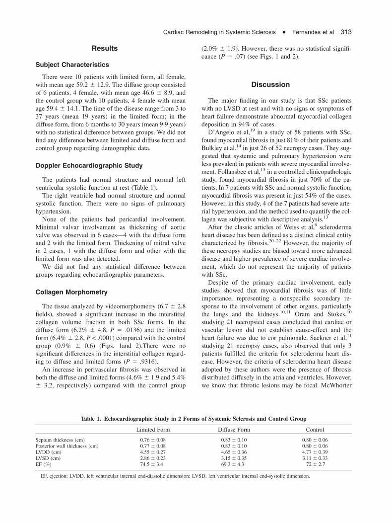
The tissue analyzed by videomorphometry (6.7 � 2.8fields), showed a significant increase in the interstitialcollagen volume fraction in both SSc forms. In thediffuse form (6.2% � 4.8, P � .0136) and the limitedform (6.4% � 2.8, P < .0001) compared with the controlgroup (0.9% � 0.6) (Figs. 1and 2).There were nosignificant differences in the interstitial collagen regard-ing to diffuse and limited forms (P � .9316).
An increase in perivascular fibrosis was observed inboth the diffuse and limited forms (4.6% � 1.9 and 5.4%� 3.2, respectively) compared with the control group
(2.0% � 1.9). However, there was no statistical signifi-cance (P � .07) (see Figs. 1 and 2).
Discussion
The major finding in our study is that SSc patientswith no LVSD at rest and with no signs or symptoms ofheart failure demonstrate abnormal myocardial collagendeposition in 94% of cases.
D’Angelo et al,19 in a study of 58 patients with SSc,found myocardial fibrosis in just 81% of their patients andBulkley et al.14 in just 26 of 52 necropsy cases. They sug-gested that systemic and pulmonary hypertension wereless prevalent in patients with severe myocardial involve-ment. Follansbee et al,13 in a controlled clinicopathologicstudy, found myocardial fibrosis in just 70% of the pa-tients. In 7 patients with SSc and normal systolic function,myocardial fibrosis was present in just 54% of the cases.However, in this study, 4 of the 7 patients had severe arte-rial hypertension, and the method used to quantify the col-lagen was subjective with descriptive analysis.13
After the classic articles of Weiss et al,9 sclerodermaheart disease has been defined as a distinct clinical entitycharacterized by fibrosis.20–22 However, the majority ofthese necropsy studies are biased toward more advanceddisease and higher prevalence of severe cardiac involve-ment, which do not represent the majority of patientswith SSc.
Despite of the primary cardiac involvement, earlystudies showed that myocardial fibrosis was of littleimportance, representing a nonspecific secondary re-sponse to the involvement of other organs, particularlythe lungs and the kidneys.10,11 Oram and Stokes,10
studying 21 necropsied cases concluded that cardiac orvascular lesion did not establish cause-effect and theheart failure was due to cor pulmonale. Sackner et al,11
studying 21 necropsy cases, also observed that only 3patients fulfilled the criteria for scleroderma heart dis-ease. However, the criteria of scleroderma heart diseaseadopted by these authors were the presence of fibrosisdistributed diffusely in the atria and ventricles. However,we know that fibrotic lesions may be focal. McWhorter
Table 1. Echocardiographic Study in 2 Forms of Systemic Sclerosis and Control Group
Limited Form Diffuse Form Control
Septum thickness (cm) 0.76 � 0.08 0.83 � 0.10 0.80 � 0.06Posterior wall thickness (cm) 0.77 � 0.08 0.83 � 0.10 0.80 � 0.06LVDD (cm) 4.55 � 0.27 4.65 � 0.36 4.77 � 0.39LVSD (cm) 2.86 � 0.23 3.15 � 0.35 3.11 � 0.33EF (%) 74.5 � 3.4 69.3 � 4.3 72 � 2.7
EF, ejection; LVDD, left ventricular internal end-diastolic dimension; LVSD, left ventricular internal end-systolic dimension.
Cardiac Remodeling in Systemic Sclerosis O Fernandes et al 313

and LeRoy2 studied 34 patients with SSc and observedthat pericardial involvement (62%) was found moreoften than intense or diffuse myocardial fibrosis (30%).Smith et al,23 in an echocardiographic study correlating
hemodynamic and necropsy data, observed that only 1 of8 cases presented myocardial fibrosis. They concludedthat fibrosis leading to cardiomyopathy was rare.
This was a systematic study done in vivo using a rightventricular endomyocardial biopsy to identify and quan-tify the myocardial interstitial fibrosis in cardiovascularasymptomatic patients with SSc. The use of a comple-mentary invasive method to diagnose myocardial dam-age with no signs or symptoms of heart failure is justifiedbecause of the occult involvement of the heart in thisdisorder.7 Noninvasive exams identified cardiac damagelater. We believe that the earlier treatment with drugsacting on collagen metabolism could avoid the laterevolution to myocardial dilatation.24
Few articles have explored right ventricular endomyo-cardial biopsy in SSc patients. Tamburino et al,8 todiagnose lesions during the early phase of myocardialdisease, described the myocardial fibrosis in only onepatient. Liangos et al25 based their studies on just 2 casesof the systemic form to suggest endomyocardial biopsywith the objective of distinguishing inflammatory fromthat of fibrotic involvement.
Fig. 1. Coronary artery and myocardium staining by Masson’s trichrome (magnification 10�) in control (female), limited form(female), and diffuse form (male) of systemic sclerosis patients. Arrows indicate fibrosis.
Fig. 2. Interstitial and perivascular collagen volume fraction incontrol and limited form and diffuse form. ICVF, interstitialcollagen volume fraction; PCVF, perivascular collagen volumefraction. *P < .0001, **P < .07 versus control group.
314 Journal of Cardiac Failure Vo. 9 No. 4 August 2003

The biopsy information obtained in our study showedmorphologic damage in the absence of clinical symp-toms of heart failure identifying a higher risk subgroup.The fibrosis may have important prognostic value in theprogression to eventual heart failure.26 We found ahistopathologic substrate that may predispose systolicand diastolic dysfunction. Further studies are needed toassess the long-term outcome of these patients. We alsoknow that not only the inadequate collagen amount, butalso the proportion between different subtypes of col-lagen,27 could be important to ventricular impairmentand increased stiffness.
Our control group had more male than female patients.However, some studies have investigated gender-relateddifferences in cardiovascular morphology with conflict-ing results.28,29 Moreover, we did not observe anydifference between male and female myocardial collagenamount in the control group.
Our patients had normal electrocardiogram and nor-mal left ventricular systolic function at rest, even thoughhistopathologic analysis showed myocardial involve-ment. Leinwand et al30 also found myocardial involve-ment, in a necropsy study, with a normal electrocardio-gram. In a study of patients with a normalelectrocardiogram, Follansbee et al31 suggested cardiacfibrosis assessed by myocardial scintigraphy that dem-onstrated perfusion defects. However, the degree ofperfusion defect did not correlate to left ventricularfunction at rest. Ten years later these patients haddeveloped increases in symptomatic cardiac disease anddeath.32 Thus the absence of symptoms or changes innoninvasive complementary study at rest may not ex-clude myocardial involvement. However, the structuraldamage that was not capable of modifying the systolicfunction at rest could be detected through other methods,such as stress echocardiogram or diastolic evaluation.Further studies are needed to confirm this.
The difficulty in studying cardiac damage in SScoccurs by renal and pulmonary involvement coexistence,which make the separation of primary and secondarycardiac involvement difficult.12 In this study, we assessedpatients without clinical and laboratory evidence ofpulmonary hypertension and renal disease. Thus intersti-tial myocardial fibrosis was characterized by primarycardiac involvement of the disease.
D’Angelo et al19 related an increased myocardialfibrosis with age. Our study, as did those of Bulkley etal14 and Ferri et al,33 showed no correlation of cardiacfibrosis with age. Internal organs were affected later inthe limited form, usually in 2nd to 5th decades of thedisease. One of our patients with a 6-month evolutionhad myocardial fibrosis, whereas another with a 30-yearevolution presented a normal histologic pattern.
With regard to the manifestation of the disease, someauthors mention that cardiac involvement is more often
in the diffuse than in the limited form.34–37 In our studywe observe interstitial myocardial fibrosis in both formsof the disease, with no statistical differences betweenthem. Follansbee et al13 also found no statistical differ-ences between the diffuse and the limited form, althoughsevere myocardial fibrosis was observed only in thediffuse form. Ferri et al33 also observed myocardialfibrosis by echocardiogram with tissular analysis in thelimited form.
Raynaud phenomenon are often found in small digitalarteries with intimal fibrosis, thinning of media layer anddermal perivascular fibrosis.38 D’Angelo et al19 andJames39 found this type of structural damage in myocar-dial vessels of patients with SSc. This vascular remod-eling may precede myocardial fibrosis. Kahan et al.40
observed decreased coronary reserve flow, which may besecondary to perivascular fibrosis and is feasible toassociate it to myocardial fibrosis. However, Sakner etal,11 Bulkley et al,14 and Follansbee et al13 did not findanatomical evidence of arterial disease. Bulkley et al.14
believed that vascular abnormalities would be morefunctional than structural. In our study, there is a trend toperivascular fibrosis. However, there was no statisticalsignificance. Therefore, a possible ischemic processcould be involved with a loss of myocyte teamed withreparative fibrosis. Moreover, a proliferative response offibroblast has been described.38 It could be anothermechanism of inappropriate collagen deposition on myo-cardial interstitial space representing a reactive fibrosis.It is probable that both mechanisms are involved.
Presently, we know that cardiac remodeling is aimportant feature in the progression of cardiac diseaseand considered as an adverse sign.41 Its evolution in-cludes a complex mechanism of myocardial fibrosis andstructural disorganization of the myocardium that resultsin cardiac dysfunction with known higher morbidity andmortality. We believe that new treatment may be adoptedearly in SSc to prevent structural cardiac remodeling,thus avoiding further progression to heart failure.
References
1. Péltonen J, Kahari L, Uitto J, Jimenez SA. Increasedexpression of type VI collagen genes in systemic sclero-sis. Arthritis Rheum 1990;33:1829-35.
2. McWhorter JE, LeRoy EC. Pericardial disease in sclero-derma (systemic sclerosis). Am J Med 1974;57:566-75.
3. Roberts NK, Cabeen WR Jr, Moss J, Clements PJ, FurstDE. The prevalence of conduction defects and cardiacarrhythmias in progressive systemic sclerosis. Ann IntMed 1981;94:38-40.
4. Clements PJ, Furst DE, Cabeen W, Tashkin D, Paulus HE,Roberts N. The relationship of arrhythmias and conduc-tion disturbances to other manifestations of cardiopulmo-
Cardiac Remodeling in Systemic Sclerosis O Fernandes et al 315
nary disease in progressive systemic sclerosis (PSS). AmJ Med 1981;71:38-46.
5. Ridolfi RL, Bulkley BH, Hutchins GM. The cardiacconduction system in progressive systemic sclerosis.Clinical and pathologic features of 35 patients. Am J Med1976;61:361-6.
6. Bulkley BH, Klacsmann PG, Hutchins GM. Anginapectoris, myocardial infarction and sudden cardiac deathwith normal coronary arteries: a clinicopathologic studyof 9 patients with progressive systemic sclerosis. AmHeart J 1978;95:563-9.
7. Deswal A, Follansbee WP. Cardiac involvement in scle-roderma. Rheum Dis Clin North Am 1996;22:841-61.
8. Tamburino C, Salomone E, Di Paola RD, Bruno G, RussoG, Tolaro S, et al. Precoce coinvolgimento miocardiconelle collagenopatie. Studio com biopsia endomiocardica.Minerva Cardioangiol 1991;39:209-12.
9. Weiss S, Stead Jr EA, Warren JV, Bailey OT. Sclerodermaheart disease with a consideration of certain other visceralmanifestations of scleroderma. Arch Intern Med 1943:71:749-76.
10. Oram S, Stokes W. The heart in scleroderma. Br Heart J1961;23;243-58.
11. Sackner MA, Heinz R, Steinberg AJ. The heart inscleroderma. Am J Cardiol 1966;17:542-60.
12. Botstein GR, LeRoy EC. Primary heart disease in sys-temic sclerosis (scleroderma): advances in clinical andpathologic features, pathogenesis, and new therapeuticapproaches. Am Heart J 1981;102:913-8.
13. Follansbee WP, Miller TR, Curtiss EI, Orie JE, BersteinRL, Kiernan J, Medsger Jr TA. A controlled clinicopatho-logic study of myocardial fibrosis in systemic sclerosis(scleroderma). J Rheumatol 1990;17:656-62.
14. Bulkley BH, Ridolfi RL, Salyer WR. Myocardial lesionsof progressive systemic sclerosis. A cause of cardiacdysfunction. Circulation 1976;53:483-90.
15. Subcommittee for Scleroderma Criteria of the AmericanRheumatism Association Diagnostic and Therapeutic Cri-teria Committee. Preliminary criteria for the classificationof systemic sclerosis (scleroderma). Arthritis Rheum1980;23:581-90.
16. Schiller NB, Shan PM, Crawford M, DeMaria A, De-vereux R, Feigenbaum H, et al. American Society ofEchocardiography Committee on Standards, subcommit-tee on quantification of two-dimensional echocardio-grams. Recommendations for quantification of the leftventricle by two-dimensional echocardiography. J AmEchocardiogr 1989;2:358-67.
17. Sahn DJ, DeMaria AN, Kisslo J, Weyman A. The Com-mittee on M-mode Standardization of the AmericanSociety of Echocardiography. Recommendations regard-ing quantitation in M-mode echocardiography: results ofa survey of echocardiographic measurements. Circulation1978;58:1072-83.
18. Ramires FJ, Sun Y, Weber KT. Myocardial fibrosisassociated with aldosterone or angiotensin II administra-tion: attenuation by calcium channel blockade. J Mol CellCardiol 1998;30:475-483.
19. D’Angelo WA, Fries JF, Masi AT, Shulman LE. Patho-logic observations in systemic sclerosis (scleroderma). A
study of fifty-eight autopsy cases and fifty-eight matchedcontrols. Am J Med 1969;46:428-40.
20. Mathisen AK, Palmer JD. Diffuse scleroderma withinvolvement of the heart. Report of a case. Am Heart J1946;33:366-74.
21. Goetz RH. The heart in generalised scleroderma. Progres-sive systemic sclerosis. Angiology 1951;2:555-78.
22. Hurly J, Billings M, Coe J, Weber L. Scleroderma heartdisease. Am Heart J 1950;42:758-65.
23. Smith JW, Clements PJ, Levisman J, Furst D, Ross M.Echocardiographic features of progressive systemic scle-rosis (PSS). Correlation with hemodynamic and postmor-tem studies. Am J Med 1979;66:28-33.
24. Schwarz F, Mall G, Zebe H, Blickle J, Derks H, MantheyJ, et al. Quantitative morphologic findings of the myocar-dium in idiopathic dilated cardiomyopathy. Am J Cardiol1982;51:501-6.
25. Liangos O, Neure L, Kühl U, Pauschinger M, Sieper J,Distler A, et al. The possible role of myocardial biopsy insystemic sclerosis. Rheumatology 2000;39:674-9.
26. Mady C, Ianni BM, Arteaga E, Montes GS, Caldini EG,Andrade G, et al. Relation between interstitial myocardialcollagen and the degree of clinical impairment in Chagas’disease. Am J Cardiol 1999;15;84:1456.
27. Weber KT. Cardiac interstitium in health and disease: thefibrillar collagen Network. J Am Coll Cardiol 1989;13:1637-52.
28. Villari B, Campbell SE, Schneider J, Vassalli G, Chiari-ello M, Hess OM. Sex-dependent differences in leftventricular function and structure in chronic pressureoverload. Eur Heart J 1995;16:1410-9.
29. Rosenkranz-Weiss P, Tomek RJ, Mathew J, Eghbali M.Gender-specific differences in expression of mRNAs forfunctional and structural proteins in rat ventricular myo-cardium. J Mol Cell Cardiol 1994;26:261-70.
30. Leinwand I, Duryee W, Richter MN. Scleroderma (basedon a study of over 150 cases). Ann Intern Med 1954;41:1003-41.
31. Follansbee WP, Curtiss EI, Rahko PS, Medsger Jr TA,Lavine SJ, Owens GR, et al. The electrocardiogram insystemic sclerosis (scleroderma). Study of 102 consecu-tive cases with functional correlations and review of theliterature. Am J Med 1985;79:183-92.
32. Steen VD, Follansbee WP, Conte CG, Medsger Jr TA.Thallium perfusion defects predict subsequent cardiacdysfunction in patients with systemic sclerosis. ArthritisRheum 1996;39:677-81.
33. Ferri C, Di Bello V, Martini A, Giorgi D, Storino FAA,Bianchini M, et al. Heart involvement in systemic scle-rosis: an ultrasonic tissue characterization study. AnnRheum Dis 1998;57:296-302.
34. Akesson A, Wollheim FA. Organ manifestations in 100patients with progressive systemic sclerosis: a compari-son between the crest syndrome and diffuse scleroderma.Br J Rheumatol 1989;28:281-6.
35. Murata I, Takenaka K, Shinohara S, Suzuki T, Sasaki T,Yamamoto K. Diversity of myocardial involvement insystemic sclerosis: an 8-year study of 95 Japanese pa-tients. Am Heart J 1998;135:960-9.
316 Journal of Cardiac Failure Vo. 9 No. 4 August 2003

36. Follansbee WP, Curtiss EI, Medsger Jr TA, Steen V,Uretsky BF, Owens GR, et al. Physiologic abnormalitiesof cardiac function in progressive systemic sclerosis withdiffuse scleroderma. N Engl J Med 1984;310:142-8.
37. Ellis WW, Baer AN, Robertson RM, Pincus T, Kronen-berg MW. Left ventricular dysfunction induced by coldexposure in patients with systemic sclerosis. Am J Med1986;80:385-92.
38. Fleischmajer R, Perlish JS, Duncan M. Scleroderma. Amodel for fibrosis. Arch Dermatol 1983;119:957-62.
39. James TN. De Subitaneis mortibus. Coronary arteries andconduction system in scleroderma heart disease. Circula-tion 1974;50:844-56.
40. Kahan A, Nitenberg A, Foult JM, et al. Decreasedcoronary reserve in primary scleroderma myocardial dis-ease. Arthritis Rheum 1985;28:637-46.
41. Cohn JN, Ferrari R, Sharpe N. Cardiac remodeling—concepts and clinical implications: a consensus paperfrom an international forum on cardiac remodeling. J AmColl Cardiol 2000;35:570-82.
Cardiac Remodeling in Systemic Sclerosis O Fernandes et al 317