Concordance between breast core needle biopsy ... - PLOS
11
RESEARCH ARTICLE A ten-year, single-center experience: Concordance between breast core needle biopsy/vacuum-assisted biopsy and postoperative histopathology in B3 and B5a cases Mohamed Elsharkawy ID 1 *, Thomas Vestring 1 , Hans-Juergen Raatschen 2 1 Diagnostic and Interventional Radiology/Neuroradiology Department, Agaplesion Diakonieklinikum, Rotenburg Wuemme, Germany, 2 Diagnostic and Interventional Radiology Department, Hannover Medical School, Hanover, Germany * [email protected] Abstract Purpose To determine the concordance rate between core needle biopsy/vacuum-assisted biopsy (CNB/VAB) and postoperative histopathology in B3 (lesions of uncertain malignant poten- tial) and B5a (in situ) lesions found on mammograms or ultrasound. Material and methods 2,029 consecutive biopsies performed over 10 years for patients who underwent mammo- grams or ultrasounds. For CNB 14G needle and for VAB 8G/10G needles were used. In all biopsies, we identified the age, BI-RADS®, histopathological biopsy results, B-category, nuclear grade for DCIS and postoperative histopathology results in B3 and B5a cases from the biopsy. Results The B-categories from CNB/VAB were as follows: B2 42.2 percent (n = 856), B3 4.5 percent (n = 91), B5a 5.7 percent (n = 115), and B5b 47.6 percent (n = 967). In the B3-category, 72/ 91 patients underwent surgical excision, with a concordance rate of 83.3 percent (n = 60/72) and a discordance rate of 16.7 percent (n = 12/72) to postoperative histopathology. From the discordant cases, 67.7 percent (n = 8/12) showed DCIS and 32.3 percent (n = 4/12) showed invasive breast cancer. The BIRADS of the discordant cases was 4b in 41.7 percent (n = 5/12) and 5 in 58.3 percent (n = 7/12). The PPVs for malignancy of B3 lesions were 0.21, with no statistical significance between subgroups. In the B5a-category, 101 of 115 patients underwent surgery in our hospital, with a concordance rate of 80.2 percent (n = 81/ 101) and a discordance rate of 19.8 percent (n = 20/101) to postoperative histopathology. PLOS ONE PLOS ONE | https://doi.org/10.1371/journal.pone.0233574 May 21, 2020 1 / 11 a1111111111 a1111111111 a1111111111 a1111111111 a1111111111 OPEN ACCESS Citation: Elsharkawy M, Vestring T, Raatschen H-J (2020) A ten-year, single-center experience: Concordance between breast core needle biopsy/ vacuum-assisted biopsy and postoperative histopathology in B3 and B5a cases. PLoS ONE 15 (5): e0233574. https://doi.org/10.1371/journal. pone.0233574 Editor: Fernando Schmitt, University of Toronto, CANADA Received: February 10, 2020 Accepted: May 7, 2020 Published: May 21, 2020 Copyright: © 2020 Elsharkawy et al. This is an open access article distributed under the terms of the Creative Commons Attribution License, which permits unrestricted use, distribution, and reproduction in any medium, provided the original author and source are credited. Data Availability Statement: All relevant data are within the paper. Funding: The author(s) received no specific funding for this work. Competing interests: The authors have declared that no competing interests exist.
-
Upload
khangminh22 -
Category
Documents
-
view
5 -
download
0
Transcript of Concordance between breast core needle biopsy ... - PLOS

RESEARCH ARTICLE
A ten-year, single-center experience:
Concordance between breast core needle
biopsy/vacuum-assisted biopsy and
postoperative histopathology in B3 and B5a
cases
Mohamed ElsharkawyID1*, Thomas Vestring1, Hans-Juergen Raatschen2
1 Diagnostic and Interventional Radiology/Neuroradiology Department, Agaplesion Diakonieklinikum,
Rotenburg Wuemme, Germany, 2 Diagnostic and Interventional Radiology Department, Hannover Medical
School, Hanover, Germany
Abstract
Purpose
To determine the concordance rate between core needle biopsy/vacuum-assisted biopsy
(CNB/VAB) and postoperative histopathology in B3 (lesions of uncertain malignant poten-
tial) and B5a (in situ) lesions found on mammograms or ultrasound.
Material and methods
2,029 consecutive biopsies performed over 10 years for patients who underwent mammo-
grams or ultrasounds. For CNB 14G needle and for VAB 8G/10G needles were used. In all
biopsies, we identified the age, BI-RADS®, histopathological biopsy results, B-category,
nuclear grade for DCIS and postoperative histopathology results in B3 and B5a cases from
the biopsy.
Results
The B-categories from CNB/VAB were as follows: B2 42.2 percent (n = 856), B3 4.5 percent
(n = 91), B5a 5.7 percent (n = 115), and B5b 47.6 percent (n = 967). In the B3-category, 72/
91 patients underwent surgical excision, with a concordance rate of 83.3 percent (n = 60/72)
and a discordance rate of 16.7 percent (n = 12/72) to postoperative histopathology. From
the discordant cases, 67.7 percent (n = 8/12) showed DCIS and 32.3 percent (n = 4/12)
showed invasive breast cancer. The BIRADS of the discordant cases was 4b in 41.7 percent
(n = 5/12) and 5 in 58.3 percent (n = 7/12). The PPVs for malignancy of B3 lesions were
0.21, with no statistical significance between subgroups. In the B5a-category, 101 of 115
patients underwent surgery in our hospital, with a concordance rate of 80.2 percent (n = 81/
101) and a discordance rate of 19.8 percent (n = 20/101) to postoperative histopathology.
PLOS ONE
PLOS ONE | https://doi.org/10.1371/journal.pone.0233574 May 21, 2020 1 / 11
a1111111111
a1111111111
a1111111111
a1111111111
a1111111111
OPEN ACCESS
Citation: Elsharkawy M, Vestring T, Raatschen H-J
(2020) A ten-year, single-center experience:
Concordance between breast core needle biopsy/
vacuum-assisted biopsy and postoperative
histopathology in B3 and B5a cases. PLoS ONE 15
(5): e0233574. https://doi.org/10.1371/journal.
pone.0233574
Editor: Fernando Schmitt, University of Toronto,
CANADA
Received: February 10, 2020
Accepted: May 7, 2020
Published: May 21, 2020
Copyright: © 2020 Elsharkawy et al. This is an
open access article distributed under the terms of
the Creative Commons Attribution License, which
permits unrestricted use, distribution, and
reproduction in any medium, provided the original
author and source are credited.
Data Availability Statement: All relevant data are
within the paper.
Funding: The author(s) received no specific
funding for this work.
Competing interests: The authors have declared
that no competing interests exist.

From the discordant cases, 55 percent (n = 11/20) showed invasive breast carcinoma of no
special type (NST).
Conclusion
Our concordance rate for B3 (83.3 percent) and B5a (80.2 percent) lesions in the biopsies to
postoperative histopathology is matching to previously published literature. Surgical exci-
sion is our recommendation for lesions biopsied with a B3 category in the histopathology
and a BIRADS category of (4b, 4c and 5). The PPVs for malignancy of B3 lesions showed
no statistical significance between subgroups. Also, the nuclear grade of DCIS was not sta-
tistically significant in terms of upgrade into invasive breast cancer.
Introduction
In Europe, the female breast was the most common cancer site in 2018 (523,000 cases) [1].
Breast biopsies are commonly performed to evaluate mammographic or palpable findings that
are of concern, and the majority reveal benign findings [2].
The breast imaging and data system (BI-RADS1) categorizes mammographic findings
from 0 to 6 [3], with categories 4 (including a- low; b- moderate; and c- high suspicion of
malignancy) and 5 requiring tissue biopsy. According to guidelines for non-operative diagnos-
tic procedures and reporting in breast cancer screening, the histological results of core needle
biopsy (CNB) and vacuum-assisted biopsy (VAB) are categorized from B1 to B5 [4]. The B1
and B2 categories respectively represent normal and benign lesions, while the B4 and B5 cate-
gories respectively represent suspicious and malignant lesions [4]. The B3 category comprise
lesions with uncertain malignant potential. The B5 category is further subdivided into B5a,
which comprise in-situ carcinomas and the B5b, which comprise the invasive carcinomas [4].
The B3 category represents a heterogeneous group of lesions (for example: atypical ductal
hyperplasia, radial scar, papillary lesions, etc.) that may be associated with malignant disease
requiring surgical intervention [5]. With the increasing use of mammographic screening, the
detection rate of B3 lesions in patients who were previously asymptomatic has increased [6, 7].
This has resulted in breast surgery for ultimately benign final histopathological diagnoses [5].
The rate of B3 lesions in biopsies ranged from 3.8 percent to 9.2 percent in screening program
or single institution [8–13]. El Sayed et.al found that B3 rate was 5 percent from all CNBs
(20.001) in screening program over 10 years [14]. Weigel et al, reported a B3 rate of 15.1 per-
cent in digital mammography screening over a period of 4 years [15]. Lee et al, found that the
B3 rate was higher in screening program compared to symptomatic patients (7.3 percent vs 2
percent) [6].
Furthermore, ductal carcinoma in-situ (DCIS), is primarily diagnosed by imaging because
it is usually clinically occult [16]. DCIS was previously an uncommonly identified breast lesion,
now it accounts for approximately 20 percent of newly diagnosed breast cancer cases [17]. As
the detection rate increased, there has also been increased discussion and controversy [18].
Because of the heterogenous nature of DCIS, the disease process is in part not well understood
[19]. Although it sometimes presents as a nonaggressive occult lesion, untreated DCIS may
progress into an aggressive, invasive cancer [20, 21].
El Sayed et al, found that underestimation rate of malignancy associated with B3 lesions is
19.1% [14] and Brennan meta-analysis showed that approximately one in four DCIS diagnoses
by CNB represents understaged, invasive breast cancer [22].
PLOS ONE Single-center experience: Concordance between breast biopsy and postoperative histopathology in B3 and DCIS
PLOS ONE | https://doi.org/10.1371/journal.pone.0233574 May 21, 2020 2 / 11
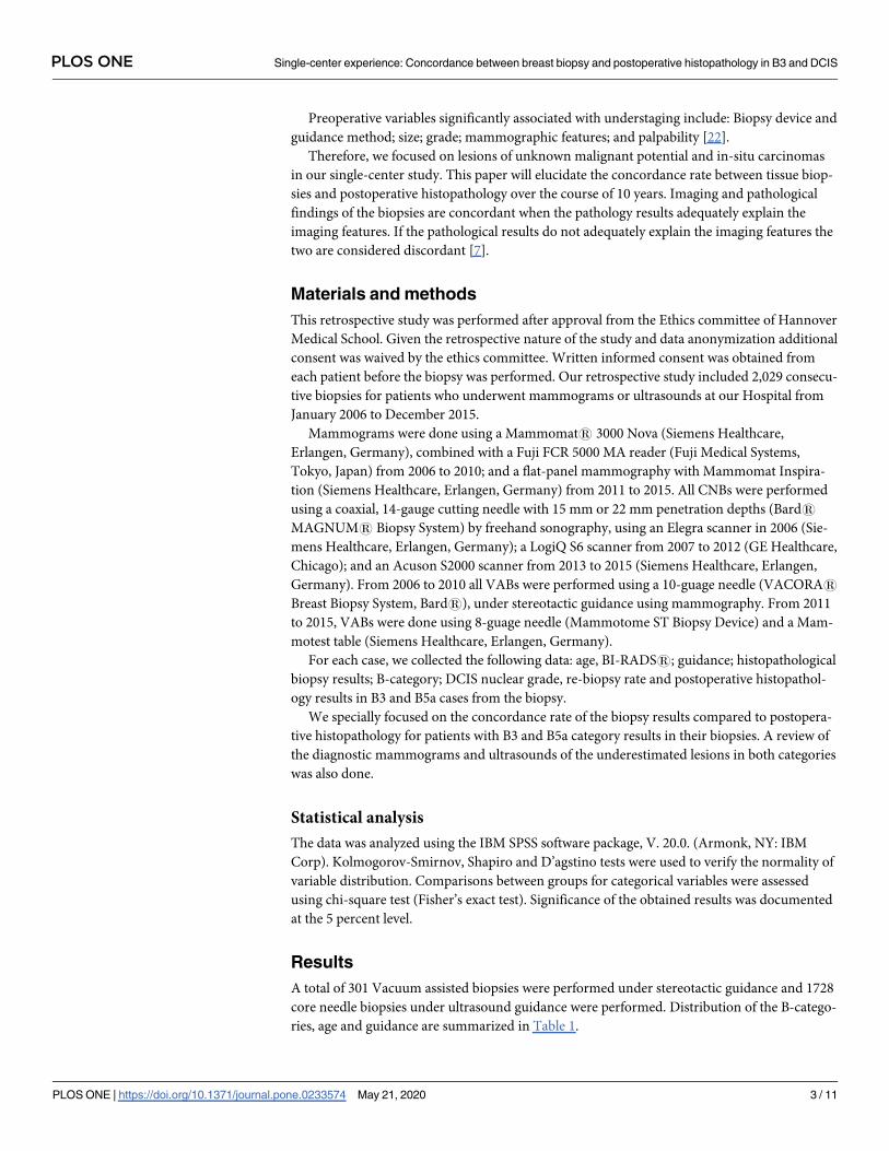
Preoperative variables significantly associated with understaging include: Biopsy device and
guidance method; size; grade; mammographic features; and palpability [22].
Therefore, we focused on lesions of unknown malignant potential and in-situ carcinomas
in our single-center study. This paper will elucidate the concordance rate between tissue biop-
sies and postoperative histopathology over the course of 10 years. Imaging and pathological
findings of the biopsies are concordant when the pathology results adequately explain the
imaging features. If the pathological results do not adequately explain the imaging features the
two are considered discordant [7].
Materials and methods
This retrospective study was performed after approval from the Ethics committee of Hannover
Medical School. Given the retrospective nature of the study and data anonymization additional
consent was waived by the ethics committee. Written informed consent was obtained from
each patient before the biopsy was performed. Our retrospective study included 2,029 consecu-
tive biopsies for patients who underwent mammograms or ultrasounds at our Hospital from
January 2006 to December 2015.
Mammograms were done using a Mammomat1 3000 Nova (Siemens Healthcare,
Erlangen, Germany), combined with a Fuji FCR 5000 MA reader (Fuji Medical Systems,
Tokyo, Japan) from 2006 to 2010; and a flat-panel mammography with Mammomat Inspira-
tion (Siemens Healthcare, Erlangen, Germany) from 2011 to 2015. All CNBs were performed
using a coaxial, 14-gauge cutting needle with 15 mm or 22 mm penetration depths (Bard1
MAGNUM1 Biopsy System) by freehand sonography, using an Elegra scanner in 2006 (Sie-
mens Healthcare, Erlangen, Germany); a LogiQ S6 scanner from 2007 to 2012 (GE Healthcare,
Chicago); and an Acuson S2000 scanner from 2013 to 2015 (Siemens Healthcare, Erlangen,
Germany). From 2006 to 2010 all VABs were performed using a 10-guage needle (VACORA1
Breast Biopsy System, Bard1), under stereotactic guidance using mammography. From 2011
to 2015, VABs were done using 8-guage needle (Mammotome ST Biopsy Device) and a Mam-
motest table (Siemens Healthcare, Erlangen, Germany).
For each case, we collected the following data: age, BI-RADS1; guidance; histopathological
biopsy results; B-category; DCIS nuclear grade, re-biopsy rate and postoperative histopathol-
ogy results in B3 and B5a cases from the biopsy.
We specially focused on the concordance rate of the biopsy results compared to postopera-
tive histopathology for patients with B3 and B5a category results in their biopsies. A review of
the diagnostic mammograms and ultrasounds of the underestimated lesions in both categories
was also done.
Statistical analysis
The data was analyzed using the IBM SPSS software package, V. 20.0. (Armonk, NY: IBM
Corp). Kolmogorov-Smirnov, Shapiro and D’agstino tests were used to verify the normality of
variable distribution. Comparisons between groups for categorical variables were assessed
using chi-square test (Fisher’s exact test). Significance of the obtained results was documented
at the 5 percent level.
Results
A total of 301 Vacuum assisted biopsies were performed under stereotactic guidance and 1728
core needle biopsies under ultrasound guidance were performed. Distribution of the B-catego-
ries, age and guidance are summarized in Table 1.
PLOS ONE Single-center experience: Concordance between breast biopsy and postoperative histopathology in B3 and DCIS
PLOS ONE | https://doi.org/10.1371/journal.pone.0233574 May 21, 2020 3 / 11

Seventy-three lesions were re-biopsied due to discordance between imaging and histopath-
ological results of the VAB/CNB biopsies. From the 73 biopsies, 10 lesions histopathology
results changed from B2 to B5b,1 lesion changed from B2 to B5a, 4 changed from B1 to B2 and
58 lesions did not change with a B2 result in both biopsies. B4 category was not included
because those were only found in preliminary pathology reports of 4 cases which was catego-
rized as B5a in the final histopathology report of the biopsy.
For the lesions with uncertain malignant potential (B3) and in-situ carcinoma (B5a) with
CNB or VAB histopathology, a comparison to postoperative histopathology was done to show
the concordance rate.
In the B3 category, 72 of 91 patients underwent surgical excision in our hospital. From the
19 non operated patients 12 had mammogram or ultrasound follow-ups for at least one year
with no radiologic changes requiring a re-biopsy or surgical excision. Seven patients did not
come for follow-up after the biopsy result.
Concordance between CNB/VAB and postoperative histopathology for B3 was found in
83.3 percent (n = 60/72 patients) cases, and discordance was documented in 16.7 percent
(n = 12/72 patients) of cases. Three patients had two lesions, i.e. a total of 75 lesions. The 12
cases that were discordant with histopathology were upgraded as follows: Eight patients with
DCIS (B5a) and four with invasive breast cancer (B5b). Table 2 summarizes the discordant
cases regarding B3 lesions in CNB/VAB histopathology, compared to postoperative
histopathology.
The most frequently excised lesion in the B3 subgroups was papillary lesion (50.7 percent),
followed by atypical ductal hyperplasia (20 percent), flat epithelial atypia (12 percent), lobular
intraepithelial neoplasia (8 percent), radial scar (5.3 percent), and phyllodes tumor (4 percent).
Table 3 summarizes the frequency of the different B3 lesions from the biopsies and the rate of
malignant diagnosis after excision.
Table 1. Distribution of the different B-categories.
B-Classification B2 B3 B5a B5b Total (n)
Age range in years 16–94 20.9–87.8 33.9–85.8 20.6–96.5
Mean Age SD 51.46 ± 14.14 56.33 ± 13.67 59.24 ± 12.69 63.29 ± 14.37
Guidance
Ultrasound 711 63 27 927 1728
Stereotactic 145 28 88 40 301
Total (n) 856 91 115 967 2029
https://doi.org/10.1371/journal.pone.0233574.t001
Table 2. Pre- and postoperative histopathology in discordant cases from the B3 category.
CNB/VAB histopathology vs.
postoperative histopathology
DCIS G1
(n)
DCIS G2
(n)
DCIS G3
(n)
Poorly differentiated
carcinoma (n)
Tubularadeno CA
(n)
NST- LCIS-
Lobular CA (n)
DCIS G2-
NST- LCIS (n)
Total
number
ADH (n) 1 1 1 3
ADH+PL (n) 1 1 1 3
PL (n) 1 1 1 3
LN (n) 1 1
FEA (n) 1 1
FEA+LN (n) 1 1
Total number 3 2 3 1 1 1 1 12
ADH = atypical ductal hyperplasia; PL = papillary lesion; LN = non-invasive lobular neoplasia; FEA = flat epithelial atypia; DCIS = ductal carcinoma in-situ;
NST = invasive breast carcinoma of no special type; LCIS = lobular carcinoma in-situ; CA = carcinoma; (n) number of patients.
https://doi.org/10.1371/journal.pone.0233574.t002
PLOS ONE Single-center experience: Concordance between breast biopsy and postoperative histopathology in B3 and DCIS
PLOS ONE | https://doi.org/10.1371/journal.pone.0233574 May 21, 2020 4 / 11
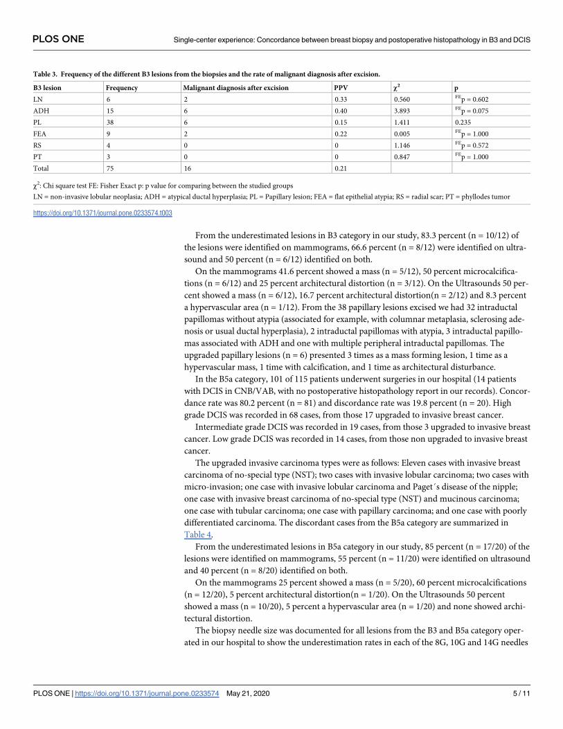
From the underestimated lesions in B3 category in our study, 83.3 percent (n = 10/12) of
the lesions were identified on mammograms, 66.6 percent (n = 8/12) were identified on ultra-
sound and 50 percent (n = 6/12) identified on both.
On the mammograms 41.6 percent showed a mass (n = 5/12), 50 percent microcalcifica-
tions (n = 6/12) and 25 percent architectural distortion (n = 3/12). On the Ultrasounds 50 per-
cent showed a mass (n = 6/12), 16.7 percent architectural distortion(n = 2/12) and 8.3 percent
a hypervascular area (n = 1/12). From the 38 papillary lesions excised we had 32 intraductal
papillomas without atypia (associated for example, with columnar metaplasia, sclerosing ade-
nosis or usual ductal hyperplasia), 2 intraductal papillomas with atypia, 3 intraductal papillo-
mas associated with ADH and one with multiple peripheral intraductal papillomas. The
upgraded papillary lesions (n = 6) presented 3 times as a mass forming lesion, 1 time as a
hypervascular mass, 1 time with calcification, and 1 time as architectural disturbance.
In the B5a category, 101 of 115 patients underwent surgeries in our hospital (14 patients
with DCIS in CNB/VAB, with no postoperative histopathology report in our records). Concor-
dance rate was 80.2 percent (n = 81) and discordance rate was 19.8 percent (n = 20). High
grade DCIS was recorded in 68 cases, from those 17 upgraded to invasive breast cancer.
Intermediate grade DCIS was recorded in 19 cases, from those 3 upgraded to invasive breast
cancer. Low grade DCIS was recorded in 14 cases, from those non upgraded to invasive breast
cancer.
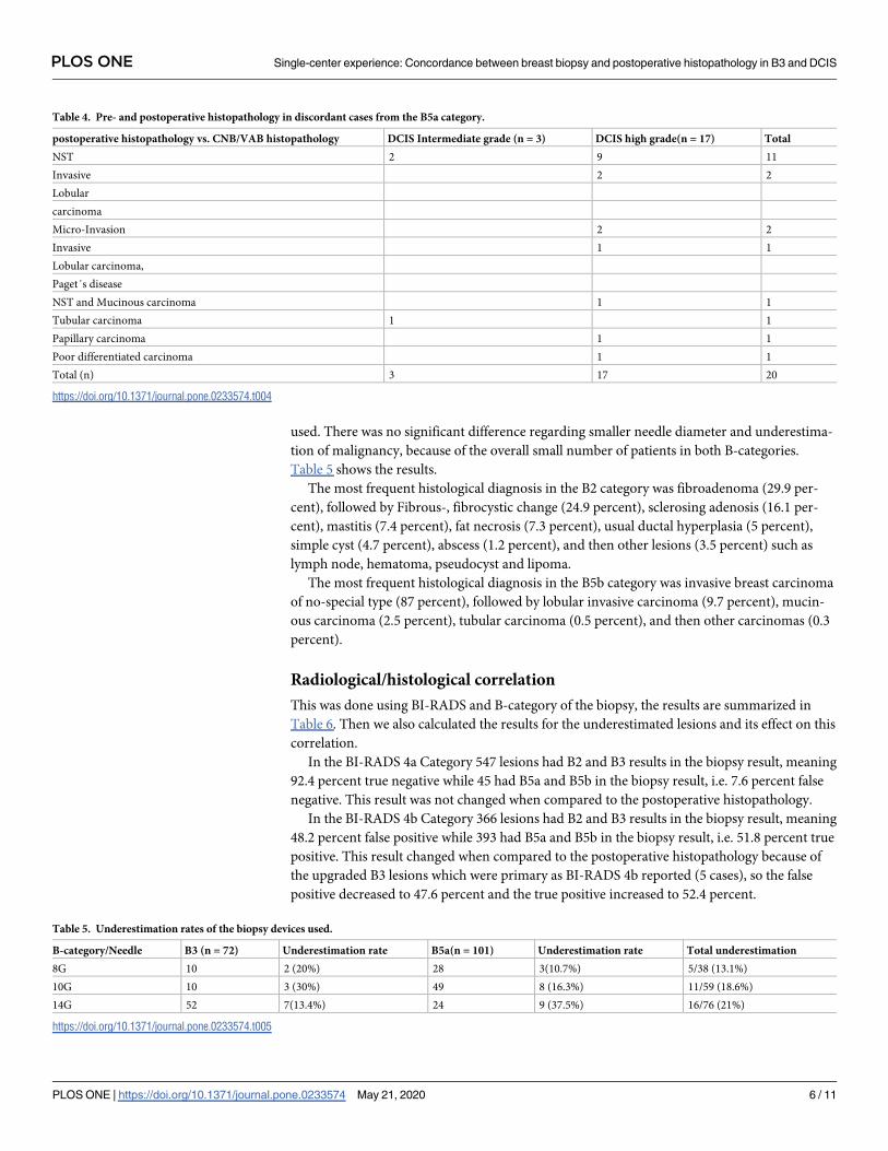
The upgraded invasive carcinoma types were as follows: Eleven cases with invasive breast
carcinoma of no-special type (NST); two cases with invasive lobular carcinoma; two cases with
micro-invasion; one case with invasive lobular carcinoma and Paget´s disease of the nipple;
one case with invasive breast carcinoma of no-special type (NST) and mucinous carcinoma;
one case with tubular carcinoma; one case with papillary carcinoma; and one case with poorly
differentiated carcinoma. The discordant cases from the B5a category are summarized in
Table 4.
From the underestimated lesions in B5a category in our study, 85 percent (n = 17/20) of the
lesions were identified on mammograms, 55 percent (n = 11/20) were identified on ultrasound
and 40 percent (n = 8/20) identified on both.
On the mammograms 25 percent showed a mass (n = 5/20), 60 percent microcalcifications
(n = 12/20), 5 percent architectural distortion(n = 1/20). On the Ultrasounds 50 percent
showed a mass (n = 10/20), 5 percent a hypervascular area (n = 1/20) and none showed archi-
tectural distortion.
The biopsy needle size was documented for all lesions from the B3 and B5a category oper-
ated in our hospital to show the underestimation rates in each of the 8G, 10G and 14G needles
Table 3. Frequency of the different B3 lesions from the biopsies and the rate of malignant diagnosis after excision.
B3 lesion Frequency Malignant diagnosis after excision PPV χ2 p
LN 6 2 0.33 0.560 FEp = 0.602
ADH 15 6 0.40 3.893 FEp = 0.075
PL 38 6 0.15 1.411 0.235
FEA 9 2 0.22 0.005 FEp = 1.000
RS 4 0 0 1.146 FEp = 0.572
PT 3 0 0 0.847 FEp = 1.000
Total 75 16 0.21
χ2: Chi square test FE: Fisher Exact p: p value for comparing between the studied groups
LN = non-invasive lobular neoplasia; ADH = atypical ductal hyperplasia; PL = Papillary lesion; FEA = flat epithelial atypia; RS = radial scar; PT = phyllodes tumor
https://doi.org/10.1371/journal.pone.0233574.t003
PLOS ONE Single-center experience: Concordance between breast biopsy and postoperative histopathology in B3 and DCIS
PLOS ONE | https://doi.org/10.1371/journal.pone.0233574 May 21, 2020 5 / 11
used. There was no significant difference regarding smaller needle diameter and underestima-
tion of malignancy, because of the overall small number of patients in both B-categories.
Table 5 shows the results.
The most frequent histological diagnosis in the B2 category was fibroadenoma (29.9 per-
cent), followed by Fibrous-, fibrocystic change (24.9 percent), sclerosing adenosis (16.1 per-
cent), mastitis (7.4 percent), fat necrosis (7.3 percent), usual ductal hyperplasia (5 percent),
simple cyst (4.7 percent), abscess (1.2 percent), and then other lesions (3.5 percent) such as
lymph node, hematoma, pseudocyst and lipoma.
The most frequent histological diagnosis in the B5b category was invasive breast carcinoma
of no-special type (87 percent), followed by lobular invasive carcinoma (9.7 percent), mucin-
ous carcinoma (2.5 percent), tubular carcinoma (0.5 percent), and then other carcinomas (0.3
percent).
Radiological/histological correlation
This was done using BI-RADS and B-category of the biopsy, the results are summarized in
Table 6. Then we also calculated the results for the underestimated lesions and its effect on this
correlation.
In the BI-RADS 4a Category 547 lesions had B2 and B3 results in the biopsy result, meaning
92.4 percent true negative while 45 had B5a and B5b in the biopsy result, i.e. 7.6 percent false
negative. This result was not changed when compared to the postoperative histopathology.
In the BI-RADS 4b Category 366 lesions had B2 and B3 results in the biopsy result, meaning
48.2 percent false positive while 393 had B5a and B5b in the biopsy result, i.e. 51.8 percent true
positive. This result changed when compared to the postoperative histopathology because of
the upgraded B3 lesions which were primary as BI-RADS 4b reported (5 cases), so the false
positive decreased to 47.6 percent and the true positive increased to 52.4 percent.
Table 4. Pre- and postoperative histopathology in discordant cases from the B5a category.
postoperative histopathology vs. CNB/VAB histopathology DCIS Intermediate grade (n = 3) DCIS high grade(n = 17) Total
NST 2 9 11
Invasive 2 2
Lobular
carcinoma
Micro-Invasion 2 2
Invasive 1 1
Lobular carcinoma,
Paget´s disease
NST and Mucinous carcinoma 1 1
Tubular carcinoma 1 1
Papillary carcinoma 1 1
Poor differentiated carcinoma 1 1
Total (n) 3 17 20
https://doi.org/10.1371/journal.pone.0233574.t004
Table 5. Underestimation rates of the biopsy devices used.
B-category/Needle B3 (n = 72) Underestimation rate B5a(n = 101) Underestimation rate Total underestimation
8G 10 2 (20%) 28 3(10.7%) 5/38 (13.1%)
10G 10 3 (30%) 49 8 (16.3%) 11/59 (18.6%)
14G 52 7(13.4%) 24 9 (37.5%) 16/76 (21%)
https://doi.org/10.1371/journal.pone.0233574.t005
PLOS ONE Single-center experience: Concordance between breast biopsy and postoperative histopathology in B3 and DCIS
PLOS ONE | https://doi.org/10.1371/journal.pone.0233574 May 21, 2020 6 / 11
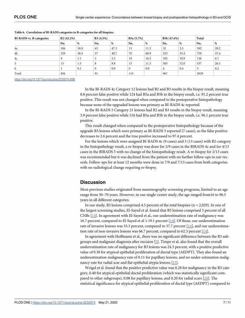
In the BI-RADS 4c Category 12 lesions had B2 and B3 results in the biopsy result, meaning
8.8 percent false positive while 124 had B5a and B5b in the biopsy result, i.e. 91.2 percent true
positive. This result was not changed when compared to the postoperative histopathology
because none of the upgraded lesions was primary as BI-RADS 4c reported.
In the BI-RADS 5 Category 21 lesions had B2 and B3 results in the biopsy result, meaning
3.9 percent false positive while 516 had B5a and B5b in the biopsy result, i.e. 96.1 percent true
positive.
This result changed when compared to the postoperative histopathology because of the
upgrade B3 lesions which were primary as BI-RADS 5 reported (7 cases), so the false positive
decreases to 2.6 percent and the true positive increased to 97.4 percent.
For the lesions which were assigned BI-RADS 4c (9 cases) and 5 (13 cases) with B2-category
in the histopathology result, a re-biopsy was done for 2/9 cases in the BIRADS 4c and for 4/13
cases in the BIRADS 5 with no change of the histopathology result. A re-biopsy for 2/13 cases
was recommended but it was declined from the patient with no further follow-ups in our rec-
ords. Follow-ups for at least 12 months were done in 7/9 and 7/13 cases from both categories
with no radiological change requiring re-biopsy.
Discussion
Most previous studies originated from mammography screening programs, limited to an age
range from 50–70 years. However, in our single-center study, the age ranged from16 to 96.5
years in all different categories.
In our study, B3 lesions comprised 4.5 percent of the total biopsies (n = 2,029). In one of
the largest screening studies, El-Sayed et al. found that B3 lesions comprised 5 percent of all
CNBs [14]. In agreement with El-Sayed et al., our underestimation rate of malignancy was
16.7 percent, compared to El-Sayed et al.’s 19.1 percent [14]. Of those, our underestimation
rate of invasive lesions was 33.3 percent, compared to 37.7 percent [14], and our underestima-
tion rate of non-invasive lesions was 66.7 percent, compared to 62.3 percent [14].
In agreement with Hoffmann et al., there was no significant difference between the B3 sub-
groups and malignant diagnosis after excision [5]. Timpe et al. also found that the overall
underestimation rate of malignancy for B3 lesions was 24.3 percent, with a positive predictive
value of 0.30 for atypical epithelial proliferation of ductal type (AEDPT). They also found an
underestimation malignancy rate of 0.11 for papillary lesions, and no under-estimation malig-
nancy rate for radial scar and flat epithelial atypia lesions [23].
Weigel et al. found that the positive predictive value was 0.28 for malignancy in the B3 cate-
gory, 0.40 for atypical epithelial ductal proliferation (which was statistically significant com-
pared to other subgroups), 0.08 for papillary lesions, and 0.20 for radial scars [24]. The
statistical significance for atypical epithelial proliferation of ductal type (AEDPT) compared to
Table 6. Correlation of BI-RADS categories to B-categories for all biopsies.
BI-RADS vs. B-categories B2 (42.2%) B3 (4.5%) B5a (5.7%) B5b (47.6%) Total
No. % No. % No. % No. % No. %
4a 504 58.9 43 47.3 13 11.3 32 3.3 592 29.2
4b 329 38.4 37 40.7 70 60.9 323 33.4 759 37.4
4c 9 1.1 3 3.3 19 16.5 105 10.9 136 6.7
5 13 1.5 8 8.8 13 11.3 503 52.0 537 26.5
6 1 0.1 0 0.0 0 0.0 4 0.4 5 0.2
Total 856 91 115 967 2029
https://doi.org/10.1371/journal.pone.0233574.t006
PLOS ONE Single-center experience: Concordance between breast biopsy and postoperative histopathology in B3 and DCIS
PLOS ONE | https://doi.org/10.1371/journal.pone.0233574 May 21, 2020 7 / 11

other subgroups in this study, was due to the percentage of the AEDPT lesions from total B3
lesions; 51.6 percent (n = 47/91). In our study the atypical ductal hyperplasia was only 20 per-
cent from all B3 lesions (n = 15/75). The frequency of papillary lesion in our cohort was 50.6
percent and 20 percent for atypical ductal hyperplasia, compared to El-Sayed et al.’s respective
findings of 24 percent and 36 percent [25]. The difference in the order of the two most fre-
quent lesions in our study is most probably because our cohort was not confined to a specific
age group.
Our results regarding the B3 subcategories, may be different from some previous studies,
but the positive predictive value for malignancy for the B3 category is very consistent and the
positive predictive value for malignancy for ADH was the highest, although it was not statisti-
cally significant.
The B5a lesions comprised 5.6 percent from all biopsied lesions and 10.6 percent from all
malignant lesions in our study. Weigel et al.’s screening program found that 82.3 percent
(n = 5,082) of their patients had invasive breast cancers, 17.4 percent (n = 1,074) had DCIS,
and 0.3 percent (n = 16) had LCIS [26]. In agreement with Weigel et al., regarding nuclear
grade distribution [26], the percentage of high-grade, in-situ ductal carcinomas in our study
was 65.2 percent, compared to 40.2 percent in Weigel et al. Moreover, 21.7 percent and 37.3
percent were intermediate grade in our study and Weigel et al.’s, respectively. Furthermore,
13.1 percent and 17.2 percent were low grade in our study and Weigel et al.’s, respectively.
Most ductal carcinoma in-situ lesions found at mammography present as microcalcifica-
tions, with approximately 75 percent of lesions presenting only as calcifications [27]. Up to 23
percent of DCIS may present as a mass or asymmetry, and approximately 12 percent are asso-
ciated with a palpable abnormality [27, 28]. Our study showed that 75.6 percent of in-situ duc-
tal carcinomas were biopsied under stereotactic-guidance and our results were consistent with
previous literature.
There were several limitations to our study. First, we used two different mammography sys-
tems–digital luminescence mammography until 2010, and then flat panel mammography after
2010. Second, stereotactic guidance before 2011 was done using mammography, with patients
seated. After 2011, we used a Mammotest table with patients in a prone position. In some
cases, posteriorly located lesions can be challenging to biopsy in the prone position leading to
technical failure. However, Wunderbaldinger found that there was no significant differences
regarding the patient position in large core biopsy [29].
Third, different needle sizes (8-, 10-gauge) were used for VABs, and 14-gauge needles were
used for CNBs. Previous studies found that malignancy underestimation rates for high-risk
lesions and DCIS using 8-gauge needles ranged from 0 percent to 17 percent. However, rates
ranged from 3 percent to 25 percent with 11-gauge needle [30–33]. This was consistent with
our results shown in Table 3. Finally, ultrasound guidance for CNBs was done with three dif-
ferent ultrasound scanners. The latter two had a tissue harmonic imaging mode, providing
sometimes the possibility for better visualization of the different lesions especially those with
cystic or fatty parts.
Conclusion
Our concordance rate for B3 (83.3 percent) and B5a (80.2 percent) lesions in the biopsies to
postoperative histopathology matching to previously published literature. Surgical excision is
our recommendation for lesions biopsied with a B3 category in the histopathology and a BIR-
ADS category of (4b, 4c and 5). The PPVs for malignancy of B3 lesions showed no statistical
significance between subgroups. Also, the nuclear grade of DCIS was not statistically signifi-
cant in terms of upgrade into invasive breast cancer.
PLOS ONE Single-center experience: Concordance between breast biopsy and postoperative histopathology in B3 and DCIS
PLOS ONE | https://doi.org/10.1371/journal.pone.0233574 May 21, 2020 8 / 11
Acknowledgments
We thank Amgad Hamza for assistance with the statistical analysis.
Author Contributions
Conceptualization: Mohamed Elsharkawy, Thomas Vestring, Hans-Juergen Raatschen.
Data curation: Mohamed Elsharkawy.
Formal analysis: Mohamed Elsharkawy.
Methodology: Mohamed Elsharkawy, Thomas Vestring, Hans-Juergen Raatschen.
Project administration: Mohamed Elsharkawy.
Software: Mohamed Elsharkawy.
Supervision: Thomas Vestring, Hans-Juergen Raatschen.
Validation: Mohamed Elsharkawy, Thomas Vestring, Hans-Juergen Raatschen.
Writing – original draft: Mohamed Elsharkawy.
Writing – review & editing: Mohamed Elsharkawy, Thomas Vestring, Hans-Juergen
Raatschen.
References1. Ferlay J, Colombet M, Soerjomataram I, Dyba T, Randi G, Bettio M, et al. Cancer incidence and mortal-
ity patterns in Europe: Estimates for 40 countries and 25 major cancers in 2018. Eur J Cancer. 2018;
103:356–87. https://doi.org/10.1016/j.ejca.2018.07.005 PMID: 30100160
2. Hartmann LC, Degnim AC, Santen RJ, Dupont WD, Ghosh K. Atypical hyperplasia of the breast—risk
assessment and management options. N Engl J Med. 2015; 372(1):78–89. https://doi.org/10.1056/
NEJMsr1407164 PMID: 25551530
3. D’Orsi CJ. ACR BI-RADS atlas: breast imaging reporting and data system: American College of Radiol-
ogy; 2013.
4. Lee A, Anderson N, Carder P, Cooke J, Deb R, Ellis IO. Guidelines for non-operative diagnostic proce-
dures and reporting in breast cancer screening. 2016:18–22.
5. Hoffmann O, Stamatis GA, Bittner AK, Arnold G, Schnabel R, Kruger K, et al. B3-lesions of the breast
and cancer risk—an analysis of mammography screening patients. Mol Clin Oncol. 2016; 4(5):705–8.
https://doi.org/10.3892/mco.2016.790 PMID: 27123266
6. Lee AH, Denley HE, Pinder SE, Ellis IO, Elston CW, Vujovic P, et al. Excision biopsy findings of patients
with breast needle core biopsies reported as suspicious of malignancy (B4) or lesion of uncertain malig-
nant potential (B3). Histopathology. 2003; 42(4):331–6. https://doi.org/10.1046/j.1365-2559.2003.
01582.x PMID: 12653944
7. Liberman L. Clinical management issues in percutaneous core breast biopsy. Radiol Clin North Am.
2000; 38(4):791–807. https://doi.org/10.1016/s0033-8389(05)70201-3 PMID: 10943278
8. Rakha EA, Lee AH, Jenkins JA, Murphy AE, Hamilton LJ, Ellis IO. Characterization and outcome of
breast needle core biopsy diagnoses of lesions of uncertain malignant potential (B3) in abnormalities
detected by mammographic screening. Int J Cancer. 2011; 129(6):1417–24. https://doi.org/10.1002/ijc.
25801 PMID: 21128240
9. Houssami N, Ciatto S, Bilous M, Vezzosi V, Bianchi S. Borderline breast core needle histology: predic-
tive values for malignancy in lesions of uncertain malignant potential (B3). Br J Cancer. 2007; 96
(8):1253–7. https://doi.org/10.1038/sj.bjc.6603714 PMID: 17438578
10. Andreu FJ, Saez A, Sentıs M, Rey M, Fernandez S, Dinarès C, et al. Breast core biopsy reporting cate-
gories—An internal validation in a series of 3054 consecutive lesions. Breast. 2007; 16(1):94–101.
https://doi.org/10.1016/j.breast.2006.06.009 PMID: 16982194
11. Dillon MF, McDermott EW, Hill AD, O’Doherty A, O’Higgins N, Quinn CM. Predictive value of breast
lesions of "uncertain malignant potential" and "suspicious for malignancy" determined by needle core
biopsy. Ann Surg Oncol. 2007; 14(2):704–11. https://doi.org/10.1245/s10434-006-9212-8 PMID:
17151788
PLOS ONE Single-center experience: Concordance between breast biopsy and postoperative histopathology in B3 and DCIS
PLOS ONE | https://doi.org/10.1371/journal.pone.0233574 May 21, 2020 9 / 11

12. Maclean GM, Courtney SP, Umeh H, Sanjeev S, McCormick C, Smith BM. Is mode of presentation of
B3 breast core biopsies (screen-detected or symptomatic) a distinguishing factor in the final histopatho-
logic result or risk of diagnosis of malignancy? World J Surg. 2013; 37(11):2607–12. https://doi.org/10.
1007/s00268-013-2191-6 PMID: 23963348
13. Richter-Ehrenstein C, Maak K, Roger S, Ehrenstein T. Lesions of "uncertain malignant potential" in the
breast (B3) identified with mammography screening. BMC Cancer. 2018; 18(1):829. https://doi.org/10.
1186/s12885-018-4742-6 PMID: 30115017
14. El-Sayed ME, Rakha EA, Reed J, Lee AH, Evans AJ, Ellis IO. Audit of performance of needle core
biopsy diagnoses of screen detected breast lesions. Eur J Cancer. 2008; 44(17):2580–6. https://doi.
org/10.1016/j.ejca.2008.05.024 PMID: 18632261
15. Weigel S, Decker T, Korsching E, Biesheuvel C, Wostmann A, Bocker W, et al. Minimal invasive biopsy
results of "uncertain malignant potential" in digital mammography screening: high prevalence but also
high predictive value for malignancy. Rofo. 2011; 183(8):743–8. https://doi.org/10.1055/s-0031-
1273330 PMID: 21506072
16. Parikh U, Chhor CM, Mercado CL. Ductal Carcinoma In Situ: The Whole Truth. AJR Am J Roentgenol.
2018; 210(2):246–55. https://doi.org/10.2214/AJR.17.18778 PMID: 29045181
17. (AFHSC) AFHSC. Incident diagnoses of breast cancer, active component service women, U.S. Armed
Forces, 2000–2012. MSMR. 2013;20(9):25–7.
18. Barrio AV, Van Zee KJ. Controversies in the Treatment of Ductal Carcinoma in Situ. Annu Rev Med.
2017; 68:197–211. https://doi.org/10.1146/annurev-med-050715-104920 PMID: 28099081
19. Park TS, Hwang ES. Current Trends in the Management of Ductal Carcinoma In Situ. Oncology (Willis-
ton Park). 2016; 30(9):823–31. PMID: 27633413
20. Sanders ME, Schuyler PA, Dupont WD, Page DL. The natural history of low-grade ductal carcinoma in
situ of the breast in women treated by biopsy only revealed over 30 years of long-term follow-up. Can-
cer. 2005; 103(12):2481–4. https://doi.org/10.1002/cncr.21069 PMID: 15884091
21. Collins LC, Tamimi RM, Baer HJ, Connolly JL, Colditz GA, Schnitt SJ. Outcome of patients with ductal
carcinoma in situ untreated after diagnostic biopsy: results from the Nurses’ Health Study. Cancer.
2005; 103(9):1778–84. https://doi.org/10.1002/cncr.20979 PMID: 15770688
22. Brennan ME, Turner RM, Ciatto S, Marinovich ML, French JR, Macaskill P, et al. Ductal carcinoma in
situ at core-needle biopsy: meta-analysis of underestimation and predictors of invasive breast cancer.
Radiology. 2011; 260(1):119–28. https://doi.org/10.1148/radiol.11102368 PMID: 21493791
23. Timpe L, Berkemeyer S, Puesken M, Tio J, Heindel W, Weigel S. Rates of presurgical underestimation
of breast cancer after standardized assessment of breast calcifications. Rofo. 2015; 187(6):445–9.
https://doi.org/10.1055/s-0034-1399273 PMID: 25877994
24. Weigel S, Decker T, Korsching E, Biesheuvel C, Wostmann A, Bocker W, et al. Minimal invasive biopsy
results of "uncertain malignant potential" in digital mammography screening: high prevalence but also
high predictive value for malignancy. Rofo. 2011; 183(8):743–8. https://doi.org/10.1055/s-0031-
1273330 PMID: 21506072
25. El-Sayed ME, Rakha EA, Reed J, Lee AH, Evans AJ, Ellis IO. Predictive value of needle core biopsy
diagnoses of lesions of uncertain malignant potential (B3) in abnormalities detected by mammographic
screening. Histopathology. 2008; 53(6):650–7. https://doi.org/10.1111/j.1365-2559.2008.03158.x
PMID: 19076681
26. Weigel S, Heindel W, Heidinger O, Berkemeyer S, Hense HW. Digital mammography screening: asso-
ciation between detection rate and nuclear grade of ductal carcinoma in situ. Radiology. 2014; 271
(1):38–44. https://doi.org/10.1148/radiol.13131498 PMID: 24475843
27. Barreau B, de Mascarel I, Feuga C, MacGrogan G, Dilhuydy MH, Picot V, et al. Mammography of ductal
carcinoma in situ of the breast: review of 909 cases with radiographic-pathologic correlations. Eur J
Radiol. 2005; 54(1):55–61. https://doi.org/10.1016/j.ejrad.2004.11.019 PMID: 15797293
28. Ikeda DM, Andersson I. Ductal carcinoma in situ: atypical mammographic appearances. Radiology.
1989; 172(3):661–6. https://doi.org/10.1148/radiology.172.3.2549563 PMID: 2549563
29. Wunderbaldinger P, Wolf G, Turetschek K, Helbich TH. Comparison of sitting versus prone position for
stereotactic large-core breast biopsy in surgically proven lesions. AJR Am J Roentgenol. 2002; 178
(5):1221–5. https://doi.org/10.2214/ajr.178.5.1781221 PMID: 11959735
30. Ruggirello I, Nori J, Desideri I, Saieva C, Giannotti E, Bicchierai G, et al. Stereotactic vacuum-assisted
breast biopsy: Comparison between 11- and 8-gauge needles. Eur J Surg Oncol. 2017; 43(12):2257–
60. https://doi.org/10.1016/j.ejso.2017.07.011 PMID: 29042074
31. Diebold T, Hahn T, Solbach C, Rody A, Balzer JO, Hansmann ML, et al. Evaluation of the stereotactic
8G vacuum-assisted breast biopsy in the histologic evaluation of suspicious mammography findings
PLOS ONE Single-center experience: Concordance between breast biopsy and postoperative histopathology in B3 and DCIS
PLOS ONE | https://doi.org/10.1371/journal.pone.0233574 May 21, 2020 10 / 11
(BI-RADS IV). Invest Radiol. 2005; 40(7):465–71. https://doi.org/10.1097/01.rli.0000167711.78180.a9
PMID: 15973139
32. Brem RF, Schoonjans JM, Goodman SN, Nolten A, Askin FB, Gatewood OM. Nonpalpable breast can-
cer: percutaneous diagnosis with 11- and 8-gauge stereotactic vacuum-assisted biopsy devices. Radi-
ology. 2001; 219(3):793–6. https://doi.org/10.1148/radiology.219.3.r01jn34793 PMID: 11376271
33. Venkataraman S, Dialani V, Gilmore HL, Mehta TS. Stereotactic core biopsy: Comparison of 11 gauge
with 8 gauge vacuum assisted breast biopsy. Eur J Radiol. 2012; 81(10):2613–9. https://doi.org/10.
1016/j.ejrad.2011.10.027 PMID: 22127375
PLOS ONE Single-center experience: Concordance between breast biopsy and postoperative histopathology in B3 and DCIS
PLOS ONE | https://doi.org/10.1371/journal.pone.0233574 May 21, 2020 11 / 11


![[Guide to best practices for Needle Exchange Programs]](https://static.fdokumen.com/doc/165x107/634561c703a48733920b56a7/guide-to-best-practices-for-needle-exchange-programs.jpg)