The nature and impact of oral disease of systemic sclerosis
279
The nature and impact of oral disease of systemic sclerosis Submitted in fulfilment of the conditions governing candidates for the degree of DOCTOR OF PHILOSOPHY University College London Ismail Abdouh BDS MSc Oral Medicine / Special Care Dentistry Eastman Dental Institute University College London United Kingdom 2020
-
Upload
khangminh22 -
Category
Documents
-
view
2 -
download
0
Transcript of The nature and impact of oral disease of systemic sclerosis
The nature and impact of oral disease of systemic sclerosis
Submitted in fulfilment of the conditions governing candidates for the degree of
DOCTOR OF PHILOSOPHY
University College London
Ismail Abdouh
BDS MSc
Oral Medicine / Special Care Dentistry
Eastman Dental Institute
University College London
United Kingdom
2020
2
DECLARATION
Except for the help listed in the acknowledgements, the contents of this thesis are
entirely my own work. This has not previously been submitted, in part or in full, for a
degree or diploma of this or any other university or examination board.
Ismail Mahmoud Y Abdouh
Oral Medicine / Special Care Dentistry
Eastman Dental Institute
University College London
256 Gray’s Inn Road
London WC1X 8LD
United Kingdom

3
ACKNOWLEDGEMENTS
I must record my sincere gratitude and appreciation to my supervisor Professor
Stephen Porter for the opportunity he gave me to be taught by him and for his valuable
suggestions and criticism. His encouragement and work have been fundamental for
my personal and professional growth. I am so grateful for his indispensable and
individual guidance throughout my project work.
I am grateful to Professor Stefano Fedele for his professional advice, valuable time
and precious assistance from the first day. His support and effort during this journey
have been essential and highly instructive.
I must record my sincere gratitude and appreciation to Doctor Richeal Ni Riordain for
her considerable help all the time. She has always been prompt to guide and support.
Many and special thanks go to Mr Adnan Ali and Mrs Nichola Faddoul for their help,
patience and sincerity.
To my parents, who supported me all the time during my studies, I will always be
praying for them.
To my wife Dalia and my little daughters Jana, Farah and Nada, I will be grateful
forever for your love.

4
ABSTRACT
Systemic Sclerosis (SSc) is a multisystem immune-mediated disorder that by virtue of
its many possible orofacial features has the potential to adversely affect oral function,
impact on oral health care and lessen the quality of life.
This thesis has sought to determine the oral and dental consequences of SSc upon a
substantial group of affected individuals in the UK. A retrospective analysis of 138
patients with SSc found that the most frequent extra-oral feature was facial skin
fibrosis followed by perioral skin tightening, 37% and 35% respectively. Intra-oral
features were common, as 73% of patients had microstomia and 47% had xerostomia
and generalised chronic periodontitis.
An assessment of the online information regarding the treatment of the oral
manifestations of SSc found that there are general scarcity of websites providing
relevant content with most sites being of poor quality and difficult to read.
A cross-sectional observational questionnaire study of the implications of SSc upon
the access to dental care services and oral health-related quality of life (OHRQoL)
indicated that SSc has a negative impact on general and OHRQoL with a high level of
psychological disability that included pain, anxiety and depression.
A detailed study of the psychometric properties of the only specific patient-reported
outcome measure Mouth Handicap in Systemic Sclerosis (MHISS) in a large group of
patients with SSc found that this instrument had good levels of validity and reliability
with respect to patients resident in the UK.
The results of this thesis indicate that many patients with SSc may have oral
manifestations that can potentially impact adversely upon their oral function, ability to
maintain good oral health and lessen OHRQoL. They will not be able to obtain reliable,

5
understandable information from the world wide web concerning oral aspects of SSc
– although their OHRQoL can be assessed well using the MHISS.

6
IMPACT STATEMENT
Systemic sclerosis is a multisystem immune-mediated disease that negatively impacts
upon the oral health and delivery of oral health care of affected individuals. While only
a few studies of the nature and impact of oral features of SSc have been detailed, one
objective of this research is to determine the orofacial complications in the largest
cohort of individuals with SSc in the UK.
The present study comprised 138 patients with different types of SSc and thus
represents the largest group of patients of SSc resident in the UK to have ever been
examined for aspects of their oral health. The orofacial manifestations and related
complications the data described have enabled better understanding and prediction of
the oral health care needs of such patients. As suggested by the present pattern of
referral, and subsequent oral health care, the oral health needs of patients with SSc
may sometimes require the skills and experience of clinicians from a variety of dental
specialities.
The present study also sought to explore the effect of the disease on access to dental
care in the UK. Although the majority of patients readily accessed dental health care
services, 32.1% expressed worries about their future dental needs and where to seek
treatment in emergency situations and reported their concern about the lack of the
appropriate level of knowledge of dentists regarding their conditions. Indeed, present
results highlighted the need to develop appropriate patient-centred protocols for oral
self-care and to ensure patients with SSc have appropriate, ready, access to dental
care to lessen the risk of related oral disease.
As it has been reported that about 85% of SSc patients are using the internet websites
seeking for information on their condition, and 58-63% of those patients were looking
for information about treatment options and management of their lifestyle. Further
7
assessment of the available online information regarding the treatment of the mouth
in SSc has been examined, and results show the reliability and quality of the online
content remain questionable to be used as a source of knowledge due to the lack of
accurate contents and difficult level of readability. Therefore, patients should be aware
of the substantial unmet needs regarding the available online information about the
treatment of the mouth and further work is required to ensure accurate,
comprehensible and relevant online content is accessible to patients with SSc.
Other objectives were to measure the impact of SSc upon the oral health-related
quality of life (OHRQoL) and explore the psychometric properties of the Mouth
Handicap in Systemic Sclerosis (MHISS) in a UK population. Given the impact of poor
OHRQoL and psychological distress on the lives of patients with SSc, health care
providers should make efforts to collaborate and develop early multidisciplinary
targeted interventions to improve the disease comorbidity in patients with SSc. Current
results demonstrate good preliminary psychometric properties of MHISS in a UK
population with further exploration of psychometric properties with an emphasis on
interpretability required.
8
TABLE OF CONTENTS
DECLARATION 2 ACKNOWLEDGEMENTS 3 ABSTRACT 4 IMPACT STATEMENT 6 TABLE OF CONTENTS 8 LIST OF TABLES 13 LIST OF FIGURES 16 ABBREVIATIONS 17 CHAPTER 1: Introduction and literature review 19 1.1 History of systemic sclerosis 19 1.2 Epidemiology 21
1.2.1 Incidence and prevalence 21 1.2.2 Age 22 1.2.3 Gender 22 1.2.4 Ethnicity 24 1.2.5 Survival and mortality 24
1.3 Classification Criteria 25 1.3.1 Localised SSc 28 1.3.2 Systemic sclerosis 31
1.4 Clinical features 33 1.4.1 Cutaneous features 33
1.4.2 Vascular features 34 1.4.3 Gastrointestinal features 34 1.4.4 Cardiopulmonary features 35 1.4.5 Renal features 36 1.4.6 Neurological features 36
1.5 Association of malignancy 37 1.6 Sine systemic sclerosis 38 1.7 Juvenile SSc 38 1.8 Overlap syndromes 41 1.9 Environmentally-induced SSc 45 1.10 Diagnostic criteria 45 1.11 Aetiopathogenesis 47 1.12 Serological features of SSc 49
9
1.12.1 SSc associated antibodies 49 1.12.2 SSc overlap-associated antibodies 51
1.13 Haematological features of SSc 52 1.14 Management 53 1.15 Orofacial features 57
1.15.1 Microstomia 59 1.15.2 Salivary gland hypofunction and xerostomia 61 1.15.3 Gingival and periodontal ligament disease 62 1.15.4 Temporomandibular joint and trismus 63 1.15.5 Neurological involvement 64 1.15.6 Tongue rigidity and ankyloses 65 1.15.7 Oral mucosal atrophy and ulceration 65 1.15.8 Dental erosion and decay 66 1.15.9 Oral infections 66 1.15.10 Treatment-related adverse effect 67 1.15.11 Telangiectasia and oral mucosa 68
1.16 Orofacial radiological features 69 1.17 Head and neck malignancy 74 1.18 Oral treatment and rehabilitation 75
1.18.1 Oral Hygiene 75 1.18.2 Dry mouth 76 1.18.3 Reduced mouth opening 77 1.18.4 Prosthetic dental care 79
1.19 Impact of systemic sclerosis upon the quality of life 82 1.19.1 Health-related quality of life measures in systemic sclerosis 83 1.19.2 Oral health-related quality of life measures in systemic sclerosis 87
AIMS AND OBJECTIVES 95 CHAPTER 2 : A retrospective analysis of orofacial features of systemic sclerosis 96 1 Introduction 96 2 Material and Methods 97
2.1 Study Group 97 2.2 Statistical Analysis 98 2.3 Ethical considerations 98
3 Results 99
10
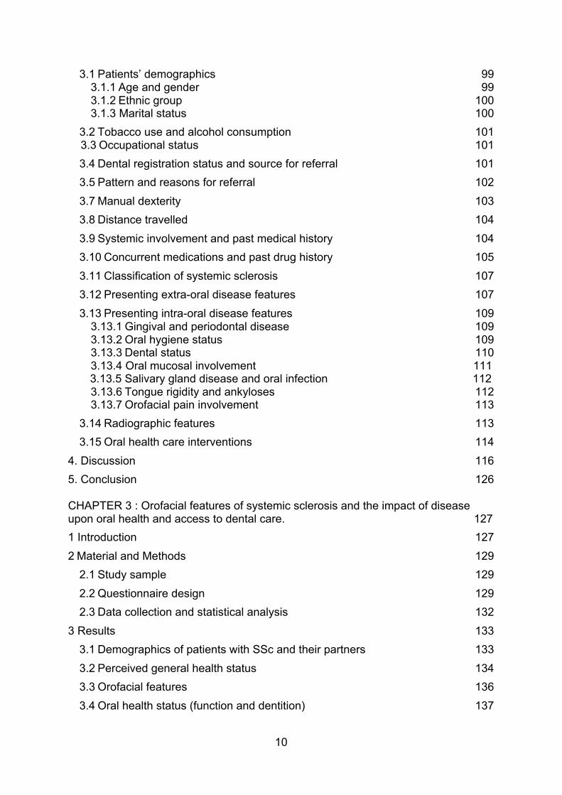
3.1 Patients’ demographics 99 3.1.1 Age and gender 99 3.1.2 Ethnic group 100 3.1.3 Marital status 100
3.2 Tobacco use and alcohol consumption 101 3.3 Occupational status 101
3.4 Dental registration status and source for referral 101 3.5 Pattern and reasons for referral 102 3.7 Manual dexterity 103 3.8 Distance travelled 104 3.9 Systemic involvement and past medical history 104 3.10 Concurrent medications and past drug history 105 3.11 Classification of systemic sclerosis 107 3.12 Presenting extra-oral disease features 107 3.13 Presenting intra-oral disease features 109
3.13.1 Gingival and periodontal disease 109 3.13.2 Oral hygiene status 109 3.13.3 Dental status 110
3.13.4 Oral mucosal involvement 111 3.13.5 Salivary gland disease and oral infection 112
3.13.6 Tongue rigidity and ankyloses 112 3.13.7 Orofacial pain involvement 113
3.14 Radiographic features 113 3.15 Oral health care interventions 114
4. Discussion 116 5. Conclusion 126 CHAPTER 3 : Orofacial features of systemic sclerosis and the impact of disease upon oral health and access to dental care. 127 1 Introduction 127 2 Material and Methods 129
2.1 Study sample 129 2.2 Questionnaire design 129 2.3 Data collection and statistical analysis 132
3 Results 133 3.1 Demographics of patients with SSc and their partners 133 3.2 Perceived general health status 134 3.3 Orofacial features 136 3.4 Oral health status (function and dentition) 137

11
3.5 Dental History, Attitude and pattern of dental attendance 138 3.6 Access to dental care 140 3.7 Oral Hygiene and behaviour 142 3.8 Additional comments provided by patients 143 3.9 Comparison to MORI study 2009 and ADHS 2009 144
4 Discussion 147 5 Conclusion 163 CHAPTER 4: Web-based information on the treatment of the mouth in systemic sclerosis 164 1 Introduction 164 2 Materials and Methods 166
2.1 Search 166 2.2 Quality assessment 166 2.3 Readability assessment 167 2.4 Statistical analyses 168
3 Results 169 3.1 Available websites 169 3.2 Quality assessment 172 3.3 Readability 173
4 Discussion 174 5 Conclusion 177
CHAPTER 5: Oral health-related quality of life and self-reported anxiety and depression in systemic sclerosis compared with the UK general population. 178 1 Introduction 178 2 Material and Methods 180
2.1 Study design and participants 180 2.2 Outcome measures 181 2.3 Data collection and statistical analysis 184
3 Results 184 3.1 Baseline characteristics of patients with SSc and controls 184 3.2 Oral health-related quality of life measures 185 3.3 General health-related quality of life measures 186 3.4 Anxiety and depression measures 187
4 Discussion 192 5 Conclusion 198
12
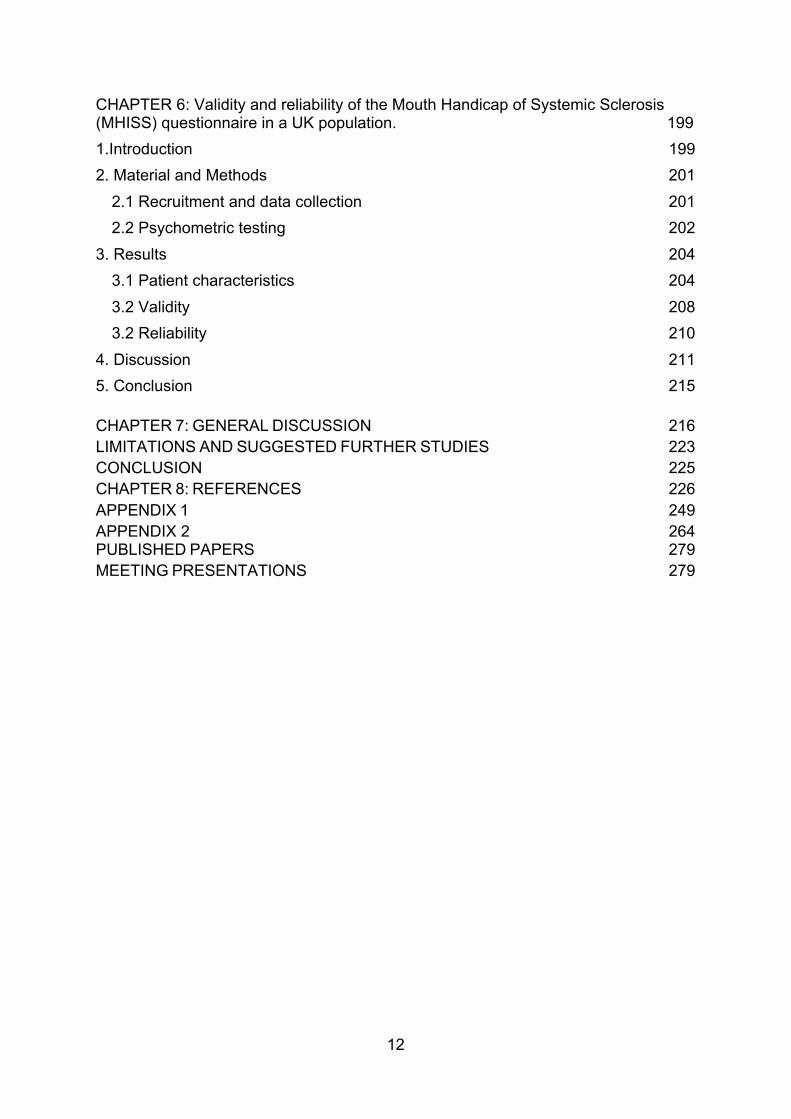
CHAPTER 6: Validity and reliability of the Mouth Handicap of Systemic Sclerosis (MHISS) questionnaire in a UK population. 199 1.Introduction 199 2. Material and Methods 201
2.1 Recruitment and data collection 201 2.2 Psychometric testing 202
3. Results 204 3.1 Patient characteristics 204 3.2 Validity 208 3.2 Reliability 210
4. Discussion 211 5. Conclusion 215 CHAPTER 7: GENERAL DISCUSSION 216 LIMITATIONS AND SUGGESTED FURTHER STUDIES 223 CONCLUSION 225 CHAPTER 8: REFERENCES 226 APPENDIX 1 249 APPENDIX 2 264 PUBLISHED PAPERS 279 MEETING PRESENTATIONS 279

13
LIST OF TABLES
Table 1.1 Incidence and prevalence according to regions…………………………….23 Table 1.2 ACR-EULAR 2013 classification criteria for systemic sclerosis…………..27 Table 1.3 Comparison of en coup de sabre and progressive hemifacial atrophy
(Parry-Romberg syndrome)………………………………………………………….30 Table 1.4 Description of the CREST Syndrome………………………………………..31 Table 1.5 The main clinical features of systemic sclerosis……………………………33 Table 1.6 A new proposed classification criteria for Juvenile systemic sclerosis…...40 Table 1.7 Preliminary proposed classification for juvenile localised systemic sclerosis
………………………………………………………………………………………….40 Table 1.8 Diagnostic features of systemic sclerosis-overlap syndrome……………..42 Table 1.9. Therapeutics strategies and treatment agents in the management of SSc
………………………………………………………………………………………….56 Table 1.10 Frequency and impact of oral symptoms experienced by patients with
SSc in five European countries .......................................................................... 59 Table 1.11 Impact on everyday activities of oral symptoms among patients with SSc
in five European countries ................................................................................. 59 Table 1.12 Radiographic oral findings of systemic sclerosis ..................................... 74 Table 1.13 The principals of the management of oral dryness ................................. 77 Table 1.14 Health-related quality of life assessment measures that have been used
in SSc ................................................................................................................. 86 Table 1.15 Mouth Handicap in Systemic Sclerosis scale .......................................... 88 Table 1.16 Reported impact of systemic sclerosis upon oral quality of life by using
MHISS scale ...................................................................................................... 94 Table 2.1 Age, gender and disease duration of 138 patients with SSc ..................... 99 Table 2.2 Ethnicity of patients with SSc 100 Table 2.3 Marital status of patients with SSc .......................................................... 100 Table 2.4 Tobacco use and alcohol consumption of patients with SSc .................. 101 Table 2.5 Employment status of patients with SSc ................................................. 101 Table 2.6 Dental registration status and source of referral for patients with SSc ... 102 Table 2.7 Clinical speciality for referral of patients with SSc ................................... 102 Table 2.8 Reasons for referral of patients with SSc to oral health care services .... 103 Table 2.9 Mobility difficulties of patients with SSc attending for oral health care .... 103 Table 2.10 Distance travelled to obtain dental care at the Eastman Dental Hospital
......................................................................................................................... 104 Table 2.11 Past medical history of patients with systemic sclerosis ....................... 105 Table 2.12 Past drug history of patients with systemic sclerosis ............................ 106 Table 2.13 Classification of patients with systemic sclerosis referred to UCLH
Eastman Dental Hospital ................................................................................. 107 Table 2.14 Extra-oral clinical findings at presentation of patients with SSc ............ 108 Table 2.15 Limited mouth opening and TMD at presentation of patients with SSc . 108 Table 2.16 Measurements of maximum mouth opening of 69 patients with SSc ... 109 Table 2.17 Gingival and periodontal features observed in patients with SSc ......... 109 Table 2.18 Oral hygiene status of patients with systemic sclerosis ........................ 110 Table 2.19 Decayed, mobility, missing and filled teeth of patients with systemic
sclerosis ........................................................................................................... 111 Table 2.20 Oral mucosal involvement of patients with systemic sclerosis .............. 111 Table 2.21 Salivary gland disease and oral infection of patients with systemic
sclerosis ........................................................................................................... 112
14
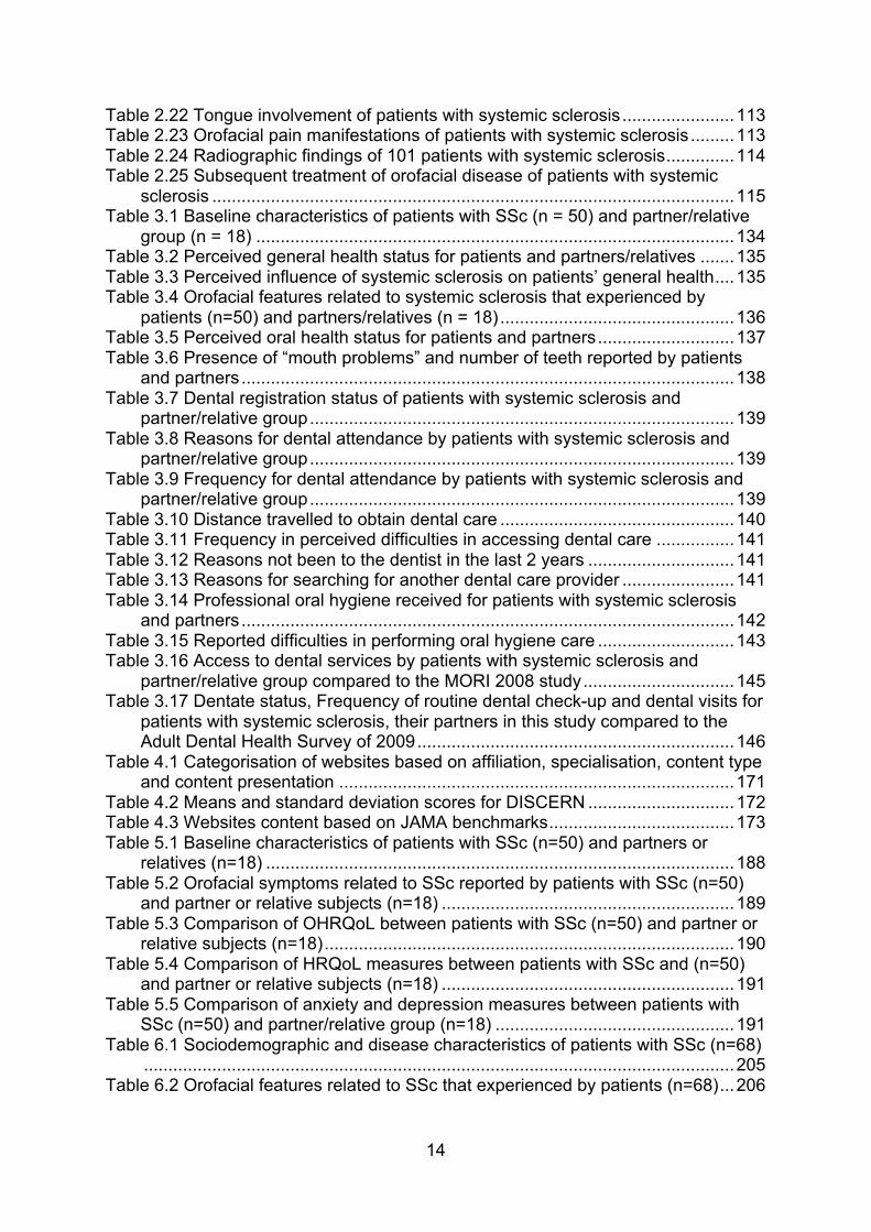
Table 2.22 Tongue involvement of patients with systemic sclerosis ....................... 113 Table 2.23 Orofacial pain manifestations of patients with systemic sclerosis ......... 113 Table 2.24 Radiographic findings of 101 patients with systemic sclerosis .............. 114 Table 2.25 Subsequent treatment of orofacial disease of patients with systemic
sclerosis ........................................................................................................... 115 Table 3.1 Baseline characteristics of patients with SSc (n = 50) and partner/relative
group (n = 18) .................................................................................................. 134 Table 3.2 Perceived general health status for patients and partners/relatives ....... 135 Table 3.3 Perceived influence of systemic sclerosis on patients’ general health .... 135 Table 3.4 Orofacial features related to systemic sclerosis that experienced by
patients (n=50) and partners/relatives (n = 18) ................................................ 136 Table 3.5 Perceived oral health status for patients and partners ............................ 137 Table 3.6 Presence of “mouth problems” and number of teeth reported by patients
and partners ..................................................................................................... 138 Table 3.7 Dental registration status of patients with systemic sclerosis and
partner/relative group ....................................................................................... 139 Table 3.8 Reasons for dental attendance by patients with systemic sclerosis and
partner/relative group ....................................................................................... 139 Table 3.9 Frequency for dental attendance by patients with systemic sclerosis and
partner/relative group ....................................................................................... 139 Table 3.10 Distance travelled to obtain dental care ................................................ 140 Table 3.11 Frequency in perceived difficulties in accessing dental care ................ 141 Table 3.12 Reasons not been to the dentist in the last 2 years .............................. 141 Table 3.13 Reasons for searching for another dental care provider ....................... 141 Table 3.14 Professional oral hygiene received for patients with systemic sclerosis
and partners ..................................................................................................... 142 Table 3.15 Reported difficulties in performing oral hygiene care ............................ 143 Table 3.16 Access to dental services by patients with systemic sclerosis and
partner/relative group compared to the MORI 2008 study ............................... 145 Table 3.17 Dentate status, Frequency of routine dental check-up and dental visits for
patients with systemic sclerosis, their partners in this study compared to the Adult Dental Health Survey of 2009 ................................................................. 146
Table 4.1 Categorisation of websites based on affiliation, specialisation, content type and content presentation ................................................................................. 171
Table 4.2 Means and standard deviation scores for DISCERN .............................. 172 Table 4.3 Websites content based on JAMA benchmarks ...................................... 173 Table 5.1 Baseline characteristics of patients with SSc (n=50) and partners or
relatives (n=18) ................................................................................................ 188 Table 5.2 Orofacial symptoms related to SSc reported by patients with SSc (n=50)
and partner or relative subjects (n=18) ............................................................ 189 Table 5.3 Comparison of OHRQoL between patients with SSc (n=50) and partner or
relative subjects (n=18) .................................................................................... 190 Table 5.4 Comparison of HRQoL measures between patients with SSc and (n=50)
and partner or relative subjects (n=18) ............................................................ 191 Table 5.5 Comparison of anxiety and depression measures between patients with
SSc (n=50) and partner/relative group (n=18) ................................................. 191 Table 6.1 Sociodemographic and disease characteristics of patients with SSc (n=68)
......................................................................................................................... 205 Table 6.2 Orofacial features related to SSc that experienced by patients (n=68) ... 206

15
Table 6.3 Descriptive statistics for the SF-36, OHIP-14 and MHISS scales of patients with SSc ........................................................................................................... 207
Table 6.4 Matrix of Spearman's rho coefficients between pairs of responses (n=68) to the MHISS and SF-36…………………………………………………………………209 Table 6.5 Matrix of Spearman’s rho coefficients between pairs of responses to the MHISS and OHIP-14 (n=68)…………………………………………………………….209 Table 6.6 Internal consistency reliability of the MHISS scales………………………210

16
LIST OF FIGURES
Figure 4.1 Number of excluded websites 170
Figure 4.2 Flowchart of the sample selection strategy 170
Figure 4.3 Flesch Reading Ease Score 173

17
ABBREVIATIONS
ACA – Anti-centromere Antibody ACE - Angiotensin-converting enzyme ACR - American College of Rheumatology ADHS – Adult dental health survey ADSC - Adipose-derived stromal cell AFGT - Autologous fat grafting ANA – Anti-nuclear Antibodies ATA – Anti-topoisomerase Antibody BMS – Burning mouth syndrome CBCT – Cone-beam computerised tomography CNS – Central nervous system CREST- Calcinosis, Raynaud’s phenomenon, Oesophageal dysmotility, Sclerodactyly and Telangiectasia CRISS - Composite Response Index in SSc CTD – Connective Tissue Disease DcSSc – Diffuse cutaneous systemic sclerosis EDH – Eastman Dental Hospital EDS - Ehlers-Danlos Syndrome ENT – Ear, Nose and Throat ESR- Erythrocyte sedimentation rate EULAR - European League Against Rheumatism FDA - Food and Drug Administration FRES - Flesch Reading Ease Scores GDP – General Dental Practitioner GIT – Gastro-intestinal tract GORD – Gastroesophageal reflux disease HADS - Hospital Anxiety and Depression scale HAMIS - Hand Mobility in SSc HAQ – Health assessment questionnaire HON – Health on the Net HRQoL – Health-Related Quality of life ILD – Interstitial lung disease JAMA - Journal of the American Medical Association jLSSc - Juvenile localised systemic sclerosis jSSc - Juvenile systemic sclerosis lcSSc – Limited cutaneous systemic sclerosis lSSc – Localised systemic sclerosis MCTD – Mixed connective tissue disease MDAS – Modified depression and anxiety scale MHC - Major histocompatibility complex MHISS – Mouth handicap in systemic sclerosis MRONJ - Medication-related osteonecrosis of the jaw

18
NHS - National Health Service NSAID – Non-steroidal anti-inflammatory drugs OA – Osteoarthritis OHIP – Oral health impact profile OHRQoL – Oral health-related quality of life OIDP - Oral Impact on Daily Performance OMFS – Oral maxillofacial surgery ONS - Office for National Statistics PAH – Pulmonary arterial hypertension PDL – Periodontal ligament PROMS - Patient-reported outcome measures QoL – Quality of life RA - Rheumatoid arthritis RP- Raynaud’s phenomenon SCTC - Scleroderma Clinical Trials Consortium SF-36 – Short form health questionnaire SLE - Systemic lupus erythematosus SS - Sjogren’s syndrome SSA - Skin Symptom Assessment SSc – Systemic Sclerosis SWAP - satisfaction with appearance scale TMD – Temporomandibular disorder TMJ - Temporomandibular joint TN - Trigeminal neuralgia UCLA SCTC GIT - Scleroderma Gastrointestinal Tract Scale UCLHT – University College London Hospital NHS Trust VAS – Visual analogue scale VEDOSS - Very Early Diagnoses clinic for Systemic Sclerosis WHO – World Health Organisation

19
CHAPTER 1: Introduction and literature review
Immunologically mediated connective tissue disorders (CTDs) comprise a group of
disorders that give rise to a wide variety of systemic and/or oral manifestations that
compromise healthy living. Scleroderma or “Systemic Sclerosis” is one of the rare
CTDs that can adversely impact upon multiple organs and in some instances can be
life-threatening. SSc can adversely impact upon a patient’s quality of life and daily
activity.
There remains no definitive cure for SSc although a wide range of different treatment
modalities is helpful in lessening or stopping disease progression. Nevertheless, many
affected individuals will have lifelong complications that may adversely impact
physically and/or psychologically upon their lives and perhaps their families and/or
carers.
1.1 History of systemic sclerosis
The term scleroderma is derived from classical Greek terminology: Skleros meaning
hard and derma meaning skin. Historically, in 450-370 BC Hippocrates described a
disorder of widespread hardening of the skin (David, 1981). In 1754, Robert Westson
translated into English the first convincing description of SSc (which was written by
Carlo Curzio in 1753) concerning a female patient complaining of excessive
generalised tension and hardness of skin that limited her movement. Curzio had
described hardening and tightness of the skin without any involvement of the
underlying muscles with constriction and tightness of the skin of the head and neck
that limited mouth opening and neck movements (Rocco and Hurd, 1986).
The term “scleroderma” was used for the first time by Giovambattista Fantonetti when
describing a patient who had thickened and dark skin lesions with loss of normal joint
mobility (Benedek and Rodnan, 1982). In 1837 eight patients with skin disease were

20
diagnosed as having SSc and in 1865 a comprehensive review by Horteloup described
several patients (Benedek and Rodnan, 1982).
The association between SSc and calcinosis was first reported by Webar in 1878. In
1865, the association of SSc and peripheral vasoconstriction was first described by
Maurice Raynaud. The changes of peripheral vasoconstriction were further described
by Ball in 1871 who detailed marked sclerosis, atrophy of the fingers and painful ulcers
which was later termed as “Sclerodactylie” (Benedek and Rodnan, 1982).
Between 1892 and 1898, Wolters and Osler concluded that SSc was a multisystem
condition that could affect different organs leading to a wide variety of complications
with high mortality and morbidity (Benedek and Rodnan, 1982). Multi-organ
involvement in systemic sclerosis was described by Matsui in 1924 while in 1942
Klemperer, Pollack and Baehr emphasised that SSc should be categorised as a
systemic connective tissue disorder. Three years later, Goetz considered that SSc is
a cutaneous phase of an underlying systemic disorder and introduce the term
“Progressive systemic sclerosis” (Klemperer et al., 1984).
CREST syndrome, which was defined by Winterbaure in 1964 as Calcinosis as
calcium deposition, Raynaud’s phenomenon, Oesophageal dysfunction, Sclerodactyly
as thickening and hardening skin and Telangiectasia (Winterbauer, 1964). Since this
time, as medical advances have been made, so the description of SSc has extended
and is considered in detail in the classification section.
21
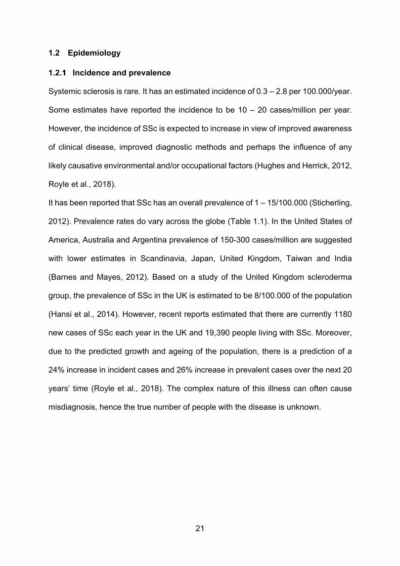
1.2 Epidemiology
Incidence and prevalence
Systemic sclerosis is rare. It has an estimated incidence of 0.3 – 2.8 per 100.000/year.
Some estimates have reported the incidence to be 10 – 20 cases/million per year.
However, the incidence of SSc is expected to increase in view of improved awareness
of clinical disease, improved diagnostic methods and perhaps the influence of any
likely causative environmental and/or occupational factors (Hughes and Herrick, 2012,
Royle et al., 2018).
It has been reported that SSc has an overall prevalence of 1 – 15/100.000 (Sticherling,
2012). Prevalence rates do vary across the globe (Table 1.1). In the United States of
America, Australia and Argentina prevalence of 150-300 cases/million are suggested
with lower estimates in Scandinavia, Japan, United Kingdom, Taiwan and India
(Barnes and Mayes, 2012). Based on a study of the United Kingdom scleroderma
group, the prevalence of SSc in the UK is estimated to be 8/100.000 of the population
(Hansi et al., 2014). However, recent reports estimated that there are currently 1180
new cases of SSc each year in the UK and 19,390 people living with SSc. Moreover,
due to the predicted growth and ageing of the population, there is a prediction of a
24% increase in incident cases and 26% increase in prevalent cases over the next 20
years’ time (Royle et al., 2018). The complex nature of this illness can often cause
misdiagnosis, hence the true number of people with the disease is unknown.

22
Age
Systemic sclerosis tends to manifest in the middle to late life with the peak age of
onset between 30-50 years of age. However, the disease can arise in the second or
third decades of life. Patient ethnic background may influence the risk of SSc within
individuals. African-American ethnic background developing the disease at an earlier
age than whites (Ranque and Mouthon, 2010, Gelber et al., 2013, Hansi et al., 2014).
Gender
Systemic sclerosis has a female predominance being 3-8 times more common in
females than males. In addition, females are more likely to develop SSc in early life
compared with males (Hughes and Herrick, 2012). Male patients may have a greater
risk of cancer development than female patients this reflecting the aetiology related to
tobacco smoking, alcohol and immune and haematological malignancies (Olesen et
al., 2010).

23
Table 1.1 Incidence and Prevalence according to regions
Region Study period
Incidence per million
Prevalence per million
Publication
USA Minnesota 1947 - 68 2.7 - (Medsger and Masi, 1971) Minnesota 1953 - 67 1.2 105 (Kurland et al., 1969) Minnesota 1950 - 79 10 138 (Michet et al., 1985)
USA 1963 - 68 2.3 19.8 (Medsger and Masi, 1978) Pennsylvania 1963 - 82 13.9 - 18.7 - (Steen et al., 1997) South carolina 1989 - 290 - 1130 (Maricq et al., 1989)
Michigan 1985 - 91 14.1 - (Laing et al., 1997) Michigan 1989 - 91 21 276 (Mayes et al., 2003) Oklahoma 1996 - 658.6 (Arnett et al., 1996) All states 2001 - 02 - 300 (Robinson et al., 2008) Quebec 2003 - 443 (Bernatsky et al., 2009)
Argentine Buenos Aires 1999 -
2004 21.2 296 (Rosa et al., 2011)
Australia Southern Half 1950 - 73 1.96 - (Wigley and Borman, 1980) New Zealand 1970 - 79 6.3 30 (Eason et al., 1981)
Sydney 1974 - 88 12 45.2 - 86.2 (Englert et al., 1999) South Australia 1987 - 93 - 208 (Chandran et al., 1995) South Australia 1993 - 99 15.1 - 22.8 200 - 233 (Roberts-Thomson et al., 2001) South Australia 1993 - 02 20.4 232.2 (Roberts-Thomson et al., 2006)
Japan Tokyo 1987 7.2 38 - 53 (Tamaki et al., 1991)
Taiwan Taiwan 2002 -
2007 10.9 56 (Kuo et al., 2011)
India North India 2006 -
2007 - 120 (Minz et al., 2012)
UK & Europe England
(West Midlands) 1986 3.7 31
(Silman et al., 1988) England
(Newcastle) 2000 - 88 (Allcock et al., 2004)
Hungary (South West)
2001 - 910 - 2370 (Czirjak et al., 2005)
Estonia (South) 1996 - 97 - 350 - 2280 (Valter et al., 1997) Iceland 1975 - 90 3.8 71 (Geirsson et al., 1994) Finland 1990 3.7 - (Kaipiainen-Seppanen and Aho,
1996) France (Seine St
Denis) 2001 - 15.8 (Le Guern et al., 2004)
Greece (Northwest)
1981 - 02 11 154 (Alamanos et al., 2005)
Spain (Northwest) 1988 - 06 23 277 (Arias-Nunez et al., 2008) Italy (Northeast) 1991 -
2007 43 341 (Lo Monaco et al., 2011)
Adapted from (Ranque and Mouthon, 2010, Barnes and Mayes, 2012)

24
Ethnicity
Systemic sclerosis arises in all ethnic groups and seems to be more common in certain
ethnic groups. In the United States, SSc is more common in African Americans than
American Whites. African American develop disease at an earlier age than Whites and
have an increased frequency of diffuse cutaneous disease and poorer prognosis
(Gelber et al., 2013). The difference between black and white ethnic groups in the
United States may involve multiple predisposing factors such as socioeconomic status
and health care access, and recently it has been suggested that genetic factors might
explain more the racial disparities such as the overexpression of profibrotic factors
and the under-expression of antifibrotic factors (Nashid et al., 2011, Silver et al., 2012).
Survival and mortality
Mortality varies widely among different patient populations and their organ
involvement, and it is increased in male patients (Nikpour and Baron, 2014). The five-
year survival rate is estimated to be 85%, and the 10-year survival rate is under 70%
(Sticherling, 2012). Similar results have been reported in a systematic review and
meta-analysis that included 17 studies and 9239 patients and revealed that the main
cause of death is related to the pulmonary involvement of the disease (Rubio-Rivas et
al., 2014). In a comprehensive study of 700 patients, African-Americans had a 43 –
60% increased risk of mortality in comparison to White Americans (Gelber et al.,
2013). Recent studies have reported that patients with SSc have survival rates 16 –
34 years less than appropriate gender and age-matched populations due presumably
to cardiopulmonary involvement and renal impairment (Nikpour and Baron, 2014,
Rubio-Rivas et al., 2014).
25
1.3 Classification Criteria
Classification criteria for SSc were not developed until 1980 when the American
Rheumatism Association (now known as the American College of Rheumatology
(ACR)) developed specific criteria to differentiate systemic sclerosis disease from
other connective tissue diseases. This included four items (SSc proximal to the
metacarpophalangeal joints, sclerodactyly, digital pitting scars and bilateral basilar
pulmonary fibrosis). This classification was judged to have been low sensitivity and
specificity in the identification of early or mild disease. The further classification was
developed in 1988, which divided the disease into two main categories (localised and
diffuse). Localised was characterised by localised skin fibrosis limited to the distal
aspects of the fingers and face without systemic involvement while diffuse disease
type was generalised skin involvement with rapid progressive internal organ
involvement (van den Hoogen et al., 2013).
In 2001, LeRoy and Medsger developed new criteria for early SSc with some new
elements of the presence of relevant autoantibodies, Raynaud’s phenomenon, skin
fibrosis and nail fold capillaroscopy. This modification gave a high score of sensitivity
(Ranque and Mouthon, 2010, Pope, 2015).
In 2013, a classification system was developed principally to aid research but also to
help clinical practice and diagnosis (Table 1.2). The new American College of
Rheumatology (ACR) and the European League Against Rheumatism (EULAR)
criteria considered three main features:
1- Fibrosis of the skin and/or internal organs.
2- Production of specific autoantibodies.
3- Presence of vasculopathy.

26
In the new diagnosis of SSc it is likely if the patient has skin thickening of the fingers
that extends proximally to the metacarpophalangeal joints. Seven factors including
skin thickening of the fingers, fingertip lesions, telangiectasia, abnormal nailfold
capillaries, interstitial lung disease and/or pulmonary arterial hypertension, Raynaud’s
phenomenon and SSc-related autoantibodies are considered if the cutaneous features
are not present. Each of these factors has a certain value, but overall a score of 9
points or more is considered to be a definitive diagnosis of SSc (Table 1.2) (van den
Hoogen et al., 2013).
The sensitivity and specificity of the new classification criteria have been assessed
and have excellent results achieving 91%, 92% respectively across different
populations. However, the ACR/EULAR criteria should not be misinterpreted as a
diagnostic tool as some recent reports indicate that there are patients for example who
have Raynaud’s phenomenon, positive SSc-related antibodies and nailfold
capillaroscopy but do not fulfil the new criteria (Pope, 2015, Pope and Johnson, 2015).
Despite the overall acceptance of this classification system further studies of different
populations may help to allow the determination of the possible power of the criteria
to predict disease progression and overall prognosis (Jordan et al., 2015, Pope and
Johnson, 2015).
27
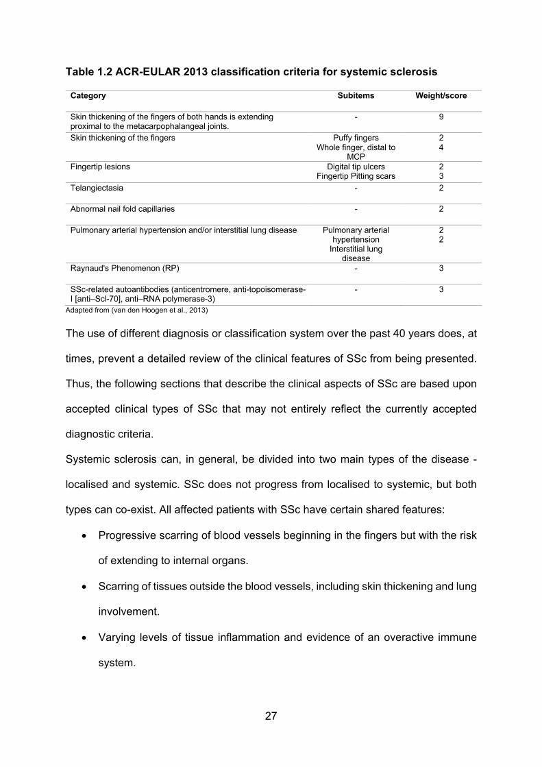
Table 1.2 ACR-EULAR 2013 classification criteria for systemic sclerosis
Category Subitems Weight/score
Skin thickening of the fingers of both hands is extending proximal to the metacarpophalangeal joints.
- 9
Skin thickening of the fingers Puffy fingers Whole finger, distal to
MCP
2 4
Fingertip lesions Digital tip ulcers Fingertip Pitting scars
2 3
Telangiectasia - 2
Abnormal nail fold capillaries - 2
Pulmonary arterial hypertension and/or interstitial lung disease Pulmonary arterial hypertension
Interstitial lung disease
2 2
Raynaud's Phenomenon (RP) - 3
SSc-related autoantibodies (anticentromere, anti-topoisomerase-I [anti–Scl-70], anti–RNA polymerase-3)
- 3
Adapted from (van den Hoogen et al., 2013) The use of different diagnosis or classification system over the past 40 years does, at
times, prevent a detailed review of the clinical features of SSc from being presented.
Thus, the following sections that describe the clinical aspects of SSc are based upon
accepted clinical types of SSc that may not entirely reflect the currently accepted
diagnostic criteria.
Systemic sclerosis can, in general, be divided into two main types of the disease -
localised and systemic. SSc does not progress from localised to systemic, but both
types can co-exist. All affected patients with SSc have certain shared features:
• Progressive scarring of blood vessels beginning in the fingers but with the risk
of extending to internal organs.
• Scarring of tissues outside the blood vessels, including skin thickening and lung
involvement.
• Varying levels of tissue inflammation and evidence of an overactive immune
system.
28
Localised form of the disease consists of two main subtypes: morphea and linear SSc.
Systemic disease includes three subclasses: limited cutaneous (lcSSc); that
previously referred to CREST syndrome, diffuse cutaneous (dcSSc) and sine SSc.
Localised systemic sclerosis
About 70% of all patients with SSc have been diagnosed with localised disease.
Localised SSc has two main subtypes (morphea and linear SSc) and rarely progresses
into systemic disease as it commonly affects the skin and subcutaneous tissues. About
10% of patients may have muscle spasm, deformities or disfiguration due to excessive
scar formation (Lachner, 2016).
Morphea SSc is a rare form of SSc that is sometimes referred to as circumscribed
SSc. It can develop during childhood commencing as violaceous skin patches of
variable size that may then progress to form fibrotic coloured skin plaques that later
evolve from a sclerotic stage to non-indurated lesions that may appear as hypo- or
hyper-pigmentation and may extend to the underlying cutaneous layers and manifest
as a depressed area below the skin level. Morphea can be differentiated from systemic
sclerosis by the absence of sclerodactyly, Raynaud’s phenomenon and nailfold
capillary changes. Morphea SSc can be limited to the hands and called acrosclerosis
however patients with morphea can present some systemic symptoms such as
malaise, fatigue, arthralgia and myalgia accompanied with a generalised form of
morphea (Bali et al., 2013).
Linear SSc is more likely to manifests in children than adults and generally manifests
as a band-like thickening that usually affects one side of the body and can extend to
the underlying skin tissue and may extend to the muscles and bones. Linear SSc in
some severe circumstances can give rise to arthritis, growth impairment and
contractures of the associated joints causing significant deformity (Lachner, 2016).
29
Linear SSc has two subtypes (En coup de sabre and Parry-Romberg syndrome). En
coup de sabre commonly affects the head and neck area resembling the stroke of a
sabre and causing hyper-pigmented skin lesions and also in severe instances can give
rise to fibrosis of the skin, subcutaneous tissues, muscles and extend to bones leading
to a sword thrust shape. The lesion is typically located paramedial on the forehead
and runs to the hairline, but it can occur on the chin and can extend intraorally. A linear
depression area or groove of the skin can be apparent and can cause loss of hair
(alopecia) if present on the scalp or eyelids and craniofacial development can be
affected in severe cases and cause hemifacial atrophy (Horberg et al., 2015,
Tolkachjov et al., 2015). In occasional patients, the defect can extend to the
peridentine and has been reported to cause root abnormalities of teeth in affected
areas (de Figueiredo et al., 2008, Horberg et al., 2015, Arroyo-Bote et al., 2017).
Progressive hemifacial atrophy or Parry-Romberg syndrome is a very rare orofacial
form of the disease characterised by involvement of both muscles and bone rather
than superficial skin layers. It can affect the body in a symmetrical manner, although
cutaneous involvement can occur, and lesions can extend to tongue, gingiva, teeth
and palate. Neurological complications such as seizures, headaches and trigeminal
neuralgia after cranial nerve neuropathies may be features. Of concern, ocular
disorders can result in a mild impairment to permanent blindness due to both muscular
and neural pathological involvement. Patients with Parry-Romberg syndrome can
have dental abnormalities such as delayed eruption, dental root exposure and/or
resorption, temporomandibular joint disorder, trismus and muscle spasm and atrophy
of the affected soft tissues such as the lips and tongue (Horberg et al., 2015,
Tolkachjov et al., 2015, Distler and Cozzio, 2016). The severity and pattern of the
disease vary between patients.
30
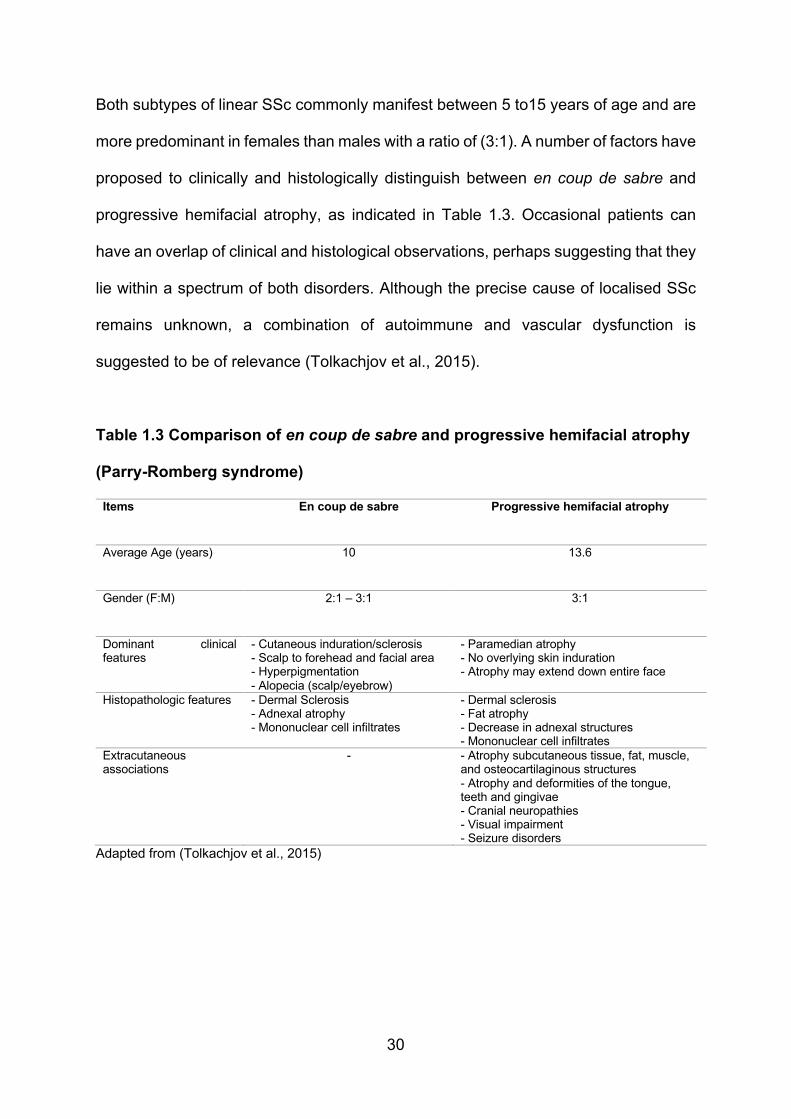
Both subtypes of linear SSc commonly manifest between 5 to15 years of age and are
more predominant in females than males with a ratio of (3:1). A number of factors have
proposed to clinically and histologically distinguish between en coup de sabre and
progressive hemifacial atrophy, as indicated in Table 1.3. Occasional patients can
have an overlap of clinical and histological observations, perhaps suggesting that they
lie within a spectrum of both disorders. Although the precise cause of localised SSc
remains unknown, a combination of autoimmune and vascular dysfunction is
suggested to be of relevance (Tolkachjov et al., 2015).
Table 1.3 Comparison of en coup de sabre and progressive hemifacial atrophy
(Parry-Romberg syndrome)
Items En coup de sabre Progressive hemifacial atrophy
Average Age (years) 10 13.6
Gender (F:M) 2:1 – 3:1 3:1
Dominant clinical features
- Cutaneous induration/sclerosis - Scalp to forehead and facial area - Hyperpigmentation - Alopecia (scalp/eyebrow)
- Paramedian atrophy - No overlying skin induration - Atrophy may extend down entire face
Histopathologic features - Dermal Sclerosis - Adnexal atrophy - Mononuclear cell infiltrates
- Dermal sclerosis - Fat atrophy - Decrease in adnexal structures - Mononuclear cell infiltrates
Extracutaneous associations
- - Atrophy subcutaneous tissue, fat, muscle, and osteocartilaginous structures - Atrophy and deformities of the tongue, teeth and gingivae - Cranial neuropathies - Visual impairment - Seizure disorders
Adapted from (Tolkachjov et al., 2015)

31
Systemic sclerosis
In contrast to localised SSc - where the affected tissues are predominantly cutaneous
without the involvement of blood vessels or viscera - systemic sclerosis comprises
cutaneous and vascular disease with the likelihood of visceral involvement. Systemic
sclerosis can have life-threatening complications affecting the gastrointestinal,
pulmonary, cardiovascular and renal systems. Systemic sclerosis can be divided into
two main groups (limited and diffuse cutaneous SSc).
Limited cutaneous systemic sclerosis is characterised by low-grade skin involvement
that is commonly limited to extremities such as the hands, forearms and feet with the
possibility of head and neck involvement. It is the most common type of systemic
sclerosis, accounting for around 60% of patients. 90 - 95% of patients with limited
cutaneous SSc have Raynaud’s phenomenon for a long time before the development
of other manifestations of the disease (Lachner, 2016).
As noted earlier, limited cutaneous SSc was previously termed CREST as a common
manifestation was this combination of cutaneous Calcinosis, Raynaud’s phenomenon,
Oesophageal dysfunction, Sclerodactyly and Telangiectasia as denoted in Table 1.4.
Table 1.4 Description of the CREST Syndrome
Calcinosis Raynaud’s Phenomenon
Oesophageal dysfunction
Sclerodactyly Telangiectasia
Accumulation of calcium salts under the skin and causes hard painful raised areas that can open and cause ulcerations of the skin. Typical sites of deposition are mainly on elbows, knees and fingers.
Vasoconstriction with intermittent loss of blood flow to fingers, toes and nose that occurs with exposure to cold and stress and causes numbness, tingling and pain. Disease progression can lead to gangrenous lesion of tissue and erosion of the terminal extremities
Weakened lower oesophagus causing reflux and swallowing difficulties. This commonly leads to heartburn, inflammation and scarring of oesophageal tissues.
Excess collagen in the skin layers causing thickening, tighten and shiny appearance and difficulty moving fingers/toes. In severe cases there is notable contraction of the fingers and toes.
Dilation of capillaries causing small blanching red spots on hands, face, chest and mouth. While harmless they may cause disfigurement and social embarrassment.
Adapted from (Lachner, 2016)
32
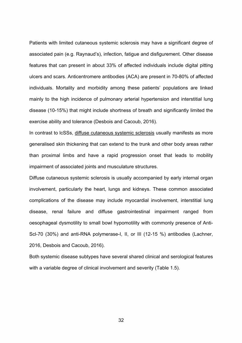
Patients with limited cutaneous systemic sclerosis may have a significant degree of
associated pain (e.g. Raynaud’s), infection, fatigue and disfigurement. Other disease
features that can present in about 33% of affected individuals include digital pitting
ulcers and scars. Anticentromere antibodies (ACA) are present in 70-80% of affected
individuals. Mortality and morbidity among these patients’ populations are linked
mainly to the high incidence of pulmonary arterial hypertension and interstitial lung
disease (10-15%) that might include shortness of breath and significantly limited the
exercise ability and tolerance (Desbois and Cacoub, 2016).
In contrast to lcSSs, diffuse cutaneous systemic sclerosis usually manifests as more
generalised skin thickening that can extend to the trunk and other body areas rather
than proximal limbs and have a rapid progression onset that leads to mobility
impairment of associated joints and musculature structures.
Diffuse cutaneous systemic sclerosis is usually accompanied by early internal organ
involvement, particularly the heart, lungs and kidneys. These common associated
complications of the disease may include myocardial involvement, interstitial lung
disease, renal failure and diffuse gastrointestinal impairment ranged from
oesophageal dysmotility to small bowl hypomotility with commonly presence of Anti-
Scl-70 (30%) and anti-RNA polymerase-I, II, or III (12-15 %) antibodies (Lachner,
2016, Desbois and Cacoub, 2016).
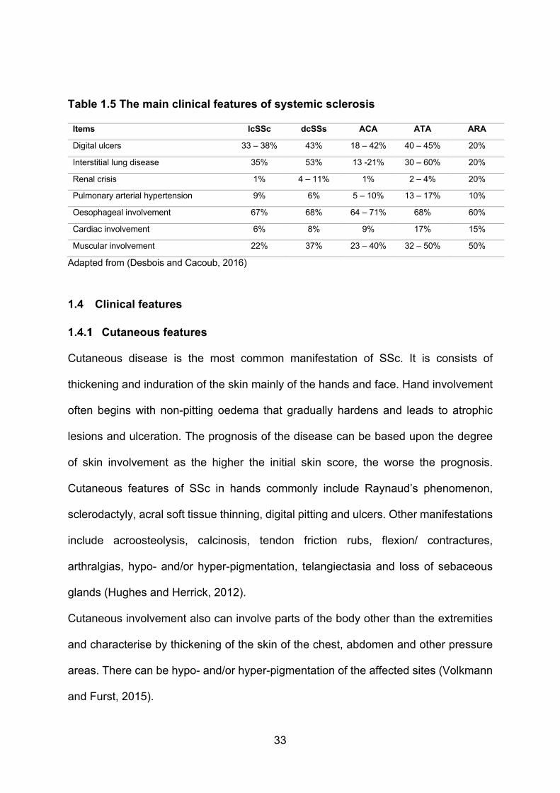
Both systemic disease subtypes have several shared clinical and serological features
with a variable degree of clinical involvement and severity (Table 1.5).
33
Table 1.5 The main clinical features of systemic sclerosis
Items lcSSc dcSSs ACA ATA ARA
Digital ulcers 33 – 38% 43% 18 – 42% 40 – 45% 20%
Interstitial lung disease 35% 53% 13 -21% 30 – 60% 20%
Renal crisis 1% 4 – 11% 1% 2 – 4% 20%
Pulmonary arterial hypertension 9% 6% 5 – 10% 13 – 17% 10%
Oesophageal involvement 67% 68% 64 – 71% 68% 60%
Cardiac involvement 6% 8% 9% 17% 15%
Muscular involvement 22% 37% 23 – 40% 32 – 50% 50%
Adapted from (Desbois and Cacoub, 2016)
1.4 Clinical features
Cutaneous features
Cutaneous disease is the most common manifestation of SSc. It is consists of
thickening and induration of the skin mainly of the hands and face. Hand involvement
often begins with non-pitting oedema that gradually hardens and leads to atrophic
lesions and ulceration. The prognosis of the disease can be based upon the degree
of skin involvement as the higher the initial skin score, the worse the prognosis.
Cutaneous features of SSc in hands commonly include Raynaud’s phenomenon,
sclerodactyly, acral soft tissue thinning, digital pitting and ulcers. Other manifestations
include acroosteolysis, calcinosis, tendon friction rubs, flexion/ contractures,
arthralgias, hypo- and/or hyper-pigmentation, telangiectasia and loss of sebaceous
glands (Hughes and Herrick, 2012).
Cutaneous involvement also can involve parts of the body other than the extremities
and characterise by thickening of the skin of the chest, abdomen and other pressure
areas. There can be hypo- and/or hyper-pigmentation of the affected sites (Volkmann
and Furst, 2015).

34
Vascular features
Raynaud’s phenomenon (RP) is the earliest and most common disease feature in 95%
of patients with SSc. This is bilateral and can also affect feet, nose, ears and, very
rarely, the tongue. It consists of white (ischaemia), blue (deoxygenation) and painfully
red decolouration (reperfusion) as a result of vascular spasm and ischemia (Desbois
and Cacoub, 2016). In most cases, the presence of RP along with both nailfold
capillary changes and SSc-specific antibodies indicating a high probability of
developing SSc with the association of irreversible tissue injury, ulceration and critical
ischaemia. In addition to ischaemia, patients may have paraesthesia, discomfort and
pain during episodes (McCray and Mayes, 2015).
The presence of RP may differ across patients with SSc. In those with limited SSc it
typically begins many years before the first onset of cutaneous disease, while in
patients with diffuse SSc, the RP usually occurs in parallel with, or even after, the
onset of the other cutaneous events (Elhai et al., 2015).
1.4.3 Gastrointestinal features
The gastrointestinal tract is the second most common site of involvement, occurring in
75% - 90% of patients with SSc (Elhai et al., 2015). It is one of the leading causes of
morbidity in SSc and the third most common cause of mortality after cardiopulmonary
and renal involvement (Hansi et al., 2014). All parts of the gastrointestinal tract can be
affected although gastro-oesophageal involvement is most common with SSc-related
disease accounting for 90% of the symptoms of heartburn, regurgitation and
dysphagia in SSc (Hansi et al., 2014). Other gastrointestinal manifestations include
poor appetite, motility abnormalities (e.g. diarrhoea, constipation, bloating, actual and
pseudo-obstruction of the bowel due to poor peristalsis). Severe gastro-oesophageal
reflux can lead to oesophageal stricture formation and narrowing, Barrett’s

35
oesophagus and risk of adenocarcinoma (McCray and Mayes, 2015). 50% of patients
with GIT involvement of SSc have gastroparesis. This delayed gastric emptying can
cause early satiety, bloating, nausea, vomiting, weight loss and subsequent
undernourishment. Other gastrointestinal disease features include rectal prolapse,
tenesmus, and pain during defecation, spontaneous colonic perforation and colonic
infarction (Barsotti et al., 2014, Gyger and Baron, 2015).
Cardiopulmonary features
Pulmonary involvement can affect 70 - 80% of all patients with SSc. Interstitial lung
disease and pulmonary arterial hypertension (PAH) are the most frequent features of
lung involvement (Lachner, 2016). Interstitial lung disease is considered as one of the
major causes of death in SSc patients while PAH is considered as one of the leading
causes of death in SSc patients, particularly in a late stage of the disease and when
associated with interstitial lung disease. The prevalence of PAH in all patients with
SSc is about 9-12% (Elhai et al., 2015).
Cardiac involvement in SSc carries a poor prognosis and is the leading cause of death.
Both the pericardium and myocardium can be affected. A wide spectrum of pericardial
diseases occurs in SSc including acute pericarditis, chronic pericarditis, pericardial
fibrosis and constrictive pericarditis (Bissell et al., 2016).
Myocardial involvement is commonly associated with vasospasm of the small vessels
and ischaemia of the coronary microcirculation that may lead to myocardial fibrosis.
Systolic and diastolic ventricular dysfunction can occur as a result of myocardial
involvement and can lead to post-capillary pulmonary hypertension. Recent reports
have found that pulmonary involvement together with factors including age, male
gender and myositis are independent risk factors for acute myocardial infarction in

36
patients with SSc. In addition, immunosuppressive therapy does not lessen the risk of
myocardial infarction (Barsotti et al., 2014).
Renal features
Renal involvement in SSc is relatively rare occurring in about 4-6% of patients.
Nevertheless, the severe manifestation can occur even in early disease. Patients with
renal involvement can have a renal crisis, normotensive renal crisis, anti-neutrophil
cytoplasmic antibody-associated glomerulonephritis, penicillamine-associated renal
disease and reduced renal function. The renal crisis is the most significant renal
complication with a prevalence of 5% in diffuse subtype and 2% in limited-SSc. Renal
crisis commonly presents as severe hypertension 90%, hypertensive encephalopathy
or cardiac failure, rapid deterioration of renal function and thrombotic microangiopathy
40%. Although the prognosis of renal involvement has been improved by using the
angiotensin-converting-enzyme inhibitors as the first line of treatment for the renal
crisis, the mortality rate remains high with overall five-year survival rates being only
30-50% (Elhai et al., 2015).
Neurological features
Various neurological disorders can arise in SSc. Central nervous system (CNS)
involvement includes headache 23.7%, seizures 13.5%, cognitive impairment 8.4%,
depression 73% and anxiety 23.9%. While peripheral nervous system (PNS)
involvement can manifests as myopathy 51.8%, trigeminal neuropathy 16.5%,
peripheral sensorimotor polyneuropathy 14.2% and carpal tunnel syndrome 6.5%
(Amaral et al., 2013). Although the cause of neurological involvement is not fully
defined, different possible theories are suggested including vascular-dependant
mechanisms due to vasculitis and/or vessel wall damage, compression-dependent
mechanisms due to oedema and/or fibrosis then progressive demyelination and
37
autoimmune-dependent mechanisms associated with anti-neuronal antibodies
(Amaral et al., 2013, Ludwig et al., 2017).
1.5 Association of malignancy
The precise association of SSc with the risk of cancer remains undetermined.
However, recent studies report that the risk of malignancy is increased in SSc. Several
reports have demonstrated an increased incidence of cancer lesions in SSc
populations with a concern about the long-term morbidities of the disease especially
among older patients with the diffuse cutaneous SSc (Barnes and Mayes, 2012,
Zeineddine et al., 2016). The reported standardised incidence ratio (i.e. the ratio of
observed to expected instances) for cancer among men was higher than women 2.2
(95% CI 1.7-2.8), 1.3 (95% CI 1.1-1.6) respectively. However, the most frequent
malignancies in SSc were found to be either smoking or alcohol-related cancers. Other
studies reported the relative risk of overall cancer among SSc patients to be 3.15 (95%
CI 1.77-5.20) with highest for haematologic and lung malignancies.
The cancer risk in some patients with systemic sclerosis may be associated with the
presence of anti-RNA polymerase III antibodies (14.2%) rather than anti-
topoisomerase I antibody and anti-centromere antibodies. In patients with positive
anti-RNA polymerase III antibodies, the risk of developing cancer (e.g. breast cancer,
melanoma, colorectal cancer, lymphoma and lung cancer) may be four to six times
greater in the 3 years since the first onset of clinical signs of SSc (Desbois and
Cacoub, 2016, Zeineddine et al., 2016). It has been suggested that severe and
progressive fibrosis, chronic inflammation and B-cell stimulation may increase the risk
of carcinogenesis with contributions by environmental factors, DNA oxidative damage,
genetic background and immunosuppressive therapy (Zeineddine et al., 2016).
38
1.6 Sine systemic sclerosis
Sine SSc is a rare form of the disease accounting for 2-8% of affected individuals and
can resemble either limited or diffuse systemic sclerosis. The diagnosis is challenging
as it commonly manifests as internal organ involvement without skin sclerosis with
early systemic changes occurring in the oesophagus (56% - 83%), lungs (25% - 57%),
kidneys (2.5% - 3.7%) and blood vessels. Patients with sine SSc may have some
features of other disease types such as mild telangiectasia, digital ulcers and
circulating autoantibodies (Desbois and Cacoub, 2016, Lachner, 2016).
Recent studies suggest that patients with sine disease form may have the same
laboratory features as other disease categories however they should be classified as
a separate subset and diagnosed as early as possible to lessen the risk of significant
morbidity and mortality associated with visceral disease (Simeon-Aznar et al., 2014).
1.7 Juvenile systemic sclerosis
It has recently been suggested that SSc be further classified into adulthood and
childhood disease as its childhood disease may differ from that in adults. New
classification criteria have now been proposed to differentiate between the localised
and systemic forms of juvenile SSc. Recent reports suggest an estimated prevalence
of less than 1 in 5000. The incidence rate per million children per year for both jSSc
and jlSSs in United Kingdom and Ireland are 0.27 (95% CI 0.1 - 0.5) and 3.4 (95% CI
2.4 - 4.1) respectively with a median time diagnosis of juvenile SSc of 0.2 to 18.8 years
delay being greater than jlSSc (Foeldvari, 2013, Foeldvari, 2015, McCann and Pain,
2016).
The new proposed classification of jSSc (Table 1.6) may be helpful to improve early
diagnosis and hence management of the disease. It consists of one major criterion
(proximal skin fibrosis/induration) and secondary factors associated with different body

39
organs. However, a further classification system considers jlSSc as a separate type of
the disease, but this has yet to be widely validated (Table 1.7) (Foeldvari, 2013).
Juvenile localised SSc is the more common form of the disease in children (93%) with
mean age onset 7.3 years old, and is characterised by skin and subcutaneous tissue
fibrosis and rarely in 20% associated with extracutaneous manifestations including
multiple internal organ involvement. Both forms of localised SSc (linear and morphea)
frequently occur in childhood-onset and may lead to significant aesthetic and function
impairment especially in growing periods which can give rise to joint contractures,
bone length malformation and facial atrophy (Trainito et al., 2012, McCann and Pain,
2016).
Juvenile systemic sclerosis is the less common variant and affects children with a
mean age around (8.1 years old). Patients with juvenile dcSSc commonly present with
a wide range of skin involvement and rapid widespread fibrosis of internal organs.
Vascular features such as Raynaud’s phenomenon, nailfold capillary changes, digital
ulcers and scars can arise in juvenile localised SSc but the visceral disease of jSSc
depending upon the severity and activity scores of the jSSc, can be considered to be
a life-threatening condition with a significant mortality rate (12%) with approximately 5
– 8% of patients dying within five years following disease diagnosis (Foeldvari, 2015,
McCann and Pain, 2016).

40
Table 1.6 A new proposed classification criteria for Juvenile systemic sclerosis
Major criterion (required): Proximal skin sclerosis/induration of the skin
Minor criterion (at least two required):
Cutaneous Sclerodactyly
Peripheral vascular - Raynaud’s phenomenon - Nailfold capillary abnormalities - Digital tip ulcers
Gastrointestinal - Dysphagia - Gastroesophageal reflux
Cardiac - Arrhythmias - Cardiac failure
Renal - Renal crisis - New-onset arterial hypertension
Respiratory - Pulmonary fibrosis (high-resolution computed tomography/radiography) - Decreased diffusing capacity of carbon monoxide - Pulmonary arterial hypertension
Neurologic - Neuropathy - Carpal tunnel syndrome
Musculoskeletal - Tendon friction rubs - Arthritis - Myositis
Serologic - Antinuclear antibodies - Systemic sclerosis–selective autoantibodies (anticentromere, anti–topoisomerase I [Scl-70], antifibrillarin, anti-PMScl, antifibrillin, or anti–RNA polymerase I or III)
Adapted from the Paediatric Rheumatology European Society/American College of Rheumatology/European League against Rheumatism (Foeldvari, 2013)
Table 1.7 Preliminary proposed classification for juvenile localised systemic sclerosis
Main Group Subtype/Definition
1- Circumscribed morphea - Superficial - Deep
2- Linear SSc - Trunk / Limbs - Head (En coup de sabre, Progressive hemifacial atrophy)
3- Generalized morphea - Four or more plaques (>3 cm) and involves at least 2 of 7 anatomic sites.
4- Pan sclerotic morphea - Circumferential involvement of the limbs affecting all tissue layers including the bone.
5- Mixed morphea - Combination of 2 or more previous types.
Adapted from (Foeldvari, 2013)

41
1.8 Overlap syndromes
Systemic sclerosis often overlaps with other autoimmune connective tissue diseases.
The diagnosis of SSc overlap syndrome is usually made upon fulfilment of specific
criteria of SSc and the presence of related clinical features and/or serological
autoantibodies (Table 1.8). The most common variants are an overlapping syndrome
with Sjogren’s syndrome, systemic lupus erythematosus, rheumatoid arthritis,
dermatomyositis or polymyositis. Approximately up to one-fifth (10-38%) of the
patients with SSc may have elements of these other disorders (Denton, 2016). This
overlap may reflect a genetic element as 16.3% of patients with overlap disease can
have a family history of autoimmune disease, this being more likely in affected children
(23.8%) than adults (10.6%) (Kreuter et al., 2016).

42
Table 1.8 Diagnostic features of systemic sclerosis-overlap syndrome
Diagnosis Clinical criteria of SSc plus SSc–polymyositis overlap syndrome Muscle weakness with elevated creatine kinase and two of
the following: - Inflammatory myositis from muscle biopsy - Abnormal electromyography (EMG) - Positive for anti-PM-Scl or anti-Ku
SSc–dermatomyositis overlap syndrome Dermatomyositis skin lesions such as (heliotrope, Gottron's sign, Gottron's papule) plus three of the following: - Muscle weakness - Elevated creatine kinase - Inflammatory myositis from muscle biopsy - Abnormal electromyography (EMG) - Positive anti-Mi2
SSc–systemic lupus erythematous overlap syndrome
≥ four of the following criteria with at least one clinical and one laboratory criteria according to Systemic Lupus International Collaborating Clinics (SLICC) Classification Criteria 2012 for SLE or kidney biopsy proven-lupus nephritis with ANA or anti-DNA positive. Clinical criteria composed of: - Acute cutaneous lupus - Chronic cutaneous lupus - Oral or nasal ulcers - Non-scarring alopecia - Arthritis - Serositis - Renal involvement - Neurological involvement - Haemolytic anaemia - Leukopenia - Thrombocytopenia Laboratory criteria compose of (ANA, Anti-DNA, Anti-Sm, Antiphospholipid antibodies, Low complement, Direct Coombs’ test in the absence of haemolytic anaemia)
SSc-rheumatoid arthritis overlap syndrome Total score ≥ 6 according to 2010 ACR-EULAR classification criteria for RA Joint distribution: - 1 large joint (score = 0) - 2–10 large joints (score = 1) - 1–3 small joints (score = 2) - 4–10 small joints (score = 3) - > 10 joints (score = 5) Serology: - Negative RF and negative anti-CCP (score = 0) - Low positive RF or low positive anti-CCP (score ==2) - High positive RF or high positive anti-CCP (score = 3) Duration of symptoms: - < 6 weeks (score = 0) - ≥ 6 weeks (score = 1) Acute phase reactants: - Normal CRP and normal ESR (score = 0) - High CRP or high ESR (score = 1)
SSc–polymyositis – systemic lupus erythematous overlap syndrome
PM plus clinical features of SLE and the specific serology for PM or SLE
Adapted from (Foocharoen et al., 2016)
43
“Sicca” symptoms (i.e. oral and ocular dryness) are suggested to be common (71.2%)
in SSc, but some patients (33.9%) might have an overlap with SS with Ro and/or La
positive antibody profiles (Kobak et al., 2013). The co-existence of systemic lupus
erythematosus (SLE) in SSs is more often reported in association with localised rather
than systemic disease and may be more likely in young populations (Nazarinia et al.,
2016).
SSc-rheumatoid arthritis overlap syndrome has been described and accounts for
about 13.2% of all overlap disease. The SSc-polymyositis overlap syndrome may be
the most common type (70.6%) as patients have features including vasculopathy, skin
fibrosis and internal organ involvement such as gastrointestinal, cardiopulmonary and
renal impairment. Multiple sclerosis had been found in some patients with SSc
(Foocharoen et al., 2016).
In addition to the aforementioned overlap conditions, other rare cutaneous disorders
have been listed as a differential diagnosis of systemic sclerosis such as eosinophilic
fasciitis, scleredema and scleromyxoedema. Eosinophilic fasciitis is very uncommon
and has symmetric skin manifestations characterised by the fibrosis of the underlying
fascia with an absence of autoantibodies, Raynaud’s phenomenon and nailfold
changes.
Moreover, sclerodema as a collagen deposition disorder can lead to a similar
appearance of SSc skin lesions, but it usually does not involve extremities.
Scleromyxederma has a common involvement of face and hands with other features
including oesophageal dysmotility, arthralgia, myopathy and Raynaud’s phenomenon.
It differs from SSc as there is no involvement of lung disease or calcinosis with the
absence of autoantibodies (McCray and Mayes, 2015).
44
The term mixed connective tissue diseases (MCTD) was proposed to describe the
overlap between systemic sclerosis with other autoimmune disorders with the
presence of serum antibody to ribonucleoprotein (Jasinska and Boczon, 2015). The
concept of MCTD has been established to classify the combination of having more
than one connective tissue disease with regards to ARA classification criteria
(Hoffmann-Vold et al., 2015). However, mixed connective tissue disease is defined as
a multisystem disorder with overlapping clinical manifestations of systemic lupus
erythematosus, systemic sclerosis and polymyositis or dermatomyositis and the
definite diagnosis is mainly determined upon the serological lab investigations and the
related specific antibody titers (Martínez-Barrio et al., 2018).
Common clinical features of MCTD can be summarised as patients have Raynaud’s
phenomenon, arthritis, sclerodactyly and myositis (Ungprasert et al., 2016). However,
the involvement of cardiac, renal and respiratory systems is common. The neurological
involvement of the MCTD may include headaches, sensorineural hearing, cerebral
haemorrhage, transverse myelitis, cauda equine syndrome, retinal vasculitis,
progressive multifocal encephalopathy and demyelinating neuropathy (Jasinska and
Boczon, 2015).
In one recent study, Melkersson-Rosenthal syndrome was suggested to be an early
neurological manifestation of the MCTD with its common symptoms including facial
nerve palsy, fissured tongue and facial oedema (Jasinska and Boczon, 2015), but
there is little evidence to strongly support this notion.
In a recent survey, the incidence of MCTD found to be 1.9/100,000 population with an
overall mortality rate ratio of 1.1 (95% CI, 0.4-2.6). The most prevalent symptoms were
arthralgia 86%, Raynaud’s phenomenon 80%, swollen hands 64%,
45
leukopenia/lymphopenia 44% and heartburn 38%. The evolution of SSc found to be
6.3% with 10 years rate of evolution (Ungprasert et al., 2016).
1.9 Environmentally-induced systemic sclerosis
Environmental risk factors have been reported to be linked with some SSc-like
disorders when there is a history of exposure to an environmental agent suspected of
causing SSc and chemical potential precipitant agents such as silica dust,
hydrocarbons, organic solvent materials, quartz salts, vinyl chloride, epoxy resins and
pesticides (Mayes et al., 2003). Moreover, some authors tended to consider systemic
sclerosis as an occupational disease in regard to some industrial exposure to
chemicals such as trichloroethylene, chlorinated solvents, aromatic solvents, ketones
and welding fumes (Barnes and Mayes, 2012, Niklas et al., 2016). Although, recent
studies concluded that there is no association between SSc and silicone breast
implants (Hong et al., 2015).
Further studies could investigate the suggested link between the different potential
agents and SSc as there is a lack of strong evidence for the majority of these case
reports to be associated with SSc (Ranque and Mouthon, 2010, Sticherling, 2012,
Dumoitier et al., 2014).
1.10 Diagnostic criteria
The diagnostic features of systemic sclerosis are mainly based on the identification of
the disease features that patients mostly have such as Raynaud’s phenomenon, puffy
swollen fingers, abnormal nail fold capillaroscopy and the presence of serum specific
antibodies.
Early diagnosis of SSc is usually challenging as patients might not show any
extracutaneous features at this early stage of the disease, or they might not have
visible skin lesions for example in the case of having sine SSc. Raynaud’s
46
phenomenon is often the first disease manifestation however it is present in 3-5% of
general population as a primary RP that may not lead to development of SSc although
79.5% of the patients having RP with other feature and/or serum antibodies might
indicate the diagnosis of SSc (Sakkas et al., 2015, Desbois and Cacoub, 2016).
In the case of patients having sine SSc the diagnosis usually made upon the presence
of the other disease manifestations such as Raynaud’s phenomenon, oesophageal
dysmotility, nail fold microvascular changes and other suspected organs involvement
including heart, lungs and kidneys. Autoantibodies are considered a highly valuable
diagnostic tool in SSc as it found in about 90-95% of SSc population however in some
rare cases 6.4%, SSc autoantibodies were negative (Diab et al., 2014, McCray and
Mayes, 2015).
Skin biopsy is not very helpful in the diagnosis of SSc as it might represent similar
features with other SSc-like disorders that are differentiated from each other by the
clinical features and the nature of involvement. Although, it might be considered for
other conditions such as eosinophilic fasciitis, sclerodema and scleromyxedema
(Hachulla and Launay, 2011). In making the diagnosis, it is essential not only to
confirm the presence of SSc but also to determine its extent and severity particularly
with regards to the involvement of the organs thus the diagnosis is usually made by
the physician through a combination of medical history, past and present symptoms,
a thorough examination, blood tests and capillaroscopy.
The diagnosis of SSc can be challenging particularly in its early stages, therefore, the
concept of Very Early Diagnoses clinic for Systemic Sclerosis (VEDOSS) initiative in
Europe 2009 aiming for early diagnostic tests for systemic sclerosis in any patient with
Raynaud's and finger swelling, using nail fold capillaroscopy and anti-nuclear antibody
tests (Jordan et al., 2015).

47
1.11 Aetiopathogenesis
A detailed discussion of the aetiopathogenesis of SSc is out with the remit of this
present work, but certainly, SSc has a strong immunological basis. Although the
precise aetiology of SSc remains unknown, several factors are thought to be
considered as risk factors such as age, sex, genetic background, environmental
elements and infectious agents (Stern and Denton, 2015).
Both autoimmune factors and vascular injury have been proposed to play a major part
in the disease pathology, while defects in cell-mediated immunity lead to fibrosis. The
abnormal activation of the fibroblast and increased production of the collagen and
extracellular matrix in the dermis layers usually results in symmetrical thickening,
tightening and induration of the skin appearance associated with other pathological
processes including narrowing of blood vessels and ischaemia. Also, the association
of high levels of non-specific and specific autoantibodies proposed the pathogenesis
autoimmune mechanism of the disease (Bali et al., 2013).
The role of the environmental and occupational risk factors in the aetiology of SSc
remains a controversial issue. As discussed previously, exposure to a variety of
different chemicals can cause SSc-like disease (Hughes and Herrick, 2012). Similarly,
certain occupations that increase the opportunity to expose to chemicals such as
trichloroethylene, chlorinated solvents, aromatic solvents, ketones and welding fumes
may increase the risk of SSc-like disease. Nevertheless, such exposures do not
account for the great hazards of individuals with SSc (Sticherling, 2012, Desbois and
Cacoub, 2016).
A variety of different infectious agents have been suggested to be potential trigger
factors for SSc such as EBV, CMV, HPV, Parvovirus B19, HBV, retrovirus,
Toxoplasmosis, Helicobacter pylori and chlamydia. This may arise as a consequence
48
of molecular mimicry with the induction of cross-reactivity between several antigens
with cellular autoantigens which might lead to autoreactive immune responses and the
initiation of the disease (Sticherling, 2012, Farina and Farina, 2016).
Many studies have demonstrated that there is an increased susceptibility to develop
SSc among family members of affected individuals with a relative risk as high as 13
(10 -16 across cohorts) and recurrence risk of 1.6% versus 0.026% in the general
population. The relative risk between siblings maybe 15 (10 - 27 across cohort)
(Dumoitier et al., 2014, Stern and Denton, 2015), although studies have reported that
the relative risk of developing SSc in families among first-degree relatives is 3.07 (95%
CI, 1.25-7.57), and 2.14 (95% CI, 1.16-3.95) in third-degree relatives (Luo et al., 2013).
Genome-wide association studies have been reported that there is a contribution
between SSc and different components of the major histocompatibility complex
(MHC), and non-HLA genes that are strongly associated with SSc (Luo et al., 2013,
Dumoitier et al., 2014, Murdaca et al., 2016).
The common pathological events of SSc are microvascular changes, inflammation and
immune system activation leading to connective tissue repair and fibrosis of affected
organs (Stern and Denton, 2015). Vascular abnormalities and endothelial dysfunction
are early events that lead to progressive obliteration of the microvessels and
ischaemia of the associated tissues. Fibrogenesis and collagen production occur as
via subsequent activation of T and B lymphocytes and secretion of the inflammatory
cytokines and chemokines causing chronic fibroblast activation, myofibroblast
formation and fibrotic accumulation (Hua-Huy and Dinh-Xuan, 2015).
49
1.12 Serological features of SSc
Systemic sclerosis is associated with several specific and non-specific autoantibodies
some of which are of diagnostic and/or prognostic value. The present classification
criteria of the American College of Rheumatology/European League against
Rheumatism in 2013 includes the presence of SSc-related autoantibodies such as
(Anti-centromere, Anti-topoisomerase I and Anti-RNA polymerase III) as a diagnostic
item for SSc. However, ANA is found to be positive in approximately 90-95% of SSc
patients with different frequencies being reported in the different disease subtypes and
populations. About 6.4% of patients with SSc may have no detectable autoantibodies
hence making early disease challenging (Hughes and Herrick, 2012, McCray and
Mayes, 2015).
SSc associated antibodies
Anti-centromere antibody (ACA):
The most frequent found autoantibodies among white individuals and females are anti-
centromere antibodies (ACA). The frequency of ACA among SSc patients with limited
cutaneous disease is 15 – 43%. Patients with positive ACA often have pulmonary
hypertension but are unlikely to have other disease features such as digital ulcers,
parenchymal lung disease, myocardial and kidney involvement. ACA positive SSc
patients tend to have a better prognosis and lower mortality rate than those with other
SSc autoantibodies (Sticherling, 2012, Desbois and Cacoub, 2016).
Anti-topoisomerase I antibody (ATA) / (anti-ScL-70):
Systemic sclerosis with positive ATA is mainly that of diffuse cutaneous type disease
(30 – 40%) but some studies reported positive values of ATA in limited cutaneous
disease (14%), and/or when there is lung fibrosis or digital ulcers. ATA positivity
suggests a poor prognosis with high mortality rate. Patients with ATA positive SSc

50
tend to develop Raynaud’s phenomenon at an early stage and to have more severe
skin disease (Hughes and Herrick, 2012, Desbois and Cacoub, 2016).
Anti-RNA Polymerase III:
Anti-RNA polymerase III antibodies are mainly associated with diffuse cutaneous
systemic sclerosis (11-19%) and rarely (3-5%) occur in limited cutaneous disease.
Overall, they are found in about 11% and their prevalence can vary across different
ethnic groups. Anti-RNA polymerase III positive patients can have an increased risk
of visceral complications such as SSc-renal crisis (OR 3.8), tendon contracture (OR
2.5) and cancer (OR 4.2) in the first five years from time of diagnosis of SSc (Desbois
and Cacoub, 2016).
A variety of other autoantibodies have been found in or associated with SSc and other
overlap-SSc syndromes. While these autoantibodies may not be of notable diagnostic
help, they can be rarely associated with certain clinical features.
Anti-Th/To antibodies:
They are present in 0.2-6% of patients with SSc and are typically associated with the
limited cutaneous type disease and those with a risk of renal crisis, pulmonary arterial
hypertension and pericarditis (but not digital ulcers). The presence of these antibodies
is usually associated with low survival rate (Nihtyanova and Denton, 2010, Desbois
and Cacoub, 2016).
Anti-fibrillarin/U3 RNP antibodies:
Anti-U3 RNP antibodies occur in about 4-18% of patients with SSc and are strongly
found in African/American males. These autoantibodies can be associated with both
main types of the disease with a greater tendency for the diffuse subtype. Their
presence indicates an increased risk of pulmonary arterial hypertension, muscular
involvement, digital ulceration, pericarditis and severe involvement of the lower
51
gastrointestinal system. Reports show conflicting results about the impact on survival
rates with Anti-U3 RNP, but the association with a younger age at onset has been
documented (Silver et al., 2012, Desbois and Cacoub, 2016).
Anti-U11/U12 RNP antibodies:
The prevalence of these antibodies and their clinical associations are not well
documented, although they may be found in about 3% of patients with SSc. It has
been reported that there is a high risk of developing pulmonary fibrosis among patients
with positive anti-U11/U12 RNP antibodies (Nihtyanova and Denton, 2010).
SSc overlap-associated antibodies
Anti-Pm/Scl antibodies:
Anti-Pm/Scl antibodies can occur in (2-3%) of SSc patients, and their positivity has
been linked to a variety of clinical features of overlap connective tissue diseases (e.g.
Raynaud phenomenon, calcinosis, sicca syndrome and myositis). Anti-Pm/Scl
antibodies are also present in 55% of other autoimmune diseases such as
polymyositis/dermatomyositis, systemic lupus erythematosus and Sjogren’s
syndrome. Up to 85% of positive anti-Pm/Scl antibodies patients developed pulmonary
fibrosis and digital ulceration (Nihtyanova and Denton, 2010).
Anti-Ku antibodies:
Anti-Ku antibodies are not specific to SSc and have been found in only 2% of patients
with SSc and other undifferentiated connective tissue disease, overlap syndromes with
features of systemic lupus erythematosus, polymyositis/dermatomyositis and
Sjogren’s syndrome. These antibodies have been strongly associated with
musculoskeletal involvement in SSc with a suggested protective role against severe
digital ulceration (Nihtyanova and Denton, 2010).

52
Anti-U1 RNP antibodies:
Anti-U1 RNP antibodies are found in about 90% of mixed connective tissue diseases
and commonly associated with the presence of other antibodies such as anti-Ro/SSA,
anti-La/SSB and anti-smith antibodies. They are more likely in patients with a limited
form of SSc and have a poor prognosis in instances of pulmonary arterial hypertension
(Nihtyanova and Denton, 2010, Desbois and Cacoub, 2016).
Anti-phospholipid antibodies:
Both anti-cardiolipin antibodies and anti- β2 glycoprotein I antibodies are the most
clinically significant anti-phospholipids antibodies. Positive results of these antibodies
have been associated with an increased risk of pulmonary arterial hypertension,
Raynaud’s phenomenon and digital ulceration. However, the frequency of these
antibodies has been reported to be 13.3% among SSc patients (Nihtyanova and
Denton, 2010, Rai and Swetha, 2015).
1.13 Haematological features of SSc
A spectrum of haematological abnormalities can arise in patients with SSc such as
raised erythrocyte sedimentation rate (ESR), mild anaemia of chronic disease, iron
deficiency anaemia, megaloblastic anaemia and autoimmune haemolytic anaemia
(Ranque and Mouthon, 2010, Hughes and Herrick, 2016). Circulating apoptotic
fragmented red blood cells might give rise to microangiopathic haemolytic anaemia
while passing though obstructed microvessels.
Thrombocytopenia can occur in SSc as a consequence of prolonged platelet activation
and deposition in the association of endothelial injury. Neutropenia is common in SSc
patients either as a result of associated anti-neutrophil autoantibodies or the
53
dysfunction in the physiological phagocytic system. Thrombotic thrombocytopenia
purpura that comprises thrombocytopenia, thrombosis and microangiopathic
haemolytic anaemia is a significant, but rare, scenario among SSc patients with the
renal crisis (Keeler et al., 2015).
1.14 Management
A detailed discussion of the different agents used in managing systemic sclerosis is
out with the scope of this review. However, as discussed previously, patients with SSc
frequently have more than one disease manifestation that might occur at the same
time. Thus, the management of SSc is often challenged. However, therapeutic
strategies have, therefore been developed that are directed towards the organ
systems involved (Table 1.9).
Among patients with Raynaud’s phenomenon, treatment should be aiming at reducing
the symptoms and preventing further progression to digital ulceration. General
measures including avoiding cold exposure, wearing warm clothes and stopping
smoking could be beneficial. Drug treatment such as calcium antagonists is
recommended as first-line therapy. Another options including Angiotensin II receptor
antagonist, angiotensin-converting enzyme inhibitors, alpha blockade and selective
serotonin-reuptake. In severe cases and the presence of digital ulcerations
intravenous prostanoids (e.g. iloprost and epoprostenol), endothelin-1 receptor
antagonist (e.g. bosentan and ambrisentan) are associated with improved
haemodynamics and significant reduction in the frequency and duration of RP attacks,
and antibiotics should be prescribed in case of recurrent ulcers and possible infections
(Hughes and Herrick, 2016). Sildenafil and other phosphodiesterase inhibitors are
recommended agents for therapy in vasculopathy related SSc as it can decrease the
expression of several pro-fibrotic factors and promote vascular smooth muscle
54
relaxation (Higuchi et al., 2015). Non-healing digital ulcerations can be treated with
digital sympathectomy to prevent amputation (Hughes and Herrick, 2012, Desbois and
Cacoub, 2016). In some cases, low doses of prednisolone or topical corticosteroid
creams can be beneficial. Other immunosuppressive agents such as mycophenolate
mofetil or methotrexate can be administered as well as local phototherapy (Teske and
Jacobe, 2016).
Cyclophosphamide is recommended as first-line therapy in SSc patients with
interstitial lung disease. Other options for immunosuppression are azathioprine and
mycophenolate mofetil sometimes in combination with anti-fibrotic agents such as
imatinib, nintedanib and nilotinib for improving and stabilising the lung disease.
However, the advanced disease in which patients are unresponsive to either
treatment, lung transplantation may be required, although surgical treatment
approaches tend to have high risk and mortality in relation to the multi-organ nature of
the disease. Patients with PAH are usually treated first by applying general measures
including rehabilitation and exercise training within symptoms limits. Other options
include anticoagulant agents, diuretics and oxygen use with receiving of calcium
channel blockers for patients with the vasoactive disease (Desbois and Cacoub,
2016).
Patients with SSc are considered at high risk of renal disease; thus, prevention should
include using angiotensin-converting enzyme inhibitors (ACE) or angiotensin receptor
blockers as an alternative. Glucocorticoids should be avoided or if necessary, given at
low doses as they associated with the renal crisis (Desbois and Cacoub, 2016). SSc
renal crisis is a serious complication with a hypertensive emergency. Early detection
with prompt initiation of treatment is crucial. For severe cases, other drugs are added

55
to ACE inhibition therapy (e.g. prostacyclin, dopamine and sympathetic blockers) with
close monitoring and regular follow-up (Lee and Pope, 2016).
Cardiac involvement in SSc patients is common, including conducting system,
myocardial and/or pericardial leading to left ventricular dysfunction and heart failure.
However, ACE inhibitors and/or diuretics are considered as standard treatment
(Hughes and Herrick, 2012).
Cases with SSc-related gastrointestinal involvement, treatment with proton-pump
inhibitors can be beneficial for oesophageal dysmotility. However, lifestyle
modifications are recommended including not eating at certain times especially before
sleeping, elevating the head position of the bed, decreasing the caffeine consumption
and other exacerbating food groups like spicy food and carbonated beverages (Hansi
et al., 2014).
Rapidly developing an understanding of the underlying pathological mechanisms of
SSc and other connective tissue diseases has aided to the development of new novel
therapies entering clinical trials such as the specific targeted biologic medicines and
molecular therapies. These therapeutic modalities are mainly targeting the profibrotic
pathway and the B-cell depletion aiming to minimise the damage from early
inflammation and autoimmunity, restore the vascular homeostasis, promote repair of
structural connective tissue and modulate scarring. These new therapeutics trends are
targeting biological agents, intracellular signalling inhibitors, stem cell biology and
epithelial regeneration (Hughes and Herrick, 2012, Denton and Ong, 2013).
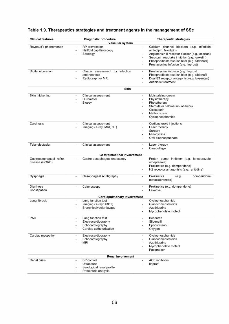
56
Table 1.9. Therapeutics strategies and treatment agents in the management of SSc
Clinical features Diagnostic procedure Therapeutic strategies Vascular system
Raynaud’s phenomenon - RP provocation - Nailfold capillaroscopy - Serology
- Calcium channel blockers (e.g. nifedipin, amlodipin, felodipin)
- Angiotensin II receptor blocker (e.g. losartan) - Serotonin reuptake inhibitor (e.g. luoxetin) - Phosphodiesterase inhibitor (e.g. sildenafil) - Prostacycline infusion (e.g. iloprost)
Digital ulceration - Clinical assessment for infection and necrosis
- Radiograph or MRI
- Prostacycline infusion (e.g. iloprost - Phosphodiesterase inhibitor (e.g. sildenafil - Dual ET receptor antagonist (e.g. bosentan) - Antibiotic treatment
Skin
Skin thickening - Clinical assessment - Durometer - Biopsy
- Moisturising cream - Physiotherapy - Phototherapy - Steroids or calcineurin inhibitors - Ciclosporin - Methotrexate - Cyclophosphamide
Calcinosis - Clinical assessment - Imaging (X-ray, MRI, CT)
- Corticosteroid injections - Laser therapy - Surgery - Minocycline - Oral bisphosphonate
Telangiectasia - Clinical assessment
- Laser therapy - Camouflage
Gastrointestinal involvement Gastroesophageal reflux disease (GORD)
- Gastro-oesophageal endoscopy - Proton pump inhibitor (e.g. lansoprazole, omeprazole)
- Prokinetics (e.g. domperidone) - H2 receptor antagonists (e.g. ranitidine)
Dysphagia - Oesophageal scintigraphy - Prokinetics (e.g. domperidone, metoclopramide)
Diarrhoea Constipation
- Colonoscopy
- Prokinetics (e.g. domperidone) - Laxative
Cardiopulmonary involvement Lung fibrosis - Lung function test
- Imaging (X-ray/HRCT) - Bronchioalveolar lavage
- Cyclophosphamide - Glucocorticosteroids - Azathioprine - Mycophenolate mofetil
PAH - Lung function test - Electrocardiography - Echocardiography - Cardiac catheterisation
- Bosentan - Sildenafil - Epoprostenol - Oxygen
Cardiac myopathy - Electrocardiography - Echocardiography - MRI
- Cyclophosphamide - Glucocorticosteroids - Azathioprine - Mycophenolate mofetil - Pacemaker
Renal involvement Renal crisis - BP control
- Ultrasound - Serological renal profile - Proteinuria analysis
- ACE inhibitors - Iloprost

57
1.15 Orofacial features
Orofacial manifestations occur in about 80-91% of patients with SSc, and it can give
rise to a range of extra-oral and intra-oral features (Marmary et al., 1981, Jagger et
al., 2006b, Bajraktari et al., 2015, Vitali et al., 2015, Hadj Said et al., 2016b, Veale et
al., 2016). The common extra-oral manifestations of SSc include hardening and
tightness of facial skin (mask-like appearance) also known as “bird’s face” or “Mona
Lisa face”, facial asymmetry, telangiectasia, small nose (peak nose), narrow eyes,
parotid salivary gland enlargement, trigeminal neuralgia and firm stretched lips.
Systemic sclerosis can give rise to a wide range of intra-oral features that include
microstomia, xerostomia, mucosal ulceration and atrophy, increased risk of caries and
dental erosion, periodontal and gingival inflammation, occasional oral infections and
rigidity of the tongue (“chicken tongue”) and lips (“fish mouth”) (Nagy et al., 1994,
Maddali-Bongi et al., 2011, Chapin and Hant, 2013).
Affected patients usually have more than one feature of the disease, and this may
lessen both the functional and aesthetic aspects of the patients and lead to a negative
impact on their emotional and social lives (Kwakkenbos et al., 2015).
Interincisal distance is determined by asking the patients to open the mouth widely
and measuring the distance between the incisal edges of both the upper and lower
incisors in millimetres (Wood and Lee, 1988). Also, the maximum mouth opening can
be determined by measuring the distance between the nearest points of the two
vermillion borders of the lips. Unsurprisingly in one study the mean interincisal
distance for patients with SSc has been found to be less than (37.68mm ± 8.36)
compared to unaffected individuals (mean 44.30mm ± 6.59) (p=0.0001) (Baron et al.,
2014).
58
It is one of the most comprehensive studies across Europe that examined a broad
range of symptoms (40 symptoms) of patients with SSc. It has been reported that more
than 70% of SSc patients complained of fatigue, Raynaud’s phenomenon, joint and
muscle pain. Some of the oral impairments of systemic sclerosis including xerostomia,
decreased mouth opening and dysphagia have been included in this study among 537
patients from five European countries (France, Netherlands, Spain, Switzerland, and
United Kingdom) (Willems et al., 2014).
The survey was translated into different languages as a self-reported questionnaire
assessing both frequencies of the symptoms in the last year and the impact of each
symptom on their daily life activities (Table 1.10 and 1.11). Most of the patients, 55%
had lcSSc, 36% had dcSSc, and 9% did not report. All three symptoms (decreased
mouth opening, dry mouth and difficulty swallowing) have been reported by all patients
with different values across countries (Willems et al., 2014).
In addition approximately 80% of patients with SSc have oral manifestations of the
disease (Del Rosso and Maddali-Bongi, 2014), another study clinically assessed 163
participants (12 patients with SSc, 151 control group), the result showed a significant
impact of SSc on the oral functions and reported oral-related decreased quality of life
as up to 30% of the patients considered having severely decreased mouth opening of
less than 30mm (Baron et al., 2014, Baron et al., 2015b). A high degree of tongue
involvement was found with a 42% reduction of the protrusion of the tongue, 23%
decreased strength and 92% lessened endurance (ability to sustain maximum
pressure on a specific period of time) which might affect a wide range of normal
functions such as speaking, chewing, saliva control and maintenance of swallowing
which can be associated with other complications such as changing in facial
appearance and resorption of the lips (Maddali Bongi et al., 2012, Vitali et al., 2015).

59
Table 1.10 Frequency and impact of oral symptoms experienced by patients with SSc
in five European countries
Symptoms France (n=111)
Netherlands (n=229)
Spain (n=61)
Switzerland (n=50)
UK (n=86)
Dry mouth 74% 69% 55% 69% 79%
Difficulty swallowing
53% 56% 56% 77% 70%
Difficulty opening mouth
39% 44% 40% 39% 44%
Adapted from (Willems et al., 2014)
Table 1.11 Impact on everyday activities of oral symptoms among patients with SSc in
five European countries
Symptoms France (n=111)
Netherlands (n=229)
Spain (n=61)
Switzerland (n=50)
UK (n=86)
Dry mouth 69% 50% 64% 46% 53%
Difficulty swallowing
79% 65% 72% 53% 73%
Difficulty opening mouth
84% 60% 67% 50% 61%
Adapted from (Willems et al., 2014)
Microstomia
Microstomia is considered to be the most common oral feature of patients with SSc
(52.0 – 80%). Reduced mouth opening plays a significant role in lessening the quality
of life of patients as it is strongly related to multiple functions such as speaking, eating,
aesthetic appearance and even maintenance of good oral hygiene. This is mainly
resulting from the pathological nature of the disease as an increased amount of
collagen deposition in orofacial tissues (Marmary et al., 1981, Fischer and Patton,
2000, Bajraktari et al., 2015).
With respect to measuring the mouth opening, different methods have been applied
as following:

60
- Inter-commissural distance: while the teeth are in occlusion, recording of the
distance between two points from one commissure to the other.
- Maximum oral aperture: by opening the mouth as much as possible, the
measurement done by tracing of all-around vermilion borders of both lips by a
non-stretchable cord.
- Inter-incisal distance: after registration of the overbite during the maximum
intercuspation, the mouth is opened wide and the distance between the upper
and lower incisal edges is measured plus the amount of overbite.
- Maximum mouth opening: by using the Willis Bite Gauge, the distance between
both upper and lower incisal edges measured while opening the mouth widely.
By applying these different methods of measuring the mouth opening, several studies
demonstrated that mouth opening was significantly reduced in patients with SSc
(Wood and Lee, 1988, Vitali et al., 2015, Baron et al., 2014). As reported by Cox et
al., the average mean of the normal maximum mouth opening among 700 healthy
individuals (98%) was 47.1mm (33.7 – 60.4mm) (Cox and Walker, 1997). However,
the mean reduced mouth opening for SSc patients found to be 34.9mm (Mouthon et
al., 2007). It has been reported that maximum mouth opening of less than 40mm can
be considered as reduced mouth opening with up to 30% of SSc patients showing a
severe form of decreased mouth opening as less than 30mm (Vitali et al., 2015).
Reduced mouth opening is suggested to be a co-factor for mandibular erosions as it
may be a cause of increased strain of the associated muscles and tightness of the
skin of the face (Baron et al., 2015a). Microstomia may be associated with an
increased risk of tooth loss and periodontal disease and even the overall disease
severity (Nagy et al., 1994, Baron et al., 2015b).

61
Salivary gland hypofunction and xerostomia
Symptoms of mouth dryness in SSc patients varies from 32-70% and is considered as
one of the most frequent oral manifestations of SSc (Wood and Lee, 1988, Bajraktari
et al., 2015). Dry mouth is found to be associated with salivary gland fibrosis, the
presence of Sjogren’s syndrome (SS) and increased deposition of collagen in the oral
mucosa. Objective estimate of the unstimulated salivary flow (using the Saxon test; an
oral equivalent of Schirmer’s tear production test) in 163 SSc patients with disease
duration 13.9 years (8.5), found saliva production to be reduced (147.52mg/min). Both
resting and stimulated whole saliva production may be reduced in SSc (Baron et al.,
2014).
Loss of salivary glands function may increase the risk of dental caries, gingival and
periodontal disease and perhaps oral malodour as well as causing some degree of
dysphagia, dysarthria and dysgeusia. It is recommended that all patients with SSc
might be investigated for Sjogren’s syndrome as it was reported that the prevalence
of SS among SSc patients ranged from 17-29% (Chu et al., 2011).
The presence of histological features of fibrosis of salivary glands in patients with SSc
has been reported in 23-48% while the histopathological features of SS occur in 65%
of examined specimens. A recent study of 118 patients with SSc against SS-related
antibodies found that sicca symptoms were present in 71.2% of patients with SSc and
33.9% were diagnosed with SS (Kobak et al., 2013). Hence, salivary hypofunction in
SSc disease is linked to SS as one of the expected underlying causes (Baron et al.,
2015b). It has been proposed that both microstomia and salivary gland dysfunction
may coexist as a consequence of common pathological fibrosis of the tissues (Nagy
et al., 1994).

62
Gingival and periodontal ligament disease
A number of studies have been reported that gingival and periodontal ligament disease
found to be common in patients with SSc (76%) with an increased likelihood of deep
periodontal pockets and high gingivitis scores (Chu et al., 2011).
A relatively recent study of patients with SSc concluded that periodontitis was more
likely and severe in SSc than appropriate control subjects. High plaque indices were
associated with sclerodactyly and more severe SSc disease status. The higher
gingival index was linked with disease duration and severity while bleeding on probing
only correlate with duration of the SSc (Elimelech et al., 2015).
The relationship between periodontal disease and SSc might reflect reduced salivary
production but periodontitis was not found to be directly related to neither the number
of missing teeth nor the SSc disease severity (Baron et al., 2015b). Other studies have
concluded that there is a significant level of periodontal disease in patients with SSc
(Wood and Lee, 1988, Baron et al., 2014). It is potentially possible that this is the
consequence of fibrotic changes and hypovascularity (Yuen et al., 2014b). Although
as patients may have reduced manual dexterity, there is a risk of this symptom
reflecting the difficulty in being able to maintain good oral hygiene (Fischer and Patton,
2000, Poole et al., 2013).
Progressive (presumably non-dental plaque-induced) gingival recession and/or
resorption of alveolar bone has been documented in SSc. A 19-year-old female patient
having had SSc (En coup de sabre) for 3 years was found to have localised 4mm
recession of the upper incisor together with a linear atrophic lesion traced from the
nose to the upper lip on the same side and extended to the gingival tissue mesial to
the affected tooth with periapical radiograph showing widening of PDL and pulp stone
(Van der Veken et al., 2015). Recently, another 10-year-old female patient was
63
diagnosed with localised SSc (morphea). On intra-oral examination revealed a white
plaque extending from the centre of upper lip mucosa to the oral vestibule with 12-
13mm PD pocket depth found to be associated with both maxillary left central and
lateral incisors. Radiographical findings revealed alveolar bone resorption between the
affected teeth. However, there are no specific recommendations for the management
of the intraoral disease involvement and so conservative intervention and maintaining
optimum oral health care is mandatory to prevent further gingival and periodontal
inflammation (Van der Veken et al., 2015, Wang et al., 2015).
Temporomandibular joint and trismus
Involvement of the temporomandibular joint can further increase the risk of trismus.
This can be due to fibrosis of masticatory muscles, muscles of facial expression, oral
and perioral tissues and the resorption of the articular bones. However, as a
consequence of this, the movement of the mandible is restricted, and there may be
articular pain and swelling due to synovitis and tendinitis. Also, as a consequence of
the masticatory muscles atrophy, due to the low blood supply and the fibrosis of the
blood vessels, this can cause trismus and increase the risk of inflammation within the
associated joint structures (Chu et al., 2011, Alantar et al., 2011, Baron et al., 2015a).
TMJ involvement in SSc can develop early in the disease onset and then give rise to
malocclusion, limited movement, speech impairment, tenderness, pain and
discomfort. Wood et al., indicated a high risk of TMJ dysfunction among patients with
severe and more active disease (Wood and Lee, 1988).
With regards to SSc patients with TMJ involvement, a recent study of 27 patients with
SSc (12 diffuse and 15 limited) revealed that 74.1% had TMJ clicking, 66.7% had
reduced mouth opening, and 77.8% had pain with muscle and TMJ tenderness with
55% reported pain lasting for six months or more. Crepitation has been reported
64
among 70.4% of the total sample. However, magnetic resonance imaging findings
were 51.8% abnormal disks without displacement, and 81.5% had disk displacement
with reduction and TMJ osteolytic bone lesions 66.6%. Moreover, flattening of the
temporal eminence function surface 48.1%, flattening of condyle 66.7%%,
osteophytes 44.4% and synovitis 37% (Matarese et al., 2016).
Bilateral TMJ involvement has been reported in SSc and recognised as a
consequence of mandibular bone resorption (MacIntosh et al., 2015). In advance
stages of bone resorption and TMJ involvement, 4 -13% might present with trigeminal
neuropathy with up to 83% involvement of mandibular and maxillary nerves (Fischoff
and Sirois, 2000, Doucet and Morrison, 2011).
Neurological involvement
Neurological disease can arise secondary to SSc in about 40%. Various forms of
neurological complications have been described in association with different subtypes
of SSc such as cranial entrapment, peripheral, cutaneous and autonomic
neuropathies, myopathy and rarely associated with the central nervous system
(Amaral et al., 2013).
Cranial neuropathy has been described in SSc with the most common being trigeminal
neuropathy that affects 5-15% of patients with SSc (Cazal et al., 2008, Vincent et al.,
2010, Amaral et al., 2013, Bajraktari et al., 2015). Although, the exact pathophysiology
of trigeminal neuropathy related to SSc, it is still unknown, some studies reported that
it can occur as a consequence of vascular-dependant neuropathy (due to vasculitis
and/or vessel wall damage), compression-dependant neuropathy (related to oedema
or fibrosis and as a consequence of progressive demyelination) or autoimmune-
dependant neuropathy (associated with antineuronal antibodies) (Fischoff and Sirois,
2000, Jagger et al., 2006b, Amaral et al., 2013).

65
Several reports have generally documented multiple neurological symptoms which
might correlate either to disease severity or as an iatrogenic cause with 83%
prevalence of maxillary and/or mandibular nerves involvement with (4 - 13%) sensory
neuropathy. Symptoms of trigeminal neuropathy can manifest bilaterally and are
frequently associated with pain that is described as throbbing, aching, scalding,
burning or lancinating with involvement of the intraoral tissues that may be provoked
by jaw movement (Farrell and Medsger, 1982, Lee et al., 1984, Fischoff and Sirois,
2000, Vincent et al., 2009, Doucet and Morrison, 2011).
It has been reported that neurological involvement in 224 patients with SSc included
seizures (13.56%), headache (23.73%) and trigeminal neuropathy (16.52%).
Moreover, depressive and anxiety symptoms are highly prevalent as the estimated
prevalence of depression (73.15%) and (23.95%) for feeling anxiety. Other very rare
features included dropped head (4%), facial weakness (4%) and (0.40%) for having
suicidal ideation (Amaral et al., 2013).
Tongue rigidity and ankyloses
Hardening of the oral soft tissue structures is common in SSc. Tongue rigidity of up to
25% prevalence has been documented and could greatly impact oral functions such
as speech, eating and swallowing (Fischer and Patton, 2000, Jagger et al., 2006b,
Cazal et al., 2008, Hajimahmoudi and Mostafavi, 2014). In one small study of 12 SSc
patients 42% had a reduction of tongue protrusion, 23% a reduction of tongue
strength, and 92% a reduction of tongue endurance (Vitali et al., 2015). A shortened
lingual frenulum was found in 0.4% of 75 patients with SSc (Bajraktari et al., 2015).
Oral mucosal atrophy and ulceration
Thinning (atrophy) of oral mucosa can occur in SSc and might reflect fibrosis and/or
local ischaemia (Poole et al., 2013). Oral ulceration due to disease-modifying drugs
66
(e.g. methotrexate, azathioprine and cyclophosphamide) can arise. Oral ulceration
may also be an adverse consequence of malnutrition, vitamins deficiency, exocrine
pancreatic insufficiency or small bowel involvement associated with bacterial
overgrowth (Nagy et al., 1994, Alantar et al., 2011). It has been suggested that
ulceration can occur due to gastroesophageal reflux disease (GORD), but there is little
supportive evidence; however oral ulceration giving rise to dysphonia and dysphagia
has been reported (Jagger et al., 2006b).
Dental erosion and decay
Patients with SSc exhibit enamel erosion that may be related to gastroesophageal
reflux disease (GORD) and less likely xerostomia (Chapin and Hant, 2013). It has
been reported that both xerostomia and GORD are responsible for a decreased
salivary pH that compromises the buffering capacity of the saliva and can induce
enamel and dentine erosion (Jung et al., 2016). The Canadian systemic sclerosis oral
health study, which describes the largest SSc cohort (163 patients with SSc), reveals
that patients with SSc had significantly more decayed teeth compared to healthy
controls (0.88 vs 0.59, P = 0.0465) (Yoshikawa et al., 2012, Baron et al., 2014, Baron
et al., 2015b). According to a study of 42 patients with SSc, examining the oral health
status in relation to SSc disease, all the study sample had dental caries with 65%
untreated decay (Chu et al., 2011). However, a trend towards increasing number of
dental decay has been reported and it would be expected that it is related to oral
dryness and difficulties with oral hygiene routine associated with reduced manual
dexterity and limitation in mouth opening (Jung et al., 2016).
Oral infections
Increased susceptibility to microbial infection would be expected in consequence to
other oral manifestation of SSc disease such as (decreased mouth opening, salivary

67
gland hypofunction and drug adverse side effects). Candida infection has been
correlated to SSc patients with low saliva flow rate (Fischer and Patton, 2000).
Furthermore, patients using immunosuppressant agents such as corticosteroids and
cyclophosphamide and/or antibiotics have a high risk of developing oral candidiasis
(Martin et al., 1997).
Treatment-related adverse side effect
A wide range of medications are often prescribed for the treatment of SSc and its
complications. Use of medicine including immunosuppressive and cytotoxic drugs
(cyclophosphamide, cyclosporine, methotrexate), antihypertensive, antidepressant
and anticoagulant drugs can give rise to some oral complications such as (mucosal
ulceration, xerostomia, gingival bleeding and hyperplasia) (Elimelech et al., 2015).
Calcium channel blockers used to treat vascular features, may give rise to gingival
hyperplasia (Seymour and Heasman, 1988, Shah and Wigley, 2008, Yuen et al., 2011,
Yuen et al., 2014b).
Using corticosteroid agents for a long time can give rise to candida infection while
cyclophosphamide has been used for cases with lung involvement and caused oral
infections and even oral ulceration if taken in high doses (Martin et al., 1997).
For treatment of joint and muscles involvement, methotrexate has been indicated and
was found to be recommended for use with folic acid supplements to decrease the risk
of mucosal ulceration. Patients with SSc have a great impact on their quality of life and
may present a wide range of psychological distress symptoms, therefore they may use
antidepressant medications with anticholinergic effects which might cause
symptomatic dry mouth (Scully, 2003, Thombs et al., 2007). Furthermore, anti-vitamin
K anticoagulants agents, mostly prescribed for SSc patients in regards to preventing
thrombosis, might results in an increased tendency to gingival bleeding and
68
inflammation and hence those patients particularly must maintain good oral hygiene
(Alantar et al., 2011).
Medication-related osteonecrosis of the jaw can be seen among patients with SSc
who are taking bisphosphonate therapy (zoledronic acid and/or pamidronate) or other
medications such as bevacizumab or sunitinib to lessen skeletal-related diseases or
metastatic cancer. MRONJ may give rise to oral infections, mucositis and impaired
wound healing. Among those patients, it has been recommended that a thorough oral
examination should be performed before starting to take the medications. Also, all
invasive dental treatment should be completed, and preventive measures should be
considered to reduce the risk of jaw osteonecrosis. Furthermore, MRONJ needs long-
term management, including a combination of systemic and topical antimicrobial
agents and surgical debridement of necrotic bone (Sigua-Rodriguez et al., 2014,
Mawardi et al., 2016).
Telangiectasia and oral mucosa
Dilatation of blood vessels, also called “Telangiectasia” can manifest in extra and intra-
oral tissues (Ramazani et al., 2015). Oral mucosal telangiectasia can occur in the
palate, lips, lateral borders of the tongue and buccal mucosa in about 56.3% and other
studies reported 70 - 80% (Nagy et al., 1994, Vincent et al., 2009, Chu et al., 2011).
Telangiectasia may cause some level of cosmetic disfigurement and psychological
impact as it may present peri-orally as well as on the face, neck and hands. Patients
can use make-up, or it can be treated conservatively by laser application (Lachner,
2016).
Assessing oral mucosal fibrosis have been done using an ultrasonography imaging
technique and found a significant increase of fibrotic components in the buccal mucosa
69
among patients with SSc with a case report of fibro-epithelial polyp on the buccal
mucosa (Jackowski et al., 1999, Chapin and Hant, 2013).
1.16 Orofacial radiological features
The radiological findings of the maxillofacial area are mainly due to the involvement of
the musculoskeletal structures in the pathological sclerosis of SSc and/or due to the
vascular ischemia possibly caused either by the deposition of abnormal collagen or as
a result of tightening of the associated soft tissue and its pressure effect (Jung et al.,
2016, Veale et al., 2016). Recently, different assessment measures are used to
determine the radiological findings in relation to systemic sclerosis such as magnetic
resonance imaging and high-frequency ultrasonography (Chapin and Hant, 2013).
Overall mandibular resorption prevalence 50% with incidence of 20-30% has been
reported in a review by Haers and Sailer of 22 publications with a sample of 52
patients, the mandibular angle was affected by 37.6%, resorption of the coronoid
process 20%, condylar head 20.8% and the resorption of the ascending ramus was
found in 14.4% and 13.7% as a bilateral condylysis (Seifert et al., 1975, Haers and
Sailer, 1995, Doucet and Morrison, 2011, Chapin and Hant, 2013, Delantoni and
Matziari, 2015).
A recent study of 159 patients with SSc find 14.5% of mandibular erosion with regards
to the range of other reports (6.6% - 46.7%) (Table 1.12) and there was up to 60.9%
of the patients had multiple sites of erosion in the mouth (Dagenais et al., 2015).
Resorption of the zygomatic arches has been reported in SSc patients in association
with other parts of bone resorption such as mandibular angles and ascending ramus
(Hopper and Giles, 1982, Wood and Lee, 1988).

70
Other patterns of mandibular erosions have been reported as osseous resorption of
the digastric region 2.53%, resorption of the posterior ramus 5.06% (Dagenais et al.,
2015). However, apart from the presence of rheumatoid factor that is found to be
positive in about 33% of SSc cases, the overall resorption pattern of the maxillofacial
bones in SSc patients especially the bilateral bone resorption in mandibular angles,
digastric region and both coronoids and condyles processes might be caused by the
long term (5-7 years disease duration) effect of increased physical pressure from the
overlying tight skin and also due to the associated sites of attachments of masticatory
muscles such as masseter, temporal, lateral pterygoid and anterior belly of digastric
muscles which may affect the blood supply to the bone itself. Also, another explanation
included in the pathological process of the disease which might cause ischemia as a
result of vasculitis and perivascular fibrosis, and this could affect the blood supply
vessels of related bone structures such as the maxillary artery to the condyle, coronoid
process, mandibular angle and both masseteric and pterygoid muscles (Ramon et al.,
1987, Jagger et al., 2006a, Jagger et al., 2006b, Rahpeyma et al., 2013, Delantoni
and Matziari, 2015, MacIntosh et al., 2015).
MRI has been used to measure the masseter musculature in a group of 15 SSc
patients in relation to mandibular osteolysis. A high ratio of fat replacement and
atrophy of the muscular tissues appeared in association with the disease (Chapin and
Hant, 2013). The reported incidence of mandibular erosion in SSc is 20%, and the
overall prevalence is 50% with almost 28.6% considered as a mild degree of
osteolysis. However, it has been suggested that the more severe mandibular erosion,
the more severe the sclerosis (MacIntosh et al., 2015).
As a consequence of TMJ involvement in individuals with SSc, MRI showed that
51.8% of the sample consisted of 27 patients with SSc (12 diffuse and 15 limited)

71
revealed abnormal disks without displacement and 81.5% diagnosed as disk
displacement with reduction. The reported symptoms are mainly TMJ clicking 63%,
crepitation in 70.4%, reduced mouth opening 66.7%, TMJ and muscle tenderness in
55.6% and 55% reported pain lasting for 6 months or more (Matarese et al., 2016).
Degenerative bone changes were reported in 66.6% with the highest percentage in
flattening of condyle anterior surface was (66.7%), followed by joint surfaces erosion
and irregularities (55.6%). Furthermore, flattening of temporal eminence functional
surface 48.1%, osteophytes (44.4%), synovitis (37%), subchondral cysts (11.1%) and
idiopathic condyle resorption (11.1%) with a significant correlation of the frequency of
bone changes and the duration of the disease (P< 0.05). However, the re-adsorption
theory might take place in different sites such as mandibular angle, condyle and/or
coronoid process as a result of atrophic ischemia and this may increase the risk of a
pathological fracture of the affected site (Matarese et al., 2016).
Also, an ultrasonographic scanner has been applied to 10 patients with SSc evaluating
the extra-oral and intraoral changes by looking specifically to a range of criteria such
as reduction in the range of facial expression, telangiectasia in both oral mucosa and
facial skin, perioral folds, changes in lip redness, impaired tongue movement,
flattening of the palate and shortening of the uvula, myosclerosis in the vestibule of
the mouth, decreased inter-incisal distance and gingival inflammation. Two patients
showed an increased amount of fibrotic deposition and sclerosis. However,
considering ultrasonography as a non-invasive investigative tool might be useful for
monitoring the disease activity and therapeutic effectiveness. Also, it has been
reported that colour Doppler ultrasound has benefits of measuring dermal blood flow
as it might indicate underlying active disease (Chapin and Hant, 2013, Tolkachjov et
al., 2015).
72
More recent studies demonstrated that the widening of the PDL is the most common
oral radiographic feature. A study sample of 159 individuals with systemic sclerosis,
38% indicated a widening of the PDL and a strong association was found between the
number of teeth included and the severity of the disease. However, widening of the
PDL in patients with SSc could be due to the generalised overproduction of collagen,
but there is no clear correlation to other periodontal diseases as it is not showing any
significant level of gingival attachment loss. Although, it has been found that there is
a strong association between smoking, widening of PDL and the severity of SSc
(Baron et al., 2015a).
In Jung et al., periodontal ligament widening in patients with SSc usually presented in
both anterior and posterior teeth with more tendency to be found around the posterior
teeth (Jung et al., 2013). Furthermore, widening of PDL has been described as a
uniform widening in up to 10% of SSc cases (Chapin and Hant, 2013) and it was
located in 163 patients with SSc adjacent to coronal and periapical areas of the roots
by using either periapical or panoramic radiographs (Dagenais et al., 2015).
As a rare case, cone-beam computerised tomography and panoramic radiograph
images have been used to report an uncommon calcification in both widening PDL
and pulp canals, which might be related to the dystrophic calcinosis pattern of the
disease. A case report with 13 years diagnosis of diffuse SSc, a history of persistant
maxillary pain lasting for several weeks, involvement of systemic organs including the
lungs, gastrointestinal and myocardial involvement and digital ulcers. Clinical
examination reported tightness of facial skin, telangiectasia, thin and sclerotic lips and
decreased mouth opening. The radiological evaluation shows generalised PDL
widening, and by utilising the CBCT, it has been found that multiple maxillary teeth
revealed calcifications in associated PDL spaces. Additionally, those affected teeth,
73
specifically the incisors and premolars also contained pulp stones and were at risk of
root canal obliteration. In this case, the calcification was associated with areas of PDL
widening and the lamina dura was intact. However, it is suggested that this clinical
scenario might be related to the nature of the sclerotic features of SSc (Jung et al.,
2013).
Rarely there may be a few reported cases with external dental root abnormalities
(resorption, dilacerations, underdevelopment) as it has been reported to be associated
with the disease with considerable high prevalence in childhood cases 27.3% (Trainito
et al., 2012). One study revealed a patient with intraoral features of SSc such as
significantly decreased mouth opening (20mm), telangiectasia on both the hard palate
and lateral border of the tongue with radiological findings of left side mandibular
erosion coincident with the external distal root resorption of the lower left third molar.
However, the resorption of the apical third of the root was found to be adjacent to the
area of the bone resorption of the mandible, and after excluding other causes, it was
suggested that it had happened as a consequence of both the erosive pathological
process of the disease and the high magnitude of the external pressure from the
adjacent muscular structure (de Figueiredo et al., 2008). However, a recent study has
reported that patients with SSc may present with an external pattern of root resorption
located cervically instead of apically (Arroyo-Bote et al., 2017).
A study of 16 patients with juvenile localised SSc showed a rare presentation of the
disease in 9 patients (81.8%) as the cephalometric analysis was applied and revealed
an overgrowth of the lower third of the face as an increased anterior face height. The
same study reported that 81.8% had skeletal malocclusion, and 18.7% had TMJ
asymmetry (Trainito et al., 2012).
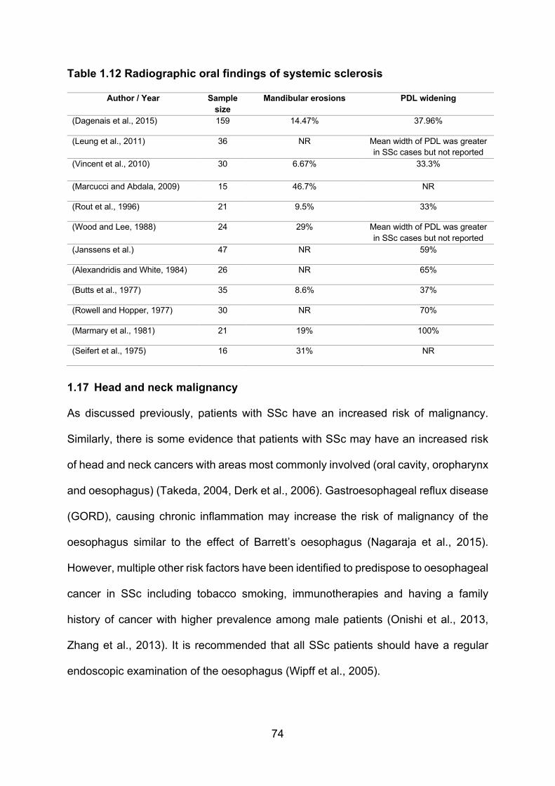
74
Table 1.12 Radiographic oral findings of systemic sclerosis
Author / Year Sample size
Mandibular erosions PDL widening
(Dagenais et al., 2015) 159 14.47% 37.96%
(Leung et al., 2011) 36 NR Mean width of PDL was greater in SSc cases but not reported
(Vincent et al., 2010)
30 6.67% 33.3%
(Marcucci and Abdala, 2009) 15 46.7% NR
(Rout et al., 1996) 21 9.5% 33%
(Wood and Lee, 1988) 24 29% Mean width of PDL was greater in SSc cases but not reported
(Janssens et al.) 47 NR 59%
(Alexandridis and White, 1984) 26 NR 65%
(Butts et al., 1977) 35 8.6% 37%
(Rowell and Hopper, 1977) 30 NR 70%
(Marmary et al., 1981) 21 19% 100%
(Seifert et al., 1975) 16 31% NR
1.17 Head and neck malignancy
As discussed previously, patients with SSc have an increased risk of malignancy.
Similarly, there is some evidence that patients with SSc may have an increased risk
of head and neck cancers with areas most commonly involved (oral cavity, oropharynx
and oesophagus) (Takeda, 2004, Derk et al., 2006). Gastroesophageal reflux disease
(GORD), causing chronic inflammation may increase the risk of malignancy of the
oesophagus similar to the effect of Barrett’s oesophagus (Nagaraja et al., 2015).
However, multiple other risk factors have been identified to predispose to oesophageal
cancer in SSc including tobacco smoking, immunotherapies and having a family
history of cancer with higher prevalence among male patients (Onishi et al., 2013,
Zhang et al., 2013). It is recommended that all SSc patients should have a regular
endoscopic examination of the oesophagus (Wipff et al., 2005).
75
The risk of oral squamous cell carcinoma in patients with SSc has been reported
particularly of the tongue and lower lips (Derk and Jimenez, 2003, Petrov et al., 2009,
Acarturk et al., 2015). The exact underlying causes of this increased risk are not
known. Regarding the poor prognosis of both systemic sclerosis and cancer lesions,
early diagnosis is highly valuable among SSc patients.
1.18 Oral treatment and rehabilitation
Oral Hygiene
As noted previously, patients with SSc have an increased risk of common plaque-
induced dental disease. Patients can have difficulty maintaining good oral hygiene in
view of a limited range of mouth opening and reduced manual dexterity. Any oral
dryness will increase the risk of plaque retention, the stiffness and rigidity of oral
mucosa and peri-oral soft tissues, restricted tongue movement and fibrosis of the
lingual frenum may reduce normal oral movements and self-cleaning mechanisms
leading to increased the susceptibility to dental caries and periodontal disease (Yuen
et al., 2014b).
Regular dental attendance and prophylaxis have been suggested to be of benefit for
SSc patients such as the regular application of 5% sodium fluoride varnish which may
decrease the risk of carious lesions (Chu et al., 2011, Gyger and Baron, 2015).
Oral hygiene tools may be required to be modified if patients have limited hand or arm
mobility. Helper-assisted tooth brushing can be useful, but clearly requires the
assistance of a partner or carer. A variety of toothbrushes are available to allow
patients to better clean their teeth such as angled brushes, altered filament length
brushes, easy-grip brushes, extended handle brushes, electric brushes and soft small-
headed toothbrushes. Recent studies recommend the use of appropriately adapted
76
flossing and/or interdental cleaning tools to prevent periodontal disease and
interdental lesions (Poole et al., 2010, Yuen et al., 2014a).
Other suggested, but little proven, therapeutic modalities include connective tissue
massage, joint mobilisation, finger stretching exercise, manual lymph drainage of the
hands and ultrasound therapy (Maddali Bongi et al., 2009, Yuen et al., 2014a, Willems
et al., 2015b).
Patients with SSc may require referral to a dietitian as they have difficulties in eating
solid food or dietary choices. Furthermore, patients with a significant level of dry mouth
and/or gastroesophageal reflux may tend to consume more frequent meals that are
softer and contain refined carbohydrates which increase the risk of caries (Alantar et
al., 2011).
Additionally, the psychological status of the patients should be monitored as those with
depression can lose interest in maintaining a high standard of oral hygiene (Yuen et
al., 2014a).
Dry mouth
Xerostomia has been linked to an increased number of missing teeth, carious lesions
and periodontal diseases among SSc patients due to low salivary flow rate and
salivary pH value. Also, it might give rise to an altered taste sensation, oral malodour
and loss of retention of dentures.
The treatment of oral dryness in relation to SSc is no different from the treatment of
other causes of oral dryness. This has been summarised in table 1.13.
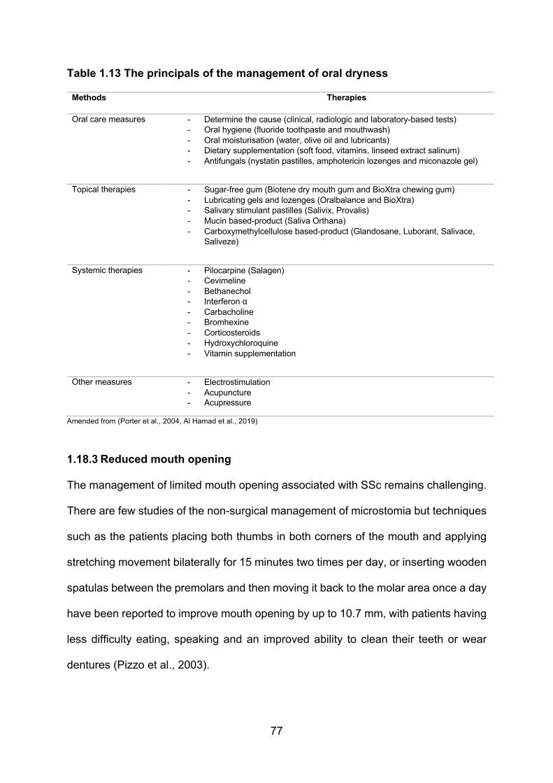
77
Table 1.13 The principals of the management of oral dryness
Methods Therapies
Oral care measures - Determine the cause (clinical, radiologic and laboratory-based tests) - Oral hygiene (fluoride toothpaste and mouthwash) - Oral moisturisation (water, olive oil and lubricants) - Dietary supplementation (soft food, vitamins, linseed extract salinum) - Antifungals (nystatin pastilles, amphotericin lozenges and miconazole gel)
Topical therapies - Sugar-free gum (Biotene dry mouth gum and BioXtra chewing gum) - Lubricating gels and lozenges (Oralbalance and BioXtra) - Salivary stimulant pastilles (Salivix, Provalis) - Mucin based-product (Saliva Orthana) - Carboxymethylcellulose based-product (Glandosane, Luborant, Salivace,
Saliveze)
Systemic therapies - Pilocarpine (Salagen) - Cevimeline - Bethanechol - Interferon α - Carbacholine - Bromhexine - Corticosteroids - Hydroxychloroquine - Vitamin supplementation
Other measures - Electrostimulation - Acupuncture - Acupressure
Amended from (Porter et al., 2004, Al Hamad et al., 2019)
Reduced mouth opening
The management of limited mouth opening associated with SSc remains challenging.
There are few studies of the non-surgical management of microstomia but techniques
such as the patients placing both thumbs in both corners of the mouth and applying
stretching movement bilaterally for 15 minutes two times per day, or inserting wooden
spatulas between the premolars and then moving it back to the molar area once a day
have been reported to improve mouth opening by up to 10.7 mm, with patients having
less difficulty eating, speaking and an improved ability to clean their teeth or wear
dentures (Pizzo et al., 2003).

78
A study of 20 patients with SSc that comprised a 9 weeks comprehensive programme
of massaging techniques of the soft tissues, facial physiotherapy and facial muscle
exercises found a significant improvement in mouth opening. Another method of trying
to increase mouth opening consists of neuromuscular stimulation of the facial muscles
in conjunction with passive and active physiotherapy to the temporomandibular joint.
However, the long-term benefits of such intervention have not been published
(Maddali-Bongi et al., 2011, Poole et al., 2013, Baron et al., 2015b).
Surgical interventions for microstomia such as bilateral commissurotomies might
increase the size of the labial opening, but these run the risk of impaired healing and
further fibrosis (Fischer and Patton, 2000, Albilia et al., 2007). Bilateral
commissurotomies may be indicated for patients who require major general surgical
operations as severely limited mouth opening might impede general anaesthesia and
oral intubation.
Perhaps the most promising treatment for orofacial tightness in SSc is autologous fat
grafting (AFGT). In a study of 20 patients with dcSSc, 2 mm of autologous fat grafting
were injected into 8 different areas around the lips. At 3-month follow-up, all patients
had a significant degree of mouth opening improvement and improved orofacial
function. Perioral skin elasticity significantly improved in this 3 month period and there
was a reduction of fibrosis (Del Papa et al., 2015).
A later study compared the two methods of AFGT with autologous adipose-derived
stromal cell (ADSC) therapy in 2 groups of patients with SSc. Both methods led to an
improvement in mouth opening and patient satisfaction. However, ADSC method was
concluded to be the more technically challenging technique as it necessitated
laboratory processing to isolate the stem cells (Onesti et al., 2016).

79
AFGT was suggested to be more effective for severe fibrotic lesions. However, in
contrast to ADSC the results of AFGT are unpredictable as it may give rise to an
asymmetrical appearance of the two sides of the face. Furthermore, AFGT requires
administration by cannula rather than the ADSC method that requires the injection of
cells using a thin needle. Hence the AFGT is more likely than the ADSC method to
cause operative scarring (Onesti et al., 2016).
Intralesional injection as a combination of decamycin corticosteroid (dexamethasone
sodium phosphate 4 mg) and hyaluronidase (Hynidase 1500IU) with the multi-
antioxidant capsule (Ricinia-LP) for 5 months has recently been found to improve
mouth opening. Intralesional injections of the lips and face were given over multiple
visits. It has been suggested that this technique may lessen hyperactivity of fibroblasts
(Kumar et al., 2016).
Prosthetic dental care
Patients with SSc commonly show some degree of reduced mouth opening, tongue
rigidity, dry mouth and impaired manual dexterity so special therapeutic modifications
should be considered as the disease nature and progression may limit the treatment
options (Baron et al., 2015b).
The increased acidity of the oral cavity as a result of decreased salivary flow rate and
GORD may greatly affect the dentition and increase the risk of developing caries
and/or erode the teeth structures. Thus, during the treatment planning, it is essential
to consider the most appropriate restorative dental material as the suitability and
longevity is arguably more important than aesthetic aspects.
Glass ionomer-based restorations have multiple advantages such as the long-term
fluoride release, the inherent adhesive properties and the similar coefficient of
expansion to enamel with low setting shrinkage. However, it might be suggested that

80
using resin-modified glass ionomer restorations is more beneficial in regard to
overcoming the physical characteristics and the dual-cure effect that allows longer
working time and rapid hardening during the clinical setting. Also, the shortened dental
arches concept indicates that the dental arches compromising the anterior teeth and
premolars can meet the requirements of a functional dentition hence compromised
treatment planning may consider the shortened dental arches concept, in so far as it
does not contradict the current theories of occlusion. However, considering the
patients’ ability to cooperate, adapt and maintain the dentition with the possible
treatment options, this may help achieve therapeutic goals and desired outcomes
(Armellini and von Fraunhofer, 2004).
Comprehensive prosthetic dental treatment may be considered for patients with SSc
with great attention to the limitations and necessity of modifications. Common
challenges while making dental treatment decisions are the degree of reduced mouth
opening, the rigidity of the tongue, dry mouth status and the manual dexterity (Yenisey
et al., 2005, Gozde Turk and Ulusoy, 2015).
With regards to overcoming the difficulties for the clinical setup, fabrication and
construction of the removable prostheses some studies have recommended using
different modifications such as sectional, collapsible or combined sectional-collapsible
designs with various connection systems such as clasp retainers, stud attachments,
telescope systems, swing-lock attachments, magnetic attachments and pins (Singh et
al., 2014, Gozde Turk and Ulusoy, 2015).
Benefits of utilising the overdenture prostheses include preserving abutment teeth that
help to lessen the alveolar bone loss, enhance the stability and comfort, provide
favourable crown-root ratio, maintain the PDL proprioception and enhance the well-
being sensation (Benetti et al., 2004). It is recommended maintaining good oral

81
hygiene and cleaning dentures with a soft brush with a mild detergent can considerably
reduce the risk of developing microbial and/or fungal infection especially on the fitted
surface of the denture (Singh et al., 2014).
The impression taking procedure is one of the most critical clinical steps during
prosthetic treatment. Sectional tray impression technique has been employed with
either locking segments along the midline or using hinges, plastic building screws,
orthodontic expansion screws, locking levers, stepped butt joints or resin blocks with
indexing technique (Prithviraj et al., 2009, Hajimahmoudi and Mostafavi, 2014, Gozde
Turk and Ulusoy, 2015).
Implant treatments are not necessarily contraindicated in SSc. Two main areas are
commonly indicated for implant placement either in the canine area of the mandible
for stabilisation and/or in the maxillary incisors and canine area for aesthetics (Oczakir
et al., 2005). However, dental implants are contraindicated among patients with severe
gingival and periodontal disease as they commonly manifest a significant degree of
tissue fibrosis and microvascular impairment (Alantar et al., 2011).
Due to the nature of the disease, it is important to consider the disease’s impact on all
of the associated tissue structures and the related patient’s quality of life by
considering all other available alternative treatment modalities including simplicity,
future repair services and financial concerns (Baptist, 2016).
82
1.19 Impact of systemic sclerosis upon the quality of life
Systemic sclerosis is a chronic disorder that can adversely impact upon an individuals
physical and psychological wellbeing. According to the World Health Organisation, the
quality of life (QoL) concept goes back to the 1947 (WHO) definition of health as a
“state of complete physical, mental and social well-being, and not merely the absence
of disease and infirmity (WHO, 1947). The use of the term “well-being” in this definition
of health has contributed significantly to the conceptual confusion about what is health
and what is QoL. Quality of life encompasses (physical, mental and social wellbeing).
Within these, there are the physical, mental and social impacts of any orofacial disease
that an individual may have. The Canadian Dental Association has defined oral health-
related quality of life as “a state of the oral and related tissues and structures that
contribute to physical, mental and social well-being and enjoyment of life’s
possibilities, by allowing the individual to speak, eat and socialise without feeling pain,
discomfort or embarrassment” (Gift and Atchison, 1995, Petersen, 2003). Over the
past two decades, there has been an acknowledgement that the management of
disease must consider the oral health-related quality of life. An assessment of the
quality of life is now considered an essential aspect of the evaluation of treatment and
management of health-related conditions. There are many good reviews of this subject
area (Ni Riordain and McCreary, 2010a), but the measurement of quality of life
remains challenging. A variety of different instruments can be employed to record QoL,
these basically fall into generic and specific. Generic instruments (typically
questionnaires) may demonstrate a change in the quality of life, but they do not detect
small clinical changes relevant to a specific disease. Specific measures may more
accurately determine changes within a particular disease - but do not allow for
comparison between diseases (Ni Riordain et al., 2011).

83
It has been recommended that a combination of both generic and specific measures
can be of clinical relevance in order to gain insight into as many dimensions of a
patient’s well-being as possible. However, it is essential to determine the
appropriateness of each instrument to ensure that they are valid, reliable and detect
responsiveness (Locker et al., 2007, Lopez-Jornet and Camacho-Alonso, 2008,
Oviedo-Joekes et al., 2010) as detailed below.
Construct validity assesses the ability of an instrument to reflect theories in relation to
the relevant disease (Ehrs et al., 2001). Convergent validity shows the degree to which
a new instrument might relate to similar ones that it is supposed to be related (Butt et
al., 2009). Internal consistency reliability refers to the degree to which items in an
instrument correlate with one other (Aaronson et al., 2002). Also, in assessing the
reliability over time, test-retest reliability can be applied at two different points in time
with a recommended interval range from 12 to 14 days (Henson et al., 2001).
Responsiveness as the ‘ability of an instrument to measure a meaningful or clinically
important change in a clinical state’ (Liang, 2000), hence more simply responsiveness
can be considered as a measure of the magnitude of any treatment (therapeutic
effects) (Terwee et al., 2003).
Health-related quality of life measures in systemic sclerosis
Perhaps unsurprisingly, in view of its relative rarity, there are few studies of the impact
of SSc, or indeed the orofacial aspects of SSc upon the QoL of affected individuals.
Certainly, SSc can affect the mental status of affected individuals. For example,
anxiety and fear have been reported by 49% of 50 patients, with social and generalised
anxiety disorders being most common. This may reflect the unpredictable course of
the disease and the patient’s (and carer’s and partner’s) worry about the future
(Baubet et al., 2011, Kwakkenbos et al., 2015).
84
Fatigue in SSc can adversely impact on the daily life activity. 89% of 464 patients
reported that they experienced fatigue at least some of the time due to the disease.
70% of a group of 537 European patients reported fatigue, Raynaud’s phenomenon,
muscle and joint pain (Willems et al., 2014).
Sleep disruption is a common complication among SSc patients due to dyspnoea,
pain, fatigue and depressive symptoms (Almeida et al., 2015). 60% to 83% of
examined SSc patients may report experiencing pain from Raynaud’s phenomenon,
gastrointestinal, joint and musculoskeletal or cutaneous disease. Pruritus was cited by
43% of 959 patients as being an upset. Unhappiness with facial appearance due to
SSc can also adversely impact upon the quality of life (Kwakkenbos et al., 2015).
Sexual dysfunction was reported by 62% of 165 female sexually active patients.
Vaginal pain can lessen the quality of life (Levis et al., 2012), although one study did
not find that sexual dysfunction has larger concern in another group of female patients.
Thickening and tightening of the skin, vaginal sclerosis and oral dryness together with
ulcers of the hands may be contributing to sexual dysfunction (Maddali Bongi et al.,
2013). Sexual dysfunction in males has been reported, with 38% of a group of 130
men reporting severe erectile dysfunction due to severe cutaneous, muscular or renal
involvement of SSc, elevated pulmonary pressures and restrictive lung disease
(Foocharoen et al., 2012). A more recent study found that erectile dysfunction was the
most common “sexual” problem in males while in females genital tract alteration are
the dominant contributors to sexual impairment and a lessening of the quality of life
(Bruni et al., 2015).
Living with a disorder such as SSc require repeated clinical attendance to professional
services, including clinical psychology units, patient support groups and self-
management programmes which are helpful in lessening any decline in the ability of
85
patients to ‘cope’ with the physical and psychological consequences of the disease
lifestyle changes (Kwakkenbos et al., 2015).
Nevertheless, in one recent study, 36% of 280 patients with SSc reported that they
are not interested in or have no perceived need of support groups, while 35% reported
that they had no access to such groups, 13% were not aware of the existence of these
support groups, 6% were facing a practical barrier to attend, 4% did not attend due to
emotional factors, 4% were uncertain about whether to attend or not and 3% have
negative perceptions about support groups (Delisle et al., 2016).
The present discussion thus reveals that there are many ways in which SSc can lessen
the quality of life of an affected individual, indeed any single physical complication of
SSc has the potential to lessen the enjoyment of life.
Expanding upon earlier discussion physical, social, psychological, emotional,
cognitive, spiritual, work-related and financial impact are all aspects of an individual’s
life which can each be affected.
A variety of generic and specific assessment measures of HRQol have been employed
or developed to be relevant to SSc (Table 1.14) covering global health-related quality
of life and life satisfaction, global disability and pain, assessment of work-related
factors and daily activities, assessment of fatigue, sleep disturbance, depression,
sexual dysfunction, assessment of specific physical disability, aesthetic and skin
impairment. Assessment of both the gastrointestinal, nutritional and pulmonary
aspects of SSc has been undertaken (Almeida et al., 2015).
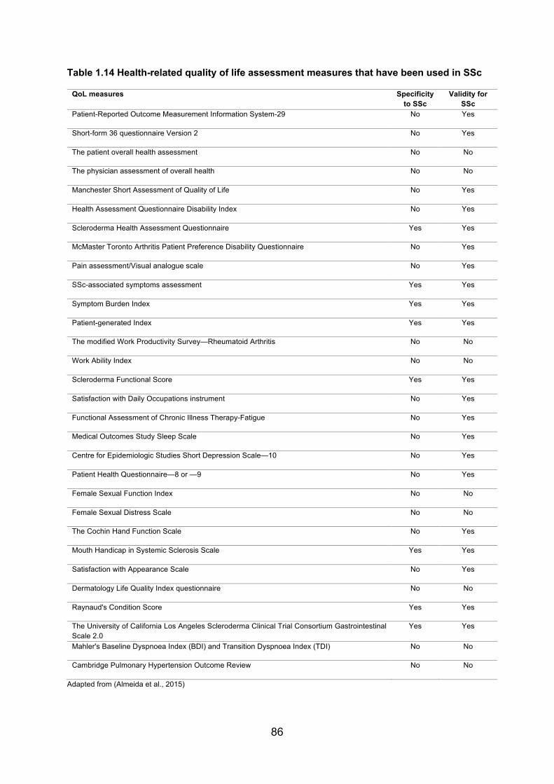
86
Table 1.14 Health-related quality of life assessment measures that have been used in SSc
QoL measures Specificity to SSc
Validity for SSc
Patient-Reported Outcome Measurement Information System-29 No Yes
Short-form 36 questionnaire Version 2 No Yes
The patient overall health assessment No No
The physician assessment of overall health No No
Manchester Short Assessment of Quality of Life No Yes
Health Assessment Questionnaire Disability Index No Yes
Scleroderma Health Assessment Questionnaire Yes Yes
McMaster Toronto Arthritis Patient Preference Disability Questionnaire No Yes
Pain assessment/Visual analogue scale No Yes
SSc-associated symptoms assessment Yes Yes
Symptom Burden Index Yes Yes
Patient-generated Index Yes Yes
The modified Work Productivity Survey—Rheumatoid Arthritis No No
Work Ability Index No No
Scleroderma Functional Score Yes Yes
Satisfaction with Daily Occupations instrument No Yes
Functional Assessment of Chronic Illness Therapy-Fatigue No Yes
Medical Outcomes Study Sleep Scale No Yes
Centre for Epidemiologic Studies Short Depression Scale—10 No Yes
Patient Health Questionnaire—8 or —9 No Yes
Female Sexual Function Index No No
Female Sexual Distress Scale No No
The Cochin Hand Function Scale No Yes
Mouth Handicap in Systemic Sclerosis Scale Yes Yes
Satisfaction with Appearance Scale No Yes
Dermatology Life Quality Index questionnaire No No
Raynaud's Condition Score Yes Yes
The University of California Los Angeles Scleroderma Clinical Trial Consortium Gastrointestinal Scale 2.0
Yes Yes
Mahler's Baseline Dyspnoea Index (BDI) and Transition Dyspnoea Index (TDI) No No
Cambridge Pulmonary Hypertension Outcome Review No No
Adapted from (Almeida et al., 2015)
87
Recently, a study has explored the relationship between different QoL measures of
skin manifestations in SSc and the skin histopathological features with clinical
examination. The informally developed new measurement, called The Skin Symptom
Assessment (SSA), evaluates six cutaneous symptoms (tight, painful, red, hard,
rigid/stiff and itchy) with a score of five-level Likert Scale (not at all, a little bit,
somewhat, moderately and severely). Among 41 patients, the SSA correlated well with
histopathological features of the biopsies of the 29 patients who had provided biopsy
material. The new SSA questionnaire requires further assessment, but it might aid the
evaluation of SSc skin disease and improve the ability to measure the effectiveness
of different treatment modalities (Ziemek et al., 2016).
Using the paper profiles of 150 patients, a new outcome measurement tool was
developed – called the Composite Response Index in SSc (CRISS). The new
composite index (CRISS) was tested in a randomised control trial of methotrexate
versus placebo (p=0.02). CRISS is a two-step process employed in a 12 months
interval period between primary and secondary measures. At present, however, this
new composite index is considered to be only at the provisional stage of use and may
require further assessment for use in clinical studies (Khanna et al., 2016).
Oral health-related quality of life measures in systemic sclerosis
As reviewed previously despite the potential impact of its orofacial features no quality
of life measure has been specifically constructed to assess the impact of SSc upon
the oral quality of life other than the Mouth Handicap scale (MHISS) (Mouthon et al.,
2007).
The MHISS was developed to assesses the degree of restriction of mouth opening,
dryness of the mouth and the aesthetic appearance. It consists of 12 items (each with
five levels of answers) divided into 3 subscales. Subscale 1 examines impact related
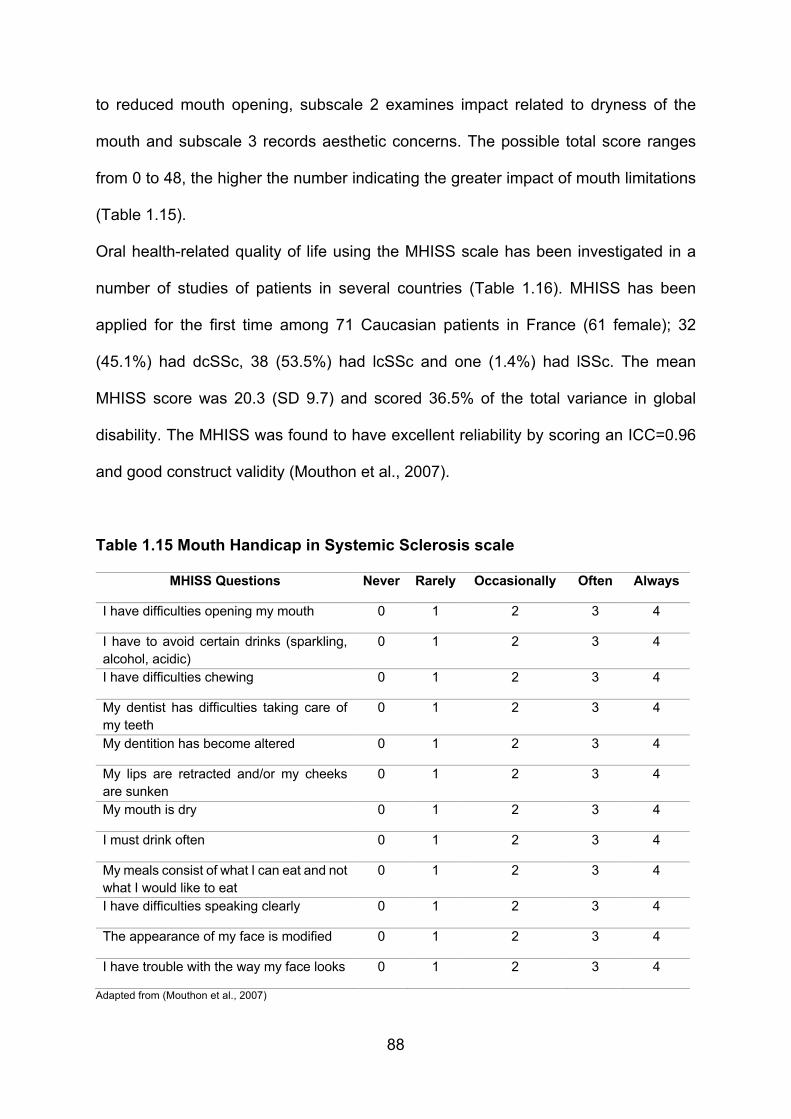
88
to reduced mouth opening, subscale 2 examines impact related to dryness of the
mouth and subscale 3 records aesthetic concerns. The possible total score ranges
from 0 to 48, the higher the number indicating the greater impact of mouth limitations
(Table 1.15).
Oral health-related quality of life using the MHISS scale has been investigated in a
number of studies of patients in several countries (Table 1.16). MHISS has been
applied for the first time among 71 Caucasian patients in France (61 female); 32
(45.1%) had dcSSc, 38 (53.5%) had lcSSc and one (1.4%) had lSSc. The mean
MHISS score was 20.3 (SD 9.7) and scored 36.5% of the total variance in global
disability. The MHISS was found to have excellent reliability by scoring an ICC=0.96
and good construct validity (Mouthon et al., 2007).
Table 1.15 Mouth Handicap in Systemic Sclerosis scale
MHISS Questions Never Rarely Occasionally Often Always
I have difficulties opening my mouth 0 1 2 3 4
I have to avoid certain drinks (sparkling, alcohol, acidic)
0 1 2 3 4
I have difficulties chewing 0 1 2 3 4
My dentist has difficulties taking care of my teeth
0 1 2 3 4
My dentition has become altered 0 1 2 3 4
My lips are retracted and/or my cheeks are sunken
0 1 2 3 4
My mouth is dry 0 1 2 3 4
I must drink often 0 1 2 3 4
My meals consist of what I can eat and not what I would like to eat
0 1 2 3 4
I have difficulties speaking clearly 0 1 2 3 4
The appearance of my face is modified 0 1 2 3 4
I have trouble with the way my face looks 0 1 2 3 4
Adapted from (Mouthon et al., 2007)

89
The MHISS has been translated into different languages including (English, French,
Italian and Dutch). The Italian version of MHISS was validated with 40 SSc patients (7
had dcSSc, 33 had lcSSc). The total MHISS score was 17.65 (SD 5.20) with good
reliability (ICC=0.93) and validity. Good external consistency was found with mouth
opening (-0.3869, p=0.0137) and good internal consistency by (Cronbach’s alpha =
0.93) (Maddali Bongi et al., 2012). A Dutch version has been employed in a study of
52 SSc patients from the Netherlands, 27 (52%) of whom had dcSSc. The mean total
score was 17.5 (SD 10.0). Good convergent validity was demonstrated, and excellent
reliability with (ICC = 0.94), Cronbach’s alpha was (0.88) (Schouffoer et al., 2013).
It has been suggested that the MHISS may be useful in assessing any improvement
of the face and mouth after physiotherapy and evaluating the outcomes after the dental
therapeutic intervention (Maddali-Bongi et al., 2011), although there is little evidence
that this has been undertaken.
The MHISS has been employed with other HRQoL measures including Short Health
Survey (SF-36), Hospital Anxiety and Depression (HADS), HAMIS (Hand Mobility In
SSc) and Health Assessment Questionnaire (HAQ), to assess a group of 46 female
patients with SSc (29 had lcSSc and 17 had dcSSc) assessing hand and mouth
disability in association with sexual function and, unsurprisingly, it was found that hand
and mouth involvement may lessen the sexual function of patients due to hardening
of face skin, digital ulceration and skin sclerosis with total MHISS (p=0.038) (Maddali
Bongi et al., 2013).
A cross-sectional survey of 381 patients with SSc from France, 187 (50.5%) had
lcSSc, 149 (40.3%) had dcSSc and 34 (9.2%) had lSSc assessed by different
measures, including the MHISS, found that mouth handicap was significantly higher
in patients with symptoms of anxiety and depression (Nguyen et al., 2014). Another

90
study of 248 SSc patients from France, 146 (58.9%) had lcSSc, 90 (36.3%) had
dcSSc, 12 (4.8%) had lSSc aimed to compare hospitalised patients with those from
patient associations using MHISS with other health-related quality of life measures.
Mouth impairment was significantly greater in the patient association group than the
group of hospitalised patients with MHISS mean scores of 20.65 (SD = 10.91) and
13.25 (SD = 9.26) p = 0.0001 respectively. It was considered that this might reflect the
non-hospitalised patients being older and having had the condition longer than
hospitalised patients. (Mestre-Stanislas et al., 2010).
Certainly, the MHISS is likely to be greater in those individuals who have more
orofacial involvement than those who do not. This was indeed found in a study of 119
patients from Italy in correlation with the global disability measures including Short
Health Survey (SF-36), Hand Mobility In SSc (HAMIS) and Health Assessment
Questionnaire (HAQ), in which the total score of MHISS was found to be higher in
dcSSc patients with reduced mouth opening score than the lcSSc group (Maddali-
Bongi et al., 2014).
Despite not being specific for SSc a number of other health-related quality of life
measures have been employed to investigate the impact of the oral aspects of SSc
upon the quality of life. For example, the Oral Health Impact Profile (OHIP) and Short
Health Survey (SF-36).
The Oral health impact profile (OHIP) is a questionnaire designed to measure oral
health-related QoL in adults with oral disease. The OHIP-49 consists of 49 items
representing seven aspects including (functional limitation, physical pain,
psychological discomfort, physical disability, psychological disability, social disability
and handicap) and has been found to be reliable and valid to evaluate changes and
to exhibit suitable cross-cultural consistency, based on a conceptual oral health model

91
outlined by Locker et al. (Slade and Spencer, 1994). The short form of OHIP (OHIP-
14) consists of two questions in each of the seven OHIP aspects. Both OHIP
questionnaires have been found to be useful tools for use in clinical practice with good
reliability, validity and precision. Each question is rated on a 5-point Likert scale, and
patients are invited to answer questions by choosing from a 0-4 scale while 0=never
and 4=very often (Allen and Locker, 1997, Slade, 1997).
OHIP was used to assess the OHRQoL of 163 patients with SSc and 231 controls.
The SSc patients had a significantly increased number of carious and missing teeth,
more periodontal inflammation, less saliva production and less interincisal distance
than the control subjects. However of relevance to OHRQoL the patients with SSc had
significantly higher scores in all seven subscales of the OHIP (p<0.01) with an overall
mean score 41.58 vs 26.67 (p<0.0001) respectively (Baron et al., 2014).
A study of 39 patients with SSc (22 in the intervention group and 17 as a control group)
completed the two OHIP versions at three different time points. An acceptable stability
was found with ICC (0.50 – 0.86) but due to the large standard error of measurement
and the large coefficient of repeatability, this indicated that OHIP was precise nor
sensitive tool to assess change (Yuen and Nelson, 2014).
Measuring pain in SSc is one of the most common global assessments in SSc. There
are, however, no standardisation measurements, although the visual analogue scale
(VAS) is commonly employed.
The Visual Analogue Scale consists of a 10-cm line with verbal anchors labelling both
ends. It is a straight line with the left end of the line, or 0, meaning no pain (none) and
the right end of the line, or 10, representing the worst pain that can happen. Patients
are invited to mark the line according to the intensity of their pain (Ni Riordain et al.,
2011). Feeling pain among SSc patients is found to be associated with other disease

92
impairments such as sleep problems, fatigue, depression and physical disability. Thus,
pain in SSc, unsurprisingly, is associated with a reduced HRQoL and being greater in
those with dcSSc than lcSSc (Pope, 2011, Kwakkenbos et al., 2015).
The 36-item short-form health survey (SF-36) is designed as a generic assessment of
health status used in a wide range of types and severity of conditions. The 36 items
includes eight concepts of health (physical functioning, role-physical, bodily pain,
general health, vitality, social functioning, role-emotional and mental health). However,
after answering the questionnaire, all scores have to be added together and
transformed on a scale from 0 (worst health) to 100 (best health) (Ni Riordain et al.,
2011). However, the SF-36 instrument is not a specific measure, but it has already
been used in SSc with good validity and responsiveness (Nguyen et al., 2014, Almeida
et al., 2015).
Patients with diffuse cutaneous SSc may have some degree of body image
dissatisfaction in relation to the disease changes. The satisfaction with appearance
scale (SWAP) was developed to assess the body image disfigurement among a
variety of disorders, including SSc. It is a 14 items questionnaire designed to focus on
two aspects of the disease (subjective body image satisfaction and social impact). It
has four subscales assessing the social distress, facial features, non-facial features
and social impact. A brief version of the SWAP was developed and consists of 6 items
with the same two subscales previously identified for SSc (Jewett et al., 2010). Both
forms of SWAP present a good to excellent level of internal consistency, reliability and
validity. However, it has been suggested that the brief SWAP might be more specific
for SSc, being useful for the identification of support (for example psychological) for
patients with SSc (Mills et al., 2015, Almeida et al., 2015).

93
Although not a measure of the quality of life, the interincisal opening is often used to
objectively assess the effects of fibrosis upon the mouth.
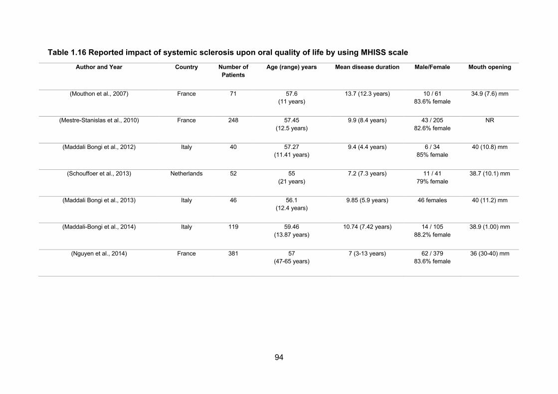
94
Table 1.16 Reported impact of systemic sclerosis upon oral quality of life by using MHISS scaleAuthor and Year Country Number of
Patients Age (range) years Mean disease duration Male/Female Mouth opening
(Mouthon et al., 2007) France 71 57.6 (11 years)
13.7 (12.3 years) 10 / 61 83.6% female
34.9 (7.6) mm
(Mestre-Stanislas et al., 2010) France 248 57.45 (12.5 years)
9.9 (8.4 years) 43 / 205 82.6% female
NR
(Maddali Bongi et al., 2012) Italy 40 57.27 (11.41 years)
9.4 (4.4 years) 6 / 34 85% female
40 (10.8) mm
(Schouffoer et al., 2013) Netherlands 52 55 (21 years)
7.2 (7.3 years) 11 / 41 79% female
38.7 (10.1) mm
(Maddali Bongi et al., 2013) Italy 46 56.1 (12.4 years)
9.85 (5.9 years) 46 females 40 (11.2) mm
(Maddali-Bongi et al., 2014) Italy 119 59.46 (13.87 years)
10.74 (7.42 years) 14 / 105 88.2% female
38.9 (1.00) mm
(Nguyen et al., 2014) France 381 57 (47-65 years)
7 (3-13 years) 62 / 379 83.6% female
36 (30-40) mm
95
AIMS AND OBJECTIVES
In view of the paucity of information regarding the oral disease of systemic sclerosis
in patients resident in the UK, the aims of this thesis are:
1. To determine the orofacial features and oral complications in a large cohort of
individuals with SSc in the UK.
2. To determine the impact of SSc upon access to dental care and oral health
care needs of SSc patients in the UK.
3. To assess the quality and readability of available online information regarding
oral health in SSc disease.
4. To determine the impact of SSc disease upon health-related quality of life
(HRQoL) and oral health-related quality of life (OHRQoL).
5. To explore the psychometric properties of the Mouth Handicap in Systemic
Sclerosis (MHISS) specifically exploring aspects of validity and reliability of
MHISS in UK SSc patients.
96
CHAPTER 2: A retrospective analysis of orofacial features of systemic
sclerosis
1 Introduction
As reviewed in chapter 1 systemic sclerosis (SSc) is a relatively rare autoimmune
connective tissue disease of middle-aged and elderly persons that may adversely
impact upon the quality of life of affected persons (Almeida et al., 2015). SSc is
characterised by three pathophysiological features: i. obstructive vasculopathy of the
small blood vessels, ii. immunological dysfunction followed by iii. cellular inflammation
(Baron et al., 2015b). Inflammation and fibrotic changes characteristically lead to
thickening of the skin and fibrosis of internal organs particularly the lungs,
gastrointestinal tract and kidneys and/or associated with high morbidity and mortality
(Rubio-Rivas et al., 2014, Royle et al., 2018). As indicated in chapter 1 patients with
SSc are suggested to have a wide range of orofacial problems such as fibrosis of the
facial skin, mask-like facial appearance, telangiectasia, stiffening of the tongue and
oral mucosa, microstomia, salivary gland dysfunction and xerostomia, dysphagia as
well as a potential risk of caries, periodontal disease and head and neck malignancy
(Jung et al., 2016, Veale et al., 2016, Crincoli et al., 2016a, Smirani et al., 2018b).
Despite its potential impact upon the mouth, little is known about the frequency of
orofacial features experienced by patients with SSc resident in the UK.
There are few detailed reports of the nature and impact of oral features of SSc, and
most studies were undertaken outside the United Kingdom. Therefore, knowledge
about the orofacial features of SSc and the related oral health care needs is needed
to clarify the oral treatment that affected individuals may require.
97
2 Material and Methods
2.1 Study Group
The current analysis incorporates data of 138 patients with SSc who attend or
previously attended, the Oral Medicine Unit of UCLHT Eastman Dental Hospital for
the care of their mouths between June 1994 and December 2018. The case records
of all patients were reviewed carefully to obtain details of the patients’ address, age,
gender, ethnicity, past medical history, details of their referral source and any
treatment received in UCLHT Eastman Dental Hospital. Data of the dental and oral
status was extracted systematically, coded and recorded on predetermined Excel data
sheets for subsequent statistical analysis.
It is appreciated that different diagnostic criteria for SSc have been applied over time
due to increased knowledge and changing diagnostic criteria. However, the diagnosis
has some consistency as most patients in this study had been referred with a diagnosis
of SSc confirmed by a specialist team in the Rheumatology Department of the Royal
Free Hospital – London. All patients were subsequently found to have clinical, and
usually histo-pathological features of SSc.
The disease categorisation divided the patients into three groups: those with diffuse
cutaneous SSc (dcSSc), limited cutaneous SSc (lcSSc) or mixed/overlap SSc. lcSSc
was defined as skin involvement distal to the elbows and knees, with or without face
involvement. dcSSc was defined as skin involvement proximal to the elbows and
knees, with or without truncal involvement. Mixed and/or overlap SSc was proposed
to describe the existance of SSc and other autoimmune connective tissue diseases
with the presence of related clinical features and/or serological autoantibodies
(Desbois and Cacoub, 2016, Denton, 2016). Orofacial features related to SSc were
98
reported based on detailed clinical examination by clinicians experienced in the field
of oral medicine, Professor Stephen Porter (SRP) or Professor Stefano Fedele (SF).
2.2 Statistical Analysis
Descriptive statistical analysis of data was summarised to describe the demographics
and characteristics of patients with SSc. Comparisons of different variables were
performed by using classical statistics, including chi-square test for categorical
variables, Student’s t-test and Mann-Whitney U test for continuous variables. All
statistical analysis was performed using SPSS 25 (version), and the significance level
was taken when the p-value was less than 0.05.
2.3 Ethical considerations
No ethical approval was required, and data management was conducted with
appropriate confidentiality.
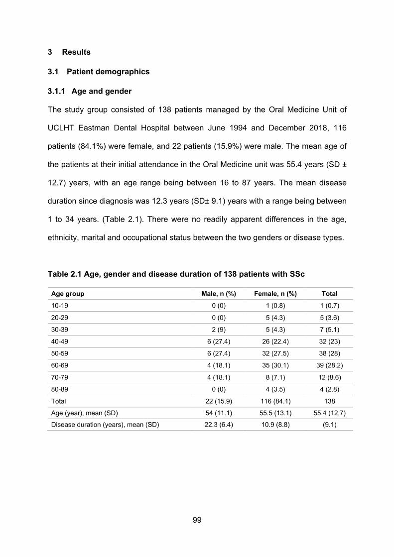
99
3 Results
3.1 Patient demographics
Age and gender
The study group consisted of 138 patients managed by the Oral Medicine Unit of
UCLHT Eastman Dental Hospital between June 1994 and December 2018, 116
patients (84.1%) were female, and 22 patients (15.9%) were male. The mean age of
the patients at their initial attendance in the Oral Medicine unit was 55.4 years (SD ±
12.7) years, with an age range being between 16 to 87 years. The mean disease
duration since diagnosis was 12.3 years (SD± 9.1) years with a range being between
1 to 34 years. (Table 2.1). There were no readily apparent differences in the age,
ethnicity, marital and occupational status between the two genders or disease types.
Table 2.1 Age, gender and disease duration of 138 patients with SSc
Age group Male, n (%) Female, n (%) Total
10-19 0 (0) 1 (0.8) 1 (0.7)
20-29 0 (0) 5 (4.3) 5 (3.6)
30-39 2 (9) 5 (4.3) 7 (5.1)
40-49 6 (27.4) 26 (22.4) 32 (23)
50-59 6 (27.4) 32 (27.5) 38 (28)
60-69 4 (18.1) 35 (30.1) 39 (28.2)
70-79 4 (18.1) 8 (7.1) 12 (8.6)
80-89 0 (0) 4 (3.5) 4 (2.8)
Total 22 (15.9) 116 (84.1) 138
Age (year), mean (SD) 54 (11.1) 55.5 (13.1) 55.4 (12.7)
Disease duration (years), mean (SD) 22.3 (6.4) 10.9 (8.8) (9.1)

100
Ethnic group:
The majority of patients were White British (81; 58.7%), 10 were Indian (7.2%),6 Black
African (4.3%) and other ethnicity backgrounds including 7 black Caribbean (5.1%),12
other white patients (8.7%), 3 Asian patients (2.2%), 2 Pakistani patients (1.4%), 1
Bangladesh patient (0.7%) and 16 patients belong to another unspecified racial group
(11.6%) (Table 2.2).
Table 2.2 Ethnicity of patients with SSc Ethnic group Number of patients % of group
British White 81 58.7
Other White 12 8.7
Indian 10 7.2
Black Caribbean 7 5.1
Black African 6 4.3
Pakistani 2 1.4
Asian 3 2.2
Bangladesh 1 0.7
Another ethnicity 16 11.6
3.1.3 Marital status:
Marital status was stated under four categories; married - which included married
patients and patients with civil partnership; single, divorced and widowed. Eighty
(58%) patients were married or living with a partner. 36 (26.1%) were single, 12 (8.7%)
were widowed, 10 (7.2%) were divorced (Table 2.3).
Table 2.3 Marital status of patients with SSc
Marital status Number of patients % of group
Married 80 58
Single 36 26.1
Widowed 12 8.7
Divorced 10 7.2
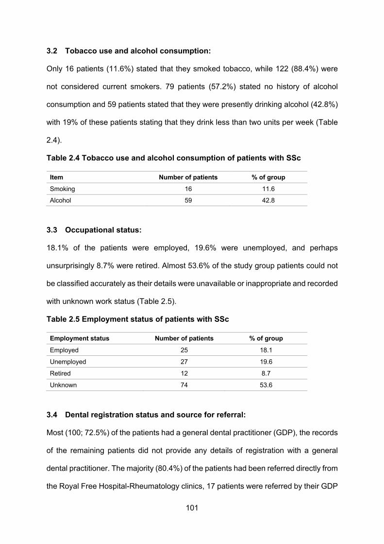
101
3.2 Tobacco use and alcohol consumption:
Only 16 patients (11.6%) stated that they smoked tobacco, while 122 (88.4%) were
not considered current smokers. 79 patients (57.2%) stated no history of alcohol
consumption and 59 patients stated that they were presently drinking alcohol (42.8%)
with 19% of these patients stating that they drink less than two units per week (Table
2.4).
Table 2.4 Tobacco use and alcohol consumption of patients with SSc
Item Number of patients % of group
Smoking 16 11.6
Alcohol 59 42.8
3.3 Occupational status:
18.1% of the patients were employed, 19.6% were unemployed, and perhaps
unsurprisingly 8.7% were retired. Almost 53.6% of the study group patients could not
be classified accurately as their details were unavailable or inappropriate and recorded
with unknown work status (Table 2.5).
Table 2.5 Employment status of patients with SSc
Employment status Number of patients % of group
Employed 25 18.1
Unemployed 27 19.6
Retired 12 8.7
Unknown 74 53.6
3.4 Dental registration status and source for referral:
Most (100; 72.5%) of the patients had a general dental practitioner (GDP), the records
of the remaining patients did not provide any details of registration with a general
dental practitioner. The majority (80.4%) of the patients had been referred directly from
the Royal Free Hospital-Rheumatology clinics, 17 patients were referred by their GDP

102
(12.3%), five patients were referred from Oral and Maxillofacial Surgery clinics (3.6%),
4 patients were referred from a General Medical Practitioner (2.9%), and one patient
was referred from an Orthodontic specialist (0.7%) (Table 2.6).
Table 2.6 Dental registration status and source of referral for patients with SSc
Item Number of patients % of group
Registered with a dentist 100 72.5
Source of referral
Rheumatology 111 80.4
General dental practitioner 17 12.3
Oral maxillofacial surgeon 5 3.6
General medical practitioner 4 2.9
Orthodontics 1 0.7
3.5 Pattern and reasons for referral:
Most referrals were primarily directed to Oral Medicine clinics 114 (82.6%), 22 cases
were referred seeking dental treatment (16%), and two cases were referred for
periodontal treatment (1.4%) (Table 2.7). Reasons for referrals were usually linked to
patients’ complaints and treatment needs including “poor dentition” (50 patients
36.3%), microstomia (30 patients 21.7%), “oral medicine consultation” (23 patients
16.7%), dry mouth (20 patients 14.5%), sore mouth (11 patients 8%) with single
patients having complaints of oral ulceration, temporomandibular joint dysfunction,
micrognathia, altered taste and a burning sensation of the mouth (Table 2.8).
Table 2.7 Clinical speciality for referral of patients with SSc
Item Number of patients % of group
Oral medicine clinics 114 82.6
Dental treatment 22 16
Periodontal treatment 2 1.4
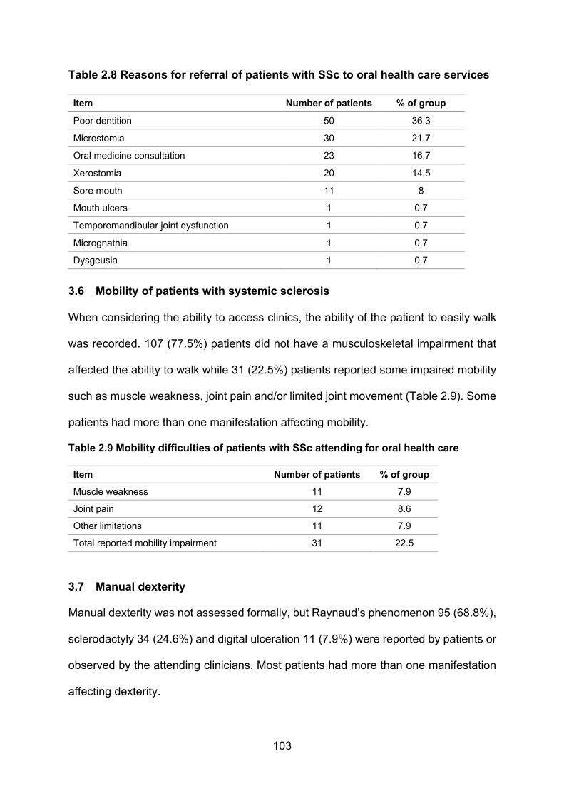
103
Table 2.8 Reasons for referral of patients with SSc to oral health care services
Item Number of patients % of group
Poor dentition 50 36.3
Microstomia 30 21.7
Oral medicine consultation 23 16.7
Xerostomia 20 14.5
Sore mouth 11 8
Mouth ulcers 1 0.7
Temporomandibular joint dysfunction 1 0.7
Micrognathia 1 0.7
Dysgeusia 1 0.7
3.6 Mobility of patients with systemic sclerosis
When considering the ability to access clinics, the ability of the patient to easily walk
was recorded. 107 (77.5%) patients did not have a musculoskeletal impairment that
affected the ability to walk while 31 (22.5%) patients reported some impaired mobility
such as muscle weakness, joint pain and/or limited joint movement (Table 2.9). Some
patients had more than one manifestation affecting mobility.
Table 2.9 Mobility difficulties of patients with SSc attending for oral health care
Item Number of patients % of group
Muscle weakness 11 7.9
Joint pain 12 8.6
Other limitations 11 7.9
Total reported mobility impairment 31 22.5
3.7 Manual dexterity
Manual dexterity was not assessed formally, but Raynaud’s phenomenon 95 (68.8%),
sclerodactyly 34 (24.6%) and digital ulceration 11 (7.9%) were reported by patients or
observed by the attending clinicians. Most patients had more than one manifestation
affecting dexterity.

104
3.8 Distance travelled
Distance travelled was estimated from the patient’s home address to UCLHT Eastman
Dental Hospital. Although some of the study participants were resident in London, the
category of 13 plus miles was the largest with around 30% of patients travelling an
estimated round trip in excess of 60 miles to access their dental care at the Hospital
(Table 2.10).
Table 2.10 Distance travelled to obtain dental care at the Eastman Dental
Hospital
Distance Number of patients % of group
1-4 miles 9 6.5
5-9 miles 21 15.2
10-19 miles 45 32.6
20-39 miles 32 23.1
More than 40 miles 31 22.6
3.9 Systemic involvement and past medical history
Cutaneous disease was the dominant non-oral feature of SSc for this group of patients
as almost two-thirds (67.4%) of the patients had or had had different types of
dermatological diseases including dermatomyositis, digital ulceration, Raynaud’s
phenomenon, sclerodactyly, calcinosis and nail fold abnormalities. Respiratory
manifestations of SSc, including pulmonary arterial hypertension (PAH) and interstitial
lung disease (ILD), were recorded in 53.6% of patients. Half of the patients had
problems related to the gastrointestinal tract such as oesophageal dysmotility,
abdominal discomfort, constipation, diverticulitis, gastric ulcers and gastroesophageal
reflux disease (GORD). However, cardiovascular and dermatological diseases were
more common among patients with dcSSc (p=.01). Additional details of past medical
history are provided in Table 2.11. Most patients had more than one feature of
systemic involvement.
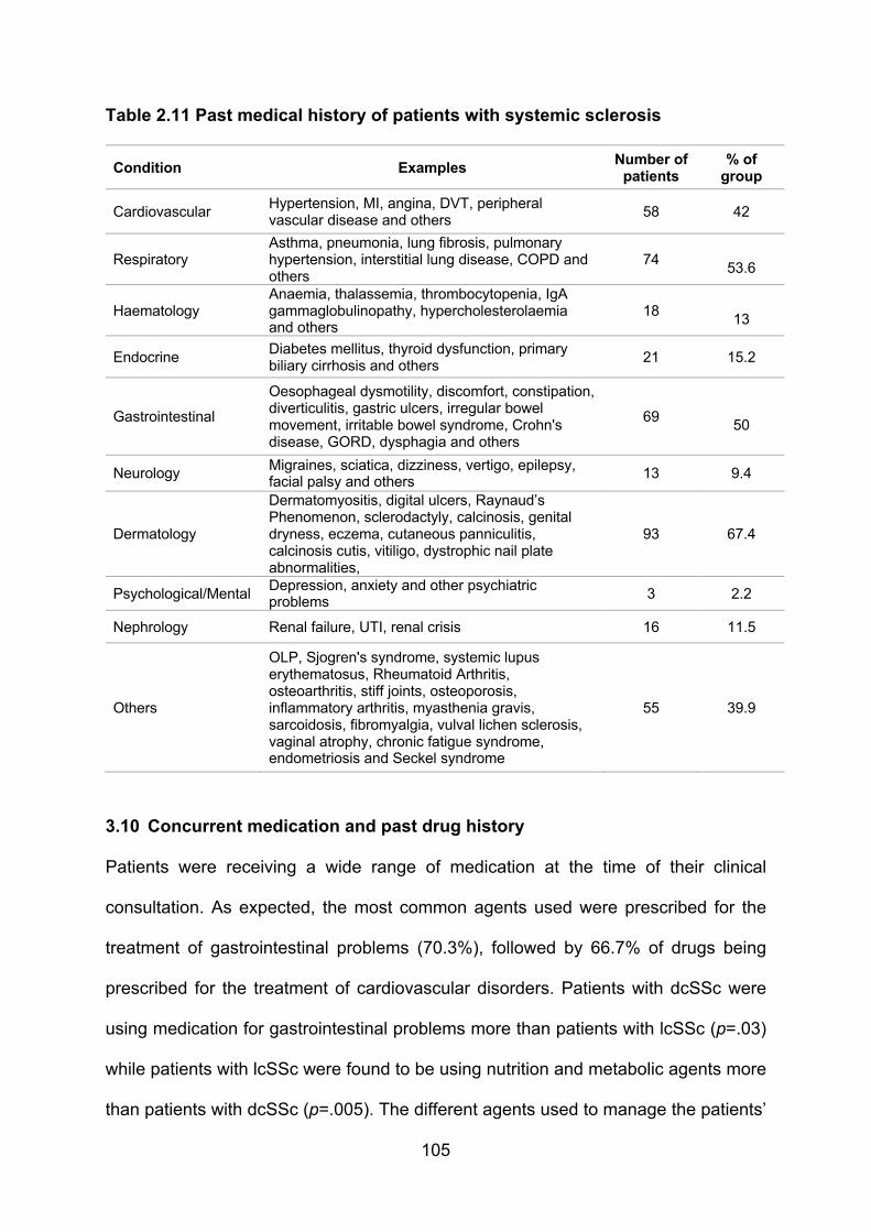
105
Table 2.11 Past medical history of patients with systemic sclerosis
Condition Examples Number of patients
% of group
Cardiovascular Hypertension, MI, angina, DVT, peripheral vascular disease and others
58 42
Respiratory Asthma, pneumonia, lung fibrosis, pulmonary hypertension, interstitial lung disease, COPD and others
74
53.6
Haematology Anaemia, thalassemia, thrombocytopenia, IgA gammaglobulinopathy, hypercholesterolaemia and others
18
13
Endocrine Diabetes mellitus, thyroid dysfunction, primary biliary cirrhosis and others
21 15.2
Gastrointestinal
Oesophageal dysmotility, discomfort, constipation, diverticulitis, gastric ulcers, irregular bowel movement, irritable bowel syndrome, Crohn's disease, GORD, dysphagia and others
69
50
Neurology Migraines, sciatica, dizziness, vertigo, epilepsy, facial palsy and others
13 9.4
Dermatology
Dermatomyositis, digital ulcers, Raynaud’s Phenomenon, sclerodactyly, calcinosis, genital dryness, eczema, cutaneous panniculitis, calcinosis cutis, vitiligo, dystrophic nail plate abnormalities,
93 67.4
Psychological/Mental Depression, anxiety and other psychiatric problems
3 2.2
Nephrology Renal failure, UTI, renal crisis 16 11.5
Others
OLP, Sjogren's syndrome, systemic lupus erythematosus, Rheumatoid Arthritis, osteoarthritis, stiff joints, osteoporosis, inflammatory arthritis, myasthenia gravis, sarcoidosis, fibromyalgia, vulval lichen sclerosis, vaginal atrophy, chronic fatigue syndrome, endometriosis and Seckel syndrome
55 39.9
3.10 Concurrent medication and past drug history
Patients were receiving a wide range of medication at the time of their clinical
consultation. As expected, the most common agents used were prescribed for the
treatment of gastrointestinal problems (70.3%), followed by 66.7% of drugs being
prescribed for the treatment of cardiovascular disorders. Patients with dcSSc were
using medication for gastrointestinal problems more than patients with lcSSc (p=.03)
while patients with lcSSc were found to be using nutrition and metabolic agents more
than patients with dcSSc (p=.005). The different agents used to manage the patients’
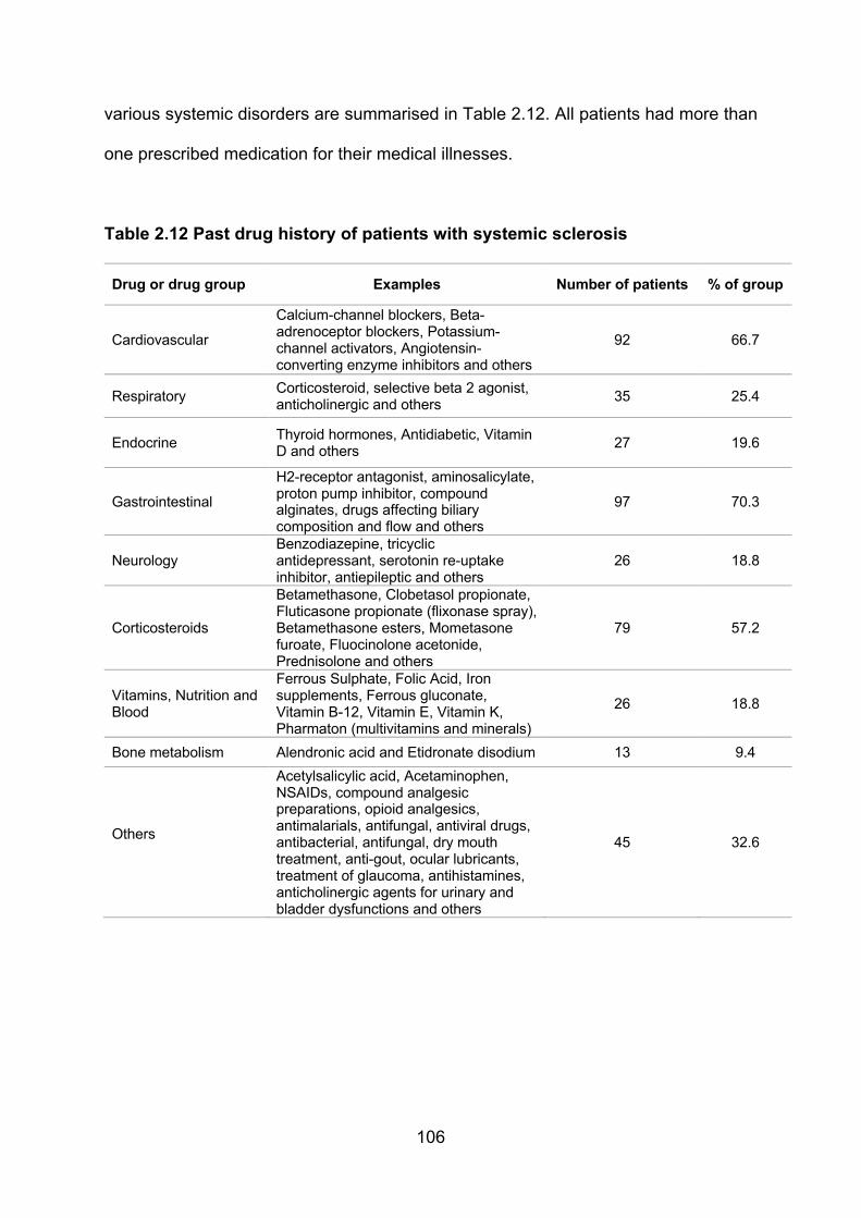
106
various systemic disorders are summarised in Table 2.12. All patients had more than
one prescribed medication for their medical illnesses.
Table 2.12 Past drug history of patients with systemic sclerosis
Drug or drug group Examples Number of patients % of group
Cardiovascular
Calcium-channel blockers, Beta-adrenoceptor blockers, Potassium-channel activators, Angiotensin-converting enzyme inhibitors and others
92 66.7
Respiratory Corticosteroid, selective beta 2 agonist, anticholinergic and others
35 25.4
Endocrine Thyroid hormones, Antidiabetic, Vitamin D and others
27 19.6
Gastrointestinal
H2-receptor antagonist, aminosalicylate, proton pump inhibitor, compound alginates, drugs affecting biliary composition and flow and others
97 70.3
Neurology Benzodiazepine, tricyclic antidepressant, serotonin re-uptake inhibitor, antiepileptic and others
26 18.8
Corticosteroids
Betamethasone, Clobetasol propionate, Fluticasone propionate (flixonase spray), Betamethasone esters, Mometasone furoate, Fluocinolone acetonide, Prednisolone and others
79 57.2
Vitamins, Nutrition and Blood
Ferrous Sulphate, Folic Acid, Iron supplements, Ferrous gluconate, Vitamin B-12, Vitamin E, Vitamin K, Pharmaton (multivitamins and minerals)
26 18.8
Bone metabolism Alendronic acid and Etidronate disodium 13 9.4
Others
Acetylsalicylic acid, Acetaminophen, NSAIDs, compound analgesic preparations, opioid analgesics, antimalarials, antifungal, antiviral drugs, antibacterial, antifungal, dry mouth treatment, anti-gout, ocular lubricants, treatment of glaucoma, antihistamines, anticholinergic agents for urinary and bladder dysfunctions and others
45 32.6
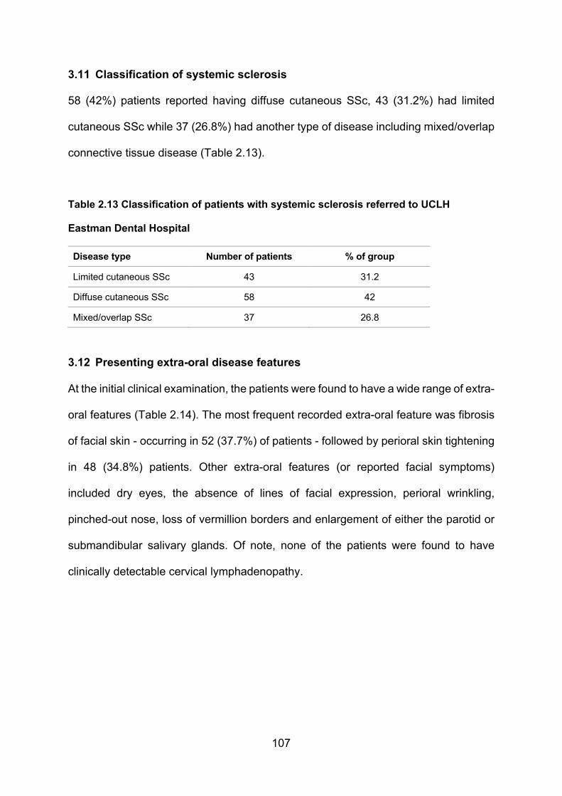
107
3.11 Classification of systemic sclerosis
58 (42%) patients reported having diffuse cutaneous SSc, 43 (31.2%) had limited
cutaneous SSc while 37 (26.8%) had another type of disease including mixed/overlap
connective tissue disease (Table 2.13).
Table 2.13 Classification of patients with systemic sclerosis referred to UCLH
Eastman Dental Hospital
Disease type Number of patients % of group
Limited cutaneous SSc 43 31.2
Diffuse cutaneous SSc 58 42
Mixed/overlap SSc 37 26.8
3.12 Presenting extra-oral disease features
At the initial clinical examination, the patients were found to have a wide range of extra-
oral features (Table 2.14). The most frequent recorded extra-oral feature was fibrosis
of facial skin - occurring in 52 (37.7%) of patients - followed by perioral skin tightening
in 48 (34.8%) patients. Other extra-oral features (or reported facial symptoms)
included dry eyes, the absence of lines of facial expression, perioral wrinkling,
pinched-out nose, loss of vermillion borders and enlargement of either the parotid or
submandibular salivary glands. Of note, none of the patients were found to have
clinically detectable cervical lymphadenopathy.
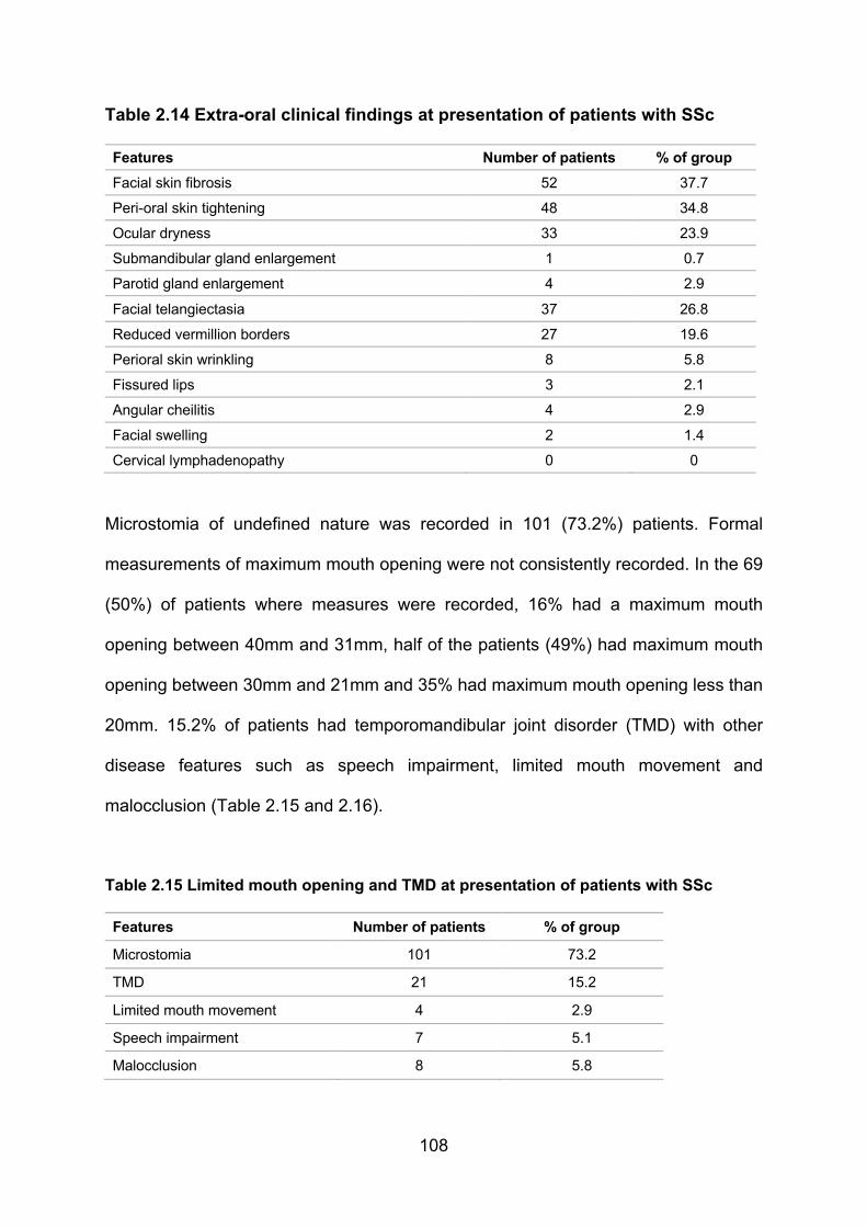
108
Table 2.14 Extra-oral clinical findings at presentation of patients with SSc
Features Number of patients % of group
Facial skin fibrosis 52 37.7
Peri-oral skin tightening 48 34.8
Ocular dryness 33 23.9
Submandibular gland enlargement 1 0.7
Parotid gland enlargement 4 2.9
Facial telangiectasia 37 26.8
Reduced vermillion borders 27 19.6
Perioral skin wrinkling 8 5.8
Fissured lips 3 2.1
Angular cheilitis 4 2.9
Facial swelling 2 1.4
Cervical lymphadenopathy 0 0
Microstomia of undefined nature was recorded in 101 (73.2%) patients. Formal
measurements of maximum mouth opening were not consistently recorded. In the 69
(50%) of patients where measures were recorded, 16% had a maximum mouth
opening between 40mm and 31mm, half of the patients (49%) had maximum mouth
opening between 30mm and 21mm and 35% had maximum mouth opening less than
20mm. 15.2% of patients had temporomandibular joint disorder (TMD) with other
disease features such as speech impairment, limited mouth movement and
malocclusion (Table 2.15 and 2.16).
Table 2.15 Limited mouth opening and TMD at presentation of patients with SSc
Features Number of patients % of group
Microstomia 101 73.2
TMD 21 15.2
Limited mouth movement 4 2.9
Speech impairment 7 5.1
Malocclusion 8 5.8
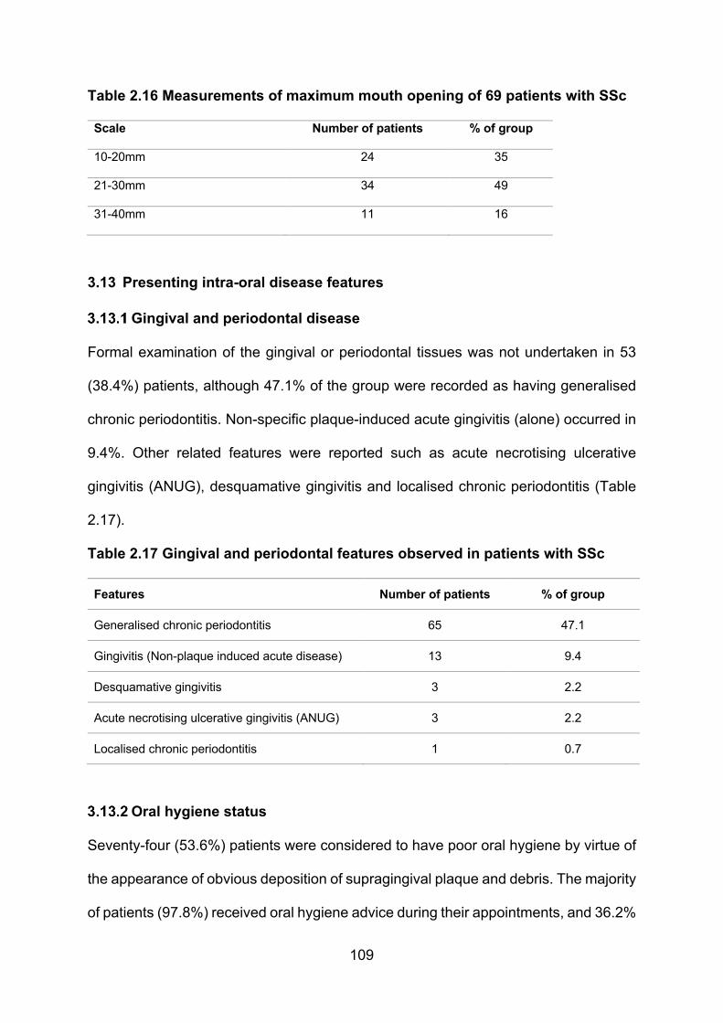
109
Table 2.16 Measurements of maximum mouth opening of 69 patients with SSc
Scale Number of patients % of group
10-20mm 24 35
21-30mm 34 49
31-40mm 11 16
3.13 Presenting intra-oral disease features
Gingival and periodontal disease
Formal examination of the gingival or periodontal tissues was not undertaken in 53
(38.4%) patients, although 47.1% of the group were recorded as having generalised
chronic periodontitis. Non-specific plaque-induced acute gingivitis (alone) occurred in
9.4%. Other related features were reported such as acute necrotising ulcerative
gingivitis (ANUG), desquamative gingivitis and localised chronic periodontitis (Table
2.17).
Table 2.17 Gingival and periodontal features observed in patients with SSc
Features Number of patients % of group
Generalised chronic periodontitis 65 47.1
Gingivitis (Non-plaque induced acute disease) 13 9.4
Desquamative gingivitis 3 2.2
Acute necrotising ulcerative gingivitis (ANUG) 3 2.2
Localised chronic periodontitis 1 0.7
Oral hygiene status
Seventy-four (53.6%) patients were considered to have poor oral hygiene by virtue of
the appearance of obvious deposition of supragingival plaque and debris. The majority
of patients (97.8%) received oral hygiene advice during their appointments, and 36.2%

110
of patients were prescribed toothpaste (usually high concentration fluoride agents)
and/or mouthwashes (usually Chlorohexidine). Furthermore, 43.5% of patients with
hypo-salivation (of known or unknown cause) were prescribed a variety of saliva
substitutes and/or oral lubricants. 64.5% of patients were referred for oral hygiene
therapy, and 26.1% were referred for specialist periodontal treatment (Table 2.18).
Table 2.18 Oral hygiene status of patients with systemic sclerosis
Item Number of patients % of group
Oral hygiene instructions and advice 135 97.8
Fluoride toothpaste and/or anti-microbial mouthwashes prescription
50 36.2
Saliva substitutes and/or lubricants 60 43.5
Hygiene therapy 89 64.5
Specialist periodontal treatment 36 26.1
Dental status
Unfortunately, no precise records of the present dental status of a substantial number
of patients were available as no formal record of the decayed, missing or filled teeth
(DMFT) was undertaken from the patient’s notes or referrals to the oral medicine unit.
The number or status of teeth decayed, missing, mobile or restored have not been
clearly indicated. However, 104 (75.4%) patients did have one or more decayed teeth,
107 (77.5%) had teeth with dental restorations, and 79 (57.2%) had missing
permanent teeth. 15.9% of patients had at least one clinically mobile tooth (the degree
of mobility was never assessed) and dental erosion was found in 8.7% of patients.
None of the patients were edentulous (Table 2.19).

111
Table 2.19 Decayed, mobility, missing and filled teeth of patients with systemic
sclerosis
Feature Number of patients % of group
Poor oral hygiene 74 53.6
Decayed teeth 104 75.4
Filled teeth 107 77.5
Mobile teeth 22 15.9
Missing teeth 79 57.2
Oral mucosal involvement
Just over half of the patients had apparently healthy oral mucosae (51.4%). 23.2%
had “dry lining mucosa”, 17.4% had erythematous mucosa, and a small number were
reported to have tight or “atrophic” mucosa 7.2% and 0.7% respectively. Mucosal
alterations were mostly manifested as a generalised involvement (87.7%) while only
12.3% of patients had localised involvement of buccal mucosae. Oral ulceration was
observed in 15.2% of patients while indentation of buccal mucosa occurred in 10.1%
of cases. Oral telangiectasia occurred in 29.7% of patients in a variety of locations
including the palate, buccal mucosa and tongue (Table 2.20).
Table 2.20 Oral mucosal involvement of patients with systemic sclerosis
Features Number of patients % of group
Dry lining mucosa 32 23.2
Erythematous mucosa 24 17.4
Tight mucosa 10 7.2
Atrophic mucosa 1 0.7
Denture stomatitis 4 2.9
Lichenoid hyperkeratosis 1 0.7
Ulceration 21 15.2
Submucosal fibrosis 1 0.7
Oral telangiectasia 41 29.7
Indentation of the buccal mucosa 14 10.1
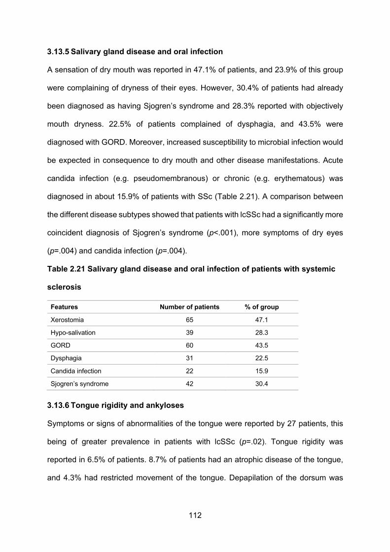
112
Salivary gland disease and oral infection
A sensation of dry mouth was reported in 47.1% of patients, and 23.9% of this group
were complaining of dryness of their eyes. However, 30.4% of patients had already
been diagnosed as having Sjogren’s syndrome and 28.3% reported with objectively
mouth dryness. 22.5% of patients complained of dysphagia, and 43.5% were
diagnosed with GORD. Moreover, increased susceptibility to microbial infection would
be expected in consequence to dry mouth and other disease manifestations. Acute
candida infection (e.g. pseudomembranous) or chronic (e.g. erythematous) was
diagnosed in about 15.9% of patients with SSc (Table 2.21). A comparison between
the different disease subtypes showed that patients with lcSSc had a significantly more
coincident diagnosis of Sjogren’s syndrome (p<.001), more symptoms of dry eyes
(p=.004) and candida infection (p=.004).
Table 2.21 Salivary gland disease and oral infection of patients with systemic
sclerosis
Features Number of patients % of group
Xerostomia 65 47.1
Hypo-salivation 39 28.3
GORD 60 43.5
Dysphagia 31 22.5
Candida infection 22 15.9
Sjogren’s syndrome 42 30.4
Tongue rigidity and ankyloses
Symptoms or signs of abnormalities of the tongue were reported by 27 patients, this
being of greater prevalence in patients with lcSSc (p=.02). Tongue rigidity was
reported in 6.5% of patients. 8.7% of patients had an atrophic disease of the tongue,
and 4.3% had restricted movement of the tongue. Depapilation of the dorsum was
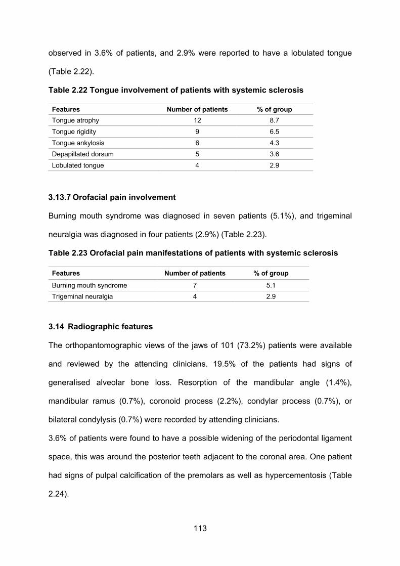
113
observed in 3.6% of patients, and 2.9% were reported to have a lobulated tongue
(Table 2.22).
Table 2.22 Tongue involvement of patients with systemic sclerosis
Features Number of patients % of group
Tongue atrophy 12 8.7
Tongue rigidity 9 6.5
Tongue ankylosis 6 4.3
Depapillated dorsum 5 3.6
Lobulated tongue 4 2.9
Orofacial pain involvement
Burning mouth syndrome was diagnosed in seven patients (5.1%), and trigeminal
neuralgia was diagnosed in four patients (2.9%) (Table 2.23).
Table 2.23 Orofacial pain manifestations of patients with systemic sclerosis
Features Number of patients % of group
Burning mouth syndrome 7 5.1
Trigeminal neuralgia 4 2.9
3.14 Radiographic features
The orthopantomographic views of the jaws of 101 (73.2%) patients were available
and reviewed by the attending clinicians. 19.5% of the patients had signs of
generalised alveolar bone loss. Resorption of the mandibular angle (1.4%),
mandibular ramus (0.7%), coronoid process (2.2%), condylar process (0.7%), or
bilateral condylysis (0.7%) were recorded by attending clinicians.
3.6% of patients were found to have a possible widening of the periodontal ligament
space, this was around the posterior teeth adjacent to the coronal area. One patient
had signs of pulpal calcification of the premolars as well as hypercementosis (Table
2.24).
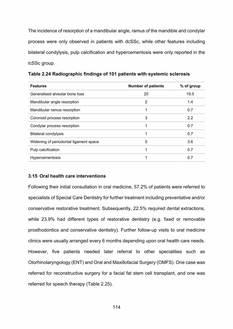
114
The incidence of resorption of a mandibular angle, ramus of the mandible and condylar
process were only observed in patients with dcSSc, while other features including
bilateral condylysis, pulp calcification and hypercementosis were only reported in the
lcSSc group.
Table 2.24 Radiographic findings of 101 patients with systemic sclerosis
Features Number of patients % of group
Generalised alveolar bone loss 20 19.5
Mandibular angle resorption 2 1.4
Mandibular ramus resorption 1 0.7
Coronoid process resorption 3 2.2
Condylar process resorption 1 0.7
Bilateral condylysis 1 0.7
Widening of periodontal ligament space 5 3.6
Pulp calcification 1 0.7
Hypercementosis 1 0.7
3.15 Oral health care interventions
Following their initial consultation in oral medicine, 57.2% of patients were referred to
specialists of Special Care Dentistry for further treatment including preventative and/or
conservative restorative treatment. Subsequently, 22.5% required dental extractions,
while 23.9% had different types of restorative dentistry (e.g. fixed or removable
prosthodontics and conservative dentistry). Further follow-up visits to oral medicine
clinics were usually arranged every 6 months depending upon oral health care needs.
However, five patients needed later referral to other specialities such as
Otorhinolaryngology (ENT) and Oral and Maxillofacial Surgery (OMFS). One case was
referred for reconstructive surgery for a facial fat stem cell transplant, and one was
referred for speech therapy (Table 2.25).

115
Table 2.25 Subsequent treatment of orofacial disease of patients with systemic
sclerosis
Interventions Number of patients % of group
Special Care Dentistry 79 57.2
Surgical treatment 31 22.5
Restorative treatment 33 23.9
OMFS 3 2.1
ENT 2 1.4
Speech therapy 1 0.7
Facial stem cell transplant 1 0.7
116
4. Discussion
The primary aim of this part of the study was to determine the key orofacial
manifestations and explore the nature of the oral health problems of a large cohort of
individuals with SSc and thus present the possible oral health care needs of such
patients. The present group of participants comprised 138 patients with different types
of SSc and thus represented the largest group of patients with SSc resident in the UK
to have ever been examined for aspects of their oral health.
The demographics of the present patient cohort confirm recent reports that SSc is
primarily a disease affecting middle to late age females (Chu et al., 2011, Willems et
al., 2014, Jung et al., 2016, Veale et al., 2016, Smirani et al., 2018b). Although SSc is
most commonly diagnosed in the middle to late age group, it can affect younger people
(Ranque and Mouthon, 2010, Hughes and Herrick, 2012, Gelber et al., 2013, Hansi et
al., 2014) as also demonstrated in the present study group. Although most of the
patients’ records did not specify employment status, a considerable number of the
patients were unemployed or retired. These data could be related to the morbidity of
the disease and/or physical disability in SSc patients (Decuman et al., 2015, Poole et
al., 2016, Morrisroe et al., 2018).
There have been several reports of the oral manifestations of SSc, and a recent large
multisite study of the Canadian systemic sclerosis oral health study group examined
several aspects of the disease characteristics, orofacial features and oral health-
related quality of life (Baron et al., 2014). The Canadian researchers analysed 163
patients with SSc. 90% were female, and the mean age of the group was 56 years,
72% had limited cutaneous subtype and 28% with diffuse cutaneous SSc (Baron et
al., 2015b). As expected, for a group of residents in the UK the patients were
predominantly female and predominantly white British (58.7%), also reflecting the

117
epidemiology of SSc in the UK (Kuo et al., 2011, Barnes and Mayes, 2012, Ingegnoli
et al., 2018). Different values have been recorded in this study with regards the
disease subtypes, 42% had a diffuse cutaneous subtype, 31.2% had limited cutaneous
SSc, and 26.8% had other mixed/overlap connective tissue diseases. Although the
data on the prevalence of SSc in the UK is limited, previous reports have shown that
the ratio of limited cutaneous SSc to diffuse cutaneous SSc was (4.7:1) (Allcock et al.,
2004, Willems et al., 2014). However, similar values have been reported recently
where dcSSc was slightly more common than other subtypes (26/50, 52%) (Gomes
da Silva et al., 2019). Differences in the distribution of disease subtype across
countries have been identified and may be attributed to different disease profiles or
different access to services. However, it is unclear to what extent the observed
differences in the disease subtypes reflect any differences in disease features or is a
consequence of sampling differences as the current results incorporate data of
patients who attend or previously attended one tertiary health care centre (the Oral
Medicine Unit of UCLHT Eastman Dental Hospital).
The majority of patients in the present study had general dental practitioners, but most
of them were referred from a (single) Rheumatology Centre in North London – Royal
Free Hospital, that provides a nationally recognised clinical service for the treatment
of SSc – managing 2000 out of approximately 12000-19000 patients living with SSc in
the UK (Royle et al., 2018). Although most of the patients were registered with general
dental clinics, the reasons for referral to the specialists clinics were generally for the
supposed difficulty in obtaining routine dental care and/or for further consultation and
treatment needs that mainly included poor dentition 36.3%, reduced mouth opening
21.7%, oral medicine consultation 16.7% and feeling dry mouth 14.5%. An alternative,
and possibly more likely reason for the present referral pattern is that it reflects the
118
rheumatology team having knowledge that the Oral Medicine unit of the EDH has an
interest in managing patients with complex connective tissue disease.
Despite supposedly having a general dental practitioner, 53.6% of the patient group
of 138 patients were considered by the oral medicine team to have poor oral hygiene,
and indeed 64.5% subsequently received oral hygiene care. More than half of the
patients were later referred to Special Care Dentistry (SCD) as well as other
specialities for further treatment. The dominant referral to SCD probably reflected the
levels of associated impaired mobility, difficult dental access due to reduced mouth
opening and abnormal manual dexterity making effective oral hygiene difficult and
hence subsequent plaque-related disease likely (Poole et al., 2010, Yuen et al., 2014a,
Alantar et al., 2011). As suggested by the present pattern of referral, and subsequent
oral health care, the oral health needs of patients with SSc may sometimes require the
skills and experience of clinicians from a variety of dental specialities (Leader et al.,
2014).
In the present study, a possible mask-like facial appearance (due to fibrosis) was
observed in 37.7% of cases. 34.8% of the patients had tightness of the perioral skin,
some within this group, and others had features such as the absence of lines of facial
expression, a perioral wrinkling, a pinched-out nose, a reduced vermillion borders
and/or facial telangiectasia. While such orofacial manifestations have been described
in about 80-90% of SSc cases (Bajraktari et al., 2015, Burchfield and Vorrasi, 2019),
it is possible that such obvious signs will impact adversely upon an individual’s
confidence in social interaction.
The present group of patients with SSc had reduced mouth opening in 73.2%, and
84% of them had objective evidence of reduced interincisal distance less than 30mm.
Similar findings have been reported in other large cohorts of patients with SSc, and
119
the mean interincisal distance was 37.7mm with more severely limited mouth opening
(mean=34.4mm) among patients with diffuse cutaneous subtype (Baron et al., 2014,
Jung et al., 2016). Microstomia has the potential to interfere considerably with oral
function, and also cause challenges with oral hygiene self-care and dental treatment,
due to the limited access (Alantar et al., 2011, Del Rosso and Maddali-Bongi, 2014).
The degree of decreased mouth opening has been found to correlate with overall
disease activity, the extent of skin involvement and specific SSc antibodies (Baron et
al., 2014). Of note, the majority of the present patients with limited oral opening had
dcSSc. Severely limited mouth opening might be more related to disease severity and
diffuse subtype of SSc.
The intra-oral clinical features of SSc have been described in several large cohorts
(Leung et al., 2011, Elimelech et al., 2015, Jung et al., 2016, Pischon et al., 2016). In
the present study, SSc gave rise to a variety of mucosal and periodontal
manifestations. 47.1% of the patients were found to have generalised chronic
periodontitis, while other gingival and soft tissue alterations included desquamative
gingivitis and localised chronic periodontitis. It has been reported that patients with
SSc can have a significant level of periodontal disease and higher periodontal
attachment loss compared with healthy controls (Baron et al., 2014, Pischon et al.,
2016). Previous reports have suggested that an increased risk of periodontal disease
may be related to the microvascular abnormalities of the disease and/or increased
collagen deposition (Chu et al., 2011). However, the cause of the periodontal disease
in the present group of patients with SSc is likely to be multifactorial in its nature and
is likely to be dominated by poor oral hygiene. Other suggested risk factors include
tobacco smoking, level of education, daily alcohol consumption and body mass index
(Isola et al., 2017). However, this is unlikely to be related for this patient group.

120
Although certain intra-oral manifestations such as thinning and atrophy of oral mucosa
can be related to the generalised fibrosis and tissue ischaemia involving both overlying
skin and gastrointestinal system 66 – 90% (Poole et al., 2013, Jung et al., 2016), the
present patients have little dryness, erythematous and tightening oral mucosa (23.2%,
17.4% and 7.2%) respectively. Mucosal alterations were mostly generalised
involvement which might be correlated to other manifestations of the disease including
malabsorption, drug-related adverse side effects or GORD (Alantar et al., 2011, Jung
et al., 2016).
Changes to the tongue, such as fibrosis leading to restricted movement of the tongue
or lingual fraenum (Chu et al., 2011, Vitali et al., 2015) have previously been observed
in patients with SSc. In the present study atrophy, ankylosis and rigidity of the tongue
were noted in a small number of patients. Atrophic tongue was significantly associated
with the lcSSc. Tongue involvement can adversely impact upon speech, eating and
swallowing (Fischer and Patton, 2000, Vitali et al., 2015, Crincoli et al., 2016b) and
hence, although uncommon, can ultimately lessen general health and quality of life.
Perhaps, as might be expected given the oral hygiene status of the patients, 75.4% of
the group had at least one carious tooth, 77.5% had filled teeth, and 57% had missing
teeth. These findings of perhaps high levels of present and past dental disease agree
with previous reports which revealed that patients with SSc have significantly worse
dental health including more missing teeth and periodontal disease than those without
SSc (Chu et al., 2011, Baron et al., 2014, Baron et al., 2015b). Also, it has been
reported that these patients are more likely to have salivary gland hypofunction and
reduced mouth opening (Baron et al., 2014, Crincoli et al., 2016b).
Interestingly, it was reported that the number of missing teeth among patients with
SSc was associated with salivary dysfunction, as for every 10 mg/min increase of
121
saliva production the number of missing teeth decreased by 3%. Furthermore, a
significant association was found between missing teeth and gastroesophageal reflux
disease (Baron et al., 2015b). These data might explain the high level of missing teeth
among the present sample as 43.5% of the patients were diagnosed with GORD.
However, it would seem that the possible difficulties in being able to clean their teeth
due to reduced mouth opening and reduced manual dexterity and/or oral dryness due
to Sjogren’s syndrome, or possibly drug therapy, resulted in these patients with SSc
having a substantial dental disease burden.
53.6% of patients were considered to have poor oral hygiene. Almost all patients
initially required oral hygiene instruction and advice and 64.5% of patients were
referred to have oral hygiene therapy, while 26.1% required advanced periodontal
treatment. The ability to maintain a good level of oral hygiene may reflect microstomia,
reduced manual dexterity and/or associated co-morbidity of the disease (Poole et al.,
2013, Del Rosso and Maddali-Bongi, 2014).
Of relevance, fatigue, Raynaud’s phenomenon, stiffness of the hands and joint pain
have been found to adversely impact upon the ability to carry out everyday activities,
as well as, somehow, lessening the opening of the mouth, and the ability to swallow
(Bassel et al., 2011, Willems et al., 2014).
Several studies have shown that patients with SSc do have difficulties in the
maintenance of oral hygiene care, while the microstomia of SSc may compromise oral
hygiene care by patients (Poole et al., 2004). Measures to enable individuals with SSc
to maintain good oral hygiene have been suggested and include regular follow-up
visits, patient education and using adaptive oral hygiene devices (e.g. angled brushes,
altered filament length brushes, easy-grip brushes, extended handle brushes, electric
brushes and soft small-headed toothbrushes (Alantar et al., 2011, Jung et al., 2016).
122
47.1% of the present group of patients with SSc reported having a feeling of dry mouth
and 22.5% having dysphagia. Indeed, salivary gland hypofunction was reported in
28.3% of patients with SSc but unfortunately not objectively measured in all the
present group of patients with SSc. These present results are in line with those of other
studies that reported that salivary gland hypofunction in patients with SSc was 33.9%
and associated with the presence of Sjogren’s syndrome-related antibodies (Kobak et
al., 2013). 29.9% of the present group of patients had been diagnosed as having
Sjogren’s syndrome. The presence of Sjogren’s syndrome in systemic sclerosis is well
known (14-33.9%), and certainly it would be important to assess all patients with SSc
and oral dryness for Sjogren’s syndrome in view of the risk of non-Hodgkin’s
lymphoma (Chu et al., 2011, Jung et al., 2016). Indeed it has been recommended that
all patients with SSc should be investigated for Sjogren’s syndrome as it was reported
that the prevalence of SS among patients with SSc is ranged from 17-29% (Kobak et
al., 2013).
The patients were receiving a wide spectrum of drugs for the management of SSc and
other disorders. Some of these agents will cause or worsen oral dryness (Elimelech
et al., 2015). Previous evidence has been reported that the use of such drug therapies
was thought to be related to increasing pre-existing xerostomia in 76.3% of the study
sample (Baron et al., 2014, Baron et al., 2015b) and hence increasing the risk of
plaque-related oral disease as well as dental decay. Treatment adverse side effects
among patients with SSc are common as certain medications (e.g.
immunosuppressive, cytotoxic drugs, antihypertensive, antidepressant and
anticoagulant drugs) can give rise to some oral complications such as (mucosal
ulceration, xerostomia, gingival bleeding and hyperplasia) (Alantar et al., 2011,
Elimelech et al., 2015, Porter et al., 2017). Other therapeutic agents (e.g. Calcium

123
channel blockers, corticosteroids and Medication-related to osteonecrosis) may be
responsible for specific symptoms such as increased risk of oral infections, mucositis,
impaired wound healing and risk of jaw osteonecrosis (Yuen et al., 2014b, Mawardi et
al., 2016, Jung et al., 2016). Considering the many adverse effects that can be
observed, such therapies should be used carefully, and all patients should undergo
clinical and radiographic examination prior to commencing any dental treatment.
Maxillofacial radiographic features of SSc have been reported among patients with
SSc in different cohort studies (Marcucci and Abdala, 2009, Dagenais et al., 2015,
Crincoli et al., 2016b, Burchfield and Vorrasi, 2019). Periodontal ligament (PDL)
widening is a reported radiographic feature of SSc that might be due to excessive
pathological collagen deposition. However, the present results showed that only 3.6%
of patients have signs of PDL widening. This value is significantly lower than results
from other studies that ranged from 10% - 38% of patients with SSc (Baron et al.,
2015a, Jung et al., 2016). Present results might be related to a relatively small sample
size having radiographic records and the measurements that have been performed,
were on panoramic radiographs that are known to be prone to image distortion.
Although no evidence of a clear correlation between PDL widening and other
periodontal diseases have been reported, careful assessment of similar radiographic
features and awareness of this specific disease manifestation can prevent misleading
diagnosis among patients with SSc.
Bone resorption is considered common (6.6% - 46.7) in patients with SSc (Jung et al.,
2016). However, there is no clear correlation between disease severity and the
incidence of bone resorption (Vincent et al., 2009, Baron et al., 2015a). Present results
showed that up to 19.5% of patients have a generalised pattern of alveolar bone loss.
Furthermore, a different pattern of mandibular bone resorption has been reported with

124
most frequently affected sites; coronoid process 2.2%, angle of the mandible 1.4%,
condylar process, ramus of the mandible and bilateral condylysis 0.7%. Higher values
have been reported from previous studies with a different range of severity that rarely
can result in facial asymmetry, malocclusion and pathological fracture (Veale et al.,
2016). Findings such as bilateral condylysis, pulp calcification and hypercementosis
were only reported in patients with lcSSc 0.7% while other features including
resorption of the angle of the mandible, ramus and condylar process were only
reported among patients with dcSSc. This heterogenicity of the pathogenesis of bone
resorption is not well understood and may be related to the nature of the disease
subtype and/or duration of disease.
Temporomandibular joint dysfunction is frequently associated with SSc disease and
can manifest as (pain, clicking, deviation, trismus, crepitation) (MacIntosh et al., 2015,
Matarese et al., 2016, Crincoli et al., 2016a). Present results showed that up to 15.2%
of patients with SSc have TMJ involvement while 5.8% present with different sort of
malocclusion, 5.1% speech impairment and 1.4% trismus. However, bilateral TMJ
involvement has been reported in multiple cases, and it has been recognised as a
consequence of mandibular bone resorption (MacIntosh et al., 2015). In the advanced
stages of bone resorption and TMJ involvement, 4 -13% might present with trigeminal
neuropathy with 83% involvement of mandibular and maxillary nerves (Fischoff and
Sirois, 2000) (Doucet and Morrison, 2011). Thus, giving a comprehensive clinical
assessment of facial involvement in the course of SSc should be performed on a
routine basis.
Current data revealed that almost all patients who participated in this study received
advice regarding their oral health, and 64.5% received dental scaling and polishing,
while 26.1% needed to undertake advanced periodontal treatment. Similarly, half of
125
the patients were referred to a special care dental unit for further treatment due to their
medically compromised health status and impaired dental access. These figures
support the previous evidence that highlights the limited access to dental treatment
along with the dental treatment challenges among patients with SSc (Alantar et al.,
2011, Poole et al., 2014, Leader et al., 2014). Thus, patients’ education and advice
should be detailed regarding the associated risk factors of oral disease and the
importance of maintaining preventative measures. The need to implement therapeutic
modalities should be explained, such as regular follow-up with appropriate preventive
measures (e.g. topical fluoride application, fissure sealing restorations, scaling and
root planning) and routine radiographic evaluation aiming to achieve early diagnosis
and management of related disease manifestations.
On the other hand, dental health care providers should be aware of the features of
SSc disease, the challenges that SSc patients have in obtaining dental care and
integrated into the multidisciplinary team. Dental health care providers play an
important role in the management of this condition and should be involved in the
diagnostic process and know the specific orofacial manifestations of SSc. However,
medical health care providers need to reinforce and acknowledge that patients need
to be regularly monitored by their dentists and familiar with the alternative oral hygiene
aids to maintain the recommended level of oral hygiene.
126
5. Conclusion
Retrospective observational studies such as the present study have many limitations
that include; missing data, high variability in the quality and quantity of the data and
differences in reporting clinical features and outcomes. Going forwards, there is also
a need to address the importance of detailed data recording when attempting a
retrospective study. The more detailed information collected, the more likely the data
are to contribute to expanding our knowledge in the future. Despite these limitations,
this study provides useful information on the oral health status of the UK people with
SSc, and it allows a rational understanding of the oral health status and dental care
needs of people with SSc in the UK. However, the results of this study reveal that UK
patients with SSc are typically middle to late age group and complain of variable
degrees of orofacial symptoms of the disease. Concerning oral management, early
multidisciplinary management based on the collaboration between different health
care providers is essential. The majority of patients will have long-term SSc, and
perhaps a high risk of more severe adverse side effects of SSc-related features that
might impact upon their ability to maintain a good standard of oral hygiene and/or
access to oral health care. Indeed some orofacial manifestations might aid for the early
diagnosis of SSc thus it is essential that such individuals are carefully monitored by
appropriately trained clinicians using standardised evaluation of orofacial involvement
during routine clinical reviews
127
CHAPTER 3: Orofacial features of systemic sclerosis and the impact of
disease upon oral health and access to dental care
1 Introduction
As previously discussed orofacial manifestations of SSc have been reported to arise
in sometimes up to 80-90% of patients including skin fibrosis, microstomia, xerostomia
and dysphagia, as well as the potential increased risk of caries, periodontal disease
and oral malignancy (Jung et al., 2016, Veale et al., 2016, Smirani et al., 2018b).
Affected individuals usually have more than one feature of the disease, and this may
lessen both the functional and aesthetic aspects of patients and lead to a negative
impact on a patient’s emotional and social life (Kwakkenbos et al., 2015). Despite its
impact upon the mouth, there are little data regarding the patients perceived adverse
oral health and access to oral health care.
The clinical course can be variable and unpredictable (Pope and Johnson, 2015,
Lachner, 2016), and the oral and facial features of SSc may vary with each type of
disease. Patient self-maintenance of oral health and the delay of professional oral
health care have the potential to be compromised as a consequence of systemic
features of the disease such as fatigue, joint and muscle pain, dysphagia, reduced
manual dexterity and cardiopulmonary involvement and perhaps limit the ability of
patients to attend for routine or essential oral health care (Alantar et al., 2011, Willems
et al., 2014).
It might thus be expected that individuals with SSc may have, or have had, oral
features that caused them or their oral health care providers concerns. In addition, the
relative rarity of SSc could perhaps affect the ability or willingness of oral health teams
to provide care (Leader et al., 2014, Willems et al., 2015a). Patients with rare diseases
such as SSc might experience challenges including gaps in knowledge of attending

128
clinician about the disease, difficulty obtaining an accurate diagnosis and limited ability
to treat affected individuals (Delisle et al., 2018). Certainly patients with SSc have
reported substantial unmet needs regarding general health care services,
management of their physical and psychological symptoms, health information and
social support (Rubenzik and Derk, 2009, Schouffoer et al., 2011, Cossu et al., 2017).
There is no information as to the perception of individuals with SSc as regards whether
SSc has impacted upon their oral health nor any published knowledge of the
experience of patients with SSc being able to access oral health care services.
The aims of the present chapter were to assess the frequency and type of oral
manifestations of SSc that patients self-report and to explore the implications of SSc
upon the ability to obtain routine dental care and.
The objectives of the present study were as follows:
1. To identify the perceived oral manifestations as reported by patients or a family
member or partner of the patient.
2. To determine dental registration status and type of provider services for adults
with SSc.
3. To determine if individuals with SSc believe that their disease status has
influenced the availability of dental care for them.

129
2 Material and Methods
2.1 Study sample
This was an observational cross-sectional study undertaken between May and July
2017. The study group comprised 50 patients with a diagnosis of systemic sclerosis
(SSc) who were registered with Scleroderma Society and participated in Scleroderma
family day – UK and/or attended the outpatient Rheumatology clinic of the Royal Free
Hospital, London; and 18 partners or relatives of these patients.
All patients and partners or relatives were over 18 years old and able to understand
and communicate in verbal and written English language. All patients were invited to
participate in the study following a verbal explanation of the aim of the study and were
asked to invite their partners or relatives to participate in the study. All completed a
self-administered questionnaire concerning their opinions of access to dental care and
the impact of SSc upon their oral and general health status.
2.2 Questionnaire design
Two questionnaires were designed and employed in this study. The first questionnaire
(A) was designed for individuals with SSc, including details of registration status with
a dentist, difficulties in accessing dental services and self-perceived oral problems due
to SSc.
The second questionnaire (B) was designed for partners or relatives of the individuals
with SSc, to obtain similar information as the patient’ questionnaire. Both
questionnaires were 8 pages; single-sided on A4 pages.
Copies of the questionnaires are provided in the appendices.
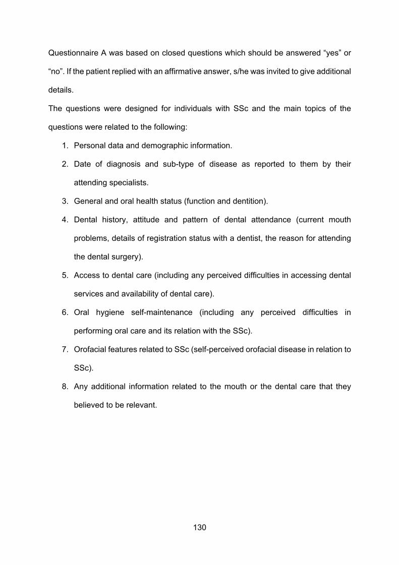
130
Questionnaire A was based on closed questions which should be answered “yes” or
“no”. If the patient replied with an affirmative answer, s/he was invited to give additional
details.
The questions were designed for individuals with SSc and the main topics of the
questions were related to the following:
1. Personal data and demographic information.
2. Date of diagnosis and sub-type of disease as reported to them by their
attending specialists.
3. General and oral health status (function and dentition).
4. Dental history, attitude and pattern of dental attendance (current mouth
problems, details of registration status with a dentist, the reason for attending
the dental surgery).
5. Access to dental care (including any perceived difficulties in accessing dental
services and availability of dental care).
6. Oral hygiene self-maintenance (including any perceived difficulties in
performing oral care and its relation with the SSc).
7. Orofacial features related to SSc (self-perceived orofacial disease in relation to
SSc).
8. Any additional information related to the mouth or the dental care that they
believed to be relevant.
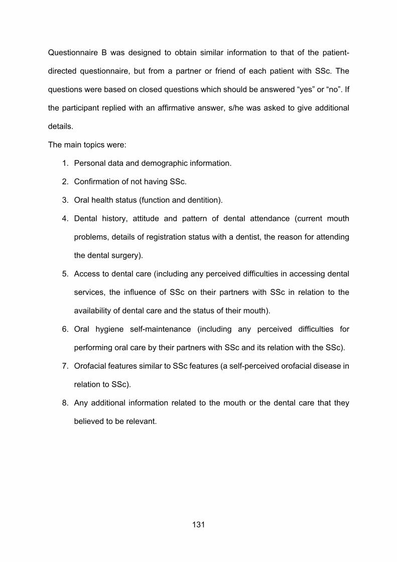
131
Questionnaire B was designed to obtain similar information to that of the patient-
directed questionnaire, but from a partner or friend of each patient with SSc. The
questions were based on closed questions which should be answered “yes” or “no”. If
the participant replied with an affirmative answer, s/he was asked to give additional
details.
The main topics were:
1. Personal data and demographic information.
2. Confirmation of not having SSc.
3. Oral health status (function and dentition).
4. Dental history, attitude and pattern of dental attendance (current mouth
problems, details of registration status with a dentist, the reason for attending
the dental surgery).
5. Access to dental care (including any perceived difficulties in accessing dental
services, the influence of SSc on their partners with SSc in relation to the
availability of dental care and the status of their mouth).
6. Oral hygiene self-maintenance (including any perceived difficulties for
performing oral care by their partners with SSc and its relation with the SSc).
7. Orofacial features similar to SSc features (a self-perceived orofacial disease in
relation to SSc).
8. Any additional information related to the mouth or the dental care that they
believed to be relevant.
132
2.3 Data collection and statistical analysis
The participants were asked to return the completed questionnaire on-site or in a
provided stamped addressed envelope. After completion of the data collection, all data
was transferred to Excel spreadsheets, tabulated and later interpreted where
appropriate using the SPSS statistical software package version 25.
The descriptive statistical analysis was undertaken for demographics and disease
features. Means, standard deviations and ranges were calculated for continuous
variables while frequency counts (number and percentage) were calculated for
categorical (ordinal and nominal) variables. In patients and controls, further statistical
analysis was performed for comparisons of different variables using the Chi-squared
test, Fisher’s exact test for categorical variables, independent t-test and Mann-Whitney
U test for continuous variables. For all statistical analyses, the threshold of significance
was set at a P-value < 0.05.
Some data concerning the dental status, access to NHS dentistry, number of teeth
and frequency of dental check-up of the study group were compared with the South of
England regional data of the Adult Dental Health Survey (2009) for age-matched
individuals and with the results of a study conducted by Ipsos MORI on behalf of
Citizen’s Advice Bureau in 2008.
Ethical approval was sought for this study; however, as this was considered to be an
evaluation of service, ethical approval was not deemed necessary.

133
3 Results
3.1 Demographics of patients with SSc and their partners
The participants in the study consisted of 50 individuals (the patient group) with likely
SSc and 18 partners or relatives (the partner/relative group). The patient group
consisted of 48 females (96%) and 2 males (4%), with a group mean age of 62.5 years
(SD = 10.8), with an age range between 29 and 81 years. The the partner/relative
group consisted of 14 males (78%) and 4 females (22%) with a group mean age of 67
years (SD = 8.8), with an age range between 46 and 78 years. The disease group had
a disease duration of 13.2 years (SD = 10.9). A slight majority of the patients were
married (54%). 70% were British white, and 88% had an educational level at degree
level or above.
The type of systemic sclerosis was based upon the diagnosis as reported by the
patient. There was no attempt to elucidate the clinical type by additional clinical
examination or alternative investigations. Twenty-four patients described themselves
as having diffuse cutaneous SSc (dcSSc) while 30 patients reported that they had
limited cutaneous SSc (lcSSc). Six patients described having mixed/overlap SSc.
However, seven patients with SSc did not report their likely type of the disease.
No significant difference in age, marital status, work status, education level and
ethnicity were found among participants’ groups. Demographics and disease
characteristics related to SSc are summarised in Table 3.1.
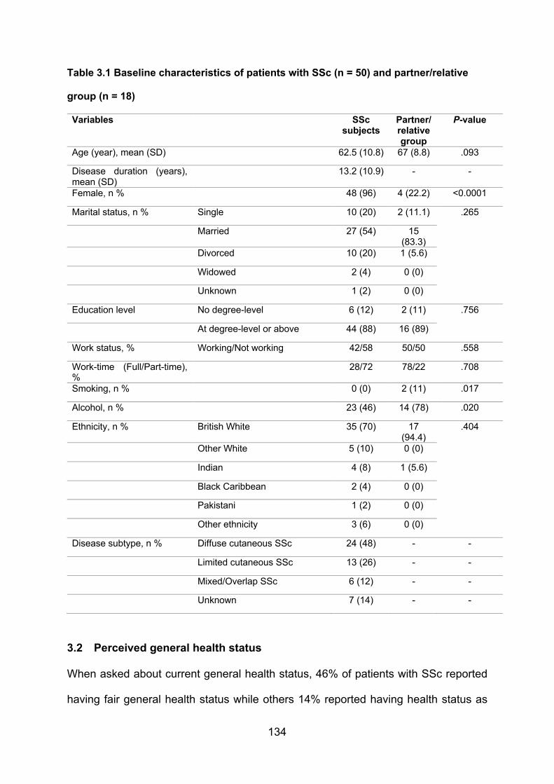
134
Table 3.1 Baseline characteristics of patients with SSc (n = 50) and partner/relative
group (n = 18)
Variables SSc subjects
Partner/relative group
P-value
Age (year), mean (SD) 62.5 (10.8) 67 (8.8) .093
Disease duration (years), mean (SD)
13.2 (10.9) - -
Female, n % 48 (96) 4 (22.2) <0.0001
Marital status, n % Single 10 (20) 2 (11.1) .265
Married 27 (54) 15 (83.3)
Divorced 10 (20) 1 (5.6)
Widowed 2 (4) 0 (0)
Unknown 1 (2) 0 (0)
Education level No degree-level 6 (12) 2 (11) .756
At degree-level or above 44 (88) 16 (89)
Work status, % Working/Not working 42/58 50/50 .558
Work-time (Full/Part-time), %
28/72 78/22 .708
Smoking, n % 0 (0) 2 (11) .017
Alcohol, n % 23 (46) 14 (78) .020
Ethnicity, n % British White 35 (70) 17 (94.4)
.404
Other White 5 (10) 0 (0)
Indian 4 (8) 1 (5.6)
Black Caribbean 2 (4) 0 (0)
Pakistani 1 (2) 0 (0)
Other ethnicity 3 (6) 0 (0)
Disease subtype, n % Diffuse cutaneous SSc 24 (48) - -
Limited cutaneous SSc 13 (26) - -
Mixed/Overlap SSc 6 (12) - -
Unknown 7 (14) - -
3.2 Perceived general health status
When asked about current general health status, 46% of patients with SSc reported
having fair general health status while others 14% reported having health status as
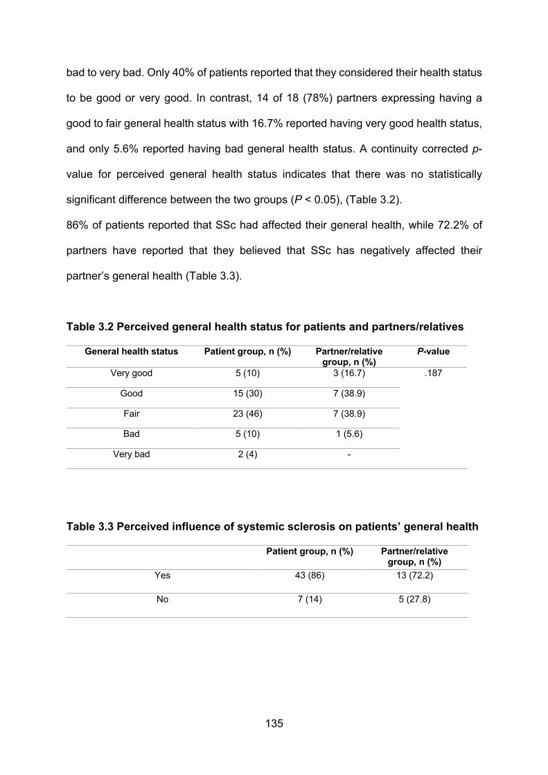
135
bad to very bad. Only 40% of patients reported that they considered their health status
to be good or very good. In contrast, 14 of 18 (78%) partners expressing having a
good to fair general health status with 16.7% reported having very good health status,
and only 5.6% reported having bad general health status. A continuity corrected p-
value for perceived general health status indicates that there was no statistically
significant difference between the two groups (P < 0.05), (Table 3.2).
86% of patients reported that SSc had affected their general health, while 72.2% of
partners have reported that they believed that SSc has negatively affected their
partner’s general health (Table 3.3).
Table 3.2 Perceived general health status for patients and partners/relatives
General health status Patient group, n (%) Partner/relative group, n (%)
P-value
Very good 5 (10) 3 (16.7) .187
Good 15 (30) 7 (38.9)
Fair 23 (46) 7 (38.9)
Bad 5 (10) 1 (5.6)
Very bad 2 (4) -
Table 3.3 Perceived influence of systemic sclerosis on patients’ general health
Patient group, n (%) Partner/relative group, n (%)
Yes 43 (86) 13 (72.2)
No 7 (14) 5 (27.8)
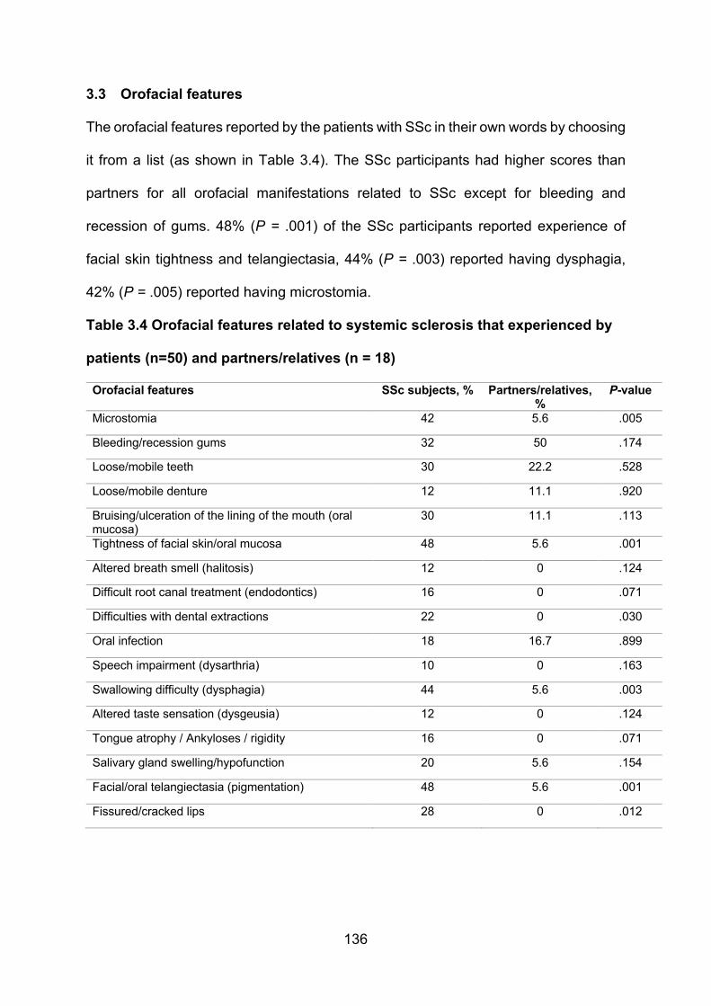
136
3.3 Orofacial features
The orofacial features reported by the patients with SSc in their own words by choosing
it from a list (as shown in Table 3.4). The SSc participants had higher scores than
partners for all orofacial manifestations related to SSc except for bleeding and
recession of gums. 48% (P = .001) of the SSc participants reported experience of
facial skin tightness and telangiectasia, 44% (P = .003) reported having dysphagia,
42% (P = .005) reported having microstomia.
Table 3.4 Orofacial features related to systemic sclerosis that experienced by
patients (n=50) and partners/relatives (n = 18)
Orofacial features SSc subjects, %
Partners/relatives, %
P-value
Microstomia 42 5.6 .005
Bleeding/recession gums 32 50 .174
Loose/mobile teeth 30 22.2 .528
Loose/mobile denture 12 11.1 .920
Bruising/ulceration of the lining of the mouth (oral mucosa)
30 11.1 .113
Tightness of facial skin/oral mucosa 48 5.6 .001
Altered breath smell (halitosis) 12 0 .124
Difficult root canal treatment (endodontics) 16 0 .071
Difficulties with dental extractions 22 0 .030
Oral infection 18 16.7 .899
Speech impairment (dysarthria) 10 0 .163
Swallowing difficulty (dysphagia) 44 5.6 .003
Altered taste sensation (dysgeusia) 12 0 .124
Tongue atrophy / Ankyloses / rigidity 16 0 .071
Salivary gland swelling/hypofunction 20 5.6 .154
Facial/oral telangiectasia (pigmentation) 48 5.6 .001
Fissured/cracked lips 28 0 .012
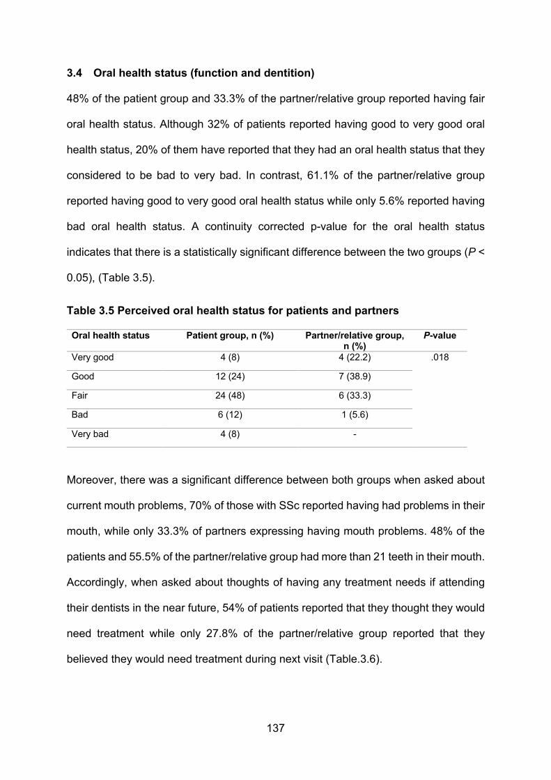
137
3.4 Oral health status (function and dentition)
48% of the patient group and 33.3% of the partner/relative group reported having fair
oral health status. Although 32% of patients reported having good to very good oral
health status, 20% of them have reported that they had an oral health status that they
considered to be bad to very bad. In contrast, 61.1% of the partner/relative group
reported having good to very good oral health status while only 5.6% reported having
bad oral health status. A continuity corrected p-value for the oral health status
indicates that there is a statistically significant difference between the two groups (P <
0.05), (Table 3.5).
Table 3.5 Perceived oral health status for patients and partners
Oral health status Patient group, n (%) Partner/relative group, n (%)
P-value
Very good 4 (8) 4 (22.2) .018
Good 12 (24) 7 (38.9)
Fair 24 (48) 6 (33.3)
Bad 6 (12) 1 (5.6)
Very bad 4 (8) -
Moreover, there was a significant difference between both groups when asked about
current mouth problems, 70% of those with SSc reported having had problems in their
mouth, while only 33.3% of partners expressing having mouth problems. 48% of the
patients and 55.5% of the partner/relative group had more than 21 teeth in their mouth.
Accordingly, when asked about thoughts of having any treatment needs if attending
their dentists in the near future, 54% of patients reported that they thought they would
need treatment while only 27.8% of the partner/relative group reported that they
believed they would need treatment during next visit (Table.3.6).
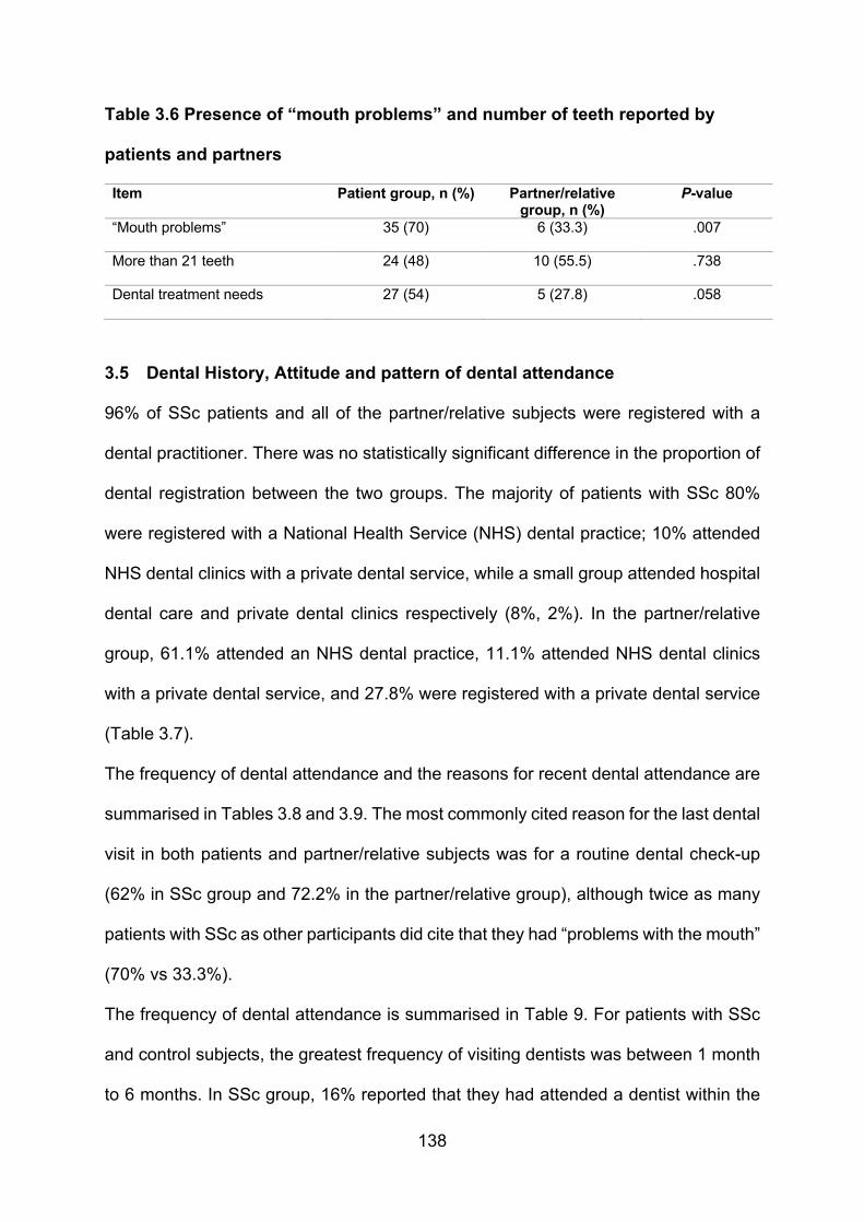
138
Table 3.6 Presence of “mouth problems” and number of teeth reported by
patients and partners
Item Patient group, n (%) Partner/relative group, n (%)
P-value
“Mouth problems” 35 (70) 6 (33.3) .007
More than 21 teeth 24 (48) 10 (55.5) .738
Dental treatment needs 27 (54) 5 (27.8) .058
3.5 Dental History, Attitude and pattern of dental attendance
96% of SSc patients and all of the partner/relative subjects were registered with a
dental practitioner. There was no statistically significant difference in the proportion of
dental registration between the two groups. The majority of patients with SSc 80%
were registered with a National Health Service (NHS) dental practice; 10% attended
NHS dental clinics with a private dental service, while a small group attended hospital
dental care and private dental clinics respectively (8%, 2%). In the partner/relative
group, 61.1% attended an NHS dental practice, 11.1% attended NHS dental clinics
with a private dental service, and 27.8% were registered with a private dental service
(Table 3.7).
The frequency of dental attendance and the reasons for recent dental attendance are
summarised in Tables 3.8 and 3.9. The most commonly cited reason for the last dental
visit in both patients and partner/relative subjects was for a routine dental check-up
(62% in SSc group and 72.2% in the partner/relative group), although twice as many
patients with SSc as other participants did cite that they had “problems with the mouth”
(70% vs 33.3%).
The frequency of dental attendance is summarised in Table 9. For patients with SSc
and control subjects, the greatest frequency of visiting dentists was between 1 month
to 6 months. In SSc group, 16% reported that they had attended a dentist within the
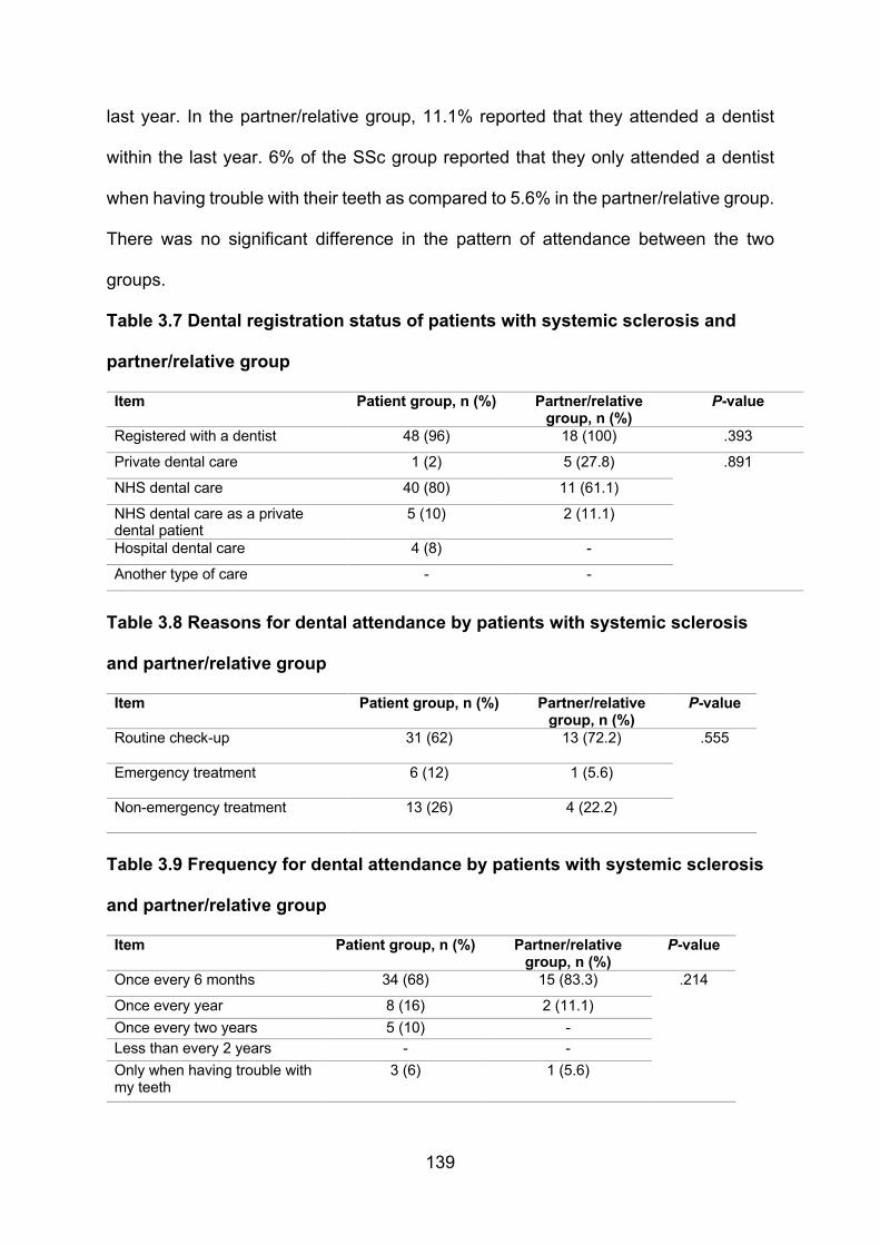
139
last year. In the partner/relative group, 11.1% reported that they attended a dentist
within the last year. 6% of the SSc group reported that they only attended a dentist
when having trouble with their teeth as compared to 5.6% in the partner/relative group.
There was no significant difference in the pattern of attendance between the two
groups.
Table 3.7 Dental registration status of patients with systemic sclerosis and
partner/relative group
Item Patient group, n (%) Partner/relative group, n (%)
P-value
Registered with a dentist 48 (96) 18 (100) .393
Private dental care 1 (2) 5 (27.8) .891
NHS dental care 40 (80) 11 (61.1)
NHS dental care as a private dental patient
5 (10) 2 (11.1)
Hospital dental care 4 (8) -
Another type of care - -
Table 3.8 Reasons for dental attendance by patients with systemic sclerosis
and partner/relative group
Item Patient group, n (%) Partner/relative group, n (%)
P-value
Routine check-up 31 (62) 13 (72.2) .555
Emergency treatment 6 (12) 1 (5.6)
Non-emergency treatment 13 (26) 4 (22.2)
Table 3.9 Frequency for dental attendance by patients with systemic sclerosis
and partner/relative group
Item Patient group, n (%) Partner/relative group, n (%)
P-value
Once every 6 months 34 (68) 15 (83.3) .214
Once every year 8 (16) 2 (11.1)
Once every two years 5 (10) -
Less than every 2 years - -
Only when having trouble with my teeth
3 (6) 1 (5.6)

140
3.6 Access to dental care
Different measures have been reported from patients and partner/relative groups
when travelling to obtain dental care from their dental health care providers. Although
the majority of the patients were using private transportation when travelling to their
dentist, 24% thought that they do require someone to assist them while travelling to
their dentist hence 42% of SSc patients and 27.8% of partner/relative group believe
that access to dental care could be improved (Table 3.10).
Table 3.10 Distance travelled to obtain dental care
Item Patient group, n (%) Partner/relative group, n (%)
P-value
1-4 miles 31 (62) 12 (66.7) .892
5-9 miles 10 (20) 3 (16.7)
10-19 miles 5 (10) 1 (5.6)
20-39 miles 2 (4) -
More than 40 miles 2 (4) 2 (11.1)
Way of travel to the dentist
Public transport 18 (36) 2 (11.1) .035
Private transport 32 (64) 16 (88.9)
Need for assistant travelling to the dentist
12 (24) - .023
Believe that access to dental care could be improved
21 (42) 5 (27.8) .215
Patients with SSc seemed to have had more difficulties in registering with dental care
(16%) compared to the partner/relative group (5.6%), and similarly, the SSc group had
had more difficulties in obtaining dental care in the past 5 years than their partners
(20% vs 5.6%), nevertheless, while there was no statistically significant difference in
individuals with SSc and their partners in obtaining dental care in the last 5 years, 24%
of the patient group and 27.8% of their partners believed that their SSc had influenced
the availability of dental care for them or affected the ability to receive dental care
(Table 3.11).

141
Table 3.11 Frequency in perceived difficulties in accessing dental care
Item Patient group, n (%) Partner/relative group, n (%)
P-value
Have difficulty in registering with GDP 8 (16) 1 (5.6) .266
Have difficulty in obtaining dental care 10 (20) 1 (5.6) .157
Believe that SSc influenced dental care 12 (24) 5 (27.8)
Patients provided written reasons as to why SSc had affected their ability to attend a
dentist in the last two years and why they might search for another dental care
provider, the most common reasons being summarised in Tables 3.12 and 3.13.
Table 3.12 Reasons not been to the dentist in the last 2 years
Item Patient group, n (%)
Difficult to get to the dentist 16 (32)
Have had bad experience with a dentist 10 (20)
Don’t see the point in going to the dentist 1 (2)
Cannot find a NHS dentist 6 (12)
Cannot afford the NHS charges 1 (2)
Afraid of going to the dentist 6 (12)
Have not got time to go to the dentist 1 (2)
Other reasons 9 (18)
Table 3.13 Reasons for searching for another dental care provider
Item Patient group, n (%)
Lack of availability of private dentist 8 (16)
Lack of availability of NHS dentist 13 (26)
Better quality of care 13 (26)
More accessible location 2 (4)
Lower waiting times 3 (6)
Better reputation 4 (8)
More specialised dentist 3 (6)
Affordability 2 (4)
No reason 2 (4)

142
3.7 Oral Hygiene and behaviour
Table 3.14 details the regular hygiene, the source and frequency of professional oral
hygiene received by both groups. 76% of the SSc patient group and 66.7% of the
partner/relative group reported that they received regular oral hygiene. Both groups
received their oral hygiene (e.g. “scale and polish”) by a dentist or a dental hygienist
equally. Both groups reported the frequency of oral hygiene to be mainly either 1 - 6
months or 6 months – 1 year. The t-test result showed the P-value to be = 0 .036,
which suggests that the SSc group received hygiene therapy more frequently than
their partners.
Table 3.14 Professional oral hygiene received for patients with systemic
sclerosis and partners
Item Patient group, n (%) Partner/relative group, n (%)
P-value
Having regular oral hygiene care 38 (76) 12 (66.7) .392
Source of oral hygiene care
Dentist 16 (42) 5 (42) .158
Dental therapist - -
Dental hygienist 22 (58) 7 (58)
Frequency of oral hygiene care
Once every 6 months 20 (52.6) 8 (66.6) .036
Once every year 14 (28) 2 (16.6)
Once every 2 years 2 (4) 2 (16.6)
Less than every 2 years 1 (2) -
Only when having trouble with my teeth 1 (2) -
52% of patients reported having difficulties in being able to maintain oral hygiene, the
main cited reasons being manual dexterity problems (e.g. cannot hold floss nor
interdental brushes or use a normal toothbrush). 44% of SSc patients and 66.7% of
their partners reported that inter-dental flossing was the most difficult aspect of oral

143
hygiene procedures followed by using interdental brushes and toothbrushes (Table
3.15). 56% of the patients believed that their condition had affected the way in which
they perform oral hygiene while 55.6% of their partners believed that SSc adversely
affected the ability of their partners to perform oral hygiene (data not shown).
Table 3.15 Reported difficulties in performing oral hygiene care
Item Patient group, n (%) Partner/relative group, n (%)
Have difficulty performing oral hygiene, n % 26 (52) -
Brushing 14 (28) 2 (11.1)
Flossing 22 (44) 12 (66.7)
Interdental brushes 14 (28) 3 (22.2)
3.8 Additional comments provided by patients
The final part of the questionnaire allowed patients or their partners/relatives to provide
any additional comments/information that was relevant to dental care or oral health.
Twenty-eight patients (56%) provided an opinion, of which 35.7% reported having had
orofacial symptoms (e.g. gingival bleeding and recession, limited mouth opening, sore
and dry mouth, teeth decay and mobility, ulcers related to the faulty prosthesis and
chronic cheek and tongue biting, TMJ disorder and muscle pain). Moreover, 35.7%
indicated that they faced difficulties accessing dental care services while 32.1%
reported they were concerned about the lack of the appropriate level of knowledge of
dentists regarding their conditions indicating that they sometimes felt neglected.
Fortunately, 17.8% of the patients were attending Eastman Dental Hospital for
receiving dental care and were happy and feeling satisfied with the level of care and
treatment provided. Finally, 32.1% of patients expressed worries about their future
dental needs and where to seek treatment in emergency situations.

144
3.9 Comparison to MORI study 2009 and ADHS 2009
Access to NHS dentistry is an issue in the UK, and it is even harder for those with a
complex medical history, with a mental or physical disability to access NHS dentistry
for various reasons. A survey on access to NHS dentistry was conducted by Ipsos
MORI on behalf of the Citizens Advice Bureau between 7th to 13th of December 2007;
the survey interviewed 1813 adults aged 15 and over throughout England and Wales
(MORI, 2008). It highlighted significant problems accessing NHS dentistry. 34% of
participants indicated that they had not been to a dentist for the last three years. The
most common reasons given were ‘lack of access to an NHS dentist’ (31%) and ‘not
needing treatment’ (30%). Amongst those who had attended a dentist, 64% had NHS
treatment, and 31% had private treatment.
The registration status with a dentist and the source of dental treatment provided for
patients and partners/relatives were compared with the Access to NHS dentistry study
2008 that was conducted by MORI for Citizens Advice Bureau. There was no
statistically significant difference between the present SSc patients and their partners
in dental registration (P=0.393), indeed more of the SSc patients and their partners
were registered with a dentist (96-100%) than that in the MORI group study (65%).
There was no statistically significant difference between the two groups as both SSc
patients and the partner/relative group in the type of dental service provider (P = .891).
However, amongst all participants (this study or MORI) NHS services were the most
common providers of dental care (Table 3.16).
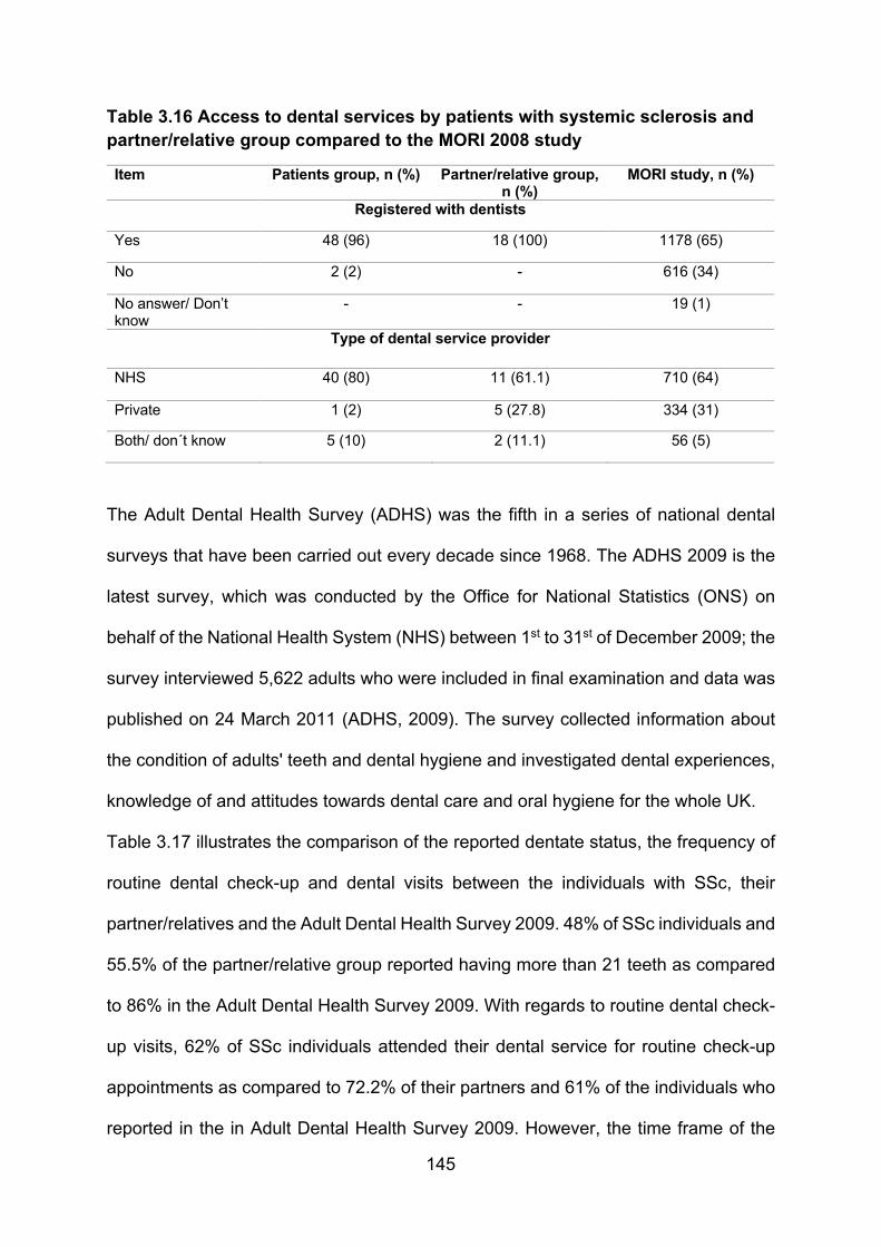
145
Table 3.16 Access to dental services by patients with systemic sclerosis and partner/relative group compared to the MORI 2008 study Item Patients group, n (%) Partner/relative group,
n (%) MORI study, n (%)
Registered with dentists
Yes 48 (96) 18 (100) 1178 (65)
No 2 (2) - 616 (34)
No answer/ Don’t know
- - 19 (1)
Type of dental service provider
NHS 40 (80) 11 (61.1) 710 (64)
Private 1 (2) 5 (27.8) 334 (31)
Both/ don´t know 5 (10) 2 (11.1) 56 (5)
The Adult Dental Health Survey (ADHS) was the fifth in a series of national dental
surveys that have been carried out every decade since 1968. The ADHS 2009 is the
latest survey, which was conducted by the Office for National Statistics (ONS) on
behalf of the National Health System (NHS) between 1st to 31st of December 2009; the
survey interviewed 5,622 adults who were included in final examination and data was
published on 24 March 2011 (ADHS, 2009). The survey collected information about
the condition of adults' teeth and dental hygiene and investigated dental experiences,
knowledge of and attitudes towards dental care and oral hygiene for the whole UK.
Table 3.17 illustrates the comparison of the reported dentate status, the frequency of
routine dental check-up and dental visits between the individuals with SSc, their
partner/relatives and the Adult Dental Health Survey 2009. 48% of SSc individuals and
55.5% of the partner/relative group reported having more than 21 teeth as compared
to 86% in the Adult Dental Health Survey 2009. With regards to routine dental check-
up visits, 62% of SSc individuals attended their dental service for routine check-up
appointments as compared to 72.2% of their partners and 61% of the individuals who
reported in the in Adult Dental Health Survey 2009. However, the time frame of the

146
routine check-up was not detailed either by the participants in the study group or the
Adult Dental Health Survey 2009.
Furthermore, 68% of SSc individuals and 83.3% of the partners attended their dental
service between 1 and 6 months as compared to 49% reported in the Adult Dental
Health Survey 2009. 16% of SSc individuals, 11.1% of the partners attended their
dental service between 6 months and 1 year as compared to 21% reported in the Adult
Dental Health Survey 2009 (Table 3.17).
Table 3.17 Dentate status, Frequency of routine dental check-up and dental visits for patients with systemic sclerosis, their partners in this study compared to the Adult Dental Health Survey of 2009
Item Patient group, n (%) Partner/relative group, n (%)
ADHS data, (%)
Number of teeth
More than 21 teeth 24 (48) 10 (55.5) (86)
Frequency of routine dental check-up
Routine check-up 31 (62) 13 (72.2) (61)
Frequency of Dental visits
1-6 months 34 (68) 15 (83.3) (49)
6 months-1 year 8 (16) 2 (11.1) (21)

147
4 Discussion
Systemic sclerosis (SSc) can give rise to a number of oral problems as a consequence
of the disease (Hughes and Herrick, 2012, Desbois and Cacoub, 2016), thus an
individual could potentially experience difficulties in access to dentistry as a result of
dentists either being unfamiliar with SSc or concerned about their ability to provide
oral care for patients with severe disease (Leader et al., 2014, Willems et al., 2015a).
Although there are no studies to confirm this, it might be expected that people with
SSc may encounter difficulties in accessing dental health care services. Individuals
with SSc may have impaired mobility and they may depend on others to travel for
hospital and dental appointments (Liem et al., 2017). The multisystem involvement of
the disease may affect an individual’s prioritisation of dental care and regular dental
attendance. The emotional effect of the disease along with fear, embarrassment,
poverty and fear of prejudice could also be significant barriers to accessing dental care
(Yuen et al., 2014a, Kwakkenbos et al., 2015, Mouthon et al., 2017). Lack of suitable
dental service provision could be a barrier to access. The dentist or dental team may
be unwilling to treat challenging disease (Leader et al., 2014). However, the provision
of special care dentistry services in the community dental service could lessen any
inequality to dental care. Since the implementation of the Disability Discrimination Act
2005, the service providers have had to make reasonable adjustments in their
premises to remove physical barriers stopping or impeding disabled access and
increased provision of domiciliary care.
By virtue of its many effects upon the skin, viscera and mouth, SSc has the potential
to adversely impact upon the oral health of and delivery of dental health care to
affected individuals (Alantar et al., 2011). The present study sought to explore the
implications of SSc upon the oral health and provision of dental care in a large group
148
of patients who have the likely diagnosis of SSc and are living in the UK. The present
study is, of course, limited by the fact that no detailed oral and/or systemic examination
was undertaken to confirm that all of the participants with SSc were indeed affected
by this disorder and had relevant orofacial features, hence there is only a presumption
that all do have SSc. Nevertheless, as is evident from the results, in which many of
the participants report oral features such as limited mouth opening and tightness of
facial skin, it would seem that these individuals are likely to have SSc-like disease.
The present group of participants comprised 50 individuals with different types of SSc
and 18 partners or relatives who were either attending a national patient group
(scleroderma society) or a nationally recognised clinic (Rheumatology clinic – Royal
Free Hospital – London) for the management of SSc and thus represents the largest
group of UK patients of SSc to have ever been asked about aspects of their oral health
or their experience of UK dentistry. As patients were all adults and had had the
diagnosis of SSc for a mean time of over 13 years with this ranging from less than one
year to more than forty years, it is likely that the opinions of this group are valid.
Despite all the limitations, this study provides a snapshot of the perceived oral health
problems and oral health care experience of a large number of individuals with SSc
and indeed represents the first such investigation of its type in the UK.
The sample included adult patients and their partners/relatives who were able to speak
and comprehend English and to complete the provided questionnaires. The study
group had a greater proportion of females (96%) than that of males (4%) which are
consistent with the previous reports such as the research carried out by Hughes and
Herrick in 2012, suggesting the 8:3 female predominance of SSc (Hughes and Herrick,
2012). The ethnicity distribution of the study group was predominantly White British in
contrast to several studies which have shown higher incidence of SSc in black
149
populations in American, French and other European population samples along with
the reported high prevalence of SSc in Caucasians (Nashid et al., 2011, Silver et al.,
2012, Gelber et al., 2013, Yuen et al., 2014a).
The geographic region of residence of individuals with SSc was mainly London in
contrast to a study done by Allcock et al. in 2004 had reported that in the United
Kingdom, SSc appears to be more common in the northeast of England (Allcock et al.,
2004). Although many participants come from London, the patients were resident in
all parts of England (data not shown) thus this does allow some broad comparisons to
be made with the last Adult Dental Health Survey of England (ADHS 2009) and the
Access to NHS dentistry study 2008 that was conducted by MORI for Citizens Advice
Bureau (MORI 2008). However, no attempts have been made to examine deprivation
scores of the patients or partners.
In view of the impact of SSc upon health, it might have been expected that patients
could have encountered difficulties in accessing dental care services. However,
access to oral health care services was higher in a sample of patients with SSc and
partners as compared to the MORI study. The majority of patients (96%) and all
partners were registered with a dentist as compared to MORI 2008 figures, 65% of
people were registered with a dentist. In the SSc group, this could be due to the fact
participants of this study were members of the Scleroderma Society, which provides
a detailed information leaflet on dental care for individuals with SSc. Therefore, the
high percentage of registration with a dentist could be a result of the sample group
being aware of the importance of oral health.
The most commonly reported type of service was NHS dental services, with 80% of
patients with SSc and 61.1% of their partners compared to data obtained from the
MORI study (64%). However, paid for NHS dental care was reported as the third most

150
common service by partners of people with SSc (11.1%). Data obtained from the
ADHS 2009 indicated that 70% of the dentate adults who participated attend to paid
NHS dental service (45%) and free NHS dental service (25%) (data not shown).
Private dental care was reported by only 10% of patients with SSc, 27.8% of their
partner as compared to the MORI study 2008, which showed 31% had access to
private dental service. Hence patients with SSc would seem to access dental services
with the same general pattern as those of their partners. Only 2% of the patient group
was not registered with a dentist as compared to 34 % as indicated in the MORI study.
Most of the patient group in this study had access to NHS dentist (80%). 2% received
private dental care, none were registered with another type of dental care such as
community dental service, and 8% received their dental care from hospital dental care.
The most commonly used dental service in the partner's group was also NHS general
dental practice with 61.1% registration, followed by 27.8% registration with the private
dental service and none of the partner's group was registered with another type of
dental care such as community dental service nor specialist hospital dental care. It is
reassuring that the majority of patients were able to attend an NHS or private dental
service as this presumably means that their service is local to their site of residency.
The high rate of attendance at a GDP, as opposed to the other type of dental care
such as community dental service and hospital dental care, may reflect the willingness
of general dental practitioners to provide treatment, limited availability of community
dental service or patients not having the appropriate clinical criteria for acceptance by
the community dental services.
The continued provision (at least at present) of special care dentistry service in the
community dental service in England and Wales and the increase in specialist training
151
posts in special care dentistry based both in hospital and community dental service
should be able to enhance the dental health care of patients with SSc.
Both the patient and the partner groups generally attended a dentist regularly, and
there was no significant difference in the frequency of attendance between the patient
and the partner/relative group (68% and 83.3% respectively). However, a lower value
has been reported in the Adult Dental Health Survey of 2009 (49%). The reason for
regular attendance was also highlighted to be mainly for a routine dental check-up in
both groups consistent with the Adult Dental Health Survey of 2009.
It would, therefore, seem that SSc does not affect the ability to attend for and receive
regular dental care as confirmed by 84% of the individuals with SSc having attended
a dentist in the preceding year (data not shown).
As might be expected, the dental status of patients with SSc did seem to have been
consequently adversely affected. When the number of patients (48%) having more
than 21 teeth was compared with either partner (55.5%) or results from the ADHS
2009 study (86%), it was evident that patients with SSc had significantly more missing
teeth than the individuals of the ADHS study. It would seem that the possible difficulties
in being able to maintain good oral hygiene due to their disorder resulted in these
patients with SSc having a substantially increased risk of hard tissue disease of the
mouth. However, the reason for loss of teeth in those with SSc could be due to a
combination of inability to keep the posterior teeth cleaned by the patients themselves
as indicated in the current result and previous study (Yuen et al., 2014a) and restricted
mouth opening, limiting the access for dentist to provide complex restorative
treatments hence causing loss of teeth. Both could have caused the prognosis of
teeth to become poor.

152
Life expectancy, in general, is increasing in the UK, and more people are also retaining
more teeth at an older age. As reported by Mayes in 2003, the life expectancy for
individuals with SSc has increased in the last few decades (Mayes et al., 2003).
Ionnidis et al. in 2005 conducted a meta-analysis and concluded that the risk of death
was not significantly different for individuals with SSc from that for the general
population in three cohorts unless there were significant internal organ involvement
and anti-topoisomerase I antibodies involvement (Ioannidis et al., 2005). Hissaria et
al. in 2011 conducted a retrospective analysis of clinical notes between 1993 and 2007
and reported that the mean age of death for patients with limited SSc (lcSSc) was 74.1
years, diffuse SSc (dcSSc) 62.9 years and overlap disease 57.8 years. They also
report that survival has improved over the 15-year study period (Hissaria et al., 2011).
However, recent studies have been reported that SSc patients have high risk of
mortality as they might have survival rates 16 – 34 years less than the sex- and age-
matched populations due to their active disease and other risk factors including
(extensive skin disease, cardiopulmonary involvement, renal impairment,
gastrointestinal involvement, older age and male gender) (Nikpour and Baron, 2014,
Rubio-Rivas et al., 2014, Richard et al., 2018).
The increase in life expectancy of patients with SSc implies that the dental care
demands of people with SSc are likely to increase. The present results point towards
a need to ensure that the access that patients presently have to dental care is
maintained and that strategies to allow patients to maintain good oral hygiene are
established.
Just over 60% of those with SSc indicated that the reason for the last dental visit was
for a routine check-up, although this dropped to 54% when those patients reported
that they think they would need treatment if visited dentist in the near future. Almost
153
72.2% of the partner's group reported a check-up to be the reason for their last dental
attendance. 61% of the dentate adults interviewed in the ADHS 2009 indicated that
the reason for their last dental attendance was for a check-up. Thus, again the SSc
group do not seem to have any different pattern of dental attendance as others without
this disease.
However, while 26% of patients with SSc indicated that their last attendance was
because of a treatment needed, while 22.2% of partners reported this as a reason.
This does not, however, imply that SSc does necessitate more frequent dental
examination or treatment sessions, as 27% of the group of ADHS 2009 participants
attended when they had a dental problem.
The difficulties that patients reported in accessing dental health care in the previous 5
years did not appear to be related to SSc. Instead cited reasons included their dentist
migrating to a private service, difficulties in being able to find an appropriate dental
service or a lack of NHS dentist in their area of residence. Seven out of 10 (14% of
the total group) who reported access difficulties did report that they believed that their
dentist had a lack of knowledge of SSc and that this may have led to difficulties in
receiving dental care – presumably under the NHS. Three out of 10 (6% of the total
group) cited failed treatment as an indication of lack of access, but this seems
contradictory as they must already have received dental care (data not shown).
It would thus seem that patients with SSc do not encounter significant difficulties in
obtaining regular dental care, as confirmed by 84% having attended a dentist in the
year preceding the study. Furthermore, although the partner/relative group comprised
fewer individuals to that of the SSc group, there were no significant differences in the
trends with regard to the type of dental service attended or recent attendance patterns
between the two groups. The present data indicate that SSc does not impact
154
significantly upon access to dental services. When comparing these results with Ipsos
MORI, Access to NHS Dentists Study (2008), patients with SSc and their partners
have a higher level of registration and attendance to dental services (96%, 100%)
respectively (both NHS and private) than the adults who participated in the latter study,
where 65% reported having attended a dentist between April 2006 and December
2007 (a 20 month-period).
The present 24% of SSc patients who reported that SSc may have affected the
availability of dental care for them cited reasons such as lack of understanding or
knowledge about SSc by dentists, psychological barriers (e.g. previous bad
experience with a dentist and feeling afraid of going to the dentist), difficulties in access
(e.g. physical limitations, cost), oral complications of SSc (limited mouth opening,
fatigue, pain and TMJ dysfunction) and the need to migrate to a private dentist.
Seventy-two per cent of the partners or relatives of SSc patients did not believe the
disease has influenced the availability of dental care for individuals with SSc. Those
who reported patients with SSc having difficulties indicated the same reasons as the
patient group: lack of understanding or knowledge about SSc by dentists, difficulties
in access, oral complications of SSc and the need to migrate to a private dentist or
referred to hospital dental services with the long waiting time.
Despite first being described over 100 years ago, this continues to be an unknown
disorder for a considerable number of healthcare workers, due perhaps to its low
prevalence. There would seem to be a need to enhance the knowledge of dental care
workers on relevant aspects of SSc to perhaps lessen the risk of patients (all be it a
small group) with SSc being unable to access dental care services.
Pokrajac-Zirojevic, Slack-Smith and Booth (2002) investigated the level of dental
attendance in a Rheumatoid arthritis (RA) and Osteoarthritis (OA) population
155
(Pokrajac-Zirojevic et al., 2002). They reported that 41% of patients with RA (51.1%
of non-RA subjects) and 42% of OA patients (51.3% of non-arthritic individuals) visited
a dentist during a previous 12 months period. These figures are lower than those of
the present group of SSc patients (84%) and their partners (94.4%). The main finding
reported by Pokrajac-Zirojevic, Slack-Smith and Booth (2002) was that chronic
diseases affect dental attendance; however, this does not seem to be necessarily the
case for individuals with SSc.
There are no published reports concerning the ability to attend a dental service in
relation to SSc. The present data indicate that SSc does not significantly influence the
ability to attend a dental practice, but this does not imply that all patients do not
encounter difficulties.
The maintenance of good oral hygiene is a fundamental component of oral disease
prevention in all groups of individuals (Yuen et al., 2014a). 76% of the SSc group and
66.7% of the partner/relative group reported regular oral hygiene care was provided
by their dental service provider. This could suggest that the dental health provider and
the patient group are aware of the implications of SSc upon their mouths. However,
as 24% of patients did not receive regular oral hygiene care, there is a concern that
they will be at risk of caries and periodontal disease especially if they also have
secondary Sjogren’s syndrome (Chu et al., 2011, Baron et al., 2015b).
76% of SSc individuals and 72.2% of the partners reported that SSc did not have an
adverse effect on the availability of dental care. Only 24% of individuals with SSc
believed that SSc had adversely influenced the availability of dental care as compared
to 27.8% in the partner/relative group. The present data indicate that there is no
significant impact of SSc upon access to dental services. However, the reason for the
lack of availability even in the small group could be due to restricted mouth opening,
156
a dentist’s lack of experience in dealing with SSc and patient’s own reluctance to
attend dental service due to physical disabilities.
There is an obligation for a special care dentist to understand the attitudinal barriers
people can encounter and are expected to demonstrate an appropriate level of
disability etiquette. This has been established to ensure that the clinical care of people
with a medical or physical disability is carried out with a progressive and positive
approach to their disability-related issues. The findings of the present study indicate
that patients with SSc are only sometimes encountering some difficulties in receiving
dental care, nevertheless, there do seem to remain some barriers to be overcome.
52% of the patients with SSc considered that their oral hygiene technique was
adversely affected by SSc involvement of hands and fingers. Problems such as hand
pain, sclerodactyly, Raynaud’s phenomena, digital ulceration and loss of manual
dexterity necessary for different methods of oral hygiene (flossing, use of a manual
toothbrush or interdental brushes) were reported by patients with SSc. A direct
relationship between reduced manual dexterity and an inability to maintain oral care
has previously been reported (Felder et al., 1994, Yuen et al., 2014a) hence the
present results are of concern as it may be that patients with SSc may be prone to
develop plaque-related oral diseases such as caries or periodontal disease as a
consequence of difficulties in oral hygiene procedures.
Although other non-orofacial disease features were not evaluated in this study, about
44% of patients with SSc indicated that the most difficult aspect of oral hygiene
procedure was related to interdental flossing hence this might be due to the disease-
related disorders of the hands (e.g. sclerodactyly, Raynaud’s phenomena, digital
ulceration and reduced manual dexterity). Patients with SSc (32%) reported that
periodontal problems such as gingival bleeding and recession might affect their ability
157
to maintain an effective oral hygiene, however, periodontal involvement may be
affected if a patient does not carry out or cannot undertake an effective oral hygiene
technique (Fischer and Patton, 2000, Poole et al., 2013, Yuen et al., 2014b, Baron et
al., 2015a). Dentists and dental hygienists should thus consider educating patients on
an appropriate method of oral hygiene and should explain that there is always an
implicit risk of periodontal problems due to the associated poor oral hygiene status.
52% of individuals with SSc reported that they encountered difficulty in performing oral
hygiene procedures. The suggested reasons for the difficulty included tightness of skin
around the mouth, difficulty in opening mouth, SSc of hands and difficulty in flossing
posterior teeth. The frequency of reported limited mouth opening, tightening of skin of
face and SSc of hands affecting the manual dexterity is consistent with the previous
literature by others (Wood and Lee, 1988, Fischer and Patton, 2000, Poole et al., 2004,
Chu et al., 2011, Baron et al., 2014, Elimelech et al., 2015). There is thus a need to
develop strategies that will ensure that patients with SSc are able to maintain an
efficient standard of oral hygiene. Such a strategy/protocol should be patient-driven to
ensure that their opinions are acknowledged, for example, developing a simple
method of interdental cleaning.
Limited mouth opening and tightness of the facial skin were the most commonly cited
perceived oral manifestation in the SSc group 42% and 48% respectively. This is
consistent with the literature in which it is indicated that approximately 52-80% of
individuals with SSc have orofacial involvement and the most striking feature to be
fibrosis of the facial skin (Marmary et al., 1981, Fischer and Patton, 2000, Bajraktari
et al., 2015).
20% of individuals reported the presence of dry mouth, the cause of this is unclear.
The study did not include any oral examination nor a reference to the patient’s medical
158
history. However, if the patients do indeed have salivary gland dysfunction, there will
be a need for them to maintain low levels of plaque to lessen any risk of caries or
gingivitis (Wolff et al., 2012, Gyger and Baron, 2015).
Oral ulceration was reported by 30% of the SSc individuals as an oral manifestation
of the disease. This might reflect anaemia due to gastrointestinal involvement of SSc,
pernicious anaemia, pancreatic insufficiency or as an adverse side effect of
therapeutics (Alantar et al., 2011, Baron et al., 2015b). Oral ulceration in the oral cavity
has also been reported due to changes in collagen structure in the mucous membrane,
making them thin and prone to ulceration (Fischer and Patton, 2000, Elimelech et al.,
2015, Mawardi et al., 2016). The patients in this study have also reported limited
tongue movement to be one of their main oral manifestations of SSc (16%) which is
also consistent with the previous literature as SSc can cause the tongue to become
fibrotic and have reduced mobility (Fischer and Patton, 2000, Vitali et al., 2015).
With regard to other Connective Tissues Disorders (CTDs), although perhaps at the
other extreme of mobility patients with Ehlers-Danlos Syndrome (EDS) have reported
difficulties in the maintenance of oral hygiene care (De Coster et al., 2005, Abel and
Carrasco, 2006). While the hypermobility and involvement of different joints (e.g.
wrists, elbows or shoulders) may compromise oral hygiene care by patients. (Poole et
al., 2004), suggested a series of measures to enable individuals with CTDs to maintain
good oral hygiene, for example, the toothbrush should have handles with a thumb stop
and soft rubber inserts, to provide a good grip and effective control. A small head and
soft bristles may also aid plaque removal. When a manual toothbrush cannot be easily
used, an electric toothbrush is advisable. Large diameter floss may be used to easily
allow cleaning of interdental areas.

159
Poole et al. (2010) developed an intervention programme for patients with SSc,
involving a dental examination, analysis of upper extremity functioning and a home
programme with patient education on brushing and flossing methods, hand and facial
exercises, adapted dental appliances and supplementation of dental products. The
researchers found that such a regimen improved the oral hygiene and periodontal
status of patients with SSc. It would thus seem logical that an appropriate patient
guidance “kit” be developed for the oral hygiene care of patients with CTDs. Of course,
this will require the active participation of patients in the developing of the “kit” (Poole
et al., 2010). Hence, Patients’ education and advice should be detailed regarding the
associated risk factors of the disease and the importance of maintaining preventative
measures with consideration of manual dexterity as a major limitation among SSc
population and the need to implement other therapeutic modalities such as connective
tissue massage, joint mobilisation, fingers stretching exercise, manual lymph drainage
of the hands and ultrasound therapy (Maddali Bongi et al., 2009, Poole et al., 2013,
Willems et al., 2015b, Thuraisingham and Sinniah, 2016).
SSc can give rise to a range of orofacial manifestations. Most will be obvious signs or
symptoms, although features such as the widening of periodontal space ligament
(PDL) are unlikely to give rise to symptoms (Baron et al., 2015a). The present group
of patients with SSc reported tightness of facial skin and/or oral mucosa and facial
and/or oral telangiectasia (48%), swallowing difficulty (44%), microstomia (42%) and
gingival recession and bleeding (32%) to be their most common orofacial concerns.
These common self-reported features accord with those expected orofacial
manifestations of SSc, however, gingival involvement is probably more likely to be due
to plaque-related gingivitis rather than directly the consequence of SSc (Baron et al.,
2015a). Some of the other reported features may reflect xerostomia (e.g. loose

160
denture, oral infection, halitosis, dysphagia, mucosal soreness and cracked lips) but it
is of interest to note that despite 48% of patients having tightness of facial skin and/or
oral mucosa and 42% having microstomia, only 10% of the patients reported
symptoms of possible dysarthria (e.g. chewing difficulties and/or speech impairment).
Of note, 24% of SSc patients who reported that SSc might have affected the availability
of dental care have cited that they had had difficulties with past therapeutic dental
procedures such as failure of endodontic therapy (16%). However, as expected
having, for example, a severe degree of reduced mouth opening might be challenging
for dentists to carry out different types of dental interventions, without a more detailed
history it is not possible to determine how real this was and what the precise causes
were for the failure of endodontic therapy. However, as dental root abnormalities (e.g.
calcification, resorption, dilacerations and underdevelopment) are dental features of
SSc, it might not be unexpected for these to compromise dental treatment outcome
such as in endodontic therapy (de Figueiredo et al., 2008, Trainito et al., 2012, Jung
et al., 2013).
Many patients with SSc commented that they felt misunderstood by health care
workers in terms of their general pain or physical difficulties which arise as a
consequence of SSc. A considerable number (32.1%) indicated that they were
depressed due to lack of understanding by dental staff about their condition. Other
concerns have been reported such as difficulties to find a dentist or the need to travel
long distances to attend a dental clinic, worries about their future dental needs and
where to seek treatments in emergency situations. Some reported being concerned
that they would have to keep their mouths open for long periods despite having
significantly limited mouth opening and/or facial skin tightness. It seemed that some
161
patients were angry about issues related to dentistry, particularly if they had been
“rejected” because of the SSc. While this study did not explore the psychological
impact of dentistry upon SSc (or vice versa), it is known that patients with SSc have
psychological upset, such as mild to moderate levels of anxiety, depression or anger
(Almeida et al., 2015). Regardless of the possible psychological implications of
dentistry and SSc, it is important that all workers of the dental team demonstrate
empathy and understanding concerning the problems that SSc will cause for affected
individuals (Delisle et al., 2018). It is important to note that only 17.8% of the present
group of patients, indicated they had been treated by a “good dentist” with an
awareness of SSc.
The majority of patients with systemic sclerosis who participated in this study were
registered with a dentist, and the main provider service they attend is an NHS dental
service. The majority of patients did not believe that their disease has influenced the
availability of dental care for them, nor their ability to attend dental services, however,
they have encountered slightly fewer difficulties in obtaining dental care in the past 5
years than their partners/relatives believed that they had. Patients with SSc indicated
that a lack of understanding or knowledge about SSc by dentists is one of the
difficulties they have encountered when attending a dental service.
Individuals with SSc have encountered problems when performing oral hygiene,
interdental cleaning being the most challenging aspect of this. The most common
orofacial features reported by patients were the tightness of facial skin and/or oral
mucosa and facial and/or oral telangiectasia, swallowing difficulty, microstomia and
gingival recession and bleeding. Patients reported having had a bad experience with
162
dental services that sometimes might be related to difficulties with past therapeutic
dental procedures and a high rate of unsuccessful outcomes.
Although this study is limited by lack of definitive diagnosis of SSc and that all
participants were not examined clinically, the relatively high numbers of participants
and generally strong trends would suggest that SSc can adversely impact upon oral
health and the provision of oral health care to affected individuals. A much more
detailed study is warranted to confirm the present observations, but there is perhaps
sufficient evidence to consider developing a specific tool to provide patients with
appropriate information and skills to maintain good oral hygiene, thereby hopefully
reducing the risk of common, usually plaque-related, oral disease.

163
5 Conclusion
Although the present study is limited by its cross-section design, the current results
suggest that oral health care providers can learn from our findings and benefit from
routine assessment of patients with SSc to develop and evolve support regarding their
oral health care needs. It is evident that although uncommon, SSc has the potential to
adversely affect oral function and compromise access to and the delivery of oral health
care. To date, there remains no study that describes the impact of SSc upon access
to dentistry in the UK.
This is the first study to show the perceived oral implications and access to dental care
service SSc patients as compared to their partners. Despite the SSc, most of the
individuals were able to access NHS dentists. The main oral implications of the SSc
were tightness and telangiectasia of facial skin and oral mucosa, dysphagia and
microstomia. The oral hygiene procedures which individuals with SSc find difficult were
brushing and flossing especially of posterior teeth. Almost half of the individuals with
SSc have reported having more than 21 teeth in the mouth. The increase in life
expectancy of patients with SSc implies that the dental care demands of people with
SSc are likely to increase. The present results point towards a need to ensure that the
access that patients presently have to dental care is maintained and improved and
that strategies to allow patients to maintain good oral hygiene are established.
This study has indicated that access to dental health care is not greatly affected by
SSc. However, patients with SSc may experience the expected oral consequences of
SSc and can have difficulties in maintaining effective oral hygiene. There is a need to
extend this work to determine more precisely the impact of SSc upon oral health, to
elucidate the influence of SSc upon the quality of life of people with SSc and to develop
simple patient-centred protocols for oral self-care.

164
CHAPTER 4: Web-based information on the treatment of the mouth in systemic
sclerosis
1 Introduction
As detailed in chapter 1, although rare, systemic sclerosis (SSc), can give rise to a
wide spectrum of manifestations that affect the skin and internal organs that may
negatively impact upon a patient’s quality of life (Almeida et al., 2015). The extra-oral
and intra-oral manifestations of SSc can be challenging to manage effectively and can
limit oral function, negatively impact upon facial aesthetics and adversely affects a
patients’ emotional and social life (Marmary et al., 1981, Nagy et al., 1994, Alantar et
al., 2011, Kwakkenbos et al., 2015).
The recent emphasis on shared decision making in a clinical setting places increased
importance upon patient education (Powell et al., 2011, Lee et al., 2014). To effectively
participate in clinical decisions regarding their healthcare patients need to be familiar
with the risks and benefits of treatment options being considered (Eysenbach and
Kohler, 2003, Andreassen et al., 2007, Trotter and Morgan, 2008). The Worldwide
Web is considered one of the most rapidly growing sources of healthcare information
and patient self-education. Although such online information is easily accessible and
plentiful, there are concerns regarding the poor quality, inaccuracy and difficult
readability of health-related information (Ni Riordain and McCreary, 2009, Dy et al.,
2012). Thus, online information could be misleading or inaccurate and hence hinder
informed shared clinical decision making (Murray et al., 2003, Lorence et al., 2006,
Singh et al., 2012, Alcorn and Madhok, 2012). In addition, poor quality information can
limit the ability of a patient with chronic illness to cope with their disease (Bremer et
al., 1997, Trento et al., 2008, Alnafea et al., 2017).

165
Due to the chronic and variable nature of the disease, individuals with SSc are likely
to require or wish to have the appropriate knowledge to help them to cope with the
impairments of the disease. Individuals with oral and/or facial disease of SSc are likely
to search for information concerning the features of the disease, their treatment
options and perhaps the complications of therapy (Almeida et al., 2015, van der Vaart
et al., 2013). There is, however, no data on how helpful online information regarding
the orofacial aspects of SSc may be for patients (or carers).
The aim of the present chapter was to categorise the content and evaluate the quality
and readability of the available web-based information concerning the treatment of the
oral aspects of SSc.

166
2 Materials and Methods
2.1 Search
An online search using the most popular international search engine (Google.com)
was conducted in November 2018 using three different search terms (“Treatment of
the mouth in scleroderma”; “Treatment of the mouth in systemic sclerosis”; “Treatment
of the mouth in scleroderma/systemic sclerosis”). The first 100 websites of each term
were assessed for duplications and screened for any non-operative link. Inclusion
criteria were included only unique websites in the English language relevant to the
employed search terms. The following exclusion criteria were then applied; scientific
articles, book reviews, websites with non-related content, non-working links, non-
English language links, membership-based websites, promotional product websites,
discussion groups, video feeds and online medical dictionaries.
The remaining websites were categorised as defined by Ni Riordain and McCreary
(2009), based upon affiliation (commercial, non-profit organisation, university/medical
centre and government), specialisation (exclusively or partly related to treatment of
the mouth in scleroderma/SSc), content type (medical facts, clinical trials, question
and answers and human interest stories) and content presentation (image, video and
audio).
2.2 Quality assessment
The quality of the online material was assessed by two reviewers (IA and RNR) using
the DISCERN instrument (Charnock et al., 1999), and the Journal of the American
Medical Association (JAMA) benchmarks for website analysis (Silberg et al., 1997).
The presence of the HON seal was also recorded.
167
The DISCERN instrument is a 16-point questionnaire developed and validated to
examine the reliability of online content and its specific information on treatment
options and overall quality scoring (Charnock et al., 1999). The JAMA benchmarks
were used to analyse the quality of websites. These benchmarks include clarity of
authorship of medical content including (authors, contributors, affiliations and relevant
credentials), inclusion of attributions (references and sources), statements of
disclosure (ownership, conflicts and interest) and indication of currency (dates of
content posted and updates) (Silberg et al., 1997). Health on the Net (HON) is a non-
profit organisation established in 1995 to guide in the evaluation of the reliability of
online information and sources in the medical field. The HON seal can be displayed
on websites that comply with eight elements ranging from the indication of authors’
qualifications to clearly distinguishing advertising from editorial content.
2.3 Readability assessment
Readability is defined as the determination by systematic formulae of the reading
comprehension level a person must possess to understand written texts (Albright et
al., 1996). The readability assessment was undertaken by using the Flesch Reading
Ease Score. This score is based upon a formula that incorporates the average
sentence length and the average number of syllables per word and the outcome score
is a number ranging from 0 to 100. The higher the score - the easier the passage is to
read (Flesch, 1948). For example, Flesch Reading Ease Scores above 90 are
considered easily understandable by an average 5th-grade student while scores
between 60 and 70 are supposed to be easily readable for 8th and 9th-grade students.
Finally, scores less than 50 represented an academic grade level and considered as
difficult level of readability.

168
2.4 Statistical analyses
Standard descriptive statistical analysis was performed by using SPSS (version 25)
and tabulated as mean ± standard deviation of the mean.
169
3 Results
3.1 Available websites
The search strategy for the term “treatment of the mouth in scleroderma/systemic
sclerosis” generated 432 000 websites, 440 000 websites for “treatment of the mouth
in scleroderma” and 338 000 by searching “treatment of the mouth in systemic
sclerosis” on the Google search engine. Of the first 300 websites of the three search
terms, 105 were scientific articles, 6 were book reviews, 12 were online medical
dictionaries, 34 were non-related websites, 10 were links of online discussion groups,
two were commercial and 24 were duplicated (Figure 4.1). Only 107 selected websites
met the inclusion criteria, however, a total of 57 unique websites remained for final
review after eliminating the duplicates between the three search terms (Figure 4.2).
Among these selected 57 sites, only 16 sites (28.1%) were exclusively dedicated to
the treatment of the mouth in SSc.

170
Figure 4.1 Number of excluded websites
Figure 4.2 Flowchart of the sample selection strategy
The search terms
•Treatment of the mouth in Scleroderma•Treatment of the mouth in Systemic sclerosis•Treatment of the mouth in Scleroderma/Systemic sclerosis
300 websites
•193 excluded websites
107 selected websites
•50 duplicated websites
Remaining websites
•57 evaluated websites
The Term: Scleroderma 23 Articles 1 Book 1 Dictionary 6 Discussion groups 2 Promotions 20 Non-related 15 Duplicated
Excluded: 68 websites
Excluded: 62 websites
Excluded: 63 websites
The Term: Scleroderma/SSc
33 Articles 1 Book 7 Dictionary 2 Discussion groups 0 Promotion 11 Non-related 9 Duplicated
The Term: Systemic sclerosis
49 Articles 4 Books 4 Dictionary 2 Discussion groups 0 Promotions 3 Non-related 0 Duplicated
Google Top 100 websites

171
Regarding the affiliation of the websites, 25 websites (43.9%) were commercial, 23
(40.4%) were non-profit websites, 5 (8.8%) were considered as governmental, and
only 4 (7%) were either universities or hospitals. The majority of the websites (82.5%)
included medical facts. However, 10 (17.5%) of the sites included clinical facts, 6
(10.5%) included human-interest stories, and only 5 (8.8%) included questions and
answers. The content presentation varied as 17 (29.8%) websites included images,
and only one website (1.8%) included an audio illustration. None of the websites
included videos. (Table 4.1) provides a summary of website categorisation.
Table 4.1 Categorisation of websites based on affiliation, specialisation,
content type and content presentation
Category Criteria Number of websites (%)
Affiliation
Commercial Non-profit organisation Governmental University/medical centre
25 (43.9%) 23 (40.4%) 5 (8.8%) 4 (7.0%)
Specialisation Exclusively related to SSc Partly dedicated to SSc
16 (28.1%)
41 (71.9%)
Content type
Medical facts Clinical trials Human interest stories Question and answer
47 (82.5%) 10 (17.5%) 6 (10.5%) 5 (8.8%)
Content presentation Image Video Audio
17 (29.8%) 0 (0%)
1 (1.8%)
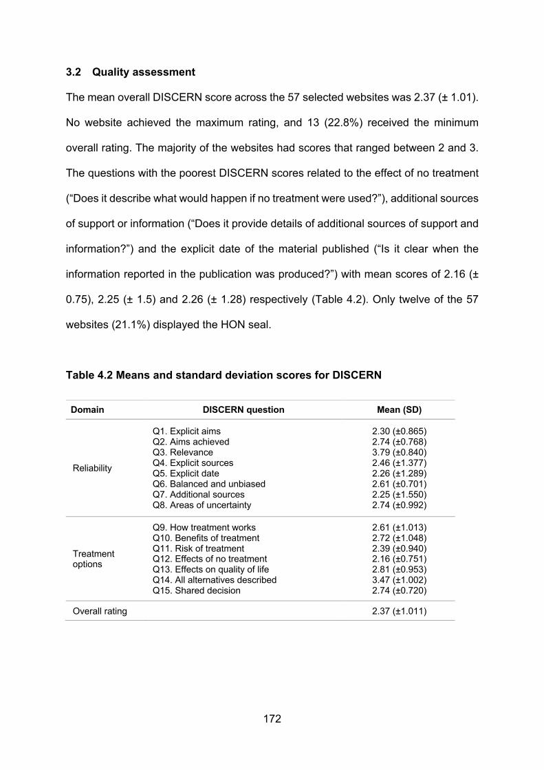
172
3.2 Quality assessment
The mean overall DISCERN score across the 57 selected websites was 2.37 (± 1.01).
No website achieved the maximum rating, and 13 (22.8%) received the minimum
overall rating. The majority of the websites had scores that ranged between 2 and 3.
The questions with the poorest DISCERN scores related to the effect of no treatment
(“Does it describe what would happen if no treatment were used?”), additional sources
of support or information (“Does it provide details of additional sources of support and
information?”) and the explicit date of the material published (“Is it clear when the
information reported in the publication was produced?”) with mean scores of 2.16 (±
0.75), 2.25 (± 1.5) and 2.26 (± 1.28) respectively (Table 4.2). Only twelve of the 57
websites (21.1%) displayed the HON seal.
Table 4.2 Means and standard deviation scores for DISCERN
Domain DISCERN question Mean (SD)
Reliability
Q1. Explicit aims Q2. Aims achieved Q3. Relevance Q4. Explicit sources Q5. Explicit date Q6. Balanced and unbiased Q7. Additional sources Q8. Areas of uncertainty
2.30 (±0.865) 2.74 (±0.768) 3.79 (±0.840) 2.46 (±1.377) 2.26 (±1.289) 2.61 (±0.701) 2.25 (±1.550) 2.74 (±0.992)
Treatment options
Q9. How treatment works Q10. Benefits of treatment Q11. Risk of treatment Q12. Effects of no treatment Q13. Effects on quality of life Q14. All alternatives described Q15. Shared decision
2.61 (±1.013) 2.72 (±1.048) 2.39 (±0.940) 2.16 (±0.751) 2.81 (±0.953) 3.47 (±1.002) 2.74 (±0.720)
Overall rating 2.37 (±1.011)
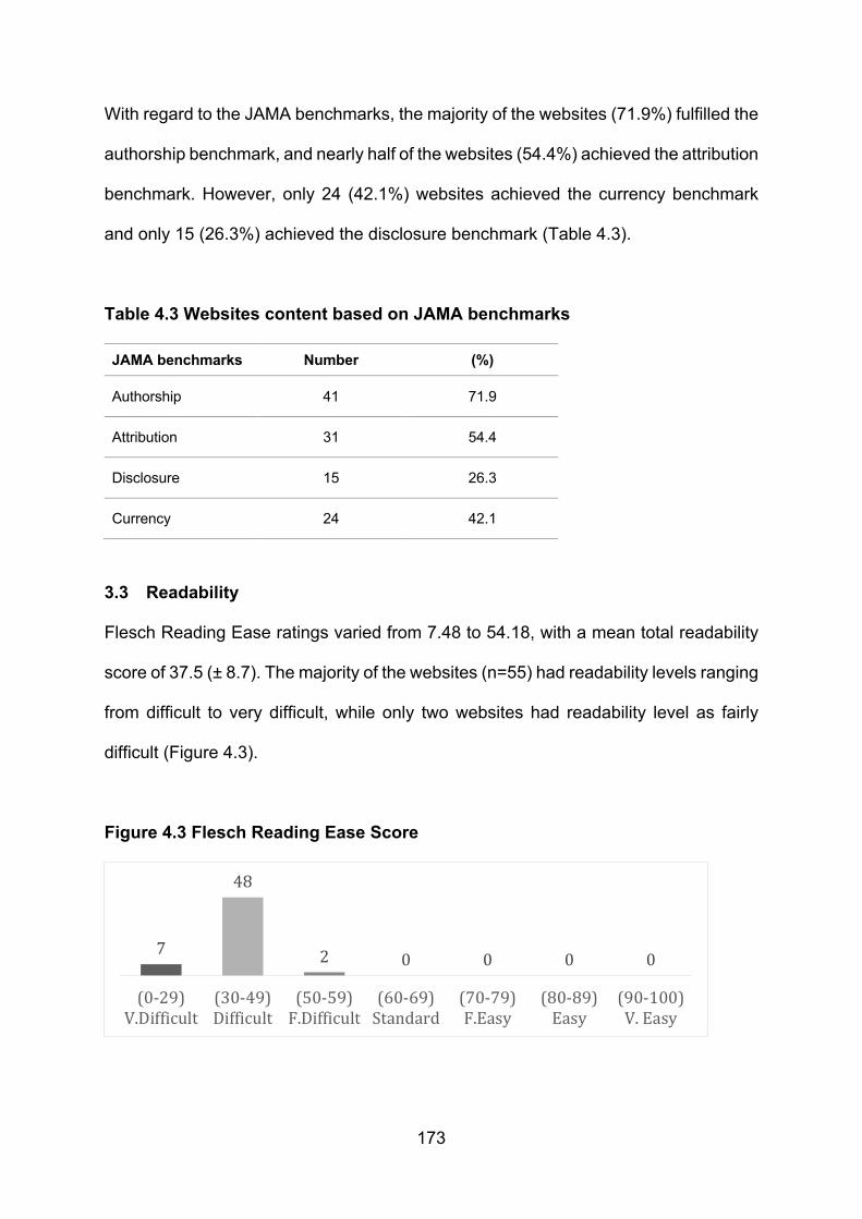
173
With regard to the JAMA benchmarks, the majority of the websites (71.9%) fulfilled the
authorship benchmark, and nearly half of the websites (54.4%) achieved the attribution
benchmark. However, only 24 (42.1%) websites achieved the currency benchmark
and only 15 (26.3%) achieved the disclosure benchmark (Table 4.3).
Table 4.3 Websites content based on JAMA benchmarks
JAMA benchmarks Number (%)
Authorship 41 71.9
Attribution 31 54.4
Disclosure 15 26.3
Currency 24 42.1
3.3 Readability
Flesch Reading Ease ratings varied from 7.48 to 54.18, with a mean total readability
score of 37.5 (± 8.7). The majority of the websites (n=55) had readability levels ranging
from difficult to very difficult, while only two websites had readability level as fairly
difficult (Figure 4.3).
Figure 4.3 Flesch Reading Ease Score
7
48
2 0 0 0 0
(0-29)V.Difficult
(30-49)Difficult
(50-59)F.Difficult
(60-69)Standard
(70-79)F.Easy
(80-89)Easy
(90-100)V.Easy
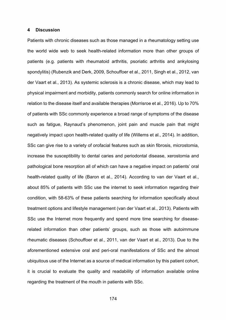
174
4 Discussion
Patients with chronic diseases such as those managed in a rheumatology setting use
the world wide web to seek health-related information more than other groups of
patients (e.g. patients with rheumatoid arthritis, psoriatic arthritis and ankylosing
spondylitis) (Rubenzik and Derk, 2009, Schouffoer et al., 2011, Singh et al., 2012, van
der Vaart et al., 2013). As systemic sclerosis is a chronic disease, which may lead to
physical impairment and morbidity, patients commonly search for online information in
relation to the disease itself and available therapies (Morrisroe et al., 2016). Up to 70%
of patients with SSc commonly experience a broad range of symptoms of the disease
such as fatigue, Raynaud’s phenomenon, joint pain and muscle pain that might
negatively impact upon health-related quality of life (Willems et al., 2014). In addition,
SSc can give rise to a variety of orofacial features such as skin fibrosis, microstomia,
increase the susceptibility to dental caries and periodontal disease, xerostomia and
pathological bone resorption all of which can have a negative impact on patients’ oral
health-related quality of life (Baron et al., 2014). According to van der Vaart et al.,
about 85% of patients with SSc use the internet to seek information regarding their
condition, with 58-63% of these patients searching for information specifically about
treatment options and lifestyle management (van der Vaart et al., 2013). Patients with
SSc use the Internet more frequently and spend more time searching for disease-
related information than other patients’ groups, such as those with autoimmune
rheumatic diseases (Schouffoer et al., 2011, van der Vaart et al., 2013). Due to the
aforementioned extensive oral and peri-oral manifestations of SSc and the almost
ubiquitous use of the Internet as a source of medical information by this patient cohort,
it is crucial to evaluate the quality and readability of information available online
regarding the treatment of the mouth in patients with SSc.

175
When considering the content of the websites reviewed in this study only 28.1% of the
examined websites were exclusively dedicated to the treatment of the mouth in SSc
with 82.5% of the sites identified in this study containing medical facts. With over four-
fifths of the material being deemed as medical facts, it is unsurprising that the Flesch
readability level in this study ranged from “difficult” to “very difficult”. Patients searching
for material specifically dedicated to the management of the oral manifestations of SSc
will not only have to delve into the website content to find content pertaining to the oral
cavity but will also have to try to interpret the extensive medical content. Concern has
previously been expressed regarding the ability of patients to accurately interpret
medical information (Ayonrinde, 1998, Sacchetti et al., 1999). Ayonrinde highlights that
although the access to high-quality specialist medical texts online is beneficial to the
medical community in the pursuit of the practice of evidence-based medicine, the
general public lacks the crucial appraisal skills to appreciate the quality of the
published material or interpret the data provided (Ayonrinde, 1998). In other studies
that have evaluated online health information-seeking behaviour of patients with
chronic or debilitating diseases a number of barriers have been reported, which
include patients being unable to find specific information and an inability of patients to
evaluate the material found (Baker et al., 2007, Lee and Hawkins, 2010, Samal et al.,
2011). Based on the findings of this study, these barriers may be relevant to patients
with oral manifestations of SSc, therefore, providing guidance to this cohort of patients
on easily accessible and comprehensible online information pertaining to the
management of the oral manifestations of SSc is worthwhile.
An alternative means of providing the material that is easily understood to patients is
to use human-interest studies or patient-based vignettes. These vignettes contain
medical content but use lay terminology and present the material often using the
176
patient voice. These human-interest vignettes have been reported to be considered
as a form of social and emotional support to patients (van Uden-Kraan et al., 2009).
Hay et al. reported that up to 9% of patients attending a rheumatology clinic searched
online, trying to find people with matching disease features and experiences (Hay et
al., 2008). In spite of the merits of this form of patient information, only 10.5% of the
websites reviewed in this study contained human-interest studies. With the permission
of patients under their care, a collaborative initiative between Rheumatologists and
Dental Practitioners could provide a series of vignettes to be included in online
material, thereby eliminating the need for critical appraisal skills needed for medical
texts and providing a form of emotional support for patients with the oral manifestations
of SSc.
In considering the reliability of the online material the overall mean DISCERN score of
the assessed websites was 2.37 (± 1.01), indicating that the quality of the available
information was low to moderate (Charnock et al., 1999). Similar results have been
reported among several studies dealing with different oral health-related conditions
(Lopez-Jornet and Camacho-Alonso, 2010, Ni Riordain and Hodgson, 2014,
Wiriyakijja et al., 2016). These poor results in the overall DISCERN score were
mirrored in the study findings for the JAMA benchmarks. Only 7% of the websites met
the full JAMA benchmarks, while the highest number of websites (30%) achieved two
benchmarks. Less than half of the websites achieved the currency benchmark
(42.1%), and only 26% achieved the disclosure benchmark while almost half of the
sites achieved the attribution benchmark (54.4%).
Recent similar findings were seen across different online sources dealing with other
oral health-related conditions, and the absence of such information could be
considered to be suspicious since patients cannot trust these online sources
177
(Wiriyakijja et al., 2016, Alnafea et al., 2017, Alsoghier et al., 2018). The American
Food and Drug Administration (FDA, 2005) suggested that the highest quality of online
information is usually administrated by governmental, non-profit and academic
institutions. However, the current results showed variations of quality among these
available online sources which might be related to a potential commercial bias as the
highest number of included websites were categorised as commercial sites (43.9%).
5 Conclusion
This study highlights the poor quality and questionable reliability of the content of the
associated online sources in relation to the treatment of the mouth in SSc. However,
when considering the significant impact of SSc upon both physical and psychological
aspects of patients, it is worrying that more high-quality patient-centred material is not
available to those searching online. Current results also suggest that the readability
level of the available online information did not meet the recommended levels to be
read and understood easily by the general population. Thus at present patients with
SSc who are seeking health-related online information should be aware of the
substantial unmet needs regarding the available information about the treatment of the
mouth and its related conditions. Based on the results of this study, further work is
required to ensure accurate, comprehensible and relevant online content is accessible
to patients with SSc.

178
CHAPTER 5: Oral health-related quality of life and self-reported anxiety and
depression in systemic sclerosis compared with the UK general population
1 Introduction
Systemic sclerosis (SSc) can give rise to a high level of physical and psychological
symptoms (e.g. pain, fatigue, depression, anxiety and fear) (Baubet et al., 2011, Del
Rosso et al., 2013). Patients with SSc have a standardised mortality ratio of 3.5 and
higher rates of psychological morbidity compared to the general population (Willems
et al., 2014, Sumpton et al., 2017, Royle et al., 2018). As a consequence with the
aforementioned adverse side effects of the disease, its chronicity and the lack of a
definitive cure, patients with SSc frequently have a reduced health-related quality of
life (HRQoL) in comparison to general population controls (Smirani et al., 2018a).
Patients with SSc usually have more than one feature of the disease, and this may
lessen both the functional and aesthetic elements that negatively impact on an
individual’s emotional and social life (Kwakkenbos et al., 2015). Despite its impact
upon the mouth, there are little data regarding the perceived adverse oral health and
impact on the oral health-related quality of life of individuals with SSc in the UK.
According to the World Health Organisation, quality of life (QoL) is defined as “the
absence of disease or physical or mental weakness as well as person’s ability to lead
a productive and enjoyable life” (WHO). By stating this, WHO outlined three specific
areas dealing with the HRQoL (physical, mental and social). The Canadian Dental
Association defines the oral health-related quality of life (OHRQoL) as “a state of the
oral and related tissues and structures that contribute to the physical, mental and
social well-being and enjoyment of life’s possibilities, by allowing the individual to
speak, eat and socialise without feeling pain, discomfort or embarrassment”(Gift and
Atchison, 1995, Petersen, 2003).

179
Patient-reported outcome measures (PROMS) are standardised measures directly
reported by the patient that characterise the patient’s perception of the impact of
disease and treatment on health and functioning. Several patient-reported quality of
life measures have been developed to measure the impact of SSc on a patient’s
health-related quality of life (Pope, 2011). PROMS can be designed to
comprehensively assess the overall HRQoL and can be a generic instrument or
alternatively designed with a specific focus on a particular aspect, disease or
population and commonly known as specific instruments. However, quality of life is
considered as a multidimensional construct that covers different impact domains of
physical, social, psychological, emotional, cognitive, spiritual, work-related and
financial aspects. Thus, it is recommended that both generic and specific QoL
measures are employed to assess relevant functioning and psychological well-being
(Almeida et al., 2015, Callahan, 2016).
With respect to the mouth, a number of different patient-reported outcome measures
(PROMS) have been used over the last decade to measure the impact of different
diseases upon the OHRQoL of patients (Lopez-Jornet et al., 2009, Ni Riordain and
McCreary, 2010a, Liu et al., 2012, Ni Riordain et al., 2016). Various reports have
explored the impact of SSc upon both HRQoL, and OHRQoL using different PROMS
such as (the oral health impact profile, the 36-item short-form health survey, hospital
anxiety and depression scale and oral impact on daily performance) and have
demonstrated a significant level of negative impact upon patients’ general and oral
health-related quality of life (Maddali Bongi et al., 2013, Maddali-Bongi et al., 2014,
Baron et al., 2014, Baron et al., 2015c, Smirani et al., 2018a).
Patients with SSc can have a spectrum of oral and maxillofacial features that may
interfere with both function and aesthetic appearance and can lessen the OHRQoL as
180
compared to the general population (Bajraktari et al., 2015, Jung et al., 2016). The
Mouth Handicap in Systemic Sclerosis (MHISS) was developed to evaluate the
limitations of the oral condition in SSc individuals by measuring the degree of
restriction of mouth opening, dryness of the mouth and aesthetic orofacial appearance.
It is the only specific PROMS for OHRQoL in SSc (Mouthon et al., 2007) and has been
employed in several studies and validated in different populations in France, Italy and
Netherlands with excellent test-retest reliability and good construct and divergent
validity (Maddali Bongi et al., 2012, Schouffoer et al., 2013, Maddali-Bongi et al.,
2014).
There are no data on the impact of SSc upon the OHRQoL among patients in the UK,
thus the aim of this study was to assess the impact of SSc upon OHRQoL and general
well-being using a number of specific and non-specific employed quality of life
instruments such as the SSc oral health-specific questionnaire (MHISS), oral health-
related questionnaires (OHIP-14, OIDP), a generic health-related questionnaire (SF-
36) and the general psychological health-related questionnaires (HADS, MDAS). Such
information should provide an understanding of whether there is a need to develop
perhaps personalised management strategies for individuals with SSc affecting the
orofacial tissues.
2 Material and Methods
2.1 Study design and participants
This was an observational cross-sectional study to evaluate the self-perceived general
and OHRQoL in patients with SSc in the UK. The study group comprised of 50 patients
and 18 partners or relatives who attended the Outpatient Rheumatology Clinic of the
Royal Free Hospital – London and Scleroderma family day – UK between May and

181
July 2017. All patients had a diagnosis of SSc confirmed by a Rheumatology team,
were over 18 years of age and had an adequate command of the spoken and written
English language to comprehend the study questionnaires. All participants were
invited to answer relevant questionnaires regarding their health and medical condition
including (SF-36, OHIP-14, MHISS, HADS, MDAS and OIDP).
Disease duration was measured as the time between the diagnosis and the time of
recruitment to the study. The disease categorisation divided into three groups: diffuse
cutaneous SSc (dcSSc), limited cutaneous SSc (lcSSc) and mixed/overlap SSc. lcSSc
was defined as skin involvement distal to the elbows and knees, with or without face
involvement. dcSSc was defined as skin involvement proximal to the elbows and
knees, with or without truncal involvement. Mixed and/or overlap SSc was proposed
to describe existing SSc and other autoimmune connective tissue diseases with the
presence of related clinical features and/or serological autoantibodies (Desbois and
Cacoub, 2016, Denton, 2016). Orofacial features related to SSc were reported in
relation to patients’ perceptions and not as a consequence of any formal clinical
examination.
2.2 Outcome measures
All participants were given detailed written information concerning the study and
requested to answer all the included questionnaires. Information collected included
sociodemographic data including age, sex, ethnicity, marital status and clinical
diagnosis. Participants were given HRQoL, OHRQoL and psychological self-
administered questionnaires including (SF-36, OHIP-14, MHISS, HADS, MDAS and
OIDP).

182
The 36-item short-form health survey (SF-36) is designed as a generic assessment
tool to measure functional health and well-being from the patient’s point of view. The
36 items are summarised into psychometrically-based physical component that
includes questions about physical functioning, role limitations due to physical health,
bodily pain and general health perceptions while mental component includes
questions about vitality, social functioning, role limitations due to emotional problems
and mental health. All health domain scales contribute to the scoring of both the
physical and mental component summary measures and transformed on a scale from
0 (worst health) to 100 (best health) with higher values reflecting better HRQoL (Del
Rosso et al., 2004).
The Oral Health Impact Profile (OHIP-14) is a modified short version instrument to
measure OHRQoL in adults with oral diseases. OHIP was originally developed as a
49-item tool representing seven domains including functional limitation, physical pain,
psychological discomfort, physical disability, psychological disability, social disability
and handicap. This was subsequently refined to the 14 item OHIP-14 by including two
questions in each of the seven OHIP aspects (Locker and Allen, 2002). Each question
is rated on a 5-point Likert scale to record the frequency of the oral problems. Patients
are invited to answer questions by choosing from 0-4 scale while 0=never and 4=very
often. The total score ranges from 0 to 56 by summing the score for all items.
The Mouth Handicap in Systemic Sclerosis (MHISS) was developed by Mouthon et al.
in 2007 to identify and evaluate the limitations of the oral manifestations of SSc on
affected individuals. It consists of 12 items (with five levels of answers), divided into
three subscales as subscale one examines impact related to reduced mouth opening,
subscale 2 examines impact related to dryness of the mouth and subscale 3 examines
183
aesthetic concerns (Mouthon et al., 2007). The total score ranges from 0 to 48 by
summing the score for all items.
The Hospital Anxiety and Depression Scale (HADS) is a psychological assessment
tool used to identify and evaluate psychological distress. It consists of 2 main domains
concerning anxiety and depression, respectively. The HADS includes seven questions
for each domain with a score range from 0 to 21 with 0 implying no depression, or
anxiety while 21 reflects the highest level of depression or anxiety. Scores ranging
between 0 – 7 are considered as normal, 8 – 10 as borderline based on a cut-off point
more or equal to 8 and sores more than 10 represent abnormal values (Honarmand
and Feinstein, 2009).
The Modified Dental Anxiety Scale (MDAS) is an assessment tool regarding anxiety
related to receiving dental care. It includes questions assessing fears associated with
visiting dentists as well as four other scenarios comprising anticipated anxiety in
relation to sitting in a dentist’s waiting room, having a scale and polish dental
procedure, having a tooth drilled or having a local anaesthetic injection. A five-point
response format is employed ranging from 1 (not anxious) to 5 (extremely anxious)
with the lowest possible score being five and the maximum possible score of 25.
Scores of 19 and above are considered to reflect extreme dental anxiety (Humphris et
al., 2013).
The Oral Impact on Daily Performance (OIDP) is an assessment tool of oral quality of
life that attempts to determine oral impacts that can significantly affect a person’s daily
life. The OIDP is based on Locker’s adaptation of the World Health Organisation’s
(WHO) classification of impairments, disability and handicap concepts model and
tends to measure the most significant oral impacts (Locker et al., 2000). The OIDP
scale assesses the frequency and severity of oral impacts among nine daily
184
performances in the past 12 months using a scale from 0 – 5, where 0 is no effect,
and 5 is a very severe effect. The total score is calculated by adding all subscores,
then divided by the maximum score (45) and multiplied by 100; the range of values is
therefore from 0 – 100. Higher total OIDP scores indicate the more severe effect of
oral impacts on daily life and represent the poorer quality of life.
2.3 Data collection and statistical analysis
Participants were asked to return the completed questionnaires on-site or in a stamped
addressed envelope to one of the authors (SRP). After completion of the data
collection, all data were transferred to Excel spreadsheets, tabulated and adjusted for
later interpretation where appropriate. Descriptive statistical analysis was calculated
for demographics and disease features. Mean, median, standard deviation and
interquartile range were calculated for continuous variables and frequency counts
(number and percentage) were calculated for ordinal and nominal variables. In
patients and controls, further statistical analysis was performed for comparisons of
different variables using Chi-squared tests, Fisher’s exact test, independent t-tests and
Mann-Whitney U tests as appropriate. For all statistical tests, the threshold of
significance was set at a P-value < 0.05. All statistical analysis was performed using
the SPSS statistical software package (version 25).
Ethical approval was sought for this study; however, as this was considered to be an
evaluation of service, ethical approval was not deemed necessary.
3 Results
3.1 Baseline characteristics of patients with SSc and controls
The SSc group comprised 48 females (96%) with a group mean age of 62.5 years (SD
= 10.8). The partner/relative group comprised only 4 females (22.2%) with a group
mean age of (67 ±8.8) years. Participants with SSc had disease duration (13.2 ±10.9)

185
years. The majority of participants in both groups were married. More than two-thirds
(70%) of the SSc patients and (94.4%) of the partner/relative group were British white.
Almost 88% of participants had an education level at degree level or above. Twenty-
four patients had diffuse cutaneous SSc (dcSSc), 13 patients had limited cutaneous
SSc (lcSSc), and six patients had mixed/overlap SSc, while 7 patients did not report
their disease type. When asked about their oral disease in the enduiring questionnaire,
48% (P = .001) of the participants reported the experience of facial skin tightness and
telangiectasia, 44% (P = .003) reported having dysphagia and 42% (P = .005) reported
having microstomia. Patient demographics, disease characteristics and self-reported
orofacial features are summarised in Table 5.1 and 5.2.
3.2 Oral health-related quality of life measures
In all OHRQoL measures (OHIP-14, MHISS and OIDP), strong statistically significant
trends in impairment were observed between patients and partners or relatives except
for the OIDP component related to problems enjoying contact with others. The total
mean OHIP-14 score was significantly lower in the patients with SSc (16.5 ±12.4)
compared with partners or relatives (6.06 ±7.6, P .001). Indeed, all the mean scores
of OHIP-14 components were significantly lower in patients with SSc than the
partner/relative subjects (Table 5.3).
The MHISS scores highlighted similar results. The total mean MHISS score was
significantly higher in patients (21.26 ±12) compared with partner/relative group (4.8
±7.3, P <.0001). Also, and perhaps unsurprisingly, patients reported significantly
higher scores in all MHISS components (mouth opening restriction, mouth dryness,
aesthetic concerns) than partner/relative group, P <.0001 (Table 5.3).

186
With regard to the oral impact of SSc upon daily performances, the total mean score
for patients was (10 ±8.7) compared with partner/relative group (1.72 ±3.4, P <.0001).
The mean scores of nine components of the OIDP reflecting the presence of the last
12 months were significantly poorer in the patients compared with partner/relative
group. The only exception was that enjoyment of contact with others was not reduced.
However, oral impacts were very frequent for most patients compared to
partner/relative group, as 88% of SSc patients reported difficulty performing at least
one element of the OIDP compared to 44.4% in partner/relative group. Overall, the
more prevalent oral impacts among SSc patients were difficulty eating (76%), difficulty
relaxing and sleeping (52%), problems smiling without embarrassment and difficulty
cleaning teeth (50%). Among partner/relative group, although the prevalence was very
low compared to patients, the most prevalent oral impacts referred to finding problems
smiling without embarrassment (6%), difficulty relaxing and sleeping (4%) and difficulty
eating (3%) (Table 5.3).
3.3 General health-related quality of life measures
Although this study was focused upon the oral aspects of SSc, general health-related
quality of life also revealed low scores. The general health mean score of SF-36 was
significantly lower in patients with SSc (43.3 ±27.2) compared with that of
partner/relative group (69.3 ±18.8, P <.0001). All subscales scores other than that of
mental health were significantly lower in patients than partner/relative group (Table
5.4).

187
3.4 Anxiety and depression measures
There were no significant differences between patients and partner/relative group for
both HADS and MDAS scales. However, there was a trend for patients with SSc to
have higher scores for both depression and anxiety of HADS and MDAS total score.
Patients with SSc had a higher mean score for HADS depression (4.8 ±3.3) and
anxiety (6 ±4.6) compared to their partners or carers (3.7 ±3.1) (4.7 ±3.9) respectively.
In patients with SSc, the rates of abnormal depression and anxiety were (6% and 18%)
respectively, higher when compared to partner/relative group (5.5%) for both
depression and anxiety. 16% of patients had borderline rates of both depression and
anxiety compared to their partners or relatives (5.5% and 16.6%) respectively.
Similarly, the mean of total MDAS score was higher among patients compared to their
partners or relatives (11.7 ±5.3), (9.5 ±4.4) respectively which indicates a moderate
trend of dental anxiety level. Overall, twelve per cent of patients had an MDAS score
of 19 or more, which suggests extreme dental anxiety and phobia compared with no
extreme level of dental anxiety among the control group. Almost half of the patients
with SSc (46%) reported a moderate level of dental anxiety compared to 38.9% in
partner/relative group. However, 38% of patients had an MDAS score of between 5
and 9, indicating low/no dental anxiety compared with 61.1% of the partner/relative
group (Table 5.5).
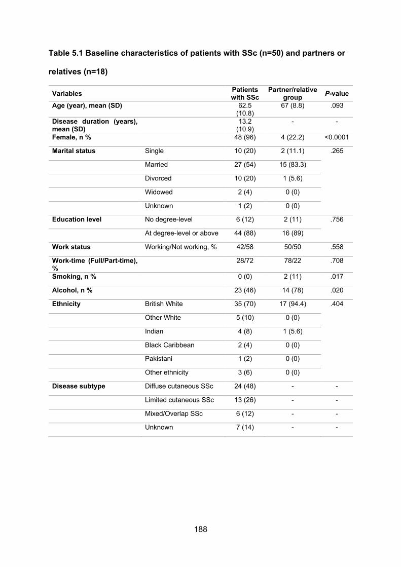
188
Table 5.1 Baseline characteristics of patients with SSc (n=50) and partners or
relatives (n=18)
Variables Patients with SSc
Partner/relative group P-value
Age (year), mean (SD) 62.5 (10.8)
67 (8.8) .093
Disease duration (years), mean (SD)
13.2 (10.9)
- -
Female, n % 48 (96) 4 (22.2) <0.0001
Marital status Single 10 (20) 2 (11.1) .265
Married 27 (54) 15 (83.3)
Divorced 10 (20) 1 (5.6)
Widowed 2 (4) 0 (0)
Unknown 1 (2) 0 (0)
Education level No degree-level 6 (12) 2 (11) .756
At degree-level or above 44 (88) 16 (89)
Work status Working/Not working, % 42/58 50/50 .558
Work-time (Full/Part-time), %
28/72 78/22 .708
Smoking, n % 0 (0) 2 (11) .017
Alcohol, n % 23 (46) 14 (78) .020
Ethnicity British White 35 (70) 17 (94.4) .404
Other White 5 (10) 0 (0)
Indian 4 (8) 1 (5.6)
Black Caribbean 2 (4) 0 (0)
Pakistani 1 (2) 0 (0)
Other ethnicity 3 (6) 0 (0)
Disease subtype Diffuse cutaneous SSc 24 (48) - -
Limited cutaneous SSc 13 (26) - -
Mixed/Overlap SSc 6 (12) - -
Unknown 7 (14) - -

189
Table 5.2 Orofacial symptoms related to SSc reported by patients with SSc
(n=50) and partner or relative subjects (n=18)
Orofacial features Patients with
SSc, % Partner/relative
group, % P-value
Microstomia 42 5.6 .005
Bleeding / recession gums 32 50 .174
Loose / mobile teeth 30 22.2 .528
Loose / mobile denture 12 11.1 .920
Bruising / ulceration of the lining of the mouth (oral mucosa)
30 11.1 .113
Tightness of facial skin / oral mucosa 48 5.6 .001
Altered breath smell (halitosis) 12 0 .124
Difficult root canal treatment (endodontics) 16 0 .071
Difficulties with dental extractions 22 0 .030
Oral infection 18 16.7 .899
Speech impairment (dysarthria) 10 0 .163
Swallowing difficulty (dysphagia) 44 5.6 .003
Altered taste sensation (dysgeusia) 12 0 .124
Tongue atrophy / ankylosis / rigidity 16 0 .071
Salivary gland swelling / hypofunction 20 5.6 .154
Facial / oral telangiectasia (pigmentation) 48 5.6 .001
Fissured / cracked lips 28 0 .012
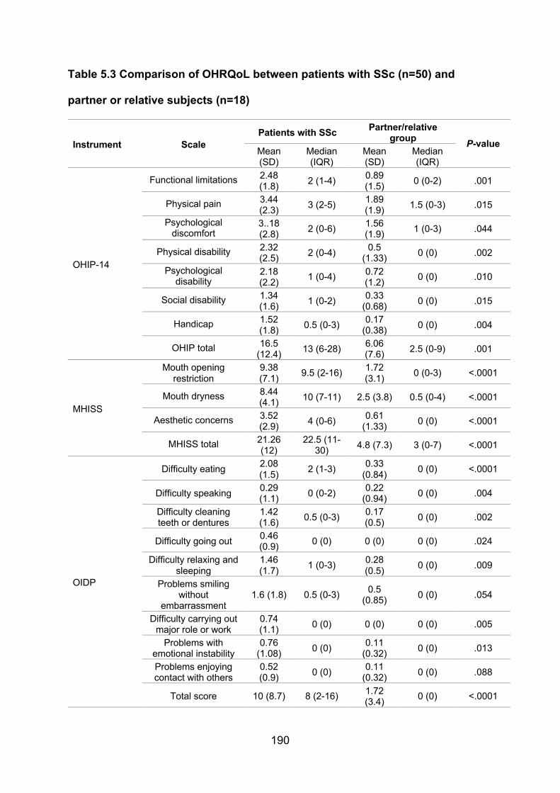
190
Table 5.3 Comparison of OHRQoL between patients with SSc (n=50) and
partner or relative subjects (n=18)
Instrument Scale Patients with SSc Partner/relative
group P-value Mean (SD)
Median (IQR)
Mean (SD)
Median (IQR)
OHIP-14
Functional limitations 2.48 (1.8)
2 (1-4) 0.89 (1.5)
0 (0-2) .001
Physical pain 3.44 (2.3)
3 (2-5) 1.89 (1.9)
1.5 (0-3) .015
Psychological discomfort
3..18 (2.8)
2 (0-6) 1.56 (1.9)
1 (0-3) .044
Physical disability 2.32 (2.5)
2 (0-4) 0.5
(1.33) 0 (0) .002
Psychological disability
2.18 (2.2)
1 (0-4) 0.72 (1.2)
0 (0) .010
Social disability 1.34 (1.6)
1 (0-2) 0.33
(0.68) 0 (0) .015
Handicap 1.52 (1.8)
0.5 (0-3) 0.17
(0.38) 0 (0) .004
OHIP total 16.5 (12.4)
13 (6-28) 6.06 (7.6)
2.5 (0-9) .001
MHISS
Mouth opening restriction
9.38 (7.1)
9.5 (2-16) 1.72 (3.1)
0 (0-3) <.0001
Mouth dryness 8.44 (4.1)
10 (7-11) 2.5 (3.8) 0.5 (0-4) <.0001
Aesthetic concerns 3.52 (2.9)
4 (0-6) 0.61
(1.33) 0 (0) <.0001
MHISS total 21.26 (12)
22.5 (11-30)
4.8 (7.3) 3 (0-7) <.0001
OIDP
Difficulty eating 2.08 (1.5)
2 (1-3) 0.33
(0.84) 0 (0) <.0001
Difficulty speaking 0.29 (1.1)
0 (0-2) 0.22
(0.94) 0 (0) .004
Difficulty cleaning teeth or dentures
1.42 (1.6)
0.5 (0-3) 0.17 (0.5)
0 (0) .002
Difficulty going out 0.46 (0.9)
0 (0) 0 (0) 0 (0) .024
Difficulty relaxing and sleeping
1.46 (1.7)
1 (0-3) 0.28 (0.5)
0 (0) .009
Problems smiling without
embarrassment 1.6 (1.8) 0.5 (0-3)
0.5 (0.85)
0 (0) .054
Difficulty carrying out major role or work
0.74 (1.1)
0 (0) 0 (0) 0 (0) .005
Problems with emotional instability
0.76 (1.08)
0 (0) 0.11
(0.32) 0 (0) .013
Problems enjoying contact with others
0.52 (0.9)
0 (0) 0.11
(0.32) 0 (0) .088
Total score 10 (8.7) 8 (2-16) 1.72 (3.4)
0 (0) <.0001
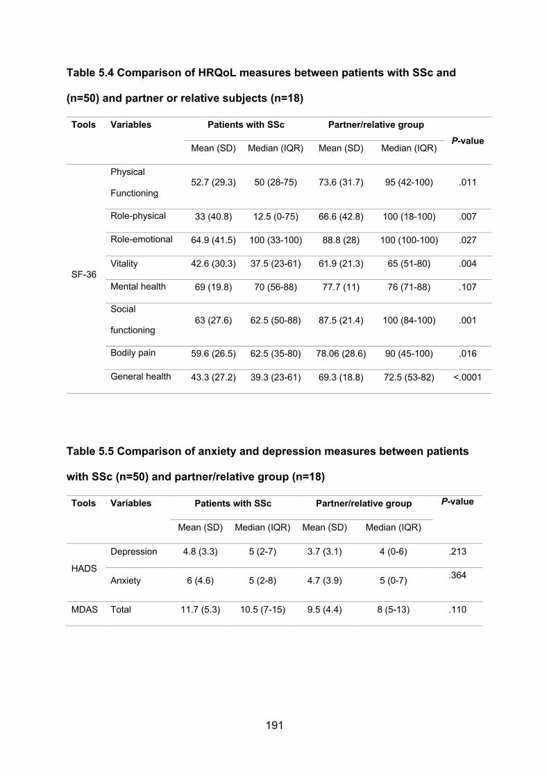
191
Table 5.4 Comparison of HRQoL measures between patients with SSc and
(n=50) and partner or relative subjects (n=18)
Tools Variables Patients with SSc Partner/relative group
P-value Mean (SD) Median (IQR) Mean (SD) Median (IQR)
SF-36
Physical
Functioning 52.7 (29.3) 50 (28-75) 73.6 (31.7) 95 (42-100) .011
Role-physical 33 (40.8) 12.5 (0-75) 66.6 (42.8) 100 (18-100) .007
Role-emotional 64.9 (41.5) 100 (33-100) 88.8 (28) 100 (100-100) .027
Vitality 42.6 (30.3) 37.5 (23-61) 61.9 (21.3) 65 (51-80) .004
Mental health 69 (19.8) 70 (56-88) 77.7 (11) 76 (71-88) .107
Social
functioning 63 (27.6) 62.5 (50-88) 87.5 (21.4) 100 (84-100) .001
Bodily pain 59.6 (26.5) 62.5 (35-80) 78.06 (28.6) 90 (45-100) .016
General health 43.3 (27.2) 39.3 (23-61) 69.3 (18.8) 72.5 (53-82) <.0001
Table 5.5 Comparison of anxiety and depression measures between patients
with SSc (n=50) and partner/relative group (n=18)
Tools Variables Patients with SSc Partner/relative group P-value
Mean (SD) Median (IQR) Mean (SD) Median (IQR)
HADS
Depression 4.8 (3.3) 5 (2-7) 3.7 (3.1) 4 (0-6) .213
Anxiety 6 (4.6) 5 (2-8) 4.7 (3.9) 5 (0-7) .364
MDAS Total 11.7 (5.3) 10.5 (7-15) 9.5 (4.4) 8 (5-13) .110
192
4 Discussion
Systemic sclerosis is a complex autoimmune disorder that gives rise to small vessel
disease and fibrosis of the mucocutaneous surfaces and viscera, particularly the lungs
and gastrointestinal tract. Co-morbidity of the disease is considered high among
affected individuals due to the multisystem involvement, the unpredictability of disease
and variable response to therapy (Stern and Denton, 2015, Royle et al., 2018). As with
all disease, management is centred upon maintaining function as well as quality and
longevity of life.
The clinical consequences of SSc can lessen the quality of life of patients through
activity limitations, impairments and/or disability, thus understanding the impact of
systemic sclerosis upon HRQoL may help to address the healthcare needs aiding
healthcare providers to better-overcome any unmet patients needs for this rare
condition. Previous evidence has reported that SSc can radically affect oral health and
impact negatively on oral and general HRQoL compared with the general population
(Veale et al., 2016, Smirani et al., 2018b). Aside from dysfunction of lungs, kidneys,
heart, gastrointestinal tract and musculoskeletal system, there is an increased risk of
malignancy (Elhai et al., 2015, McCray and Mayes, 2015, Lachner, 2016, Zeineddine
et al., 2016). The orofacial manifestations of SSc have been ranked by patients as
amongst the most worrying aspects of their disease as they may alter the patient’s
OHRQoL and hinder multiple functions such as eating, speaking and interfere with
daily hygiene and dental treatment procedures (Alantar et al., 2011, Del Rosso and
Maddali-Bongi, 2014, Jung et al., 2016). Despite their high frequency, the precise
impact of the oral manifestations of SSc patients upon the patient’s quality of life has
rarely been investigated. Certainly, HRQoL can be significantly impaired in SSc
patients as demonstrated using (SF-36, OHIP-14, MHISS and OIDP) and OHRQoL is
193
associated with global HRQoL (Baron et al., 2015c). Due to the heterogeneity of SSc,
different disease-specific PROMS have been developed such as Scleroderma Health
Assessment Questionnaire (Steen and Medsger, 1997, Johnson et al., 2005, Pope,
2011), UK Scleroderma Functional Score (Silman et al., 1998), Symptom Burden
Index (Kallen et al., 2010), Scleroderma Gastrointestinal Tract Scale and UCLA SCTC
GIT (Khanna et al., 2007, Khanna et al., 2009) and Raynaud’s Condition Score (Merkel
et al., 2002, Pope, 2011) but there remains only one SSc-specific PROM that
measures the mouth-related disability – the MHSS (Mouthon et al., 2007). Another
study has assessed the psychometric properties, including validity and reliability of
MHISS scale in the same cohort of patients with SSc making it the first study to validate
the MHISS in the UK population. Both SF-36 and OHIP-14 have been reported to be
used commonly among different OHRQoL studies of different oral disorders perhaps
of relevance to SSc with excellent measurement properties (Ni Riordain and
McCreary, 2010a, Baron et al., 2015c) and hence the inclusion of these in the present
study has some relevance.
In this study, the generic and specific HRQoL was measured in a cohort of patients
with SSc compared to non-SSc participants as a control group using a number of
PROMS including (SF-36, OHIP-14, MHISS, HADS, MDAS and OIDP). The current
results indicate that SSc disease and associated orofacial features radically affect the
HRQoL and may affect psychological factors including depression and anxiety.
Although using generic instruments such as SF-36 might be less responsive to
changes due to the focus on general aspects of the related-condition, it assesses the
influence of the disease on overall well-being rather than specific manifestations and
may detect an unexpected clinical event which would otherwise be missed by
measures that are more specific.

194
According to present results, SSc patients present a significantly high level of disability
in all subscales scores of SF-36 other than that of mental health compared to
partner/relative group. These results reflect those of a recent meta-analysis that
suggests that physical health measured by SF-36 was more likely to be affected than
mental well-being in systemic sclerosis (Li et al., 2018). The present results also
demonstrate that the effect of emotional impact, mental health and social functioning
are remarkable with the highest scores in the SF-36 questionnaire. Previous studies
have reported similar results with a high impact of psychological disability on the
HRQoL of the SSc patients including pain, anxiety and depression (Mestre-Stanislas
et al., 2010, Maddali Bongi et al., 2013, van der Vaart et al., 2013, Maddali-Bongi et
al., 2014, Bragazzi et al., 2019). Moreover, in this group of patients with SSc
psychological discomfort and disability as recorded in the OHIP-14 questionnaire had
high scores, patients felt ‘self-conscious’, ‘tense’, ‘distressed’, ‘not able to relax’ and/or
‘embarrassed’ as an outcome of their oral condition as well as experiencing increased
physical pain and functioning limitation. Baron et al. similarly reported that SSc had
impacted greatly upon functional limitation, physical pain and disability, psychological
discomfort and disability in the OHIP (Baron et al., 2014).
Apart from the multisystemic nature of the disease, patients with SSc are commonly
affected by a variety of orofacial features that can impact upon OHRQoL including
increased susceptibility to dental decay and perhaps periodontal disease, decreased
saliva production, limited mouth opening and tightening of the facial skin (Veale et al.,
2016, Smirani et al., 2018b). Thus patients with SSc can have significant mouth
disability of the orofacial tissues (Schouffoer et al., 2013, Maddali-Bongi et al., 2014),
as well as a reduction in global well-being (Nguyen et al., 2014, Kwakkenbos et al.,
2015). The present results have revealed that patients with SSc do report significantly

195
higher scores in all MHISS components (mouth opening restriction, mouth dryness,
aesthetic concerns) and thus have significantly impaired overall OHRQoL. As perhaps
expected the present patients often reported facial disability and reduced mouth
opening – as recorded in the MHISS. Indeed, tightness of facial skin and/or oral
mucosa and reduced mouth opening were among the most commonly reported SSc-
specific features (48% and 42%). However, several studies have found that patients
reported experiencing dry mouth as the second most common feature with
comparatively lower scores for aesthetic change related to their disease. (Maddali
Bongi et al., 2012, Maddali-Bongi et al., 2014, Smirani et al., 2018b, Basta et al., 2019).
According to the most recent Adult Dental Health Survey (ADHS) of the UK, 33% of
participants report having difficulty with at least one item on the OIDP compared to
88% of patients with SSc. However, the most prevalent oral impacts among SSc
patients were difficulty eating (76%), difficulty relaxing and sleeping (52%), problems
smiling without embarrassment and difficulty cleaning teeth (50%) reflecting much
higher values comparing to Adult Dental Health Survey (ADHS, 2009). Patients with
SSc scored a significant level of impact in all scores except for “problems enjoying
contact with others”. This perhaps may be related to the benefits of perceived help
and support from either relatives and friends and/or support groups (Milette et al.,
2019). However, these findings are in line with results among other groups of patients
with chronic disease particularly some of the rheumatological disorders (Gumuchian
et al., 2016, Gumuchian et al., 2017, Poole et al., 2018).
Other aspects, such as anxiety and depression, were assessed using the hospital
anxiety and depression scale (HADS). A direct relationship has been reported
between depression and anxiety and reduced HRQoL among patients with SSc
(Kwakkenbos et al., 2015, Almeida et al., 2015, Lisitsyna et al., 2018). Similar to the
196
results in an Italian population, the present data show that up to 16% of patients with
SSc had ‘borderline’ depression and anxiety while 18% have had an ‘abnormal’
anxiety scores and 6% have had an ‘abnormal’ depression score (Del Rosso et al.,
2013, Nguyen et al., 2014). These results of the HADS are lower compared to a
previous French study, where depression and anxiety were detected in 40%, 58%,
respectively whereas 66% displayed symptoms of anxiety and/or depression (Nguyen
et al., 2014). The high prevalence of anxiety and depression among patients with SSc
may be related to various factors (e.g. pain, fatigue, body image dissatisfaction) and
may be associated with increased disability and poorer HRQoL. Therefore,
psychological and emotional impact upon patients’ quality of life should not be
underestimated. Dental anxiety is considered to be one of the most important
psychological barriers to patients accessing dental care (Hill et al., 2013). Current
results highlighted that up to 12% of SSc patients have extreme dental anxiety, while
46% reported a moderate level of dental anxiety.
Interestingly, results from the latest dental health survey (ADHS, 2009) indicated a
relationship between dental anxiety and dental attendance. Participants with extreme
dental anxiety were found to be less likely to attend unless having problems with their
teeth 22% than attending for a routine dental check-up 8%. Therefore dental anxiety
may act as a psychological barrier to seeking dental care and might lead to a negative
impact on OHRQoL.
Some oral features are more painful while others have only an aesthetic burden, but
together they build a complex figure of impairments to patients OHRQoL. Oral
disorders may impact daily living through work disability, reduced productivity and
decreased social interaction (Poole et al., 2016, Morrisroe et al., 2018). Although
patients with SSc reported higher levels of impact upon all physical and functional

197
domains, the present results indicated no significant difference between patients and
controls in relation to work status and employment. This might be related to the
relatively small sample size. A precise evaluation of the disease severity is important
to determine whether health care can easily be accessed and performed. Due to the
chronic and progressive nature of the disease patients with SSc might need to be
continuously assessed by their health care providers (Rubenzik and Derk, 2009).
Compared to other chronic diseases, patients with rare conditions such as SSc tend
to receive more help and support regarding their chronic illnesses (Joachim and Acorn,
2003). Therefore, more efforts is needed towards disease-specific evaluation and
support that could help to improve the quality of life of affected patients’ groups.

198
5 Conclusion
Systemic sclerosis has a negative impact on both general and OHRQoL of the affected
individuals that might not be routinely captured by healthcare assessment of disease
severity. Although, as mentioned above, not all HRQoL measures have been validated
specifically in SSc, the present data suggest that patients with SSc have significantly
impaired global and oral health-related quality of life. Indeed, there is a high level of
anxiety and depression compared to their partners or relatives. This study is not
without limitations as it has a cross-sectional design without detailed clinical
evaluation. However, the strengths of this study include the assessment of both oral
and global health-related quality of life at the same time along with the evaluation of
associated psychological impact, including anxiety and depression symptoms. Given
the impact of poor HRQoL, OHRQoL and psychological distress on the lives of
patients, health care providers should make efforts to collaborate and develop early
multidisciplinary targeted interventions to improve the disease comorbidity in patients
with SSc. Research is required to better understand, monitor and evaluate patients
with SSc in any health care setting and clinical trial.
199
CHAPTER 6: Validity and reliability of the Mouth Handicap of Systemic
Sclerosis (MHISS) questionnaire in a UK population
1. Introduction
Due to the nature of the disease and the lack of a definitive cure, the management of
SSc can be challenging, and patients may face significant physical, social and
psychological impacts on daily life (Almeida et al., 2015). SSc is commonly associated
with anxiety, fear and depression that might be related to the unpredictable course of
the disease and the concern about the future (Baubet et al., 2011) not only due to the
disease manifestation but also due to the increased mortality reported in this cohort of
patients (Lee, 2018). Living and coping with the disease itself and its subsequent
complications usually need both pharmacological and non-pharmacological
therapeutic efforts and referral to multiple healthcare clinicians. The goal of treatment
is to improve health-related quality of life (HRQoL) and reduce the morbidity of the
disease.
As previously discussed quality of life is a multidimensional concept incorporating
areas such as physical function, social function, psychological, emotional, cognitive,
spiritual, work-related and financial aspects of health and disease (Callahan, 2016).
Since early 1990, the utility of patient-reported outcome measures (PROMS) in the
medical field, both in clinical practice and in research has grown significantly. Efforts
have been made over the last two decades to enhance the acceptance of PROMS in
routine clinical practice and research (Callahan, 2016, Ni Riordain et al., 2016).
However, more than 250 PROMS exist in the field of rheumatology (Pellar et al., 2016).
Although the use of PROMS is thought to be helpful in recording the impact of the
disease from the perspective of the patient, further research is needed to refine
disease-specific PROMS in SSc and to investigate psychometric properties.

200
As highlighted in the literature and the previous chapter, the majority of patients with
SSc have impairment of health-related quality of life (HRQoL) and oral health-related
quality of life (OHRQoL) that should not be underestimated when considering the
spectrum of systemic manifestations of the disease (Baron et al., 2014). Generic and
specific oral health-related quality of life measures have been used to assess various
aspects of SSc (e.g. OHIP and SF-36). Although these have been used frequently,
they may not successfully assess the magnitude and impact of the oral features of the
disease as they were not developed in a population with SSc.
The Mouth Handicap in Systemic Sclerosis questionnaire (MHISS) was developed in
a French population with SSc and has proven to be a valid and reliable oral health-
specific QoL instrument on psychometric testing (Mouthon et al., 2007). However,
given the necessity to explore the cross-cultural sensitivity of health-related quality of
life measures researchers have examined the psychometric properties of MHISS in
Italian and Dutch populations with supporting outcomes (Maddali Bongi et al., 2012,
Schouffoer et al., 2013).
The MHISS had not been assessed in the UK population, hence the aims of this
chapter were to explore the psychometric properties of MHISS, namely validity and
reliability, in an English-speaking UK population.
201
2. Material and Methods
2.1 Recruitment and data collection
This observational study had a cross-sectional design. A total of 150 questionnaires
were distributed at 3 sites: Oral Medicine Department of UCLHT Eastman Dental
Hospital, the Outpatient Rheumatology Clinic of the Royal Free Hospital and
Scleroderma Family, between May and July 2017. Patients over the age of 18 years
with a diagnosis of SSc and who had an adequate command of the English language
to comprehend the questionnaires were invited to complete the study questionnaire
pack. This pack included SF 36, OHIP-14 and MHISS.
The Mouth Handicap in Systemic Sclerosis questionnaire (MHISS) was developed by
Mouthon et al. in 2007 to identify and evaluate the limitations of the oral manifestations
of the condition of SSc on individuals and aims to improve the oral health care mainly
by measuring the degree of restriction of mouth opening, dryness of the mouth and
the aesthetic appearance (Mouthon et al., 2007). It consists of 12 items (with five levels
of answers), divided into 3 subscales as subscale one examines impact related to
reduced mouth opening, subscale 2 examines impact related to dryness of the mouth
and subscale 3 examines aesthetic concerns. The total score ranges from 0 to 48 by
summing the score for all items.
The Oral Health Impact Profile (OHIP) is a questionnaire designed by Slade and
Spencer to measure OHRQoL in adults with oral disease (Slade and Spencer, 1994).
It was originally developed as a 49-item tool representing seven domains including
functional limitation, physical pain, psychological discomfort, physical disability,
psychological disability, social disability and handicap. This was subsequently refined
to the 14 item OHIP-14 by including two questions in each of the seven OHIP aspects
(Locker and Allen, 2002). Each question is rated on a 5-point Likert scale to record the

202
frequency of the oral problems. Patients are invited to answer questions by choosing
from a 0-4 scale while 0=never and 4=very often. The total score ranges from 0 to 56
by summing the score for all items.
The 36-item Short-Form Health Survey (SF-36) is designed as a generic assessment
of health status with a wide range of types and severity of conditions. The 36 items
measure eight aspects of health which includes physical functioning, role-physical,
bodily pain, general health, vitality, social functioning, role-emotional and mental
health. All scores are added together and transformed on a scale from 0 (worst health)
to 100 (best health) as a higher score (Del Rosso et al., 2004).
Participants were asked to return the completed questionnaire on-site or in a stamped
addressed envelope. After completion of the data collection, all data were transferred
to Excel spreadsheets, tabulated and later interpreted where appropriate using the
SPSS statistical software package (version 25).
Ethical approval was sought for this study; however, as this was considered to be an
evaluation of service, ethical approval was not deemed necessary.
2.2 Psychometric testing
Convergent validity, a type of construct validity, was assessed. Construct validity
explores whether the instrument can reflect theories and traits of SSc. Convergent
validity is the extent to which MHISS relates to other instruments of a similar construct
to which it is proposed to be related (Butt et al., 2009). The initial proposal explored in
this study was that patients with poor general HRQoL, as measured by SF-36, would
have poorer scores as recorded by MHISS. Secondly, we anticipated that patients with
poor OHRQoL, as measured by OHIP-14, would have poorer scores as recorded by
MHISS. Spearman’s rho correlations were used to assess the relationship between
MHISS and SF-36 and between MHISS and OHIP-14. The following grading of the

203
degree of correlation was applied 0.1 – 0.29 low correlation, 0.3 – 0.49 moderate
correlation, 0.5 – 1.0 good high correlation (Anastasi and Urbina, 1997, Schouffoer et
al., 2013).
Internal consistency reliability refers to the degree to which items in an instrument
correlate with each other and should also correlate with the overall scale score
(Aaronson et al., 2002). Cronbach’s alpha coefficient was used to determine internal
consistency reliability which can range from 0.0 to 1.0. In this study, Cronbach’s alpha
of 0.6 to 0.69 was considered acceptable, 0.7 or higher is required before an
instrument is considered adequate reliability, however, a minimum value of 0.8 is
necessary for ‘good’ internal consistency reliability (Gravetter and Forzano, 2008,
Schouffoer et al., 2013).
204
3. Results
3.1 Patient characteristics
Of the 150 questionnaires distributed to patients, 68 responded, giving a response rate
of 45.3%. Of the 68 study participants, 64 were female (94%), and 4 were male. The
mean age was 62.5 (±10) years and mean disease duration was 13.2 (±11) years.
Most of the study sample were White British (73.5%). All the participants were non-
smokers, and 33.8% of the sample consumed alcohol. Approximately a third of the
patients had diffuse cutaneous SSc (dcSSc) (35.3%), 19.1% had limited cutaneous
SSc (lcSSc), 7.4% had overlap SSc, and 38.2% were reported with unknown disease
types as they did not know the subset of their disease. With regard to orofacial
manifestations of SSc, 48% of the participants reported experience of facial skin
tightness and telangiectasia, 44% reported having dysphagia, 42% reported having
microstomia, 32% reported having gingival diseases, and 30% reported having teeth
mobility and oral ulceration. Patient demographics, disease characteristics and
symptoms are summarised in Table 6.1 and 6.2.
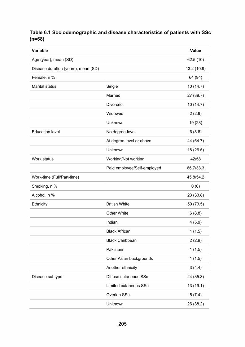
205
Table 6.1 Sociodemographic and disease characteristics of patients with SSc (n=68)
Variable Value
Age (year), mean (SD) 62.5 (10)
Disease duration (years), mean (SD) 13.2 (10.9)
Female, n % 64 (94)
Marital status Single 10 (14.7)
Married 27 (39.7)
Divorced 10 (14.7)
Widowed 2 (2.9)
Unknown 19 (28)
Education level No degree-level 6 (8.8)
At degree-level or above 44 (64.7)
Unknown 18 (26.5)
Work status Working/Not working 42/58
Paid employee/Self-employed 66.7/33.3
Work-time (Full/Part-time) 45.8/54.2
Smoking, n % 0 (0)
Alcohol, n % 23 (33.8)
Ethnicity British White 50 (73.5)
Other White 6 (8.8)
Indian 4 (5.9)
Black African 1 (1.5)
Black Caribbean 2 (2.9)
Pakistani 1 (1.5)
Other Asian backgrounds 1 (1.5)
Another ethnicity 3 (4.4)
Disease subtype Diffuse cutaneous SSc 24 (35.3)
Limited cutaneous SSc 13 (19.1)
Overlap SSc 5 (7.4)
Unknown 26 (38.2)

206
Table 6.2 Orofacial features related to SSc that experienced by patients (n=68)
Item %
Microstomia 42
Bleeding/recession gums 32
Loose/mobile teeth 30
Loose/mobile denture 12
Bruising/ulceration of the lining of the mouth (oral mucosa) 30
Tightness of facial skin/oral mucosa 48
Altered breath smell (halitosis) 12
Difficult root canal treatment (endodontics) 16
Difficulties with dental extractions 22
Oral infection 18
Speech impairment (dysarthria) 10
Swallowing difficulty (dysphagia) 44
Altered taste sensation (dysgeusia) 12
Tongue atrophy / ankylosis / rigidity 16
Salivary gland swelling/hypofunction 20
Facial/oral telangiectasia (pigmentation) 48
Fissured/cracked lips 28
The response frequencies to MHISS, OHIP-14 and SF-36 tended towards skewed
distributions, with some subscales demonstrating normal distribution. Due to the
mixture between normal and skewed data the median, mean, standard deviation,
standard error and interquartile range scores for each subscale in MHISS, and the
OHIP-14 and for SF-36 have been reported in Table 6.3.

207
Table 6.3 Descriptive statistics for the SF-36, OHIP-14 and MHISS scales of patients with SSc
Instrument Scale Median IQR Mean (SD) Std. Error
SF-36 Physical Functioning 45 20-70 48.05 (28.6) 3.47
Role-physical 25 0-69 32.72 (40.3) 4.89
Role-emotional 100 33-100 63.53 (41.4) 5.02
Vitality 34 15-55 39.34 (28.2) 3.43
Mental health 72 57-87 70.04 (20.4) 2.48
Social functioning 62.5 41-88 61.95 (27.5) 3.33
Bodily pain 56.2 37.5-78 58.04 (24.9) 3.02
General health 30 20-60 40.59 (26.1) 3.17
OHIP-14 Functional limitations 3 1-4 2.97 (2.1) 0.26
Physical pain 4 2-5 3.63 (2.2) 0.27
Psychological discomfort 4 0.25-7 3.97 (2.9) 0.36
Physical disability 2.5 0-4 2.85 (2.5) 0.31
Psychological disability 3 0-4 2.81 (2.4) 0.29
Social disability 1 0-3 1.63 (1.8) 0.22
Handicap 1.5 0-3 1.79 (1.9) 0.23
OHIP total 20 7.25-31 19.74 (13.0) 1.57
MHISS Mouth opening restriction 12 4.25-18 11.31 (7.3) 0.89
Mouth dryness 10 8-12 9.16 (4.0) 0.48
Aesthetic concerns 4 1-6 4.03 (2.8) 0.34
MHISS total 27 12.5-35 24.59 (12.1) 1.47

208
3.2 Validity
With respect to convergent validity, all Spearman’s rho correlation coefficients were
significant at the p<0.05 and p<0.01 levels as seen in Table 6.4 and 6.5. The negative
correlation seen in Table 6.4 reflects the fact that the scores of SF-36 and MHISS are
inversely related; a higher score in SF-36 reflects a better HRQoL while a higher score
in MHISS indicates a worse impact of SSc on the patient. Results show a low
correlation between the following - all MHISS domains and SF-36 Role-physical
domain; MHISS Mouth opening restriction and SF-36 Social functioning; MHISS
Mouth dryness and both SF-36 Vitality and Bodily pain; MHISS Aesthetic concerns
and SF-36 Role-emotional and Vitality. A moderate correlation was found between
MHISS Mouth opening restriction and SF-36 Role-emotional, Vitality and Bodily pain
domains. Also, a moderate correlation was found between MHISS Mouth dryness and
both SF-36 Role-emotional and Social-functioning. MHISS total scores were found
moderately correlated to all SF-36 except for both Physical functioning and Mental
health domains. However, Spearman’s rho correlation coefficients indicated that all
MHISS subscales were moderately correlated to SF-36 General health domain.

209
Table 6.4 Matrix of Spearman’s rho coefficients between pairs of responses (n=68) to the MHISS and SF-36
Instrument SF-36 Physical Functioning
SF-36 Role-physical
SF-36 Role-emotional
SF-36 Mental health
SF-36 Vitality
SF-36 Social functioning
SF-36 Bodily pain
SF-36 General health
MHISS Mouth opening restriction
-.158 -.262* -.307* -.046 -.300* -.254* -.331** -.362**
MHISS Mouth dryness
-.164 -.276* -.335** -.001 -.275* -.341** -.250* -.353**
MHISS Aesthetic concerns
-.136 -.257* -.247* -.033 -.283* -.187 -.092 -.316**
MHISS total score
-.221 -.315** -.365** -.058 -.372** -.351** -.310** -.446**
**. Correlation is significant at the 0.01 level (2-tailed). *. Correlation is significant at the 0.05 level (2-tailed). Table 6.5 Matrix of Spearman’s rho coefficients between pairs of responses to the MHISS and OHIP-14 (n=68)
Instrument OHIP Functional limitations
OHIP Physical pain
OHIP Psychological discomfort
OHIP Physical disability
OHIP Psychological disability
OHIP Social disability
OHIP Handicap
OHIP total
MHISS Mouth opening restriction
.487** .595** .618** .604** .622** .455** .551** .689**
MHISS Mouth dryness
.573** .513** .281* .548** .325** .234 .453** .489**
MHISS Aesthetics
.383** .399** .562** .407** .585** .449** .438** .582**
MHISS total score
.575** .628** .609** .652** .623** .456** .602** .725**
**. Correlation is significant at the 0.01 level (2-tailed). *. Correlation is significant at the 0.05 level (2-tailed).
Most of the MHISS domains were found to positively and highly correlated to the OHIP-
14 subscales with Spearman’s rho correlation coefficients between 0.543 and 0.725
at the p<0.01 level as seen in (Table 6.5). However, data shows some degree of
moderate correlation was found between MHISS Mouth opening restriction and both

210
OHIP-14 Functional limitation and Social disability and between MHISS Mouth
dryness and OHIP-14 Psychological disability, Handicap and OHIP-14 total score.
MHISS Aesthetic concerns domain was only highly correlated to OHIP-14
(Psychological discomfort, disability and total score). The total MHISS score was
highly correlated to all OHIP-14 subscales except the Social disability score that
reflects a moderate level of correlation.
3.2 Reliability
With regard to internal consistency reliability, the mouth dryness domain with
Cronbach’s alpha value of 0.76 demonstrated adequate internal consistency reliability.
The other two domains, including mouth opening restriction and aesthetic concerns,
indicated good internal consistency reliability level with Cronbach’s alpha values of
0.878 and 0.874, respectively. The overall MHISS Cronbach’s alpha value of 0.894
indicated very good internal consistency reliability of the 12-item instrument (Table
6.6).
Table 6.6 Internal consistency reliability of the MHISS scales Items Scale Number of items (n=68) Cronbach’s alpha
MO1-MO6 Mouth opening restriction 6 0.878
MD1-MD4 Mouth dryness 4 0.760
AC1-AC2 Aesthetic concerns 2 0.874
All the above MHISS total score 12 0.894

211
4. Discussion
SSc is a heterogeneous disease with high morbidity and impacts upon patients’
HRQoL (Almeida et al., 2015, Jung et al., 2016). Given the impact of the widespread
orofacial manifestations on both HRQoL and OHRQoL, continuous clinical evaluation
should be performed routinely in parallel to the continuous assessment of disease-
related mouth disability (Veale et al., 2016).
Although the value and merit of PROMS use in chronic disease have long been
established, the importance of selecting and administering appropriately valid and
reliable instruments cannot be underestimated. It can be challenging for clinicians and
researchers to select appropriate tools. Streiner and Norman (2006) recommended
the use of both a generic and a disease-specific outcome measure when evaluating
the impact of a disease on the daily life of a patient (Streiner et al., 2014). Generic
instruments can demonstrate a change in the HRQoL, but they cannot detect small
clinical important changes in a specific disease. On the other hand, specific measures
can more accurately predict most of the clinical changes but do not allow for
comparison between diseases. However, evidence shows that using both generic and
specific HRQoL measures when evaluating the impact of a disease is recommended
(Ni Riordain and McCreary, 2010a, Baron et al., 2015c).
Oral health-related quality of life measures have been applied among different groups
of oral diseases demonstrating valid and reliable outcomes (Lopez-Jornet and
Camacho-Alonso, 2008, Ni Riordain et al., 2016, Pacheco-Pereira et al., 2018). Both
SF-36 and OHIP were found to be the most frequently used PROMS when assessing
the impact of different oral diseases. It is reasonable to consider using these tools
when evaluating the impact of the oral manifestations of SSc on patients (Ni Riordain
and McCreary, 2010b). However, concerns have been expressed regarding the
212
suitability of OHIP-14 as an OHRQoL measure in certain patient cohorts (Locker and
Allen, 2002, Allen and Locker, 2002). Therefore, using specific disease HRQoL
measures could offer more accurate and reliable results in both clinical and research
settings as seen in recent reports (Maddali-Bongi et al., 2014, Bennani et al., 2016,
Blezien et al., 2017, Gheisari et al., 2018).
MHISS was developed as a disease-specific tool to specifically evaluate the ‘Mouth
Handicap’ in patients with SSc. This may be a viable alternative to OHIP-14 in this
patient population. The oral health-related quality of life has been assessed by MHISS
scale in many studies (Mestre-Stanislas et al., 2010, Maddali Bongi et al., 2013,
Nguyen et al., 2014, Bennani et al., 2016, Gheisari et al., 2018). MHISS has been
reported to have acceptable validity and reliability in different languages over different
countries (Mouthon et al., 2007, Maddali Bongi et al., 2012, Schouffoer et al., 2013).
To the best of our knowledge, there is no study that has investigated the psychometric
properties of the MHISS questionnaire among patients with SSc in the UK. Therefore
the aim of this study was to explore the psychometric properties of MHISS in a cohort
of UK patients with SSc.
Current results demonstrate a moderate correlation between MHISS total score and
SF-36 general health (r=-0.446) and other domains. However, the low correlation level
that has been reported among different SF-36 subscales highlighted that the SF-36
questionnaire could not comprehensively capture and evaluate specific orofacial
impairments related to SSc including mouth opening reduction, mouth dryness and
impaired aesthetics. Therefore, OHRQoL should be evaluated using disease-specific
HRQoL measures.
213
Also, the low to moderate level of correlation between the MHISS and the SF-36
different subscales was found to be similar to the previous studies (Maddali-Bongi et
al., 2014). However, this lack of correlation might be related to the nature of the generic
properties of the SF-36 as it is proposed to evaluate the HRQoL with no specific
assessment of the mouth disability in SSc patients. Despite the fact that generic
measures might be less responsive due to their focus on general aspects of HRQoL,
implementing such generic measures might help to detect an unexpected clinical
event that may be out of the scope of other specific tools.
Furthermore, current results demonstrated that the total MHISS score has a high
correlation with total OHIP-14 (r=0.725) and most of its domains. However, low to
moderate level of correlation has been noticed between different MHISS and OHIP
subscales, and this might emphasise the need for using more HRQoL measures
related to mouth disability among patients with SSc.
Although these overall values reflect moderate to high convergent validity, variations
among different subscales could be further explored in a more general study sample
of patients with SSc undertaking qualitative assessment regarding the MHISS
instrument and the impact of the disease on their lives and daily activities.
Cronbach's alpha should approach a good level for the instrument to be considered
reliable for use in clinical practice. MHISS has shown a good level of internal
consistency reliability as the values of Cronbach’s alpha for the MHISS total score was
0.894 and this was also mirrored in the study of the Dutch version of MHISS reporting
a good internal consistency reliability level of MHISS with a total MHISS score of 0.88
(Schouffoer et al., 2013). Although these values are considered good they are lower
than the values found in a previous study using an Italian translated version of MHISS
in patients with SSc which demonstrated excellent reliability (Cronbach’s alpha = 0.99)

214
(Maddali Bongi et al., 2012). However, these values of reliability could be further
investigated in a different setting to obtain precise estimates as it is dependent on the
number of items as well as the level of correlation between them.
MHISS is suggested not only to measure the face and mouth handicap among patients
with SSc but is also found to be useful in assessing the improvement of the face and
mouth after different therapeutic interventions including a facial rehabilitation
programme and physiotherapy, laser therapy, autologous fat grafting treatment
modalities, and evaluating the outcomes after dental therapeutic intervention
(Maddali-Bongi et al., 2011, Bennani et al., 2016, Maddali-Bongi and Del Rosso, 2016,
Blezien et al., 2017). Because of this, disease-specific measures are believed to be
useful in routine in clinical practice as well as research trials.
In this current study, the validity and internal consistency reliability of MHISS have
been tested, and the MHISS has been used to assess the impact of SSc on the HRQoL
of English-speaking patients living in the UK. This study has demonstrated that SSc
can affect the physical, social, emotional and psychological aspects of the patients.
Thus, patients with SSc could require detailed evaluation and specific oral
management that might involve a multidisciplinary team with a comprehensive
healthcare approach. Therefore additional research is required to refine SSc general
and specific PROMS to comprehensively assess the overall HRQoL with a specific
focus on particular aspects and manifestations of OHRQoL.
215
5. Conclusion
Given the impact of the widespread orofacial manifestations of SSc on HRQoL,
healthcare providers caring for patients with SSc should be aware of the availability of
such a reliable and valid instrument and its essential role to improve OHRQoL. Current
results suggest that the MHISS is a valid and reliable instrument to evaluate mouth-
related disability in patients with SSc and can be considered potentially useful in daily
clinical practice. However, further exploration of related psychometric properties of the
questionnaire and its individual domains could facilitate better cross-cultural
adaptation among different groups of patients with SSc in different populations.

216
CHAPTER 7: GENERAL DISCUSSION
Systemic sclerosis (SSc) is a rare autoimmune connective tissue disorder of middle-
aged and elderly persons giving rise to wide range of often clinically significant
systemic and oral problems that have the capacity to adversely affect the patients’
health-related quality of life (HRQoL) and adversely impact upon oral health and the
delivery of oral health care (Almeida et al., 2015, Salaffi et al., 2019). Orofacial features
can significantly affect the ability to speak, eat and swallow, and ultimately can lessen
oral health-related quality of life (OHRQoL). Additionally, SSc can also affect the profile
of oral and facial appearance and thus has the potential to cause patient
embarrassment, upset and psychological morbidity (Baubet et al., 2011, Bragazzi et
al., 2019).
In the past decade, there has been a considerable advance in the knowledge of
various aspects of this disorder, but there are still only a few detailed epidemiological
studies of SSc which have been carried out, particularly in the UK population. A recent
study estimated that there are currently more than one thousand new cases of SSc
each year in the UK and approximately 19,000 people living with SSc (Royle et al.,
2018). Therefore, the incidence and prevalence of SSc that affects the orofacial region
is largely unknown. However, the true prevalence has been suggested to be higher
than that suggested previously in the literature. The establishment of a national and
international register for this rare disorder would help researchers and practitioners to
better study and understand the related clinical features and prevalence of this disease
resulting in earlier diagnosis and better management.
The first part of this study retrospectively examined the nature and clinical features of
a large cohort of patients with SSc living in the UK. The group comprised of 138
patients with SSc referred to a single London hospital dental clinic, Oral Medicine Unit

217
of UCLHT Eastman Dental Hospital. It was noted that only one case of childhood SSc
was encountered. As expected, the patients were predominantly female in middle to
late life and predominantly white British, thus reflecting the epidemiology of SSc (Jung
et al., 2016, Smirani et al., 2018b). The proportions of patients with different SSc
subtypes varied from previous studies. However the proportions in the present cohort
were within the expected ranges as present data demonstrated that 42% of patients
had a diffuse cutaneous subtype, 31.2% had limited cutaneous SSc, and 26.8% had
other mixed/overlap connective tissue diseases (Willems et al., 2014, Gomes da Silva
et al., 2019). Although the results of the present study indicate that most of the patients
with SSc were registered with general dental clinics, the reasons for referral of those
patients were generally for the supposed difficulty in obtaining routine dental care
and/or for further consultation and treatment needs. The present data confirm previous
studies that patients with SSc tend to have distinct characteristics of orofacial
involvement such as more decayed and/or missing teeth, increased risk of periodontal
disease, microstomia, xerostomia, oral infections, dysphagia, oral ulceration and
temporomandibular joint dysfunction than systemically well individuals (Nagy et al.,
1994, Maddali Bongi et al., 2009, Maddali-Bongi et al., 2011, Chu et al., 2011, Chapin
and Hant, 2013, Baron et al., 2014, Del Rosso and Maddali-Bongi, 2014, Jung et al.,
2016, Hadj Said et al., 2016a, Burchfield and Vorrasi, 2019). These findings
emphasise the need for close working collaboration between specialists as early
diagnosis and management of orofacial changes can prevent at least some oral
complications of the disease, such as caries and periodontal disease.
The treatment of SSc is likely to be long-term and at present does not lead to complete
resolution of most of the orofacial disorders. Previous non-pharmacological
therapeutic trials have reported some degree of positive effects regarding
218
temporomandibular joint dysfunction, reduced mouth opening and decreased manual
dexterity in patients with SSc. However, although there have been relatively small
numbers of reports on the use of such treatments (Poole et al., 2004, Alantar et al.,
2011, Maddali-Bongi et al., 2011, Poole et al., 2013, Willems et al., 2015b, Maddali-
Bongi and Del Rosso, 2016, Khanna et al., 2018) there remain no published well-
designed randomised controlled trials for the prevention and treatment of oral disease
of SSc. New surgical treatment such as autologous fat grafting therapeutic technique
(AFGT), autologous adipose-derived stromal cells (ADSCs) and intralesional
injections while possibly promising, are costly, necessitate detailed clinical monitoring
and carry a risk of significant adverse side effects (Del Papa et al., 2015, Kumar et al.,
2016, Onesti et al., 2016, Del Papa et al., 2016).
Oral treatment and rehabilitation for patients with SSc are important as the orofacial
disease can adversely impact upon the quality of life. A multidisciplinary team
approach is essential as most of the patients were referred to the special needs dental
clinic as well as another speciality for further treatment. This might be related to the
severity of the disease and the relatively high levels of associated impaired mobility,
difficult dental access due to reduced mouth opening and abnormal manual dexterity
making effective oral hygiene difficult for them. Due to the nature of the disease, it is
important to consider the impact of the disease on all of the associated tissue
structures and the health-related quality of life bearing in mind all other available
alternative treatment modalities including simplicity, future repair services and
financial concerns. Most of the reported studies used to manage patients with oral
and/or dental problems have comprised small patient groups; as a result, the
management of such disease is largely based on clinical experience not controlled
research.
219
There is a need for detailed, well-designed studies to provide high-quality evidence on
the efficacy of treatment of the orofacial consequences of SSc. However, one problem
with implementing such research is the limited number of patients that attend individual
clinical units and the associated co-morbidity of the disease that makes it difficult for
patients to commit to long-term clinical studies.
The present study sought to determine the perceived oral health problems and oral
health care experience of a large number of individuals with SSc, and indeed,
represents the first such investigation of its type in the UK. The results of the patients
were compared to their partners or relatives as well as to the access for NHS dentistry
study by MORI 2008 and adult dental health survey (ADHS 2009). Although the
majority of individuals in the study group, both patients and partners did have access
to dental services, the greatest number of patients believed that SSc has adversely
affected their oral and general health. While 20% of patients reported difficulties in
obtaining dental care, 42% of patients and 27.8% of partners believed that access to
dental care could be improved. However, the present results point towards a need to
ensure that the access that patients presently have to dental care is enhanced and
that the strategies to ensure that patients are able to maintain good oral hygiene are
well established.
It might be expected in the 21st century that patients with SSc, or indeed their carers
or relatives or partners would seek information from the World Wide Web. The study
has sought to evaluate the related online information for the treatment of oral disability
in SSc as part of the patient information needs. This part of the study included
categorisation of the content and assessment of the quality and readability of included
websites concerning oral health and treatment of the mouth in SSc by using the
DISCERN instrument, the Journal of the American Medical Association (JAMA)
220
benchmarks and the HON seal in a standardised data collection document. However,
included websites indicated poor quality and questionable reliability of the content of
the associated online sources in relation to the treatment of the mouth in SSc. The
readability level of the available online information did not meet the recommended
levels to be read and understood easily by a general population. This availability of
reliable online sources may be due to the rarity and relatively low prevalence of the
disease.
Nevertheless, more efforts should be directed to construct a well informed and
knowledgeable level of online sources that can help to facilitate the patients’ education
and overall healthcare management. Patient education is crucial in the management
of rheumatology disorders such as SSc as most of the relevant disorders are chronic,
requiring long term treatment and follow-up. Sources include the clinician, support
groups, internet sites and brochures. Detailed information on clinical presentation,
diagnosis, treatment options and prognosis should be provided as well as information
on the presumed adverse therapeutic side effects and/or increased risk of cancer
either from the disease or treatment agents and more general advice on any habits
(e.g. tobacco smoking cessation, alcohol consumption, maintaining a healthy
balanced diet, maintaining good oral hygiene and regular review by a general dental
practitioner).
As SSc can sometimes give rise to disease that can compromises orofacial features,
it would be anticipated that the quality of life of affected individuals will be
compromised. This study has explored the health-related quality of life (HRQoL) and
oral health-related quality of life (OHRQoL) of a substantial group of patients with SSc.
Aiming to assess the impact of SSc upon health and well-being, different patient-
reported outcome measures (PROMs) have been employed (e.g. SF-36, OHIP-14,

221
MHISS, HADS, MDAS and OIDP) with results indicating that SSc has a negative
impact on both general and oral health-related quality of life of the affected individuals.
The Mouth Handicap Scale in Systemic Sclerosis (MHISS) was found to be the only
tool that specifically measured patient-reported mouth problems in SSc and was found
to be both valid and reliable for use in English speaking patients with SSc resident in
the UK.
As the patient is considered the centre of the healthcare system, patient-centred care
is considered the best approach able to reflect the quality of life of the affected
individual (Scambler et al., 2016). According to the Food and Drug Administration
(FDA), a patient-reported outcome is any report of the status of a patient’s health
condition that comes directly from the patient, without interpretation of the patient’s
response by a clinician or anyone else. Indeed, PROMs have been used as effective
assessment tools in both clinical practice and research-based trials (Pauling et al.,
2017). Systemic sclerosis is known to be chronic in its course and is associated with
significant morbidity and mortality as one of the most severe connective tissue
disorders (Pearson et al., 2018). Perhaps the most common features of SSc is
associated with progressive skin thickening and fibrosis that classically involves the
orofacial tissues and may also affect other parts of the body and internal organs
causing substantial disfigurement and disability thus impacting patients throughout
many domains of their well-being (Denton, 2015, Desbois and Cacoub, 2016). A high
level of physical and psychological complications (e.g. pain, fatigue, depression,
anxiety and fear) can arise in patients with SSc (Baubet et al., 2011, Del Rosso et al.,
2013). Present results highlighted that SSc disease and associated orofacial features
radically affected the health-related quality of life. Patients with SSc have a higher
prevalence of psychological problems including depression and anxiety.
222
Oral health-related quality of life has been assessed by a specific tool (MHISS) that
provides unique insight into the impact of the disease on mouth involvement. MHISS
demonstrate a moderate to high convergent validity and a good level of reliability; thus,
it can be considered for use in routine clinical practice in the UK. The assessment of
disease status, disease impact, multi-organ involvement and other relevant outcomes
such as oral health-related quality of life is particularly challenging in a multi-faceted
disease like SSc thus the development and validation of specific tools measuring
patient-reported outcomes in SSc are necessary. Scleroderma Clinical Trials
Consortium (SCTC), consists of 11 working groups dealing with a variety of aspects
of SSc, experts from worldwide and international multicentre disease sites have
established its research work aiming to improve clinical trials and observational studies
by improving or developing better, more sensitive ways of measuring various aspects
of the disease. A recent report has raised concerns regarding SSc morbidity that leads
to disability and poor quality of life. It has been concluded that careful attention should
be paid to the outcome measures in SSc to improve and enhance the scoring system
of various patient-reported outcome measures that deal with SSc disease and its
future clinical applications that include; use as an outcome measure and enrichment
tool for patient selection in clinical trials, a tool to describe the course of the disease in
observational studies and an instrument to quantify disease burden for epidemiological
studies and policy-making (Baron et al., 2019).

223
LIMITATIONS AND SUGGESTED FURTHER STUDIES
There are several factors which may have affected the validity of the data in this study.
The analysis of the orofacial features of SSc (Chapter 2) had a retrospective design;
hence the results must be interpreted with caution. While retrospective studies are
less expensive and time-consuming than prospective studies, can cover extended
periods and are often used to report rare diseases they might be limited by bias, lack
of agreement on exclusion and inclusion criteria, incomplete data, differences in
reporting clinical features and outcomes, variations in diagnostic and monitoring
procedures and definition of some terms (Mann, 2003). It is thus important to
recognise the importance of detailed data recording when attempting a retrospective
study. The more detailed information collected, the more likely these data are to
contribute to expanding our knowledge in the future.
Other parts of the study were observational cross-sectional designed investigations,
therefore, were limited by the lack of definitive diagnosis of SSc and that all participants
were not examined clinically. Furthermore, the sample size was relatively small and
may have introduced bias as the individuals who attended the Scleroderma family day
event on the weekend were from all across England, may possibly not have mobility
issues and be highly motivated as regards their general oral health. This might explain
the reason for having a high level of registration status with the dentist. The quality
and quantity of the data were variable, and it would be erroneous to draw definitive
conclusions from data that lack the statistical power to validate them.
Despite these limitations, this study provides useful information on the oral health
status of people with SSc, and it allows a rational understanding of the oral health-
related quality of life and dental care needs of people with SSc in the UK. However,
suggestions for future work would be to design a much more detailed prospective
224
longitudinal study with a larger cohort, possibly at more than one centre in the UK to
confirm the present observations. Also, a further survey to evaluate and provide an
indication of the level of awareness of SSc among general dental practitioners and the
willingness of these practitioners to treat patients with SSc is warranted. In view of the
present results, there is perhaps sufficient evidence to consider developing packages
that provide patients and partners with information about the self-maintenance of oral
health and sources of professional oral health care.
225
CONCLUSION
Although SSc is considered rare multisystem connective tissue disease, patients with
SSc as reported in this study, commonly have orofacial involvement such as
microstomia, xerostomia, maxillofacial bone resorption, increased risk of caries and
periodontal disease. The hallmark signs of this disorder involve excess collagen
deposition affecting the connective tissues and vascular hyper-reactivity of the skin
and internal organs. Patients with SSc in this study frequently presented with systemic
diseases and concurrent medications, making medical assessment and management
more complex.
There are many challenges in the management of the orofacial aspects of SSc for
both patients and their attendant clinicians. Patients with SSc have reported difficulty
accessing dental care, registering with dentists and/or maintaining good oral hygiene
due to disease co-morbidity. The results indicate that there is a need to ensure proper
access to dental services and improve the management of associated oral disability.
Part of this study highlights the poor quality, questionable reliability and difficult
readability of the content of the available online information regarding the treatment of
the mouth in SSc. Therefore, patients should be aware of the current shortcomings
while searching for online health advice of the treatment of the mouth in SSc. However,
clinicians should provide guidance to patients regarding the most reliable information
sources. Furthermore, the responses of the SSc patients included in this study
revealed a high level of psychological disability and a significant negative impact on
health-related quality of life. However, the present results indicated good psychometric
properties of MHISS among the patients with SSc in the UK, and hence this could be
readily introduced into clinical practice in the UK.
226
CHAPTER 8: REFERENCES
AARONSON, N., ALONSO, J., BURNAM, A., LOHR, K. N., PATRICK, D. L., PERRIN, E. & STEIN, R. E. 2002. Assessing health status and quality-of-life instruments: attributes and review criteria. Qual Life Res, 11, 193-205.
ABEL, M. D. & CARRASCO, L. R. 2006. Ehlers-Danlos syndrome: classifications, oral manifestations, and dental considerations. Oral Surg Oral Med Oral Pathol Oral Radiol Endod, 102, 582-90.
ACARTURK, T. O., MALDONADO, A. A. & ERESO, A. 2015. Intraoral reconstruction with "thinned" peroneal artery perforator flaps: An alternative to classic donor areas in comorbid patients. Microsurgery, 35, 399-402.
ADHS. 2009. ADHS [Online]. Available: https://digital.nhs.uk/data-and-information/areas-of-interest/public-health/adult-dental-health-survey [Accessed 29/11/2018 2018].
AL HAMAD, A., LODI, G., PORTER, S., FEDELE, S. & MERCADANTE, V. 2019. Interventions for dry mouth and hyposalivation in Sjogren's syndrome: A systematic review and meta-analysis. Oral Dis, 25, 1027-1047.
ALAMANOS, Y., TSIFETAKI, N., VOULGARI, P. V., SIOZOS, C., TSAMANDOURAKI, K., ALEXIOU, G. A. & DROSOS, A. A. 2005. Epidemiology of systemic sclerosis in northwest Greece 1981 to 2002. Semin Arthritis Rheum, 34, 714-20.
ALANTAR, A., CABANE, J., HACHULLA, E., PRINC, G., GINISTY, D., HASSIN, M., SOREL, M., MAMAN, L., PILAT, A. & MOUTHON, L. 2011. Recommendations for the care of oral involvement in patients with systemic sclerosis. Arthritis Care Res (Hoboken), 63, 1126-33.
ALBILIA, J. B., LAM, D. K., BLANAS, N., CLOKIE, C. M. & SANDOR, G. K. 2007. Small mouths ... Big problems? A review of scleroderma and its oral health implications. J Can Dent Assoc, 73, 831-6.
ALBRIGHT, J., DE GUZMAN, C., ACEBO, P., PAIVA, D., FAULKNER, M. & SWANSON, J. 1996. Readability of patient education materials: implications for clinical practice. Appl Nurs Res, 9, 139-43.
ALCORN, N. & MADHOK, R. 2012. Health information on the internet. J Rheumatol, 39, 883-4.
ALEXANDRIDIS, C. & WHITE, S. C. 1984. Periodontal ligament changes in patients with progressive systemic sclerosis. Oral Surgery, Oral Medicine, Oral Pathology, 58, 113-118.
ALLCOCK, R. J., FORREST, I., CORRIS, P. A., CROOK, P. R. & GRIFFITHS, I. D. 2004. A study of the prevalence of systemic sclerosis in northeast England. Rheumatology (Oxford), 43, 596-602.
ALLEN, F. & LOCKER, D. 2002. A modified short version of the oral health impact profile for assessing health-related quality of life in edentulous adults. Int J Prosthodont, 15, 446-50.
ALLEN, P. F. & LOCKER, D. 1997. Do item weights matter? An assessment using the oral health impact profile. Community Dent Health, 14, 133-8.
ALMEIDA, C., ALMEIDA, I. & VASCONCELOS, C. 2015. Quality of life in systemic sclerosis. Autoimmun Rev, 14, 1087-96.
ALNAFEA, S., FEDELE, S., PORTER, S. & NI RIORDAIN, R. 2017. Online Information on the Treatment of Burning Mouth Syndrome: Quality and Readability. J Oral Facial Pain Headache, 31, 147-151.

227
ALSOGHIER, A., NI RIORDAIN, R., FEDELE, S. & PORTER, S. 2018. Web-based information on oral dysplasia and precancer of the mouth – Quality and readability. Oral Oncology, 82, 69-74.
AMARAL, T. N., PERES, F. A., LAPA, A. T., MARQUES-NETO, J. F. & APPENZELLER, S. 2013. Neurologic involvement in scleroderma: a systematic review. Semin Arthritis Rheum, 43, 335-47.
ANASTASI, A. & URBINA, S. 1997. Psychological Testing, Prentice Hall. ANDREASSEN, H. K., BUJNOWSKA-FEDAK, M. M., CHRONAKI, C. E., DUMITRU,
R. C., PUDULE, I., SANTANA, S., VOSS, H. & WYNN, R. 2007. European citizens' use of E-health services: a study of seven countries. BMC Public Health, 7, 53.
ARIAS-NUNEZ, M. C., LLORCA, J., VAZQUEZ-RODRIGUEZ, T. R., GOMEZ-ACEBO, I., MIRANDA-FILLOY, J. A., MARTIN, J., GONZALEZ-JUANATEY, C. & GONZALEZ-GAY, M. A. 2008. Systemic sclerosis in northwestern Spain: a 19-year epidemiologic study. Medicine (Baltimore), 87, 272-80.
ARMELLINI, D. & VON FRAUNHOFER, J. A. 2004. The shortened dental arch: a review of the literature. J Prosthet Dent, 92, 531-5.
ARNETT, F. C., HOWARD, R. F., TAN, F., MOULDS, J. M., BIAS, W. B., DURBAN, E., CAMERON, H. D., PAXTON, G., HODGE, T. J., WEATHERS, P. E. & REVEILLE, J. D. 1996. Increased prevalence of systemic sclerosis in a Native American tribe in Oklahoma. Association with an Amerindian HLA haplotype. Arthritis Rheum, 39, 1362-70.
ARROYO-BOTE, S., BUCCHI, C. & MANZANARES, M. C. 2017. External Cervical Resorption: A New Oral Manifestation of Systemic Sclerosis. J Endod, 43, 1740-1743.
AYONRINDE, O. 1998. Patients in cyberspace: information or confusion? Postgrad Med J, 74, 449-50.
BAJRAKTARI, I. H., KRYEZIU, A., SHERIFI, F., BAJRAKTARI, H., LAHU, A. & BAJRAKTARI, G. 2015. Oral manifestations of Systemic Sclerosis and Correlation with anti-Topoisomerase I Antibodies (SCL-70). Med Arch, 69, 153-6.
BAKER, D. W., WOLF, M. S., FEINGLASS, J., THOMPSON, J. A., GAZMARARIAN, J. A. & HUANG, J. 2007. Health literacy and mortality among elderly persons. Arch Intern Med, 167, 1503-9.
BALI, V., DABRA, S., BEHL, A. B. & BALI, R. 2013. A rare case of hidebound disease with dental implications. Dent Res J (Isfahan), 10, 556-61.
BAPTIST, B. A. 2016. Fixed Implant Supported Rehabilitation of Partially Edentulous Posterior Maxilla in a Patient With Systemic Scleroderma: A Case Report. Implant Dent, 25, 155-9.
BARNES, J. & MAYES, M. 2012. Epidemiology of systemic sclerosis: incidence, prevalence, survival, risk factors, malignancy, and environmental triggers. Current opinion in rheumatology, 24, 165-70.
BARON, M., HUDSON, M., DAGENAIS, M., MACDONALD, D., GYGER, G., EL SAYEGH, T., POPE, J., FONTAINE, A., MASETTO, A., MATTHEWS, D., SUTTON, E., THIE, N., JONES, N., COPETE, M., KOLBINSON, D., MARKLAND, J., NOGUEIRA-FILHO, G., ROBINSON, D., FRITZLER, M. & GORNITSKY, M. 2015a. The Canadian Systemic Sclerosis Oral Health Study V: Relationship Between Disease Characteristics and Oral Radiologic Findings in Systemic Sclerosis. Arthritis Care Res (Hoboken).
228
BARON, M., HUDSON, M., TATIBOUET, S., STEELE, R., LO, E., GRAVEL, S., GYGER, G., EL SAYEGH, T., POPE, J., FONTAINE, A., MASETTO, A., MATTHEWS, D., SUTTON, E., THIE, N., JONES, N., COPETE, M., KOLBINSON, D., MARKLAND, J., NOGUEIRA, G., ROBINSON, D., FRITZLER, M. & GORNITSKY, M. 2015b. Relationship between disease characteristics and orofacial manifestations in systemic sclerosis: Canadian Systemic Sclerosis Oral Health Study III. Arthritis Care Res (Hoboken), 67, 681-90.
BARON, M., HUDSON, M., TATIBOUET, S., STEELE, R., LO, E., GRAVEL, S., GYGER, G., EL SAYEGH, T., POPE, J., FONTAINE, A., MASETTO, A., MATTHEWS, D., SUTTON, E., THIE, N., JONES, N., COPETE, M., KOLBINSON, D., MARKLAND, J., NOGUEIRA-FILHO, G., ROBINSON, D. & GORNITSKY, M. 2015c. The Canadian systemic sclerosis oral health study II: the relationship between oral and global health-related quality of life in systemic sclerosis. Rheumatology (Oxford), 54, 692-6.
BARON, M., HUDSON, M., TATIBOUET, S., STEELE, R., LO, E., GRAVEL, S., GYGER, G., EL SAYEGH, T., POPE, J., FONTAINE, A., MASSETO, A., MATTHEWS, D., SUTTON, E., THIE, N., JONES, N., COPETE, M., KOLBINSON, D., MARKLAND, J., NOGUEIRA-FILHO, G., ROBINSON, D. & GORNITSKY, M. 2014. The Canadian systemic sclerosis oral health study: orofacial manifestations and oral health-related quality of life in systemic sclerosis compared with the general population. Rheumatology (Oxford), 53, 1386-94.
BARON, M., KAHALEH, B., BERNSTEIN, E. J., CHUNG, L., CLEMENTS, P. J., DENTON, C., DOMSIC, R. T., FERDOWSI, N., FOELDVARI, I., FRECH, T., GORDON, J. K., HUDSON, M., JOHNSON, S. R., KHANNA, D., MCMAHAN, Z., MERKEL, P. A., NARAIN, S., NIKPOUR, M., PAULING, J. D., ROSS, L., VALENZUELA VERGARA, A. M. & VACCA, A. 2019. An Interim Report of the Scleroderma Clinical Trials Consortium Working Groups. J Scleroderma Relat Disord, 4, 17-27.
BARSOTTI, S., BELLANDO RANDONE, S., GUIDUCCI, S. & DELLA ROSSA, A. 2014. Systemic sclerosis: a critical digest of the recent literature. Clin Exp Rheumatol, 32, S194-205.
BASSEL, M., HUDSON, M., TAILLEFER, S. S., SCHIEIR, O., BARON, M. & THOMBS, B. D. 2011. Frequency and impact of symptoms experienced by patients with systemic sclerosis: results from a Canadian National Survey. Rheumatology (Oxford), 50, 762-7.
BASTA, F., MARGIOTTA, D. P. E., MAZZUCA, C., BATANI, V., DOLCINI, G., MORAS, P., VADACCA, M. & AFELTRA, A. 2019. Factors related to alexithymia in patients with systemic sclerosis: a tight relationship with facial image dissatisfaction. Rheumatol Int, 39, 461-467.
BAUBET, T., RANQUE, B., TAIEB, O., BEREZNE, A., BRICOU, O., MEHALLEL, S., MORONI, C., BELIN, C., PAGNOUX, C., MORO, M. R., GUILLEVIN, L. & MOUTHON, L. 2011. Mood and anxiety disorders in systemic sclerosis patients. Presse Med, 40, e111-9.
BENEDEK, T. G. & RODNAN, G. P. 1982. The early history and nomenclature of scleroderma and of its differentiation from sclerema neonatorum and scleroedema. Seminars in Arthritis and Rheumatism, 12, 52-67.
BENETTI, R., ZUPI, A. & TOFFANIN, A. 2004. Prosthetic rehabilitation for a patient with microstomia: a clinical report. J Prosthet Dent, 92, 322-7.

229
BENNANI, I., LOPEZ, R., BONNET, D., PREVOT, G., CONSTANTIN, A., CHAUVEAU, D., PAUL, C. & BULAI LIVIDEANU, C. 2016. Improvement of Microstomia in Scleroderma after Carbon Dioxide Laser Treatment. Case Rep Dermatol, 8, 142-50.
BERNATSKY, S., JOSEPH, L., PINEAU, C. A., BELISLE, P., HUDSON, M. & CLARKE, A. E. 2009. Scleroderma prevalence: demographic variations in a population-based sample. Arthritis Rheum, 61, 400-4.
BISSELL, L. A., MD YUSOF, M. Y. & BUCH, M. H. 2016. Primary myocardial disease in scleroderma-a comprehensive review of the literature to inform the UK Systemic Sclerosis Study Group cardiac working group. Rheumatology (Oxford).
BLEZIEN, O., D'ANDREA, F., NICOLETTI, G. F. & FERRARO, G. A. 2017. Effects of Fat Grafting Containing Stem Cells in Microstomia and Microcheilia Derived from Systemic Sclerosis. Aesthetic Plast Surg, 41, 839-844.
BRAGAZZI, N. L., WATAD, A., GIZUNTERMAN, A., MCGONAGLE, D., MAHAGNA, H., COMANESHTER, D., AMITAL, H., COHEN, A. D. & AMITAL, D. 2019. The burden of depression in systemic sclerosis patients: a nationwide population-based study. J Affect Disord, 243, 427-431.
BREMER, B. A., MOORE, C. T., BOURBON, B. M., HESS, D. R. & BREMER, K. L. 1997. Perceptions of control, physical exercise, and psychological adjustment to breast cancer in South African women. Ann Behav Med, 19, 51-60.
BRUNI, C., RAJA, J., DENTON, C. P. & MATUCCI-CERINIC, M. 2015. The clinical relevance of sexual dysfunction in systemic sclerosis. Autoimmun Rev, 14, 1111-5.
BURCHFIELD, C. & VORRASI, J. 2019. Maxillofacial Implications of Scleroderma and Systemic Sclerosis: A Case Report and Literature Review. J Oral Maxillofac Surg.
BUTT, M. L., PINELLI, J., BOYLE, M. H., THOMAS, H., HUNSBERGER, M., SAIGAL, S., LEE, D. S., FANNING, J. K. & AUSTIN, P. 2009. Development and evaluation of an instrument to measure parental satisfaction with quality of care in neonatal follow-up. J Dev Behav Pediatr, 30, 57-65.
BUTTS, H. C., GOETZ, J. B., SPENCER, S. H., MORREY, L. W., WHITE, S. C., FREY, N. W., BLASCHKE, D. D., ROSS, M. D., CLEMENTS, P. J., FURST, D. E. & PAULUS, H. E. 1977. Oral radiographic changes in patients with progressive systemic sclerosis (scleroderma). The Journal of the American Dental Association, 94, 1178-1182.
CALLAHAN, L. F. 2016. The History of Patient-Reported Outcomes in Rheumatology. Rheum Dis Clin North Am, 42, 205-17.
CAZAL, C., SOBRAL, A. P. V., NEVES, R. F. S. N., FREIRE FILHO, F. W. V., CARDOSO, A. & DA SILVEIRA, M. M. F. 2008. Oral complaints in progressive systemic sclerosis: two cases report. Medicina oral, patología oral y cirugía bucal, 13, E114-8.
CHANDRAN, G., SMITH, M., AHERN, M. J. & ROBERTS-THOMSON, P. J. 1995. A study of scleroderma in South Australia: prevalence, subset characteristics and nailfold capillaroscopy. Aust N Z J Med, 25, 688-94.
CHAPIN, R. & HANT, F. N. 2013. Imaging of scleroderma. Rheum Dis Clin North Am, 39, 515-46.
CHARNOCK, D., SHEPPERD, S., NEEDHAM, G. & GANN, R. 1999. DISCERN: an instrument for judging the quality of written consumer health information on treatment choices. J Epidemiol Community Health, 53, 105-11.
230
CHU, C. H., YEUNG, C. M., LAI, I. A., LEUNG, W. K. & MOK, M. Y. 2011. Oral health of Chinese people with systemic sclerosis. Clin Oral Investig, 15, 931-9.
COSSU, M., BERETTA, L., MOSTERMAN, P., DE HAIR, M. J. H. & RADSTAKE, T. 2017. Unmet Needs in Systemic Sclerosis Understanding and Treatment: the Knowledge Gaps from a Scientist's, Clinician's, and Patient's Perspective. Clin Rev Allergy Immunol.
COX, S. C. & WALKER, D. M. 1997. Establishing a normal range for mouth opening: its use in screening for oral submucous fibrosis. Br J Oral Maxillofac Surg, 35, 40-2.
CRINCOLI, V., FATONE, L., FANELLI, M., ROTOLO, R., CHIALÀ, A., FAVIA, G. & LAPADULA, G. 2016a. Orofacial Manifestations and Temporomandibular Disorders of Systemic Scleroderma: An Observational Study. International Journal of Molecular Sciences, 17, 1189.
CRINCOLI, V., FATONE, L., FANELLI, M., ROTOLO, R. P., CHIALA, A., FAVIA, G. & LAPADULA, G. 2016b. Orofacial Manifestations and Temporomandibular Disorders of Systemic Scleroderma: An Observational Study. Int J Mol Sci, 17.
CZIRJAK, L., KISS, C. G., LOVEI, C., SUTO, G., VARJU, C., FUZESI, Z., ILLES, T. & NAGY, Z. 2005. Survey of Raynaud's phenomenon and systemic sclerosis based on a representative study of 10,000 south-Transdanubian Hungarian inhabitants. Clin Exp Rheumatol, 23, 801-8.
DAGENAIS, M., MACDONALD, D., BARON, M., HUDSON, M., TATIBOUET, S., STEELE, R., GRAVEL, S., MOHIT, S., EL SAYEGH, T., POPE, J., FONTAINE, A., MASSETO, A., MATTHEWS, D., SUTTON, E., THIE, N., JONES, N., COPETE, M., KOLBINSON, D., MARKLAND, J., NOGUEIRA-FILHO, G., ROBINSON, D. & GORNITSKY, M. 2015. The Canadian Systemic Sclerosis Oral Health Study IV: oral radiographic manifestations in systemic sclerosis compared with the general population. Oral Surg Oral Med Oral Pathol Oral Radiol, 120, 104-11.
DAVID, M. 1981. A case of scleroderma mentioned by Hippocrates in his aphorisms. Qôrôt̲, 8, 61-3.
DE COSTER, P. J., MARTENS, L. C. & DE PAEPE, A. 2005. Oral health in prevalent types of Ehlers-Danlos syndromes. J Oral Pathol Med, 34, 298-307.
DE FIGUEIREDO, M. A., DE FIGUEIREDO, J. A. & PORTER, S. 2008. Root resorption associated with mandibular bone erosion in a patient with scleroderma. J Endod, 34, 102-3.
DECUMAN, S., SMITH, V., GRYPDONCK, M., DE KEYSER, F. & VERHAEGHE, S. 2015. Factors influencing the occupational trajectory of patients with systemic sclerosis: a qualitative study. Clin Exp Rheumatol, 33, S26-30.
DEL PAPA, N., CAVIGGIOLI, F., SAMBATARO, D., ZACCARA, E., VINCI, V., DI LUCA, G., PARAFIORITI, A., ARMIRAGLIO, E., MAGLIONE, W., POLOSA, R., KLINGER, F. & KLINGER, M. 2015. Autologous fat grafting in the treatment of fibrotic perioral changes in patients with systemic sclerosis. Cell Transplant, 24, 63-72.
DEL PAPA, N., ONIDA, F., ZACCARA, E., SAPORITI, G., MAGLIONE, W., TAGLIAFERRI, E., ANDRACCO, R., VINCENTI, D., MONTEMURRO, T., MIRCOLI, L., VITALI, C. & CORTELEZZI, A. 2016. Autologous hematopoietic stem cell transplantation has better outcomes than conventional therapies in patients with rapidly progressive systemic sclerosis. Bone Marrow Transplant.
DEL ROSSO, A., BOLDRINI, M., D'AGOSTINO, D., PLACIDI, G. P., SCARPATO, A., PIGNONE, A., GENERINI, S., KONTTINEN, Y., ZOPPI, M., VLAK, T.,
231
PLACIDI, G. & MATUCCI-CERINIC, M. 2004. Health-related quality of life in systemic sclerosis as measured by the Short Form 36: relationship with clinical and biologic markers. Arthritis Rheum, 51, 475-81.
DEL ROSSO, A. & MADDALI-BONGI, S. 2014. Oral health in patients with systemic sclerosis. Rheumatology (Oxford), 53, 1355-6.
DEL ROSSO, A., MIKHAYLOVA, S., BACCINI, M., LUPI, I., MATUCCI CERINIC, M. & MADDALI BONGI, S. 2013. In systemic sclerosis, anxiety and depression assessed by hospital anxiety depression scale are independently associated with disability and psychological factors. Biomed Res Int, 2013, 507493.
DELANTONI, A. & MATZIARI, E. 2015. Osteolysis Affecting the Jaws in Systemic Sclerosis: Clinical and Osseous Changes Based on a Case Presentation. Clin Med Insights Arthritis Musculoskelet Disord, 8, 65-7.
DELISLE, V. C., GUMUCHIAN, S. T., EL-BAALBAKI, G., KORNER, A., MALCARNE, V. L., PELAEZ, S., CARRIER, M. E., PEPIN, M. & THOMBS, B. D. 2018. Training and support needs of scleroderma support group facilitators: the North American Scleroderma Support Group Facilitators Survey. Disabil Rehabil, 1-6.
DELISLE, V. C., GUMUCHIAN, S. T., PELAEZ, S., MALCARNE, V. L., EL-BAALBAKI, G., KORNER, A., HUDSON, M., BARON, M. & THOMBS, B. D. 2016. Reasons for non-participation in scleroderma support groups. Clin Exp Rheumatol.
DENTON, C. P. 2015. Advances in pathogenesis and treatment of systemic sclerosis. Clin Med (Lond), 15 Suppl 6, s58-63.
DENTON, C. P. 2016. Advances in pathogenesis and treatment of systemic sclerosis. Clin Med (Lond), 16, 55-60.
DENTON, C. P. & ONG, V. H. 2013. Targeted therapies for systemic sclerosis. Nat Rev Rheumatol, 9, 451-64.
DERK, C. T. & JIMENEZ, S. A. 2003. Systemic sclerosis: current views of its pathogenesis. Autoimmun Rev, 2, 181-91.
DERK, C. T., RASHEED, M., ARTLETT, C. M. & JIMENEZ, S. A. 2006. A cohort study of cancer incidence in systemic sclerosis. J Rheumatol, 33, 1113-6.
DESBOIS, A. C. & CACOUB, P. 2016. Systemic sclerosis: An update in 2016. Autoimmun Rev, 15, 417-26.
DIAB, S., DOSTROVSKY, N., HUDSON, M., TATIBOUET, S., FRITZLER, M. J., BARON, M. & KHALIDI, N. 2014. Systemic sclerosis sine scleroderma: a multicenter study of 1417 subjects. J Rheumatol, 41, 2179-85.
DISTLER, O. & COZZIO, A. 2016. Systemic sclerosis and localized scleroderma--current concepts and novel targets for therapy. Semin Immunopathol, 38, 87-95.
DOUCET, J. C. & MORRISON, A. D. 2011. Bilateral mandibular condylysis from systemic sclerosis: case report of surgical correction with bilateral total temporomandibular joint replacement. Craniomaxillofac Trauma Reconstr, 4, 11-8.
DUMOITIER, N., LOFEK, S. & MOUTHON, L. 2014. Pathophysiology of systemic sclerosis: state of the art in 2014. Presse Med, 43, e267-78.
DY, C. J., TAYLOR, S. A., PATEL, R. M., KITAY, A., ROBERTS, T. R. & DALUISKI, A. 2012. The effect of search term on the quality and accuracy of online information regarding distal radius fractures. J Hand Surg Am, 37, 1881-7.
EASON, R. J., TAN, P. L. & GOW, P. J. 1981. Progressive systemic sclerosis in Auckland: a ten year review with emphasis on prognostic features. Aust N Z J Med, 11, 657-62.
232
EHRS, P. O., ABERG, H. & LARSSON, K. 2001. Quality of life in primary care asthma. Respir Med, 95, 22-30.
ELHAI, M., AVOUAC, J., KAHAN, A. & ALLANORE, Y. 2015. Systemic sclerosis: Recent insights. Joint Bone Spine, 82, 148-53.
ELIMELECH, R., MAYER, Y., BRAUN-MOSCOVICI, Y., MACHTEI, E. E. & BALBIR-GURMAN, A. 2015. Periodontal Conditions and Tumor Necrosis Factor-Alpha Level in Gingival Crevicular Fluid of Scleroderma Patients. Isr Med Assoc J, 17, 549-53.
ENGLERT, H., SMALL-MCMAHON, J., DAVIS, K., O'CONNOR, H., CHAMBERS, P. & BROOKS, P. 1999. Systemic sclerosis prevalence and mortality in Sydney 1974-88. Aust N Z J Med, 29, 42-50.
EYSENBACH, G. & KOHLER, C. 2003. What is the prevalence of health-related searches on the World Wide Web? Qualitative and quantitative analysis of search engine queries on the internet. AMIA Annu Symp Proc, 225-9.
FARINA, A. & FARINA, G. A. 2016. Fresh Insights into Disease Etiology and the Role of Microbial Pathogens. Curr Rheumatol Rep, 18, 1.
FARRELL, D. A. & MEDSGER, T. A., JR. 1982. Trigeminal neuropathy in progressive systemic sclerosis. Am J Med, 73, 57-62.
FELDER, R., JAMES, K., BROWN, C., LEMON, S. & REVEAL, M. 1994. Dexterity testing as a predictor of oral care ability. J Am Geriatr Soc, 42, 1081-6.
FISCHER, D. J. & PATTON, L. L. 2000. Scleroderma: oral manifestations and treatment challenges. Spec Care Dentist, 20, 240-4.
FISCHOFF, D. K. & SIROIS, D. 2000. Painful trigeminal neuropathy caused by severe mandibular resorption and nerve compression in a patient with systemic sclerosis: case report and literature review. Oral Surg Oral Med Oral Pathol Oral Radiol Endod, 90, 456-9.
FLESCH, R. 1948. A new readability yardstick. J Appl Psychol, 32, 221-33. FOELDVARI, I. 2013. New developments in juvenile systemic and localized
scleroderma. Rheum Dis Clin North Am, 39, 905-20. FOELDVARI, I. 2015. Update on juvenile systemic sclerosis. Curr Rheumatol Rep, 17,
18. FOOCHAROEN, C., NETWIJITPAN, S., MAHAKKANUKRAUH, A., SUWANNAROJ,
S. & NANAGARA, R. 2016. Clinical characteristics of scleroderma overlap syndromes: comparisons with pure scleroderma. Int J Rheum Dis.
FOOCHAROEN, C., TYNDALL, A., HACHULLA, E., ROSATO, E., ALLANORE, Y., FARGE-BANCEL, D., CARAMASCHI, P., AIRO, P., NIKOLAEVNA, S. M., PEREIRA DA SILVA, J. A., STAMENKOVIC, B., RIEMEKASTEN, G., REDNIC, S., SIBILIA, J., WILAND, P., TARNER, I., SMITH, V., ONKEN, A. T., ABDEL ATTY MOHAMED, W. A., DISTLER, O., MOROVIC-VERGLES, J., HIMSEL, A., DE LA PENA LEFEBVRE, P. G., HUGLE, T. & WALKER, U. A. 2012. Erectile dysfunction is frequent in systemic sclerosis and associated with severe disease: a study of the EULAR Scleroderma Trial and Research group. Arthritis Res Ther, 14, R37.
GEIRSSON, A. J., STEINSSON, K., GUTHMUNDSSON, S. & SIGURTHSSON, V. 1994. Systemic sclerosis in Iceland. A nationwide epidemiological study. Ann Rheum Dis, 53, 502-5.
GELBER, A., MANNO, R., SHAH, A., WOODS, A., LE, E., BOIN, F., HUMMERS, L. & WIGLEY, F. 2013. Race and association with disease manifestations and mortality in scleroderma: a 20-year experience at the Johns Hopkins Scleroderma Center and review of the literature. Medicine, 92, 191-205.
233
GHEISARI, M., AHMADZADEH, A., NOBARI, N., IRANMANESH, B. & MOZAFARI, N. 2018. Autologous Fat Grafting in the Treatment of Facial Scleroderma. Dermatol Res Pract, 2018, 6568016.
GIFT, H. C. & ATCHISON, K. A. 1995. Oral health, health, and health-related quality of life. Med Care, 33, Ns57-77.
GOMES DA SILVA, G. S., MAYMONE DE MELO, M. L., LEAO, J. C., CARVALHO, A. T., PORTER, S., DUARTE, A., DANTAS, A. T. & GUEIROS, L. A. 2019. Oral features of systemic sclerosis: A case-control study. Oral Dis, 25, 1995-2002.
GOZDE TURK, A. & ULUSOY, M. 2015. A Collapsible Partial Denture for a Patient with Limited Mouth Opening Induced by Scleroderma: A Clinical Report. J Prosthodont, 24, 334-8.
GRAVETTER, F. & FORZANO, L. A. 2008. Research Methods for the Behavioral Sciences, Cengage Learning.
GUMUCHIAN, S. T., DELISLE, V. C., KWAKKENBOS, L., PEPIN, M., CARRIER, M. E., MALCARNE, V. L., PELAEZ, S., EL-BAALBAKI, G. & THOMBS, B. D. 2017. Reasons for attending support groups and organizational preferences: the European scleroderma support group members survey. Disabil Rehabil, 1-9.
GUMUCHIAN, S. T., PELAEZ, S., DELISLE, V. C., CARRIER, M. E., JEWETT, L. R., EL-BAALBAKI, G., FORTUNE, C., HUDSON, M., IMPENS, A., KORNER, A., PERSMANN, J., KWAKKENBOS, L., BARTLETT, S. J. & THOMBS, B. D. 2016. Exploring Sources of Emotional Distress among People Living with Scleroderma: A Focus Group Study. PLoS One, 11, e0152419.
GYGER, G. & BARON, M. 2015. Systemic Sclerosis: Gastrointestinal Disease and Its Management. Rheum Dis Clin North Am, 41, 459-73.
HACHULLA, E. & LAUNAY, D. 2011. Diagnosis and classification of systemic sclerosis. Clin Rev Allergy Immunol, 40, 78-83.
HADJ SAID, M., FOLETTI, J. M., GRAILLON, N., GUYOT, L. & CHOSSEGROS, C. 2016a. Orofacial manifestations of scleroderma. A literature review. Rev Stomatol Chir Maxillofac Chir Orale, 117, 322-326.
HADJ SAID, M., FOLETTI, J. M., GRAILLON, N., GUYOT, L. & CHOSSEGROS, C. 2016b. Orofacial manifestations of scleroderma. A literature review. Rev Stomatol Chir Maxillofac Chir Orale.
HAERS, P. E. & SAILER, H. F. 1995. Mandibular resorption due to systemic sclerosis. Case report of surgical correction of a secondary open bite deformity. Int J Oral Maxillofac Surg, 24, 261-7.
HAJIMAHMOUDI, M. & MOSTAFAVI, A. S. 2014. A simple and effective method for prosthetic rehabilitation in scleroderma patients: a clinical report. Int J Prosthodont, 27, 169-73.
HANSI, N., THOUA, N., CARULLI, M., CHAKRAVARTY, K., LAL, S., SMYTH, A., HERRICK, A., OGUNBIYI, O., SHAFFER, J., MCLAUGHLIN, J., DENTON, C., ONG, V., EMMANUEL, A. V. & MURRAY, C. D. 2014. Consensus best practice pathway of the UK scleroderma study group: gastrointestinal manifestations of systemic sclerosis. Clin Exp Rheumatol, 32, S-214-21.
HAY, M. C., CADIGAN, R. J., KHANNA, D., STRATHMANN, C., LIEBER, E., ALTMAN, R., MCMAHON, M., KOKHAB, M. & FURST, D. E. 2008. Prepared patients: internet information seeking by new rheumatology patients. Arthritis Rheum, 59, 575-82.
HENSON, B. S., INGLEHART, M. R., EISBRUCH, A. & SHIP, J. A. 2001. Preserved salivary output and xerostomia-related quality of life in head and neck cancer patients receiving parotid-sparing radiotherapy. Oral Oncol, 37, 84-93.
234
HIGUCHI, T., KAWAGUCHI, Y., TAKAGI, K., TOCHIMOTO, A., OTA, Y., KATSUMATA, Y., ICHIDA, H., HANAOKA, M., KAWASUMI, H., TOCHIHARA, M. & YAMANAKA, H. 2015. Sildenafil attenuates the fibrotic phenotype of skin fibroblasts in patients with systemic sclerosis. Clin Immunol, 161, 333-8.
HILL, K. B., CHADWICK, B., FREEMAN, R., O'SULLIVAN, I. & MURRAY, J. J. 2013. Adult Dental Health Survey 2009: relationships between dental attendance patterns, oral health behaviour and the current barriers to dental care. Br Dent J, 214, 25-32.
HISSARIA, P., LESTER, S., HAKENDORF, P., WOODMAN, R., PATTERSON, K., HILL, C., AHERN, M. J., SMITH, M. D., WALKER, J. G. & ROBERTS-THOMSON, P. J. 2011. Survival in scleroderma: results from the population-based South Australian Register. Intern Med J, 41, 381-90.
HOFFMANN-VOLD, A. M., GUNNARSSON, R., GAREN, T., MIDTVEDT, O. & MOLBERG, O. 2015. Performance of the 2013 American College of Rheumatology/European League Against Rheumatism Classification Criteria for Systemic Sclerosis (SSc) in large, well-defined cohorts of SSc and mixed connective tissue disease. J Rheumatol, 42, 60-3.
HONARMAND, K. & FEINSTEIN, A. 2009. Validation of the Hospital Anxiety and Depression Scale for use with multiple sclerosis patients. Mult Scler, 15, 1518-24.
HONG, C., SANGLE, S. R., COGHLAN, J. G. & D'CRUZ, D. P. 2015. Scleroderma and breast implants. Qjm, 108, 569-70.
HOPPER, F. E. & GILES, A. D. 1982. Orofacial changes in systemic sclerosis--report of a case of resorption of mandibular angles and zygomatic arches. Br J Oral Surg, 20, 129-34.
HORBERG, M., LAUESEN, S. R., DAUGAARD-JENSEN, J. & KJAER, I. 2015. Linear scleroderma en coup de sabre including abnormal dental development. Eur Arch Paediatr Dent, 16, 227-31.
HUA-HUY, T. & DINH-XUAN, A. T. 2015. Cellular and molecular mechanisms in the pathophysiology of systemic sclerosis. Pathologie Biologie, 63, 61-68.
HUGHES, M. & HERRICK, A. 2012. Systemic sclerosis. British journal of hospital medicine, 73, 509-10, 511.
HUGHES, M. & HERRICK, A. L. 2016. Raynaud's phenomenon. Best Practice & Research Clinical Rheumatology, 30, 112-132.
HUMPHRIS, G., CRAWFORD, J. R., HILL, K., GILBERT, A. & FREEMAN, R. 2013. UK population norms for the modified dental anxiety scale with percentile calculator: adult dental health survey 2009 results. BMC Oral Health, 13, 29.
INGEGNOLI, F., UGHI, N. & MIHAI, C. 2018. Update on the epidemiology, risk factors, and disease outcomes of systemic sclerosis. Best Pract Res Clin Rheumatol, 32, 223-240.
IOANNIDIS, J. P., VLACHOYIANNOPOULOS, P. G., HAIDICH, A. B., MEDSGER, T. A., JR., LUCAS, M., MICHET, C. J., KUWANA, M., YASUOKA, H., VAN DEN HOOGEN, F., TE BOOME, L., VAN LAAR, J. M., VERBEET, N. L., MATUCCI-CERINIC, M., GEORGOUNTZOS, A. & MOUTSOPOULOS, H. M. 2005. Mortality in systemic sclerosis: an international meta-analysis of individual patient data. Am J Med, 118, 2-10.
ISOLA, G., RAMAGLIA, L., WILLIAMS, R. C. & MATARESE, G. 2017. Letter to the Editor: Re: Increased Periodontal Attachment Loss in Patients With Systemic Sclerosis. J Periodontol, 88, 615-616.
235
JACKOWSKI, J., JOHREN, P., MULLER, A. M., KRUSE, A. & DIRSCHKA, T. 1999. Imaging of fibrosis of the oral mucosa by 20 MHz sonography. Dentomaxillofac Radiol, 28, 290-4.
JAGGER, R., ABSI, E., JEREMIAH, H. & SUGAR, A. 2006a. Bilateral mandibular condylysis in a patient with systemic sclerosis. Dentomaxillofacial Radiology, 35, 461-463.
JAGGER, R. G., ABSI, E. G., JEREMIAH, H. G. & SUGAR, A. W. 2006b. Bilateral mandibular condylysis in a patient with systemic sclerosis. Dentomaxillofac Radiol, 35, 461-3.
JANSSENS, X., HERMAN, L., MIELANTS, H., VERBRUGGEN, G. & VEYS, E. M. Disease manifestations of progressive systemic sclerosis: sensitivity and specificity. Clinical Rheumatology, 6, 532-538.
JASINSKA, D. & BOCZON, J. 2015. Melkersson-Rosenthal syndrome as an early manifestation of mixed connective tissue disease. Eur J Med Res, 20, 100.
JEWETT, L. R., HUDSON, M., HAYTHORNTHWAITE, J. A., HEINBERG, L., WIGLEY, F. M., BARON, M., THOMBS, B. D. & CANADIAN SCLERODERMA RESEARCH, G. 2010. Development and validation of the brief-satisfaction with appearance scale for systemic sclerosis. Arthritis Care Res (Hoboken), 62, 1779-86.
JOACHIM, G. & ACORN, S. 2003. Life with a rare chronic disease: the scleroderma experience. J Adv Nurs, 42, 598-606.
JOHNSON, S. R., HAWKER, G. A. & DAVIS, A. M. 2005. The health assessment questionnaire disability index and scleroderma health assessment questionnaire in scleroderma trials: an evaluation of their measurement properties. Arthritis Rheum, 53, 256-62.
JORDAN, S., MAURER, B., TONIOLO, M., MICHEL, B. & DISTLER, O. 2015. Performance of the new ACR/EULAR classification criteria for systemic sclerosis in clinical practice. Rheumatology (Oxford), 54, 1454-8.
JUNG, S., MARTIN, T., SCHMITTBUHL, M. & HUCK, O. 2016. The spectrum of orofacial manifestations in systemic sclerosis: a challenging management. Oral Dis.
JUNG, S., MINOUX, M., MANIERE, M. C., MARTIN, T. & SCHMITTBUHL, M. 2013. Previously undescribed pulpal and periodontal ligament calcifications in systemic sclerosis: a case report. Oral Surg Oral Med Oral Pathol Oral Radiol, 115, e47-51.
KAIPIAINEN-SEPPANEN, O. & AHO, K. 1996. Incidence of rare systemic rheumatic and connective tissue diseases in Finland. J Intern Med, 240, 81-4.
KALLEN, M. A., MAYES, M. D., KRISEMAN, Y. L., DE ACHAVAL, S. B., COX, V. L. & SUAREZ-ALMAZOR, M. E. 2010. The symptom burden index: development and initial findings from use with patients with systemic sclerosis. J Rheumatol, 37, 1692-8.
KEELER, E., FIORAVANTI, G., SAMUEL, B. & LONGO, S. 2015. Scleroderma renal crisis or thrombotic thrombocytopenic purpura: seeing through the masquerade. Lab Med, 46, e39-44.
KHANNA, D., BERROCAL, V. J., GIANNINI, E. H., SEIBOLD, J. R., MERKEL, P. A., MAYES, M. D., BARON, M., CLEMENTS, P. J., STEEN, V., ASSASSI, S., SCHIOPU, E., PHILLIPS, K., SIMMS, R. W., ALLANORE, Y., DENTON, C. P., DISTLER, O., JOHNSON, S. R., MATUCCI-CERINIC, M., POPE, J. E., PROUDMAN, S. M., SIEGEL, J., WONG, W. K., WELLS, A. U. & FURST, D. E. 2016. The American College of Rheumatology Provisional Composite

236
Response Index for Clinical Trials in Early Diffuse Cutaneous Systemic Sclerosis. Arthritis Rheumatol, 68, 299-311.
KHANNA, D., HAYS, R. D., MARANIAN, P., SEIBOLD, J. R., IMPENS, A., MAYES, M. D., CLEMENTS, P. J., GETZUG, T., FATHI, N., BECHTEL, A. & FURST, D. E. 2009. Reliability and validity of the University of California, Los Angeles Scleroderma Clinical Trial Consortium Gastrointestinal Tract Instrument. Arthritis Rheum, 61, 1257-63.
KHANNA, D., HAYS, R. D., PARK, G. S., BRAUN-MOSCOVICI, Y., MAYES, M. D., MCNEARNEY, T. A., HSU, V., CLEMENTS, P. J. & FURST, D. E. 2007. Development of a preliminary scleroderma gastrointestinal tract 1.0 quality of life instrument. Arthritis Rheum, 57, 1280-6.
KHANNA, D., SERRANO, J., BERROCAL, V. J., SILVER, R. M., CUENCAS, P., NEWBILL, S. L., BATTYANY, J., MAXWELL, C., ALORE, M., DYAS, L., RIGGS, R., CONNOLLY, K., KELLNER, S., FISHER, J. J., BUSH, E., SACHDEVA, A., EVNIN, L., RAISCH, D. W. & POOLE, J. L. 2018. A Randomized Controlled Trial to Evaluate an Internet-Based Self-Management Program in Systemic Sclerosis. Arthritis Care Res (Hoboken).
KLEMPERER, P., POLLACK, A. D. & BAEHR, G. 1984. Landmark article May 23, 1942: Diffuse collagen disease. Acute disseminated lupus erythematosus and diffuse scleroderma. By Paul Klemperer, Abou D. Pollack and George Baehr. JAMA: the Journal of the American Medical Association, 251, 1593-4.
KOBAK, S., OKSEL, F., AKSU, K. & KABASAKAL, Y. 2013. The frequency of sicca symptoms and Sjogren's syndrome in patients with systemic sclerosis. Int J Rheum Dis, 16, 88-92.
KREUTER, A., KRIEG, T., WORM, M., WENZEL, J., MOINZADEH, P., KUHN, A., ABERER, E., SCHARFFETTER-KOCHANEK, K., HORNEFF, G., REIL, E., WEBERSCHOCK, T. & HUNZELMANN, N. 2016. German guidelines for the diagnosis and therapy of localized scleroderma. J Dtsch Dermatol Ges, 14, 199-216.
KUMAR, M. H., KUMAR, M. S., KUMAR, S. H. & KUMAR, K. S. 2016. A neoteric multidrug combination: novel approach to limited cutaneous systemic scleroderma involving the face. BMJ Case Rep, 2016.
KUO, C. F., SEE, L. C., YU, K. H., CHOU, I. J., TSENG, W. Y., CHANG, H. C., SHEN, Y. M. & LUO, S. F. 2011. Epidemiology and mortality of systemic sclerosis: a nationwide population study in Taiwan. Scand J Rheumatol, 40, 373-8.
KURLAND, L. T., HAUSER, W. A., FERGUSON, R. H. & HOLLEY, K. E. 1969. Epidemiologic features of diffuse connective tissue disorders in Rochester, Minn., 1951 through 1967, with special reference to systemic lupus erythematosus. Mayo Clin Proc, 44, 649-63.
KWAKKENBOS, L., DELISLE, V. C., FOX, R. S., GHOLIZADEH, S., JEWETT, L. R., LEVIS, B., MILETTE, K., MILLS, S. D., MALCARNE, V. L. & THOMBS, B. D. 2015. Psychosocial Aspects of Scleroderma. Rheum Dis Clin North Am, 41, 519-28.
LACHNER, K. D. 2016. Caring for the Patient With Limited Systemic Scleroderma. Orthop Nurs, 35, 5-10.
LAING, T. J., GILLESPIE, B. W., TOTH, M. B., MAYES, M. D., GALLAVAN, R. H., JR., BURNS, C. J., JOHANNS, J. R., COOPER, B. C., KEROACK, B. J., WASKO, M. C., LACEY, J. V., JR. & SCHOTTENFELD, D. 1997. Racial differences in scleroderma among women in Michigan. Arthritis Rheum, 40, 734-42.
237
LE GUERN, V., MAHR, A., MOUTHON, L., JEANNERET, D., CARZON, M. & GUILLEVIN, L. 2004. Prevalence of systemic sclerosis in a French multi-ethnic county. Rheumatology (Oxford), 43, 1129-37.
LEADER, D., PAPAS, A. & FINKELMAN, M. 2014. A survey of dentists' knowledge and attitudes with respect to the treatment of scleroderma patients. J Clin Rheumatol, 20, 189-94.
LEE, J. J. & POPE, J. E. 2016. Diagnosis and Management of Systemic Sclerosis: A Practical Approach. Drugs, 76, 203-13.
LEE, K., HOTI, K., HUGHES, J. D. & EMMERTON, L. 2014. Dr Google and the consumer: a qualitative study exploring the navigational needs and online health information-seeking behaviors of consumers with chronic health conditions. J Med Internet Res, 16, e262.
LEE, P., BRUNI, J. & SUKENIK, S. 1984. Neurological manifestations in systemic sclerosis (scleroderma). J Rheumatol, 11, 480-3.
LEE, S. Y. & HAWKINS, R. 2010. Why do patients seek an alternative channel? The effects of unmet needs on patients' health-related Internet use. J Health Commun, 15, 152-66.
LEE, Y. H. 2018. Overall and sex- and disease subtype-specific mortality in patients with systemic sclerosis : An updated meta-analysis. Z Rheumatol.
LEUNG, W. K., CHU, C. H., MOK, M. Y., YEUNG, K. W. & NG, S. K. 2011. Periodontal status of adults with systemic sclerosis: case-control study. J Periodontol, 82, 1140-5.
LEVIS, B., HUDSON, M., KNAFO, R., BARON, M., NIELSON, W. R., HILL, M. & THOMBS, B. D. 2012. Rates and correlates of sexual activity and impairment among women with systemic sclerosis. Arthritis Care Res (Hoboken), 64, 340-50.
LI, L., CUI, Y., CHEN, S., ZHAO, Q., FU, T., JI, J., LI, L. & GU, Z. 2018. The impact of systemic sclerosis on health-related quality of life assessed by SF-36: A systematic review and meta-analysis. Int J Rheum Dis, 21, 1884-1893.
LIANG, M. H. 2000. Longitudinal construct validity: establishment of clinical meaning in patient evaluative instruments. Med Care, 38, Ii84-90.
LIEM, S. I. E., MEESSEN, J., WOLTERBEEK, R., AJMONE MARSAN, N., NINABER, M. K., VLIET VLIELAND, T. P. M. & DE VRIES-BOUWSTRA, J. K. 2017. Physical activity in patients with systemic sclerosis. Rheumatol Int.
LISITSYNA, T. A., VELTISHCHEV, D. Y., SERAVINA, O. F., KOVALEVSKAYA, O. B., STAROVOYTOVA, M. N., DESINOVA, O. V., ABRAMKIN, A. A., OVCHAROV, P. S., VASIL'EV, V. I., ALEKBEROVA, Z. S., KRASNOV, V. N. & NASONOV, E. L. 2018. Comparative analysis of anxiety-depressive spectrum disorders in patients with rheumatic diseases. Ter Arkh, 90, 30-37.
LIU, L. J., XIAO, W., HE, Q. B. & JIANG, W. W. 2012. Generic and oral quality of life is affected by oral mucosal diseases. BMC Oral Health, 12, 2.
LO MONACO, A., BRUSCHI, M., LA CORTE, R., VOLPINARI, S. & TROTTA, F. 2011. Epidemiology of systemic sclerosis in a district of northern Italy. Clin Exp Rheumatol, 29, S10-4.
LOCKER, D. & ALLEN, P. F. 2002. Developing short-form measures of oral health-related quality of life. J Public Health Dent, 62, 13-20.
LOCKER, D., BERKA, E., JOKOVIC, A. & TOMPSON, B. 2007. Does self-weighting of items enhance the performance of an oral health-related quality of life questionnaire? Community Dent Oral Epidemiol, 35, 35-43.

238
LOCKER, D., CLARKE, M. & PAYNE, B. 2000. Self-perceived oral health status, psychological well-being, and life satisfaction in an older adult population. J Dent Res, 79, 970-5.
LOPEZ-JORNET, P. & CAMACHO-ALONSO, F. 2008. Quality of life in patients with Sjogren's syndrome and sicca complex. J Oral Rehabil, 35, 875-81.
LOPEZ-JORNET, P. & CAMACHO-ALONSO, F. 2010. The quality of internet information relating to oral leukoplakia. Med Oral Patol Oral Cir Bucal, 15, e727-31.
LOPEZ-JORNET, P., CAMACHO-ALONSO, F. & LUCERO BERDUGO, M. 2009. Measuring the impact of oral mucosa disease on quality of life. Eur J Dermatol, 19, 603-6.
LORENCE, D. P., PARK, H. & FOX, S. 2006. Assessing health consumerism on the Web: a demographic profile of information-seeking behaviors. J Med Syst, 30, 251-8.
LUDWIG, R. J., VANHOORELBEKE, K., LEYPOLDT, F., KAYA, Z., BIEBER, K., MCLACHLAN, S. M., KOMOROWSKI, L., LUO, J., CABRAL-MARQUES, O., HAMMERS, C. M., LINDSTROM, J. M., LAMPRECHT, P., FISCHER, A., RIEMEKASTEN, G., TERSTEEG, C., SONDERMANN, P., RAPOPORT, B., WANDINGER, K.-P., PROBST, C., EL BEIDAQ, A., SCHMIDT, E., VERKMAN, A., MANZ, R. A. & NIMMERJAHN, F. 2017. Mechanisms of Autoantibody-Induced Pathology. Frontiers in immunology, 8, 603-603.
LUO, Y., WANG, Y., WANG, Q., XIAO, R. & LU, Q. 2013. Systemic sclerosis: genetics and epigenetics. J Autoimmun, 41, 161-7.
MACINTOSH, R. B., SHIVAPUJA, P. K. & NAQVI, R. 2015. Scleroderma and the temporomandibular joint: reconstruction in 2 variants. J Oral Maxillofac Surg, 73, 1199-210.
MADDALI BONGI, S., DEL ROSSO, A., GALLUCCIO, F., TAI, G., SIGISMONDI, F., PASSALACQUA, M., LANDI, G., BACCINI, M., CONFORTI, M. L., MINIATI, I. & MATUCCI-CERINIC, M. 2009. Efficacy of a tailored rehabilitation program for systemic sclerosis. Clin Exp Rheumatol, 27, 44-50.
MADDALI BONGI, S., DEL ROSSO, A., MIKHAYLOVA, S., BACCINI, M. & MATUCCI CERINIC, M. 2013. Sexual function in Italian women with systemic sclerosis is affected by disease-related and psychological concerns. J Rheumatol, 40, 1697-705.
MADDALI BONGI, S., DEL ROSSO, A., MINIATI, I., GALLUCCIO, F., LANDI, G., TAI, G. & MATUCCI-CERINIC, M. 2012. The Italian version of the Mouth Handicap in Systemic Sclerosis scale (MHISS) is valid, reliable and useful in assessing oral health-related quality of life (OHRQoL) in systemic sclerosis (SSc) patients. Rheumatol Int, 32, 2785-90.
MADDALI-BONGI, S. & DEL ROSSO, A. 2016. Systemic sclerosis: rehabilitation as a tool to cope with disability. Clin Exp Rheumatol, 34 Suppl 100, 162-169.
MADDALI-BONGI, S., DEL ROSSO, A., MIKHAYLOVA, S., FRANCINI, B., BRANCHI, A., BACCINI, M. & MATUCCI-CERINIC, M. 2014. Impact of hand and face disabilities on global disability and quality of life in systemic sclerosis patients. Clin Exp Rheumatol, 32, S-15-20.
MADDALI-BONGI, S., LANDI, G., GALLUCCIO, F., DEL ROSSO, A., MINIATI, I., CONFORTI, M. L., CASALE, R. & MATUCCI-CERINIC, M. 2011. The rehabilitation of facial involvement in systemic sclerosis: efficacy of the combination of connective tissue massage, Kabat's technique and kinesitherapy: a randomized controlled trial. Rheumatol Int, 31, 895-901.
239
MANN, C. J. 2003. Observational research methods. Research design II: cohort, cross sectional, and case-control studies. Emerg Med J, 20, 54-60.
MARCUCCI, M. & ABDALA, N. 2009. Clinical and radiographic study of orofacial alterations in patients with systemic sclerosis. Braz Oral Res, 23, 82-8.
MARICQ, H. R., WEINRICH, M. C., KEIL, J. E., SMITH, E. A., HARPER, F. E., NUSSBAUM, A. I., LEROY, E. C., MCGREGOR, A. R., DIAT, F. & ROSAL, E. J. 1989. Prevalence of scleroderma spectrum disorders in the general population of South Carolina. Arthritis Rheum, 32, 998-1006.
MARMARY, Y., GLAISS, R. & PISANTY, S. 1981. Scleroderma: Oral manifestations. Oral Surgery, Oral Medicine, Oral Pathology, 52, 32-37.
MARTIN, F., LAUWERYS, B., LEFEBVRE, C., DEVOGELAER, J. P. & HOUSSIAU, F. A. 1997. Side-effects of intravenous cyclophosphamide pulse therapy. Lupus, 6, 254-7.
MARTÍNEZ-BARRIO, J., VALOR, L. & LÓPEZ-LONGO, F. J. 2018. Facts and controversies in mixed connective tissue disease. Med Clin (Barc), 150, 26-32.
MATARESE, G., ISOLA, G., ALIBRANDI, A., LO GULLO, A., BAGNATO, G., CORDASCO, G. & PERILLO, L. 2016. Occlusal and MRI characterizations in systemic sclerosis patients: A prospective study from Southern Italian cohort. Joint Bone Spine, 83, 57-62.
MAWARDI, H., GLOTZBECKER, B., RICHARDSON, P. & WOO, S. B. 2016. Hematopoietic Cell Transplantation in Patients with Medication-Related Osteonecrosis of the Jaws. Biol Blood Marrow Transplant, 22, 344-8.
MAYES, M. D., LACEY, J. V., JR., BEEBE-DIMMER, J., GILLESPIE, B. W., COOPER, B., LAING, T. J. & SCHOTTENFELD, D. 2003. Prevalence, incidence, survival, and disease characteristics of systemic sclerosis in a large US population. Arthritis Rheum, 48, 2246-55.
MCCANN, L. J. & PAIN, C. E. 2016. A Practical Approach to Juvenile Dermatomyositis and Juvenile Scleroderma. Indian J Pediatr, 83, 163-71.
MCCRAY, C. J. & MAYES, M. D. 2015. Update on systemic sclerosis. Curr Allergy Asthma Rep, 15, 25.
MEDSGER, T. A., JR. & MASI, A. T. 1971. Epidemiology of systemic sclerosis (scleroderma). Ann Intern Med, 74, 714-21.
MEDSGER, T. A., JR. & MASI, A. T. 1978. The epidemiology of systemic sclerosis (scleroderma) among male U.S. veterans. J Chronic Dis, 31, 73-85.
MERKEL, P. A., HERLYN, K., MARTIN, R. W., ANDERSON, J. J., MAYES, M. D., BELL, P., KORN, J. H., SIMMS, R. W., CSUKA, M. E., MEDSGER, T. A., JR., ROTHFIELD, N. F., ELLMAN, M. H., COLLIER, D. H., WEINSTEIN, A., FURST, D. E., JIMENEZ, S. A., WHITE, B., SEIBOLD, J. R. & WIGLEY, F. M. 2002. Measuring disease activity and functional status in patients with scleroderma and Raynaud's phenomenon. Arthritis Rheum, 46, 2410-20.
MESTRE-STANISLAS, C., POIRAUDEAU, S., BEREZNE, A., RANNOU, F., PAGNOUX, C., REVEL, M., GUILLEVIN, L. & MOUTHON, L. 2010. Differences in patients with systemic sclerosis recruited from associations and tertiary care settings. Presse Med, 39, e205-9.
MICHET, C. J., JR., MCKENNA, C. H., ELVEBACK, L. R., KASLOW, R. A. & KURLAND, L. T. 1985. Epidemiology of systemic lupus erythematosus and other connective tissue diseases in Rochester, Minnesota, 1950 through 1979. Mayo Clin Proc, 60, 105-13.
240
MILETTE, K., THOMBS, B. D., DEWEZ, S., KORNER, A. & PELAEZ, S. 2019. Scleroderma patient perspectives on social support from close social relationships. Disabil Rehabil, 1-11.
MILLS, S. D., FOX, R. S., MERZ, E. L., CLEMENTS, P. J., KAFAJA, S., MALCARNE, V. L., FURST, D. E. & KHANNA, D. 2015. Evaluation of the Satisfaction with Appearance Scale and Its Short Form in Systemic Sclerosis: Analysis from the UCLA Scleroderma Quality of Life Study. J Rheumatol, 42, 1624-30.
MINZ, R. W., KUMAR, Y., ANAND, S., SINGH, S., BAMBERI, P., VERMA, S. & SHEGAL, S. 2012. Antinuclear antibody positive autoimmune disorders in North India: an appraisal. Rheumatol Int, 32, 2883-8.
MORI, I. 2008. Access To NHS Dentists [Online]. Available: https://www.ipsos.com/ipsos-mori/en-uk/access-nhs-dentists [Accessed 29/11/2018 2018].
MORRISROE, K., FRECH, T., SCHNIERING, J., MAURER, B. & NIKPOUR, M. 2016. Systemic sclerosis: The need for structured care. Best Pract Res Clin Rheumatol, 30, 3-21.
MORRISROE, K., SUDARARAJAN, V., STEVENS, W., SAHHAR, J., ZOCHLING, J., RODDY, J., PROUDMAN, S. & NIKPOUR, M. 2018. Work productivity in systemic sclerosis, its economic burden and association with health-related quality of life. Rheumatology (Oxford), 57, 73-83.
MOUTHON, L., ALAMI, S., BOISARD, A.-S., CHAIGNE, B., HACHULLA, E. & POIRAUDEAU, S. 2017. Patients’ views and needs about systemic sclerosis and its management: a qualitative interview study. BMC Musculoskeletal Disorders, 18, 230.
MOUTHON, L., RANNOU, F., BEREZNE, A., PAGNOUX, C., ARENE, J. P., FOIS, E., CABANE, J., GUILLEVIN, L., REVEL, M., FERMANIAN, J. & POIRAUDEAU, S. 2007. Development and validation of a scale for mouth handicap in systemic sclerosis: the Mouth Handicap in Systemic Sclerosis scale. Ann Rheum Dis, 66, 1651-5.
MURDACA, G., CONTATORE, M., GULLI, R., MANDICH, P. & PUPPO, F. 2016. Genetic factors and systemic sclerosis. Autoimmun Rev, 15, 427-32.
MURRAY, E., LO, B., POLLACK, L., DONELAN, K., CATANIA, J., LEE, K., ZAPERT, K. & TURNER, R. 2003. The impact of health information on the Internet on health care and the physician-patient relationship: national U.S. survey among 1.050 U.S. physicians. J Med Internet Res, 5, e17.
NAGARAJA, V., MCMAHAN, Z. H., GETZUG, T. & KHANNA, D. 2015. Management of gastrointestinal involvement in scleroderma. Curr Treatm Opt Rheumatol, 1, 82-105.
NAGY, G., KOVACS, J., ZEHER, M. & CZIRJAK, L. 1994. Analysis of the oral manifestations of systemic sclerosis. Oral Surg Oral Med Oral Pathol, 77, 141-6.
NASHID, M., KHANNA, P. P., FURST, D. E., CLEMENTS, P. J., MARANIAN, P., SEIBOLD, J., POSTLETHWAITE, A. E., LOUIE, J. S., MAYES, M. D., AGRAWAL, H. & KHANNA, D. 2011. Gender and ethnicity differences in patients with diffuse systemic sclerosis--analysis from three large randomized clinical trials. Rheumatology (Oxford), 50, 335-42.
NAZARINIA, M. A., ESMAEILZADEH, E., EMAMI, Y. & SALEHI, A. 2016. One decade distinct features, morbidity and mortality of scleroderma: a cross-sectional study. Clin Exp Rheumatol.
241
NGUYEN, C., RANQUE, B., BAUBET, T., BEREZNE, A., MESTRE-STANISLAS, C., RANNOU, F., PAPELARD, A., MORELL-DUBOIS, S., REVEL, M., MORO, M. R., GUILLEVIN, L., POIRAUDEAU, S. & MOUTHON, L. 2014. Clinical, functional and health-related quality of life correlates of clinically significant symptoms of anxiety and depression in patients with systemic sclerosis: a cross-sectional survey. PLoS One, 9, e90484.
NI RIORDAIN, R. & HODGSON, T. 2014. Content and quality of website information on the treatment of oral ulcers. Br Dent J, 217, E15.
NI RIORDAIN, R., HODGSON, T., PORTER, S. & FEDELE, S. 2016. Validity and reliability of the Chronic Oral Mucosal Diseases Questionnaire in a UK population. J Oral Pathol Med, 45, 613-6.
NI RIORDAIN, R. & MCCREARY, C. 2009. Dental patients' use of the Internet. Br Dent J, 207, 583-6; 575.
NI RIORDAIN, R. & MCCREARY, C. 2010a. The use of quality of life measures in oral medicine: a review of the literature. Oral Dis, 16, 419-30.
NI RIORDAIN, R. & MCCREARY, C. 2010b. The use of quality of life measures in oral medicine: a review of the literature. Oral Dis, 16, 419-430.
NI RIORDAIN, R., MOLONEY, E., O'SULLIVAN, K. & MCCREARY, C. 2011. Validity and reliability of patient-centered outcome measures in oral dysesthesia. Oral Surg Oral Med Oral Pathol Oral Radiol Endod, 112, 597-601.e1.
NIHTYANOVA, S. I. & DENTON, C. P. 2010. Autoantibodies as predictive tools in systemic sclerosis. Nat Rev Rheumatol, 6, 112-6.
NIKLAS, K., NIKLAS, A. A., MAJEWSKI, D. & PUSZCZEWICZ, M. J. 2016. Rheumatic diseases induced by drugs and environmental factors: the state-of-the-art - part two. Reumatologia, 54, 165-169.
NIKPOUR, M. & BARON, M. 2014. Mortality in systemic sclerosis: lessons learned from population-based and observational cohort studies. Current opinion in rheumatology, 26, 131-7.
OCZAKIR, C., BALMER, S. & MERICSKE-STERN, R. 2005. Implant-prosthodontic treatment for special care patients: a case series study. Int J Prosthodont, 18, 383-9.
OLESEN, A. B., SVAERKE, C., FARKAS, D. K. & SORENSEN, H. T. 2010. Systemic sclerosis and the risk of cancer: a nationwide population-based cohort study. Br J Dermatol, 163, 800-6.
ONESTI, M. G., FIORAMONTI, P., CARELLA, S., FINO, P., MARCHESE, C. & SCUDERI, N. 2016. Improvement of Mouth Functional Disability in Systemic Sclerosis Patients over One Year in a Trial of Fat Transplantation versus Adipose-Derived Stromal Cells. Stem Cells Int, 2016, 2416192.
ONISHI, A., SUGIYAMA, D., KUMAGAI, S. & MORINOBU, A. 2013. Cancer incidence in systemic sclerosis: meta-analysis of population-based cohort studies. Arthritis Rheum, 65, 1913-21.
OVIEDO-JOEKES, E., GUH, D., BRISSETTE, S., MARCHAND, K., MARSH, D., CHETTIAR, J., NOSYK, B., KRAUSZ, M., ANIS, A. & SCHECHTER, M. T. 2010. Effectiveness of diacetylmorphine versus methadone for the treatment of opioid dependence in women. Drug Alcohol Depend, 111, 50-7.
PACHECO-PEREIRA, C., BRANDELLI, J. & FLORES-MIR, C. 2018. Patient satisfaction and quality of life changes after Invisalign treatment. Am J Orthod Dentofacial Orthop, 153, 834-841.
242
PAULING, J. D., FRECH, T. M., DOMSIC, R. T. & HUDSON, M. 2017. Patient participation in patient-reported outcome instrument development in systemic sclerosis. Clin Exp Rheumatol, 35 Suppl 106, 184-192.
PEARSON, D. R., WERTH, V. P. & PAPPAS-TAFFER, L. 2018. Systemic sclerosis: Current concepts of skin and systemic manifestations. Clin Dermatol, 36, 459-474.
PELLAR, R. E., TINGEY, T. M. & POPE, J. E. 2016. Patient-Reported Outcome Measures in Systemic Sclerosis (Scleroderma). Rheum Dis Clin North Am, 42, 301-16.
PETERSEN, P. E. 2003. The World Oral Health Report 2003: continuous improvement of oral health in the 21st century--the approach of the WHO Global Oral Health Programme. Community Dent Oral Epidemiol, 31 Suppl 1, 3-23.
PETROV, I., GANTCHEVA, M., MITEVA, L., VASSILEVA, S. & PRAMATAROV, K. 2009. Lower lip squamous cell carcinoma in disabling pansclerotic morphea of childhood. Pediatr Dermatol, 26, 59-61.
PISCHON, N., HOEDKE, D., KURTH, S., LEE, P., DOMMISCH, H., STEINBRECHER, A., PISCHON, T., BURMESTER, G. R., BUTTGEREIT, F., DETERT, J. & RIEMEKASTEN, G. 2016. Increased Periodontal Attachment Loss in Patients With Systemic Sclerosis. J Periodontol, 87, 763-71.
PIZZO, G., SCARDINA, G. A. & MESSINA, P. 2003. Effects of a nonsurgical exercise program on the decreased mouth opening in patients with systemic scleroderma. Clin Oral Investig, 7, 175-8.
POKRAJAC-ZIROJEVIC, V., SLACK-SMITH, L. M. & BOOTH, D. 2002. Arthritis and use of dental services: a population based study. Aust Dent J, 47, 208-13.
POOLE, J., CONTE, C., BREWER, C., GOOD, C. C., PERELLA, D., ROSSIE, K. M. & STEEN, V. 2010. Oral hygiene in scleroderma: The effectiveness of a multi-disciplinary intervention program. Disabil Rehabil, 32, 379-84.
POOLE, J. L., ANWAR, S., MENDELSON, C. & ALLAIRE, S. 2016. Workplace barriers encountered by employed persons with systemic sclerosis. Work, 55, 923-929.
POOLE, J. L., HAYGOOD, D. & MENDELSON, C. 2018. "I'm still dad": The Impact of Scleroderma on being a Father. Occup Ther Health Care, 32, 1-13.
POOLE, J. L., MACINTYRE, N. J. & DEBOER, H. N. 2013. Evidence-based management of hand and mouth disability in a woman living with diffuse systemic sclerosis (scleroderma). Physiother Can, 65, 317-20.
POOLE, J. L., MENDELSON, C., SKIPPER, B. & KHANNA, D. 2014. Taking charge of systemic sclerosis: a pilot study to assess the effectiveness of an internet self-management program. Arthritis Care Res (Hoboken), 66, 778-82.
POOLE, J. L., WATZLAF, V. J. & D'AMICO, F. 2004. A five-year followup of hand function and activities of daily living in systemic sclerosis (scleroderma). J Hand Ther, 17, 407-11.
POPE, J. 2011. Measures of systemic sclerosis (scleroderma): Health Assessment Questionnaire (HAQ) and Scleroderma HAQ (SHAQ), physician- and patient-rated global assessments, Symptom Burden Index (SBI), University of California, Los Angeles, Scleroderma Clinical Trials Consortium Gastrointestinal Scale (UCLA SCTC GIT) 2.0, Baseline Dyspnea Index (BDI) and Transition Dyspnea Index (TDI) (Mahler's Index), Cambridge Pulmonary Hypertension Outcome Review (CAMPHOR), and Raynaud's Condition Score (RCS). Arthritis Care Res (Hoboken), 63 Suppl 11, S98-111.
POPE, J. E. 2015. Systemic sclerosis classification: a rose by any other name would smell as sweet? J Rheumatol, 42, 11-3.

243
POPE, J. E. & JOHNSON, S. R. 2015. New Classification Criteria for Systemic Sclerosis (Scleroderma). Rheum Dis Clin North Am, 41, 383-98.
PORTER, S. R., MERCADANTE, V. & FEDELE, S. 2017. Oral manifestations of systemic disease. Br Dent J, 223, 683-691.
PORTER, S. R., SCULLY, C. & HEGARTY, A. M. 2004. An update of the etiology and management of xerostomia. Oral Surg Oral Med Oral Pathol Oral Radiol Endod, 97, 28-46.
POWELL, J., INGLIS, N., RONNIE, J. & LARGE, S. 2011. The characteristics and motivations of online health information seekers: cross-sectional survey and qualitative interview study. J Med Internet Res, 13, e20.
PRITHVIRAJ, D. R., RAMASWAMY, S. & ROMESH, S. 2009. Prosthetic rehabilitation of patients with microstomia. Indian J Dent Res, 20, 483-6.
RAHPEYMA, A., ZARCH, S. H. & KHAJEHAHMADI, S. 2013. Severe osteolysis of the mandibular angle and total condylolysis in progressive systemic sclerosis. Case Rep Dent, 2013, 948042.
RAI, R. & SWETHA, T. 2015. Association of anti-phospholipid antibodies with connective tissue diseases. Indian Dermatol Online J, 6, 89-91.
RAMAZANI, M., ZARENEJAD, N. & EBRAHIMI, K. 2015. The Help of Hypnodontics to a Patient With Scleroderma Case Report. Iran J Psychiatry Behav Sci, 9, e211.
RAMON, Y., SAMRA, H. & OBERMAN, M. 1987. Mandibular condylosis and apertognathia as presenting symptoms in progressive systemic sclerosis (scleroderma). Pattern of mandibular bony lesions and atrophy of masticatory muscles in PSS, presumably caused by affected muscular arteries. Oral Surg Oral Med Oral Pathol, 63, 269-74.
RANQUE, B. & MOUTHON, L. 2010. Geoepidemiology of systemic sclerosis. Autoimmunity reviews, 9, A311-8.
RICHARD, N., HUDSON, M., WANG, M., GYGER, G., PROUDMAN, S., STEVENS, W., NIKPOUR, M. & BARON, M. 2018. Severe gastrointestinal disease in very early systemic sclerosis is associated with early mortality. Rheumatology (Oxford).
ROBERTS-THOMSON, P. J., JONES, M., HAKENDORF, P., KENCANA DHARMAPATNI, A. A., WALKER, J. G., MACFARLANE, J. G., SMITH, M. D. & AHERN, M. J. 2001. Scleroderma in South Australia: epidemiological observations of possible pathogenic significance. Intern Med J, 31, 220-9.
ROBERTS-THOMSON, P. J., WALKER, J. G., LU, T. Y., ESTERMAN, A., HAKENDORF, P., SMITH, M. D. & AHERN, M. J. 2006. Scleroderma in South Australia: further epidemiological observations supporting a stochastic explanation. Intern Med J, 36, 489-97.
ROBINSON, D., JR., EISENBERG, D., NIETERT, P. J., DOYLE, M., BALA, M., PARAMORE, C., FRAEMAN, K. & RENAHAN, K. 2008. Systemic sclerosis prevalence and comorbidities in the US, 2001-2002. Curr Med Res Opin, 24, 1157-66.
ROCCO, V. K. & HURD, E. R. 1986. Scleroderma and scleroderma-like disorders. Seminars in arthritis and rheumatism, 16, 22-69.
ROSA, J. E., SORIANO, E. R., NARVAEZ-PONCE, L., DEL CID, C. C., IMAMURA, P. M. & CATOGGIO, L. J. 2011. Incidence and prevalence of systemic sclerosis in a healthcare plan in Buenos Aires. J Clin Rheumatol, 17, 59-63.
ROUT, P. G., HAMBURGER, J. & POTTS, A. J. 1996. Orofacial radiological manifestations of systemic sclerosis. Dentomaxillofac Radiol, 25, 193-6.

244
ROWELL, N. R. & HOPPER, F. E. 1977. The periodontal membrane in systemic sclerosis. British Journal of Dermatology, 96, 15-20.
ROYLE, J. G., LANYON, P. C., GRAINGE, M. J., ABHISHEK, A. & PEARCE, F. A. 2018. The incidence, prevalence, and survival of systemic sclerosis in the UK Clinical Practice Research Datalink. Clin Rheumatol, 37, 2103-2111.
RUBENZIK, T. T. & DERK, C. T. 2009. Unmet patient needs in systemic sclerosis. J Clin Rheumatol, 15, 106-10.
RUBIO-RIVAS, M., ROYO, C., SIMEON, C. P., CORBELLA, X. & FONOLLOSA, V. 2014. Mortality and survival in systemic sclerosis: systematic review and meta-analysis. Semin Arthritis Rheum, 44, 208-19.
SACCHETTI, P., ZVARA, P. & PLANTE, M. K. 1999. The Internet and patient education--resources and their reliability: focus on a select urologic topic. Urology, 53, 1117-20.
SAKKAS, L. I., SIMOPOULOU, T., KATSIARI, C., BOGDANOS, D. & CHIKANZA, I. C. 2015. Early systemic sclerosis-opportunities for treatment. Clin Rheumatol, 34, 1327-31.
SALAFFI, F., DI CARLO, M., CAROTTI, M., FARAH, S., CIAPETTI, A. & GUTIERREZ, M. 2019. The impact of different rheumatic diseases on health-related quality of life: a comparison with a selected sample of healthy individuals using SF-36 questionnaire, EQ-5D and SF-6D utility values. Acta Biomed, 89, 541-557.
SAMAL, L., SAHA, S., CHANDER, G., KORTHUIS, P. T., SHARMA, R. K., SHARP, V., COHN, J., MOORE, R. D. & BEACH, M. C. 2011. Internet health information seeking behavior and antiretroviral adherence in persons living with HIV/AIDS. AIDS Patient Care STDS, 25, 445-9.
SCAMBLER, S., DELGADO, M. & ASIMAKOPOULOU, K. 2016. Defining patient-centred care in dentistry? A systematic review of the dental literature. Br Dent J, 221, 477-484.
SCHOUFFOER, A. A., STRIJBOS, E., SCHUERWEGH, A. J., MOUTHON, L. & VLIET VLIELAND, T. P. 2013. Translation, cross-cultural adaptation, and validation of the Mouth Handicap in Systemic Sclerosis questionnaire (MHISS) into the Dutch language. Clin Rheumatol, 32, 1649-55.
SCHOUFFOER, A. A., ZIRKZEE, E. J., HENQUET, S. M., CALJOUW, M. A., STEUP-BEEKMAN, G. M., VAN LAAR, J. M. & VLIELAND, T. P. 2011. Needs and preferences regarding health care delivery as perceived by patients with systemic sclerosis. Clin Rheumatol, 30, 815-24.
SCULLY, C. 2003. Drug effects on salivary glands: dry mouth. Oral Dis, 9, 165-76. SEIFERT, M. H., STEIGERWALD, J. C. & CLIFF, M. M. 1975. Bone resorption of the
mandible in progressive systemic sclerosis. Arthritis Rheum, 18, 507-12. SEYMOUR, R. A. & HEASMAN, P. A. 1988. Drugs and the periodontium. J Clin
Periodontol, 15, 1-16. SHAH, A. A. & WIGLEY, F. M. 2008. Often forgotten manifestations of systemic
sclerosis. Rheum Dis Clin North Am, 34, 221-38; ix. SIGUA-RODRIGUEZ, E. A., DA COSTA RIBEIRO, R., DE BRITO, A. C., ALVAREZ-
PINZON, N. & DE ALBERGARIA-BARBOSA, J. R. 2014. Bisphosphonate-related osteonecrosis of the jaw: a review of the literature. Int J Dent, 2014, 192320.
SILBERG, W. M., LUNDBERG, G. D. & MUSACCHIO, R. A. 1997. Assessing, controlling, and assuring the quality of medical information on the Internet: Caveant lector et viewor--Let the reader and viewer beware. JAMA, 277, 1244-5.
245
SILMAN, A., AKESSON, A., NEWMAN, J., HENRIKSSON, H., SANDQUIST, G., NIHILL, M., PALFREY, S., LOMAS, R., WOLLHEIM, F. & BLACK, C. 1998. Assessment of functional ability in patients with scleroderma: a proposed new disability assessment instrument. J Rheumatol, 25, 79-83.
SILMAN, A., JANNINI, S., SYMMONS, D. & BACON, P. 1988. An epidemiological study of scleroderma in the West Midlands. Br J Rheumatol, 27, 286-90.
SILVER, R., BOGATKEVICH, G., TOURKINA, E., NIETERT, P. & HOFFMAN, S. 2012. Racial differences between blacks and whites with systemic sclerosis. Current opinion in rheumatology, 24, 642-8.
SIMEON-AZNAR, C. P., TOLOSA-VILELLA, C., GABARRO-JULIA, L., CAMPILLO-GRAU, M., GUILLEN DEL CASTILLO, A., FONOLLOSA-PLA, V. & VILARDELL-TARRES, M. 2014. Systemic sclerosis sine scleroderma and limited cutaneous systemic sclerosis: similarities and differences. Clin Exp Rheumatol, 32, S-33-40.
SINGH, A. G., SINGH, S. & SINGH, P. P. 2012. YouTube for information on rheumatoid arthritis--a wakeup call? J Rheumatol, 39, 899-903.
SINGH, K., GUPTA, N., GUPTA, R. & ABRAHM, D. 2014. Prosthetic rehabilitation with collapsible hybrid acrylic resin and permanent silicone soft liner complete denture of a patient with scleroderma-induced microstomia. J Prosthodont, 23, 412-6.
SLADE, G. D. 1997. Derivation and validation of a short-form oral health impact profile. Community Dent Oral Epidemiol, 25, 284-90.
SLADE, G. D. & SPENCER, A. J. 1994. Development and evaluation of the Oral Health Impact Profile. Community Dent Health, 11, 3-11.
SMIRANI, R., TRUCHETET, M. E., POURSAC, N., NAVEAU, A., SCHAEVERBEKE, T. & DEVILLARD, R. 2018a. Impact of systemic sclerosis oral manifestations on patients' health-related quality of life: a systematic review. J Oral Pathol Med.
SMIRANI, R., TRUCHETET, M. E., POURSAC, N., NAVEAU, A., SCHAEVERBEKE, T. & DEVILLARD, R. 2018b. Impact of systemic sclerosis oral manifestations on patients' health-related quality of life: A systematic review. J Oral Pathol Med, 47, 808-815.
STEEN, V. D. & MEDSGER, T. A., JR. 1997. The value of the Health Assessment Questionnaire and special patient-generated scales to demonstrate change in systemic sclerosis patients over time. Arthritis Rheum, 40, 1984-91.
STEEN, V. D., ODDIS, C. V., CONTE, C. G., JANOSKI, J., CASTERLINE, G. Z. & MEDSGER, T. A., JR. 1997. Incidence of systemic sclerosis in Allegheny County, Pennsylvania. A twenty-year study of hospital-diagnosed cases, 1963-1982. Arthritis Rheum, 40, 441-5.
STERN, E. P. & DENTON, C. P. 2015. The Pathogenesis of Systemic Sclerosis. Rheum Dis Clin North Am, 41, 367-82.
STICHERLING, M. 2012. Systemic sclerosis-dermatological aspects. Part 1: Pathogenesis, epidemiology, clinical findings. Journal der Deutschen Dermatologischen Gesellschaft, 10, 705-18; quiz 716.
STREINER, D. L. & NORMAN, G. R. 2006. Health Measurement Scales
A practical guide to their development and Use, Oxford, Oxford University Press. STREINER, D. L., NORMAN, G. R. & CAIRNEY, J. 2014. Health Measurement
Scales: A practical guide to their development and use, OUP Oxford. SUMPTON, D., THAKKAR, V., O'NEILL, S., SINGH-GREWAL, D., CRAIG, J. C. &
TONG, A. 2017. "It's Not Me, It's Not Really Me." Insights From Patients on

246
Living With Systemic Sclerosis: An Interview Study. Arthritis Care Res (Hoboken), 69, 1733-1742.
TAKEDA, S. 2004. [Systemic sclerosis (scleroderma) and malignancy]. Nihon Rinsho Meneki Gakkai Kaishi, 27, 389-96.
TAMAKI, T., MORI, S. & TAKEHARA, K. 1991. Epidemiological study of patients with systemic sclerosis in Tokyo. Arch Dermatol Res, 283, 366-71.
TERWEE, C. B., DEKKER, F. W., WIERSINGA, W. M., PRUMMEL, M. F. & BOSSUYT, P. M. 2003. On assessing responsiveness of health-related quality of life instruments: guidelines for instrument evaluation. Qual Life Res, 12, 349-62.
TESKE, N. M. & JACOBE, H. T. 2016. Phototherapy for sclerosing skin conditions. Clin Dermatol, 34, 614-22.
THOMBS, B. D., TAILLEFER, S. S., HUDSON, M. & BARON, M. 2007. Depression in patients with systemic sclerosis: a systematic review of the evidence. Arthritis Rheum, 57, 1089-97.
THURAISINGHAM, C. & SINNIAH, D. 2016. Stiff hands and feet, facial deformities. J Fam Pract, 65, 121-4.
TOLKACHJOV, S. N., PATEL, N. G. & TOLLEFSON, M. M. 2015. Progressive hemifacial atrophy: a review. Orphanet J Rare Dis, 10, 39.
TRAINITO, S., FAVERO, L., MARTINI, G., PEDERSEN, T. K., FAVERO, V., HERLIN, T. & ZULIAN, F. 2012. Odontostomatologic involvement in juvenile localised scleroderma of the face. Journal of Paediatrics and Child Health, 48, 572-576.
TRENTO, M., TOMELINI, M., BASILE, M., BORGO, E., PASSERA, P., MISELLI, V., TOMALINO, M., CAVALLO, F. & PORTA, M. 2008. The locus of control in patients with Type 1 and Type 2 diabetes managed by individual and group care. Diabet Med, 25, 86-90.
TROTTER, M. I. & MORGAN, D. W. 2008. Patients' use of the Internet for health related matters: a study of Internet usage in 2000 and 2006. Health Informatics J, 14, 175-81.
UNGPRASERT, P., CROWSON, C. S., CHOWDHARY, V. R., ERNSTE, F. C., MODER, K. G. & MATTESON, E. L. 2016. Epidemiology of Mixed Connective Tissue Disease 1985-2014: A Population Based Study. Arthritis Care Res (Hoboken).
VALTER, I., SARETOK, S. & MARICQ, H. R. 1997. Prevalence of scleroderma spectrum disorders in the general population of Estonia. Scand J Rheumatol, 26, 419-25.
VAN DEN HOOGEN, F., KHANNA, D., FRANSEN, J., JOHNSON, S. R., BARON, M., TYNDALL, A., MATUCCI-CERINIC, M., NADEN, R. P., MEDSGER, T. A., JR., CARREIRA, P. E., RIEMEKASTEN, G., CLEMENTS, P. J., DENTON, C. P., DISTLER, O., ALLANORE, Y., FURST, D. E., GABRIELLI, A., MAYES, M. D., VAN LAAR, J. M., SEIBOLD, J. R., CZIRJAK, L., STEEN, V. D., INANC, M., KOWAL-BIELECKA, O., MULLER-LADNER, U., VALENTINI, G., VEALE, D. J., VONK, M. C., WALKER, U. A., CHUNG, L., COLLIER, D. H., CSUKA, M. E., FESSLER, B. J., GUIDUCCI, S., HERRICK, A., HSU, V. M., JIMENEZ, S., KAHALEH, B., MERKEL, P. A., SIERAKOWSKI, S., SILVER, R. M., SIMMS, R. W., VARGA, J. & POPE, J. E. 2013. 2013 classification criteria for systemic sclerosis: an American College of Rheumatology/European League against Rheumatism collaborative initiative. Arthritis Rheum, 65, 2737-47.
VAN DER VAART, R., REPPING-WUTS, H., DROSSAERT, C. H., TAAL, E., KNAAPEN-HANS, H. K. & VAN DE LAAR, M. A. 2013. Need for online
247
information and support of patients with systemic sclerosis. Arthritis Care Res (Hoboken), 65, 594-600.
VAN DER VEKEN, D., DE HAES, P., HAUBEN, E., TEUGHELS, W. & LAMBRECHTS, P. 2015. A rare cause of gingival recession: morphea with intra-oral involvement. Oral Surg Oral Med Oral Pathol Oral Radiol, 119, e257-64.
VAN UDEN-KRAAN, C. F., DROSSAERT, C. H., TAAL, E., SEYDEL, E. R. & VAN DE LAAR, M. A. 2009. Participation in online patient support groups endorses patients' empowerment. Patient Educ Couns, 74, 61-9.
VEALE, B. J., JABLONSKI, R. Y., FRECH, T. M. & PAULING, J. D. 2016. Orofacial manifestations of systemic sclerosis. Br Dent J, 221, 305-10.
VINCENT, C., AGARD, C., BARBAROT, S., N'GUYEN J, M., PLANCHON, B., DURANT, C., PISTORIUS, M. A., DRENO, B., PONGE, T., STALDER, J. F., MERCIER, J. M. & HAMIDOU, M. 2010. [Orofacial manifestations of systemic sclerosis: A study of 30 consecutive patients]. Rev Stomatol Chir Maxillofac, 111, 128-34.
VINCENT, C., AGARD, C., BARBAROT, S., N'GUYEN, J. M., PLANCHON, B., DURANT, C., PISTORIUS, M. A., DRENO, B., PONGE, T., STALDER, J. F., MERCIER, J. M. & HAMIDOU, M. 2009. [Orofacial manifestations of systemic sclerosis: a study of 30 consecutive patients]. Rev Med Interne, 30, 5-11.
VITALI, C., BALDANZI, C., POLINI, F., MONTESANO, A., AMMENTI, P. & CATTANEO, D. 2015. Instrumented Assessment of Oral Motor Function in Healthy Subjects and People with Systemic Sclerosis. Dysphagia, 30, 286-95.
VOLKMANN, E. R. & FURST, D. E. 2015. Management of Systemic Sclerosis-Related Skin Disease: A Review of Existing and Experimental Therapeutic Approaches. Rheum Dis Clin North Am, 41, 399-417.
WANG, P., GUO, W. & LIU, S. 2015. A rare case of juvenile localised scleroderma with intra-oral and dental involvement. Exp Ther Med, 10, 2213-2215.
WHO 1947. CONSTITUTION of the World Health Organization. Chron World Health Organ, 1, 29-43.
WIGLEY, R. & BORMAN, B. 1980. Medical geography and the aetiology of the rare connective tissue diseases in New Zealand. Soc Sci Med Med Geogr, 14, 175-83.
WILLEMS, L. M., KWAKKENBOS, L., LEITE, C. C., THOMBS, B. D., VAN DEN HOOGEN, F. H., MAIA, A. C., VLIET VLIELAND, T. P. & VAN DEN ENDE, C. H. 2014. Frequency and impact of disease symptoms experienced by patients with systemic sclerosis from five European countries. Clin Exp Rheumatol, 32, S-88-93.
WILLEMS, L. M., REDMOND, A. C., STAMM, T. A., BOSTROM, C., DECUMAN, S., KENNEDY, A. T., BROZD, J., ROSKAR, S., SMITH, V., VLIET VLIELAND, T. P. & VAN DEN ENDE, C. H. 2015a. Content of non-pharmacological care for systemic sclerosis and educational needs of European health professionals: a EUSHNet survey. Clin Exp Rheumatol, 33, S153-9.
WILLEMS, L. M., VRIEZEKOLK, J. E., SCHOUFFOER, A. A., POOLE, J. L., STAMM, T. A., BOSTROM, C., KWAKKENBOS, L., VLIET VLIELAND, T. P. & VAN DEN ENDE, C. H. 2015b. Effectiveness of Nonpharmacologic Interventions in Systemic Sclerosis: A Systematic Review. Arthritis Care Res (Hoboken), 67, 1426-39.
WINTERBAUER, R. H. 1964. MULTIPLE TELANGIECTASIA, RAYNAUD'S PHENOMENON, SCLERODACTYLY, AND SUBCUTANIOUS CALCINOSIS:
248
A SYNDROME MIMICKING HEREDITARY HEMORRHAGIC TELANGIECTASIA. Bulletin of the Johns Hopkins Hospital, 114, 361-83.
WIPFF, J., ALLANORE, Y., SOUSSI, F., TERRIS, B., ABITBOL, V., RAYMOND, J., CHAUSSADE, S. & KAHAN, A. 2005. Prevalence of Barrett's esophagus in systemic sclerosis. Arthritis Rheum, 52, 2882-8.
WIRIYAKIJJA, P., FEDELE, S., PORTER, S. & NI RIORDAIN, R. 2016. Web-based information on the treatment of oral leukoplakia - quality and readability. J Oral Pathol Med, 45, 617-20.
WOLFF, A., FOX, P. C., PORTER, S. & KONTTINEN, Y. T. 2012. Established and novel approaches for the management of hyposalivation and xerostomia. Curr Pharm Des, 18, 5515-21.
WOOD, R. E. & LEE, P. 1988. Analysis of the oral manifestations of systemic sclerosis (scleroderma). Oral Surg Oral Med Oral Pathol, 65, 172-8.
YENISEY, M., KULUNK, T., KURT, S. & URAL, C. 2005. A prosthodontic management alternative for scleroderma patients. J Oral Rehabil, 32, 696-700.
YOSHIKAWA, H., FURUTA, K., UENO, M., EGAWA, M., YOSHINO, A., KONDO, S., NARIAI, Y., ISHIBASHI, H., KINOSHITA, Y. & SEKINE, J. 2012. Oral symptoms including dental erosion in gastroesophageal reflux disease are associated with decreased salivary flow volume and swallowing function. J Gastroenterol, 47, 412-20.
YUEN, H. K., HANT, F. N., HATFIELD, C., SUMMERLIN, L. M., SMITH, E. A. & SILVER, R. M. 2014a. Factors associated with oral hygiene practices among adults with systemic sclerosis. Int J Dent Hyg, 12, 180-6.
YUEN, H. K. & NELSON, S. L. 2014. Test--retest reliability of Oral Health Impact Profile (OHIP-49) in adults with systemic sclerosis. Spec Care Dentist, 34, 27-33.
YUEN, H. K., WENG, Y., BANDYOPADHYAY, D., REED, S. G., LEITE, R. S. & SILVER, R. M. 2011. Effect of a multi-faceted intervention on gingival health among adults with systemic sclerosis. Clin Exp Rheumatol, 29, S26-32.
YUEN, H. K., WENG, Y., REED, S. G., SUMMERLIN, L. M. & SILVER, R. M. 2014b. Factors associated with gingival inflammation among adults with systemic sclerosis. Int J Dent Hyg, 12, 55-61.
ZEINEDDINE, N., KHOURY, L. E. & MOSAK, J. 2016. Systemic Sclerosis and Malignancy: A Review of Current Data. J Clin Med Res, 8, 625-32.
ZHANG, J. Q., WAN, Y. N., PENG, W. J., YAN, J. W., LI, B. Z., MEI, B., CHEN, B., YAO, H., YANG, G. J., TAO, J. H. & WANG, J. 2013. The risk of cancer development in systemic sclerosis: a meta-analysis. Cancer Epidemiol, 37, 523-7.
ZIEMEK, J., MAN, A., HINCHCLIFF, M., VARGA, J., SIMMS, R. W. & LAFYATIS, R. 2016. The relationship between skin symptoms and the scleroderma modification of the health assessment questionnaire, the modified Rodnan skin score, and skin pathology in patients with systemic sclerosis. Rheumatology (Oxford).

249
APPENDIX 1
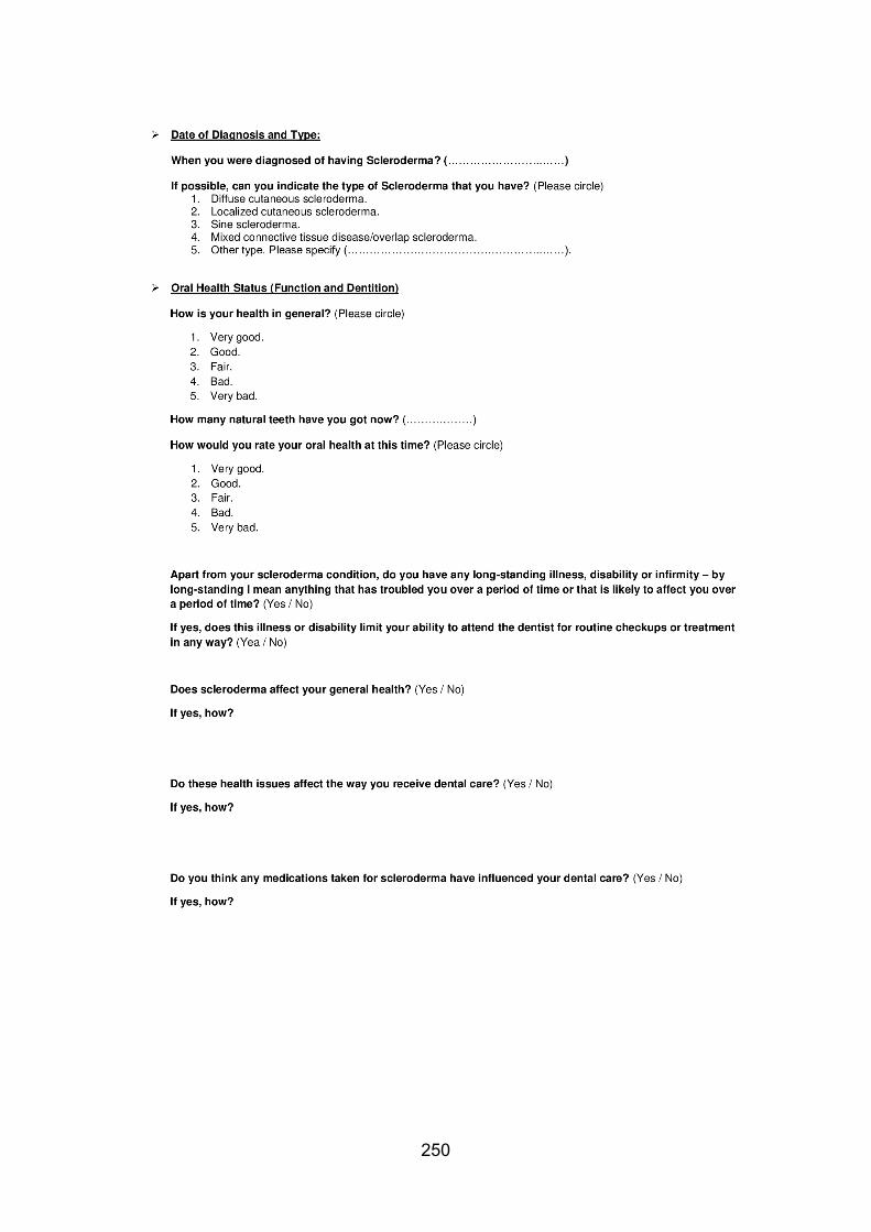
250

251
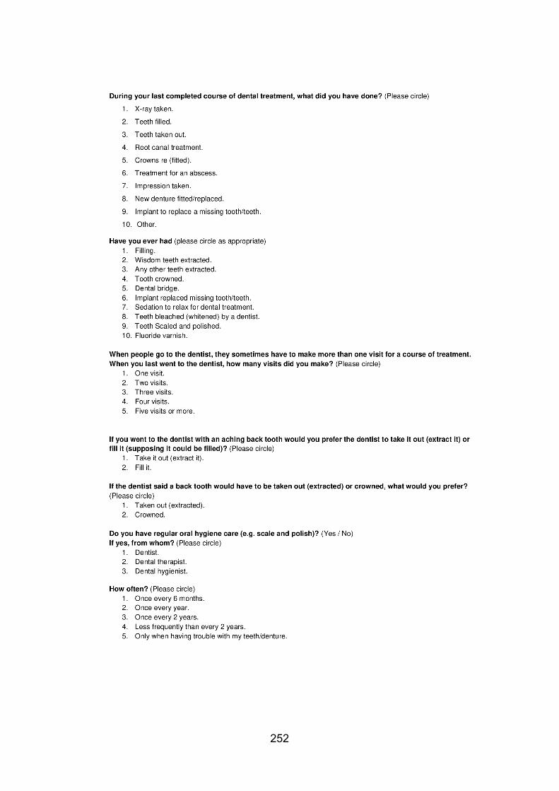
252

253

254
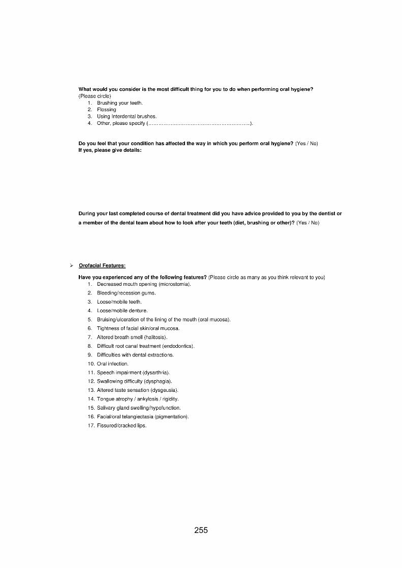
255
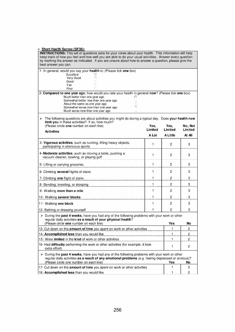
256
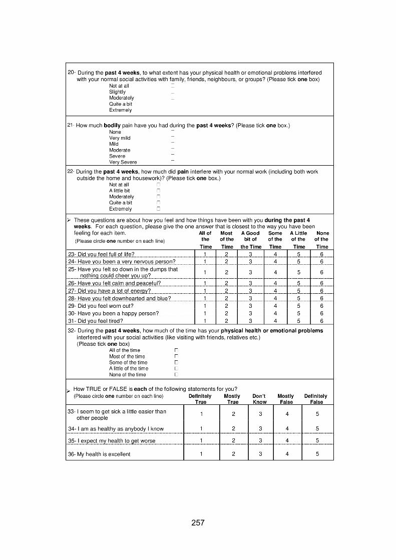
257

258

259
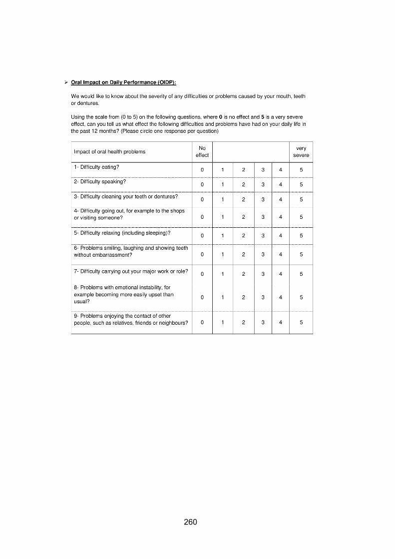
260

261
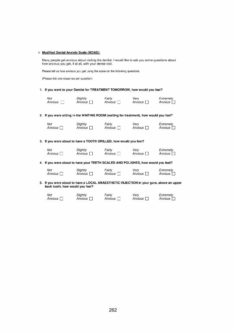
262

263

264
APPENDIX 2
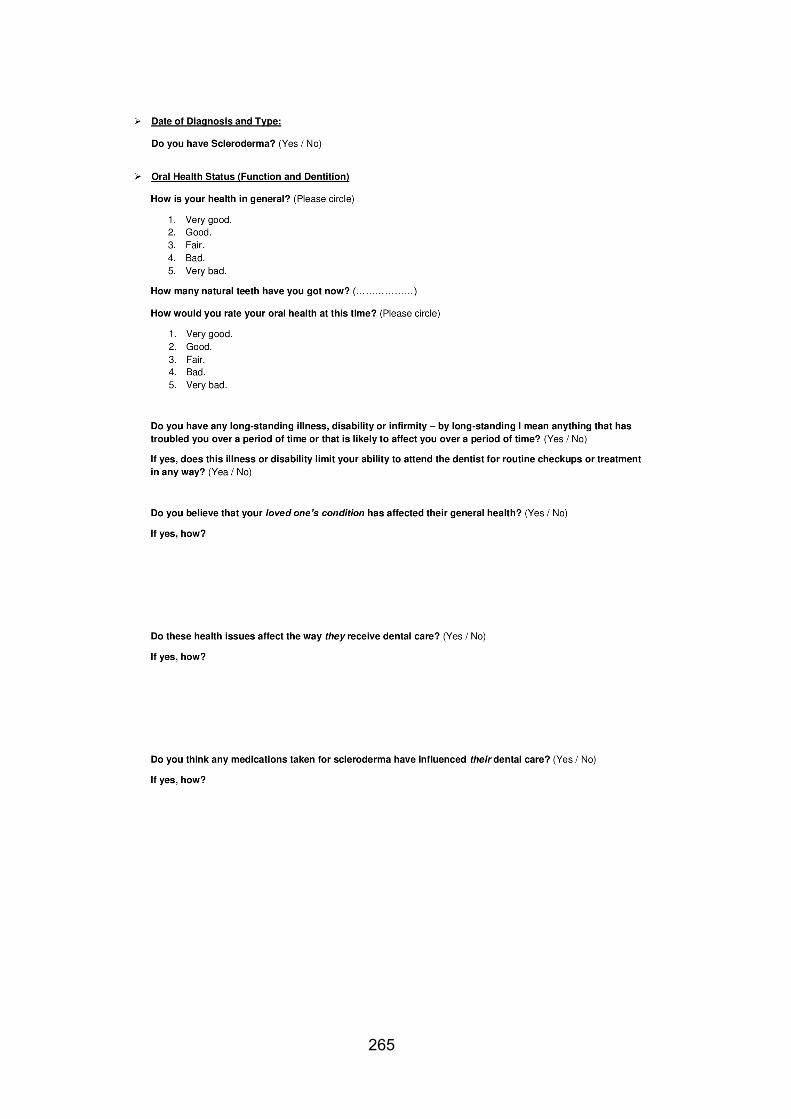
265
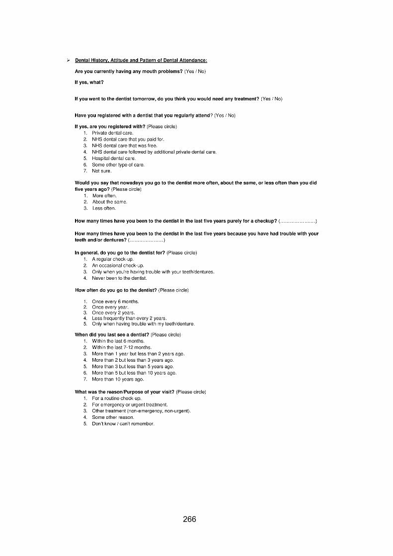
266
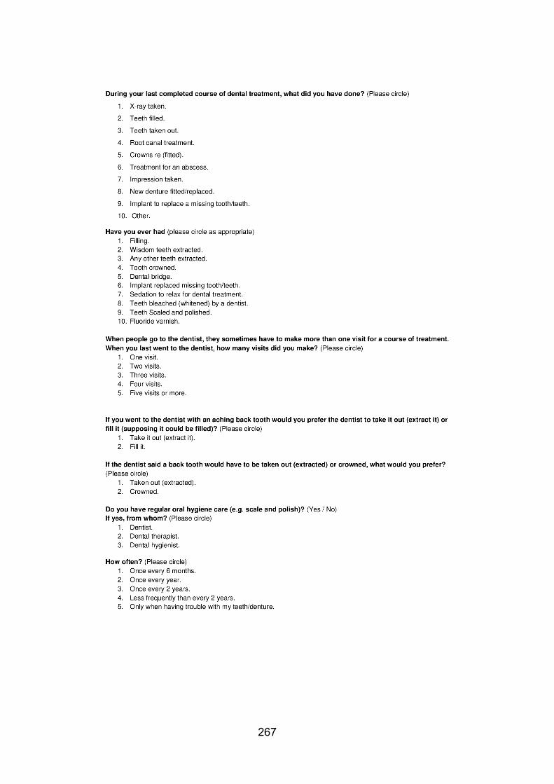
267

268
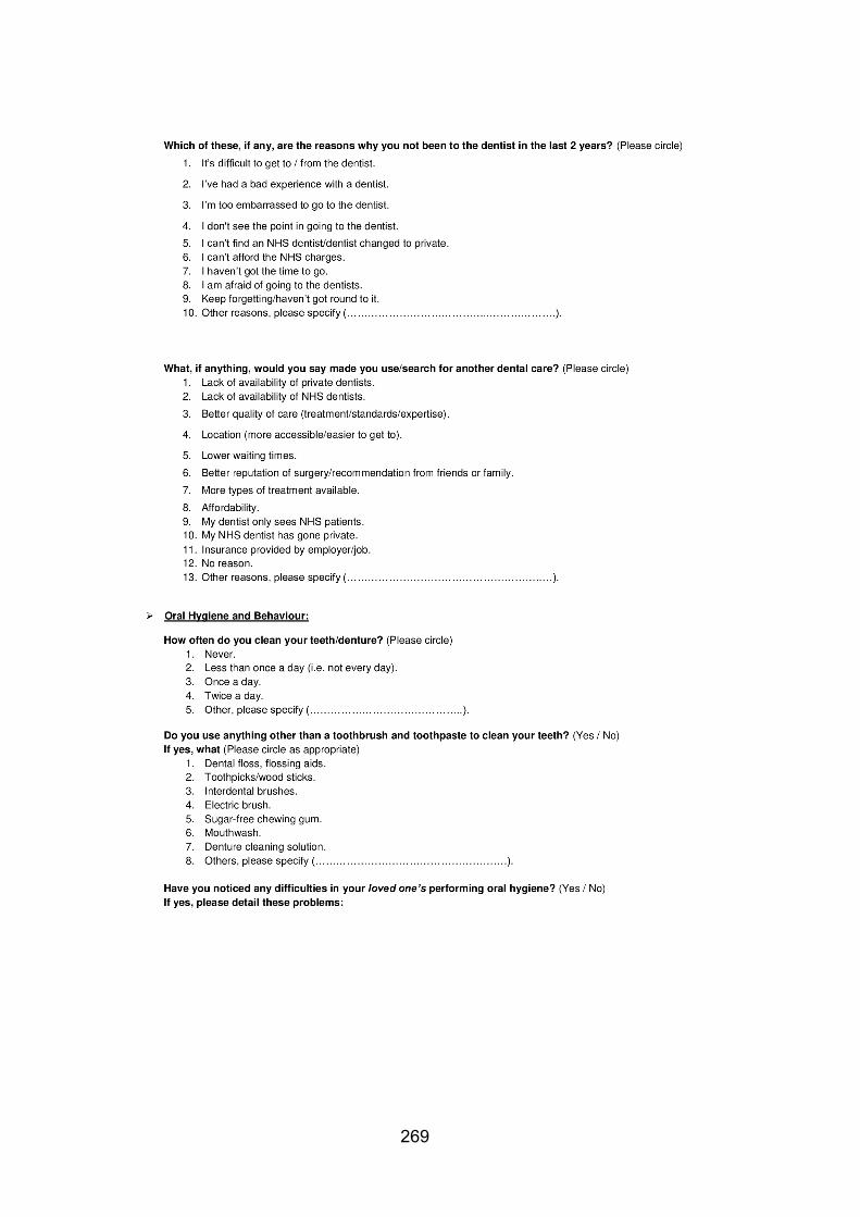
269

270

271

272
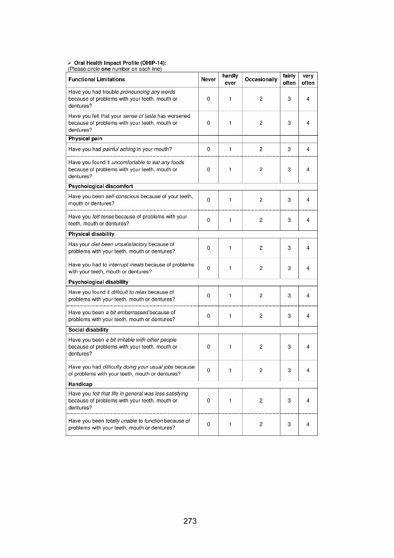
273
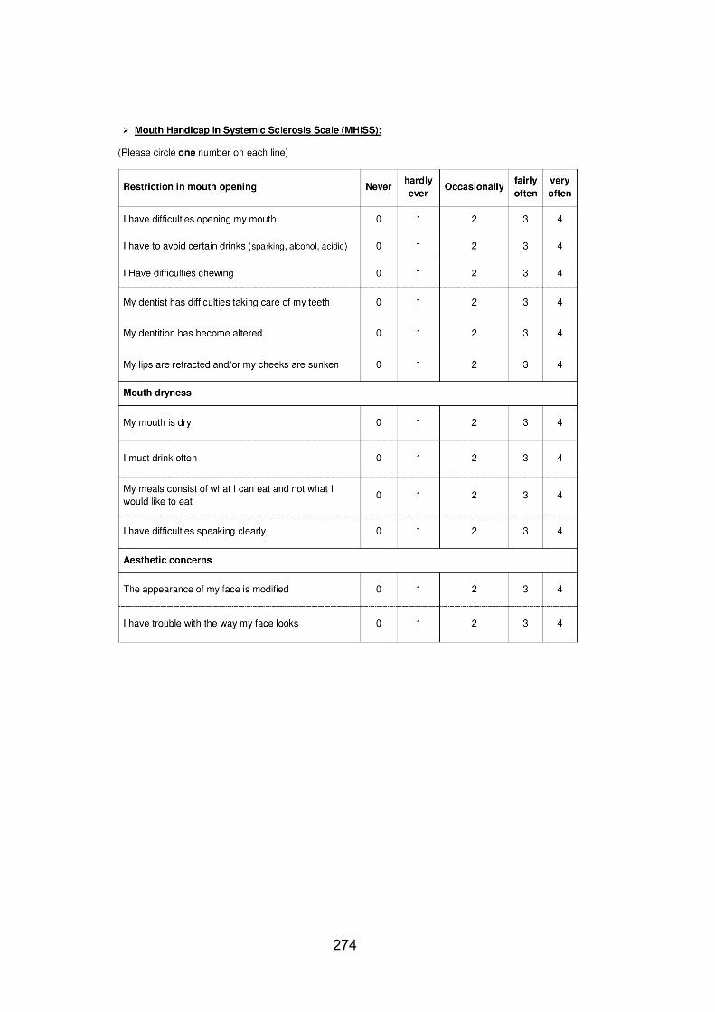
274

275

276
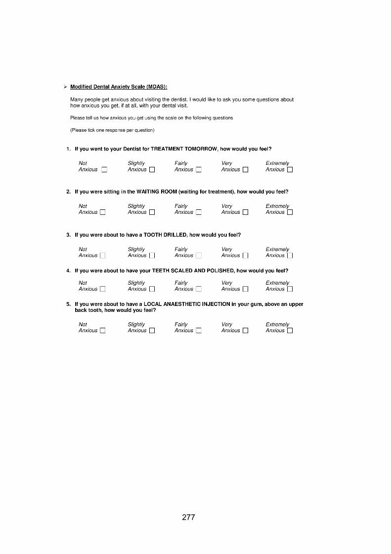
277

278
279
Published papers
Web-based information on the treatment of the mouth in systemic sclerosis. Validity and reliability of the Mouth Handicap of Systemic Sclerosis questionnaire in a UK population.
Meeting presentation
Web-based information on the treatment of the mouth in systemic sclerosis –
IADR/PER July 2018 in London, UK.
Self-reported oral and dental experience of patients with systemic sclerosis -
EAOM September 2018 in Gothenburg, Sweden.
Web-based information on the treatment of the mouth in systemic sclerosis – IADR
Saudi Arabia Division Annual meeting - April 2019 in Makkah, KSA.
Self-reported oral and dental experience of patients with systemic sclerosis - IADR
Saudi Arabia Division Annual meeting - April 2019 in Makkah, KSA.
Validity and reliability of the Mouth Handicap in Systemic Sclerosis (MHISS)
questionnaire in a UK population – Scleroderma family day – May 2019 in London,
UK.
Measuring the impact of systemic sclerosis on oral health-related quality of life in
the UK – World Dental and Oral Health Congress – July 2019 in London, UK.