Rapid efficient synthesis and characterization of silver, gold, and bimetallic nanoparticles from...
19
© 2014 Salunke et al. This work is published by Dove Medical Press Limited, and licensed under Creative Commons Attribution – Non Commercial (unported, v3.0) License. The full terms of the License are available at http://creativecommons.org/licenses/by-nc/3.0/. Non-commercial uses of the work are permitted without any further permission from Dove Medical Press Limited, provided the work is properly attributed. Permissions beyond the scope of the License are administered by Dove Medical Press Limited. Information on how to request permission may be found at: http://www.dovepress.com/permissions.php International Journal of Nanomedicine 2014:9 2635–2653 International Journal of Nanomedicine Dovepress submit your manuscript | www.dovepress.com Dovepress 2635 ORIGINAL RESEARCH open access to scientific and medical research Open Access Full Text Article http://dx.doi.org/10.2147/IJN.S59834 Rapid efficient synthesis and characterization of silver, gold, and bimetallic nanoparticles from the medicinal plant Plumbago zeylanica and their application in biofilm control Correspondence: Balu A Chopade Department of Microbiology, University of Pune, Ganeshkhind, Pune-411007, Maharashtra, India Tel +91 20 2569 0643 Fax +91 20 2569 0087 Email [email protected]n Gayatri R Salunke 1 Sougata Ghosh 1 RJ Santosh Kumar 2 Samiksha Khade 1 Priya Vashisth 3 Trupti Kale 4 Snehal Chopade 5 Vikas Pruthi 3 Gopal Kundu 4 Jayesh R Bellare 6 Balu A Chopade 1,5 1 Institute of Bioinformatics and Biotechnology, University of Pune, 2 National Chemical Laboratory, Pune, 3 Department of Biotechnology, Indian Institute of Technology, Roorkee, 4 National Centre for Cell Science, Pune University Complex, 5 Department of Microbiology, University of Pune, Pune, 6 Department of Chemical Engineering, Indian Institute of Technology Bombay, Powai, Mumbai, India Background: Nanoparticles (NPs) have gained significance in medical fields due to their high surface-area-to-volume ratio. In this study, we synthesized NPs from a medicinally important plant – Plumbago zeylanica. Materials and methods: Aqueous root extract of P. zeylanica (PZRE) was analyzed for the presence of flavonoids, sugars, and organic acids using high-performance thin-layer chroma- tography (HPTLC), gas chromatography-time of flight-mass spectrometry (GC-TOF-MS), and biochemical methods. The silver NPs (AgNPs), gold NPs (AuNPs), and bimetallic NPs (AgAuNPs) were synthesized from root extract and characterized using ultraviolet-visible spectra, X-ray diffraction (XRD), energy-dispersive spectrometry (EDS), transmission electron microscopy (TEM), and dynamic light scattering (DLS). The effects of these NPs on Acinetobacter baumannii, Staphylococcus aureus, and Escherichia coli biofilms were studied using quantitative biofilm inhibition and disruption assays, as well as using fluorescence, scanning electron microscopy, and atomic force microscopy. Results: PZRE showed the presence of phenolics, such as plumbagin, and flavonoids, in addition to citric acid, sucrose, glucose, fructose, and starch, using HPTLC, GC-TOF-MS, and quantitative analysis. Bioreduction of silver nitrate (AgNO 3 ) and chloroauric acid (HAuCl 4 ) were confirmed at absorbances of 440 nm (AgNPs), 570 nm (AuNPs), and 540 nm (AgAuNPs), respectively. The maximum rate of synthesis at 50°C was achieved with 5 mM AgNO 3 within 4.5 hours for AgNPs; and with 0.7 mM HAuCl 4 within 5 hours for AuNPs. The synthesis of AgAuNPs, which completed within 90 minutes with 0.7 mM AgNO 3 and HAuCl 4 , was found to be the fastest. Fourier-transform infrared spectroscopy confirmed bioreduction, while EDS and XRD patterns confirmed purity and the crystalline nature of the NPs, respectively. TEM micrographs and DLS showed about 60 nm monodispersed Ag nanospheres, 20–30 nm Au nanospheres adhering to form Au nanotriangles, and about 90 nm hexagonal blunt-ended AgAuNPs. These NPs also showed antimicrobial and antibiofilm activity against E. coli, A. baumannii, S. aureus, and a mixed culture of A. baumannii and S. aureus. AgNPs inhibited biofilm in the range of 96%–99% and AgAuNPs from 93% to 98% in single-culture biofilms. AuNPs also showed biofilm inhibi- tion, with the highest of 98% in S. aureus. AgNPs also showed good biofilm disruption, with the highest of 88% in A. baumannii. Conclusion: This is the first report on rapid and efficient synthesis of AgNPs, AuNPs and AgAuNPs from P. zeylanica and their effect on quantitative inhibition and disruption of bacterial biofilms. Keywords: P. zeylanica, AgNPs, AuNPs, AgAuNPs, biofilm inhibition and disruption, GC-TOF-MS
Transcript of Rapid efficient synthesis and characterization of silver, gold, and bimetallic nanoparticles from...

© 2014 Salunke et al. This work is published by Dove Medical Press Limited, and licensed under Creative Commons Attribution – Non Commercial (unported, v3.0) License. The full terms of the License are available at http://creativecommons.org/licenses/by-nc/3.0/. Non-commercial uses of the work are permitted without any further
permission from Dove Medical Press Limited, provided the work is properly attributed. Permissions beyond the scope of the License are administered by Dove Medical Press Limited. Information on how to request permission may be found at: http://www.dovepress.com/permissions.php
International Journal of Nanomedicine 2014:9 2635–2653
International Journal of Nanomedicine Dovepress
submit your manuscript | www.dovepress.com
Dovepress 2635
O r I g I N a l r e s e a r c h
open access to scientific and medical research
Open access Full Text article
http://dx.doi.org/10.2147/IJN.S59834
Journal name: International Journal of NanomedicineJournal Designation: Original ResearchYear: 2014Volume: 9Running head verso: Salunke et alRunning head recto: P. zeylanica-mediated synthesis of NPs and antibiofilm activityDOI: http://dx.doi.org/10.2147/IJN.S59834
Rapid efficient synthesis and characterization of silver, gold, and bimetallic nanoparticles from the medicinal plant Plumbago zeylanica and their application in biofilm control
Correspondence: Balu A ChopadeDepartment of Microbiology, University of Pune, Ganeshkhind, Pune-411007, Maharashtra, IndiaTel +91 20 2569 0643Fax +91 20 2569 0087email [email protected]
Gayatri R Salunke1
Sougata Ghosh1
RJ Santosh Kumar2
Samiksha Khade1
Priya Vashisth3
Trupti Kale4
Snehal Chopade5
Vikas Pruthi3
Gopal Kundu4
Jayesh R Bellare6
Balu A Chopade1,5
1Institute of Bioinformatics and Biotechnology, University of Pune, 2National Chemical Laboratory, Pune, 3Department of Biotechnology, Indian Institute of Technology, Roorkee, 4National centre for cell science, Pune University Complex, 5Department of Microbiology, University of Pune, Pune, 6Department of Chemical engineering, Indian Institute of Technology Bombay, Powai, Mumbai, India
Background: Nanoparticles (NPs) have gained significance in medical fields due to their high
surface-area-to-volume ratio. In this study, we synthesized NPs from a medicinally important
plant – Plumbago zeylanica.
Materials and methods: Aqueous root extract of P. zeylanica (PZRE) was analyzed for the
presence of flavonoids, sugars, and organic acids using high-performance thin-layer chroma-
tography (HPTLC), gas chromatography-time of flight-mass spectrometry (GC-TOF-MS), and
biochemical methods. The silver NPs (AgNPs), gold NPs (AuNPs), and bimetallic NPs
(AgAuNPs) were synthesized from root extract and characterized using ultraviolet-visible
spectra, X-ray diffraction (XRD), energy-dispersive spectrometry (EDS), transmission
electron microscopy (TEM), and dynamic light scattering (DLS). The effects of these NPs on
Acinetobacter baumannii, Staphylococcus aureus, and Escherichia coli biofilms were studied
using quantitative biofilm inhibition and disruption assays, as well as using fluorescence, scanning
electron microscopy, and atomic force microscopy.
Results: PZRE showed the presence of phenolics, such as plumbagin, and flavonoids, in addition
to citric acid, sucrose, glucose, fructose, and starch, using HPTLC, GC-TOF-MS, and quantitative
analysis. Bioreduction of silver nitrate (AgNO3) and chloroauric acid (HAuCl
4) were confirmed
at absorbances of 440 nm (AgNPs), 570 nm (AuNPs), and 540 nm (AgAuNPs), respectively.
The maximum rate of synthesis at 50°C was achieved with 5 mM AgNO3 within 4.5 hours
for AgNPs; and with 0.7 mM HAuCl4 within 5 hours for AuNPs. The synthesis of AgAuNPs,
which completed within 90 minutes with 0.7 mM AgNO3 and HAuCl
4, was found to be
the fastest. Fourier-transform infrared spectroscopy confirmed bioreduction, while EDS and XRD
patterns confirmed purity and the crystalline nature of the NPs, respectively. TEM micrographs
and DLS showed about 60 nm monodispersed Ag nanospheres, 20–30 nm Au nanospheres
adhering to form Au nanotriangles, and about 90 nm hexagonal blunt-ended AgAuNPs. These NPs
also showed antimicrobial and antibiofilm activity against E. coli, A. baumannii, S. aureus, and a
mixed culture of A. baumannii and S. aureus. AgNPs inhibited biofilm in the range of 96%–99%
and AgAuNPs from 93% to 98% in single-culture biofilms. AuNPs also showed biofilm inhibi-
tion, with the highest of 98% in S. aureus. AgNPs also showed good biofilm disruption, with
the highest of 88% in A. baumannii.
Conclusion: This is the first report on rapid and efficient synthesis of AgNPs, AuNPs and
AgAuNPs from P. zeylanica and their effect on quantitative inhibition and disruption of bacterial
biofilms.
Keywords: P. zeylanica, AgNPs, AuNPs, AgAuNPs, biofilm inhibition and disruption,
GC-TOF-MS
International Journal of Nanomedicine 2014:9submit your manuscript | www.dovepress.com
Dovepress
Dovepress
2636
Salunke et al
IntroductionNanomaterial synthesis and characterization have been an
extensive area of research since the last decade. Biological
synthesis of nanoparticles (NPs) is considered environmentally
friendly, nontoxic, and cost-effective.1 Therefore, biological
synthesis of NPs from various plant extracts2,3 and microor-
ganisms like bacteria,4 yeasts,5 fungi,6 and algae7 have proven
to be promising sources. However, plant extracts are preferred
over microbial sources, as they do not require sterile condi-
tions. Also, bioreduction of NPs using plant extracts is rapid,
ecofriendly, biocompatible, and nontoxic.8,9
Silver has been exploited as antimicrobials from ancient
period.10 With the evolution of nanomedicine as a study
for treating infections, metallic silver in the form of NPs
has regained its significance.11,12 Several bacteria have
developed resistance against antibiotics, which has chal-
lenged the treatment of human infections.13–15 Therefore,
silver NPs (AgNPs) as an antimicrobial agent seem to be
beneficial compared to antibiotics.16 Moreover, few reports
show synergistic enhancement of activity of antibiotics with
AgNPs.17 There are a number of reports on green synthesis
of gold NPs (AuNPs) by plants and microorganisms, and
they are being exploited for research on drug delivery and
cancer studies.18 However, as gold is not known to have an
antimicrobial property, very few reports are available on
the antimicrobial nature of AuNPs. It is of interest to some
researchers whether AuNPs can inhibit biofilm formation
and kill bacteria as they show surface bioconjugation.19
So far, syntheses of bimetallic NPs (AgAuNPs) from Diospy-
ros kaki and Azadirachta indica extracts have been reported
by Song and Kim20 and Shankar et al21 respectively, with an
Au core and Ag shell observed in case of A. indica. However,
research on their application as antimicrobial agents is still
in its infancy.
Plumbago zeylanica, commonly known as agni
(meaning fire) in Sanskrit, is an important medicinal
plant used in ayurveda, an Indian system of traditional
medicine.22 Ahmad and Beg23 reported on the antimicrobial
activity of aqueous and alcoholic extracts from roots of
P. zeylanica against Staphylococcus aureus, Pseudomonas
aeruginosa, Proteus vulgaris, Bacillus subtilis, and Candida
albicans. Plumbagin (5-hydroxy-2-methyl-1,4-naphthoqui-
none) is a major flavonoid present in P. zeylanica, especially
in roots, showing antifungal and antimicrobial properties.24,25
Plumbagin is also well known for its plasmid-curing
property.26 Plumbagin has cured plasmid-encoding resistance
to silver, cadmium, antimony, streptomycin, and ampicillin
in Acinetobacter baumannii species.27
The phytochemical analysis of an aqueous root extract
of P. zeylanica (PZRE) showed the presence of alkaloids,
glycoside, reducing sugars, phenolics, tannins, lignin,
saponins, and flavonoids.28 The majority of these compounds
are known to act as reducing as well as stabilizing agents
in the bioreduction of silver and gold salt.29–32 Moreover,
there have been no reports on the synthesis of metal NPs by
P. zeylanica to date. Therefore, we propose that P. zeylanica
may synthesize novel metal NPs exhibiting antimicrobial and
biofilm inhibition/disruption properties.
Materials and methodsPlant material and preparation of extractP. zeylanica roots were collected from the natural landscape
of Western Ghats, on Dr Homi Bhabha Road, Pune,
Maharashtra, India. They were thoroughly washed with dis-
tilled water, shade-dried, chopped into thin slices, and ground
to make fine powder. The aqueous extract was prepared by
boiling 5 g of the powder in 100 mL sterile distilled water,
followed by centrifugation at 8,000 rpm for 10 minutes. The
supernatant of PZRE was filtered through a 0.45 μm mem-
brane filter and stored at 4°C until further use.
Preparation of the PZRE samples for chromatographic analysisPZRE was sequentially extracted along a polarity gradient
using ethyl acetate and methanol for high-performance
thin-layer chromatography (HPTLC; semiautomated
Linomat 5; Camag, Muttenz, Switzerland) fingerprinting
and gas chromatography-time of flight-mass spectrometry
(GC-TOF-MS). The sequential extraction was done by sus-
pending 5 mL of root extract in 5 mL of high-performance
liquid chromatography-grade ethyl acetate, followed by
stirring for 3 hours and centrifugation at 10,000 rpm at room
temperature (RT). The supernatant was recovered, and 5 mL
methanol was added to the pellet and redispersed. The solu-
tion was vigorously mixed by vortexing for 15 minutes, and
then methanol extract was recovered. The ethyl acetate and
methanol extracts were concentrated.
Phytochemical analysis of P. zeylanicaHPTLC analysis of PZRE was performed according
to the protocol given in earlier studies.33,34 Plumbagin,
quercetin, diosgenin, glucose, and fructose (Sigma-Aldrich,
St Louis, MO, USA) were taken as standard for the analyses.
Similarly, 5 μL of the ethyl acetate extract of PZRE was
subjected to GC-TOF-MS (Pegasus 4D GCxGC-TOFMS
system with Agilent 6890 gas chromatograph equipped with
International Journal of Nanomedicine 2014:9 submit your manuscript | www.dovepress.com
Dovepress
Dovepress
2637
P. zeylanica-mediated synthesis of NPs and antibiofilm activity
a dual-jet thermal modulator between primary and secondary
columns and a Pegasus IV TOFMS as a detector; LECO,
St Joseph, MI, USA). The program used was as follows:
primary column, 5% phenyl polysilphenylene-siloxane
HP-5MS capillary, 30 m ×0.32 mm, 0.25 μm; secondary
column, 1.00 m ×0.10 mm ID ×0.10 μm of Rxi®-17 ms
(Restek, Bellefonte, PA, USA ) in GC oven (primary oven
program, 100°C for 0.5 minute, followed by an increase
up to 215°C at an increment of 20°C/minute, held for
0.5 minute, further to 270°C at 25°C/minute and held
for 10 min; secondary oven program, rate and duration were
identical to primary oven, target temperature was set at 30°C
above primary oven). Sample injection: Gerstel PTV (Gerstel,
Mülheim an der Ruhr, Germany) using solvent vent mode;
modulator temperature +20°C. Carrier gas: helium at ramped
pressure mode; transfer line 240°C. MS parameters: electron
impact ionization 70 eV; ion source temperature 250°C;
detector voltage 1,700 V; data-acquisition mass range
m/z 50–500 at an acquisition rate of ten spectra; software
ChromaTOF® 3.34 (LECO). Identification of components
was based on comparison of their mass spectra with those
of National Institute of Standards and Technology library
spectra (v. 2.0).35 Citric acid, plumbagin, starch, and total
reducing sugar content of PZRE were determined according
to the methodology mentioned in the following sections.
Total reducing sugarTo 50 μL of PZRE, we added the same volume of 80% phenol
(volume/volume) and vortexed. To this mixture, 2 mL of
sulfuric acid was added and incubated for 10 minutes at RT,
followed by recording absorbance at 490 nm. Total reducing
sugar content was estimated using a standard glucose curve.36
Total phenolic contentPZRE (0.125 mL) was added to 0.5 mL of deionized water.
To this mixture, 0.125 mL Folin–Ciocalteu reagent was added
and incubated for 5 minutes at RT. After incubation, 1.25 mL
of 7% Na2CO
3 solution was added, and the volume was made
up to 3 mL with distilled water. This reaction mixture was
then incubated for 90 minutes at RT, and absorbance was
recorded at 760 nm. Total phenolic content was estimated
using a standard gallic acid curve.37
Total flavonoid contentHalf a milliliter of 2% AlCl
3 in methanol was mixed with
a 0.5 mL sample of PZRE and incubated for 10 minutes at
RT. After 10 minutes, absorbance was recorded at 368 nm.
The total flavonoid content was determined using a standard
quercetin curve.37
citric acid contentOne milliliter of PZRE was evaporated in order to dry it
completely, and reconstituted in 1 mL 5% trichloroacetic acid.
Dropwise addition of 8 mL of anhydrous acetic anhydride
was done, followed by incubation at 60°C for 10 minutes.
The tubes were then stoppered. After 10 minutes, 1 mL of
pyridine was added to each tube and restoppered. The tube
was then incubated at 60°C for 40 minutes, followed by
incubation in an ice-water bath for 5 minutes. Absorbance
was recorded at 420 nm. The total citric acid content was
determined from a standard citric acid curve.37
Starch contentP. zeylanica root powder was weighed, and 5 g of the powder
was washed with 70% ethanol. The washes were repeated to
ensure the removal of sugars until it did not develop color
when treated with anthrone reagent. The residue was dried
and boiled in 100 mL distilled water. Then 1 mL of PZRE
was evaporated completely and reconstituted in 60% per-
chloric acid. To this, 4 mL of anthrone reagent was added
and was boiled in a water bath for 8 minutes. The intensity of
the dark green color was recorded at 630 nm, and the starch
content was estimated by comparing the absorbance with the
standard glucose curve.37
Plumbagin contentPlumbagin was extracted using acetone, chloroform, and hep-
tane according to the method described by Israni et al.38 In a
mortar and pestle, 5 g root powder was macerated using
acetone (3×50 mL). The acetone extract was then evaporated
to dryness at RT and 25 mL chloroform was added, followed
by an equal volume of distilled water (3×) to remove water-
soluble impurities. The chloroform extract was evaporated
till a dark brown oily residue was obtained. This residue was
treated with 10 mL of phosphoric acid (10%) for 30 minutes
followed by extraction with 5 mL n-heptane. To 0.2 mL of
this yellow solution, 10% alcoholic KOH was added, the
volume was made up to 5 mL using absolute ethanol, and
absorbance was recorded at 520 nm. The concentration of
plumbagin was determined using a standard plumbagin
curve.
Synthesis and characterization of nanoparticlesUV-visible spectroscopy and FTIR analysisSynthesis and optimization studies of AgNPs and AuNPs
were carried out according to the methods given
earlier.3,37 AgAuNPs were prepared using 1 mM silver

International Journal of Nanomedicine 2014:9submit your manuscript | www.dovepress.com
Dovepress
Dovepress
2638
Salunke et al
nitrate (AgNO3) and 1 mM chloroauric acid (HAuCl
4)
salt solutions in a 1:1 ratio. To 5 mL PZRE, 47.5 mL of
HAuCl4 solution was added, followed by addition of 47.5 mL
AgNO3. Synthesis of NPs was monitored by recording the
spectra on an ultraviolet (UV)-visible spectrophotometer
(SpectraMax M5®; Molecular Devices, Sunnyvale, CA,
USA) operating at a resolution of 1 nm. Optimization stud-
ies for rate of NP synthesis were carried out at different
temperatures ranging from 4°C to 50°C, and at varying
concentrations from 0.1 mM to 5 mM. For AgAuNPs, the
salt mixture of AgNO3 and HAuCl
4 was mixed in the ratio
of 1:1, with concentrations differing from 0.1 mM to 5 mM.
AgNPs, AuNPs, and AgAuNPs were also chemically synthe-
sized and characterized according to the protocol reported
earlier.39 Dried PZRE before and after bioreduction was
subjected to Fourier-transform infrared spectroscopy (FTIR;
Affinity-1; Shimadzu, Tokyo, Japan) using the potassium
bromide pellet technique, and was exposed to an IR source
of 500–4,000 cm−1. For application purposes, the NPs were
synthesized, followed by washing twice with double-distilled
water in order to use for antimicrobial and biofilm assays.
EDS and XRD analysisNPs were studied using energy-dispersive spectrometry
(EDS; JSM 6360A analytical scanning electron microscope
[SEM]; JEOL, Tokyo, Japan) at an energy range 0–20 keV
for elemental confirmation. Phase formation of the biore-
duced NPs was studied using X-ray diffraction (XRD;
D8 Advanced; Bruker Optik, Ettlingen, Germany) with a
CuKα (1.54 Å) source.
DLS measurement and TEM analysisFreshly prepared 3 mL AgNPs, AuNPs, and AgAuNPs were
taken in polystyrene cuvettes and subjected to particle-size
analysis employing dynamic light scattering (DLS; Zetasizer
Nano-2590; Malvern Instruments, Malvern, UK). Zeta-
potential values of each sample were also recorded on the same
instrument. The surface morphology and size of the NPs were
determined using transmission electron microscopy (TEM;
Tecnai™ G2 20 U-Twin; FEI, Hillsboro, OR, USA).
Antimicrobial assayThe effects of NPs synthesized using PZRE were tested
against A. baumannii AIIMS 7, Escherichia coli NCIM
(National Collection of Industrial Microorganisms) 2931 and
S. aureus MTCC (Microbial Type Culture Collection) 3160 on
Mueller–Hinton (MH) agar plates using a disk-diffusion
assay. Different concentrations of freshly prepared AgNPs,
AuNPs, and AgAuNPs were prepared from 2,048 μg
to 0.25 μg/disk by the double-dilution method. After
incubation at 37°C for 18 hours, the zones of inhibition
were measured.40,17 Chemically synthesized NPs were used
as controls. All assays were performed in triplicate.
Determination of biofilm inhibition and disruptionBiofilm-inhibition assays were performed by coincubation
of 5 μL of culture (optical density adjusted to 0.05 at 600 nm)
with AgNPs, AuNPs, and AgAuNPs (1,024 μg/well) in 200 μL
MH broth for 24 hours at 37°C.40 In the case of biofilm-
disruption assays, bacterial biofilm was allowed to form
for 24 hours, followed by washing with sterile phos-
phate-buffered saline (PBS; NaCl 8 g/L, KCl 0.2 g/L,
Na2HPO
4 1.44 g/L, KH
2PO
4 0.24 g/L). Biofilm disruption
was initiated by the addition of 1,024 μg/well of freshly
synthesized NPs.41,42 Additionally, the effect of NPs was
checked against a mixed-biofilm formation by A. baumannii
and S. aureus. Biofilm formation in the absence of NPs served
as a positive control. The extent of biofilm inhibition and
disruption was determined using the crystal violet staining
method as reported earlier.43 Chemically synthesized NPs
were used as controls. Biofilm inhibition and disruption
assays were carried out in triplicate.
Visualization of biofilm employing fluorescence microscopy, SEM, and AFMThe biofilm inhibition and disruption assays were carried
out on grease-free, sterile glass slides with tenfold-higher
volumes to make the final volume 2 mL in 12-well plates
and kept at 37°C for incubation. The concentration of the
NPs was 1,024 μg/well. After 24 hours, the glass slides were
washed with sterile PBS. The glass slides were stained with
acridine orange and observed under a Zeiss fluorescence
microscope using 465–495 nm excitation filters (AxioScope
A1; Carl Zeiss Meditec, Jena, Germany). Glass slides fixed
with glutaraldehyde were dehydrated sequentially using ethyl
alcohol and analyzed using SEM (JSM 6360A; JEOL). Slides
washed with PBS and air-dried were used for image analysis
at a resolution of 2×2 μm under semi-contact atomic force
microscopy (AFM; NTEGRA, NT-MDT, Moscow, Russia).
ResultsPhytochemical analysis of P. zeylanicaHPTLC fingerprints for PZRE showed the bands cor-
responding to the standard plumbagin and diosgenin
in the methanolic and ethyl acetate fractions of PZRE
International Journal of Nanomedicine 2014:9 submit your manuscript | www.dovepress.com
Dovepress
Dovepress
2639
P. zeylanica-mediated synthesis of NPs and antibiofilm activity
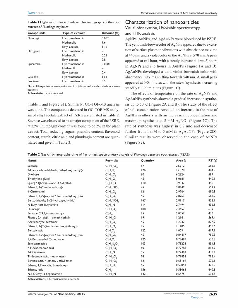
(Table 1 and Figure S1). Similarly, GC-TOF-MS analysis
was done. The compounds detected in GC-TOF-MS analy-
sis of ethyl acetate extract of PZRE are enlisted in Table 2.
Sucrose was observed to be a major component of the PZRE,
at 22%. Plumbagin content was found to be 2% in the plant
extract. Total reducing sugars, phenolic content, flavonoid
content, starch, citric acid and plumbagin content are quan-
titated and given in Table 3.
Characterization of nanoparticlesVisual observation, UV-visible spectroscopy, and FTIR analysisAgNPs, AuNPs, and AgAuNPs were bioreduced by PZRE.
The yellowish-brown color of AgNPs appeared due to excita-
tion of surface plasmon vibrations with absorbance maxima
at 440 nm and a violet color of the AuNPs at 570 nm. A peak
appeared at t=1 hour, with a steady increase till t=4.5 hours
in AgNPs and t=5 hours in AuNPs (Figure 1A and B).
AgAuNPs developed a dark-violet brownish color with
absorbance maxima shifting towards 540 nm. A small peak
appeared at t=0 minutes with the rate of synthesis increasing
steadily till 90 minutes (Figure 1C).
The effects of temperature on the rate of AgNPs and
AgAuNPs synthesis showed a gradual increase in synthe-
sis up to 50°C (Figure 2A and B). The study of the effect
of salt concentration revealed an increase in the rate of
AgNPs synthesis with an increase in concentration and
maximum synthesis at 5 mM AgNO3 (Figure 2C). The
rate of synthesis was highest in 0.7 mM and decreased
further from 1 mM to 5 mM in AgAuNPs (Figure 2D).
Similar results were observed in the case of AuNPs
(Figure S2).
Table 1 High-performance thin-layer chromatography of the root extract of Plumbago zeylanica
Compounds Type of extract Amount (%)
Plumbagin Hydromethanolic 0.002Methanolic 1.6Ethyl acetate 11.2
Diosgenin Hydromethanolic –Methanolic 0.21Ethyl acetate 2.8
Quercetin Hydromethanolic 0.0005Methanolic –Ethyl acetate 0.4
glucose Hydromethanolic 14.3Fructose Hydromethanolic 0.5
Note: all experiments were performed in triplicate, and standard deviations were negligible.Abbreviation: –, not detected.
Table 2 Gas chromatography-time of flight-mass spectrometry analysis of Plumbago zeylanica root extract (PZRE)
Name Formula Quantity Area % RT (s)
sucrose c12h22O11 57 21.912 558.32-Furancarboxaldehyde, 5-(hydroxymethyl)- c6h6O3 126 19.378 444.9D-Allose c6h12O6 60 6.2624 587Triethylene glycol c6h14O4 45 5.0681 440.1Spiro[2.3]hexan-5-one, 4,4-diethyl- c10h16O 110 3.9047 446Ethanol, 2-(2-aminoethoxy)- c4h11NO2 45 3.8949 559.74-Chromanol c9h10O2 121 2.9764 690.5Ethanol, 2,2′-[oxybis(2,1-ethanediyloxy)]bis- c8h18O5 45 2.8263 568.9Benzothiazole, 2-(2-hydroxyethylthio)- c9h9NOs2 167 2.8117 832.1N-Butyl-tert-butylamine c8h19N 114 2.7494 432.3Plumbagin c11h8O3 188 2.4452 639Pentane, 2,3,3,4-tetramethyl- c9h20 85 2.0557 430Phenol, 2,4-bis(1,1-dimethylethyl)- c14h22O 191 1.214 569.4Acetaldehyde, tetramer c8h16O4 45 1.2032 877.2Ethanol, 2-[2-(2-ethoxyethoxy)ethoxy]- c8h18O4 45 1.1105 456.6Benzoic acid c7h6O2 122 1.003 417.1Ethanol, 2,2′-[oxybis(2,1-ethanediyloxy)]bis- c8h18O5 45 0.84417 750.81,4-Benzenediol, 2-methoxy- c7h8O3 125 0.78487 530.8Semioxamazide c2h5N3O2 103 0.73226 454.8n-Hexadecanoic acid c16h32O2 60 0.72788 814.72-Octanamine c8h19N 55 0.72463 438.4Tridecanoic acid, methyl ester c14h28O2 74 0.71858 792.4Benzoic acid, 4-ethoxy-, ethyl ester c11h14O3 121 0.65169 576.1Ethane, 1,1′-oxybis, 2-methoxy- c6h14O3 45 0.59053 428.9Ethane, iodo- c2h5I 156 0.58063 640.3N,3-Diethyl-3-heptanamine c11h25N 142 0.5475 633.5
Abbreviations: rT, reaction time; s, seconds.

International Journal of Nanomedicine 2014:9submit your manuscript | www.dovepress.com
Dovepress
Dovepress
2640
Salunke et al
The plant extracts before and after bioreduction were
subjected to FTIR analysis. The sample of plant extract
before bioreduction showed a peak at 3,300 cm−1, signifying
the presence of hydroxyl groups (Figure 3). In samples after
reduction, a peak at 3,300 cm−1 was not observed and the
free hydroxyls were observed at 3,390 cm−1, indicating that
hydroxyl groups present in polyphenols from P. zeylanica
were responsible for the bioreduction of the AgNO3 and
HAuCl4 solutions to form respective NPs. A significant peak
at 1,741 cm−1of the symmetric stretch of C=O indicated the
presence of aldehydes, ketones, esters, or carboxylic acids.
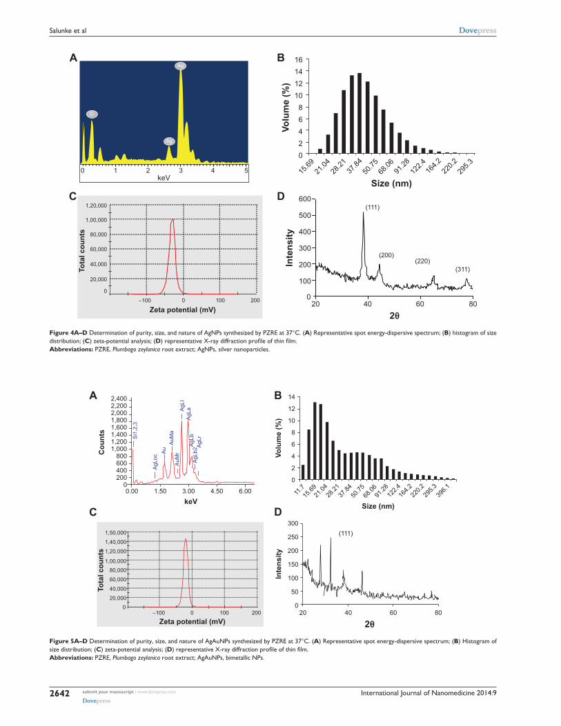
EDS, XRD, and DLS analysisIn EDS analysis, the characteristic peak at 3 keV confirmed
the presence of AgNPs, whereas particle-size analysis
employing DLS showed that the bioreduced AgNPs had an
average size of 63.92 nm (Figure 4A and B). Stability of the
particles was confirmed from the zeta-potential value, which
was found to be −31, whereas phase formation was observed
using XRD patterns. Phase formation was confirmed from
characteristic peaks, such as (111), (200), (220), and (311).
The data matched with the standard Joint Committee for Pow-
der Diffraction Set, card 040783, confirming a face-centered
cubic structure for the AgNPs (Figure 4C and D).
EDS of AgAuNPs showed that peaks were obtained at
around 2 and 3 keV, confirming the presence of silver and
gold (Figure 5A). The silver content was around 66%, and
that of gold was 14%. The average size of the AgAuNPs was
recorded to be 93.04 nm, with a zeta-potential value of −21
(Figure 5B and C). In XRD analysis, the position of the
Table 3 Phytochemical analysis of Plumbago zeylanica root extract (PZRE)
Sample Phytochemicals (mg/mL)
PZRE Plumbagin Total reducing sugars
Total phenolic content
Total flavonoid content
Starch citric acid
0.12±0.04 1.25±0.04 0.73±0.02 0.95±0.05 0.93±0.02 3.38±0.01
Note: The data is indicated as the means ± standard deviation, n=3.
200 400 600 800Wavelength (nm)
0.2
0.4
0.6
0.8
1
1.2
1.4
0
C
OD
90 minutes75 minutes60 minutes45 minutes30 minutes15 minutes
0 minute
200 400 600 800 200 400 600 800
Wavelength (nm) Wavelength (nm)
1
0.9
0.80.7
0.60.50.40.30.20.1
0
0.8
0.7
0.6
0.5
0.4
0.3
0.2
0.1
0
A B
OD OD
4.5 hours4 hours
3.5 hours3 hours
2.5 hours2 hours
1.5 hours1 hour
0.5 hour0 hour
4.5 hours5 hours
4 hours3.5 hours
3 hours2.5 hours
2 hours1.5 hours
1 hour0.5 hour
0 hour
Figure 1A–C Ultraviolet-visible spectra of nanoparticles synthesized from PZRE at 37°c as a function of time. (A) AgNPs from PZRE in 1 mM AgNO3; (B) AuNPs from PZRE in 1 mM HAuCl4; (C) AgAuNPs from PZRE in 1 mM AgNO3 and haucl4 solution in the ratio 1:1.Abbreviations: haucl4, chloroauric acid; AgNO3, silver nitrate; PZRE, Plumbago zeylanica root extract; agNPs, silver nanoparticles; auNPs, gold NPs; agauNPs, bimetallic NPs; OD, optical density.

International Journal of Nanomedicine 2014:9 submit your manuscript | www.dovepress.com
Dovepress
Dovepress
2641
P. zeylanica-mediated synthesis of NPs and antibiofilm activity
Time (hours)0 2 4 6
0
0.2
0.4
0.6
0.8
1
1.2
4ºC
20ºC
30ºC
40ºC
50ºC
0 50 1000
0.2
0.4
0.6
0.8
1
1.2
1.4
Abs
orba
nce
at 5
40 n
m
Time (minutes)
4ºC
20ºC
30ºC
40ºC
50ºC
B
0 2 4 6
1.8
2
1.6
1.4
1.2
1
0.8
0.6
0.4
0.2
0
Time (hours)
0.1 mM0.3 mM0.5 mM0.7 mM1 mM2 mM3 mM4 mM5 mM
Abs
orba
nce
at 4
40 n
m
A
Abs
orba
nce
at 4
40 n
m
C D
0 50 100
1.6
1.4
1.2
1
0.8
0.6
0.4
0.2
0
Abs
orba
nce
at 5
40 n
m
Time (minutes)
0.1 mM0.3 mM0.5 mM0.7 mM1 mM2 mM3 mM4 mM5 mM
Figure 2A–D Bioreduction of AgNPs and AgAuNPs from PZRE at varying reaction temperatures and concentrations of the salts as a function of time. (A) Time course of AgNPs synthesis with varying reaction temperatures; (B) time course of AgAuNPs synthesis against reaction temperature; (C) time course of AgNPs formation obtained at different agNO3 concentrations; (D) time course of AgAuNPs formation obtained at 1:1 AgNO3 and haucl4.Abbreviations: haucl4, chloroauric acid; AgNO3, silver nitrate; PZRE, Plumbago zeylanica root extract; agNPs, silver nanoparticles; agauNPs, bimetallic NPs.
4,000 3,000 2,000 1,000Wavenumber cm−1
% tr
ansm
ittan
ce
D
C
B
A
Figure 3A–D Fourier-transform infrared absorptive spectra before and after bioreduction of dried PZRE. (A) PZRE; (B) AgNPs; (C) AuNPs; (D) AgAuNPs.Abbreviations: PZRE, Plumbago zeylanica root extract; agNPs, silver nanoparticles; auNPs, gold NPs; agauNPs, bimetallic NPs.
peak (38.9°C), located between the (111) reflections of Ag
and Au, as seen in Figure 5D, represents the formation of
alloy-like AgAuNPs. Similarly, characterization of AuNPs
was carried out using the aforementioned techniques, and
results are given in Figure S3.
TEM analysisThe TEM micrograph showed spherically shaped AgNPs
synthesized by PZRE (Figure 6A). The TEM micrographs
revealed some unique features of AuNPs (Figure 6B and C).
They were found to be anisotropic in nature, with spheres,
triangles, and hexagons exhibiting an interesting and rare
phenomenon of shape evolution. Figure 6B demonstrates
an arrangement of gold nanospheres forming a gold nano-
triangle. Furthermore, the shape evolution continued, with
nanospheres adhering to the triangle, leading to the growth
of nanotriangles with attached nanospheres (Figure 6C).
In Figure 6D, the arrows indicate nanohexagons and nano-
spheres of AgAuNPs, but the majority of them were nano-
hexagons. A unique feature of AgAuNPs was their shape:
blunt ended polygonal NPs. Chemically synthesized NPs
were also characterized using EDS, XRD, TEM, and DLS
(Figures S4–S6).
Antibacterial activityThe minimum inhibitory concentration (MIC) values of the
NPs against Gram-negative and Gram-positive bacteria are
International Journal of Nanomedicine 2014:9submit your manuscript | www.dovepress.com
Dovepress
Dovepress
2642
Salunke et al
C
Ag
Ag
0 1 2keV
3 4 5 15.69
21.04
28.21
37.84
50.75
68.06
91.28
122.4
164.2
220.2
295.3
Size (nm)
Volu
me
(%)
1614121086420
A
C D
B
0
20,000
40,000
60,000
80,000
1,00,000
1,20,000
Tota
l cou
nts
Inte
nsity
Zeta potential (mV)
...............................................................................
...............................................................................
...............................................................................
...............................................................................
...............................................................................
...............................................................................
...............................................
...............................................
...............................................
...............................................
−100 0 100 200
100
0
2θ20 40 60 80
200
300
400
500
600(111)
(200)(220)
(311)
Figure 4A–D Determination of purity, size, and nature of AgNPs synthesized by PZRE at 37°c. (A) Representative spot energy-dispersive spectrum; (B) histogram of size distribution; (C) zeta-potential analysis; (D) representative X-ray diffraction profile of thin film.Abbreviations: PZRE, Plumbago zeylanica root extract; agNPs, silver nanoparticles.
0.00 1.50 3.00 4.50 6.000
200400600800
1,0001,2001,4001,6001,8002,0002,2002,400
Cou
nts
keV
Volu
me
(%)
AgL
ocA
uA
uMa
AgL
IA
gLa
AgL
bA
gLr
AgL
b2
AuM
r
Si1
,2,3
A
C D
B
11.7
0
2
4
6
8
10
12
14
15.69
21.04
28.21
37.84
50.75
68.06
91.28
122.4
164.2
220.2
295.3
396.1
1,50,000
1,40,000
1,20,000
1,00,00080,000
60,000
40,000
20,0000
−100 0 100 200
Zeta potential (mV)
Inte
nsity
2θ
.................................................................................................
.................................................................................................
.................................................................................................
..........................................................
..........................................................
..........................................................
..........................................................
.................................................................................................
.................................................................................................
.................................................................................................
.................................................................................................
.................................................................................................
(111)300
250
200
150
100
50
020 40 60 80
Tota
l cou
nts
Size (nm)
Figure 5A–D Determination of purity, size, and nature of AgAuNPs synthesized by PZRE at 37°c. (A) Representative spot energy-dispersive spectrum; (B) Histogram of size distribution; (C) zeta-potential analysis; (D) representative X-ray diffraction profile of thin film.Abbreviations: PZRE, Plumbago zeylanica root extract; agauNPs, bimetallic NPs.

International Journal of Nanomedicine 2014:9 submit your manuscript | www.dovepress.com
Dovepress
Dovepress
2643
P. zeylanica-mediated synthesis of NPs and antibiofilm activity
A
50 nm
B
100 nm
C
100 nm
D
50 nm
Figure 6A–D Characterization of nanoparticles synthesized by PZRE using transmission electron microscopy. (A) Spherical silver nanospheres; (B) shape evolution of gold nanotriangles; (C) assembly of gold nanospheres forming a gold nanotriangle; (D) arrows indicating blunt-ended AgAu nanopolygons.Abbreviation: PZRE, Plumbago zeylanica root extract.
given in Table 4. PZRE-mediated AgNPs showed effective
antimicrobial activity compared to PZRE-mediated AuNPs
and AgAuNPs, as well as the chemically synthesized
AgNPs when used as a control. AgNPs exhibited a lower
MIC value against E. coli (2 μg/disk) in comparison with
A. baumannii and S. aureus strains (8 μg/disk). AuNPs
showed the highest antimicrobial activity against S. aureus,
with an MIC of 8 μg/disk, whereas chemically synthesized
AuNPs did not show antimicrobial activity. Moreover,
bioreduced AgAuNPs were found to show more effective
antimicrobial activity compared to AgAuNPs synthesized
chemically.
Table 4 Minimum inhibitory concentration of silver nanoparticles (AgNPs), gold NPs (AuNPs) and bimetallic NPs (AgAuNPs) against bacterial cultures
Name of the microorganism Biologically synthesized Chemically synthesized
AgNPs AuNPs AgAuNPs AgNPs AuNPs AgAuNPs
A. baumannii AIIMS 7 8 128 16 512 – 256E. coli NcIM 2931 2 256 4 512 – 32S. aureus aureus MTcc 3160 8 8 8 256 – 512
Note: all experiments were performed in triplicate and standard deviations were negligible.Abbreviations: AIIMS, All India Institute of Medical Sciences; NCIM, National Collection of Industrial Microorganisms; MTCC, Microbial Type Culture Collection; –, No antimicrobial activity observed.
International Journal of Nanomedicine 2014:9submit your manuscript | www.dovepress.com
Dovepress
Dovepress
2644
Salunke et al
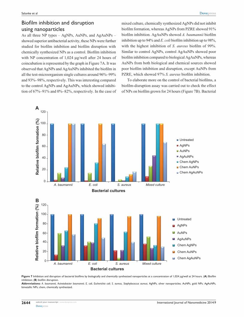
Biofilm inhibition and disruption using nanoparticlesAs all three NP types – AgNPs, AuNPs, and AgAuNPs –
showed superior antibacterial activity, these NPs were further
studied for biofilm inhibition and biofilm disruption with
chemically synthesized NPs as a control. Biofilm inhibition
with NP concentration of 1,024 μg/well after 24 hours of
coincubation is represented by the graph in Figure 7A. It was
observed that AgNPs and AgAuNPs inhibited the biofilm in
all the test-microorganism single cultures around 96%–99%
and 93%–98%, respectively. This was interesting compared
to the control AgNPs and AgAuNPs, which showed inhibi-
tion of 67%–91% and 0%–82%, respectively. In the case of
mixed culture, chemically synthesized AgNPs did not inhibit
biofilm formation, whereas AgNPs from PZRE showed 91%
biofilm inhibition. AgAuNPs showed A. baumannii biofilm
inhibition up to 94% and E. coli biofilm inhibition up to 98%,
with the highest inhibition of S. aureus biofilm of 99%.
Similar to control AgNPs, control AgAuNPs showed poor
biofilm inhibition compared to biological AgAuNPs, whereas
AuNPs from both biological and chemical sources showed
poor biofilm inhibition and disruption, except AuNPs from
PZRE, which showed 97% S. aureus biofilm inhibition.
To elaborate more on the control of bacterial biofilms, a
biofilm-disruption assay was carried out to check the effect
of NPs on biofilm grown for 24 hours (Figure 7B). Bacterial
A. baumannii E. coli S. aureus Mixed culture
Bacterial cultures
Rel
ativ
e bi
ofilm
form
atio
n (%
)
0
20
40
60
80
100
120A
Untreated
AgNPs
AuNPs
AgAuNPsChem AgNPsChem AuNPsChem AgAuNPs
B
Rel
ativ
e bi
ofilm
form
atio
n (%
)
0
20
40
60
80
100
120
A. baumannii E. coli S. aureus Mixed culture
Bacterial cultures
Untreated
AgNPs
AuNPs
AgAuNPs
Chem AgNPs
Chem AuNPs
Chem AgAuNPs
Figure 7 Inhibition and disruption of bacterial biofilms by biologically and chemically synthesized nanoparticles at a concentration of 1,024 μg/well at 24 hours. (A) Biofilm inhibition; (B) biofilm disruption.Abbreviations: A. baumannii, Acinetobacter baumannii; E. coli, Escherichia coli; S. aureus, Staphylococcus aureus; agNPs, silver nanoparticles; auNPs, gold NPs; agauNPs, bimetallic NPs; chem, chemically synthesized.
International Journal of Nanomedicine 2014:9 submit your manuscript | www.dovepress.com
Dovepress
Dovepress
2645
P. zeylanica-mediated synthesis of NPs and antibiofilm activity
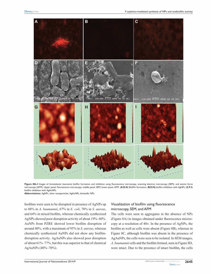
A
D E F
G H I
B C
Figure 8A–I Images of Acinetobacter baumannii biofilm formation and inhibition using fluorescence microscopy, scanning electron microscopy (SEM), and atomic force microscopy (AFM). Upper panel, fluorescence microscopy; middle panel, SEM; lower panel, AFM. (A,D,G) Biofilm formation; (B,E,H) biofilm inhibition with AgNPs; (C,F,I) biofilm inhibition with AgAuNPs.Abbreviations: agNPs, silver nanoparticles; agauNPs, bimetallic NPs.
biofilms were seen to be disrupted in presence of AgNPs up
to 88% in A. baumannii, 67% in E. coli, 78% in S. aureus,
and 64% in mixed biofilm, whereas chemically synthesized
AgNPs showed poor disruption activity of about 19%–60%.
AuNPs from PZRE showed lower biofilm disruption of
around 40%, with a maximum of 95% in S. aureus, whereas
chemically synthesized AuNPs did not show any biofilm-
disruption activity. AgAuNPs also showed poor disruption
of about 61%–77%, but this was superior to that of chemical
AgAuNPs (40%–70%).
Visualization of biofilm using fluorescence microscopy, SEM, and AFMThe cells were seen in aggregates in the absence of NPs
( Figure 8A) in images obtained under fluorescence micros-
copy at a resolution of 40×. In the presence of AgNPs, the
biofilm as well as cells were absent (Figure 8B), whereas in
Figure 8C, although biofilm was absent in the presence of
AgAuNPs, the cells were seen to be isolated. In SEM images,
A. baumannii cells and the biofilm formed, seen in Figure 8D,
were intact. Due to the presence of intact biofilm, the cells
International Journal of Nanomedicine 2014:9submit your manuscript | www.dovepress.com
Dovepress
Dovepress
2646
Salunke et al
were present in colonies, and no change in the morphology
was observed. Coincubation of the bacterial cells with AgNPs
led to a change in the cell morphology and disrupted the bio-
film of A. baumannii (Figure 8E), whereas cells were seen to
be lysed in the presence of AgAuNPs (Figure 8F).
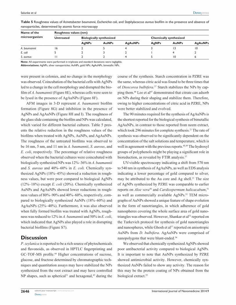
AFM images in 3-D represent A. baumannii biofilm
formation (Figure 8G) and inhibition in the presence of
AgNPs and AgAuNPs (Figure 8H and I). The roughness of
the glass slide containing the biofilm and NPs was calculated,
which varied for different bacterial cultures. Table 5 pres-
ents the relative reduction in the roughness values of the
biofilms when treated with AgNPs, AuNPs, and AgAuNPs.
The roughness of the untreated biofilms was observed to
be 16 nm, 5 nm, and 11 nm in A. baumannii, S. aureus, and
E. coli, respectively. The percentage of relative roughness
observed when the bacterial cultures were coincubated with
biologically synthesized NPs was 12%–36% in A. baumannii
and S. aureus and 40%–60% in E. coli. Chemically syn-
thesized AgNPs (18%–45%) showed a reduction in rough-
ness values, but were poor compared to biological AgNPs
(12%–18%) except E. coli (20%). Chemically synthesized
AuNPs and AgAuNPs showed lower reductions in rough-
ness values of 80%–90% and 40%–60%, respectively, com-
pared to biologically synthesized AuNPs (18%–60%) and
AgAuNPs (25%–40%). Furthermore, it was also observed
when fully formed biofilm was treated with AgNPs, rough-
ness was reduced to 12% in A. baumannii and 50% in E. coli,
which indicated that AgNPs also played a role in disrupting
bacterial biofilms (Figure S7).
DiscussionP. zeylanica is reported to be a rich source of phytochemicals
and flavonoids, as observed in HPTLC fingerprinting and
GC-TOF-MS profile.44 Higher concentrations of sucrose,
glucose, and fructose determined by chromatographic tech-
niques and quantitation assays may have stabilized the NPs
synthesized from the root extract and may have controlled
NP shapes, such as spherical31 and hexagonal,45 during the
course of the synthesis. Starch concentration in PZRE was
the same, whereas citric acid was found to be three times that
of Dioscorea bulbifera.37 Starch stabilizes the NPs by cap-
ping them.46 Lee et al47 demonstrated that citrate can adsorb
on NPs during their shaping and stabilize them. Therefore,
owing to higher concentrations of citric acid in PZRE, NPs
were better stabilized and evolved.
The 90 minutes required for the synthesis of AgAuNPs is
the shortest reported for the biological synthesis of bimetallic
AgAuNPs, in contrast to those reported from neem extract,
which took 296 minutes for complete synthesis.21 The rate of
synthesis was observed to be significantly dependent on the
concentration of the salt solutions and temperature, which is
well in agreement with the previous reports.48,49 The hydroxyl
groups of polyphenols might be playing a significant role in
bioreduction, as revealed by FTIR analysis.37
UV-visible spectroscopy indicating a shift from 570 nm
to 540 nm in synthesis of AgAuNPs, as well as EDS analysis
indicating a lower percentage of gold compared to silver,
may be attributed to the Au core and Ag shell.21 The size
of AgNPs synthesized by PZRE was comparable to earlier
reports on Aloe vera50 and Cardiospermum halicacabum,51
as well as commercially available AgNPs.52 TEM micro-
graphs of AuNPs showed a unique feature of shape evolution
in the form of nanotriangles, in which adherence of gold
nanospheres covering the whole surface area of gold nano-
triangles was observed. However, Shankar et al53 reported on
the Turkevich protocol for synthesis of gold nanotriangles
and nanospheres, while Ghosh et al37 reported on anisotropic
AuNPs from D. bulbifera. AgAuNPs were comprised of
nanopolygons that were blunt-ended.54
We observed that chemically synthesized AgNPs showed
poor antibacterial activity compared to biological AgNPs.
It is important to note that AuNPs synthesized by PZRE
showed antimicrobial activity. However, chemically syn-
thesized AuNPs failed to show any activity. The reason for
this may be the protein coating of NPs obtained from the
biological extract.55
Table 5 Roughness values of Acinetobacter baumannii, Escherichia coli, and Staphylococcus aureus biofilm in the presence and absence of nanoparticles, determined by atomic force microscopy
Name of the microorganism
Roughness values (nm)
Untreated Biologically synthesized Chemically synthesized
AgNPs AuNPs AgAuNPs AgNPs AuNPs AgAuNPs
A. baumannii 16 2 5 4 3 13 10E. coli 5 2 3 2 1 4 2S. aureus 11 2 2 4 5 10 6
Note: all experiments were performed in triplicate and standard deviations were negligible.Abbreviations: agNPs, silver nanoparticles; auNPs, gold NPs; agauNPs, bimetallic NPs.
International Journal of Nanomedicine 2014:9 submit your manuscript | www.dovepress.com
Dovepress
Dovepress
2647
P. zeylanica-mediated synthesis of NPs and antibiofilm activity
Biofilm formation by human pathogenic microbes
causing severe chronic lung, urinary tract, and various
nosocomial infections is attributed to lower penetration and
activity of drugs, altered genotype of bacteria exposed to
drugs, lower oxygen, and acidic pH.56–59 Therefore, research
on biogenic NPs for control of bacterial biofilms is very
promising. Various attempts have been made to control
biofilms by biosurfactants, rhamnolipids, and enzyme-based
detergents.60,61 Recently, Thawal et al62 demonstrated 80%
disruption of biofilm by a phage from the family Podoviridae
isolated from A. baumannii.
Control of bacterial biofilms using NPs like chitosan,
zinc oxide, and silver are interesting, because of their high
surface-to-volume ratio.42,63,64 Our results are in agreement
with the report of Kalishwaralal et al65 where 50 nM of
AgNPs significantly arrested P. aeruginosa biofilm for-
mation without affecting cell viability, whereas 100 nM
inhibited the growth of the organism itself and resulted in
a 95%–98% decrease in biofilm. Also, AgNPs and AuNPs
showed better biofilm inhibition than disruption; however,
in the case of mixed cultures, AgAuNPs showed better
biofilm disruption than biofilm inhibition. This shows the
potential of AgAuNPs to disrupt mixed biofilms, which
could be because of the synergistic effect of silver and
gold components. Mixed biofilms, however, were inhibited
and disrupted by AgNPs and AuNPs to a lesser extent
compared to single bacterial biofilm, which might be due
to more resistance to the penetrability of NPs challenging
their potential for biofilm disruption.
The novelty of the study lies in the fact that PZRE-
synthesized NPs were found to be antimicrobial against
A. baumannii and able to control its biofilm. Although
A. baumannii is found in the human skin microbiome, it is
also considered to be a dreadful nosocomial pathogen, due
to its high resistance to antibiotics and metal salts leading
to severe mortality (17%–62%) among immunocompro-
mised patients because of bacteremia.26,58,66–72 Deformity in
A. baumannii cells treated with AgNPs, as observed in fluo-
rescence and SEM micrographs, might be due to enhanced
penetrability of AgNPs within the biofilm, unlike metallic
silver.15,40,42 This can be a powerful strategy for sensitizing
A. baumannii with high metal resistance.
This is the first report of its kind where inhibition as well
as disruption of bacterial biofilms have been demonstrated
employing AFM after treatment with NPs synthesized by
PZRE. Significant reduction of roughness confirmed the
efficacy of NPs in biofilm inhibition and disruption. This
proved to be advantageous, owing to semi-contact mode,
better resolution, biofilms observed in their own physiological
condition with no addition of chemicals, direct contact with
the surface with minimum use of lenses and electron beams,
and deduction of roughness values based upon variation in
height and texture.73,74
ConclusionThis is the first report of rapid and efficient synthesis
of AgNPs, AuNPs, and AgAuNPs by P. zeylanica. All the three
types of NPs had unique features: being spherical in case of
AgNPs, revealing shape evolution in case of AuNPs, and show-
ing blunt-ended hexagonal nanostructures in AgAuNPs. These
diverse properties were obtained due to phenolics, flavonoids,
sugars, starch, and citric acid present in PZRE. Bioreduced NPs
were able to inhibit to significant levels, particularly in the case
of E. coli and S. aureus. S. aureus biofilm was inhibited and
disrupted significantly by AuNPs, which shows that AuNPs can
be more effective against Gram-positive bacteria than Gram-
negative bacteria. Therefore, the NPs synthesized, particularly
AgNPs from P. zeylanica, proved to be very efficient in the
control of bacterial biofilms.
AcknowledgmentsGS acknowledges the financial support for this work from
the Institute of Bioinformatics and Biotechnology, University
of Pune, Pune, India. SG thanks the Council of Scientific
and Industrial Research (CSIR, Government of India) for
a senior research fellowship (09/137[0516]/2012-EMR-I).
Part of this work was supported by UPE Phase I and II,
awarded to the University of Pune by the University Grant
Commission (UGC), New Delhi, India. We are thankful to
Dr Charegaonkar and Mr Naidu, Anchrome Enterprises (I)
Pvt Ltd, Mulund, Mumbai, India for HPTLC analysis. We
also acknowledge Professor SI Patil, Head of the Department
of Physics, University of Pune for use of the TEM facility;
and Purkayashta P, Khanka V, Banerjee S, and Koya R from
India LECO instruments Pvt. Ltd. (Mumbai, Maharashtra,
India) for GC-TOF-MS identification.
DisclosureThe authors report no conflicts of interest in this work.
References1. Tran QH, Nguyen VQ, Le A. Silver nanoparticles: synthesis, properties,
toxicology, applications and perspectives. Adv Nat Sci Nanosci Nanotechnol. 2013;4(3):033001.
2. Elavazhagan T, Arunachalam K. Memecylon edule leaf extract mediated green synthesis of silver and gold nanoparticles. Int J Nanomedicine. 2011;6:1265–1278.
3. Ghosh S, Patil S, Ahire M, et al. Synthesis of gold nanoanisotrops using Dioscorea bulbifera tuber extract. J Nanomater. 2011;2011:354793.
International Journal of Nanomedicine 2014:9submit your manuscript | www.dovepress.com
Dovepress
Dovepress
2648
Salunke et al
4. He S, Zhirui G, Zhang Y, Zhang S, Wang J, Gu N. Biosynthesis of gold nanoparticles using the bacteria Rhodopseudomonas capsulata. Mater Lett. 2007;61(18):3984–3987.
5. Mourato A, Gadanho M, Lino AR, Tenreiro R. Biosynthesis of crys-talline silver and gold nanoparticles by extremophilic yeasts. Bioinorg Chem Appl. 2011;2011:546074.
6. Mukherjee P, Ahmad A, Mandal D, et al. Fungus-mediated synthesis of silver nanoparticles and their immobilization in the mycelial matrix: a novel biological approach to nanoparticle synthesis. Nano Lett. 2001;1(10):515–519.
7. Govindaraju K, Kiruthuga V, Kumar VG, Singaravelu G. Extracel-lular synthesis of silver nanoparticles by a marine alga, Sargassum wightii Grevilli and their antibacterial effects. J Nanosci Nanotechnol. 2009;9(9):5497–5501.
8. Chopade NB, Ghosh S, More P, et al. Biogenic copper nanoparticles as novel antidiabetic and antioxidant in nanomedicine. Poster presented at: Second International Translational Nanomedicine Conference; July 26–28, 2013; Boston, MA.
9. Khan M, Khan M, Adil SF, et al. Green synthesis of silver nanopar-ticles mediated by Pulicaria glutinosa extract. Int J Nanomedicine. 2013:81507–81516.
10. Woo KJ, Hye CK, Ki WK, Sook S, So HK, Yong HP. Antibacterial activ-ity and mechanism of action of the silver ion in Staphylococcus aureus and Escherichia coli. Appl Env Microbiol. 2008;74(7):2171–2178.
11. He Y, Du Z, Lv H, et al. Green synthesis of silver nanoparticles by Chrysanthemum morifolium Ramat. Extract and their application in clinical ultrasound gel. Int J Nanomedicine. 2013;8:1809–1815.
12. Rai M, Yadav A, Gade A. Silver nanoparticles as a new generation of antimicrobials. Biotechnol Adv. 2009;27(1):76–83.
13. Shakibaie MR, Dhakephalkar P, Kapadnis BP, Chopade BA. Removal of silver from photographic wastewater effluent using Acinetobacter baumannii BL54. Can J Microbiol. 1999;45(12):995–1000.
14. Waters AE, Contente-Cuomo T, Buchhagen J, et al. Multidrug-resistant Staphylococcus aureus in US meat and poultry. Clin Infect Dis. 2011; 52(10):1227–1230.
15. Dhakephalkar PK, Chopade BA. High levels of multiple metal resistance and its correlation to antibiotic resistance in environmental isolates of Acinetobacter. Biometals. 1994;7(1):67–74.
16. Chopra I. The increasing use of silver-based products as antimicrobial agents: a useful development or a cause for concern? J Antimicrob Chemother. 2007;59(4):587–590.
17. Singh R, Wagh P, Wadhwani S, et al. Synthesis, optimization, and characterization of silver nanoparticles from Acinetobacter calcoa-ceticus and their enhanced antibacterial activity when combined with antibiotics. Int J Nanomedicine. 2013;8:4277–4290.
18. Shebdadkar U, Singh R, Wadhawani S, Chopade BA. Microbial synthesis of gold nanoparticles: current status and future prospects. Adv Colloid Interface Sci. Epub 2014 Jan 2.
19. Glomm WR. Functionalized gold nanoparticles for applications in bionanotechnology. J Dispers Sci Technol. 2005;26(3):389–414.
20. Song JY, Kim BS. Biological synthesis of bimetallic Au/Ag nanopar-ticles using persimmon (Diopyros kaki) leaf extract. Korean J Chem Eng. 2008;25(4):808–811.
21. Shankar SS, Rai A, Ahmad A, Sastry M. Rapid synthesis of Au, Ag, and bimetallic Au core-Ag shell nanoparticles using neem (Azadirachta indica) leaf broth. J Colloid Interface Sci. 2004;275(2): 496–502.
22. Patwardhan RB, Dhakephalkar P, Chopade BA. Medicinal importance of chitraka plant: a review. Shrushti Health Bull. 2005;4:4–5.
23. Ahmad I, Beg AZ. Antimicrobial and phytochemical studies on 45 Indian medicinal plants against multi-drug resistant human patho-gens. J Ethnopharmacol. 2001;74(2):113–123.
24. van der Vijver LM. Distribution of plumbagin in the mplumbaginaceae. Phytochemistry. 1972;11(11):3247–3248.
25. de Paiva SR, Figueiredo M, Aragão TV, Kaplan MA. Antimicrobial activity in vitro of plumbagin isolated from Plumbago species. Mem Inst Oswaldo Cruz. 2003;98(7):959–961.
26. Shakibaie MR, Dhakephalkar PK, Kapadnis BP, Salayaghe G, Chopade BA. Plasmid mediated silver and antibiotic resistance in Acinetobacter baumannii BL54. Iran J Med Sci. 1998;23(12):30–36.
27. Deshpande LM, Chopade BA. Plasmid mediated silver resistance in Acinetobacter baumannii BL54. Biometals. 1994;7(1):49–56.
28. Dhale DA, Markandeya SK. Antimicrobial and phytochemical screen-ing of Plumbago zeylanica Linn. (Plumbaginaceae) leaf. J Exp Sci. 2011;2(3):4–6.
29. Asharani PV, Mun GLK, Hande MP, Valiyaveettil S. Cytotoxicity and genotoxicity of silver nanoparticles in human cells. ACS Nano. 2009; 3(2):279–290.
30. Egorova EM, Revina AA. Synthesis of metallic nanoparticles in reverse micelles in the presence of quercetin. Colloids Surf A Physicochem Eng Asp. 2000;168(1):87–96.
31. Panigrahi S, Kundu S, Ghosh SK, Nath S, Pal T. General method of synthesis for metal nanoparticles. J Nanopart Res. 2004;6(4): 411–414.
32. Zhao X, Shi Y, Wang T, Cai Y, Jiang G. Preparation of silica-magnetite nanoparticle mixed hemimicelle sorbents for extraction of several typical phenolic compounds from environmental water samples. J Chromatogr A. 2008;1188(2):140–147.
33. Chothani DL, Patel MB, Mishra SH. HPTLC fingerprint profile and isolation of marker compound of Ruellia tuberosa. Chromatogr Res Int. 2012;2012:180103.
34. Skalska-Kamińska A, Matysik G, Wójciak-Kosior M, Donica H, Sowa I. Thin-layer chromatography of sugars in plant material. Curr Issue Pharm Med Sci. 2009;22(4):17–24.
35. Parveen I, Moorby JM, Fraser MD, Allison GG, Kopka J. Application of GCMS-TOF metabolite profiling techniques to the analysis of heathland plant diets of sheep. J Agric Food Chem. 2007;55(4):1129–1138.
36. Dubois M, Gilles DA, Hamilton JK, Rebers PA, Smith F. Colorimetric method for the determination of sugars and related substances. Anal Chem. 1956;28(3):350–356.
37. Ghosh S, Patil S, Ahire M, et al. Synthesis of silver nanoparticles using Dioscorea bulbifera tuber extract and evaluation of its synergistic potential in combination with antimicrobial agents. Int J Nanomedicine. 2012;7:483–496.
38. Israni SA, Kapadia NS, Lahiri SK, Yadav G, Shah MB. An UV-visible spectrophotometric method for the estimation of plumbagin. Int J Chem Tech Res. 2010;2(2):856–859.
39. Augustine R, Rajarathinam R. Synthesis and characterization of silver nanoparticles and its immobilization on alginate coated sutures for the prevention of surgical wound infections and the in vitro release studies. Int J Nano Dimens. 2012;2(3):205–212.
40. Deshpande LM, Kapadnis, Chopade BA. Metal resistance in Acine-tobacter and its relation to β-lactamase production. Biometals. 1993;6(1):55–59.
41. Sahu PK, Iyer PS, Oak A, Pardeshi K, Chopade BA. Characteriza-tion of eDNA from the clinical strain Acinetobacter baumannii AIIMS 7 and its role in biofilm formation. Scientific World Journal. 2012;2012:973436.
42. Gaidhani SV, Singh R, Singh D, et al. Biofilm disruption activity of silver nanoparticles synthesized by Acinetobacter calcoaceticus PUCM 1005. Mater Lett. 2013;108:324–327.
43. Stepanovic S, Vukovic D, Dakic I, Savic B, Svabic-Vlahovic M. A modified microtiter-plate test for quantification of staphy-lococcal biofilm formation. J Microbiol Methods. 2000;40(2): 175–179.
44. Kishore N, Mishra BB, Tiwari V, Tripathi V. An account of phytochemi-cals from Plumbago zeylanica (Family: Plumbaginaceae): a natural gift to human being. Chron Young Sci. 2012;3(3):178–198.
45. Swarnavalli GCJ, Joseph V, Kannappan V, Roopsingh D. A simple approach to the synthesis of hexagonal-shaped silver nanoplates. J Nanomater. 2011;2011:825637.
46. Gao XH, Wei LQ, Wang J, Xu BS. Green synthesis of starch-stabilized silver nanoparticles and their antibacterial properties. Adv Mater Res. 2011;236–238:1945–1948.
International Journal of Nanomedicine 2014:9 submit your manuscript | www.dovepress.com
Dovepress
Dovepress
2649
P. zeylanica-mediated synthesis of NPs and antibiofilm activity
47. Lee GP, Bignell LJ, Romeo TC, et al. The citrate-mediated shape evolution of transforming photomorphic silver nanoparticles. Chem Commun. 2010;46(41):7807–7809.
48. Jiang XC, Chen WM, Chen CY, Xiong SX, Yu AB. Role of temperature in the growth of silver nanoparticles through a synergetic reduction approach. Nanoscale Res Lett. 2011;6(1):32.
49. Zabetakis K, Ghann WE, Kumar S, Daniel M. Effect of high gold salt concentrations on the size and polydispersity of gold nanopar-ticles prepared by an extended Turkevich-Frens method. Gold Bull. 2012;45(4):203–211.
50. Chandran SP, Chaudhary M, Pasricha R, Ahmad A, Sastry M. Synthesis of gold nanotriangles and silver nanoparticles using Aloe vera plant extract. Biotechnol Prog. 2006;22(2):577–583.
51. Mitra B, Vishnudas D, Sant SB, Annamalai A. Green-synthesis and characterization of silver nanoparticles by aqueous leaf extracts of Cardiospermum helicacabum leaves. Drug Invent Today. 2012; 4(2):340–344.
52. Cytodiagnostics [website on the Internet]. Burlington, ON, Canada: Cytodiagnostics. Available from: http://www.cytodiagnostics.com. Accessed March 2, 2013.
53. Shankar SS, Bhargava S, Sastry M. Synthesis of gold nanospheres and nanotriangles by the Turkevich approach. J Nanosci Nanotechnol. 2005;5(10):1721–1725.
54. Mukherjee P, Nandi AK. Bimetallic Au(core)-Ag(shell) nanoparticles from interfacial redox process using poly(o-methoxyaniline). J Colloid Interface Sci. 2010;344(1):30–36.
55. Klein J. Probing the interactions of proteins and nanoparticles. Proc Natl Acad Sci U S A. 2007;104(7):2029–2030.
56. Costertan JW, Montataro L, Arciola R. Biofilm in implant infec-tions: its production and regulation. Int J Artif Organs. 2005;28(11): 1062–1068.
57. Pour NK, Dusane DH, Dhakephalkar PK, Zamin FR, Zinjarde SS, Chopade BA. Biofilm formation by Acinetobacter baumannii strains isolated from urinary tract infection and urinary catheters. FEMS Immunol Med Microbiol. 2011;62(3):328–338.
58. Høiby N, Bjarnsholt T, Givskov M, Molin S, Ciofu O. Antibiotic resistance of bacterial biofilms. Int J Antimicrob Agents. 2010;35(4): 322–332.
59. National Institutes of Health. Targeted research on oral microbial bio-films. 1998. Available from: http://grants.nih.gov/grants/guide/rfa-files/RFA-DE-98-006.html. Accessed April 3, 2013.
60. Rodrigues L, van der Mei HC, Teixeira JA, Oliveira R. Biosurfactant from Lactococcus lactis 53 inhibits microbial adhesion on silicone rubber. Appl Microbiol Biotechnol. 2004;66(3):306–311.
61. Augustine N, Ali-Vehmas T, Atroshi F. Assesment of enzymatic cleaning agents and disinfectants against bacterial biofilms. J Pharm Pharm Sci. 2004;7(1):55–64.
62. Thawal ND, Yele AB, Sahu PK, Chopade BA. Effect of a novel podophage AB7-IBB2 Poster presented at: Second International Translational Nanomedicine Conference; July 26–28, 2013; Boston, MA.
63. Chopade BA, Ghosh S, Jagtap S, et al. Dioscorea bulbifera mediated synthesis of AgNPs exhibiting novel antibiofilm and antileishmanial activity. In: International Translational Nanomedicine: ITNANO; July 26–28, 2013; Northeastern University, Boston, USA. Abstract 59.
64. Shrestha A, Shi Z, Neoh KG, Kishen A. Nanoparticulates for antibiofilm treatment and effect of aging on its antibacterial activity. Basic Res Technol. 2010;36(6):1030–1035.
65. Kalishwaralal K, BaraManiKanth S, Pandian SR, Deepak V, Gurunathan S. Silver nanoparticles impede the biofilm formation by Pseudomonas aeruginosa and Staphylococcus epidermidis. Colloids Surf B Biointerfaces. 2010;79(2):340–344.
66. Chopade BA, Patwardhan RB, Dakephalkar PK. Acinetobacter infections in India: genetic and molecular biology studies and some approaches to the problem. In: Sushil Kumar, Sen AK, editors. Tropi-cal Diseases: Molecular Biology and Control strategies. New Delhi: Council of Scientific and Industrial Research; 1994:704–716.
67. Patil JR, Jog NR, Chopade BA. Isolation and characterization of Acine-tobacter from upper respiratory tract of healthy humans and demonstra-tion of lectin activity. Indian J Med Microbiol. 2001;19(1):30–35.
68. Pardesi KR, Yavankar SP, Chopade BA. Plasmid distribution and antimicrobial susceptibility patterns of Acinetobacter genospecies from healthy skin of a tribal population in western India. Indian J Med Res. 2007;125(1):79–88.
69. Patil JR, Copade BA. Studies on bioemulsifier production by Acine-tobacter strains isolated from healthy human skin. J Appl Microbiol. 2000;91(2):290–298.
70. Robenshtok E, Paul M, Leibovici L, et al. The significance of Acinetobacter baumannii bacteraemia compared with Klebsiella pneumoniae bacteraemia: risk factors and outcomes. J Hosp Infect. 2006;64(3):282–287.
71. Sunenshine RH, Wright MO, Maragakis LL, et al. Multidrug-resistant Acinetobacter infection mortality rate and length of hospitalization. Emerg Infect Dis. 2007;13(1):97–103.
72. Dent LL, Marshall DR, Pratap S, Hulette RB. Multidrug resistant Acinetobacter baumannii: a descriptive study in a city hospital. BMC Infect Dis. 2010;10(196).
73. Oh YJ, Jo W, Yang Y, Park S. Influence of culture conditions on Escherichia coli O157: H7 biofilm formation by atomic force micros-copy. Ultramicroscopy. 2007;107(10–11):869–874.
74. Dufrene YF. Using nanotechniques to explore microbial techniques. Nat Rev Microbiol. 2004;2(6):451–460.

International Journal of Nanomedicine 2014:9submit your manuscript | www.dovepress.com
Dovepress
Dovepress
2650
Salunke et al
Table of contents
Serial number
Contents Page number
1. Supplementary figure 1 HPTLC fingerprint of PZRE. (A) PZRE ethyl acetate extract, and (B) standard mixture of plumbagin (P), diosgenin (D), quercetin (Q) and catechin (C).
2650
2. Supplementary figure 2 Bioreduction of AuNPs from PZRE at varying reaction temperatures and concentrations of the salts as a function of time. (A) Time course of AuNPs synthesis against reaction temperature, and (B) time course of auNPs formation at different haucl4 concentration.
2650
3. Supplementary figure 3 Determination of purity, size and nature of the AuNPs synthesized by PZRE at 37°c. (A) Representative spot energy-dispersive spectrum, (B) histogram of size distribution, (C) zeta potential analysis, and (D) representative X-ray diffraction profile of thin film.
2651
4. Supplementary figure 4 Determination of purity, size and nature of the AgNPs synthesized using 1% trisodium citrate. (A) Representative X-ray diffraction profile of thin film, (B) representative spot energy-dispersive spectrum, (C) transmission electron micrograph, and (D) histogram of size distribution.
2651
5. Supplementary figure 5 Determination of purity, size and nature of the AuNPs synthesized using 1% trisodium citrate. (A) Representative X-ray diffraction profile of thin film, (B) representative spot energy-dispersive spectrum, (C) transmission electron micrograph, and (D) histogram of size distribution.
2652
6. Supplementary figure 6 Determination of purity, size and nature of the AgAuNPs synthesized using 1% trisodium citrate. (A) Representative X-ray diffraction profile of thin film, (B) representative spot energy-dispersive spectrum, (C) transmission electron micrograph, and (D) histogram of size distribution.
2652
7. Supplementary figure 7 AFM micrographs for biofilm disruption. (A) A. baumannii biofilm disruption using agNPs using aFM, and (B) amplitude mode of AFM image.
2653
8. Materials and methods for cytotoxicity assay 26539. Supplementary figure 8 Cytotoxicity of the nanoparticles synthesized from PZRE and 1% trisodim citrate
determined using MCF7 cells showing no inhibition upto the concentration of 0.5 mg/well. 2653
Abbreviations: hPTlc, high-performance thin-layer chromatography; PZRE, Plumbago zeylanica root extract; agNPs, silver nanoparticles; auNPs, gold NPs; haucl4, chloroauric acid; AgAuNPs, bimetallic NPs; AFM, atomic force microscopy.
A B
C
Q
D
P
Figure S1 HPTLC fingerprint of PZRE.Notes: (A) PZRE ethyl acetate extract, and (B) standard mixture of plumbagin (P), diosgenin (D), quercetin (Q) and catechin (C).Abbreviations: hPTlc, high-performance thin-layer chromatography; PZRE, Plumbago zeylanica root extract.
0
0.1
0.2
0.3
0.4
0.5
0.6
0.7
0.8
0 2 4 6Time (h)
Abs
orba
nce
at 5
70 n
m
A
4°C
20°C
30°C
40°C
50°C
1.6
1.4
1.2
0.8
0.6
0.4
0.2
0
1
0 2 4 6Time (h)
Abs
orba
nce
at 5
70 n
m
B0.1 mM0.3 mM0.5 mM0.7 mM1 mM2 mM3 mM4 mM5 mM
Figure S2 Bioreduction of AuNPs from PZRE at varying reaction temperatures and concentrations of the salts as a function of time.Notes: (A) Time course of AuNPs synthesis against reaction temperature, and (B) time course of AuNPs formation at different HAuCl4 concentration.Abbreviations: PZRE, Plumbago zeylanica root extract; auNPs, gold nanoparticles; haucl4, chloroauric acid; h, hours.
Supplementary materials

International Journal of Nanomedicine 2014:9 submit your manuscript | www.dovepress.com
Dovepress
Dovepress
2651
P. zeylanica-mediated synthesis of NPs and antibiofilm activity
A
C D
B
43.82
58.77
105.7
141.8
190.1 25
534
245
8.761
5.1 825
78.82
Size (nm)
Volu
me
(%)
8
7
6
5
4
3
2
1
0
0
20,000
40,000
60,000
80,000
1,00,000
1,20,000 2,500
2,000
1,500
1,000
500
020 40 60 80
Tota
l cou
nts
Inte
nsity
Zeta potential (mV)
...............................................................................
......
......
......
......
......
......
......
..
......
......
......
......
......
......
......
.....
......
......
......
......
......
......
......
.....
......
......
......
......
......
......
......
.....
............................................................................
............................................................................
............................................................................
............................................................................
............................................................................
−100 0 100 200
C
Au
Au
0 1 2keV
3 4 5
(111)
(200) (220) (311)
2θ
Figure S3 Determination of purity, size and nature of the AuNPs synthesized by PZRE at 37°c.Notes: (A) Representative spot energy-dispersive spectrum, (B) histogram of size distribution, (C) zeta potential analysis, and (D) representative X-ray diffraction profile of thin film.Abbreviations: PZRE, Plumbago zeylanica root extract; auNPs, gold nanoparticles.
Figure S4 Determination of purity, size and nature of the AgNPs synthesized using 1% trisodium citrate. Notes: (A) Representative X-ray diffraction profile of thin film, (B) representative spot energy-dispersive spectrum, (C) transmission electron micrograph, and (D) histogram of size distribution.Abbreviation: agNPs, silver nanoparticles.
200
500
1,000
1,500
2,000
2,500
3,000
40 60 80
Inte
nsity
BA
C
1.73
62.
012.
328
2.69
6
3.12
2
4.18
74.
849
5.61
56.
503
7.53
18.
721
10.1
11.7
3.61
5
Size (nm)
D
Volu
me
(%)
3025
2015
10
50
0.000
300600900
1,2001,5001,8002,1002,4002,700
1.00 2.00 3.00 4.00 5.00 6.00 7.00 8.00 9.00 10.00
KeV
Cou
nts
SL1
,2,3
Agl
csc
SiK
a
AgL
IA
gLa
AgL
bA
gLb2
AgLr
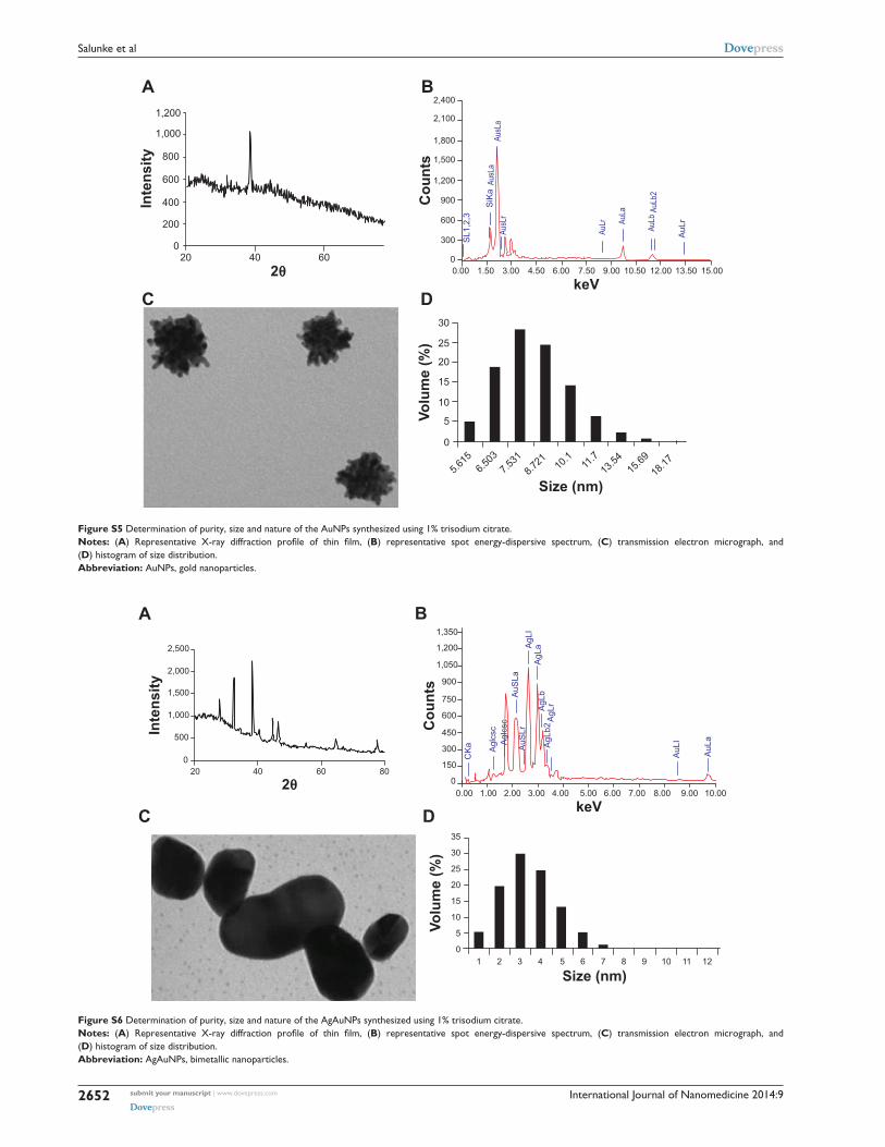
International Journal of Nanomedicine 2014:9submit your manuscript | www.dovepress.com
Dovepress
Dovepress
2652
Salunke et al
Inte
nsity
1,200
1,000
800
600
400
200
020 40 60
2θ
BA
DC
0
300
600
900
1,200
1,500
1,800
2,100
2,400
0.00 1.50 3.00 4.50 6.00 7.50 9.00 10.50 12.00 13.50 15.00
keV
Cou
nts
SL1
,2,3
SiK
a
AuL
r
AuLbAu
La
AuLr
AusL
aAu
sLa
AusL
r
AuLb
2
Size (nm)
Volu
me
(%)
30
25
20
15
10
5
0
5.615
6.503
7.531
8.721 10
.111
.713
.5415
.6918
.17
Figure S5 Determination of purity, size and nature of the AuNPs synthesized using 1% trisodium citrate.Notes: (A) Representative X-ray diffraction profile of thin film, (B) representative spot energy-dispersive spectrum, (C) transmission electron micrograph, and (D) histogram of size distribution.Abbreviation: auNPs, gold nanoparticles.
Inte
nsity
2,500
2,000
1,500
1,000
500
020 40 60 80
1,350
1,200
1,050
900
750
600
450
300
150
00.00 1.00 2.00 3.00 4.00 5.00 6.00 7.00 8.00 9.00 10.00
keV
Size (nm)
Volu
me
(%)
Agl
csc
Agl
csc
CK
a
AuS
LaA
uSLr
AgL
aAgL
IA
gLb
AgL
rA
gLb2
AuL
I
AuL
a
Cou
nts
35
30
25
20
15
10
5
01 2 3 4 5 6 7 8 9 10 11 12
BA
DC
2θ
Figure S6 Determination of purity, size and nature of the AgAuNPs synthesized using 1% trisodium citrate.Notes: (A) Representative X-ray diffraction profile of thin film, (B) representative spot energy-dispersive spectrum, (C) transmission electron micrograph, and (D) histogram of size distribution.Abbreviation: agauNPs, bimetallic nanoparticles.

International Journal of Nanomedicine
Publish your work in this journal
Submit your manuscript here: http://www.dovepress.com/international-journal-of-nanomedicine-journal
The International Journal of Nanomedicine is an international, peer-reviewed journal focusing on the application of nanotechnology in diagnostics, therapeutics, and drug delivery systems throughout the biomedical field. This journal is indexed on PubMed Central, MedLine, CAS, SciSearch®, Current Contents®/Clinical Medicine,
Journal Citation Reports/Science Edition, EMBase, Scopus and the Elsevier Bibliographic databases. The manuscript management system is completely online and includes a very quick and fair peer-review system, which is all easy to use. Visit http://www.dovepress.com/testimonials.php to read real quotes from published authors.
Dovepress
International Journal of Nanomedicine 2014:9 submit your manuscript | www.dovepress.com
Dovepress
Dovepress
2653
P. zeylanica-mediated synthesis of NPs and antibiofilm activity
A B
Figure S7 AFM micrographs for biofilm disruption.Notes: (A) A. baumannii biofilm disruption using AgNPs using AFM, and (B) amplitude mode of AFM image.Abbreviations: agNPs, silver nanoparticles; AFM, atomic force microscopy.
Reference1. Pawar VU, Ghosh S, Chopade BA, Shinde VS. Design and synthesis
of harzialactone analogues: Promising anticancer agents. Bioorg Med Chem Lett. 2010;20(24):7243–7245.
Materials and methods for cytotoxicity assayMCF7 (human breast adenocarcinoma), was obtained
from National Center for Cell Sciences, India and cultured
in a humidified atmosphere (37°C, 5% CO2) in Roswell
Park Memorial Institute1640 medium (Life Technologies,
Waltham, MA, USA) supplemented with 10% fetal bovine
serum.1 Nanoparticles were evaluated for cytotoxicity against
MCF7 cancer cell lines. The compounds were evaluated
in vitro at a various concentration. MTT colorimetric assay
was used to determine growth inhibition. 100 μL of cell
suspension (2×10⁴ cells) were plated in 96-well plates and
allowed to attach for 24 hours. Cells were exposed in tripli-
cate wells to AgNPs, AuNPs and AgAuNPs both synthesized
chemically as well as by PZRE for 48 hours. After 48 hours,
20 μL MTT (3-(4,5-dimethylthiazol-2-yl)-2,5-diphenyl
tetrazolium bromide) solution (5 mg/mL,) was added to each
well. After 4 hours of incubation the formazan precipitate
was dissolved in 100 μL of solubilization buffer (10% SDS
in 0.1 N HCl) and kept overnight for complete dissolution.
The absorbance at 576 nm was determined on a 96 well
microplate reader (SpectraMax M5®, Molecular Devices
Corporation, Sunnyvale, CA). The absorbance values were
used to calculate % inhibition.
results
Nanoparticles
ChemAgNPs
0
10
20
30
40
50
60
70
80
ChemAuNPs
ChemAgAuNPs
PZREAgNPs
PZREAuNPs
PZREAgAuNPs
% in
hibi
tion
Figure S8 Cytotoxicity of the nanoparticles synthesized from PZRE and 1% trisodim citrate determined using MCF7 cells showing no inhibition upto the concentration of 0.5 mg/ml.Abbreviations: PZRE, Plumbago zeylanica root extract; agNPs, silver nanoparticles; AuNPs, gold NPs; AgAuNPs, bimetallic NPs; Chem, chemically synthesized.