
Market Interactions in Gold and Stock Markets: Evidences from ...
Upload
independentCategory
view
1download
0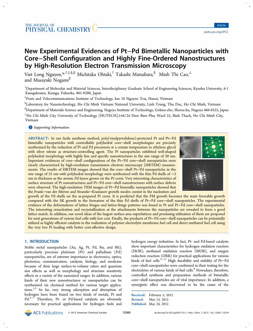
New Experimental Evidences of Pt−Pd Bimetallic Nanoparticles withCore−Shell Configuration and Highly Fine-Ordered Nanostructuresby High-Resolution Electron Transmission MicroscopyViet Long Nguyen,*,†,‡,§,∥ Michitaka Ohtaki,† Takashi Matsubara,∥ Minh Thi Cao,⊥
and Masayuki Nogami∥
†Department of Molecular and Material Sciences, Interdisciplinary Graduate School of Engineering Sciences, Kyushu University, 6-1Kasugakouen, Kasuga, Fukuoka, 861-8580, Japan‡Posts and Telecommunications Institute of Technology, km 10 Nguyen Trai, Hanoi, Vietnam§Laboratory for Nanotechnology, Ho Chi Minh Vietnam National University, Linh Trung, Thu Duc, Ho Chi Minh, Vietnam∥Department of Materials Science and Engineering, Nagoya Institute of Technology, Gokiso-cho, Showa-ku, Nagoya 466-8555, Japan⊥Ho Chi Minh City University of Technology (HUTECH),144/24 Dien Bien Phu, Ward 25, Binh Thach, Ho Chi Minh City,Vietnam
*S Supporting Information
ABSTRACT: In our facile synthesis method, poly(vinylpyrrolidone)-protected Pt and Pt−Pdbimetallic nanoparticles with controllable polyhedral core−shell morphologies are preciselysynthesized by the reduction of Pt and Pd precursors at a certain temperature in ethylene glycolwith silver nitrate as structure-controlling agent. The Pt nanoparticles exhibited well-shapedpolyhedral morphology with highly fine and specific nanostructures in the size range of 20 nm.Important evidences of core−shell configurations of the Pt−Pd core−shell nanoparticles wereclearly characterized by high-resolution transmission electron microscopy (HRTEM) measure-ments. The results of HRTEM images showed that the core−shell Pt−Pd nanoparticles in thesize range of 25 nm with polyhedral morphology were synthesized with the thin Pd shells of ∼3nm in thickness as the atomic Pd layers grown on the Pt cores. Very interesting characteristics ofsurface structure of Pt nanostructures and Pt−Pd core−shell nanostructures with surface defectswere observed. The high-resolution TEM images of Pt−Pd bimetallic nanoparticles showed thatthe Frank−van der Merwe and Stranski−Krastanov growth modes coexist in the nucleation andgrowth of the Pd shells on the as-prepared Pt cores. It is predicted that the FM growth becomes the main favorable growthcompared with the SK growth in the formation of the thin Pd shells of Pt−Pd core−shell nanoparticles. The experimentalevidence of the deformations of lattice fringes and lattice-fringe patterns was found in Pt and Pt−Pd core−shell nanoparticles.The interesting renucleation and recrystallization at the attachments between the nanoparticles are revealed to form a goodlattice match. In addition, our novel ideas of the largest surface-area superlattices and promising utilization of them are proposedfor next generations of various fuel cells with low cost. Finally, the products of Pt−Pd core−shell nanoparticles can be potentiallyutilized as highly efficient catalysts in the realization of polymer electrolyte membrane fuel cell and direct methanol fuel cell usingthe very low Pt loading with better cost-effective design.
1. INTRODUCTIONNoble metal nanoparticles (Au, Ag, Pt, Pd, Ru, and Rh),particularly precious platinum (Pt) and palladium (Pd)nanoparticles, are of extreme importance in electronics, optics,photonics, communication, catalysis, biology, and medicinebecause of their large surface-to-volume ratios and quantumsize effects as well as morphology and structure sensitivityeffects in a variety of the nanosized ranges. In addition, variouskinds of their new nanostructures or nanoparticles can besynthesized via chemical method for various target applica-tions.1,2 So far, very strong adsorption and absorption ofhydrogen have been found on two kinds of metals, Pt andPd.2−7 Therefore, Pt- or Pd-based catalysts are obviouslynecessary for practical applications for hydrogen fuels and
hydrogen energy industries. In fact, Pt- and Pd-based catalystsshow important characteristics for hydrogen oxidation reaction(HOR), methanol oxidation reaction (MOR), and oxygenreduction reaction (ORR) for practical applications for variouskinds of fuel cells.3−11 High durability and stability of Pt−Pdcore−shell nanoparticles were confirmed in their testing for theelectrolytes of various kinds of fuel cells.8 Nowadays, therefore,controlled synthesis and preparation methods of bimetalliccore−shell nanoparticles are of vital importance. In addition, asynergistic effect was discovered to be the cause of the
Received: February 5, 2012Revised: May 13, 2012Published: May 24, 2012
Article
pubs.acs.org/JPCC
© 2012 American Chemical Society 12265 dx.doi.org/10.1021/jp303117y | J. Phys. Chem. C 2012, 116, 12265−12274
significantly enhanced catalytic activity originating from theirspecific core−shell configuration.12 Therefore, Pt−Pd core−shell nanoparticles can also be very attractive catalysts forSuzuki and Heck cross-coupling reactions in the chemicalsynthesis.13 Moreover, the Pt−Pd bimetallic nanoparticles havebeen studied with efforts for confirming the engineered core−shell nanostructures with the size and morphology controls ofthe cores as well as the shells.14,15 Through the epitaxial seededgrowth, Pt−Pd nanoparticles with cubic core−shell morphol-ogy were controllably engineered in the size range of 30−50nm using K2PtCl4, TTAB, NaBH4, H2PtCl6·6H2O, K2PdCl4, L-ascorbic acid, and other experimental conditions, evidencingthe core−shell nanostructures.16In general, the nucleation and growth of the metal shells on
the metal or oxide cores depend on the Frank−van der Merwe(FM) layer-by-layer and Stranski−Krastanov (SK) island-on-wetting-layer growth modes.17,18 However, little has beenknown and confirmed for Pt−Pd core−shell nanoparticles, forexample, faceted Pt−Pd core−shell nanoparticles observed bySTEM tomography method.19 At present, experimentalevidence of high-resolution TEM images of Pt−Pd core−shellnanoparticles are still relatively rare and less, and moreconvincing evidence is needed. Therefore, more experimentalevidence of core−shell configurations will be of very mean-ingful contributions for scientific society. However, well-controlled syntheses of Pt- or Pd-based bimetallic nanoparticleswith polyhedral morphology by chemical polyol reduction arestill scarce. Furthermore, little evidence with high-resolutionTEM images of the Pt−Pd core−shell nanoparticles has beenreported so far despite their great promises for practicalapplications in various fuel cells such as polymer electrolytemembrane fuel cell (PEMFC) and direct methanol fuel cell(DMFC). It should be noted that the successful preparationprocesses of Pt−Pd core−shell nanoparticles by polyol methodor other chemical techniques should continue in further studiesin the control and optimization of nanosystems of the core−shell configurations.In this research, the controlled syntheses of Pt and Pt−Pd
core−shell nanoparticles have been presented with respect tothe issues of utilizing modified polyol method. The role ofAgNO3 is important to the controlled syntheses of Pt and Pt−Pd core−shell nanoparticles with polyhedral morphology.High-resolution TEM images of Pt−Pd core−shell config-uration are presented. Clearly, the thin Pd shells in the range of1−3 nm thickness are epitaxially grown in solutions on the Ptcores in the formation of Pt−Pd core−shell nanoparticles withthe size range of 25 nm, according to layer-to-layer mechanismand the favorable FM growth mode. The experimentalevidences of high-resolution TEM images enable us tounderstand the homogeneous nucleation and growth of boththe Pt cores and the Pd shells in the homogeneous ethyleneglycol (EG) solution in our preparation procedures. Thenanostructures of the Pt nanoparticles and the Pt−Pd core−shell nanoparticles showed the deformations of lattice fringesand lattice-fringe patterns. Therefore, the critical issues of highdurability and stability of various core−shell catalysts with thevery thin Pt or Pd shells need to be intensively studied. Ourproposals of Pt- or Pd-based superlattices by self-assembly withcontrolling the attachment modes can lead to novelapplications or discoveries in catalysis, particularly in PEMFCand DMFC. Finally, the uses of a low Pt or Pd loading in thePt-based catalysts are some of the best economical ways forlarge-scale commercialization of fuel cells.
2. EXPERIMENTAL SECTION
2.1. Synthesis. 2.1.1. Chemical. Chemicals from Aldrichand Sigma-Aldrich were used in our controlled synthesis of Ptnanoparticles as well as Pt−Pd bimetallic nanoparticles withcore−shell structure. They were polyvinylpyrrolidone (FW =55 000), sodium tetrachloropalladate (II) hydrate (ACSreagent), and chloroplantinic acid hexahydrate (ACS reagent)as precursors. EG was used as both the solvent and thereducing agent. Silver nitrate (metal basis, 99.9999%) was usedas a modifying agent. All of our chemicals used were ofanalytical grade and were used without any further purification.However, the successes of preparation processes havesignificantly depended on the experimental experiences.
2.1.2. Synthesis of Polyhedral Pt Nanoparticles. In a typicalprocess of the controlled synthesis of the Pt nanoparticles, 3mL of EG, 1.5 mL of 0.0625 M H2PtCl6, 3 mL of 0.375 M PVP,and 0.5 mL of 0.04 M AgNO3 were used. The details and stepsof our process procedures were previously presented.20,25 Ingeneral, H2PtCl6 was completely reduced with EG. As a result, adark-brown solution containing Pt nanoparticles was obtainedas the final product.
2.1.3. Synthesis of Polyhedral Pt−Pd Core−Shell Nano-particles. In a typical process, the successive reduction ofNa2PdCl4 with EG after complete reduction of H2PtCl6 withEG was used for the controlled synthesis of as-prepared Pt−Pdcore−shell nanoparticles. Here 3 mL of EG, 1.5 mL of 0.0625M H2PtCl6, 3 mL of 0.375 M PVP, and 0.5 mL of 0.04 MAgNO3 were used. A dark-brown solution of the as-prepared Ptnanoparticles thus obtained was used as the Pt cores for thecontrolled synthesis of Pt−Pd core−shell nanoparticles. Then,1.5 mL of 0.0625 M Na2PdCl4 and 3 mL of 0.375 M PVP wereadded for making the Pd shells on the as-prepared Pt cores. Inthis step, Na2PdCl4 was reduced with EG for the formation ofthe thin Pd shells on the as-prepared Pt cores. The details ofour experimental steps and procedures were previouslypresented. By using this process, we have obtained a dark-brown solution of the as-prepared Pt−Pd core−shell nano-particles as a product.
2.2. Material Characterization. 2.2.1. UV−vis-NIR Spec-troscopy. A ∼0.5 mL portion of the reaction mixtures of ourprecursor solutions and as-prepared products was collected viaa 0.5 mL standard pipet during the synthesis. They wereinvestigated by UV−vis-NIR spectroscopy (an Ubest 570 UV−vis-NIR spectrotometer) for the investigations of kinetics,mechanisms, and final formation of Pt nanoparticles from theEG solution. UV−vis absorption spectra were recorded for theprepared Pt nanoparticles as well as prepared Pt−Pd core−shellnanoparticles without the centrifugation as follows. TheSamples include Sample 1 (3 mL of ethanol and 30 μL ofthe product of the dark-brown solution of Pt nanoparticles),Sample 2 (3 mL of ethanol and 30 μL of the product of thedark-brown solution of Pt−Pd core−shell nanoparticles),Sample 3 (3 mL of ethanol and 30 μL of H2PtCl6 in EG),Sample 4 (3 mL of ethanol and 30 μL of Na2PdCl4 in EG), andSample 5 (3 mL of ethanol and 30 μL of the mixture ofH2PtCl6.6H2O and Na2PdCl4).
2.2.2. X-ray Diffraction. In X-ray diffraction (XRD)measurements, 1.5 mL of the dark-brown solution productwas used for the pure Pt nanoparticles and the pure core−shellPt−Pd bimetallic nanoparticles by removing PVP polymer andimpurities using ethanol, acetone, and hexane. Then, the purenanoparticles were homogeneously dispersed in 3 mL of
The Journal of Physical Chemistry C Article
dx.doi.org/10.1021/jp303117y | J. Phys. Chem. C 2012, 116, 12265−1227412266
ethanol by ultrasonication. After that, the volumes of 3 mL ofethanol of the pure Pt nanoparticles or the pure Pt−Pd core−shell nanoparticles were used. They were set onto a planar glasssubstrate for the XRD measurements with a small area of ∼1cm2. The ethanol containing the pure nanoparticles was veryslowly poured on the glass substrate dropwisely by taking somehours. The glass substrates with the pure nanoparticles weredried at 80 °C for 6 h prior to the measurement. The XRDpatterns were recorded in the range of 2θ of 5−90° by adiffractometer (X’Pert-Phillips) operating at 45 kV/40 mA withCu Kα radiation (1.54056 Å).2.2.3. Transmission Electron Microscopy. To analyze the as-
prepared Pt nanoparticles as well as the as-prepared Pt−Pdcore−shell nanoparticles, we have used three kinds oftransmission electron microscopes (TEMs) (JEOL-JEM-2100F, JEOL-JEM-2010, JEOL-JEM-2010XII) operated at200 kV by using CCD camera or detector. The details of thesample preparation for microanalysis by TEM methods werediscussed in our previous works.25 In addition, Thespecifications and spatial resolutions of JEOL-JEM-2100F arearound 0.1 nm (lattice resolution) and around 0.19 nm (pointresolution), and those of JEOL-JEM-2010 are around 0.14 nm(lattice resolution) and around 0.23 nm (point resolution) at200 kV. In the good and stable operations of TEM for high-resolution images, we have chosen experimental conditions ofthe camera length of 1000 mm and beam energy of 100 kV or200 kV to obtain the best quality and results of high-resolutionimages. In HRTEM modes, the spot size and brightness wereautomatically set. The issues of beam shift and tilt wereautomatically set. In particular, the modes for the exactobjection of images can be suitably selected in the diffractionmode, imaging mode or STEM mode for the best images. Inmost of HRTEM measurements, the operation modes of TEMwere selected in diffraction mode. In addition, the patterns ofselected area electron diffraction (SAED) were taken at 200 kV,and nanobeam diffraction (NBD) patterns were determined forour evaluations of the nanostructure of Pt−Pd core−shellnanoparticles.
3. RESULTS AND DISCUSSION3.1. Formation of PVP−Pt and Pt−Pd Core−Shell
Nanoparticles. 3.1.1. UV−vis Absorption Spectra of PVP−Ptand Pt−Pd Core−Shell Nanoparticles. Figure 1 shows UV−vis absorption spectra of the five samples including Sample 1,the as-prepared Pt nanoparticles, Sample 2, the prepared Pt−Pdcore−shell nanoparticles, Sample 3, H2PtCl6 in EG, Sample 4,Na2PdCl4 in EG, and Sample 5, the mixture of H2PtCl6 andNa2PdCl4 in EG. The strong decrease in the peak intensity ofthe ligand-to-metal charge-transfer absorption at ∼300 nm is animportant experimental evidence of the final formation of Ptnanoclusters and nanoparticles by the complete reduction ofthe [PtCl6]
2− ions with EG in our experimental process. Thesimilar evidence of complete reductions of the [PtCl6]
2− and[PdCl4]
2− ions with EG in the final formation of the dark-brown product solution was indicated by the strong decrease inthe absorption intensity of the dark-brown product solution ofthe as-prepared Pt−Pd core−shell nanoparticles. The UV−visabsorption spectrum of the product of the Pt nanoparticlesshowed the strong absorption at ∼258 nm. This showed thatthe fast reduction of the [PtCl6]
2− ions happened in theformation of Pt nanoparticles protected by PVP in the excessEG solvent. There were the strong absorption peaks at around258 and 285 nm due to the ligand-to-metal charge-transfer
transition of the [PtCl6]2− ions in the homogeneous solution of
H2PtCl6 in EG (285 nm) and complex formation of Ptnanoparticles in the homogeneous solution of EG and PVP(258 nm). The sample of H2PtCl6 exhibited the highestabsorbance in comparison with that of the two other samplescontaining Pt nanoparticles. The growth and shape formationmechanisms of platinum nanoparticles were studied.20−22 Inthe case of Na2PdCl4 in EG, there is evidence of the threeabsorption peaks located at around 272, 327, and 443 nmbecause of the [PdCl4]
2− complexes of Na2PdCl4 in EG (curve(a) in Figure 1). In contrast, the mixture of Na2PdCl4 andH2PtCl6 showed only the two absorption peaks located ataround 272 and 443 nm, and the peak at 443 nm was very weak(curve (c) in Figure 1). However, for the as-prepared productof PVP protected Pt−Pd bimetallic nanoparticles, the UV−visabsorption spectrum showed only one strong absorption peaklocated at 258 nm as the same as the case of PVP-protected Ptnanoparticles. Therefore, the strong decrease in the absorptionintensity in the UV−vis spectra clearly indicated the finalformation of the as-prepared PVP protected Pt and Pt−Pdnanoparticles.
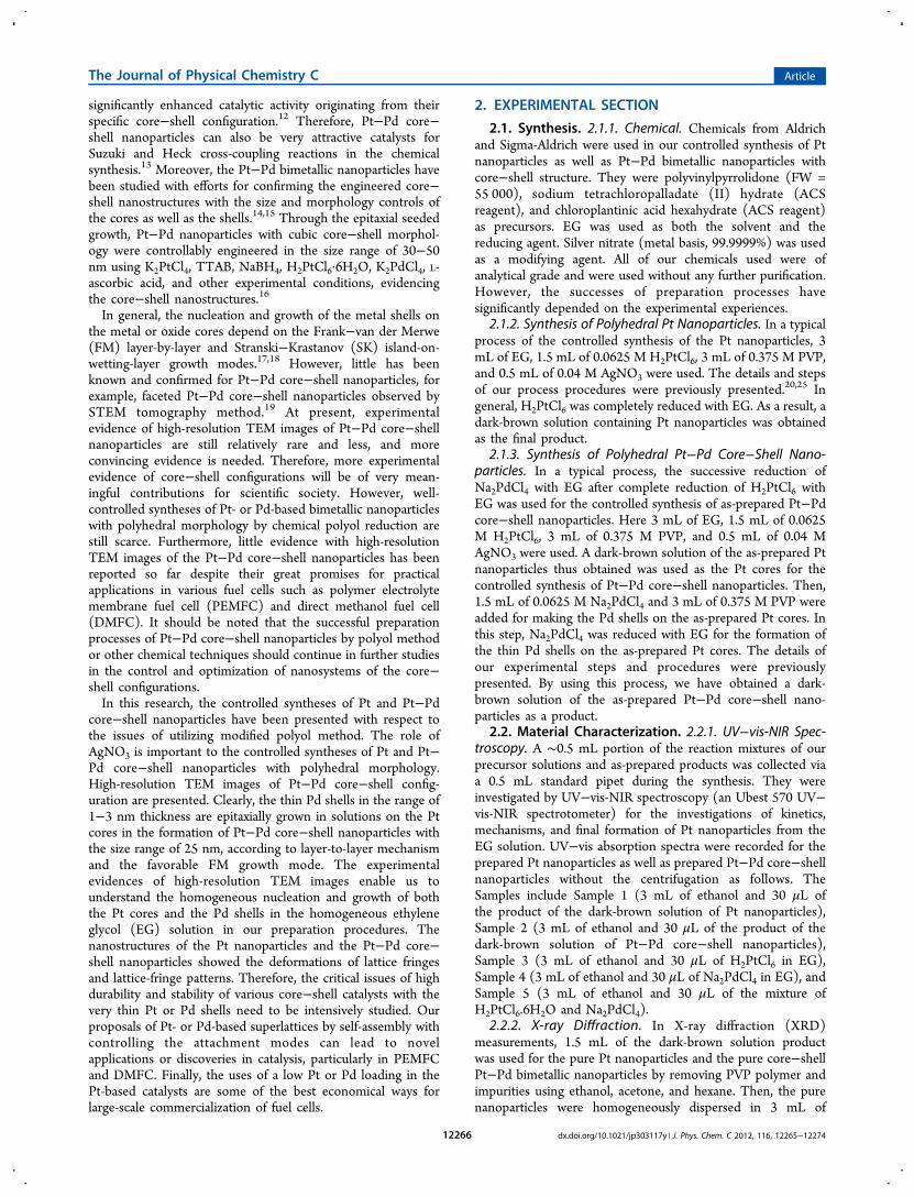
3.1.2. Structure of Pt and Pt−Pd Core−Shell Nano-particles. Figure 2 shows the XRD patterns of the as-preparedPt nanoparticles (line a) and the as-prepared Pt−Pd core−shellbimetallic nanoparticles (line b). The interesting structure ofthe crystalline face-centered cubic (fcc) phase was confirmed inboth the as-prepared Pt nanoparticles and the Pt−Pd core−shell bimetallic nanoparticles. The diffraction peaks werecharacterized by the (111), (200), (220), (311), and (222)reflections peaks in comparison with the fcc phase of bulk Pt(or bulk Pd) with respect to 2θ values of around 39, 46, 67, 81,and 85°, respectively.20,21 The XRD patterns in Figure 2 looksimilar to their appearance, shape, and XRD peak locations attheir XRD diagrams, but the diffraction intensities and widthsof peaks are significantly different. The intensity of the XRDpattern of the as-prepared Pt−Pd core−shell bimetallicnanoparticles is much stronger than that of the as-preparedPt nanoparticles. The ratios of diffraction intensity between thePt nanoparticles and the Pt−Pd core−shell bimetallic nano-
Figure 1. UV−vis spectra of samples: (a) Na2PdCl4, (b) H2PtCl6, (c)Na2PdCl4 and H2PtCl6, (d) PVP protected Pt nanoparticles withcontrol method using AgNO3, and (e) PVP protected Pt−Pdbimetallic nanoparticles with control method using AgNO3.
The Journal of Physical Chemistry C Article
dx.doi.org/10.1021/jp303117y | J. Phys. Chem. C 2012, 116, 12265−1227412267
particles are calculated as 1.758, 1.499, 1.367, 1.518, and 1.308,respectively. The important XRD evidence showed that thediffraction intensity was strongly enhanced by the core−shellPt−Pd bimetallic nanoparticles in comparison with the Ptnanoparticles from 1.3 to 1.75. The typical XRD peaks of Ptnanoparticles were used to compare with those of thecorresponding bulk Pt material. The average size of Ptnanocrystallites can be calculated by the width of the diffractionpeaks according to the Debye−Scherrer equation: D = 0.9λ/(βcos θ), where β is the full width at half-maximum (fwhm) of thepeak, θ is the angle of diffraction, and λ is the wavelength of theX-ray radiation. However, only five peaks assigned to a singlefcc structure were observed in the limited 2θ range of 5−90° bythe XRD method. Therefore, the core−shell morphology canbe only exactly determined by the TEM and HRTEM methods.3.2. Characterization of Pt and Pt−Pd Core−Shell
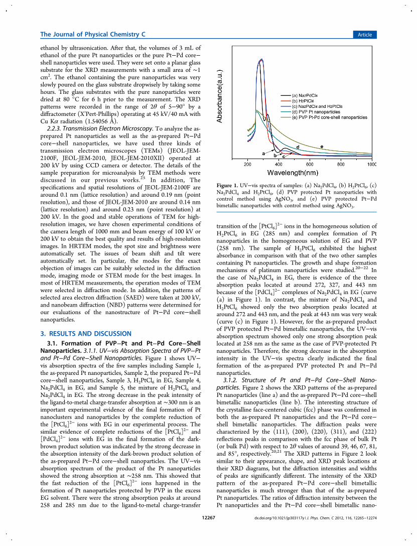
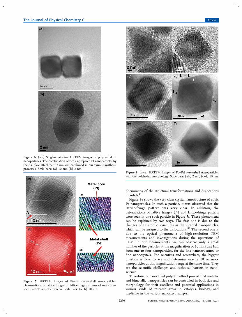
Nanoparticles. Figures 3, 5, and 6 present TEM and HRTEMimages of as-prepared Pt nanoparticles with the mostcharacteristic polyhedral morphologies and shapes. It shouldbe noted that the as-prepared nanoparticles were observed inthe polyhedral morphology, typically such as cubes, octahedra,and tetrahedra. Therefore, the favorable nucleation and growthof Pt nanoparticles are the homogeneous modes, and theanisotropic inhomogeneous nucleation and growth areunfavorable. The size was well-controlled in the range of 20nm. The surface of Pt nanostructures with surface defects suchas kinks, atomic steps, islands, and terraces can be clearly seenin the high-resolution images as shown in Figures 3b and 5. Inmany works, they were considered to be the most efficientcatalytic centers for the significant improvements of catalyticactivity of the Pt catalysts. Therefore, we need to find a methodto determine the surface concentrations of kinks, atomic steps,islands, and terraces in various Pt-based catalysts for electro-catalysis.23,46,47
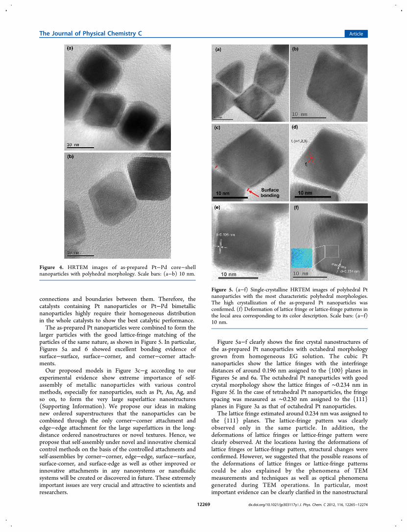
Figures 4 and 7−9 show the HRTEM images of as-preparedPt−Pd core−shell nanoparticles with the most characteristicpolyhedral morphology and shape. The thin Pd shells grownover the Pd cores have led to form the core−shell configurationwith the well-controlled size in the nanosized range of about15−25 nm. The thickness or the coated shell was well-controlled in the range of 1−3 nm. The Pt−Pd core−shell
nanoparticles also showed characteristic polyhedral morphol-ogy and shape, typically such as cubes, octahedra, andtetrahedra. Similarly, the dominant nucleation and growthmode of Pt−Pd core−shell nanoparticles are the homogeneousmodes, and the anisotropic inhomogeneous nucleation andgrowth modes are negligible.Most of the as-prepared polyhedral Pt nanoparticles as well
as the Pt−Pd core−shell nanoparticles including cubes,octahedra, tetrahedral, and polyhedra or truncated polyhedraexhibit the low-index facets of {111}, {110}, and {100} planes.So far, the {111}, {110}, and {100} low-index facets are someexperimental evidence and observations of the most stable anddurable planes of polyhedral morphology and shape observedin cyclic voltammetry (CV) experiments for specific catalyticactivity, sensitivity, and selectivity of various Pt-based catalysts.On the basis of our careful observations and analyses as well
as excellent experimental evidence in the reactions of the as-prepared nanoparticles of both Pt nanoparticles and Pt−Pdcore−shell nanoparticles, we have proposed the models of theinteractions among them. There are various kinds of randomand direct attachments or bondings between the nanoparticlesincluding corner−corner, edge−edge, surface−surface, surface−corner, and surface−edge attachments and any possible kindsof various arbitrary and direct attachments. The importantevidence of the combinations among the polyhedral Ptnanoparticles is seen in Figures 3, 5, and 6. It is predictedthat renucleation and recrystallization easily occur at the
Figure 2. Two XRD patterns: (a) Pt nanoparticles and (b) Pt−Pdcore−shell bimetallic nanoparticles.
Figure 3. (a,b) TEM and HRTEM images of as-prepared Ptnanoparticles. (c,d) Important models of random and directattachments or bondings of two particles for novel applications incatalysis, medicine, and biology. Scale bars: (a) 50 nm and (b) 10 nm.
The Journal of Physical Chemistry C Article
dx.doi.org/10.1021/jp303117y | J. Phys. Chem. C 2012, 116, 12265−1227412268
connections and boundaries between them. Therefore, thecatalysts containing Pt nanoparticles or Pt−Pd bimetallicnanoparticles highly require their homogeneous distributionin the whole catalysts to show the best catalytic performance.The as-prepared Pt nanoparticles were combined to form the
larger particles with the good lattice-fringe matching of theparticles of the same nature, as shown in Figure 5. In particular,Figures 5a and 6 showed excellent bonding evidence ofsurface−surface, surface−corner, and corner−corner attach-ments.Our proposed models in Figure 3c−g according to our
experimental evidence show extreme importance of self-assembly of metallic nanoparticles with various controlmethods, especially for nanoparticles, such as Pt, Au, Ag, andso on, to form the very large superlattice nanostructures(Supporting Information). We propose our ideas in makingnew ordered superstructures that the nanoparticles can becombined through the only corner−corner attachment andedge−edge attachment for the large superlattices in the long-distance ordered nanostructures or novel textures. Hence, wepropose that self-assembly under novel and innovative chemicalcontrol methods on the basis of the controlled attachments andself-assemblies by corner−corner, edge−edge, surface−surface,surface-corner, and surface-edge as well as other improved orinnovative attachments in any nanosystems or nanofluidicsystems will be created or discovered in future. These extremelyimportant issues are very crucial and attractive to scientists andresearchers.
Figure 5a−f clearly shows the fine crystal nanostructures ofthe as-prepared Pt nanoparticles with octahedral morphologygrown from homogeneous EG solution. The cubic Ptnanoparticles show the lattice fringes with the interfringedistances of around 0.196 nm assigned to the {100} planes inFigures 5e and 6a. The octahedral Pt nanoparticles with goodcrystal morphology show the lattice fringes of ∼0.234 nm inFigure 5f. In the case of tetrahedral Pt nanoparticles, the fringespacing was measured as ∼0.230 nm assigned to the {111}planes in Figure 3a as that of octahedral Pt nanoparticles.The lattice fringe estimated around 0.234 nm was assigned to
the {111} planes. The lattice-fringe pattern was clearlyobserved only in the same particle. In addition, thedeformations of lattice fringes or lattice-fringe pattern wereclearly observed. At the locations having the deformations oflattice fringes or lattice-fringe pattern, structural changes wereconfirmed. However, we suggested that the possible reasons ofthe deformations of lattice fringes or lattice-fringe patternscould be also explained by the phenomena of TEMmeasurements and techniques as well as optical phenomenagenerated during TEM operations. In particular, mostimportant evidence can be clearly clarified in the nanostructural
Figure 4. HRTEM images of as-prepared Pt−Pd core−shellnanoparticles with polyhedral morphology. Scale bars: (a−b) 10 nm.
Figure 5. (a−f) Single-crystalline HRTEM images of polyhedral Ptnanoparticles with the most characteristic polyhedral morphologies.The high crystallization of the as-prepared Pt nanoparticles wasconfirmed. (f) Deformation of lattice fringe or lattice-fringe patterns inthe local area corresponding to its color description. Scale bars: (a−f)10 nm.
The Journal of Physical Chemistry C Article
dx.doi.org/10.1021/jp303117y | J. Phys. Chem. C 2012, 116, 12265−1227412269
phenomena of the structural transformations and dislocationsin solids.24
Figure 5e shows the very clear crystal nanostructure of cubicPt nanoparticles. In such a particle, it was observed that thelattice-fringe pattern was very clear. In addition, thedeformations of lattice fringes ( f i) and lattice-fringe patternwere seen in one such particle in Figure 5f. These phenomenacan be explained by two ways. The first one is due to thechanges of Pt atomic structures in the internal nanoparticles,which can be assigned to the dislocations.24 The second one isdue to the optical phenomena of high-resolution TEMmeasurements and investigations during the operations ofTEM. In our measurements, we can observe only a smallnumber of the particles at the magnification of 10 nm scale bar,from one to four nanoparticles, for the fine nanostructures orfine nanocrystals. For scientists and researchers, the biggestquestion is how to see and determine exactly 10 or morenanoparticles at this magnification range at the same time. Theyare the scientific challenges and technical barriers in nano-science.Therefore, our modified polyol method proved that metallic
and bimetallic nanoparticles can be controlled in both size andmorphology for their excellent and potential applications invarious kinds of research areas in catalysis, biology, andmedicine in the various nanosized ranges.
Figure 6. (a,b) Single-crystalline HRTEM images of polyhedral Ptnanoparticles. The combination of two as-prepared Pt nanoparticles bytheir surface attachment 2 nm was confirmed in our various synthesisprocesses. Scale bars: (a) 10 and (b) 2 nm.
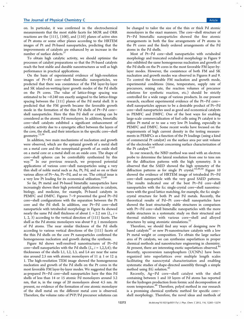
Figure 7. HRTEM images of Pt−Pd core−shell nanoparticles.Deformations of lattice fringes or latticefringe patterns of one core−shell particle are clearly seen. Scale bars: (a−b) 10 nm.
Figure 8. (a−e) HRTEM images of Pt−Pd core−shell nanoparticleswith the polyhedral morphology. Scale bars: (a,b) 2 nm, (c−f) 10 nm.
The Journal of Physical Chemistry C Article
dx.doi.org/10.1021/jp303117y | J. Phys. Chem. C 2012, 116, 12265−1227412270
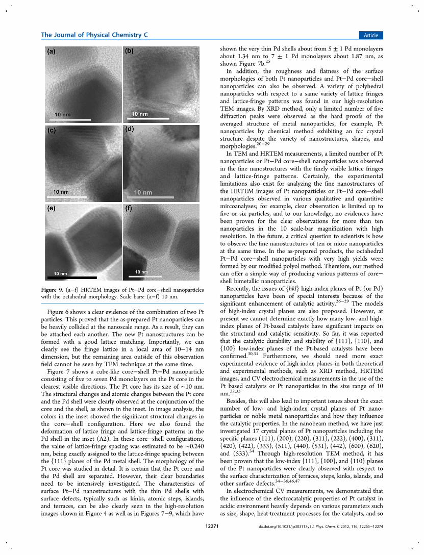
Figure 6 shows a clear evidence of the combination of two Ptparticles. This proved that the as-prepared Pt nanoparticles canbe heavily collided at the nanoscale range. As a result, they canbe attached each another. The new Pt nanostructures can beformed with a good lattice matching. Importantly, we canclearly see the fringe lattice in a local area of 10−14 nmdimension, but the remaining area outside of this observationfield cannot be seen by TEM technique at the same time.Figure 7 shows a cube-like core−shell Pt−Pd nanoparticle
consisting of five to seven Pd monolayers on the Pt core in theclearest visible directions. The Pt core has its size of ∼10 nm.The structural changes and atomic changes between the Pt coreand the Pd shell were clearly observed at the conjunction of thecore and the shell, as shown in the inset. In image analysis, thecolors in the inset showed the significant structural changes inthe core−shell configuration. Here we also found thedeformation of lattice fringe and lattice-fringe patterns in thePd shell in the inset (A2). In these core−shell configurations,the value of lattice-fringe spacing was estimated to be ∼0.240nm, being exactly assigned to the lattice-fringe spacing betweenthe {111} planes of the Pd metal shell. The morphology of thePt core was studied in detail. It is certain that the Pt core andthe Pd shell are separated. However, their clear boundariesneed to be intensively investigated. The characteristics ofsurface Pt−Pd nanostructures with the thin Pd shells withsurface defects, typically such as kinks, atomic steps, islands,and terraces, can be also clearly seen in the high-resolutionimages shown in Figure 4 as well as in Figures 7−9, which have
shown the very thin Pd shells about from 5 ± 1 Pd monolayersabout 1.34 nm to 7 ± 1 Pd monolayers about 1.87 nm, asshown Figure 7b.25
In addition, the roughness and flatness of the surfacemorphologies of both Pt nanoparticles and Pt−Pd core−shellnanoparticles can also be observed. A variety of polyhedralnanoparticles with respect to a same variety of lattice fringesand lattice-fringe patterns was found in our high-resolutionTEM images. By XRD method, only a limited number of fivediffraction peaks were observed as the hard proofs of theaveraged structure of metal nanoparticles, for example, Ptnanoparticles by chemical method exhibiting an fcc crystalstructure despite the variety of nanostructures, shapes, andmorphologies.20−29
In TEM and HRTEM measurements, a limited number of Ptnanoparticles or Pt−Pd core−shell nanoparticles was observedin the fine nanostructures with the finely visible lattice fringesand lattice-fringe patterns. Certainly, the experimentallimitations also exist for analyzing the fine nanostructures ofthe HRTEM images of Pt nanoparticles or Pt−Pd core−shellnanoparticles observed in various qualitative and quantitivemircoanalyses; for example, clear observation is limited up tofive or six particles, and to our knowledge, no evidences havebeen proven for the clear observations for more than tennanoparticles in the 10 scale-bar magnification with highresolution. In the future, a critical question to scientists is howto observe the fine nanostructures of ten or more nanoparticlesat the same time. In the as-prepared products, the octahedralPt−Pd core−shell nanoparticles with very high yields wereformed by our modified polyol method. Therefore, our methodcan offer a simple way of producing various patterns of core−shell bimetallic nanoparticles.Recently, the issues of {hkl} high-index planes of Pt (or Pd)
nanoparticles have been of special interests because of thesignificant enhancement of catalytic activity.26−29 The modelsof high-index crystal planes are also proposed. However, atpresent we cannot determine exactly how many low- and high-index planes of Pt-based catalysts have significant impacts onthe structural and catalytic sensitivity. So far, it was reportedthat the catalytic durability and stability of {111}, {110}, and{100} low-index planes of the Pt-based catalysts have beenconfirmed.30,31 Furthermore, we should need more exactexperimental evidence of high-index planes in both theoreticaland experimental methods, such as XRD method, HRTEMimages, and CV electrochemical measurements in the use of thePt based catalysts or Pt nanoparticles in the size range of 10nm.32,33
Besides, this will also lead to important issues about the exactnumber of low- and high-index crystal planes of Pt nano-particles or noble metal nanoparticles and how they influencethe catalytic properties. In the nanobeam method, we have justinvestigated 17 crystal planes of Pt nanoparticles including thespecific planes (111), (200), (220), (311), (222), (400), (311),(420), (422), (333), (511), (440), (531), (442), (600), (620),and (533).34 Through high-resolution TEM method, it hasbeen proven that the low-index {111}, {100}, and {110} planesof the Pt nanoparticles were clearly observed with respect tothe surface characterization of terraces, steps, kinks, islands, andother surface defects.34−36,46,47
In electrochemical CV measurements, we demonstrated thatthe influence of the electrocatalytic properties of Pt catalyst inacidic environment heavily depends on various parameters suchas size, shape, heat-treatment processes for the catalysts, and so
Figure 9. (a−f) HRTEM images of Pt−Pd core−shell nanoparticleswith the octahedral morphology. Scale bars: (a−f) 10 nm.
The Journal of Physical Chemistry C Article
dx.doi.org/10.1021/jp303117y | J. Phys. Chem. C 2012, 116, 12265−1227412271
on. In particular, it was confirmed in the electrochemicalmeasurements that the most stable facets for MOR and ORRreactions are the {111}, {100}, and {110} planes of active sitesof Pt atoms or some other planes according to the HRTEMimages of Pt and Pt-based nanoparticles, predicting that theimprovements of catalysts are enhanced by an increase in thenumber of surface defects.37
To obtain high catalytic activity, we should optimize theprocesses of catalyst preparations so that the Pt-based catalystsreach the best stable and durable nanostructures as well as highperformance in practical applications.On the basis of experimental evidence of high-resolution
images of Pt−Pd core−shell bimetallic nanoparticles, wepredicted that there was coexistence of the FM layer-by-layerand SK island-on-wetting-layer growth modes of the Pd shellson the Pt cores. The value of lattice-fringe spacing wasestimated to be ∼0.240 nm exactly assigned to the lattice-fringespacing between the {111} planes of the Pd metal shell. It ispredicted that the FM growth became the favorable growthmode in the formation of the thin Pd shells of Pt−Pd core−shell nanoparticles. Here the thin Pd shell or coating can beconsidered as the atomic Pd monolayers. In addition, bimetalliccore−shell catalysts exhibited a significant enhancement ofcatalytic activity due to a synergetic effect between the layers ofthe core, the shell, and their interfaces in the specific core−shellgeometry.12,38
In addition, two main mechanisms of nucleation and growthwere observed, which are the epitaxial growth of a metal shellon a metal core and the nonepitaxial growth of an oxide shellon a metal core or a metal shell on an oxide core. The shape ofcore−shell spheres can be controllably synthesized by thisway.39 In our previous research, we proposed potentialapplications of various core−shell nanoparticles using a verythin shell of noble metal such as Au, Pt, Pd, and so on or theirvarious alloys of Pt−Au, Pt−Pd, and so on. The critical issue isa very low Pt loading for its economical utilization.25,40
So far, Pt metal and Pt-based bimetallic nanoparticles haveincreasingly shown their high potential applications in catalysis,biology, and medicine, for example, Pt-based catalysts inPEMFC and DMFC. Figures 7−9 presented the good Pt−Pdcore−shell configurations with the separation between the Ptcore and the Pd shell. In addition, one Pt−Pd core−shellnanoparticle with tetrahedral morphology in Figure 8a showednearly the same Pd shell thickness of about L = 2.2 nm (Li, i =1, 2, 3) according to the vertical direction of {111} facets. Theshell as the Pd atomic monolayers was about 9 ± 1 monolayersof Pd atoms. The near similar thickness of the Pd shellsaccording to various vertical directions of the {111} facets ofthe thin Pd shells on the core Pt nanoparticles confirmed thehomogeneous nucleation and growth during the synthesis.Figure 8d shows well-resolved nanostructures of Pt−Pd
core−shell nanoparticles with the Pd shells (Li, i = 1,2,3,4); thethicknesses of the shells L1, L2, L3, and L4 are near the samesize around 2.5 nm with atomic monolayers of 11 ± 1 or 12 ±1. The high-resolution TEM image showed the homogeneousnucleation and growth of the Pd shells on the Pt cores in themost favorable FM layer-by-layer modes. We suggested that theas-prepared Pt−Pd core−shell nanoparticles have the thin Pdshells of less than 14 or 15 atomic Pd monolayers around 3.2nm, that is, in the range of 20 monolayers about 4.5 nm. Atpresent, no evidence of the formation of one atomic monolayerof the shell metal on the different metal core is observed.Therefore, the volume ratio of PVP/Pd precursor solutions can
be changed to tailor the size of the thin or thick Pd atomicmonolayers in the exact manners. The core−shell structure ofPt−Pd bimetallic nanoparticles showed the fine atomicarrangements in both the fine arrangements of Pt atoms inthe Pt cores and the finely ordered arrangements of the Pdatoms in the Pd shells.Most of Pt−Pd core−shell nanoparticles with octahedral
morphology and truncated octahedral morphology in Figure 9also exhibited the same homogeneous nucleation and growth ofthe Pd shells on the Pt cores in the most favorable FM layer-by-layer modes. However, the coexistence of both FM and SKnucleation and growth modes was observed in Figures 8 and 9.To control the favorable FM nucleation and growth mode,experimental conditions (time, temperature, supply rate ofprecursors, mixing rate, the reaction volumes of precursorsolutions for synthetic reaction, etc.) should be strictlycontrolled for a wide range of various products. In our presentresearch, excellent experimental evidence of the Pt−Pd core−shell nanoparticles appears to be a desirable product of Pt−Pdcore−shell nanoparticles used as good and economical catalystsin PEMFC and DMFC. One of the best ways for enablinglarge-scale commercialization of fuel cells using Pt catalyst is toutilize Pt metal or to use a very low Pt loading, especially inPEMFC and DMFC. Some recent works have focused on therequirements of high current density in the testing measure-ments in PEMFCs as a function of the Pt loadings (using a kindof commercial Pt catalyst) on the surface area per unit volumeof the electrodes without concerning surface characterization ofthe Pt catalyst.41,42
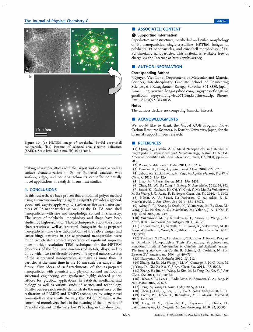
In our research, the NBD method was used with an electronprobe to determine the lateral resolution from one to tens nmfor the diffraction patterns with the high symmetry. It isobserved that the SAED showed the high symmetry of thediffraction patterns as for single Pt crystal.43,49,50 Figure 10showed the evidence of HRTEM image of tetrahedral Pt−Pdcore−shell nanoparticle with the very good SAED patterns.The results indicated one of the best Pt−Pd core−shellnanoparticles with the fcc single-crystal core−shell nanostruc-tures with the good lattice matching, for example, the fcc single-crystal structure for both Pt and Pd metals. Recently, thetheoretical results of Pd−Pt core−shell nanoparticles haveshowed the least structurally stable structures in comparisonwith Pt−Pd core−shell bimetallic nanoparticles with the morestable structures in a systematic study on their structural andthermal stabilities with various core−shell and alloyedstructures by using atomistic simulations.51
Therefore, we should find any ways of designing new Ptbased catalysts43 or new Pt-nanostructure catalysts with a lowPt metal weight or composition. To obtain the large surfacearea of Pt catalysts, we can synthesize superlattices in properchemical methods and nanostructure engineering in chemistry.At present, there are interesting exotic superlattices observed.44
Recently, upconversion nanophosphors (UCNPs) have beenorganized into superlattices over multiple length scalesfacilitating the nanocrystal characterization and enablingsystematic studies of shape-directed assembly through a simplemethod using EG solution.45
Recently, Ag−Pd core−shell catalyst with the shellcontaining between 1 and 10 layers of Pd atoms has reportedfor the hydrogen production from formic acid decomposition atroom temperature.48 Therefore, polyol method in our researchis a promising chemical synthetic method for specific core−shell morphology. Therefore, the novel ideas and methods of
The Journal of Physical Chemistry C Article
dx.doi.org/10.1021/jp303117y | J. Phys. Chem. C 2012, 116, 12265−1227412272
making new superlattices with the largest surface area as well assurface characterization of Pt- or Pd-based catalysts withsurface-, edge-, and corner-attachments can offer potentiallynovel applications in catalysis in our next studies.
4. CONCLUSIONSIn this research, we have proven that a modified polyol methodusing a structure-modifying agent as AgNO3 provides a general,good, and easy-to-apply way to synthesize the fine nanostruc-tures of Pt nanoparticles as well as the Pt−Pd core−shellnanoparticles with size and morphology control in chemistry.The issues of polyhedral morphology and shape have beenstudied by high-resolution TEM techniques to show the surfacecharacteristics as well as structural changes in the as-preparednanoparticles. The clear deformations of the lattice fringes andlattice-fringe patterns in the as-prepared nanoparticles werefound, which also showed importance of significant improve-ment in high-resolution TEM techniques for the HRTEMobjections of the fine nanostructures of metals, alloys, and soon by which we can directly observe fine crystal nanostructuresof the as-prepared nanoparticles as many as more than 10particles at the same time in the 10 nm scale-bar range in thefuture. Our ideas of self-attachments of the as-preparednanoparticles with chemical and physical control methods instructural engineering can synthesize highly ordered super-lattices for practical applications in catalysis, medicine, andbiology as well as various kinds of science and technology.Finally, our research results demonstrate the importance of therealization of PEMFC and DMFC technology by using novelcore−shell catalysts with the very thin Pd or Pt shells as thecontrolled monolayers shells in the meaning of the utilization ofPt metal element in the very low Pt loading in this direction.
■ ASSOCIATED CONTENT*S Supporting InformationSuperlattice nanostructures, octahedral and cubic morphologyof Pt nanoparticles, single-crystalline HRTEM images ofpolyhedral Pt nanoparticles, and core-shell morphology of Pt-Pd bimetallic nanoparticles. This material is available free ofcharge via the Internet at http://pubs.acs.org.
■ AUTHOR INFORMATIONCorresponding Author*Nguyen Viet Long; Department of Molecular and MaterialSciences, Interdisciplinary Graduate School of EngineeringSciences, 6-1 Kasugakouen, Kasuga, Fukuoka, 861-8580, Japan;E-mail: [email protected]; [email protected]; [email protected]. Phone/Fax: +81-(0)92-583-8835.
NotesThe authors declare no competing financial interest.
■ ACKNOWLEDGMENTSWe would like to thank the Global COE Program, NovelCarbon Resource Sciences, in Kyushu University, Japan, for thefinancial support in our research.
■ REFERENCES(1) Qiang, Q.; Ostafin, A. E. Metal Nanoparticles in Catalysis. InEncyclopedia of Nanoscience and Nanotechnology; Nalwa, H. S., Ed.;American Scientific Publishers: Stevenson Ranch, CA, 2004; pp 475−503.(2) Polarz, S. Adv. Funct. Mater. 2011, 21, 3214.(3) Duncan, H.; Lasia, A. J. Electroanal. Chem. 2008, 621, 62.(4) Lebon, A.; García-Fuente, A.; Vega, A.; Aguilera-Granja, F. J. Phys.Chem. C 2012, 116, 126.(5) Shao, M. J. Power Sources 2011, 196, 2433.(6) Chen, M.; Wu, B.; Yang, J.; Zheng, N. Adv. Mater. 2012, 24, 862.(7) Sasaki, K.; Naohara, H.; Cai, Y.; Choi, Y. M.; Liu, P.; Vukmirovic,M. B.; Wang, J. X.; Adzic, R. R. Angew. Chem., Int. Ed. 2010, 49, 8602.(8) Nilelar, A. U.; Sasaki, K.; Farberow, C. A.; Adzic, R. R.;Mavrikakis, M. J. Am. Chem. Soc. 2011, 133, 18574.(9) Adzic, R. R.; Zhang, J.; Sasaki, K.; Vukmirovic, M. B.; Shao, M.;Wang, J. X.; Nilekar, A. U.; Mavrikakis, M.; Valerio, J. A.; Uribe, F.Top. Catal 2007, 46, 249.(10) Vukmirovic, M. B.; Bliznakov, S. T.; Sasaki, K.; Wang, J. X.;Adzic, R. R. Electrochem. Soc. Interface 2011, 20, 33.(11) Koenigsmann, C.; Santulli, A. C.; Gong, K.; Vukmirovic, M. B.;Zhou, W.; Sutter, E.; Wong, S. S.; Adzic, R. R. J. Am. Chem. Soc. 2011,133, 9783.(12) Toshima, N.; Yan, H.; Shiraishi, Y. Chapter 3: Recent Progressin Bimetallic Nanoparticles: Their Preparation, Structures andFunctions. In Metal Nanoclusters in Catalysis and Materials Science:The Issue of Size Control; Corain, B., Schmid, G., Toshima, N., Eds;Elsevier BV: Amsterdam, 2004; pp 49−75.(13) Narayanan, R. Molecules 2010, 15, 2124.(14) Zhang, H.; Jin, M.; Wang, J.; Li, W.; Camargo, P. H. C.; Kim, M.J.; Yang, D.; Xie, Z.; Xia, Y. J. Am. Chem. Soc. 2011, 133, 6078.(15) Zhang, H.; Jin, M.; Wang, J.; Kim, M. J.; Yang, D.; Xia, Y. J. Am.Chem. Soc. 2011, 133, 10422.(16) Habas, S. E.; Lee, H.; Radmilovic, V.; Somorjai, G. A.; Yang, P.Nat. Mater. 2007, 6, 692.(17) Peng, Z.; Yang, H. Nano Today 2009, 4, 143.(18) Chen, J.; Lim, B.; Lee, E. P.; Xia, Y. Nano Today 2008, 4, 81.(19) Ercius, P.; Duden, T.; Radmilovic, V. R. Microsc. Microanal.2010, 16, 1650.(20) Long, N. V.; Chien, N. D.; Hayakawa, T.; Hirata, H.;Lakshminarayana, G.; Nogami, M. Nanotechnology 2010, 21, 35605.
Figure 10. (a) HRTEM image of tetrahedral Pt−Pd core−shellnanoparticle. (b,c) Patterns of selected area electron diffraction(SAED). Scale bars: (a) 2 nm, (b) 10 (1/nm).
The Journal of Physical Chemistry C Article
dx.doi.org/10.1021/jp303117y | J. Phys. Chem. C 2012, 116, 12265−1227412273

(21) Song, H.; Kim, F.; Connor, S.; Somorjai, S. A.; Yang, P. J. Phys.Chem. B. 2005, 109, 188.(22) Teranishi, T.; Hosoe, M.; Tanaka, T.; Miyake, M. J. Phys. Chem.B 1999, 103, 3818.(23) Somorjai, G. A.; Contreras, A. M.; Montano, M.; Rioux, R. M.Proc. Natl. Acad. Sci. U.S.A. 2006, 103, 10577.(24) Miyabayashi, K.; Higashimoto, M.; Shen, Z.; Miyake, M. Chem.Lett. 2011, 40, 705.(25) Long, N. V.; Asaka, T.; Matsubara, T.; Nogami, M. Acta Mater.2011, 59, 2901.(26) Tian, N.; Zhou, Z.; Sun, S.; Ding, Y.; Wang, Z. L. Science 2007,316, 732.(27) Tian, N.; Zhou, Z.; Yu, N.; Wang, L.; Sun, S. J. Am. Chem. Soc.2010, 132, 7580.(28) Tian, N.; Zhou, Z.; Sun, S. J. Phys. Chem. C 2008, 112, 19801.(29) Huang, X.; Zhao, Z.; Fan, J.; Tan, Y.; Zheng, N. J. Am. Chem.Soc. 2011, 133, 4718.(30) Furuya, N.; Koide, S. Surf. Sci. 1989, 220, 18.(31) Subhramannia, M.; Pillai, V. K. J. Mater. Chem. 2008, 18, 5858.(32) Bi, Y.; Lu, G. Electrochem. Commun. 2009, 11, 45.(33) Rodríguez-Lopez, M; Solla-Gullon, J; Herrero, E; Tuno, P;Feliu, J M; Aldaz, A; Carrasquillo, A. J. Am. Chem. Soc. 2010, 132,2233.(34) Long, N. V.; Ohtaki, M.; Hien, T. D.; Jalem, R.; Nogami, M. J.Nanopart. Res. 2011, 13, 5177.(35) Long, N. V.; Ohtaki, M.; Hien, T. D. Colloid Polym. Sci. 2011,289, 1373.(36) Long, N. V.; Ohtaki, M.; Hien, T. D.; Nogami, M. Electrochim.Acta 2011, 56, 9133.(37) Lee, S. W.; Chen, S.; Suntivich, J.; Sasaki, K.; Adzic, R. R.; Shao-Horn, Y. J. Phys. Chem. Lett. 2010, 1, 1316.(38) Bai, F.; Sun, Z.; Wu, H.; Haddad, R. E.; Xiao, X.; Fan, H. NanoLett. 2011, 11, 3759.(39) Zhang, J.; Tang, Y.; Lee, K.; Ouyang, M. Science 2010, 327,1634.(40) Long, N. V.; Hien, T. D.; Ohtaki, M.; Nogami, M. Int. J.Hydrogen Energy 2011, 36, 8478.(41) Stevens, D. A.; Wang, S.; Sanderson, R. J.; Liu, G. C. K.;Vernstrom, G. D.; Atanasoski, R. T.; Debe, M. K.; Dahn, J. R. J.Electrochem. Soc. 2011, 158, 899.(42) Debe, M. K. J. Electrochem. Soc. 2012, 159, 54.(43) Long, N. V.; Ohtaki, M.; Uchida, M.; Jalem, R.; Hirata, H.;Chien, N. D.; Nogami, M. J. Colloid Interface Sci. 2011, 359, 339.(44) Henzie, J.; Grunwald, M.; Widmer-Cooper, A.; Geissler, P. L.;Yang, P. Nat. Mater. 2012, 11, 131.(45) Ye, X.; Collins, J. E.; Kang, Y.; Chen, J.; Chen, D. T. N.; Yodh,A. G.; Murray, C. B. Proc. Natl. Acad. Sci. U. S. A. 2010, 107, 22430.(46) Gontard, L. C.; Chang, L.; Hetherington, C. J. D.; Kirkland, A.I.; Ozkaya, D.; Dunin-Borkowski, R. E. Angew. Chem. 2007, 119, 3757.(47) Chang, L. Y.; Barnard, A. S.; Gontard, L. C.; Dunin-Borkowski,R. E. Nano Lett. 2010, 10, 3073.(48) Tedsree, K.; Li, T.; Jones, S.; Chan, C. W. A.; Yu, K. M. K.;Bagot, P. A. J.; Marquis, E. A.; Smith, G. D. W.; Tsang, S. C. E. Nat.Nanotechnol 2011, 6, 302.(49) Goodhew, P. J.; Humphreys, F. J.; Beanland, R. Chapter 3:Electron Diffraction. In Electron Microscopy and Analysis; Goodhew, P.J., Humphreys, F. J., Beanland, R., Eds; Taylor & Francis: New York,2001; pp 40−65.(50) Fultz, B.; Howe, J. A. Appendix: A.6 Indexed Single CrystalDiffraction Patterns. A.7 Stereographic Projections. In TransmissionElectron Microscopy, and Diffractometry of Materials; Fultz, B., Howe, J.,Eds; Springer-Verlag: Berlin, 2008; pp 703−716.(51) Huang, R.; Wen, Y.; Zhu, Z.; Sun, S. J. Phys. Chem. C 2012, 116,8664.
■ NOTE ADDED AFTER ASAP PUBLICATIONThis article was published ASAP on May 24, 2012. Figure 10has been updated. The correct version was published on May25, 2012.
The Journal of Physical Chemistry C Article
dx.doi.org/10.1021/jp303117y | J. Phys. Chem. C 2012, 116, 12265−1227412274
Copyright © 2022 FDOKUMEN