Quantifying Susceptibility of CD4+ Stem Memory T-Cells to Infection by Laboratory Adapted and...
18
Viruses 2014, 6, 709-726; doi:10.3390/v6020709 viruses ISSN 1999-4915 www.mdpi.com/journal/viruses Article Quantifying Susceptibility of CD4 + Stem Memory T-Cells to Infection by Laboratory Adapted and Clinical HIV-1 Strains Jacqueline K. Flynn 1,2 , Geza Paukovics 3 , Kieran Cashin 1,4 , Katharina Borm 1,5 , Anne Ellett 1 , Michael Roche 1,2 , Martin R. Jakobsen 6 , Melissa J. Churchill 1,7,8 and Paul R. Gorry 1,2,4, * 1 Center for Biomedical Research, Burnet Institute, Melbourne, Victoria 3004, Australia; E-Mails: [email protected] (J.K.F.); [email protected] (K.C.); [email protected] (K.B.); [email protected] (A.E.); [email protected] (M.R.); [email protected] (M.J.C.) 2 Department of Infectious Diseases, Monash University, Melbourne, Victoria 3004, Australia 3 Alfred Medical Research and Education Precinct and Burnet Institute Flow Cytometry Core Facility, Melbourne, Victoria 3004, Australia; E-Mail: [email protected] 4 Department of Microbiology and Immunology, University of Melbourne, Parkville, Victoria 3010, Australia 5 Department of Microbiology, La Trobe University, Melbourne, Victoria 3086, Australia 6 Department of Biomedicine, Aarhus University, Aarhus 237551, Denmark; E-Mail: [email protected] 7 Department of Medicine, Monash University, Melbourne, Victoria 3004, Australia 8 Department of Microbiology, Monash University, Melbourne, Victoria 3010, Australia * Author to whom correspondence should be addressed: [email protected]; Tel.: +61-3-9282-2129; Fax: +61-3-9282-2100. Received: 20 December 2013; in revised form: 5 February 2014 / Accepted: 6 February 2014 / Published: 10 February 2014 Abstract: CD4 + T cells are principal targets for human immunodeficiency virus type 1 (HIV-1) infection. CD4 + T cell subsets are heterogeneous cell populations, divided by functional and phenotypic differences into naïve and memory T cells. The memory CD4 + T cells are further segregated into central, effector and transitional memory cell subsets by functional, phenotypic and homeostatic characteristics. Defining the distribution of HIV-1 infection in different T cell subsets is important, as this can play a role in determining the size and composition of the viral reservoir. Both central memory and transitional memory CD4 + T cells have been described as long-lived viral reservoirs for HIV. Recently, the newly described stem memory T cell subset has also been implicated as a long-lived HIV OPEN ACCESS
-
Upload
independent -
Category
Documents
-
view
3 -
download
0
Transcript of Quantifying Susceptibility of CD4+ Stem Memory T-Cells to Infection by Laboratory Adapted and...
Viruses 2014, 6, 709-726; doi:10.3390/v6020709
virusesISSN 1999-4915
www.mdpi.com/journal/viruses
Article
Quantifying Susceptibility of CD4+ Stem Memory T-Cells to
Infection by Laboratory Adapted and Clinical HIV-1 Strains
Jacqueline K. Flynn 1,2
, Geza Paukovics 3, Kieran Cashin
1,4, Katharina Borm
1,5, Anne Ellett
1,
Michael Roche 1,2
, Martin R. Jakobsen 6, Melissa J. Churchill
1,7,8 and Paul R. Gorry
1,2,4,*
1 Center for Biomedical Research, Burnet Institute, Melbourne, Victoria 3004, Australia;
E-Mails: [email protected] (J.K.F.); [email protected] (K.C.);
[email protected] (K.B.); [email protected] (A.E.); [email protected] (M.R.);
[email protected] (M.J.C.) 2
Department of Infectious Diseases, Monash University, Melbourne, Victoria 3004, Australia 3
Alfred Medical Research and Education Precinct and Burnet Institute Flow Cytometry Core
Facility, Melbourne, Victoria 3004, Australia; E-Mail: [email protected] 4
Department of Microbiology and Immunology, University of Melbourne, Parkville, Victoria 3010,
Australia 5
Department of Microbiology, La Trobe University, Melbourne, Victoria 3086, Australia 6
Department of Biomedicine, Aarhus University, Aarhus 237551, Denmark;
E-Mail: [email protected] 7
Department of Medicine, Monash University, Melbourne, Victoria 3004, Australia 8
Department of Microbiology, Monash University, Melbourne, Victoria 3010, Australia
* Author to whom correspondence should be addressed: [email protected];
Tel.: +61-3-9282-2129; Fax: +61-3-9282-2100.
Received: 20 December 2013; in revised form: 5 February 2014 / Accepted: 6 February 2014 /
Published: 10 February 2014
Abstract: CD4+ T cells are principal targets for human immunodeficiency virus type 1
(HIV-1) infection. CD4+ T cell subsets are heterogeneous cell populations, divided by
functional and phenotypic differences into naïve and memory T cells. The memory CD4+ T
cells are further segregated into central, effector and transitional memory cell subsets by
functional, phenotypic and homeostatic characteristics. Defining the distribution of HIV-1
infection in different T cell subsets is important, as this can play a role in determining the
size and composition of the viral reservoir. Both central memory and transitional memory
CD4+ T cells have been described as long-lived viral reservoirs for HIV. Recently, the
newly described stem memory T cell subset has also been implicated as a long-lived HIV
OPEN ACCESS
Viruses 2014, 6
710
reservoir. Using green fluorescent protein (GFP) reporter strains of HIV-1 and multi
parameter flow cytometry, we developed an assay to simultaneously quantify the
susceptibility of stem memory (TSCM), central memory, effector memory, transitional
memory and naïve CD4+ T cell subsets, to HIV-1 infection in vitro. We show that TSCM
are susceptible to infection with laboratory adapted and clinical HIV-1 strains. Our system
facilitates the quantitation of HIV-1 infection in alternative T cell subsets by CCR5- and
CXCR4-using viruses across different HIV-1 subtypes, and will be useful for studies of
HIV-1 pathogenesis and viral reservoirs.
Keywords: HIV-1; stem memory T cells; CD4+ T cells; T cell subsets; envelope;
viral reservoir
1. Introduction
Memory T cells play an important part of the adaptive immune response to infection [1–3]. Upon
antigen encounter, naïve T cells undergo proliferation and differentiation into different memory T cell
subsets which culminate into terminally differentiated effector T cells [4,5]. During the process of
maturation, T cells progressively acquire effector functions but also lose the capacity for self-renewal
and survival [1]. A small proportion of memory T cells survives the contraction phase and
become long-lived memory T cells which have the ability to rapidly acquire effector functions upon
reinfection [3,4].
Memory CD4+ T cells have conventionally been divided into central memory (CM) and effector
memory (EM) based on their surface receptor expression (CCR7, CD62L) and the level and type of
cytokine secretion (IFN-γ, IL-2, IL-4) [6,7]. CM CD4+ T cells are relatively long-lived memory cells
which are able to undergo differentiation into a shorter lived EM T cells upon antigen stimulation [1,5,7]
and, although to a lesser extent, in response homeostatic cytokines (for example IL-7 and IL-15) [8,9].
Transitional memory (TM) CD4+ T cells have functional and transcriptional characteristics which are
in between those of CM and EM T cells [10] and can be distinguished through the additional use of
CD27 surface receptor expression [8]. More recently a novel T cell subset, stem memory T cells
(TSCM) has been detected in both CD4+ and CD8
+ T cell populations in humans and nonhuman
primates [11,12].
This novel T cell subset comprises ~2%–3% of the CD4
+ and CD8
+ T cell population in healthy
individuals, and exhibits a gene profile between naïve and CM T cells [11,12]. These cells constitute a
small proportion of the memory T cell subset and display stem cell-like properties, representing the
earliest and longest lasting developmental stage of memory T cells [12]. TSCM cells display common
surface markers characteristic of naïve T cells (CD45RA+, CD45RO
−, CCR7
+, CD27
+), but can be
distinguished from naïve T cells by high expression of CD95 and CD122 (IL-2R) on their
cellular surface, markers which are also expressed by memory T cell subsets [11]. TSCM are
antigen-experienced and upon TCR stimulation, exhibit effector activity and are able to differentiate
into CM and EM subsets. They have the ability to self-renew in the presence of IL-15 homeostatic
signals, and are able to survive for longer periods than CM or EM populations [12].
Viruses 2014, 6
711
Previous studies have demonstrated the importance of memory T cells in immune responses to viral
infections [2,13]. In HIV-1, CD4+ T cells are a key target of infection, where depletion of these cells
results in deterioration of the immune system and progression to AIDS [14,15]. Importantly, both CM
and TM CD4+ T cell subsets have been demonstrated as major HIV-1 cellular reservoirs where the
maintenance of these reservoirs is associated with T cell survival and homeostatic proliferation
(antigen-driven and IL-7-mediated, respectively) [8]. Additionally, the TSCM subset has been
demonstrated to support long-lived T cell memory, with the potential to be a cellular reservoir of
HIV-1 [12,16,17].
Preliminary studies have demonstrated promising results for TSCM with a particular interest in the
role of these cells in cancer and HIV-1. In mouse models, TSCM have exhibited increased anti-tumor
activity and are being considered for adoptive T cell therapies for cancer patients [1,12,18].
Importantly for HIV-1 research, recent studies [17] have demonstrated CD4+ TSCM to contain high
per-cell levels of HIV-1 DNA and contribute to the total CD4+ T cell reservoir. Thus, long-lived
TSCM have the potential to promote HIV-1 viral persistence [17,19].
The cellular tropism of HIV-1 can influence the size and composition of the viral reservoir, with
particular CD4+ T cell subsets described as cellular reservoirs for HIV-1 [8,20]. How the cellular
tropism of HIV-1 for different T cell subtypes alters during disease pathogenesis is largely unknown.
Thus, the development of an assay system which has the ability to detect changes in HIV-1 tropism for
T cells during disease pathogenesis, and also characterizes changes in tropism between CCR5- and
CXCR4-using viruses is important for the design of new therapeutic targets and for characterizing the
cellular reservoir of HIV-1.
2. Results and Discussion
2.1. Development and Validation of a T Cell Assay for the Detection of Infected TSCM
We previously developed an assay to quantify HIV-1 infection in non-TSCM CD4+ T cell subsets
in vitro [21]. This assay detected and quantified HIV-1 infection in CM, TM, EM, naïve and effector
memory RA (EMRA) CD4+ T cells [21]. In this previous system the CD4
+ T cells were activated with
anti-CD3 and anti-CD28 (5 μg/mL) prior to infection with Env-pseudotyped GFP reporter viruses. The
CD4+ T cells were cultured in media supplemented with IL-2 (20 U/mL) at all stages of the experiment
(described in [21]). Since the recent description of TSCM cells, we have developed a new assay
system which incorporates quantitation of HIV-1 infection in the TSCM subset.
TSCM cells are the least differentiated of the memory T cell populations [11]. They express many
naïve markers and are relatively rare, comprising approximately 2%–4% of the total CD4+ T cells in
the blood [11]. They can be differentiated from naïve T cells by the use of the memory marker CD95
and CD122 [11]. In developing the new assay, we first ensured detection of all CD4+ T cell subsets in
uninfected CD4+ T cells from peripheral blood using a panel of cytometry antibodies (Table 1,
Figure 1).
Viruses 2014, 6
712
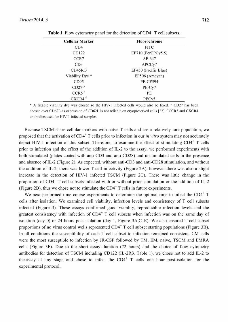
Table 1. Flow cytometry panel for the detection of CD4+ T cell subsets.
Cellular Marker Fluorochrome
CD4 FITC
CD122 EF710 (PerCPCy5.5)
CCR7 AF-647
CD3 APCCy7
CD45RO EF450 (Pacific Blue)
Viability Dye * EF506 (Amcyan)
CD95 PE-CF594
CD27 ^ PE-Cy7
CCR5 # PE
CXCR4 # PECy5
* A fixable viability dye was chosen so the HIV-1 infected cells would also be fixed. ^ CD27 has been
chosen over CD62L as expression of CD62L is not reliable on cryopreserved cells [22]. # CCR5 and CXCR4
antibodies used for HIV-1 infected samples.
Because TSCM share cellular markers with naïve T cells and are a relatively rare population, we
proposed that the activation of CD4+ T cells prior to infection in our in vitro system may not accurately
depict HIV-1 infection of this subset. Therefore, to examine the effect of stimulating CD4+ T cells
prior to infection and the effect of the addition of IL-2 to the assay, we performed experiments with
both stimulated (plates coated with anti-CD3 and anti-CD28) and unstimulated cells in the presence
and absence of IL-2 (Figure 2). As expected, without anti-CD3 and anti-CD28 stimulation, and without
the addition of IL-2, there was lower T cell infectivity (Figure 2A), however there was also a slight
increase in the detection of HIV-1 infected TSCM (Figure 2C). There was little change in the
proportion of CD4+ T cell subsets infected with or without prior stimulation or the addition of IL-2
(Figure 2B), thus we chose not to stimulate the CD4+ T cells in future experiments.
We next performed time course experiments to determine the optimal time to infect the CD4+ T
cells after isolation. We examined cell viability, infection levels and consistency of T cell subsets
infected (Figure 3). These assays confirmed good viability, reproducible infection levels and the
greatest consistency with infection of CD4+ T cell subsets when infection was on the same day of
isolation (day 0) or 24 hours post isolation (day 1, Figure 3A,C–E). We also ensured T cell subset
proportions of no virus control wells represented CD4+ T cell subset starting populations (Figure 3B).
In all conditions the susceptibility of each T cell subset to infection remained consistent. CM cells
were the most susceptible to infection by JR-CSF followed by TM, EM, naïve, TSCM and EMRA
cells (Figure 3F). Due to the short assay duration (72 hours) and the choice of flow cytometry
antibodies for detection of TSCM including CD122 (IL-2Rβ Table 1), we chose not to add IL-2 to
the assay at any stage and chose to infect the CD4+ T cells one hour post-isolation for the
experimental protocol.
Viruses 2014, 6
713
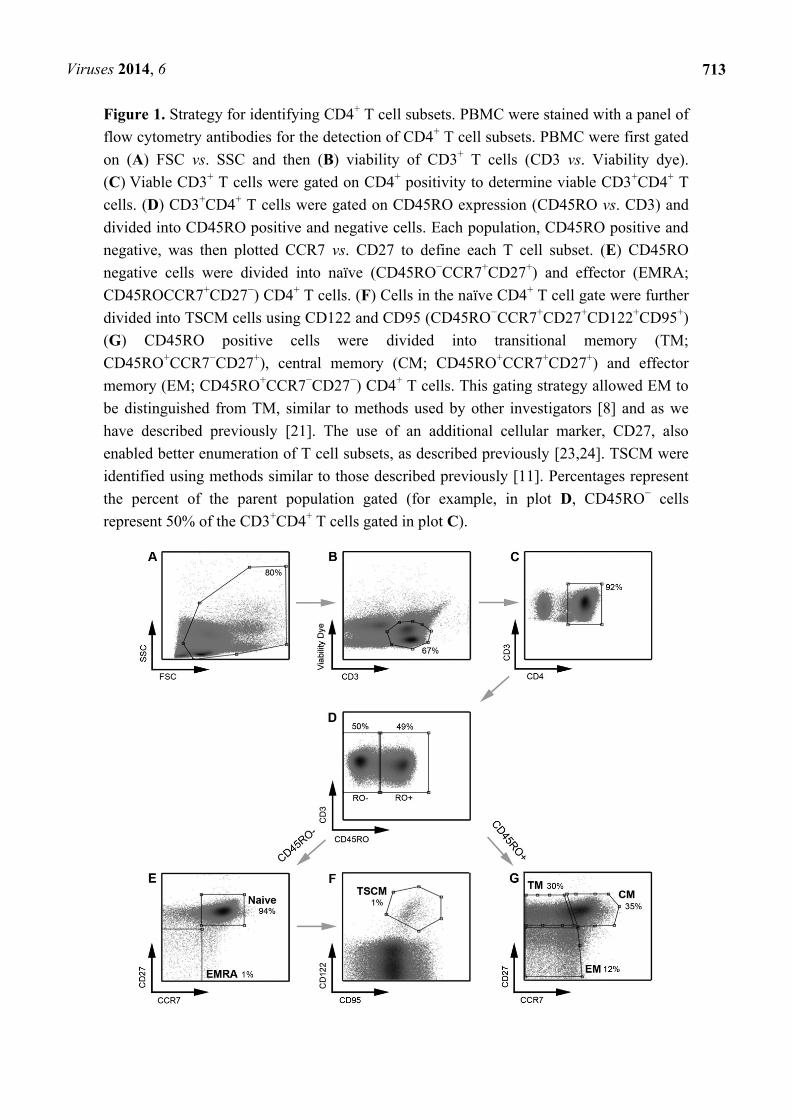
Figure 1. Strategy for identifying CD4+ T cell subsets. PBMC were stained with a panel of
flow cytometry antibodies for the detection of CD4+ T cell subsets. PBMC were first gated
on (A) FSC vs. SSC and then (B) viability of CD3+ T cells (CD3 vs. Viability dye).
(C) Viable CD3+ T cells were gated on CD4
+ positivity to determine viable CD3
+CD4
+ T
cells. (D) CD3+CD4
+ T cells were gated on CD45RO expression (CD45RO vs. CD3) and
divided into CD45RO positive and negative cells. Each population, CD45RO positive and
negative, was then plotted CCR7 vs. CD27 to define each T cell subset. (E) CD45RO
negative cells were divided into naïve (CD45RO−CCR7
+CD27
+) and effector (EMRA;
CD45ROCCR7+CD27
−) CD4
+ T cells. (F) Cells in the naïve CD4
+ T cell gate were further
divided into TSCM cells using CD122 and CD95 (CD45RO−CCR7
+CD27
+CD122
+CD95
+)
(G) CD45RO positive cells were divided into transitional memory (TM;
CD45RO+CCR7
−CD27
+), central memory (CM; CD45RO
+CCR7
+CD27
+) and effector
memory (EM; CD45RO+CCR7
−CD27
−) CD4
+ T cells. This gating strategy allowed EM to
be distinguished from TM, similar to methods used by other investigators [8] and as we
have described previously [21]. The use of an additional cellular marker, CD27, also
enabled better enumeration of T cell subsets, as described previously [23,24]. TSCM were
identified using methods similar to those described previously [11]. Percentages represent
the percent of the parent population gated (for example, in plot D, CD45RO− cells
represent 50% of the CD3+CD4
+ T cells gated in plot C).
Viruses 2014, 6
714
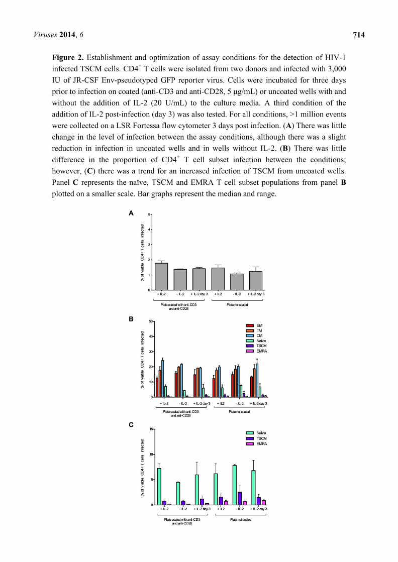
Figure 2. Establishment and optimization of assay conditions for the detection of HIV-1
infected TSCM cells. CD4+ T cells were isolated from two donors and infected with 3,000
IU of JR-CSF Env-pseudotyped GFP reporter virus. Cells were incubated for three days
prior to infection on coated (anti-CD3 and anti-CD28, 5 μg/mL) or uncoated wells with and
without the addition of IL-2 (20 U/mL) to the culture media. A third condition of the
addition of IL-2 post-infection (day 3) was also tested. For all conditions, >1 million events
were collected on a LSR Fortessa flow cytometer 3 days post infection. (A) There was little
change in the level of infection between the assay conditions, although there was a slight
reduction in infection in uncoated wells and in wells without IL-2. (B) There was little
difference in the proportion of CD4+ T cell subset infection between the conditions;
however, (C) there was a trend for an increased infection of TSCM from uncoated wells.
Panel C represents the naïve, TSCM and EMRA T cell subset populations from panel B
plotted on a smaller scale. Bar graphs represent the median and range.
Viruses 2014, 6
715
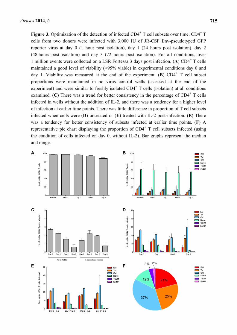
Figure 3. Optimization of the detection of infected CD4+ T cell subsets over time. CD4
+ T
cells from two donors were infected with 3,000 IU of JR-CSF Env-pseudotyped GFP
reporter virus at day 0 (1 hour post isolation), day 1 (24 hours post isolation), day 2
(48 hours post isolation) and day 3 (72 hours post isolation). For all conditions, over
1 million events were collected on a LSR Fortessa 3 days post infection. (A) CD4+ T cells
maintained a good level of viability (>95% viable) in experimental conditions day 0 and
day 1. Viability was measured at the end of the experiment. (B) CD4+ T cell subset
proportions were maintained in no virus control wells (assessed at the end of the
experiment) and were similar to freshly isolated CD4+ T cells (isolation) at all conditions
examined. (C) There was a trend for better consistency in the percentage of CD4+ T cells
infected in wells without the addition of IL-2, and there was a tendency for a higher level
of infection at earlier time points. There was little difference in proportion of T cell subsets
infected when cells were (D) untreated or (E) treated with IL-2 post-infection. (E) There
was a tendency for better consistency of subsets infected at earlier time points. (F) A
representative pie chart displaying the proportion of CD4+ T cell subsets infected (using
the condition of cells infected on day 0, without IL-2). Bar graphs represent the median
and range.
Viruses 2014, 6
716
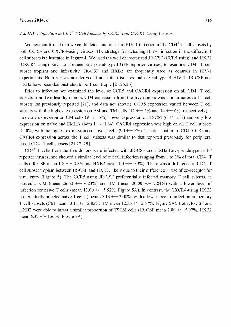
2.2. HIV-1 Infection in CD4+ T Cell Subsets by CCR5- and CXCR4-Using Viruses
We next confirmed that we could detect and measure HIV-1 infection of the CD4+ T cell subsets by
both CCR5- and CXCR4-using viruses. The strategy for detecting HIV-1 infection in the different T
cell subsets is illustrated in Figure 4. We used the well characterized JR-CSF (CCR5-using) and HXB2
(CXCR4-using) Envs to produce Env-pseudotyped GFP reporter viruses, to examine CD4+ T cell
subset tropism and infectivity. JR-CSF and HXB2 are frequently used as controls in HIV-1
experiments. Both viruses are derived from patient isolates and are subtype B HIV-1. JR-CSF and
HXB2 have been demonstrated to be T cell tropic [21,25,26].
Prior to infection we examined the level of CCR5 and CXCR4 expression on all CD4+ T cell
subsets from five healthy donors. CD4 expression from the five donors was similar across all T cell
subsets (as previously reported [21], and data not shown). CCR5 expression varied between T cell
subsets with the highest expression on EM and TM cells (17 +/− 5% and 14 +/− 6%, respectively), a
moderate expression on CM cells (9 +/− 5%), lower expression on TSCM (6 +/− 5%) and very low
expression on naïve and EMRA (both 1 +/−1 %). CXCR4 expression was high on all T cell subsets
(>70%) with the highest expression on naïve T cells (90 +/− 5%). The distribution of CD4, CCR5 and
CXCR4 expression across the T cell subsets was similar to that reported previously for peripheral
blood CD4+ T cell subsets [21,27–29].
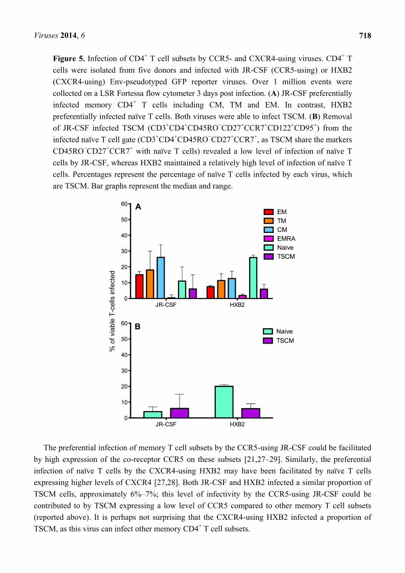
CD4+ T cells from the five donors were infected with JR-CSF and HXB2 Env-pseudotyped GFP
reporter viruses, and showed a similar level of overall infection ranging from 1 to 2% of total CD4+ T
cells (JR-CSF mean 1.8 +/− 0.8% and HXB2 mean 1.0 +/− 0.3%). There was a difference in CD4+ T
cell subset tropism between JR-CSF and HXB2, likely due to their difference in use of co-receptor for
viral entry (Figure 5). The CCR5-using JR-CSF preferentially infected memory T cell subsets, in
particular CM (mean 26.60 +/− 6.23%) and TM (mean 20.00 +/− 7.84%) with a lower level of
infection for naïve T cells (mean 12.00 +/− 5.52%, Figure 5A). In contrast, the CXCR4-using HXB2
preferentially infected naïve T cells (mean 25.13 +/− 2.00%) with a lower level of infection in memory
T cell subsets (CM mean 13.11 +/− 2.85%, TM mean 12.35 +/− 2.57%, Figure 5A). Both JR-CSF and
HXB2 were able to infect a similar proportion of TSCM cells (JR-CSF mean 7.80 +/− 5.07%, HXB2
mean 6.32 +/− 1.65%, Figure 5A).
Viruses 2014, 6
717
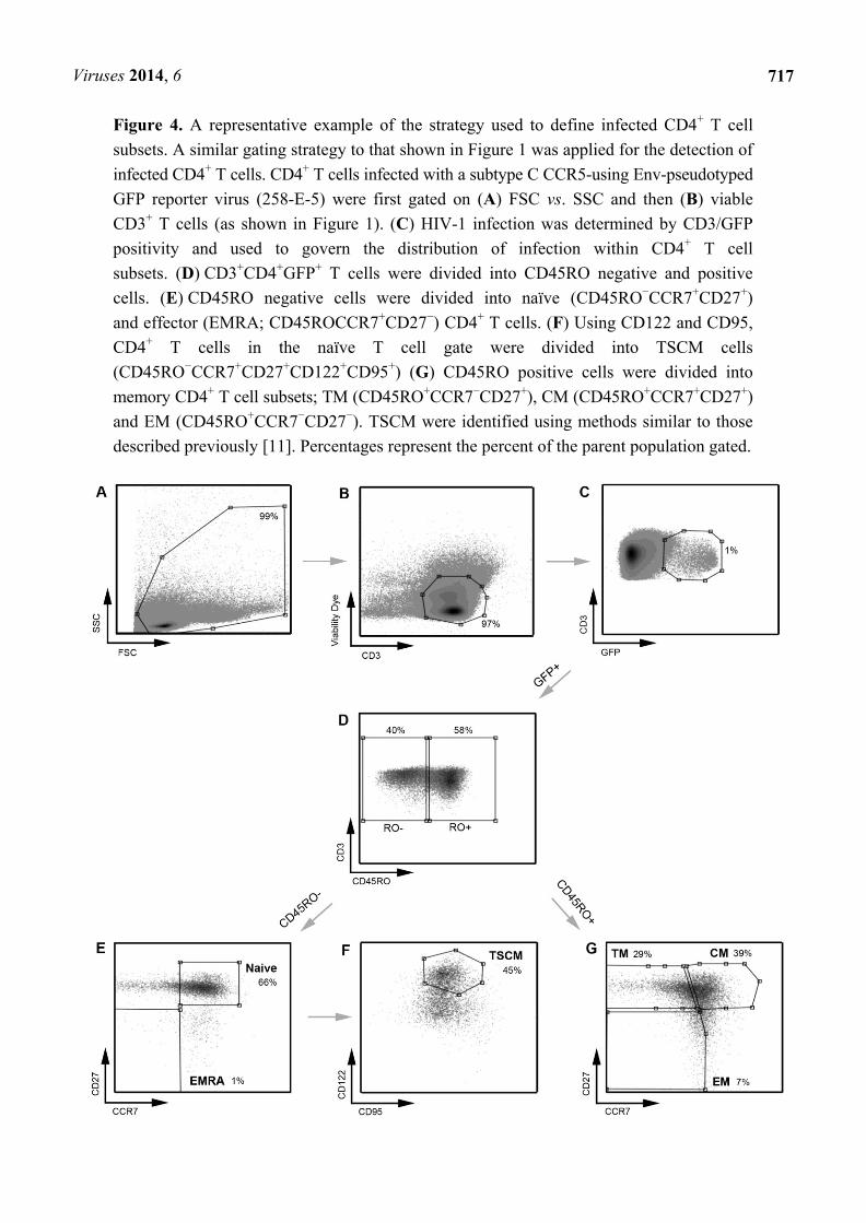
Figure 4. A representative example of the strategy used to define infected CD4+ T cell
subsets. A similar gating strategy to that shown in Figure 1 was applied for the detection of
infected CD4+ T cells. CD4
+ T cells infected with a subtype C CCR5-using Env-pseudotyped
GFP reporter virus (258-E-5) were first gated on (A) FSC vs. SSC and then (B) viable
CD3+ T cells (as shown in Figure 1). (C) HIV-1 infection was determined by CD3/GFP
positivity and used to govern the distribution of infection within CD4+ T cell
subsets. (D) CD3+CD4
+GFP
+ T cells were divided into CD45RO negative and positive
cells. (E) CD45RO negative cells were divided into naïve (CD45RO−CCR7
+CD27
+)
and effector (EMRA; CD45ROCCR7+CD27
−) CD4
+ T cells. (F) Using CD122 and CD95,
CD4+ T cells in the naïve T cell gate were divided into TSCM cells
(CD45RO−CCR7
+CD27
+CD122
+CD95
+) (G) CD45RO positive cells were divided into
memory CD4+ T cell subsets; TM (CD45RO
+CCR7
−CD27
+), CM (CD45RO
+CCR7
+CD27
+)
and EM (CD45RO+CCR7
−CD27
−). TSCM were identified using methods similar to those
described previously [11]. Percentages represent the percent of the parent population gated.
Viruses 2014, 6
718
Figure 5. Infection of CD4+ T cell subsets by CCR5- and CXCR4-using viruses. CD4
+ T
cells were isolated from five donors and infected with JR-CSF (CCR5-using) or HXB2
(CXCR4-using) Env-pseudotyped GFP reporter viruses. Over 1 million events were
collected on a LSR Fortessa flow cytometer 3 days post infection. (A) JR-CSF preferentially
infected memory CD4+ T cells including CM, TM and EM. In contrast, HXB2
preferentially infected naïve T cells. Both viruses were able to infect TSCM. (B) Removal
of JR-CSF infected TSCM (CD3+CD4
+CD45RO
−CD27
+CCR7
+CD122
+CD95
+) from the
infected naïve T cell gate (CD3+CD4
+CD45RO
−CD27
+CCR7
+, as TSCM share the markers
CD45RO−CD27
+CCR7
+ with naïve T cells) revealed a low level of infection of naïve T
cells by JR-CSF, whereas HXB2 maintained a relatively high level of infection of naïve T
cells. Percentages represent the percentage of naïve T cells infected by each virus, which
are TSCM. Bar graphs represent the median and range.
The preferential infection of memory T cell subsets by the CCR5-using JR-CSF could be facilitated
by high expression of the co-receptor CCR5 on these subsets [21,27–29]. Similarly, the preferential
infection of naïve T cells by the CXCR4-using HXB2 may have been facilitated by naïve T cells
expressing higher levels of CXCR4 [27,28]. Both JR-CSF and HXB2 infected a similar proportion of
TSCM cells, approximately 6%–7%; this level of infectivity by the CCR5-using JR-CSF could be
contributed to by TSCM expressing a low level of CCR5 compared to other memory T cell subsets
(reported above). It is perhaps not surprising that the CXCR4-using HXB2 infected a proportion of
TSCM, as this virus can infect other memory CD4+ T cell subsets.
Viruses 2014, 6
719
Investigation into the proportion of infected CD45RO−CD27
+CCR7
+ T cells which are TSCM
(CD45RO−CD27
+CCR7
+CD95
+CD122
+) revealed that the majority of CD45RO
−CD27
+CCR7
+ cells
infected by CCR5-using JR-CSF were TSCM (infected mean CD45RO−CD27
+CCR7
+ 12.00 +/− 5.52%,
TSCM 7.80 +/− 5.07%, 62 +/− 16% of CD45RO−CD27
+CCR7
+ T cells are TSCM, Figure 5B).
In contrast, the minority of CD45RO−CD27
+CCR7
+ T cells infected by CXCR4-using HXB2 were
TSCM (infected mean CD45RO−CD27
+CCR7
+ 5.13 +/− 2.00%, TSCM 6.32 +/− 1.65%, 25 +/− 7% of
CD45RO−CD27
+CCR7
+ T cells are TSCM, Figure 5B). The overall percentages of TSCM infected by
JR-CSF and HXB2 were similar; suggesting that it is the number of naïve T cells infected which
increases with CXCR4-using viruses compared to CCR5-using viruses.
Infection of TSCM by CCR5- and CXCR4-using viruses in vitro has not been previously reported.
This finding, combined with the reported potential of TSCM to be a long-lived reservoir for HIV-1
[12,16], is potentially important for the development of therapeutics targeting the HIV-1 reservoir for
both CCR5- and CXCR4-using viruses. It is also important knowledge for therapies targeting subtype
B HIV-1 strains, as approximately 40%–50% of subtype B viruses undergo a co-receptor switch during
progression to advanced stages of infection [30,31], suggesting infection of TSCM could potentially be
maintained throughout HIV-1 disease progression.
2.3. Measurement of Infection in CD4+ T Cell Subsets by HIV-1 Subtype C Viruses
We next tested the assay system using GFP reporter viruses pseudotyped with CCR5-using HIV-1
Envs isolated from two subjects infected with HIV-1 subtype C (subjects 258 and 1136). These Envs,
and the clinical characteristics of the subjects have been described in detail recently [32,33]. The Env
clones from subject 258 used here were 258-E-5, 258-E-6, 258-E-20 and 258-E-23, and those from
subject 1136 were 1136-E-1, 1136-E-4, 1136-E-11 and 1136-E-12 [32,33]. All of the subtype C Envs
exclusively used the CCR5 co-receptor [21,22].
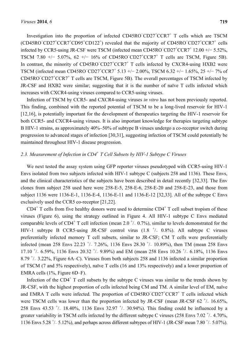
CD4+ T cells from five healthy donors were used to determine CD4
+ T cell subset tropism of these
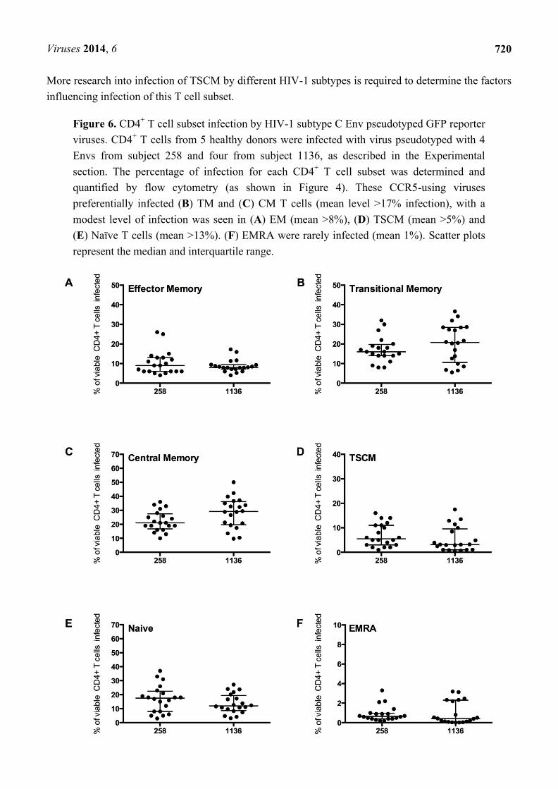
viruses (Figure 6), using the strategy outlined in Figure 4. All HIV-1 subtype C Envs mediated
comparable levels of CD4+ T cell infection (mean 2.0
+/− 0.7%), similar to levels demonstrated for the
HIV-1 subtype B CCR5-using JR-CSF control virus (1.8 +/− 0.8%). All subtype C viruses
preferentially infected memory T cell subsets, similar to JR-CSF; CM T cells were preferentially
infected (mean 258 Envs 22.23 +/− 7.26%, 1136 Envs 28.30
+/− 10.89%), then TM (mean 258 Envs
17.10 +/− 6.59%, 1136 Envs 20.32
+/− 9.89%) and EM (mean 258 Envs 10.26
+/− 6.18%, 1136 Envs
8.79 +/− 3.22%, Figure 6A–C). Viruses from both subjects 258 and 1136 infected a similar proportion
of TSCM (7 and 5% respectively), naïve T cells (16 and 13% respectively) and a lower proportion of
EMRA cells (1%, Figure 6D–F).
Infection of the CD4+ T cell subsets by the subtype C viruses was similar to the trends shown by
JR-CSF, with the highest proportion of cells infected being CM and TM. A similar level of EM, naïve
and EMRA T cells were infected. The proportion of CD45RO−CD27
+CCR7
+ T cells infected which
were TSCM cells was lower than the proportion infected by JR-CSF (mean JR-CSF 62 +/− 16.65%,
258 Envs 43.53 +/− 18.40%, 1136 Envs 32.97
+/− 30.94%). This finding could be influenced by a
greater variability in TSCM cells infected by the different subtype C viruses (258 Envs 7.02 +/− 4.70%,
1136 Envs 5.28 +/− 5.12%), and perhaps across different subtypes of HIV-1 (JR-CSF mean 7.80
+/− 5.07%).
Viruses 2014, 6
720
More research into infection of TSCM by different HIV-1 subtypes is required to determine the factors
influencing infection of this T cell subset.
Figure 6. CD4+ T cell subset infection by HIV-1 subtype C Env pseudotyped GFP reporter
viruses. CD4+ T cells from 5 healthy donors were infected with virus pseudotyped with 4
Envs from subject 258 and four from subject 1136, as described in the Experimental
section. The percentage of infection for each CD4+ T cell subset was determined and
quantified by flow cytometry (as shown in Figure 4). These CCR5-using viruses
preferentially infected (B) TM and (C) CM T cells (mean level >17% infection), with a
modest level of infection was seen in (A) EM (mean >8%), (D) TSCM (mean >5%) and
(E) Naïve T cells (mean >13%). (F) EMRA were rarely infected (mean 1%). Scatter plots
represent the median and interquartile range.

Viruses 2014, 6
721
3. Experimental Section
3.1. Cells
293T cells, JC53 cells [34] and TZM-bl cells [35] were maintained as described previously [21].
Peripheral blood mononuclear cells (PBMC) were purified from the blood of healthy HIV-1 negative
donors by density gradient centrifugation.
3.2. HIV-1 Env Clones
All Envs used in this study are expressed from the pSVIII-Env mammalian expression plasmid [36].
For assay validation and as controls for each assay, the well characterized CCR5-using JR-CSF Env
and CXCR4-using HXB2 Env were used, as described previously [21,25,26]. The HIV-1 subtype C
Envs are derived from plasma of two subjects (subjects 258 and 1136) with chronic subtype C
infection [33]. Four independent Envs from each subject were used in this study. The Env clones from
subject 258 were 258-E-5, 258-E-6, 258-E-20 and 258-E-23, and those from subject 1136 were
1136-E-1, 1136-E-4, 1136-E-11 and 1136-E-12 [32,33]. These Envs were shown to be specific for the
CCR5 co-receptor by phenotypic entry assays [33]and also by the recently developed CoRSeqV3-C
co-receptor usage prediction algorithm, that was designed specifically for HIV-1 subtype C Envs [37].
3.3. Production and Quantitation of Env Pseudotyped GFP Reporter Viruses
Env pseudotyped, GFP reporter viruses were produced as described previously [21]. Briefly, 293T
cells were transfected with pNL4-3Env-GFP [38] and pSVIII-Env plasmids using Lipofectamine 2000
(Invitrogen, Carlsbad, CA, USA) at a ratio of 4:1. Supernatants were harvested 48 h later and filtered
through 0.45 μm filters. Viruses were concentrated through a 20% (vol/vol) sucrose cushion, and
stored at −80 °C. The TCID50 of virus stocks was determined by titration in TZM-bl cells, as described
previously [35,39].
3.4. Enumeration of HIV-1 Infection in CD4+ T Cell Subsets
Forty-eight well tissue culture plates were seeded with 500 μL of 4 × 10⁶/mL of purified CD4
+ T
cells (2 × 10⁶ cells in each well) that were isolated from healthy donors using a RosetteSepCD4
+ T-cell
kit [Stemcell Technologies (Vancouver, BC, Canada), >95% purity of CD3+CD4
+ T cells in each
experiment]. Cells were suspended in RPMI 1640 medium containing 10% (vol/vol) FCS at all stages
of the experiment. CD4+ T cells were incubated for 1 hour prior to infection with 3,000 infectious units
of CCR5-using Env pseudotyped GFP reporter virus, or 1,250 infectious units of CXCR4-using virus
by spinoculation (1,200 × g for 2 h) in V-bottom 96-well tissue culture plates. We empirically
determined that this virus inoculum was within the linear range of infection for the CCR5- and
CXCR4-using viruses used (data not shown).
Cells were then transferred to 48-well tissue culture plates and incubated for 3 days at 37 °C prior to
staining with flow cytometry antibodies (Table 1). Flow cytometry antibodies were obtained from
BD Biosciences (San Jose, CA, USA) with the exception of CD45RO eFlour450, CD122 PerCP-eFlour710
and the fixable viability dye eFlour506 which were from eBiosciences (San Diego, CA, USA). Cells
Viruses 2014, 6
722
were fixed for three hours in 4% (wt/vol) paraformaldehyde, then washed and resuspended in FACS
buffer [filtered PBS with 2 mM EDTA and 0.5% (wt/vol) BSA]. We washed and suspended the cells
in FACS buffer, as paraformaldehyde can cause changes in the emission of some fluorochromes,
particularly APC-Cy7 dyes [40]. OneComp ebeads were used with the flow cytometry antibodies as
compensation controls (eBiosciences).
HIV-1 infection was determined by CD3/GFP positivity and used to determine the distribution of
infection within CD4+ T cell subsets, which were defined as naïve (CD45RO
−CCR7
+CD27
+), TSCM
(CD45RO−CCR7
+CD27
+CD95
+CD122
+), effector memory RA (EMRA, CD45RO
−CCR7
−CD27
−),
central memory (CM, CD45RO+CCR7
+CD27
+) effector memory (EM, CD45RO
+CCR7
−CD27
−) and
transitional memory (TM, CD45RO+CCR7
−CD27
+) T cells. This gating strategy allowed EM
to be distinguished from TM [8,21] and superior enumeration of T cell subsets, as described
previously [23,24]. TSCM were identified using methods similar to those described recently [11].
For these analyses, >1,000,000 events were collected on a LSR Fortessa flow cytometer
(BD Biosciences) and analyzed with Flowlogic software (eBiosciences). The strategy for measuring
HIV-1 infection in CD4+ T cell subsets is shown in Figure 1 and Figure 4, which are representative
experiments of uninfected CD4+ T cells (Figure 1) and CD4
+ T cells infected with GFP reporter virus
pseudotyped with 258-E-5 Env (Figure 4), respectively.
4. Conclusions
We developed a novel in vitro CD4+ T cell infection assay to quantify the level and distribution of
HIV-1 infection in CD4+ T cell subsets including the newly described TSCM subset. This assay was
validated with CCR5-using and CXCR4-using viruses, and was able to distinguish distinct patterns of
CD4+ T cell tropism associated with different co-receptor specificities. We further show that our assay
can be used to measure CD4+ T cell subset infection by clinical isolates, specifically HIV-1 subtype
C strains.
Our assay permits the simultaneous detection and quantification of HIV-1 infection in naïve,
EMRA, TSCM, CM, TM, EM CD4+ T cell subsets. Investigation of changes in CD4
+ T cell tropism by
viruses isolated from longitudinal cohorts could potentially predict the establishment of viral reservoirs
in vivo, and changes in cellular tropism that may be important for HIV-1 pathogenesis.
Acknowledgments
We thank J. Sodroski and D.F.J. Purcell for supplying pSVIII-HXB2 and pNL4-3Env-GFP
plasmids, respectively. This study was supported by a grant from the Australian National Health and
Medical Research Council to PRG and MJC (1022066). KC is supported by an Australian
Postgraduate Award administered through the University of Melbourne. KB is supported by a
Victorian International Research Scholarship administered through La Trobe University. MRJ is
supported by a Danish Research Council Sapere Aude Fellowship. PRG is supported by an Australian
Research Council Future Fellowship (FT2). The authors gratefully acknowledge the contribution to
this work of the Victorian Operational Infrastructure Support Program received by the Burnet Institute.

Viruses 2014, 6
723
Author Contributions
JKF, GP and KC developed the flow cytometry strategies; JKF, KC, AE, and MR performed the
experiments; KB and MRJ provided critical reagents; JKF and PRG designed the experiments; JKF,
GP, MJC and PRG analyzed the data; JKF and PRG wrote the manuscript; all authors helped
edit the manuscript.
Conflicts of Interest
The authors declare no conflicts of interest.
References and Notes
1. Gattinoni, L.; Restifo, N.P. Moving T memory stem cells to the clinic. Blood 2013, 121, 567–578.
2. Kalia, V.; Sarkar, S.; Ahmed, R. CD8 T-cell memory differentiation during acute and chronic
viral infections. Adv. Exp. Med. Biol. 2010, 684, 79–95.
3. Youngblood, B.; Hale, J.S.; Ahmed, R. T-cell memory differentiation: Insights from
transcriptional signatures and epigenetics. Immunology 2013, 139, 277–284.
4. Luckey, C.J.; Weaver, C.T. Stem-cell-like qualities of immune memory; CD4+ T cells join the
party. Cell Stem Cell 2012, 10, 107–108.
5. Fritsch, R.D.; Shen, X.; Sims, G.P.; Hathcock, K.S.; Hodes, R.J.; Lipsky, P.E. Stepwise
differentiation of CD4 memory T cells defined by expression of CCR7 and CD27. J. Immunol.
2005, 175, 6489–6497.
6. Sallusto, F.; Lenig, D.; Forster, R.; Lipp, M.; Lanzavecchia, A. Two subsets of memory T
lymphocytes with distinct homing potentials and effector functions. Nature 1999, 401, 708–712.
7. Sallusto, F.; Geginat, J.; Lanzavecchia, A. Central memory and effector memory T cell subsets:
function, generation, and maintenance. Ann. Rev. Immunol. 2004, 22, 745–763.
8. Chomont, N.; El-Far, M.; Ancuta, P.; Trautmann, L.; Procopio, F.A.; Yassine-Diab, B.; Boucher,
G.; Boulassel, M.R.; Ghattas, G.; Brenchley, J.M.; et al. HIV reservoir size and persistence are
driven by T cell survival and homeostatic proliferation. Nat. Med. 2009, 15, 893–900.
9. Geginat, J.; Sallusto, F.; Lanzavecchia, A. Cytokine-driven proliferation and differentiation of
human naive, central memory, and effector memory CD4(+) T cells. J. Exp. Med. 2001, 194,
1711–1719.
10. Riou, C.; Yassine-Diab, B.; Van grevenynghe, J.; Somogyi, R.; Greller, L.D.; Gagnon, D.;
Gimmig, S.; Wilkinson, P.; Shi, Y.; Cameron, M.J.; et al. Convergence of TCR and cytokine
signaling leads to FOXO3a phosphorylation and drives the survival of CD4+ central memory T
cells. J. Exp. Med. 2007, 204, 79–91.
11. Lugli, E.; Gattinoni, L.; Roberto, A.; Mavilio, D.; Price, D.A.; Restifo, N.P.; Roederer, M.
Identification, isolation and in vitro expansion of human and nonhuman primate T stem cell
memory cells. Nat. Protocol. 2013, 8, 33–42.
12. Gattinoni, L.; Lugli, E.; Ji, Y.; Pos, Z.; Paulos, C.M.; Quigley, M.F.; Almeida, J.R.; Gostick, E.;
Yu, Z.; Carpenito, C.; et al. A human memory T cell subset with stem cell-like properties.
Nat. Med. 2011, 17, 1290–1297.
Viruses 2014, 6
724
13. Sant, A.J.; McMichael, A. Revealing the role of CD4+ T cells in viral immunity. J. Exp. Med.
2012, 209, 1391–1395.
14. Hazenberg, M.D.; Otto, S.A.; van Benthem, B.H.; Roos, M.T.; Coutinho, R.A.; Lange, J.M.;
Hamann, D.; Prins, M.; Miedema, F. Persistent immune activation in HIV-1 infection is
associated with progression to AIDS. AIDS 2003, 17, 1881–1888.
15. Douek, D.C.; Picker, L.J.; Koup, R.A. T Cell Dynamics in HIV-1 Infection. Ann. Rev. Immunol.
2003, 21, 265–304.
16. Lugli, E.; Dominguez, M.; Gattinoni, L.; Chattopadhyay, P.; Bolton, D.; Song, K.; Klatt, N.;
Brenchley, J.; Vaccari, M.; Gostick, E.; et al. Superior T memory stem cell persistence supports
long-lived T cell memory. J. Clin. Invest. 2013, 123, 594–599.
17. Buzon, M.J.; Sun, H.; Li, C.; Shaw, A.; Seiss, K.; Ouyang, Z.; Martin-Gayo, E.; Leng, J.;
Henrich, T.J.; Li, J.Z.; et al. HIV-1 persistence in CD4+ T cells with stem cell-like properties.
Nat. Med. 2014, doi:10.1038/nm.3445.
18. Cieri, N.; Camisa, B.; Cocchiarella, F.; Forcato, M.; Oliveira, G.; Provasi, E.; Bondanza, A.;
Bordignon, C.; Peccatori, J.; Ciceri, F.; et al. IL-7 and IL-15 instruct the generation of human
memory stem T cells from naive precursors. Blood 2013, 121, 573–584.
19. Buzon, M. T memory stem cells: A long-term reservoir for HIV-1. In Proceedings of the ID Week
2012 Meeting, San Diego, CA, USA, 17–21 October 2012; Paper #594.
20. Embretson, J.; Zupancic, M.; Ribas, J.; Burke, A.; Racz, P.; Tenner-Racz, K.; Haase, A. Massive
covert infection of helper T lymphocytes and macrophages by HIV during the incubation period
of AIDS. Nature 1993, 262, 359–362.
21. Flynn, J.K.; Paukovics, G.; Moore, M.S.; Ellett, A.; Gray, L.R.; Duncan, R.; Salimi, H.; Jubb, B.;
Westby, M.; Purcell, D.F.; et al. The magnitude of HIV-1 resistance to the CCR5 antagonist
maraviroc may impart a differential alteration in HIV-1 tropism for macrophages and T-cell
subsets. Virology 2013, 442, 51–58.
22. Perfetto, S.P.; Chattopadhyay, P.K.; Roederer, M. Seventeen-colour flow cytometry: unravelling
the immune system. Nat. Rev. Immunol. 2004, 4, 648–655.
23. Appay, V.; van Lier, R.A.; Sallusto, F.; Roederer, M. Phenotype and function of human T
lymphocyte subsets: consensus and issues. Cytometry A 2008, 73, 975–983.
24. De Rosa, S.C.; Herzenberg, L.A.; Herzenberg, L.A.; Roederer, M. 11-color, 13-parameter flow
cytometry: Identification of human naive T cells by phenotype, function, and T-cell receptor
diversity. Nat. Med. 2001, 7, 245–248.
25. Roche, M.; Jakobsen, M.R.; Ellett, A.; Salimiseyedabad, H.; Jubb, B.; Westby, M.; Lee, B.;
Lewin, S.R.; Churchill, M.J.; Gorry, P.R. HIV-1 predisposed to acquiring resistance to maraviroc
(MVC) and other CCR5 antagonists in vitro has an inherent, low-level ability to utilize
MVC-bound CCR5 for entry. Retrovirology 2011, 8, 89.
26. Roche, M.; Jakobsen, M.R.; Sterjovski, J.; Ellett, A.; Posta, F.; Lee, B.; Jubb, B.; Westby, M.;
Lewin, S.R.; Ramsland, P.A.; et al. HIV-1 escape from the CCR5 antagonist maraviroc associated
with an altered and less efficient mechanism of gp120-CCR5 engagement that attenuates
macrophage-tropism. J. Virol. 2011, 85, 4330–4342.
27. Gorry, P.R.; Ancuta, P. Coreceptors and HIV-1 pathogenesis. Curr. HIV AIDS Rep. 2011, 8,
45–53.
Viruses 2014, 6
725
28. Lee, B.; Sharron, M.; Montaner, L.J.; Weissman, D.; Doms, R.W. Quantification of CD4, CCR5,
and CXCR4 levels on lymphocyte subsets, dendritic cells, and differentially conditioned
monocyte-derived macrophages. Proc. Natl. Acad. Sci. USA 1999, 96, 5215–5220.
29. Pfaff, J.M.; Wilen, C.B.; Harrison, J.E.; Demarest, J.F.; Lee, B.; Doms, R.W.; Tilton, J.C. HIV-1
resistance to CCR5 antagonists associated with highly efficient use of CCR5 and altered tropism
on primary CD4+ T cells. J. Virol. 2010, 84, 6505–6514.
30. Bjorndal, A.; Deng, H.; Jansson, M.; Fiore, J.R.; Colognesi, C.; Karlsson, A.; Albert, J.; Scarlatti,
G.; Littman, D.R.; Fenyo, E.M. Coreceptor usage of primary human immunodeficiency virus type
1 isolates varies according to biological phenotype. J. Virol. 1997, 71, 7478–7487.
31. Connor, R.I.; Sheridan, K.E.; Ceradini, D.; Choe, S.; Landau, N.R. Change in coreceptor use
coreceptor use correlates with disease progression in HIV-1—Infected individuals. J. Exp. Med.
1997, 185, 621–628.
32. Cashin, K.; Jakobsen, M.R.; Sterjovski, J.; Roche, M.; Ellett, A.; Flynn, J.K.; Borm, K.; Gouillou,
M.; Churchill, M.J.; Gorry, P.R. Linkages between HIV-1 specificity for CCR5 or CXCR4 and in
vitro usage of alternative coreceptors during progressive HIV-1 subtype C infection.
Retrovirology 2013, 10, 98.
33. Jakobsen, M.; Cashin, K.; Roche, M.; Sterjovski, J.; Ellett, A.; Borm;, K.; Flynn, J.; Erikstrup, C.;
Gouillou, M.; Gray, L.; et al. Longitudinal analysis of CCR5 and CXCR4 usage in a cohort of
antiretroviral therapy-naïve subjects with progressive HIV-1 subtype C infection. PLoS One 2013,
8, e65950.
34. Platt, E.J.; Wehrly, K.; Kuhmann, S.E.; Chesebro, B.; Kabat, D. Effects of CCR5 and CD4 cell
surface concentrations on infections by macrophagetropic isolates of human immunodeficiency
virus type 1. J. Virol. 1998, 72, 2855–2864.
35. Wei, X.; Decker, J.M.; Liu, H.; Zhang, Z.; Arani, R.B.; Kilby, J.M.; Saag, M.S.; Wu, X.;
Shaw, G.M.; Kappes, J.C. Emergence of resistant human immunodeficiency virus type 1 in
patients receiving fusion inhibitor (T-20) monotherapy. Antimicrob. Agents Chemother. 2002, 46,
1896–1905.
36. Gao, F.; Morrison, S.G.; Robertson, D.L.; Thornton, C.L.; Craig, S.; Karlsson, G.; Sodroski, J.;
Morgado, M.; Galvao-Castro, B.; von Briesen, H.; et al. Molecular cloning and analysis of
functional envelope genes from human immunodeficiency virus type 1 sequence subtypes A
through G. The WHO and NIAID Networks for HIV Isolation and Characterization. J. Virol.
1996, 70, 1651–1667.
37. Cashin, K.; Gray, L.R.; Jakobsen, M.R.; Sterjovski, J.; Churchill, M.J.; Gorry, P.R. CoRSeqV3-C:
a novel HIV-1 subtype C specific V3 sequence based coreceptor usage prediction algorithm.
Retrovirology 2013, 10, 24.
38. Center, R.J.; Wheatley, A.K.; Campbell, S.M.; Gaeguta, A.J.; Peut, V.; Alcantara, S.; Siebentritt,
C.; Kent, S.J.; Purcell, D.F. Induction of HIV-1 subtype B and AE-specific neutralizing antibodies
in mice and macaques with DNA prime and recombinant gp140 protein boost regimens. Vaccine
2009, 27, 6605–6612.
39. Yap, S.H.; Sheen, C.W.; Fahey, J.; Zanin, M.; Tyssen, D.; Lima, V.D.; Wynhoven, B.; Kuiper,
M.; Sluis-Cremer, N.; Harrigan, P.R.; et al. N348I in the connection domain of HIV-1 reverse
transcriptase confers zidovudine and nevirapine resistance. PLoS Med. 2007, 4, e335.

Viruses 2014, 6
726
40. BD Biosciences. Multicolor Flow Cytometry Absorption and Emission Spectra. Available online:
http://www.bdbiosciences.com/research/multicolor/spectrumguide/ (accessed on 10 February 2014).
© 2014 by the authors; licensee MDPI, Basel, Switzerland. This article is an open access article
distributed under the terms and conditions of the Creative Commons Attribution license
(http://creativecommons.org/licenses/by/3.0/).